Filters
Clonality
Type
Reactivity
Gene Name
Isotype
Host
Application
Clone
3487 results for " Protein Detection Antibody" - showing 3200-3250


 Application Data
(Staining of human peripheral blood lymphocytes with Mouse anti Human CD40:RPE)
Application Data
(Staining of human peripheral blood lymphocytes with Mouse anti Human CD40:RPE)
CD40, Monoclonal Antibody (Cat# AAA12090)
Full Name
MOUSE ANTI HUMAN CD40
Gene Names
CD40; p50; Bp50; CDW40; TNFRSF5
Applications
Immunohistochemistry, Flow Cytometry, Immunoprecipitation, Immunohistochemistry
Pricing
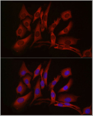 IF (Immunofluorescence)
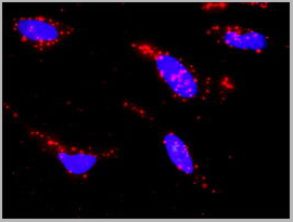
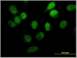
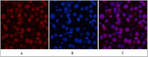
(Immunofluorescence analysis of U2OS cells using cGAS Rabbit pAb (AAA28207) at dilution of 1:50 (40x lens).Secondary antibody: Cy3-conjugated Goat anti-Rabbit IgG (H+L) at 1:500 dilution. Blue: DAPI for nuclear staining.)
IF (Immunofluorescence)
(Immunofluorescence analysis of U2OS cells using cGAS Rabbit pAb (AAA28207) at dilution of 1:50 (40x lens).Secondary antibody: Cy3-conjugated Goat anti-Rabbit IgG (H+L) at 1:500 dilution. Blue: DAPI for nuclear staining.)
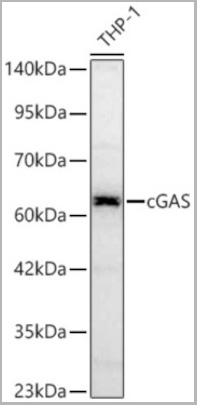
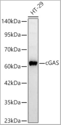
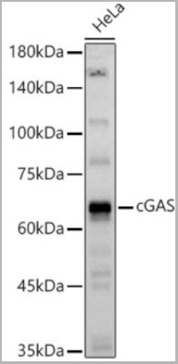
MB21D1, Polyclonal Antibody (Cat# AAA28207)
Full Name
MB21D1 Polyclonal Antibody
Gene Names
MB21D1; cGAS; h-cGAS; C6orf150
Applications
Western Blot, Immunohistochemistry, Immunofluorescence
Purity
Affinity Purification
Pricing
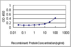 Application Data
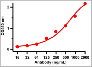
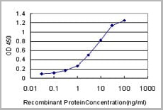
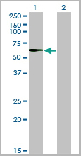
(Detection limit for recombinant GST tagged PLK1 is ~1ng/ml as a capture antibody.)
Application Data
(Detection limit for recombinant GST tagged PLK1 is ~1ng/ml as a capture antibody.)
PLK1, Monoclonal Antibody (Cat# AAA24321)
Full Name
PLK1 (STPK13, Polo-like Kinase 1, Serine/Threonine-protein Kinase 13, Serine/Threonine-protein Kinase PLK1, PLK, PLK-1) (AP)
Gene Names
PLK1; PLK; STPK13
Reactivity
Human
Applications
Immunoprecipitation, Western Blot
Purity
Purified by Protein A Affinity Chromatography.
Pricing
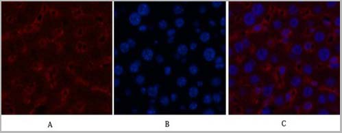
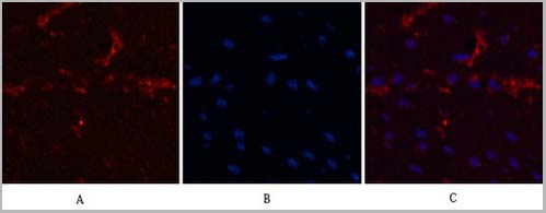
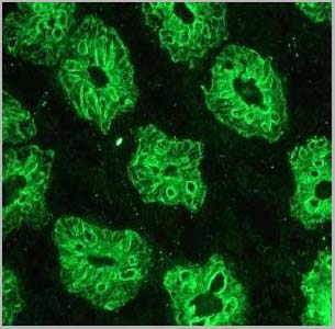
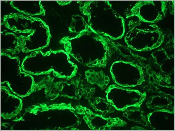
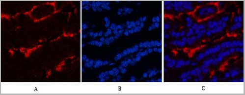
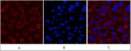
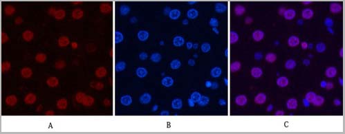
 IF (Immunofluorescence)
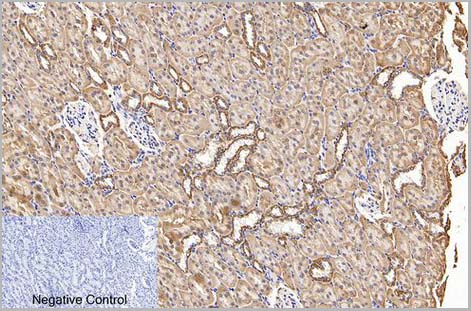
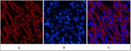
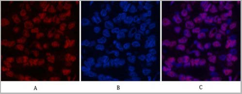
(Fig.6. Immunofluorescence analysis of rat kidney tissue. 1, COX IV Monoclonal Antibody (14Y2) (red) was diluted at 1:200 (4°C, overnight). 2, Cy3 Labeled secondary antibody was diluted at 1:300 (room temperature, 50min). 3, Picture B: DAPI (blue) 10min. Picture A: Target. Picture B: DAPI. Picture C: merge of A+B.)
IF (Immunofluorescence)
(Fig.6. Immunofluorescence analysis of rat kidney tissue. 1, COX IV Monoclonal Antibody (14Y2) (red) was diluted at 1:200 (4°C, overnight). 2, Cy3 Labeled secondary antibody was diluted at 1:300 (room temperature, 50min). 3, Picture B: DAPI (blue) 10min. Picture A: Target. Picture B: DAPI. Picture C: merge of A+B.)
COX IV, Monoclonal Antibody (Cat# AAA31542)
Full Name
Anti-COX IV Mouse Monoclonal Antibody (14Y2)
Gene Names
COX4I1; COX4; COXIV; COX4-1
Reactivity
Human, Mouse, Rat
Applications
Western Blot, Immunohistochemistry, Immunofluorescence
Purity
The antibody was affinity-purified from mouse ascites by affinity-chromatography using specific immunogen
Pricing
 IHC (Immunohistochemistry)
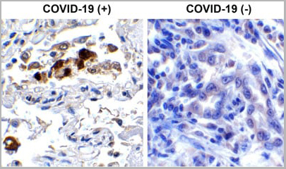
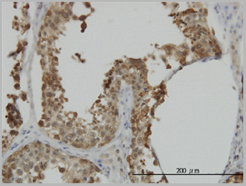
(Immunohistochemistry Validation of SARSCoV-2 (COVID-19) Spike RBD in COVID-19 Patient Lung Immunohistochemical analysis of paraffin-embedded COVID-19 patient lung tissue using anti- SARS-CoV-2 (COVID-19) Spike RBD antibody (AAA11030, 0.5 µg/mL). Tissue was fixed with formaldehyde and blocked with 10% serum for 1 h at RT; antigen retrieval was by heat mediation with a citrate buffer (pH6). Samples were incubated with primary antibody overnight at 4°C. A goat anti-rabbit IgG H&L (HRP) at 1/250 was used as secondary. Counter stained with Hematoxylin. Strong signal of SARS-COV-2 Spike RBD protein was observed in macrophage of COVID-19 patient lung, but not in non-COVID-19 patient lung.)
IHC (Immunohistochemistry)
(Immunohistochemistry Validation of SARSCoV-2 (COVID-19) Spike RBD in COVID-19 Patient Lung Immunohistochemical analysis of paraffin-embedded COVID-19 patient lung tissue using anti- SARS-CoV-2 (COVID-19) Spike RBD antibody (AAA11030, 0.5 µg/mL). Tissue was fixed with formaldehyde and blocked with 10% serum for 1 h at RT; antigen retrieval was by heat mediation with a citrate buffer (pH6). Samples were incubated with primary antibody overnight at 4°C. A goat anti-rabbit IgG H&L (HRP) at 1/250 was used as secondary. Counter stained with Hematoxylin. Strong signal of SARS-COV-2 Spike RBD protein was observed in macrophage of COVID-19 patient lung, but not in non-COVID-19 patient lung.)
COVID 19 Spike RBD Coronavirus, Polyclonal Antibody (Cat# AAA11030)
Full Name
SARS-CoV-2 (COVID-19) Spike RBD Antibody
Gene Names
S; spike glycoprotein
Reactivity
Virus
Applications
Immunofluorescence, Immunohistochemistry, Western Blot
Purity
SARS-CoV-2 (COVID-19) Spike RBD antibody is affinity chromatography purified via peptide column.
Pricing


 Application Data
(Staining of human peripheral blood lymphocytes with Mouse anti Human CD40:RPE)
Application Data
(Staining of human peripheral blood lymphocytes with Mouse anti Human CD40:RPE)
CD40, Monoclonal Antibody (Cat# AAA11867)
Full Name
MOUSE ANTI HUMAN CD40:FITC
Gene Names
CD40; p50; Bp50; CDW40; TNFRSF5
Applications
Flow Cytometry
Pricing


 Application Data
(Staining of human peripheral blood lymphocytes with Mouse anti Human CD40:RPE)
Application Data
(Staining of human peripheral blood lymphocytes with Mouse anti Human CD40:RPE)
CD40, Monoclonal Antibody (Cat# AAA11936)
Full Name
MOUSE ANTI HUMAN CD40
Gene Names
CD40; p50; Bp50; CDW40; TNFRSF5
Reactivity
Dog
Applications
Immunohistochemistry, Flow Cytometry, Immunoprecipitation, Immunohistochemistry
Pricing
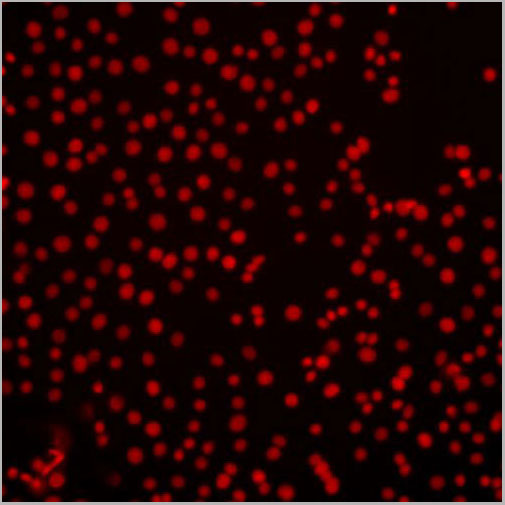
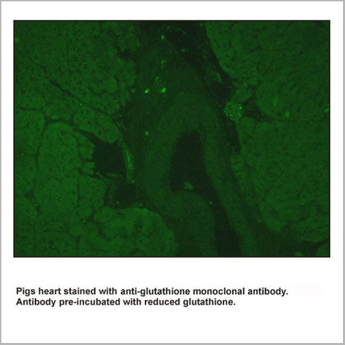
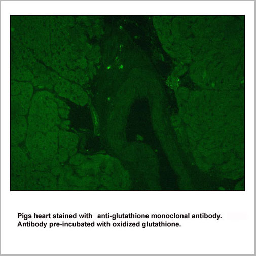
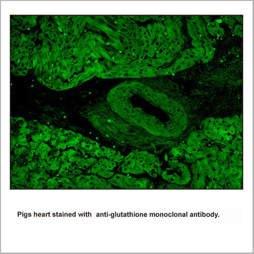
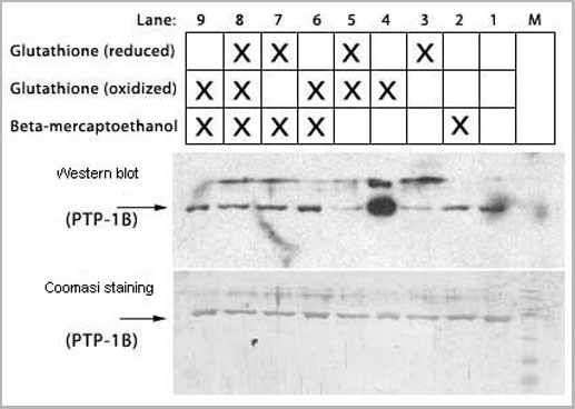
 Application Data
Glutathione IgG1, Monoclonal Antibody (Cat# AAA14178)
Full Name
anti-Glutathione monoclonal antibody IgG1
Applications
Western Blot, Immunoprecipitation
Purity
Protein A purified.
Pricing
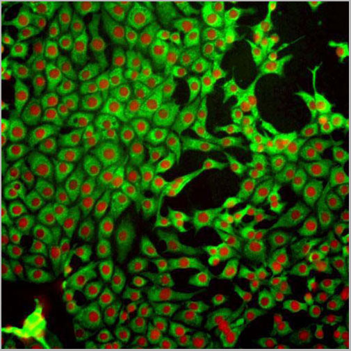
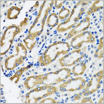
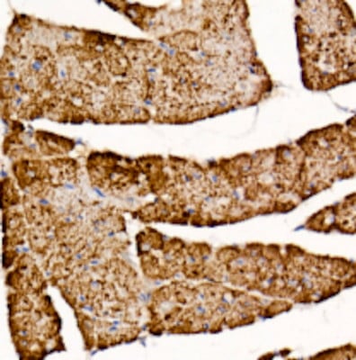
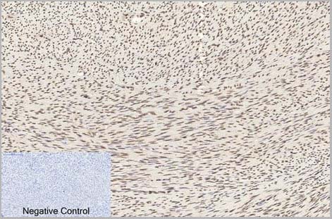
 IHC (Immunohistochemistry)
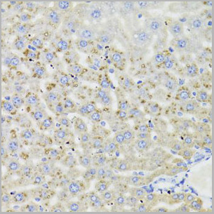
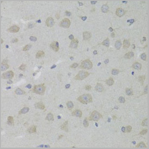
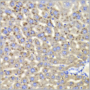
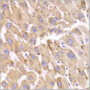
(Immunohistochemistry of paraffin-embedded rat liver using HSD17B13 Rabbit pAb (AAA28095) at dilution of 1:50 (40x lens).)
IHC (Immunohistochemistry)
(Immunohistochemistry of paraffin-embedded rat liver using HSD17B13 Rabbit pAb (AAA28095) at dilution of 1:50 (40x lens).)
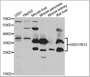
HSD17B13, Polyclonal Antibody (Cat# AAA28095)
Full Name
HSD17B13 Polyclonal Antibody
Gene Names
HSD17B13; SCDR9; NIIL497; SDR16C3; HMFN0376
Reactivity
Human, Mouse, Rat
Applications
Western Blot, Immunohistochemistry
Purity
Affinity Purification
Pricing
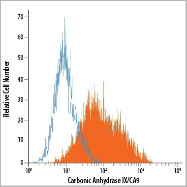 FCM (Flow Cytometry)
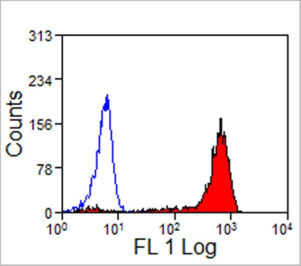
(Detection of Carbonic Anhydrase IX/CA9 in U‑87 MG Human Cell Line by Flow Cytometry using AAA14680.)
FCM (Flow Cytometry)
(Detection of Carbonic Anhydrase IX/CA9 in U‑87 MG Human Cell Line by Flow Cytometry using AAA14680.)
Carbonic Anhydrase IX, Monoclonal Antibody (Cat# AAA14680)
Full Name
Carbonic Anhydrase IX (CA9) (PE)
Gene Names
CA9; MN; CAIX
Applications
Flow Cytometry
Purity
Affinity Purified
Purified by Protein A affinity chromatography from hybridoma culture supernant.
Purified by Protein A affinity chromatography from hybridoma culture supernant.
Pricing


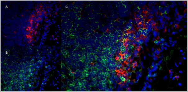
 Application Data
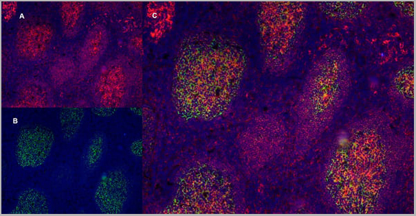
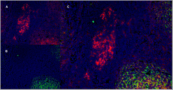
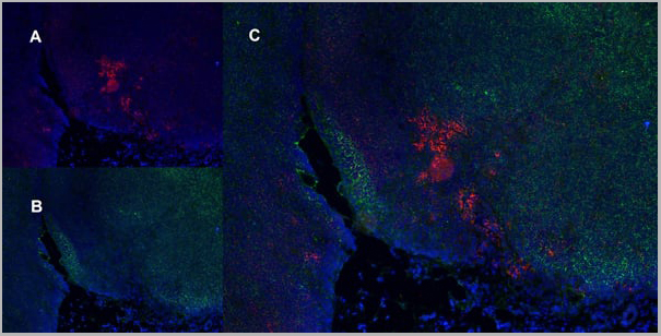
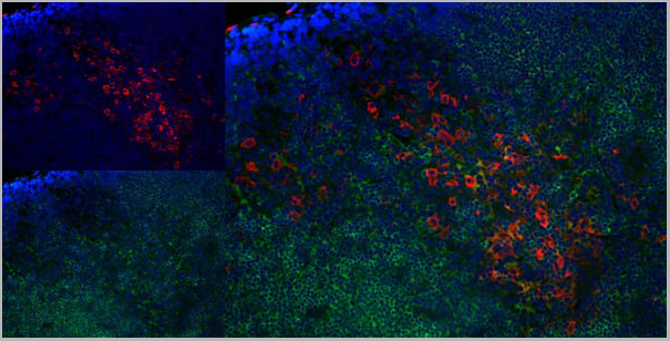
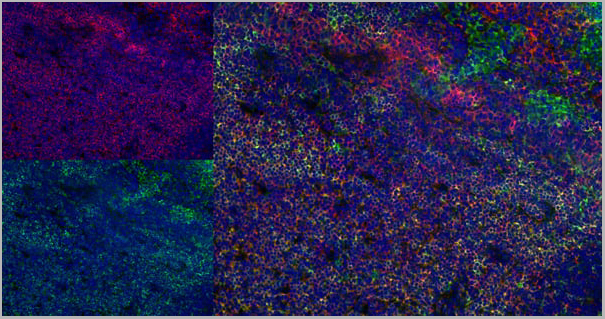
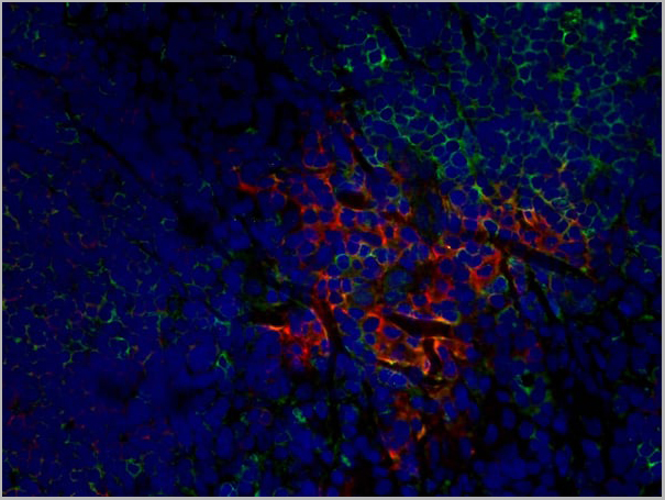
(Immunofluorescence staining of rat lymph node cryosection with Mouse anti Rat CD163 antibody , red an A and Mouse anti Rat CD8 MCA48), green in B. C is the merged image with nuclei counter-stained blue using DAPI. High power)
Application Data
(Immunofluorescence staining of rat lymph node cryosection with Mouse anti Rat CD163 antibody , red an A and Mouse anti Rat CD8 MCA48), green in B. C is the merged image with nuclei counter-stained blue using DAPI. High power)
CD8 ALPHA, Monoclonal Antibody (Cat# AAA11982)
Full Name
MOUSE ANTI RAT CD8 ALPHA
Applications
Immunohistochemistry, Flow Cytometry, Immunoprecipitation, Immunohistochemistry
Pricing


 Application Data
(Staining of human peripheral blood lymphocytes with Mouse anti Human CD40:RPE)
Application Data
(Staining of human peripheral blood lymphocytes with Mouse anti Human CD40:RPE)
CD40, Monoclonal Antibody (Cat# AAA12089)
Full Name
MOUSE ANTI HUMAN CD40:RPE
Gene Names
CD40; p50; Bp50; CDW40; TNFRSF5
Applications
Flow Cytometry
Pricing
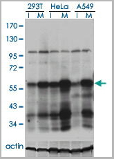
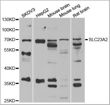
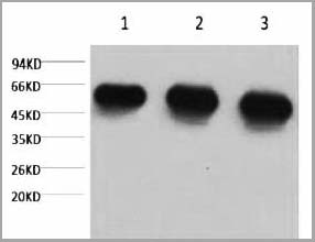
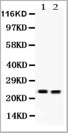
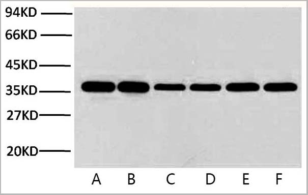
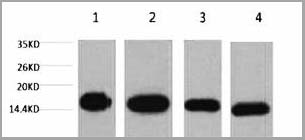
 WB (Western Blot)
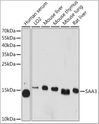
(Western blot analysis of extracts of various cell lines, using SAA3 Rabbit pAb (AAA28247) at 1:1000 dilution.Secondary antibody: HRP Goat Anti-Rabbit IgG (H+L) at 1:10000 dilution.Lysates/proteins: 25ug per lane.Blocking buffer: 3% nonfat dry milk in TBST.Detection: ECL Basic Kit.Exposure time: 5min.)
WB (Western Blot)
(Western blot analysis of extracts of various cell lines, using SAA3 Rabbit pAb (AAA28247) at 1:1000 dilution.Secondary antibody: HRP Goat Anti-Rabbit IgG (H+L) at 1:10000 dilution.Lysates/proteins: 25ug per lane.Blocking buffer: 3% nonfat dry milk in TBST.Detection: ECL Basic Kit.Exposure time: 5min.)
SAA3P, Polyclonal Antibody (Cat# AAA28247)
Full Name
SAA3P Polyclonal Antibody
Gene Names
Saa3; l7R3; Saa-3; AV098916
Applications
Western Blot
Purity
Affinity Purification
Pricing


 WB (Western Blot)
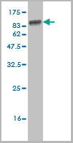
(SPP1 monoclonal antibody (M09), clone 3E11. Western Blot analysis of SPP1 expression in NIH/3T3 (Cat # L018V1).)
WB (Western Blot)
(SPP1 monoclonal antibody (M09), clone 3E11. Western Blot analysis of SPP1 expression in NIH/3T3 (Cat # L018V1).)
SPP1, Monoclonal Antibody (Cat# AAA26437)
Full Name
SPP1 (Secreted Phosphoprotein 1, BNSP, BSPI, ETA-1, MGC110940, OPN) (HRP)
Gene Names
SPP1; OPN; BNSP; BSPI; ETA-1
Applications
Western Blot
Purity
Purified
Pricing
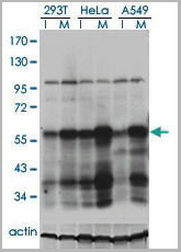
 WB (Western Blot)
(Western blot analysis of various lysates using ZDHHC9 Rabbit pAb (AAA28212) at 1:1000 dilution.Secondary antibody: HRP Goat Anti-Rabbit IgG (H+L) at 1:10000 dilution.Lysates/proteins: 25ug per lane.Blocking buffer: 3% nonfat dry milk in TBST.Detection: ECL Enhanced Kit.Exposure time: 90s.)
WB (Western Blot)
(Western blot analysis of various lysates using ZDHHC9 Rabbit pAb (AAA28212) at 1:1000 dilution.Secondary antibody: HRP Goat Anti-Rabbit IgG (H+L) at 1:10000 dilution.Lysates/proteins: 25ug per lane.Blocking buffer: 3% nonfat dry milk in TBST.Detection: ECL Enhanced Kit.Exposure time: 90s.)
ZDHHC9, Polyclonal Antibody (Cat# AAA28212)
Full Name
ZDHHC9 Polyclonal Antibody
Gene Names
ZDHHC9; CGI89; DHHC9; MMSA1; MRXSZ; ZNF379; ZNF380; CXorf11; ZDHHC10
Applications
Western Blot
Purity
Affinity Purification
Pricing
ERO1L, Monoclonal Antibody (Cat# AAA24499)
Full Name
ERO1L (ERO1-like Protein alpha, ERO1-L, ERO1-L-alpha, Endoplasmic Oxidoreductin-1-like Protein, Oxidoreductin-1-L-alpha, UNQ434/PRO865) APC
Gene Names
ERO1A; ERO1L; ERO1-L; ERO1LA; Ero1alpha; ERO1-alpha; ERO1-L-alpha
Reactivity
Human, Rat
Applications
EIA, IHC, WB
Purity
Purified by Protein A Affinity Chromatography.
Pricing

 Application Data
(Bovine lipoprotein lipase detected with Mouse anti lipoprotein lipase:HRP)
Application Data
(Bovine lipoprotein lipase detected with Mouse anti lipoprotein lipase:HRP)
LIPOPROTEIN LIPASE, Monoclonal Antibody (Cat# AAA12249)
Full Name
MOUSE ANTI LIPOPROTEIN LIPASE:Biotin
Applications
Western Blot, Immunoprecipitation
Purity
Purified IgG prepared by affinity chromatography on Protein G from tissue culture supernatant
Pricing
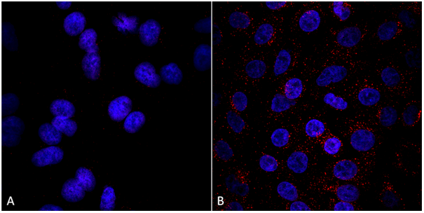
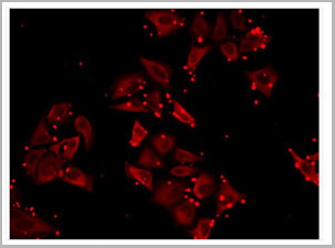 IF (Imunofluorescence)
(Immunofluorescence analysis of MCF7 cells using SLC23A2 antibody (AAA28167))
IF (Imunofluorescence)
(Immunofluorescence analysis of MCF7 cells using SLC23A2 antibody (AAA28167))
SLC23A2, Polyclonal Antibody (Cat# AAA28167)
Full Name
SLC23A2 Polyclonal Antibody
Gene Names
SLC23A2; NBTL1; SVCT2; YSPL2; SLC23A1
Reactivity
Human, Mouse, Rat
Applications
Western Blot, Immunohistochemistry, Immunofluorescence
Purity
Affinity Purification
Pricing


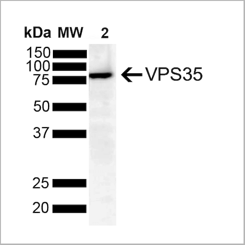
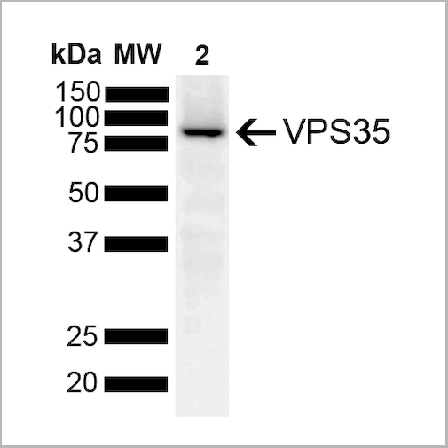
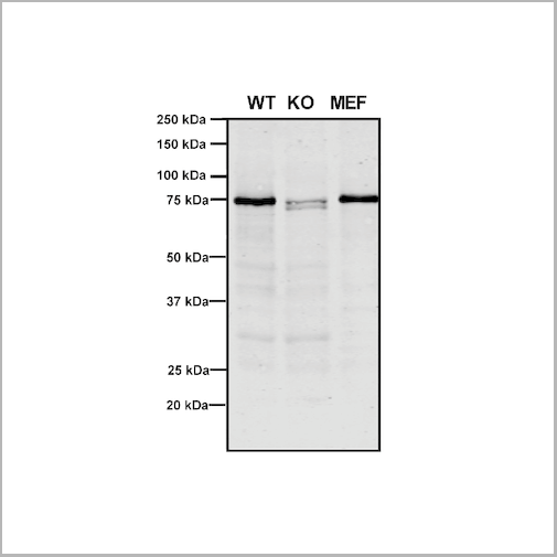
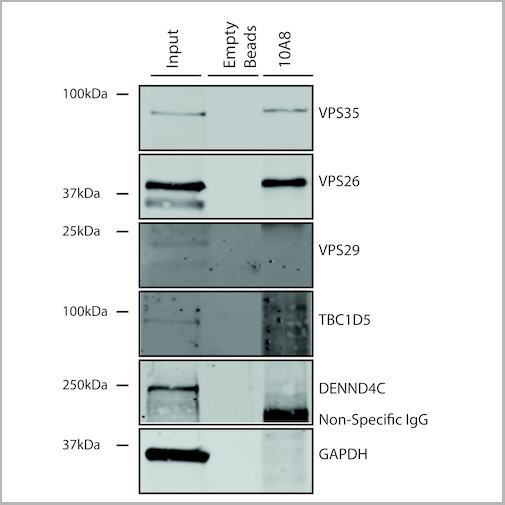
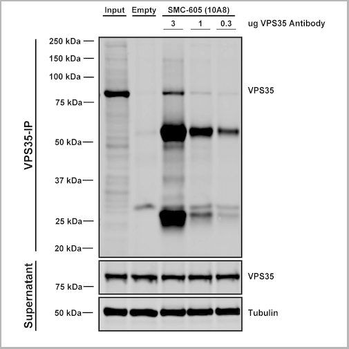
 IP (Immunoprecipitation)
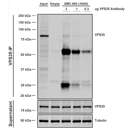
(Immunoprecipitation analysis using Mouse Anti-VPS35 Monoclonal Antibody, Clone 10A8. Tissue: embryonic fibroblast. Species: Mouse. Primary Antibody: Mouse Anti-VPS35 Monoclonal Antibody. Three amounts of (3, 1 and 0.3 ug) were non-covalently coupled to 10uL of A/G sepharose beads for 1 hour at 4 degree C and next incubated with 250ug of MEF lysate for 2 hours at 4 degree C.)
IP (Immunoprecipitation)
(Immunoprecipitation analysis using Mouse Anti-VPS35 Monoclonal Antibody, Clone 10A8. Tissue: embryonic fibroblast. Species: Mouse. Primary Antibody: Mouse Anti-VPS35 Monoclonal Antibody. Three amounts of (3, 1 and 0.3 ug) were non-covalently coupled to 10uL of A/G sepharose beads for 1 hour at 4 degree C and next incubated with 250ug of MEF lysate for 2 hours at 4 degree C.)
VPS35, Monoclonal Antibody (Cat# AAA27694)
Full Name
VPS35 Antibody, Clone 10A8: ATTO 594
Gene Names
VPS35; MEM3; PARK17
Reactivity
Human, Mouse, Rat
Applications
Western Blot, Immunocytochemistry, Immunofluorescence, Immunoprecipitation
Purity
Protein G Purified
Pricing
 Application Data
(Staining of human peripheral blood monocytes with Mouse anti Human CD14:RPE)
Application Data
(Staining of human peripheral blood monocytes with Mouse anti Human CD14:RPE)
CD14, Monoclonal Antibody (Cat# AAA11997)
Full Name
MOUSE ANTI HUMAN CD14
Applications
Immunohistochemistry, Flow Cytometry, Immunoprecipitation
Pricing
 Application Data
(Staining of human peripheral blood lymphocytes with Mouse anti Human CD4: Pacific Blue)
Application Data
(Staining of human peripheral blood lymphocytes with Mouse anti Human CD4: Pacific Blue)
CD4, Monoclonal Antibody (Cat# AAA12071)
Full Name
MOUSE ANTI HUMAN CD4:FITC
Gene Names
CD4; CD4mut
Reactivity
Human
Applications
Flow Cytometry
Pricing


 Application Data
(Staining of human peripheral blood lymphocytes with Mouse anti Human CD40:RPE)
Application Data
(Staining of human peripheral blood lymphocytes with Mouse anti Human CD40:RPE)
CD40, Monoclonal Antibody (Cat# AAA12031)
Full Name
MOUSE ANTI HUMAN CD40:RPE
Gene Names
CD40; p50; Bp50; CDW40; TNFRSF5
Applications
Flow Cytometry
Pricing
Clostridium difficile Glutamate Dehydrogenase (GDH), Recombinant Protein (Cat# AAA13444)
Full Name
Clostridium difficile Glutamate Dehydrogenase (GDH) Recombinant
Applications
Lateral Flow
Purity
> 95% Pure. (Multi-step procedure including affinity chromatography.)
Pricing
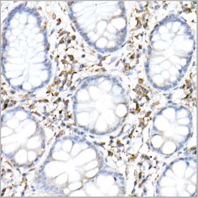
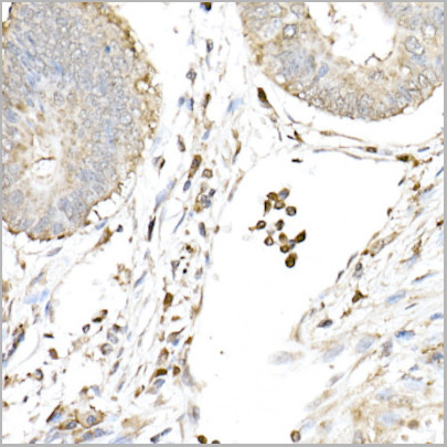
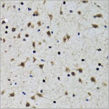
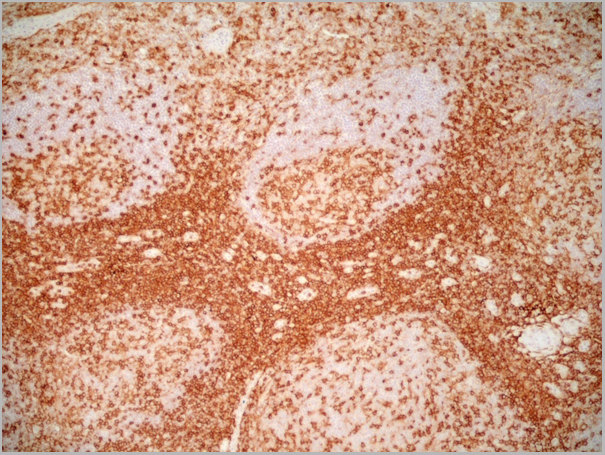
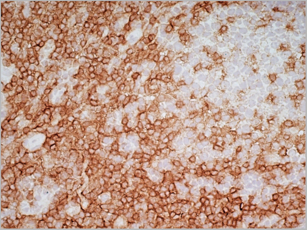
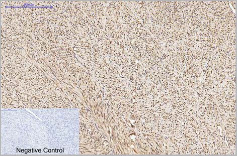
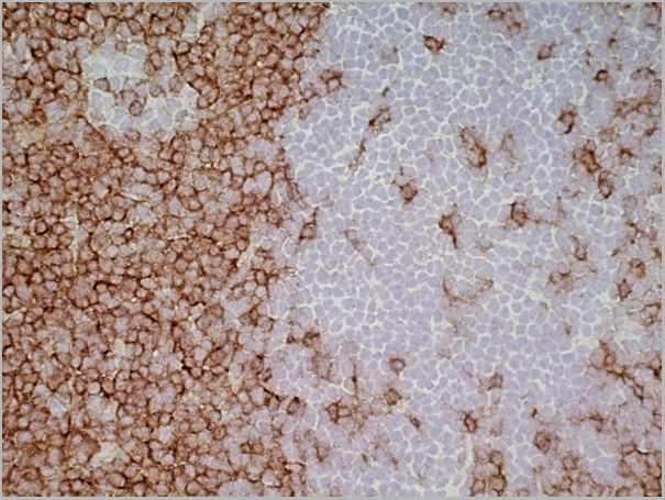
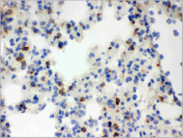
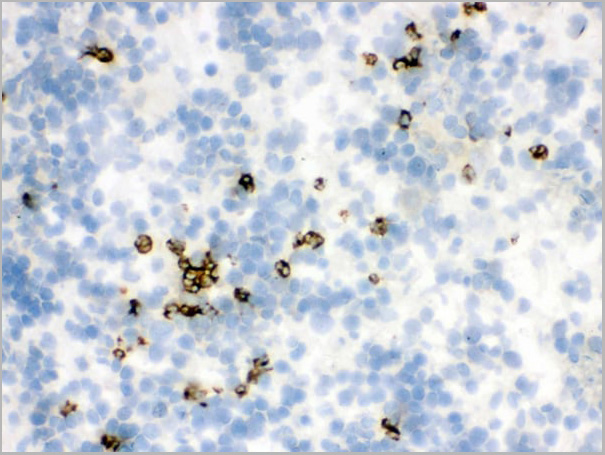
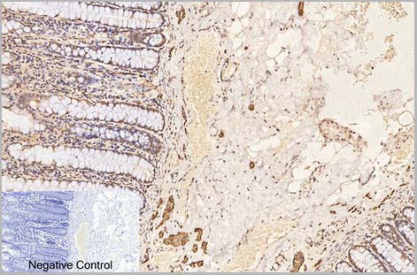
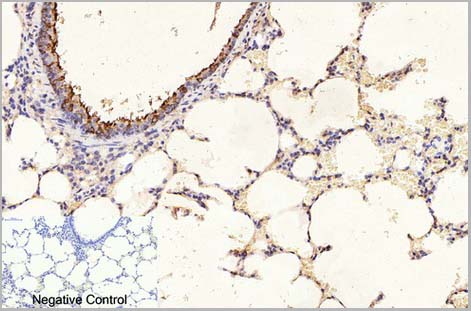
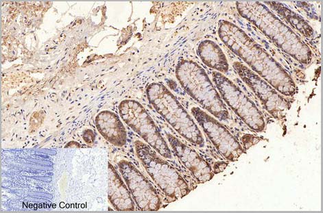
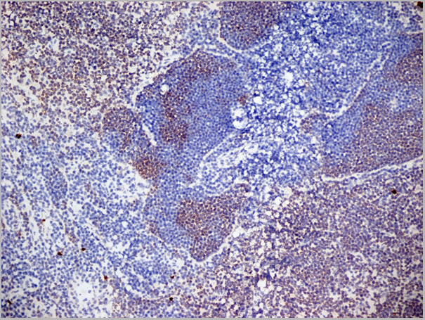
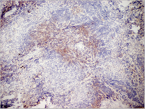
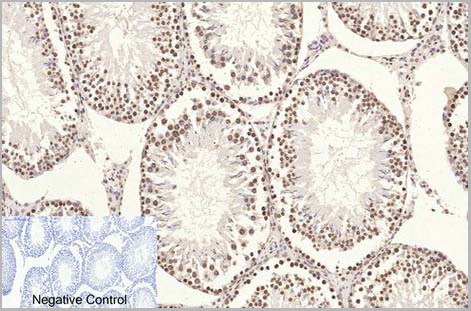
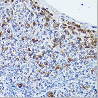 IHC (Immunohistochemistry)
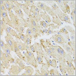
(Immunohistochemistry of paraffinembedded human tonsil using HCAR2 Rabbit pAb (AAA28288) at dilution of 1:25 (40x lens).)
IHC (Immunohistochemistry)
(Immunohistochemistry of paraffinembedded human tonsil using HCAR2 Rabbit pAb (AAA28288) at dilution of 1:25 (40x lens).)
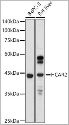
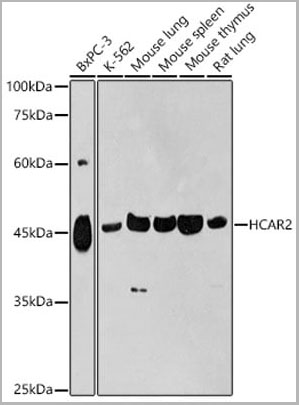
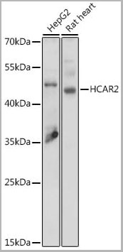
HCAR2, Polyclonal Antibody (Cat# AAA28288)
Full Name
HCAR2 Polyclonal Antibody
Gene Names
HCAR2; HCA2; HM74a; HM74b; PUMAG; NIACR1; Puma-g; GPR109A
Applications
Western Blot, Immunofluorescence, Immunohistochemistry, Immunocytochemistry
Purity
Affinity Purification
Pricing


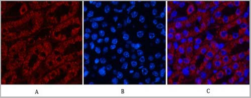
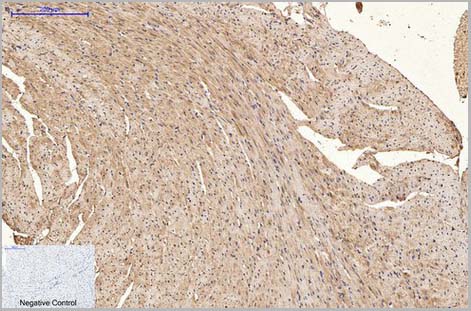
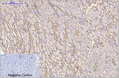
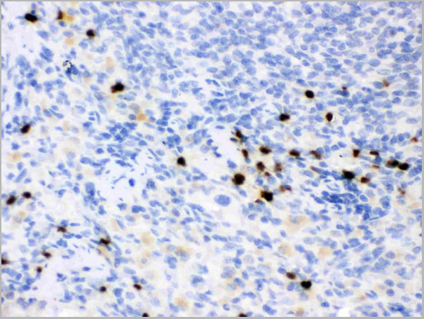
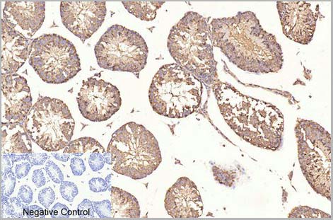
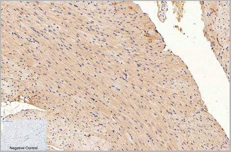
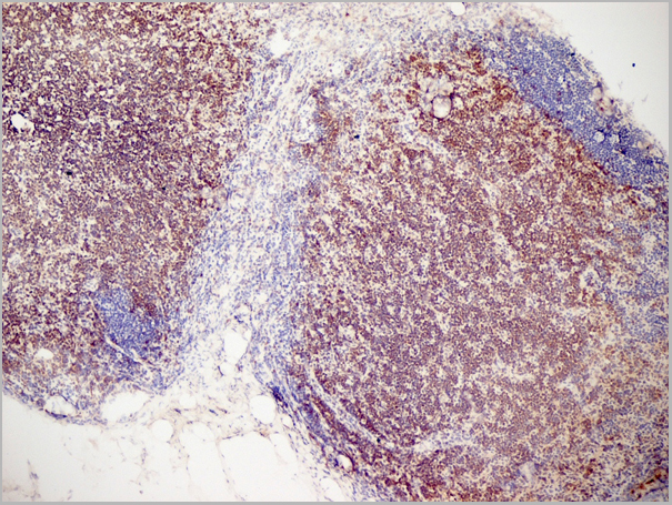
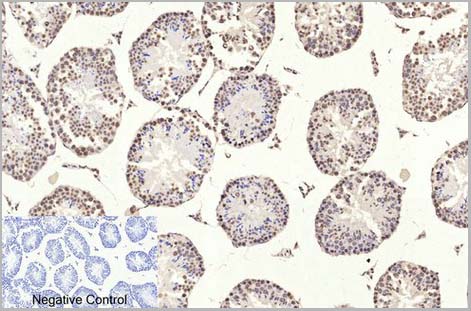
 IHC (Immunohistochemistry)
(Immunohistochemistry of paraffin-embedded rat heart using TOM20 Rabbit mAb (AAA28304) at dilution of 1:50(40xlens).Perform microwave antigen retrieval with 10 mM Tris/EDTA buffer pH 9.0 before commencing with IHC staining protocol.)
IHC (Immunohistochemistry)
(Immunohistochemistry of paraffin-embedded rat heart using TOM20 Rabbit mAb (AAA28304) at dilution of 1:50(40xlens).Perform microwave antigen retrieval with 10 mM Tris/EDTA buffer pH 9.0 before commencing with IHC staining protocol.)
TOM20, Monoclonal Antibody (Cat# AAA28304)
Full Name
TOM20 Rabbit mAb
Applications
Western Blot, Immunohistochemistry, Immunofluorescence, Immunocytochemistry, Immunoprecipitation
Purity
Affinity purification
Pricing
 IHC (Immunohistchemistry)
(Fig.6. Immunohistochemical analysis of paraffin-embedded rat kidney tissue. 1, alpha-tubulin Monoclonal Antibody (3G5) was diluted at 1:200 (4°C, overnight). 2, Sodium citrate pH 6.0 was used for antibody retrieval (>98°C, 20min). 3, secondary antibody was diluted at 1:200 (room temperature, 30min). Negative control was used by secondary antibody only.)
IHC (Immunohistchemistry)
(Fig.6. Immunohistochemical analysis of paraffin-embedded rat kidney tissue. 1, alpha-tubulin Monoclonal Antibody (3G5) was diluted at 1:200 (4°C, overnight). 2, Sodium citrate pH 6.0 was used for antibody retrieval (>98°C, 20min). 3, secondary antibody was diluted at 1:200 (room temperature, 30min). Negative control was used by secondary antibody only.)
alpha-Tubulin, Monoclonal Antibody (Cat# AAA31544)
Full Name
Anti-alpha-Tubulin Monoclonal Antibody (3G5)
Gene Names
TUBA1A; LIS3; TUBA3; B-ALPHA-1
Reactivity
Human, Mouse, Rat
Applications
Western Blot, Immunohistochemistry, Immunofluorescence, Immunoprecipitation
Purity
The antibody was affinity-purified from mouse ascites by affinity-chromatography using specific immunogen
Pricing
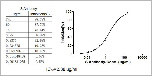

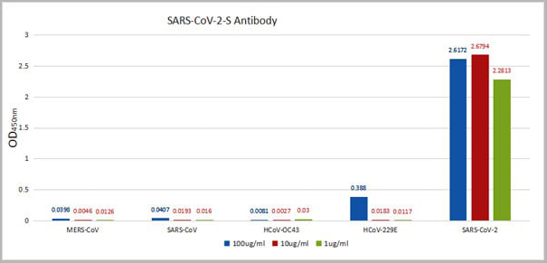 ELISA
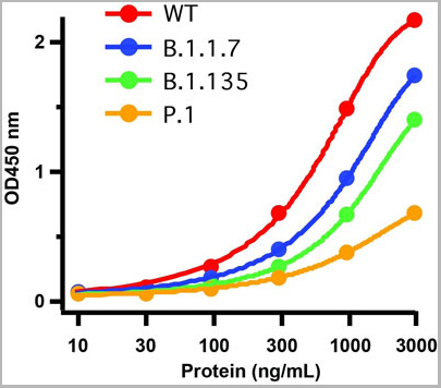
(ELISA: Immobilize various types of SARS proteins at concentration of 2mg/ml on solid substrate, then react with SARS-CoV-2-S Antibody at concentration of 100mg/ml, 10mg/ml and 1mg/ml. It shows the SARS-CoV-2-S Antibody (AAA27036) is specific for SARS-CoV-2-S1-RBD protein, without any cross-reactivity with MERS-CoV, SARS-CoV, HCoV-OC43 or HCoV-229E.)
ELISA
(ELISA: Immobilize various types of SARS proteins at concentration of 2mg/ml on solid substrate, then react with SARS-CoV-2-S Antibody at concentration of 100mg/ml, 10mg/ml and 1mg/ml. It shows the SARS-CoV-2-S Antibody (AAA27036) is specific for SARS-CoV-2-S1-RBD protein, without any cross-reactivity with MERS-CoV, SARS-CoV, HCoV-OC43 or HCoV-229E.)
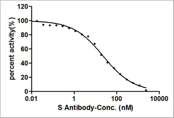
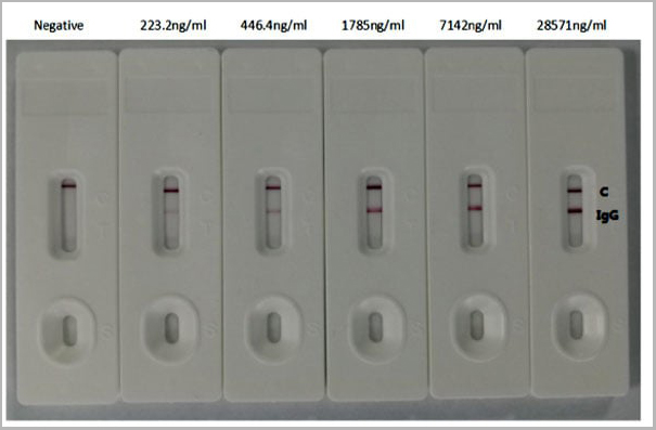
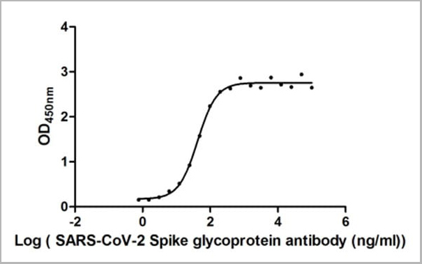
COVID 19 Spike glycoprotein (S) Coronavirus, Monoclonal Recombinant Antibody (Cat# AAA27036)
Full Name
Human Novel Coronavirus Spike glycoprotein (S) (16-685aa) Antibody
Reactivity
Human Novel Coronavirus (SARS-CoV-2/ 2019-nCoV)
Applications
Colloidal Gold Immunochromatographic Assay, Neutralization Assay
Purity
Affinity-chromatography
Pricing
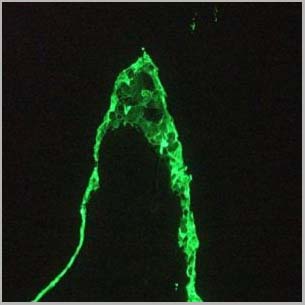
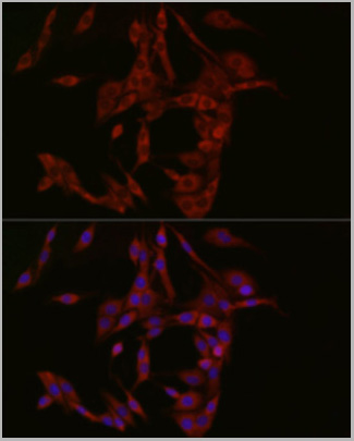
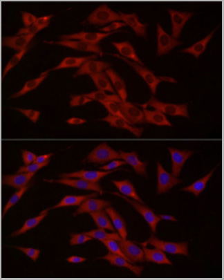
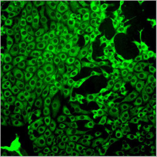
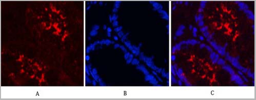
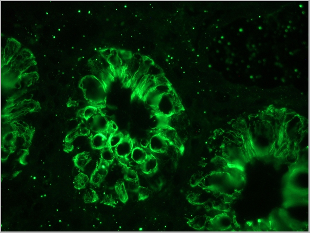 IF (Immunofluorescence)
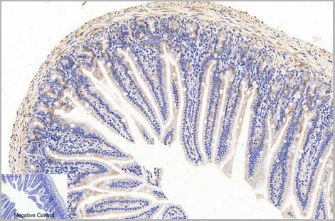
(Figure 6. immunofluorescence staining of human colon epithelium.)
IF (Immunofluorescence)
(Figure 6. immunofluorescence staining of human colon epithelium.)
CytokeRatin 18 / KeRatin K18, Monoclonal Antibody (Cat# AAA14563)
Full Name
Mouse anti CytokeRatin 18 / KeRatin K18
Gene Names
KRT18; K18; CYK18
Reactivity
Canine, Chicken,Dog,Hamster, Human, Mouse, Rabbit, Rat, Swine, Zebrafish
Applications
Flow Cytometry, Immunoblot, Immunocytochemistry, Immunohistochemistry, Western Blot
Pricing


 Application Data
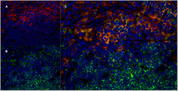
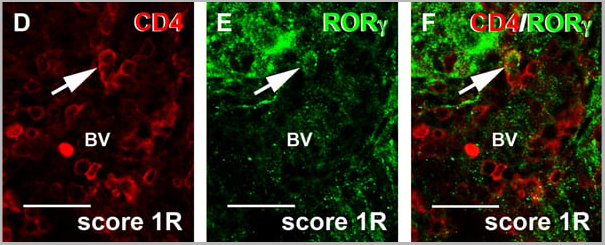
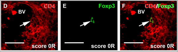
(Published customer image: Dynamics of Th17 cells. D -F) Photographs of double immunolabelled sections showing a representative CD4+ROR?+ cell (arrow) observed around blood vessels (BV). Bar scale = 30 um.Almolda B, Costa M, Montoya M, Gonz¡lez B, Castellano B (2011) Increase in Th17 and T-reg Lymphocytes and Decrease of IL22 Correlate with the Recovery Phase of Acute EAE IN Rat. PLoS ONE 6(11): e27473.)
Application Data
(Published customer image: Dynamics of Th17 cells. D -F) Photographs of double immunolabelled sections showing a representative CD4+ROR?+ cell (arrow) observed around blood vessels (BV). Bar scale = 30 um.Almolda B, Costa M, Montoya M, Gonz¡lez B, Castellano B (2011) Increase in Th17 and T-reg Lymphocytes and Decrease of IL22 Correlate with the Recovery Phase of Acute EAE IN Rat. PLoS ONE 6(11): e27473.)
CD4, Monoclonal Antibody (Cat# AAA11995)
Full Name
MOUSE ANTI RAT CD4 (DOMAIN 1)
Gene Names
Cd4; p55; W3/25
Applications
Immunohistochemistry, Flow Cytometry, Immunohistochemistry
Pricing

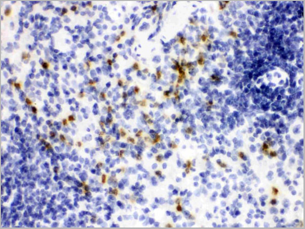
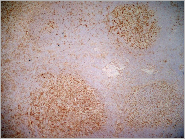
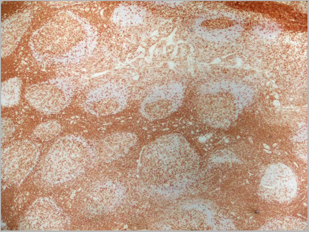
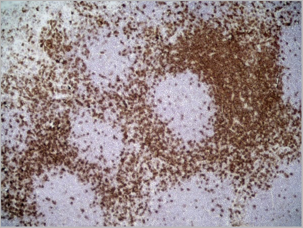
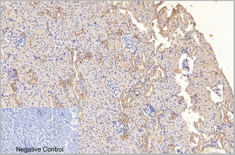
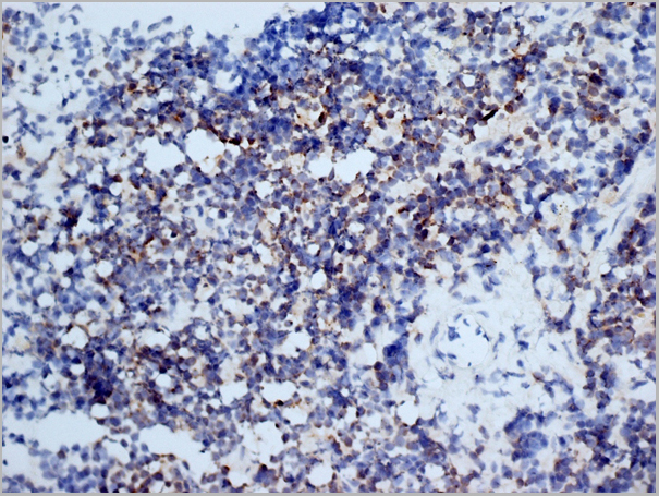
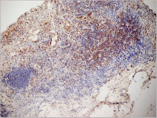
 IHC (Immunohistochemistry)
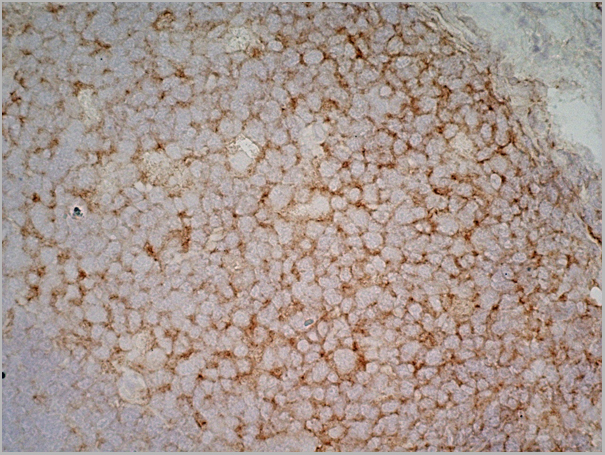
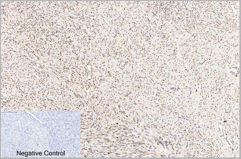
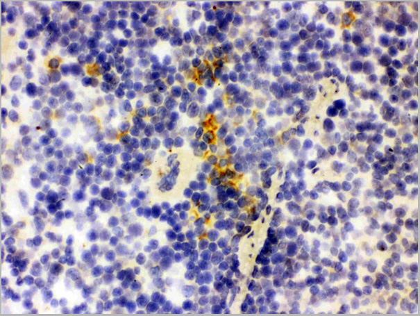
(Figure 7. IHC analysis of Lipocalin 2 using anti-Lipocalin 2 antibody (AAA11663).Lipocalin 2 was detected in frozen section of rat spleen tissue. Heat mediated antigen retrieval was performed in citrate buffer (pH6, epitope retrieval solution) for 20 mins. The tissue section was blocked with 10% goat serum. The tissue section was then incubated with 1ug/ml rabbit anti-Lipocalin 2 Antibody (AAA11663) overnight at 4 degree C. Biotinylated goat anti-rabbit IgG was used as secondary antibody and incubated for 30 minutes at 37 degree C. The tissue section was developed using Strepavidin-Biotin-Complex (SABC) with DAB as the chromogen.)
IHC (Immunohistochemistry)
(Figure 7. IHC analysis of Lipocalin 2 using anti-Lipocalin 2 antibody (AAA11663).Lipocalin 2 was detected in frozen section of rat spleen tissue. Heat mediated antigen retrieval was performed in citrate buffer (pH6, epitope retrieval solution) for 20 mins. The tissue section was blocked with 10% goat serum. The tissue section was then incubated with 1ug/ml rabbit anti-Lipocalin 2 Antibody (AAA11663) overnight at 4 degree C. Biotinylated goat anti-rabbit IgG was used as secondary antibody and incubated for 30 minutes at 37 degree C. The tissue section was developed using Strepavidin-Biotin-Complex (SABC) with DAB as the chromogen.)
Lipocalin 2, Polyclonal Antibody (Cat# AAA11663)
Full Name
Anti-Lipocalin 2 Antibody
Gene Names
Lcn2; Sip24
Reactivity
Human, Mouse, Rat
Applications
Western Blot, Immunohistochemistry, Flow Cytometry
Purity
Immunogen Affinity Purified
Pricing


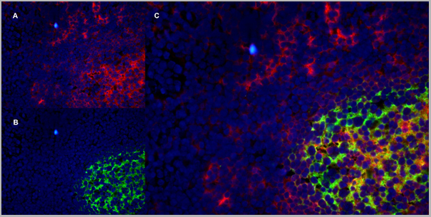
 Application Data
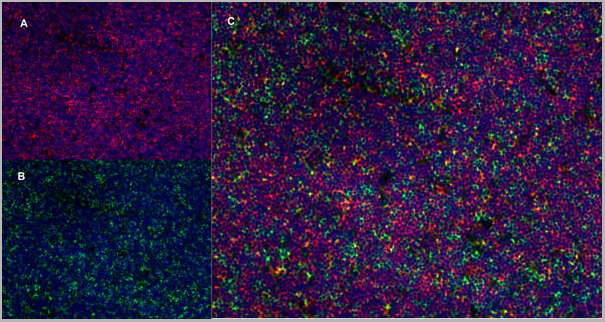
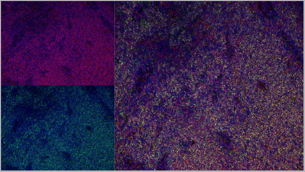
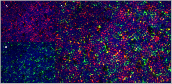
(Immunofluorescence staining of rat lymph node cryosection with Mouse anti Rat CD163 antibody , red an A and Mouse anti Rat CD8 MCA48), green in B. C is the merged image with nuclei counter-stained blue using DAPI. High power)
Application Data
(Immunofluorescence staining of rat lymph node cryosection with Mouse anti Rat CD163 antibody , red an A and Mouse anti Rat CD8 MCA48), green in B. C is the merged image with nuclei counter-stained blue using DAPI. High power)
CD8 ALPHA, Monoclonal Antibody (Cat# AAA11983)
Full Name
MOUSE ANTI RAT CD8 ALPHA
Applications
Immunohistochemistry, Flow Cytometry, Immunofluorescence, Immunoprecipitation, Immunohistochemistry, Western Blot
Pricing
 Application Data
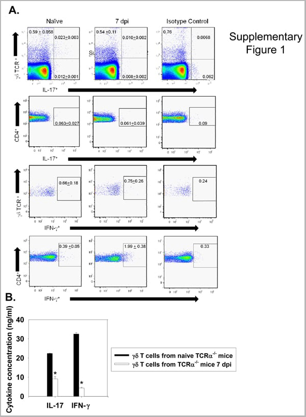
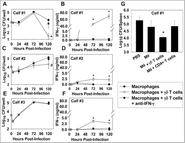
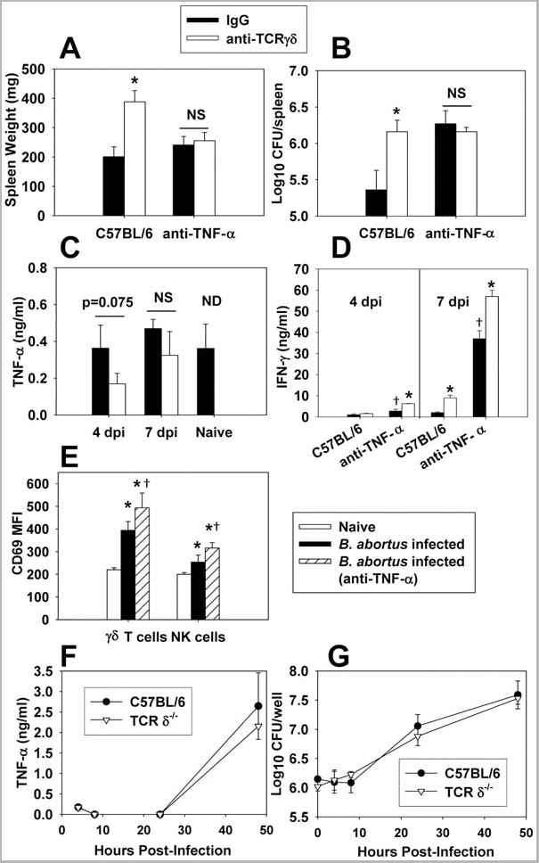
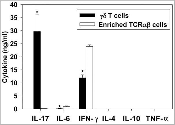
(Published customer image: gamma delta T cells are the primary source of IL-17 during B. abortus infection. C57BL/6 mice were infected i.p. with 5x10⁴ CFUs of B. abortus 2308, and two weeks later gamma delta T cells (>95% purity) and an enriched TCRalphabeta (~55% CD4+, 25% CD8+) cell fraction were isolated from the spleens of infected mice. Cells were stimulated with 500 ng/ml ionomycin and 50 ng/ml PMA for three days, and cell-free supernatants from triplicate wells were assayed for cytokine production via ELISA. The mean +/- SD is shown; * P)
Application Data
(Published customer image: gamma delta T cells are the primary source of IL-17 during B. abortus infection. C57BL/6 mice were infected i.p. with 5x10⁴ CFUs of B. abortus 2308, and two weeks later gamma delta T cells (>95% purity) and an enriched TCRalphabeta (~55% CD4+, 25% CD8+) cell fraction were isolated from the spleens of infected mice. Cells were stimulated with 500 ng/ml ionomycin and 50 ng/ml PMA for three days, and cell-free supernatants from triplicate wells were assayed for cytokine production via ELISA. The mean +/- SD is shown; * P)
IFN GAMMA, Monoclonal Antibody (Cat# AAA12093)
Full Name
MOUSE ANTI BOVINE INTERFERON GAMMA
Reactivity
Dog, Dolphin, Ferret, Fin Whale, Goat, Horse, Human, Mink, Rabbit, Pig, Sheep.
Based on sequence similarity, is expected to react with: Mustelid
N.B. Antibody reactivity and working conditions may vary between species.
Based on sequence similarity, is expected to react with: Mustelid
N.B. Antibody reactivity and working conditions may vary between species.
Applications
Flow Cytometry
Pricing
 WB (Western Blot)
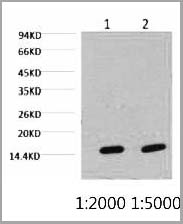
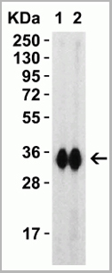
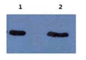
(The following image is the result of using mAb 5B8 ascites at 1:5000 dilution to detect Abeta40 and Abeta42 on Western blot. Lane 1: Abeta42, Lane 2: Abeta40.)
WB (Western Blot)
(The following image is the result of using mAb 5B8 ascites at 1:5000 dilution to detect Abeta40 and Abeta42 on Western blot. Lane 1: Abeta42, Lane 2: Abeta40.)
amyloid beta peptide N-terminal, Monoclonal Antibody (Cat# AAA14629)
Full Name
mAb anti-human amyloid beta peptide N-terminus
Reactivity
Human
Applications
Western Blot
Purity
Protein G affinity purified
Pricing
 IHC (Immunohistchemistry)
(Fig.6. Immunohistochemical analysis of paraffin-embedded rat lung tissue. 1, beta-Tubulin Monoclonal Antibody (3G6) was diluted at 1:400 (4°C, overnight). Negative control was used by secondary antibody only.)
IHC (Immunohistchemistry)
(Fig.6. Immunohistochemical analysis of paraffin-embedded rat lung tissue. 1, beta-Tubulin Monoclonal Antibody (3G6) was diluted at 1:400 (4°C, overnight). Negative control was used by secondary antibody only.)
beta-Tubulin, Monoclonal Antibody (Cat# AAA31540)
Full Name
Anti-beta-Tubulin Mouse Monoclonal Antibody (3G6)
Gene Names
TUBB3; CDCBM; TUBB4; beta-4; CFEOM3A
Reactivity
Human, Mouse, Rat, Monkey, Dog, Chicken, Rabbit, Sheep, Insect, Yeast, Hamster
Applications
Western Blot, Immunohistochemistry, Immunofluorescence
Purity
The antibody was affinity-purified from mouse ascites by affinity-chromatography using specific immunogen
Pricing
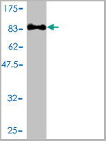
 WB (Western Blot)
(HSPA1L monoclonal antibody Western Blot analysis of HSPA1L expression in PC-12.)
WB (Western Blot)
(HSPA1L monoclonal antibody Western Blot analysis of HSPA1L expression in PC-12.)
HSPA1L, Monoclonal Antibody (Cat# AAA24833)
Full Name
HSPA1L (Heat Shock 70kD Protein 1-like, HSP70-Hom, Heat shock 70kD protein 1L, Heat Shock 70kD Protein 1-Hom) (Biotin)
Gene Names
HSPA1L; HSP70T; hum70t; HSP70-1L; HSP70-HOM
Reactivity
Human, Rat
Applications
Immunohistochemistry, Western Blot
Purity
Purified by Protein A Affinity Chromatography.
Pricing
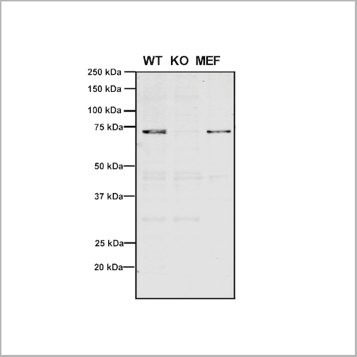
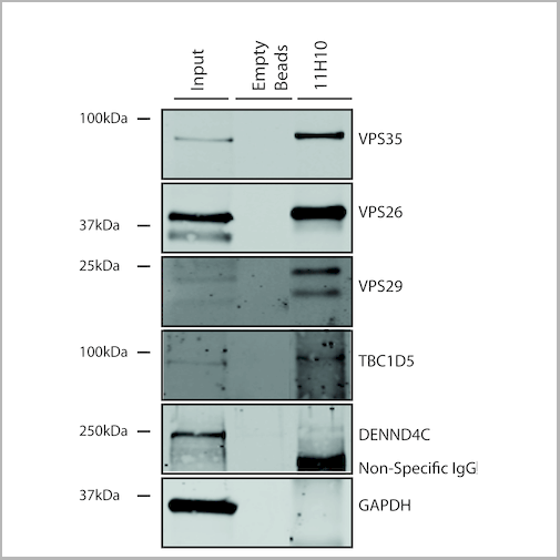
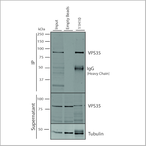
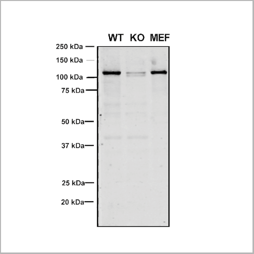
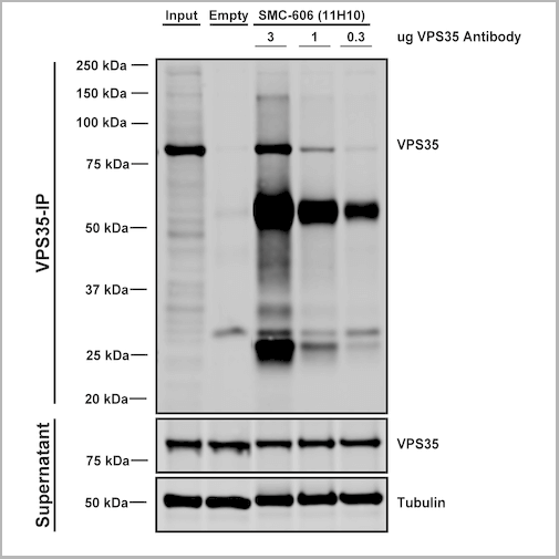 IP (Immunoprecipitation)
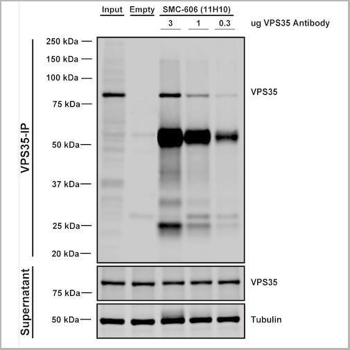
(Immunoprecipitation analysis using Mouse Anti-VPS35 Monoclonal Antibody, Clone 11H10. Tissue: embryonic fibroblast. Species: Mouse. Primary Antibody: Mouse Anti-VPS35 Monoclonal Antibody. Three amounts of (3, 1 and 0.3 ug) were non-covalently coupled to 10uL of A/G sepharose beads for 1 hour at 4 degree C and next incubated with 250ug of MEF lysate for 2 hours at 4 degree C.)
IP (Immunoprecipitation)
(Immunoprecipitation analysis using Mouse Anti-VPS35 Monoclonal Antibody, Clone 11H10. Tissue: embryonic fibroblast. Species: Mouse. Primary Antibody: Mouse Anti-VPS35 Monoclonal Antibody. Three amounts of (3, 1 and 0.3 ug) were non-covalently coupled to 10uL of A/G sepharose beads for 1 hour at 4 degree C and next incubated with 250ug of MEF lysate for 2 hours at 4 degree C.)
VPS35, Monoclonal Antibody (Cat# AAA27705)
Full Name
VPS35 Antibody, Clone 11H10: FITC
Gene Names
VPS35; MEM3; PARK17
Reactivity
Human, Mouse, Rat
Applications
Western Blot, Immunoprecipitation
Purity
Protein G Purified
Pricing


 Application Data
(Published customer image: Dynamics of Th17 cells. D -F) Photographs of double immunolabelled sections showing a representative CD4+ROR?+ cell (arrow) observed around blood vessels (BV). Bar scale = 30 um.Almolda B, Costa M, Montoya M, Gonz¡lez B, Castellano B (2011) Increase in Th17 and T-reg Lymphocytes and Decrease of IL22 Correlate with the Recovery Phase of Acute EAE IN Rat. PLoS ONE 6(11): e27473.)
Application Data
(Published customer image: Dynamics of Th17 cells. D -F) Photographs of double immunolabelled sections showing a representative CD4+ROR?+ cell (arrow) observed around blood vessels (BV). Bar scale = 30 um.Almolda B, Costa M, Montoya M, Gonz¡lez B, Castellano B (2011) Increase in Th17 and T-reg Lymphocytes and Decrease of IL22 Correlate with the Recovery Phase of Acute EAE IN Rat. PLoS ONE 6(11): e27473.)
CD4, Monoclonal Antibody (Cat# AAA11994)
Full Name
MOUSE ANTI RAT CD4 (DOMAIN 1)
Gene Names
Cd4; p55; W3/25
Applications
Immunohistochemistry, Flow Cytometry, Immunohistochemistry
Pricing
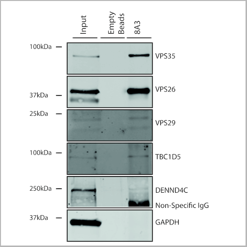
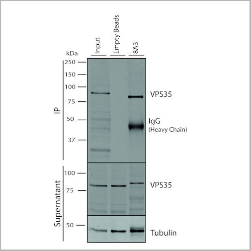
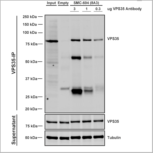 IP (Immunoprecipitation)
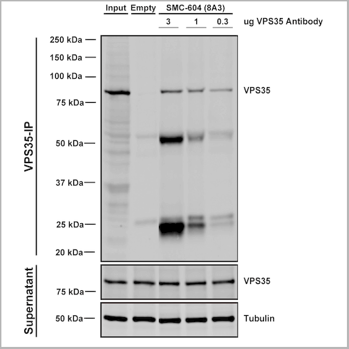
(Immunoprecipitation analysis using Mouse Anti-VPS35 Monoclonal Antibody, Clone 8A3. Tissue: embryonic fibroblast. Species: Mouse. Primary Antibody: Mouse Anti-VPS35 Monoclonal Antibody. Three amounts of (3, 1 and 0.3 ug) were non-covalently coupled to 10uL of A/G sepharose beads for 1 hour at 4 degree C and next incubated with 250ug of MEF lysate for 2 hours at 4 degree C.)
IP (Immunoprecipitation)
(Immunoprecipitation analysis using Mouse Anti-VPS35 Monoclonal Antibody, Clone 8A3. Tissue: embryonic fibroblast. Species: Mouse. Primary Antibody: Mouse Anti-VPS35 Monoclonal Antibody. Three amounts of (3, 1 and 0.3 ug) were non-covalently coupled to 10uL of A/G sepharose beads for 1 hour at 4 degree C and next incubated with 250ug of MEF lysate for 2 hours at 4 degree C.)
VPS35, Monoclonal Antibody (Cat# AAA27685)
Full Name
VPS35 Antibody, Clone 8A3: ATTO 594
Gene Names
VPS35; MEM3; PARK17
Reactivity
Human, Mouse, Rat
Applications
Western Blot, Immunocytochemistry, Immunofluorescence, Immunoprecipitation
Purity
Protein G Purified
Pricing
 Application Data
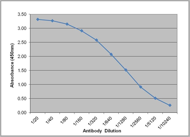
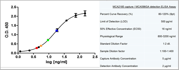
(Sandwich ELISA analysis of CD178 binding using Mouse anti Human CD178 as a capture reagent and biotinylated Mouse anti Human CD178 as a detection reagent with purified human CD178 as antigen for the generation of a standard curve. Detection is by HRP conjugated Streptavidin and substrate. Microtitre plate is read at O.D. 450 nm on the iMark Microplate Absorbance Reader . Plasma (orange) sample is displayed at 1:2 dilution.)
Application Data
(Sandwich ELISA analysis of CD178 binding using Mouse anti Human CD178 as a capture reagent and biotinylated Mouse anti Human CD178 as a detection reagent with purified human CD178 as antigen for the generation of a standard curve. Detection is by HRP conjugated Streptavidin and substrate. Microtitre plate is read at O.D. 450 nm on the iMark Microplate Absorbance Reader . Plasma (orange) sample is displayed at 1:2 dilution.)
CD178, Monoclonal Antibody (Cat# AAA11962)
Full Name
MOUSE ANTI HUMAN CD178
Gene Names
FASLG; APTL; FASL; CD178; CD95L; ALPS1B; CD95-L; TNFSF6; APT1LG1
Reactivity
Human
Applications
Flow Cytometry, Immunoprecipitation
Pricing


 Application Data
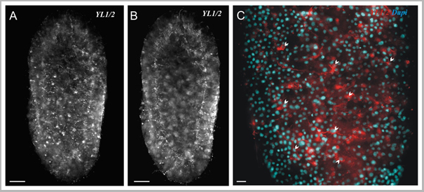
(Published customer image: Spindle abnormalities in embryos derived from imp-a2D14/imp-betaKetRE34 and imp-a2D14/imp-betac02473; NLSB-/+ females. (A -D) Wild-type and mutant embryos stained for a-tubulin (green) and DNA (blue). (A) Mitotic spindles in wild-type embryos at metaphase and anaphase. (B, C) Categories of spindle abnormalities found in embryos derived from (B) imp-a2D14/imp-betaKetRE34 and (C) imp-a2D14/imp-betac02743; NLSB-/+ females. (D) Formation of aster networks found in both genotypes. Scale bar: 10 um. (E) Frequency of spindle defects in embryos from both types of mutant females. Female genotypes are displayed at the upper right corner. At least 200 spindles were scored for both genotypes.From: Specific Cooperation Between Imp-a2 and Imp-beta/Ketel in Spindle Assembly During Drosophila Early Nuclear Divisions Erika Vir¡gh, M¡ty¡s Gorj¡n¡cz, Istv¡n T¶r¶k, Tolga Eichhorn, Sowjanya Kallakuri, Tam¡s Szlanka, Istv¡n Kiss, and Bernard M. Mechler G3 January 2012 2:1-14.)
Application Data
(Published customer image: Spindle abnormalities in embryos derived from imp-a2D14/imp-betaKetRE34 and imp-a2D14/imp-betac02473; NLSB-/+ females. (A -D) Wild-type and mutant embryos stained for a-tubulin (green) and DNA (blue). (A) Mitotic spindles in wild-type embryos at metaphase and anaphase. (B, C) Categories of spindle abnormalities found in embryos derived from (B) imp-a2D14/imp-betaKetRE34 and (C) imp-a2D14/imp-betac02743; NLSB-/+ females. (D) Formation of aster networks found in both genotypes. Scale bar: 10 um. (E) Frequency of spindle defects in embryos from both types of mutant females. Female genotypes are displayed at the upper right corner. At least 200 spindles were scored for both genotypes.From: Specific Cooperation Between Imp-a2 and Imp-beta/Ketel in Spindle Assembly During Drosophila Early Nuclear Divisions Erika Vir¡gh, M¡ty¡s Gorj¡n¡cz, Istv¡n T¶r¶k, Tolga Eichhorn, Sowjanya Kallakuri, Tam¡s Szlanka, Istv¡n Kiss, and Bernard M. Mechler G3 January 2012 2:1-14.)
TUBULIN ALPHA, Monoclonal Antibody (Cat# AAA12232)
Full Name
RAT ANTI TUBULIN ALPHA:HRP
Applications
Immunohistochemistry, Western Blot
Pricing

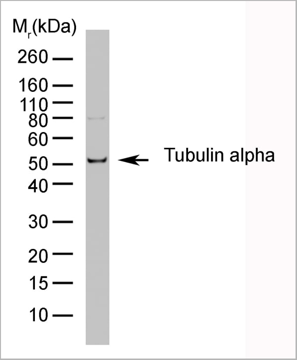

 Application Data
(Published customer image: Spindle abnormalities in embryos derived from imp-a2D14/imp-betaKetRE34 and imp-a2D14/imp-betac02473; NLSB-/+ females. (A -D) Wild-type and mutant embryos stained for a-tubulin (green) and DNA (blue). (A) Mitotic spindles in wild-type embryos at metaphase and anaphase. (B, C) Categories of spindle abnormalities found in embryos derived from (B) imp-a2D14/imp-betaKetRE34 and (C) imp-a2D14/imp-betac02743; NLSB-/+ females. (D) Formation of aster networks found in both genotypes. Scale bar: 10 um. (E) Frequency of spindle defects in embryos from both types of mutant females. Female genotypes are displayed at the upper right corner. At least 200 spindles were scored for both genotypes.From: Specific Cooperation Between Imp-a2 and Imp-beta/Ketel in Spindle Assembly During Drosophila Early Nuclear Divisions Erika Vir¡gh, M¡ty¡s Gorj¡n¡cz, Istv¡n T¶r¶k, Tolga Eichhorn, Sowjanya Kallakuri, Tam¡s Szlanka, Istv¡n Kiss, and Bernard M. Mechler G3 January 2012 2:1-14.)
Application Data
(Published customer image: Spindle abnormalities in embryos derived from imp-a2D14/imp-betaKetRE34 and imp-a2D14/imp-betac02473; NLSB-/+ females. (A -D) Wild-type and mutant embryos stained for a-tubulin (green) and DNA (blue). (A) Mitotic spindles in wild-type embryos at metaphase and anaphase. (B, C) Categories of spindle abnormalities found in embryos derived from (B) imp-a2D14/imp-betaKetRE34 and (C) imp-a2D14/imp-betac02743; NLSB-/+ females. (D) Formation of aster networks found in both genotypes. Scale bar: 10 um. (E) Frequency of spindle defects in embryos from both types of mutant females. Female genotypes are displayed at the upper right corner. At least 200 spindles were scored for both genotypes.From: Specific Cooperation Between Imp-a2 and Imp-beta/Ketel in Spindle Assembly During Drosophila Early Nuclear Divisions Erika Vir¡gh, M¡ty¡s Gorj¡n¡cz, Istv¡n T¶r¶k, Tolga Eichhorn, Sowjanya Kallakuri, Tam¡s Szlanka, Istv¡n Kiss, and Bernard M. Mechler G3 January 2012 2:1-14.)
TUBULIN ALPHA, Monoclonal Antibody (Cat# AAA12009)
Full Name
RAT ANTI TUBULIN ALPHA
Applications
Immunohistochemistry, Immunofluorescence, Immunoprecipitation, Radioimmunoassay, Western Blot
Pricing


 Application Data
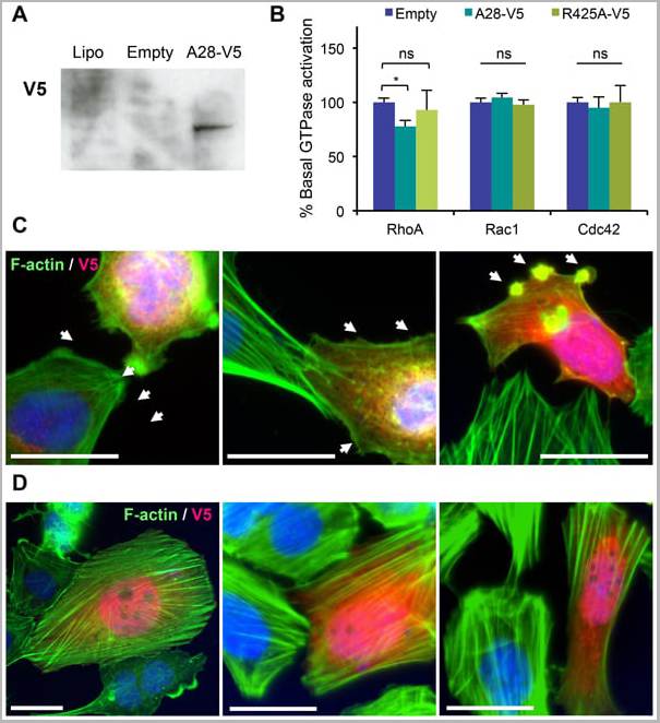
(Published customer image: Mouse anti V5 tag antibody, clone SV5-Pk1 used for the detection of V5 tagged WEEV_nsP3 protein by western blotting and immunofluorescenceImage caption: WEEV nsP3 interaction with host IKKbeta. A) U87MGs were transfected in a 6-well plate with 5 ug of pUC19 and WEEV_nsP3_HA for 24 hours. Cell lysates were resolved using SDS-PAGE and subsequently immunoblotted with V5 antibody and beta-actin served as a loading control. B) U87MGs were transfected with WEEV_nsP3_V5; cells were fixed after 24 hours and stained with antibodies against the endogenous IKKbeta and the V5 tag. Cells were incubated with appropriate secondary Alexa Fluor antibodies and the nuclei stained with DAPI. Co-localization of IKKbeta with WEEV_nsP3_V5 (yellow) was observed as shown by the arrows. B) Panels E -H serve as an example of transfected cells in a given field of view that show co-localization of IKKbeta and WEEV_nsP3_V5 24 hours post transfection. Panels I-L represent magnified images of other cells showing co-localization of IKKbeta and WEEV_nsP3_V5. Panel M is a magnified image of panel L. The co-localization was confirmed by Z-stack analysis. Co-localization was calculated to be approximately in 61% of cells (163 cells were counted of which 44% demonstrated expression of nsP3. Of those cells that expressed nsP3, 61% showed co-localization of both proteins). Images were taken using Nikon Eclipse TE2000-U at 60x magnification and are representative of 2 independent experiments.From: Amaya M, Voss K, Sampey G, Senina S, de la Fuente C, et al. (2014) The Role of IKKbeta in Venezuelan Equine Encephalitis Virus Infection. PLoS ONE 9(2): e86745.)
Application Data
(Published customer image: Mouse anti V5 tag antibody, clone SV5-Pk1 used for the detection of V5 tagged WEEV_nsP3 protein by western blotting and immunofluorescenceImage caption: WEEV nsP3 interaction with host IKKbeta. A) U87MGs were transfected in a 6-well plate with 5 ug of pUC19 and WEEV_nsP3_HA for 24 hours. Cell lysates were resolved using SDS-PAGE and subsequently immunoblotted with V5 antibody and beta-actin served as a loading control. B) U87MGs were transfected with WEEV_nsP3_V5; cells were fixed after 24 hours and stained with antibodies against the endogenous IKKbeta and the V5 tag. Cells were incubated with appropriate secondary Alexa Fluor antibodies and the nuclei stained with DAPI. Co-localization of IKKbeta with WEEV_nsP3_V5 (yellow) was observed as shown by the arrows. B) Panels E -H serve as an example of transfected cells in a given field of view that show co-localization of IKKbeta and WEEV_nsP3_V5 24 hours post transfection. Panels I-L represent magnified images of other cells showing co-localization of IKKbeta and WEEV_nsP3_V5. Panel M is a magnified image of panel L. The co-localization was confirmed by Z-stack analysis. Co-localization was calculated to be approximately in 61% of cells (163 cells were counted of which 44% demonstrated expression of nsP3. Of those cells that expressed nsP3, 61% showed co-localization of both proteins). Images were taken using Nikon Eclipse TE2000-U at 60x magnification and are representative of 2 independent experiments.From: Amaya M, Voss K, Sampey G, Senina S, de la Fuente C, et al. (2014) The Role of IKKbeta in Venezuelan Equine Encephalitis Virus Infection. PLoS ONE 9(2): e86745.)
V5-TAG, Monoclonal Antibody (Cat# AAA12081)
Full Name
MOUSE ANTI V5-TAG:HRP
Applications
Western Blot
Pricing
COVID 19 Spike RBD Coronavirus, Recombinant Protein (Cat# AAA13542)
Full Name
Recombinant SARS-Cov-2 Spike RBD Protein (Detection)
Applications
Lateral Flow
Purity
>95% by SDS-PAGE
Pricing
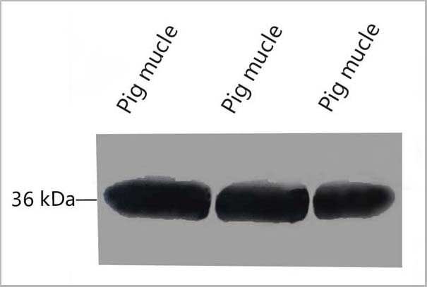
 WB (Western Blot)
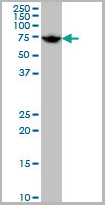
((2ug/ml) staining of Human Liver lysate (35ug protein in RIPA buffer). Primary incubation was 1 hour. Detected by chemiluminescence.)
WB (Western Blot)
((2ug/ml) staining of Human Liver lysate (35ug protein in RIPA buffer). Primary incubation was 1 hour. Detected by chemiluminescence.)
CLEC16A, Polyclonal Antibody (Cat# AAA13667)
Full Name
Goat anti-CLEC16A (aa1040-53) Antibody
Gene Names
CLEC16A; Gop-1; KIAA0350
Reactivity
Tested: Human; Expected from sequence similarity: Human
Applications
Peptide ELISA, Western Blot
Purity
Purified from goat serum by ammonium sulphate precipitation followed by antigen affinity chromatography using the immunizing peptide.
Pricing
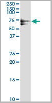
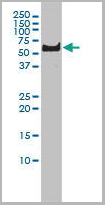
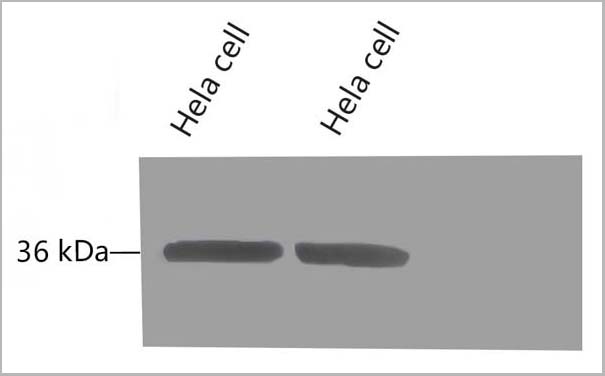
 WB (Western Blot)
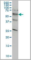
(STK33 monoclonal antibody, Western Blot analysis of STK33 expression in HeLa.)
WB (Western Blot)
(STK33 monoclonal antibody, Western Blot analysis of STK33 expression in HeLa.)
STK33, Monoclonal Antibody (Cat# AAA25856)
Full Name
STK33 (Serine/Threonine Kinase 33) (PE)
Reactivity
Human
Applications
Immunofluorescence, Immunoprecipitation, Western Blot
Purity
Purified by Protein A Affinity Chromatography.
Pricing
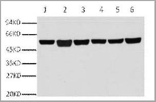
 WB (Western Blot)
(Fig.8 Western blot analysis of Hela, GAPDH Monoclonal Antibody (2B5) was diluted at 1:10000 (25°C, 3h).)
WB (Western Blot)
(Fig.8 Western blot analysis of Hela, GAPDH Monoclonal Antibody (2B5) was diluted at 1:10000 (25°C, 3h).)
GAPDH, Monoclonal Antibody (Cat# AAA31539)
Full Name
Anti-GAPDH Mouse Monoclonal Antibody (2B5)
Gene Names
GAPDH; G3PD; GAPD
Reactivity
Human, Mouse, Rat, Monkey, Dog, Chicken, Rabbit, Pig, Sheep, Yeast, Hamster
Applications
Western Blot, Immunohistochemistry, Immunofluorescence
Purity
The antibody was affinity-purified from mouse ascites by affinity-chromatography using specific immunogen
Pricing
 Application Data
(Staining of mouse spleen cells with Rat anti Mouse CD3 Epsilon (T3): APC)
Application Data
(Staining of mouse spleen cells with Rat anti Mouse CD3 Epsilon (T3): APC)
CD3, Monoclonal Antibody (Cat# AAA11984)
Full Name
RAT ANTI MOUSE CD3
Gene Names
Cd3e; CD3; T3e; AI504783; CD3epsilon
Applications
Immunohistochemistry, Flow Cytometry
Pricing
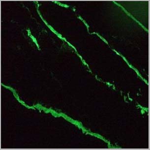
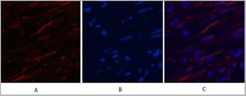
 IF (Immunofluorescence)
(Fig.7. Immunofluorescence analysis of rat liver tissue. 1, Histone H3 Monoclonal Antibody (2D10) (red) was diluted at 1:200 (4°C, overnight). 2, Cy3 Labeled secondary antibody was diluted at 1:300 (room temperature, 50min). 3, Picture B: DAPI (blue) 10min. Picture A: Target. Picture B: DAPI. Picture C: merge of A+B.)
IF (Immunofluorescence)
(Fig.7. Immunofluorescence analysis of rat liver tissue. 1, Histone H3 Monoclonal Antibody (2D10) (red) was diluted at 1:200 (4°C, overnight). 2, Cy3 Labeled secondary antibody was diluted at 1:300 (room temperature, 50min). 3, Picture B: DAPI (blue) 10min. Picture A: Target. Picture B: DAPI. Picture C: merge of A+B.)
Histone H3, Monoclonal Antibody (Cat# AAA31543)
Full Name
Anti-Histone H3 Mouse Monoclonal Antibody (2D10)
Gene Names
HIST1H3A; H3/A; H3FA
Reactivity
Human, Mouse, Rat, Yeast
Applications
Western Blot, Immunohistochemistry, Immunofluorescence, Immunoprecipitation
Pricing
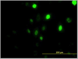
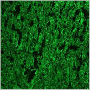
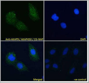 IF (Immunofluorescence)
(AAA13648 Immunofluorescence analysis of paraformaldehyde fixed U2OS cells, permeabilized with 0.15% Triton. Primary incubation 1hr (10ug/ml) followed by Alexa Fluor 488 secondary antibody (4ug/ml), showing cytoplasmic staining. The nuclear stain is DAPI (blue). Negative control: Unimmunized goat IgG (10ug/ml) followed by Alexa Fluor 488 secondary antibody (4ug/ml).)
IF (Immunofluorescence)
(AAA13648 Immunofluorescence analysis of paraformaldehyde fixed U2OS cells, permeabilized with 0.15% Triton. Primary incubation 1hr (10ug/ml) followed by Alexa Fluor 488 secondary antibody (4ug/ml), showing cytoplasmic staining. The nuclear stain is DAPI (blue). Negative control: Unimmunized goat IgG (10ug/ml) followed by Alexa Fluor 488 secondary antibody (4ug/ml).)
AKAP9/AKAP450/CG-NAP, Polyclonal Antibody (Cat# AAA13648)
Full Name
Goat anti-AKAP9/AKAP450/CG-NAP Antibody
Gene Names
AKAP9; LQT11; PRKA9; AKAP-9; CG-NAP; YOTIAO; AKAP350; AKAP450; PPP1R45; HYPERION; MU-RMS-40.16A
Reactivity
Tested: Human; Expected from sequence similarity: Human, Mouse
Applications
Peptide ELISA, Western Blot, Immunofluorescence
Purity
Purified from goat serum by ammonium sulphate precipitation followed by antigen affinity chromatography using the immunizing peptide.
Pricing


 IP (Immunoprecipitation)
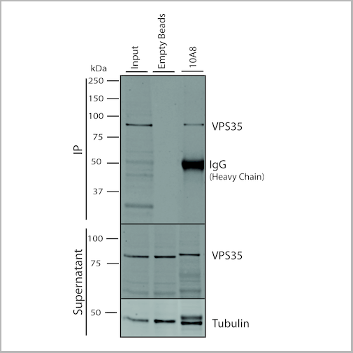
(Immunoprecipitation analysis using Mouse Anti-VPS35 Monoclonal Antibody, Clone 10A8. Tissue: embryonic fibroblast. Species: Mouse. Primary Antibody: Mouse Anti-VPS35 Monoclonal Antibody. Three amounts of (3, 1 and 0.3 ug) were non-covalently coupled to 10uL of A/G sepharose beads for 1 hour at 4 degree C and next incubated with 250ug of MEF lysate for 2 hours at 4 degree C.)
IP (Immunoprecipitation)
(Immunoprecipitation analysis using Mouse Anti-VPS35 Monoclonal Antibody, Clone 10A8. Tissue: embryonic fibroblast. Species: Mouse. Primary Antibody: Mouse Anti-VPS35 Monoclonal Antibody. Three amounts of (3, 1 and 0.3 ug) were non-covalently coupled to 10uL of A/G sepharose beads for 1 hour at 4 degree C and next incubated with 250ug of MEF lysate for 2 hours at 4 degree C.)
VPS35, Monoclonal Antibody (Cat# AAA27698)
Full Name
VPS35 Antibody, Clone 10A8: PerCP
Gene Names
VPS35; MEM3; PARK17
Reactivity
Human, Mouse, Rat
Applications
Western Blot, Immunocytochemistry, Immunofluorescence, Immunoprecipitation
Purity
Protein G Purified
Pricing