Filters
Clonality
Type
Reactivity
Gene Name
Isotype
Host
Application
Clone
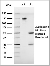
298 results for "Mouse IgG Isotype Control" - showing 250-298


 Application Data
(Staining of mouse peritoneal macrophages with Rat anti Mouse Beta-glucan Receptor: FITC)
Application Data
(Staining of mouse peritoneal macrophages with Rat anti Mouse Beta-glucan Receptor: FITC)
DECTIN-1, Monoclonal Antibody (Cat# AAA12133)
Full Name
RAT ANTI MOUSE DECTIN-1:FITC
Gene Names
Clec7a; BGR; beta-GR; Clecsf12
Applications
Flow Cytometry
Pricing


 Application Data
(Staining of mouse peritoneal macrophages with Rat anti Mouse Beta-glucan Receptor: FITC)
Application Data
(Staining of mouse peritoneal macrophages with Rat anti Mouse Beta-glucan Receptor: FITC)
DECTIN-1, Monoclonal Antibody (Cat# AAA12135)
Full Name
RAT ANTI MOUSE DECTIN-1:RPE
Gene Names
Clec7a; BGR; beta-GR; Clecsf12
Applications
Flow Cytometry
Pricing


 Application Data
(Staining of human peripheral blood lymphocytes with Mouse anti Human CD40:RPE)
Application Data
(Staining of human peripheral blood lymphocytes with Mouse anti Human CD40:RPE)
CD40, Monoclonal Antibody (Cat# AAA12089)
Full Name
MOUSE ANTI HUMAN CD40:RPE
Gene Names
CD40; p50; Bp50; CDW40; TNFRSF5
Applications
Flow Cytometry
Pricing


 Application Data
(Staining of mouse peritoneal macrophages with Rat anti Mouse Beta-glucan Receptor: FITC)
Application Data
(Staining of mouse peritoneal macrophages with Rat anti Mouse Beta-glucan Receptor: FITC)
DECTIN-1, Monoclonal Antibody (Cat# AAA12126)
Full Name
RAT ANTI MOUSE DECTIN-1
Gene Names
Clec7a; BGR; beta-GR; Clecsf12
Applications
Immunohistochemistry, Flow Cytometry, Immunoprecipitation
Pricing


 Application Data
(Staining of human peripheral blood lymphocytes with Mouse anti Human CD40:RPE)
Application Data
(Staining of human peripheral blood lymphocytes with Mouse anti Human CD40:RPE)
CD40, Monoclonal Antibody (Cat# AAA12031)
Full Name
MOUSE ANTI HUMAN CD40:RPE
Gene Names
CD40; p50; Bp50; CDW40; TNFRSF5
Applications
Flow Cytometry
Pricing
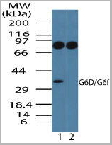
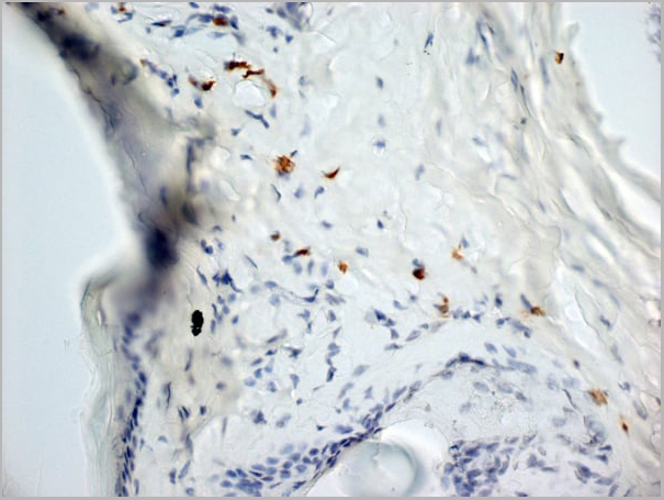
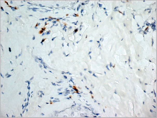
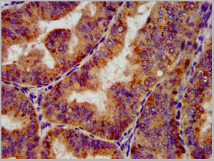
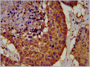
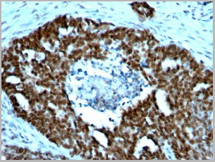
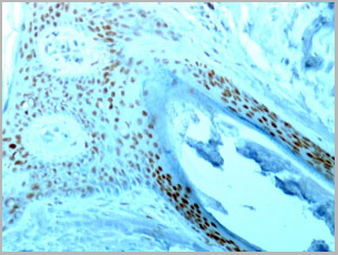
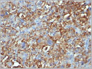
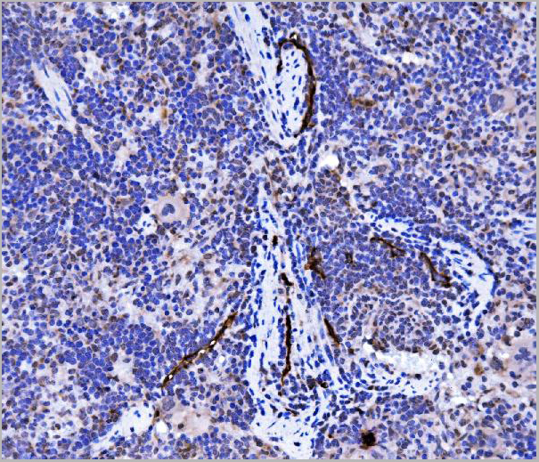
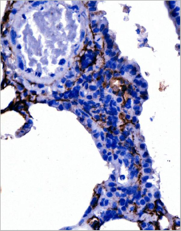
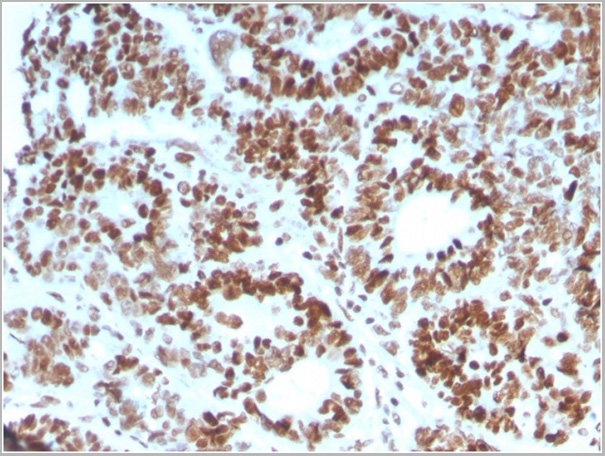
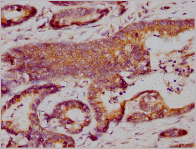
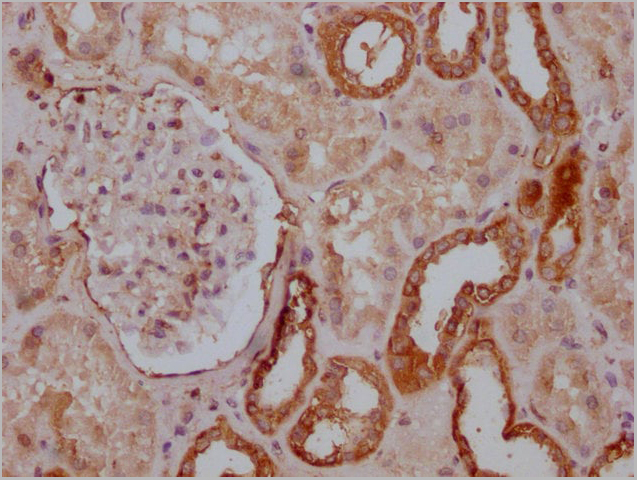
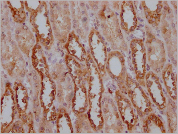
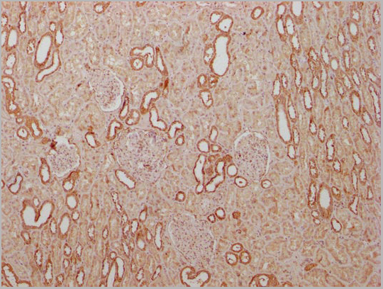
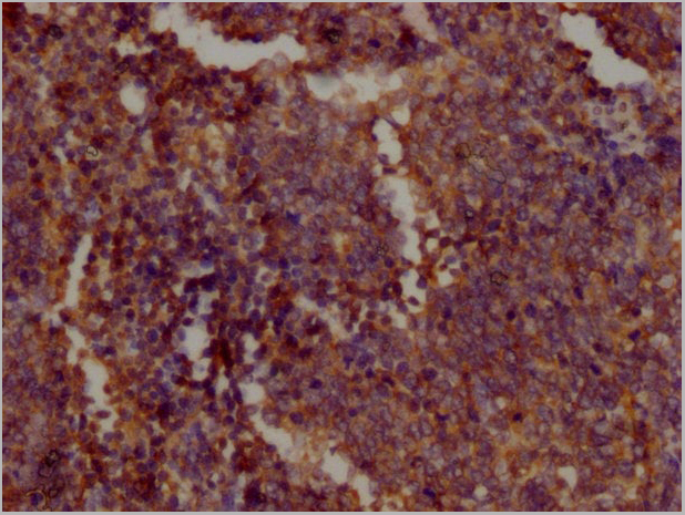
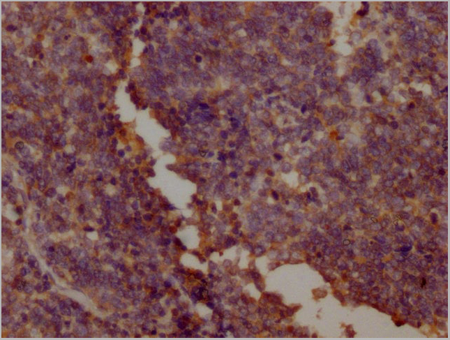
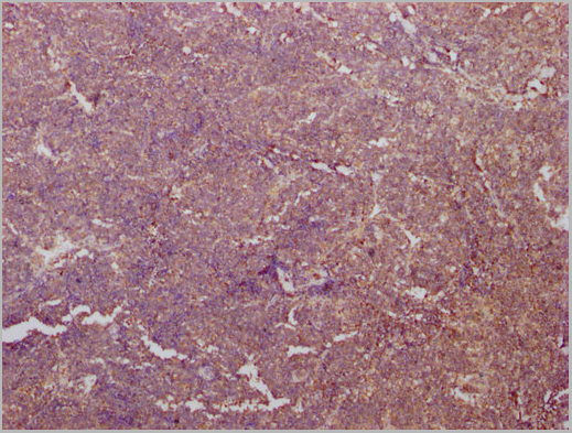
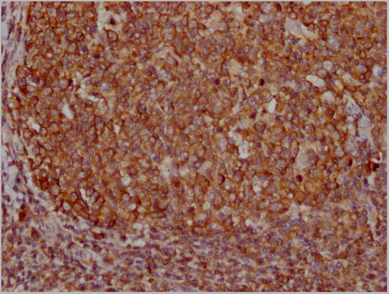
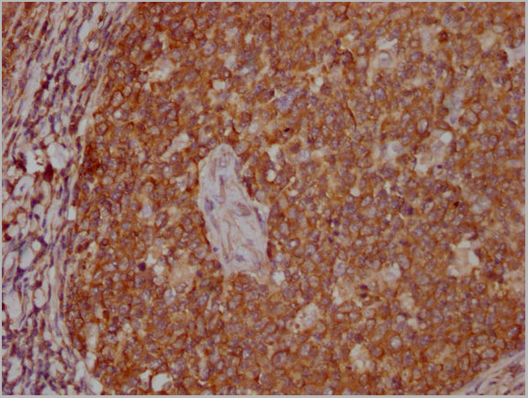
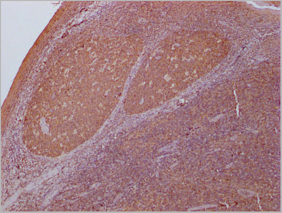
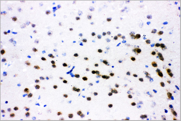
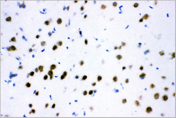
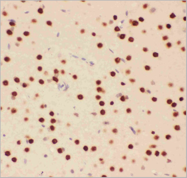
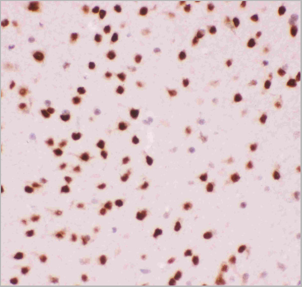
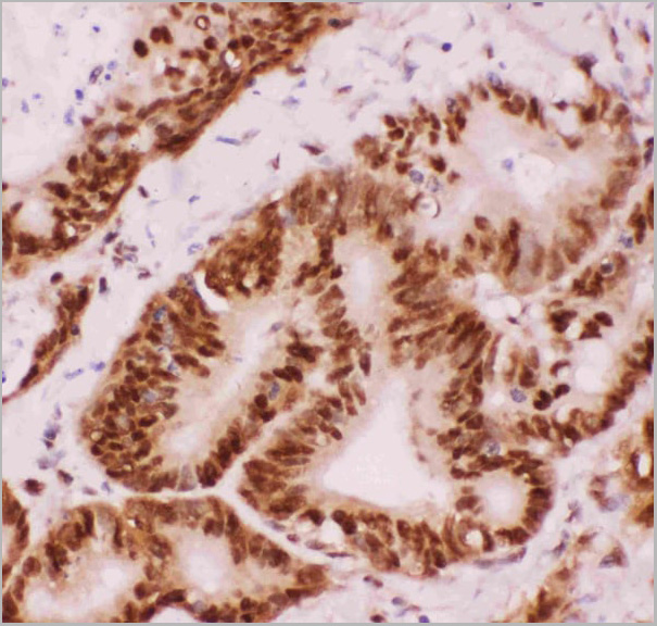
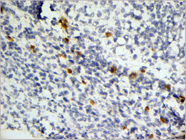
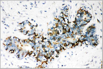
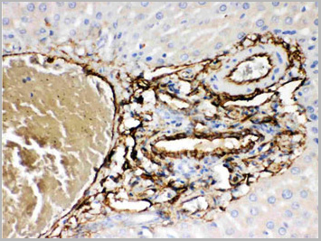
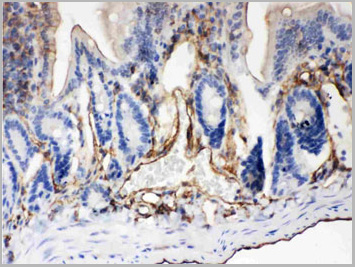
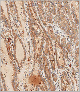
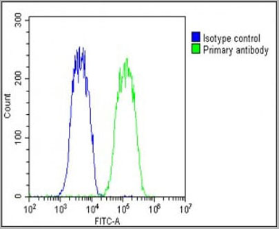
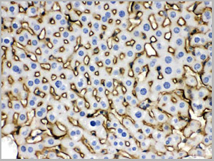
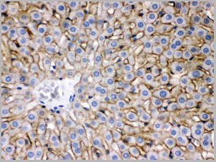
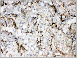
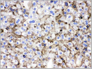
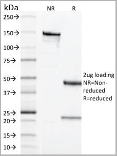
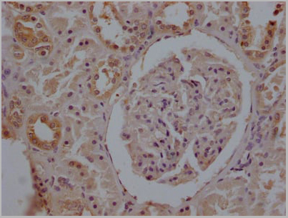
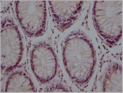
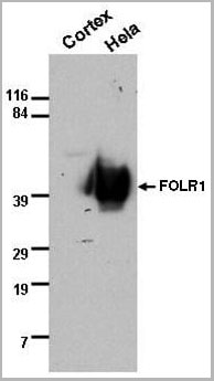
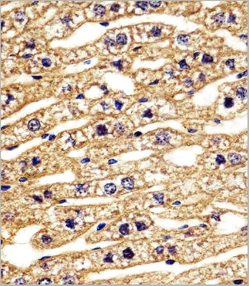
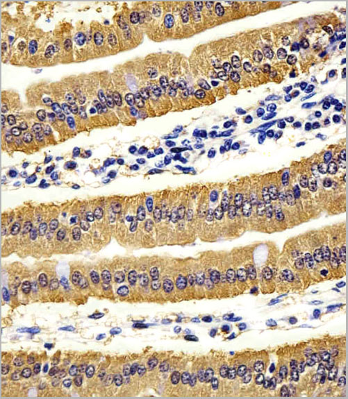
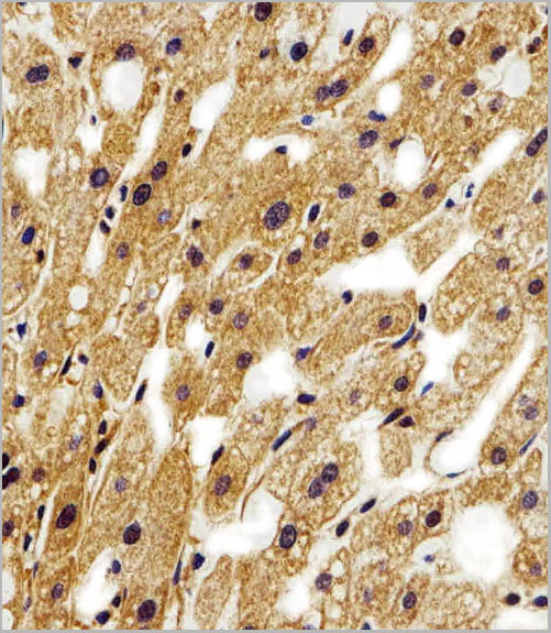
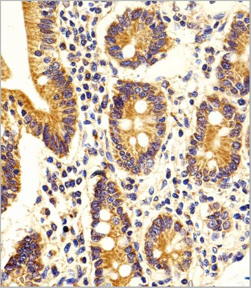
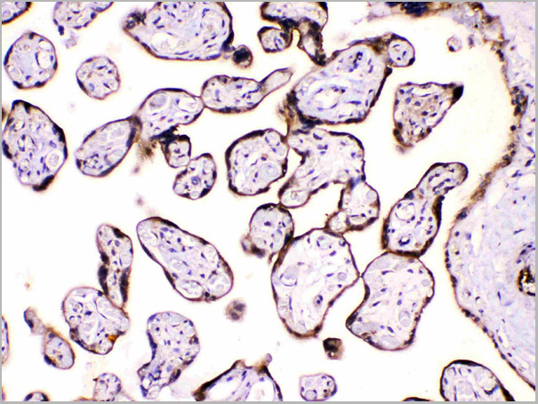
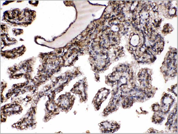
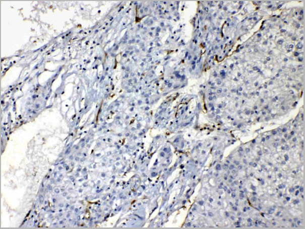
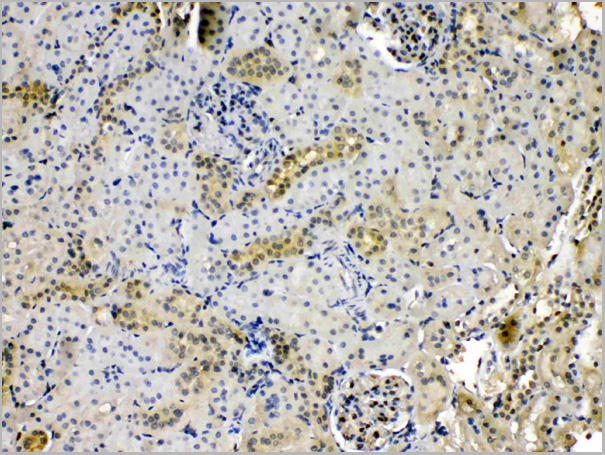
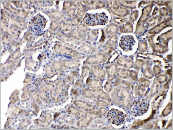
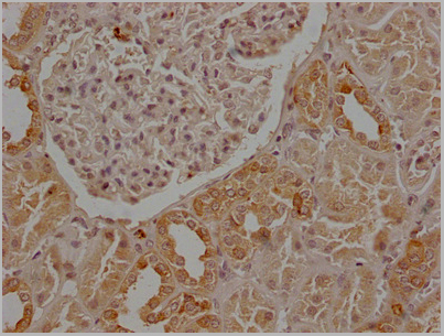
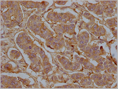
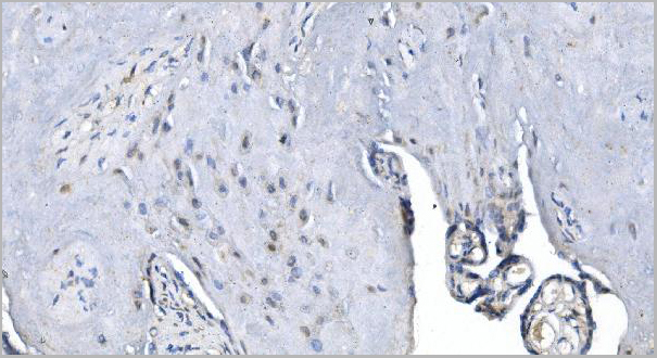
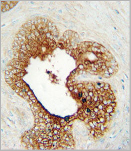
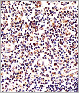
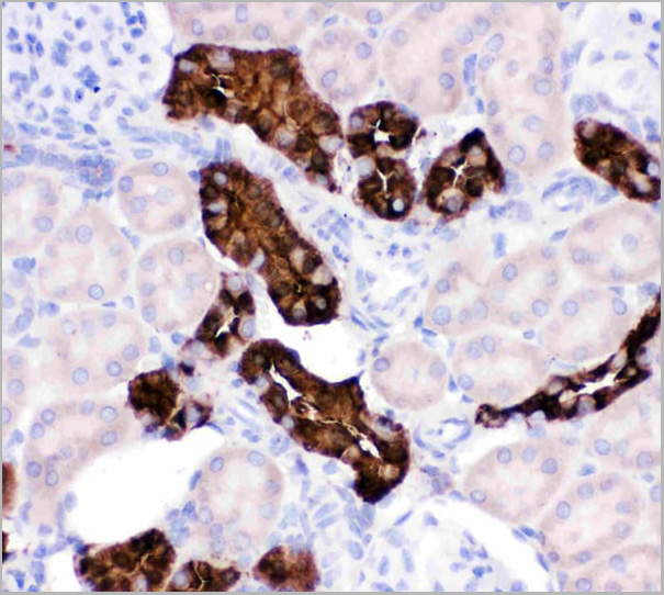
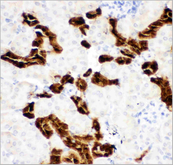
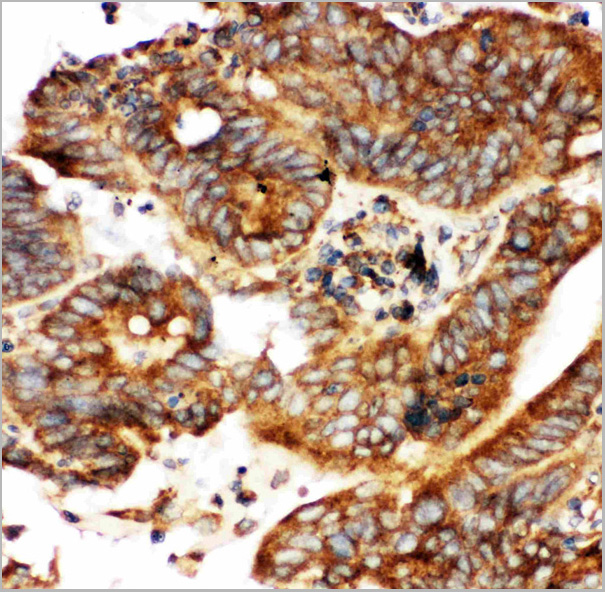
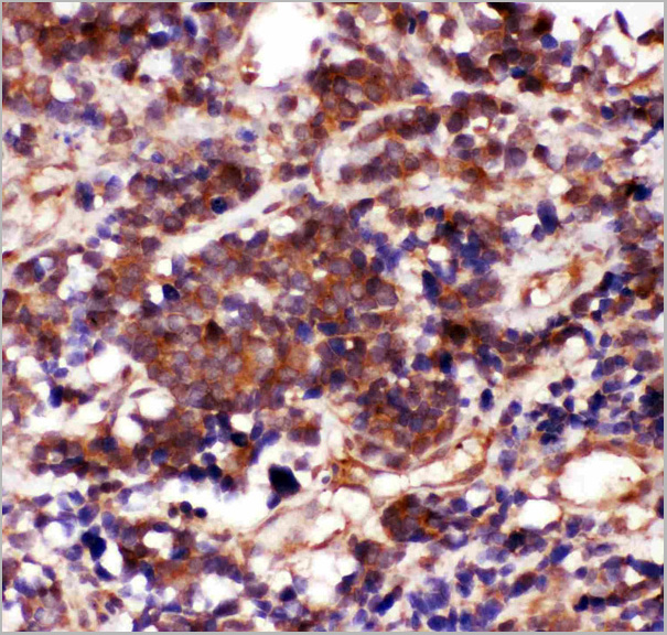
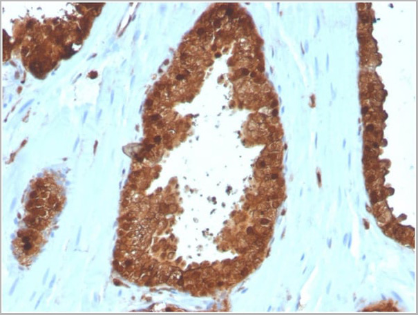
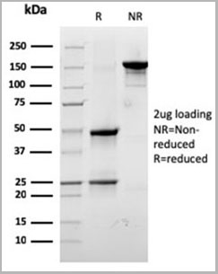
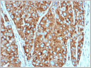
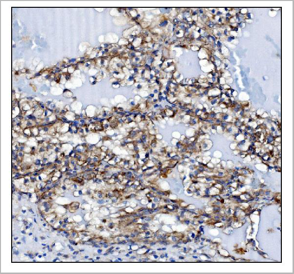
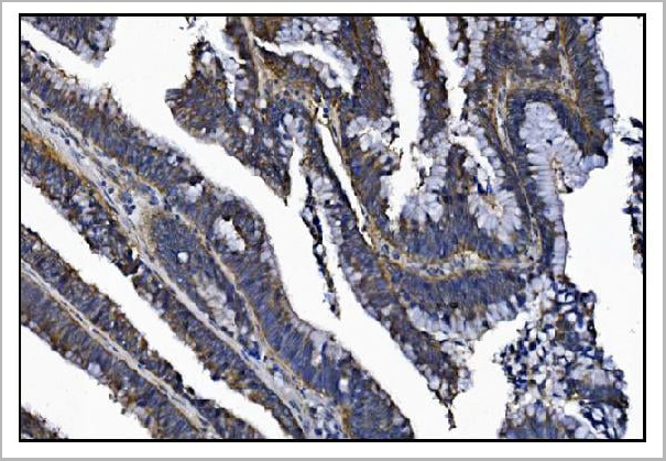
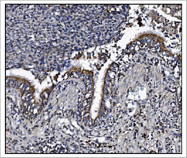
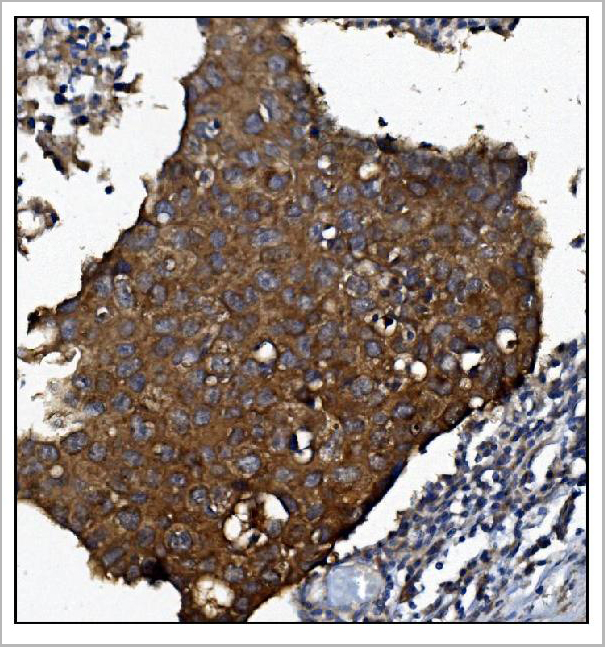
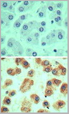 IHC (Immunohistochemistry)
(Immunohistochemical analysis of G6D in formalin-fixed, paraffin-embedded human liver tissue using an isotype control (top) and (bottom) AAA24064 at 5ug/ml.)
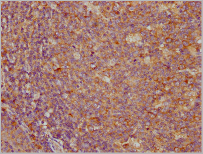
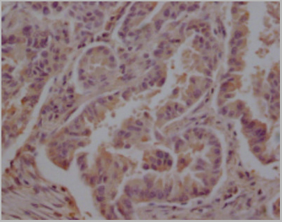
IHC (Immunohistochemistry)
(Immunohistochemical analysis of G6D in formalin-fixed, paraffin-embedded human liver tissue using an isotype control (top) and (bottom) AAA24064 at 5ug/ml.)
LY6G6D, Polyclonal Antibody (Cat# AAA24064)
Full Name
LY6G6D (G6D, Lymphocyte Antigen 6 Complex Locus Protein G6f, C6orf21, G6F, LY6G6D, NG32)
Gene Names
LY6G6D; G6D; NG25; LY6-D; MEGT1; C6orf23
Reactivity
Human
Applications
Western Blot, Immunohistochemistry
Purity
Purified by Protein A affinity chromatography.
Pricing
 Application Data
(Analysis of Protein Array containing more than 19,000 full-length human proteins using Estrogen Receptor alpha Mouse Monoclonal Antibody (ESR1/3557) Z- and S- Score: The Z-score represents the strength of a signal that a monoclonal antibody (MAb) (in combination with a fluorescently-tagged anti-IgG secondary antibody) produces when binding to a particular protein on the HuProtTM array. Z-scores are described in units of standard deviations (SD’s) above the mean value of all signals generated on that array. If targets on HuProtTM are arranged in descending order of the Z-score, the S-score is the difference (also in units of SD’s) between the Z-score. S-score therefore represents the relative target specificity of a MAb to its intended target. A MAb is considered to specific to its intended target, if the MAb has an S-score of at least 2.5. For example, if a MAb binds to protein X with a Z-score of 43 and to protein Y with a Z-score of 14, then the S-score for the binding of that MAb to protein X is equal to 29.)
Application Data
(Analysis of Protein Array containing more than 19,000 full-length human proteins using Estrogen Receptor alpha Mouse Monoclonal Antibody (ESR1/3557) Z- and S- Score: The Z-score represents the strength of a signal that a monoclonal antibody (MAb) (in combination with a fluorescently-tagged anti-IgG secondary antibody) produces when binding to a particular protein on the HuProtTM array. Z-scores are described in units of standard deviations (SD’s) above the mean value of all signals generated on that array. If targets on HuProtTM are arranged in descending order of the Z-score, the S-score is the difference (also in units of SD’s) between the Z-score. S-score therefore represents the relative target specificity of a MAb to its intended target. A MAb is considered to specific to its intended target, if the MAb has an S-score of at least 2.5. For example, if a MAb binds to protein X with a Z-score of 43 and to protein Y with a Z-score of 14, then the S-score for the binding of that MAb to protein X is equal to 29.)
Estrogen Receptor, alpha, Monoclonal Antibody (Cat# AAA23915)
Full Name
Estrogen Receptor, alpha (Marker of Estrogen Dependence)
Gene Names
ESR1; ER; ESR; Era; ESRA; ESTRR; NR3A1
Reactivity
Human
Applications
Immunofluorescence, Flow Cytometry, Immunohistochemistry
Purity
Purified Ab with BSA and Azide at 200ug/ml OR Purified Ab WITHOUT BSA and Azide at 1.0mg/ml
Pricing


 Application Data
(Staining of mouse peritoneal macrophages with Rat anti Mouse Beta-glucan Receptor: FITC)
Application Data
(Staining of mouse peritoneal macrophages with Rat anti Mouse Beta-glucan Receptor: FITC)
DECTIN-1, Monoclonal Antibody (Cat# AAA12134)
Full Name
RAT ANTI MOUSE DECTIN-1
Gene Names
Clec7a; BGR; beta-GR; Clecsf12
Applications
Immunohistochemistry, Flow Cytometry, Immunoprecipitation
Pricing
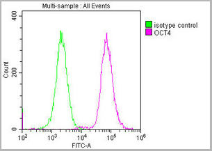
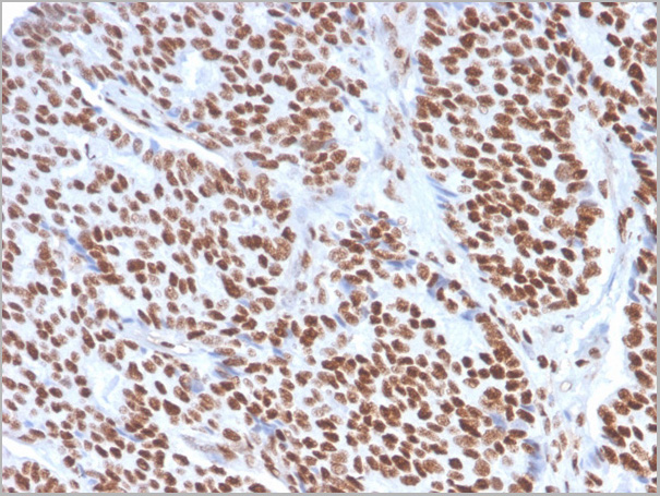
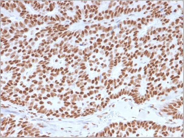
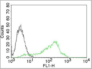
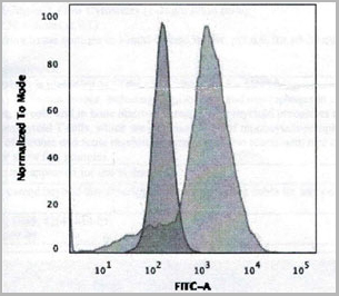
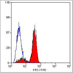
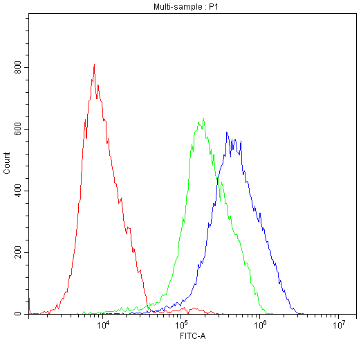
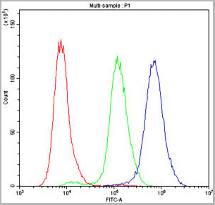
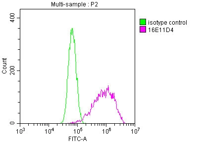
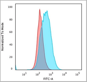
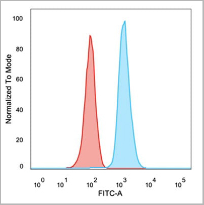
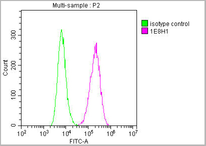
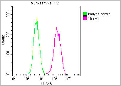
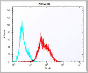
 FCM (Flow Cytometry)
(Overlay histogram showing Ntera-2 cells stained with CSB-MA018403A0m (red line) at 1:100. The cells were incubated in 1x PBS /10% normal goat serum to block non-specific protein-protein interactions followed by primary antibody for 1 h at 4 degree C. The secondary antibody used was FITC goat anti-mouse IgG(H+L) at 1/200 dilution for 1 h at 4 degree C. Isotype control antibody (green line) was used under the same conditions. Acquisition of >10,000 events was performed.)
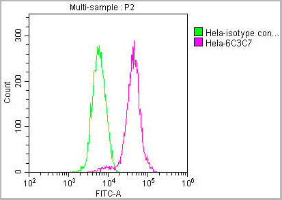
FCM (Flow Cytometry)
(Overlay histogram showing Ntera-2 cells stained with CSB-MA018403A0m (red line) at 1:100. The cells were incubated in 1x PBS /10% normal goat serum to block non-specific protein-protein interactions followed by primary antibody for 1 h at 4 degree C. The secondary antibody used was FITC goat anti-mouse IgG(H+L) at 1/200 dilution for 1 h at 4 degree C. Isotype control antibody (green line) was used under the same conditions. Acquisition of >10,000 events was performed.)
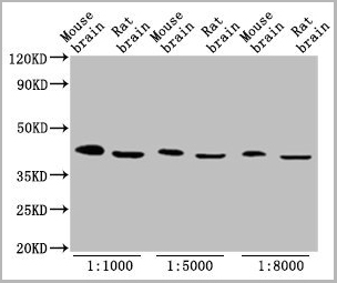
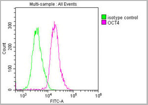
OCT4, Monoclonal Antibody (Cat# AAA27015)
Full Name
OCT4 Monoclonal Antibody
Gene Names
POU5F1; OCT3; OCT4; OTF3; OTF4; OTF-3; Oct-3; Oct-4
Reactivity
Human, Mouse, Rat
Applications
Western Blot, Immunohistochemistry, Immunofluorescence, Flow Cytometry
Purity
>95%
Protein G Purified
Protein G Purified
Pricing
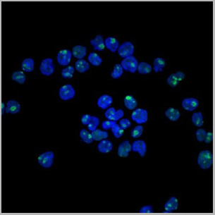

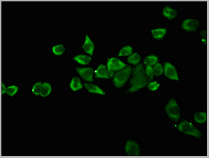
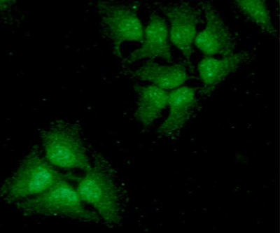
 IF (Immunofluorescence)
Nucleolin, Monoclonal Antibody (Cat# AAA13823)
Full Name
Nucleolin (Marker of Human Cells) Mouse Monoclonal Antibody
Gene Names
NCL; C23
Reactivity
Human.
Does not react with Mouse, Rat and Cow
Does not react with Mouse, Rat and Cow
Applications
Flow Cytometry, Immunofluorescence, Western Blot, Immunohistochemistry
Pricing


 Application Data
(Staining of mouse peritoneal macrophages with Rat anti Mouse Beta-glucan Receptor: FITC)
Application Data
(Staining of mouse peritoneal macrophages with Rat anti Mouse Beta-glucan Receptor: FITC)
DECTIN-1, Monoclonal Antibody (Cat# AAA12137)
Full Name
RAT ANTI MOUSE DECTIN-1
Gene Names
Clec7a; BGR; beta-GR; Clecsf12
Applications
Immunohistochemistry, Flow Cytometry, Immunoprecipitation
Pricing
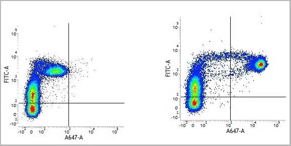
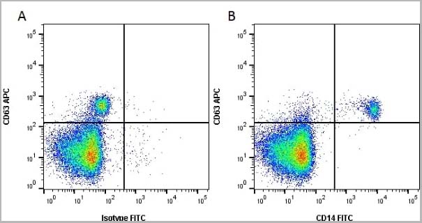
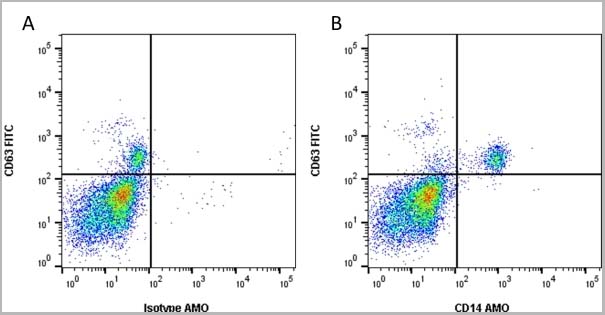
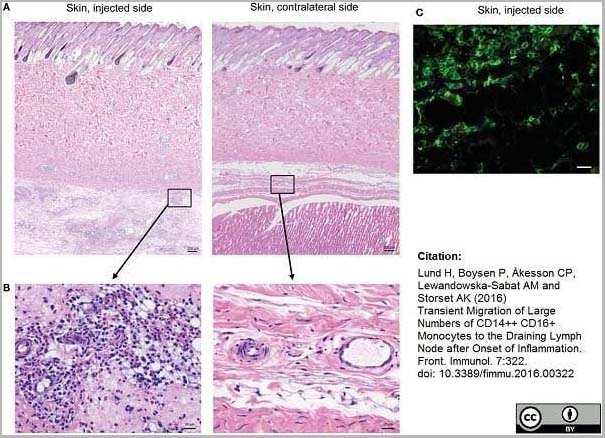 Application Data
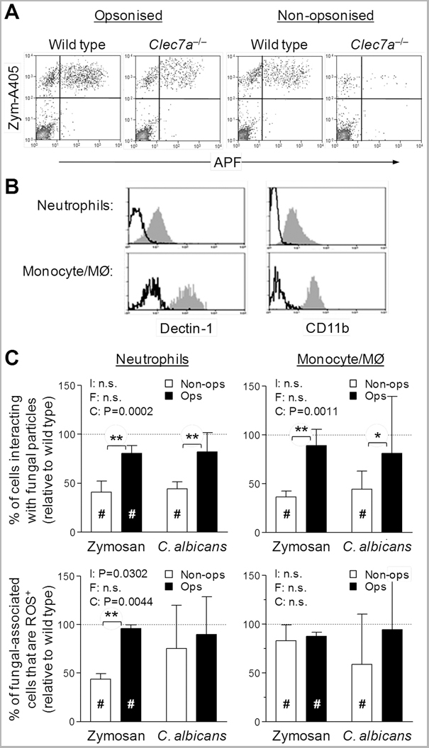
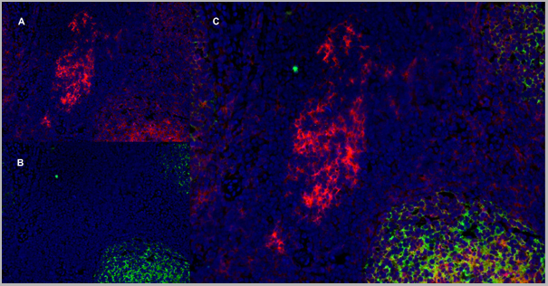
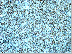
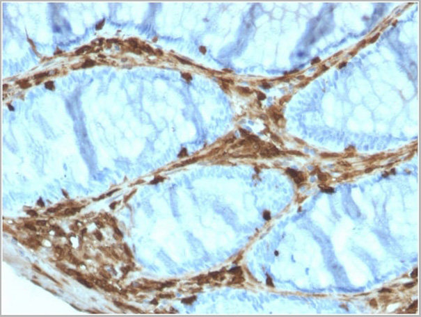
(Mouse anti Human CD14 antibody, clone Tük4 used to identify bovine monocytes in the skin and subcutaneous tissue of adjuvant injected calves by immunofluorescence.Image caption:Cellular recruitment to skin and subcutaneous tissues. (A) HE stained sections of skin with subcutaneous tissue from the side injected with adjuvant and the contralateral side, at 24 h post-injection. Scale bars: 200 ?m. (B) Enlargement of outlined areas in A, as indicated. Scale bars: 20 ?m. (C) Immunofluorescent labeling of subcutaneous tissue on the injected side with antibody against CD14 (green). Scale bar: 20 ?m.From: Lund H, Boysen P, Åkesson CP, Lewandowska-Sabat AM and Storset AK (2016)Transient Migration of Large Numbers of CD14++ CD16+ Monocytes to the Draining Lymph Node after Onset of Inflammation.Front. Immunol. 7:322.This is from an open access article distributed under the terms of the Creative Commons Attribution License.)
Application Data
(Mouse anti Human CD14 antibody, clone Tük4 used to identify bovine monocytes in the skin and subcutaneous tissue of adjuvant injected calves by immunofluorescence.Image caption:Cellular recruitment to skin and subcutaneous tissues. (A) HE stained sections of skin with subcutaneous tissue from the side injected with adjuvant and the contralateral side, at 24 h post-injection. Scale bars: 200 ?m. (B) Enlargement of outlined areas in A, as indicated. Scale bars: 20 ?m. (C) Immunofluorescent labeling of subcutaneous tissue on the injected side with antibody against CD14 (green). Scale bar: 20 ?m.From: Lund H, Boysen P, Åkesson CP, Lewandowska-Sabat AM and Storset AK (2016)Transient Migration of Large Numbers of CD14++ CD16+ Monocytes to the Draining Lymph Node after Onset of Inflammation.Front. Immunol. 7:322.This is from an open access article distributed under the terms of the Creative Commons Attribution License.)
CD14, Monoclonal Antibody (Cat# AAA12266)
Full Name
Mouse Anti Human CD14: Amethyst Orange
Reactivity
Human
Applications
Flow Cytometry
Purity
Purified IgG prepared by affinity chromatography on Protein A from tissue culture supernatant.
Pricing


 Application Data
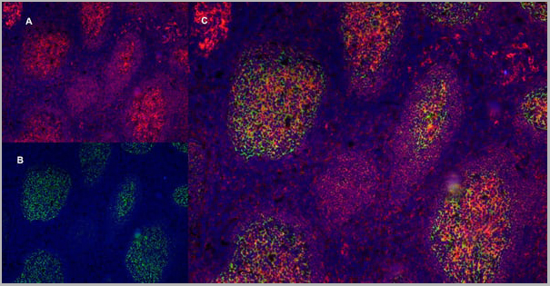
(PE conjugatedMouse anti Human CD169 antibody, clone 7-239 used to block CD169 function on myeloid cells.Image caption:Siglec-1 mediates HIV-1 uptake into a storage compartment and enhances HIV-1 trans-infection specially in IFN?-treated monocytes and DCs. A. Uptake of HIV-1NL4–3 by different myeloid cells exposed to IFN?. Cells were cultured with HIV-1 to measure p24Gag by ELISA. Mean values and SEM from four experiments include cells from 12 donors. B. Fold change in HIV-1NL4–3 uptake of cells treated with bafilomycin A1 compared to untreated cells. Mean values and SEM include cells from three donors. C. Relative uptake of HIV-1NL4–3 by IFN?-treated myeloid cells pre-incubated with the indicated mAbs. Values are normalized to the level of HIV-1 uptake by mock-treated cells (set at 100%). Mean values and SEM from two experiments include cells from six donors. D. Confocal microscopy analysis of different IFN?-treated myeloid cells pulsed with HIV-1Cherry and stained for Siglec-1 (Alexa 488), HLA-DR (Alexa 647) and DAPI. (Top) Representative viral pattern for each kind of myeloid cell analyzed, showing maximum fluorescence intensity of four channels. (Bottom) Percentage of myeloid cells with distinct viral patterns: random distribution, polarized accumulation, and sac-like compartment formation, as illustrated in the left drawing. Mean values of 50 cells from two different donors are shown. E. HIV-1 transmission from IFN?-treated myeloid cells to a luciferase reporter CD4+ cell line. HIV-1 infection was determined by induced luciferase activity in relative light units (RLUs). Mean values and SEM from four experiments include cells from 12 donors. F. Relative HIV-1 transmission from IFN?-treated myeloid cells pre-incubated with the indicated mAbs. Values are normalized to the level of HIV-1 trans-infected by mock-treated cells. Mean values and SEM from two experiments include cells from six donors. Statistical differences were assessed with a paired t test in A and E, and with a one sample t-test in B, C and F.From: Pino M, Erkizia I, Benet S, Erikson E, Fernández-Figueras MT, Guerrero D, Dalmau J, Ouchi D, Rausell A, Ciuffi A, Keppler OT, Telenti A, Kräusslich HG, Martinez-Picado J, Izquierdo-Useros N.HIV-1 immune activation induces Siglec-1 expression and enhances viral trans-infection in blood and tissue myeloid cells.Retrovirology. 2015 May 7;12:37.This image is from an open access article distributed under the terms of the Creative Commons Attribution License.)
Application Data
(PE conjugatedMouse anti Human CD169 antibody, clone 7-239 used to block CD169 function on myeloid cells.Image caption:Siglec-1 mediates HIV-1 uptake into a storage compartment and enhances HIV-1 trans-infection specially in IFN?-treated monocytes and DCs. A. Uptake of HIV-1NL4–3 by different myeloid cells exposed to IFN?. Cells were cultured with HIV-1 to measure p24Gag by ELISA. Mean values and SEM from four experiments include cells from 12 donors. B. Fold change in HIV-1NL4–3 uptake of cells treated with bafilomycin A1 compared to untreated cells. Mean values and SEM include cells from three donors. C. Relative uptake of HIV-1NL4–3 by IFN?-treated myeloid cells pre-incubated with the indicated mAbs. Values are normalized to the level of HIV-1 uptake by mock-treated cells (set at 100%). Mean values and SEM from two experiments include cells from six donors. D. Confocal microscopy analysis of different IFN?-treated myeloid cells pulsed with HIV-1Cherry and stained for Siglec-1 (Alexa 488), HLA-DR (Alexa 647) and DAPI. (Top) Representative viral pattern for each kind of myeloid cell analyzed, showing maximum fluorescence intensity of four channels. (Bottom) Percentage of myeloid cells with distinct viral patterns: random distribution, polarized accumulation, and sac-like compartment formation, as illustrated in the left drawing. Mean values of 50 cells from two different donors are shown. E. HIV-1 transmission from IFN?-treated myeloid cells to a luciferase reporter CD4+ cell line. HIV-1 infection was determined by induced luciferase activity in relative light units (RLUs). Mean values and SEM from four experiments include cells from 12 donors. F. Relative HIV-1 transmission from IFN?-treated myeloid cells pre-incubated with the indicated mAbs. Values are normalized to the level of HIV-1 trans-infected by mock-treated cells. Mean values and SEM from two experiments include cells from six donors. Statistical differences were assessed with a paired t test in A and E, and with a one sample t-test in B, C and F.From: Pino M, Erkizia I, Benet S, Erikson E, Fernández-Figueras MT, Guerrero D, Dalmau J, Ouchi D, Rausell A, Ciuffi A, Keppler OT, Telenti A, Kräusslich HG, Martinez-Picado J, Izquierdo-Useros N.HIV-1 immune activation induces Siglec-1 expression and enhances viral trans-infection in blood and tissue myeloid cells.Retrovirology. 2015 May 7;12:37.This image is from an open access article distributed under the terms of the Creative Commons Attribution License.)
CD169, Monoclonal Antibody (Cat# AAA12265)
Full Name
Mouse Anti Human CD169: RPE
Gene Names
SIGLEC1; SN; CD169; SIGLEC-1
Reactivity
Human
Applications
Flow Cytometry
Purity
>95% by SDS PAGE
Purified IgG prepared by affinity chromatography on Protein A from tissue culture supernatant.
Purified IgG prepared by affinity chromatography on Protein A from tissue culture supernatant.
Pricing
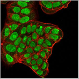
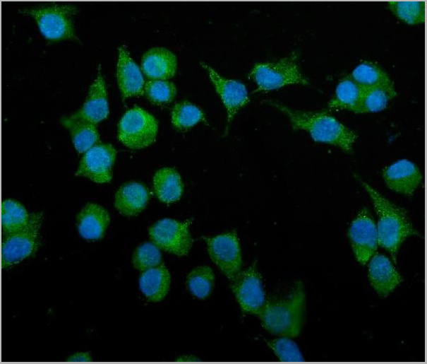
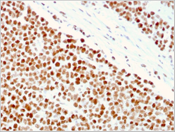
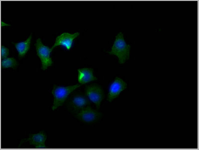
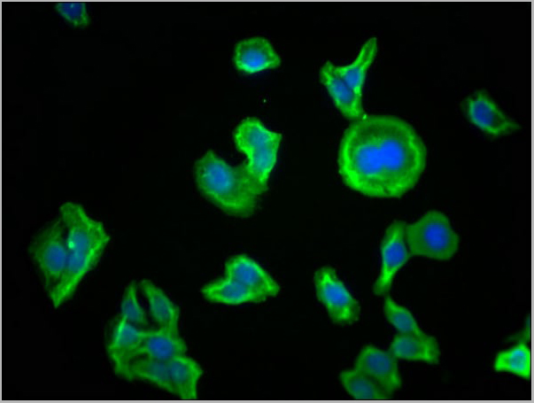
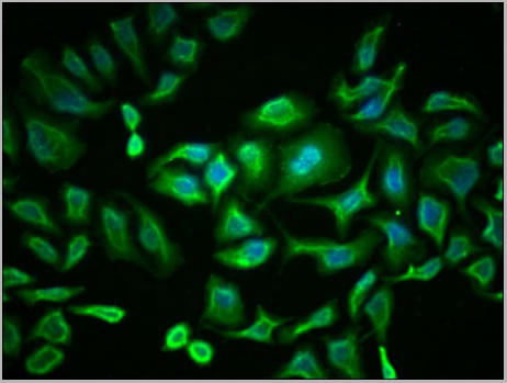
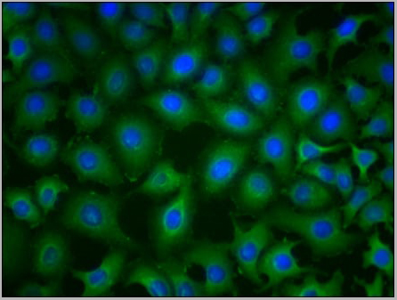
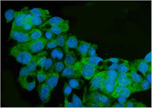
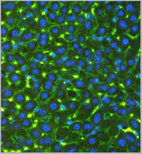
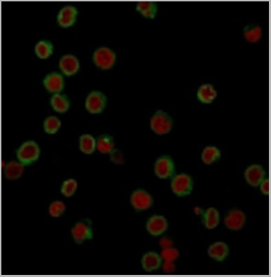
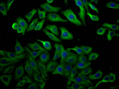
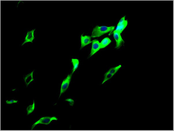
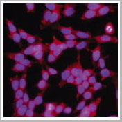
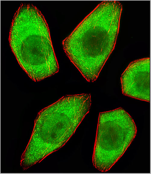
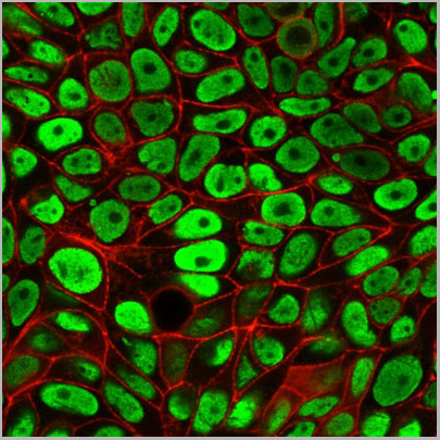
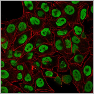
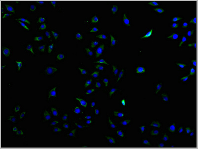
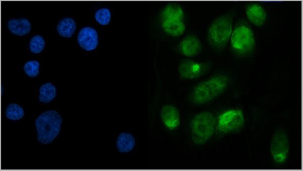
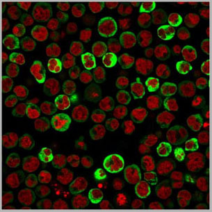
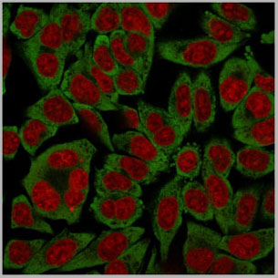
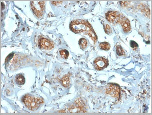
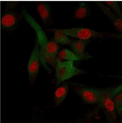 ICC (Immunocytochemistry)
(Immunofluorescence staining of U87MG cells using CD68 Mouse Monoclonal Antibody (C68/684) followed by goat anti-Mouse IgG conjugated to (green). Nuclei are stained with Red dot)
ICC (Immunocytochemistry)
(Immunofluorescence staining of U87MG cells using CD68 Mouse Monoclonal Antibody (C68/684) followed by goat anti-Mouse IgG conjugated to (green). Nuclei are stained with Red dot)
CD68, Antibody (Cat# AAA13807)
Full Name
CD68 (Macrophage Marker) Mouse Monoclonal Antibody
Gene Names
CD68; GP110; LAMP4; SCARD1
Reactivity
Human, Monkey, Mouse, and Rat. Others not known.
Applications
Western Blot, Immunofluorescence, Flow Cytometry, Immunohistochemistry
Pricing
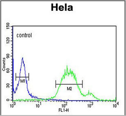
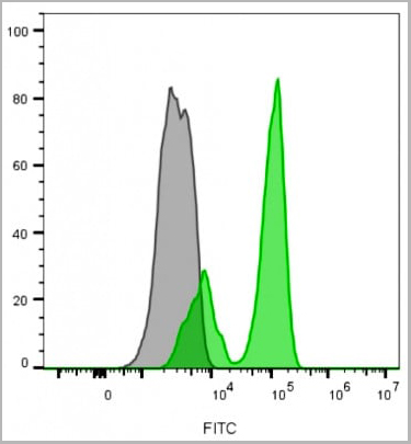
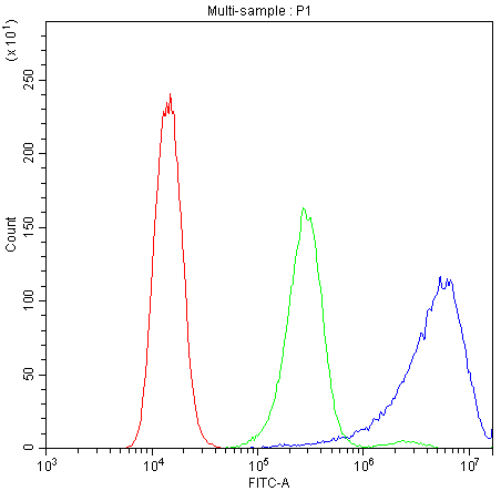
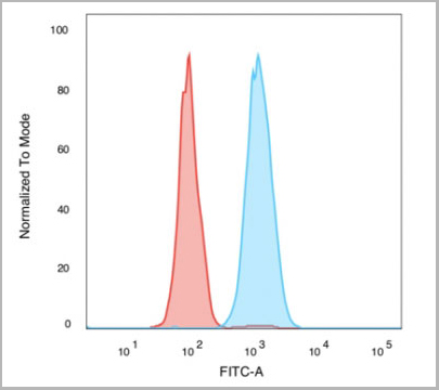
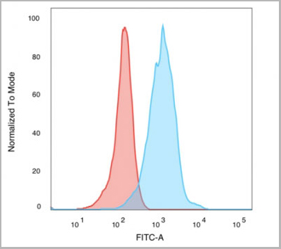
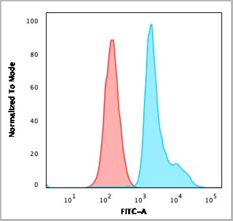
 FCM (Flow Cytometry)
(Flow Cytometric Analysis of Human Raji Cells. using HLA-Mouse Monoclonal Antibody followed by Goat anti-mouse IgG (Blue); Isotype control (Red).)
FCM (Flow Cytometry)
(Flow Cytometric Analysis of Human Raji Cells. using HLA-Mouse Monoclonal Antibody followed by Goat anti-mouse IgG (Blue); Isotype control (Red).)
HLA-A, Monoclonal Antibody (Cat# AAA13821)
Full Name
HLA-A (MHC I) Mouse Monoclonal Antibody
Gene Names
HLA-B; AS; HLAB; Bw-47; Bw-50; SPDA1; B-4901; B-5001; HLA-Cw; HLA-B15; HLA-B39; HLA-B49; HLA-B50; HLA-B55; HLA-B59; HLA-B61; HEL-S-83; HLA-DRB1; HLA-B*45ZJ; HLA-B-3506; HLA-B-3905; HLA-B-5502; HLA-B-5602
Reactivity
Human
Applications
Flow Cytometry, Immunofluorescence
Pricing
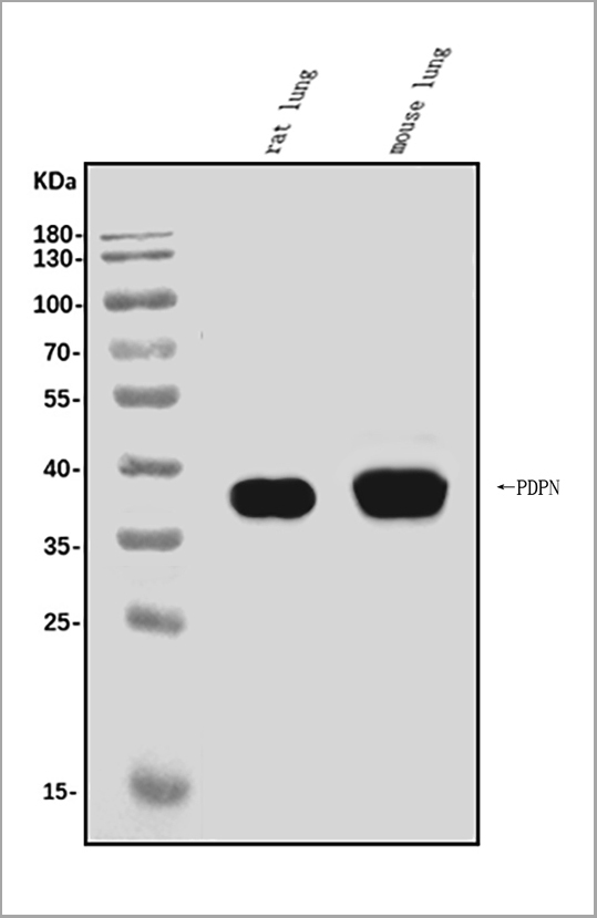
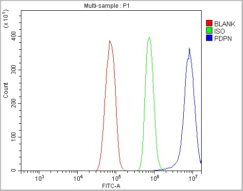 FCM (Flow Cytometry)
(Figure 5. Flow Cytometry analysis of RH35 cells using anti-Podoplanin/gp36/Pdpn antibody (AAA19233).Overlay histogram showing RH35 cells stained with AAA19233 (Blue line). The cells were blocked with 10% normal goat serum. And then incubated with rabbit anti-Podoplanin/gp36/Pdpn Antibody (AAA19233, 1μg/1x106 cells) for 30 min at 20 degree C. DyLight®488 conjugated goat anti-rabbit IgG (5-10μg/1x106 cells) was used as secondary antibody for 30 minutes at 20 degree C. Isotype control antibody (Green line) was rabbit IgG (1μg/1x106) used under the same conditions. Unlabelled sample (Red line) was also used as a control.)
FCM (Flow Cytometry)
(Figure 5. Flow Cytometry analysis of RH35 cells using anti-Podoplanin/gp36/Pdpn antibody (AAA19233).Overlay histogram showing RH35 cells stained with AAA19233 (Blue line). The cells were blocked with 10% normal goat serum. And then incubated with rabbit anti-Podoplanin/gp36/Pdpn Antibody (AAA19233, 1μg/1x106 cells) for 30 min at 20 degree C. DyLight®488 conjugated goat anti-rabbit IgG (5-10μg/1x106 cells) was used as secondary antibody for 30 minutes at 20 degree C. Isotype control antibody (Green line) was rabbit IgG (1μg/1x106) used under the same conditions. Unlabelled sample (Red line) was also used as a control.)
Podoplanin/gp36/Pdpn, Polyclonal Antibody (Cat# AAA19233)
Full Name
Anti-Podoplanin/gp36/Pdpn Antibody
Gene Names
Pdpn; T1a; Gp38; OTS-8; T1alpha; RANDAM-2; T1-alpha
Reactivity
Mouse, Rat
Applications
Western Blot, Immunohistochemistry, Immunocytochemistry, Immunofluorescence, Flow Cytometry, Direct ELISA
Purity
Immunogen affinity purified.
Pricing

 Application Data
(Analysis of Protein Array containing >19,000 full-length human proteins using MSH6 Mouse Monoclonal Antibody (MSH6/3086) Z- and S- Score: The Z-score represents the strength of a signal that a monoclonal antibody (MAb) (in combination with a fluorescently-tagged anti-IgG secondary antibody) produces when binding to a particular protein on the HuProtTM array. Z-scores are described in units of standard deviations (SD’s) above the mean value of all signals generated on that array. If targets on HuProtTM are arranged in descending order of the Z-score, the S-score is the difference (also in units of SD’s) between the Z-score. S-score therefore represents the relative target specificity of a MAb to its intended target. A MAb is considered to specific to its intended target, if the MAb has an S-score of at least 2.5. For example, if a MAb binds to protein X with a Z-score of 43 and to protein Y with a Z-score of 14, then the S-score for the binding of that MAb to protein X is equal to 29.)
Application Data
(Analysis of Protein Array containing >19,000 full-length human proteins using MSH6 Mouse Monoclonal Antibody (MSH6/3086) Z- and S- Score: The Z-score represents the strength of a signal that a monoclonal antibody (MAb) (in combination with a fluorescently-tagged anti-IgG secondary antibody) produces when binding to a particular protein on the HuProtTM array. Z-scores are described in units of standard deviations (SD’s) above the mean value of all signals generated on that array. If targets on HuProtTM are arranged in descending order of the Z-score, the S-score is the difference (also in units of SD’s) between the Z-score. S-score therefore represents the relative target specificity of a MAb to its intended target. A MAb is considered to specific to its intended target, if the MAb has an S-score of at least 2.5. For example, if a MAb binds to protein X with a Z-score of 43 and to protein Y with a Z-score of 14, then the S-score for the binding of that MAb to protein X is equal to 29.)
MSH6, Monoclonal Antibody (Cat# AAA23917)
Full Name
MSH6 (DNA Mismatch Repair Protein)
Gene Names
MSH6; GTBP; HSAP; p160; GTMBP; HNPCC5
Reactivity
Human
Applications
Flow Cytometry, Immunofluorescence, Western Blot, Immunohistochemistry
Purity
Purified Ab with BSA and Azide at 200ug/ml OR Purified Ab WITHOUT BSA and Azide at 1.0mg/ml
Pricing


 Application Data
(Staining of mouse peritoneal macrophages with Rat anti Mouse Beta-glucan Receptor: FITC)
Application Data
(Staining of mouse peritoneal macrophages with Rat anti Mouse Beta-glucan Receptor: FITC)
DECTIN-1, Monoclonal Antibody (Cat# AAA12128)
Full Name
RAT ANTI MOUSE DECTIN-1:Biotin
Gene Names
Clec7a; BGR; beta-GR; Clecsf12
Applications
Flow Cytometry
Pricing
 FCM (Flow Cytometry)
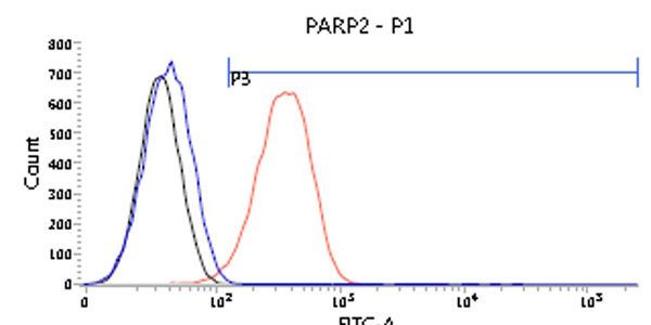
(Flow cytometry analysis of PARP2 in U87MG cells. The cell was stained with AAA11732 at 2-5ug for 1x10^6cells (red). A Goat anti mouse IgG (Alexa fluor 488) was used as the secondary antibody. Mouse monoclonal IgG was used as the isotype control (blue), cells without incubation with primary and secondary antibody was used as the negative control (black))
FCM (Flow Cytometry)
(Flow cytometry analysis of PARP2 in U87MG cells. The cell was stained with AAA11732 at 2-5ug for 1x10^6cells (red). A Goat anti mouse IgG (Alexa fluor 488) was used as the secondary antibody. Mouse monoclonal IgG was used as the isotype control (blue), cells without incubation with primary and secondary antibody was used as the negative control (black))
PARP2, Monoclonal Antibody (Cat# AAA11732)
Full Name
PARP2 antibody
Gene Names
PARP2; ARTD2; ADPRT2; PARP-2; ADPRTL2; ADPRTL3; pADPRT-2
Reactivity
Human
Applications
Western Blot, Flow Cytometry, Immunocytochemistry, Immunofluorescence
Purity
By protein-A affinity chromatography
Pricing
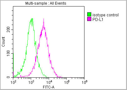 FCM (Flow Cytometry)
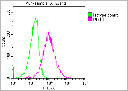
(Overlay histogram showing Hela cells stained with AAA27016 (red line) at 1:150. The cells were incubated in 1x PBS /10% normal goat serum to block non-specific protein-protein interactions followed by primary antibody for 1 h at 4 degree C. The secondary antibody used was FITC goat anti-mouse IgG(H+L) at 1/200 dilution for 1 h at 4 degree C. Isotype control antibody (green line) was used under the same conditions. Acquisition of >10,000 events was performed.)
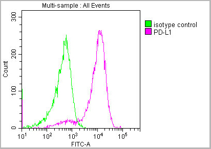
FCM (Flow Cytometry)
(Overlay histogram showing Hela cells stained with AAA27016 (red line) at 1:150. The cells were incubated in 1x PBS /10% normal goat serum to block non-specific protein-protein interactions followed by primary antibody for 1 h at 4 degree C. The secondary antibody used was FITC goat anti-mouse IgG(H+L) at 1/200 dilution for 1 h at 4 degree C. Isotype control antibody (green line) was used under the same conditions. Acquisition of >10,000 events was performed.)
PD-L1, Monoclonal Antibody (Cat# AAA27016)
Full Name
PD-L1 Monoclonal Antibody
Gene Names
CD274; B7-H; B7H1; PDL1; PD-L1; PDCD1L1; PDCD1LG1
Reactivity
Human
Applications
Western Blot, Immunohistochemistry, Immunofluorescence, Flow Cytometry
Purity
>95%, Protein G purified
Pricing
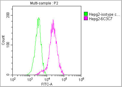 FCM (Flow Cytometry)
(Overlay histogram showing HepG2 cells stained with AAA27041 (red line) at 1:100. The cells were incubated in 1x PBS /10% normal goat serum to block non-specific protein-protein interactions followed by primary antibody for 1 h at 4 degree C. The secondary antibody used was FITC goat anti-mouse IgG(H+L) at 1/200 dilution for 1 h at 4 degree C. Isotype control antibody (green line) was used under the same conditions. Acquisition of >10,000 events was performed.)
FCM (Flow Cytometry)
(Overlay histogram showing HepG2 cells stained with AAA27041 (red line) at 1:100. The cells were incubated in 1x PBS /10% normal goat serum to block non-specific protein-protein interactions followed by primary antibody for 1 h at 4 degree C. The secondary antibody used was FITC goat anti-mouse IgG(H+L) at 1/200 dilution for 1 h at 4 degree C. Isotype control antibody (green line) was used under the same conditions. Acquisition of >10,000 events was performed.)
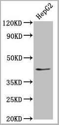
PKM, Monoclonal Antibody (Cat# AAA27041)
Full Name
PKM Monoclonal Antibody
Reactivity
Human, Rat, Mouse, Rabbit
Applications
Western Blot, Immunohistochemistry, Immunofluorescence, Flow Cytometry, Immunoprecipitation
Purity
>95%, Protein G purified
Pricing
 FCM (Flow Cytometry)
(Figure 8. Flow Cytometry analysis of HepG2 cells using anti-ATF2 antibody (AAA11634).Overlay histogram showing HepG2 cells stained with AAA11634 (Blue line).The cells were blocked with 10% normal goat serum. And then incubated with rabbit anti-ATF2 Antibody (AAA11634,1ug/1x10^6 cells) for 30 min at 20 degree C. DyLight ®488 conjugated goat anti-rabbit IgG (5-10ug/1x10^6 cells) was used as secondary antibody for 30 minutes at 20 degree C. Isotype control antibody (Green line) was rabbit IgG (1ug/1x106) used under the same conditions. Unlabelled sample (Red line) was also used as a control.)
FCM (Flow Cytometry)
(Figure 8. Flow Cytometry analysis of HepG2 cells using anti-ATF2 antibody (AAA11634).Overlay histogram showing HepG2 cells stained with AAA11634 (Blue line).The cells were blocked with 10% normal goat serum. And then incubated with rabbit anti-ATF2 Antibody (AAA11634,1ug/1x10^6 cells) for 30 min at 20 degree C. DyLight ®488 conjugated goat anti-rabbit IgG (5-10ug/1x10^6 cells) was used as secondary antibody for 30 minutes at 20 degree C. Isotype control antibody (Green line) was rabbit IgG (1ug/1x106) used under the same conditions. Unlabelled sample (Red line) was also used as a control.)
Cyclic AMP-dependent transcription factor ATF-2, Polyclonal Antibody (Cat# AAA11634)
Full Name
Anti-ATF2 Antibody
Gene Names
ATF2; HB16; CREB2; TREB7; CREB-2; CRE-BP1
Reactivity
Human, Mouse, Rat. No cross reactivity with other proteins.
Applications
Western Blot, Immunohistochemistry, Immunohistochemistry
Purity
Immunogen affinity purified.
Pricing


 Application Data
(Staining of mouse peritoneal macrophages with Rat anti Mouse Beta-glucan Receptor: FITC)
Application Data
(Staining of mouse peritoneal macrophages with Rat anti Mouse Beta-glucan Receptor: FITC)
DECTIN-1, Monoclonal Antibody (Cat# AAA12127)
Full Name
RAT ANTI MOUSE DECTIN-1:Biotin
Gene Names
Clec7a; BGR; beta-GR; Clecsf12
Applications
Flow Cytometry
Pricing
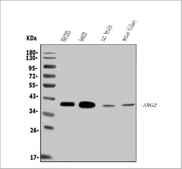
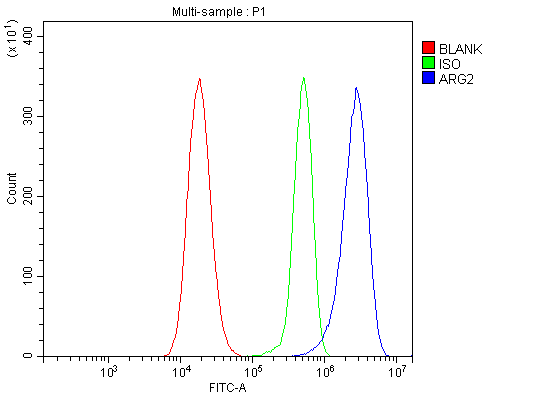 FCM (Flow Cytometry)
(Figure 7. Flow Cytometry analysis of 293T cells using anti-ARG2 antibody (AAA19256).Overlay histogram showing 293T cells stained with AAA19256 (Blue line). The cells were blocked with 10% normal goat serum. And then incubated with rabbit anti-ARG2 Antibody (AAA19256, 1μg/1x106 cells) for 30 min at 20 degree C. DyLight®488 conjugated goat anti-rabbit IgG (5-10μg/1x106 cells) was used as secondary antibody for 30 minutes at 20 degree C. Isotype control antibody (Green line) was rabbit IgG (1μg/1x106) used under the same conditions. Unlabelled sample (Red line) was also used as a control.)
FCM (Flow Cytometry)
(Figure 7. Flow Cytometry analysis of 293T cells using anti-ARG2 antibody (AAA19256).Overlay histogram showing 293T cells stained with AAA19256 (Blue line). The cells were blocked with 10% normal goat serum. And then incubated with rabbit anti-ARG2 Antibody (AAA19256, 1μg/1x106 cells) for 30 min at 20 degree C. DyLight®488 conjugated goat anti-rabbit IgG (5-10μg/1x106 cells) was used as secondary antibody for 30 minutes at 20 degree C. Isotype control antibody (Green line) was rabbit IgG (1μg/1x106) used under the same conditions. Unlabelled sample (Red line) was also used as a control.)
ARG2, Polyclonal Antibody (Cat# AAA19256)
Full Name
Anti-ARG2 Antibody
Reactivity
Human, Mouse, Rat
Applications
Western Blot, Immunohistochemistry, Immunocytochemistry, Immunofluorescence, Flow Cytometry, Direct ELISA
Purity
Immunogen affinity purified.
Pricing
 FCM (Flow Cytometry)
(Figure 6. Flow Cytometry analysis of A549 cells using anti-MDR3 antibody (AAA11652).Overlay histogram showing A549 cells stained with AAA11652 (Blue line).The cells were blocked with 10% normal goat serum. And then incubated with rabbit anti-MDR3 Antibody (AAA11652,1ug/1x106 cells) for 30 min at 20°C. DyLight®488 conjugated goat anti-rabbit IgG (5-10ug/1x106 cells) was used as secondary antibody for 30 minutes at 20°C. Isotype control antibody (Green line) was rabbit IgG (1ug/1x106) used under the same conditions. Unlabelled sample (Red line) was also used as a control.)
FCM (Flow Cytometry)
(Figure 6. Flow Cytometry analysis of A549 cells using anti-MDR3 antibody (AAA11652).Overlay histogram showing A549 cells stained with AAA11652 (Blue line).The cells were blocked with 10% normal goat serum. And then incubated with rabbit anti-MDR3 Antibody (AAA11652,1ug/1x106 cells) for 30 min at 20°C. DyLight®488 conjugated goat anti-rabbit IgG (5-10ug/1x106 cells) was used as secondary antibody for 30 minutes at 20°C. Isotype control antibody (Green line) was rabbit IgG (1ug/1x106) used under the same conditions. Unlabelled sample (Red line) was also used as a control.)
ABCB4, Polyclonal Antibody (Cat# AAA11652)
Full Name
Anti-ABCB4 Antibody
Gene Names
ABCB4; GBD1; ICP3; MDR2; MDR3; PGY3; ABC21; MDR2/3; PFIC-3
Reactivity
Human, Mouse, Rat
Applications
Western Blot, Immunohistochemistry, Immunofluorescence, Flow Cytometry
Purity
Immunogen Affinity Purified
Pricing
 FCM (Flow Cytometry)
(Overlay histogram showing Hela cells stained with AAA28750 (green line). The cells were fixed with 2% paraformaldehyde (10 min) and then permeabilized with 90% methanol for 10 min. The cells were then icubated in 2% bovine serum albumin to block non-specific protein-protein interactions followed by the antibody (AAA28750, 1:25 dilution) for 60 min at 37ºC. The secondary antibody used was Goat-Anti-Rabbit IgG, DyLight® 488 Conjugated Highly Cross-Adsorbed (1583138) at 1/200 dilution for 40 min at 37°C. Isotype control antibody (blue line) was rabbit IgG1 (1ug/1x106 cells) used under the same conditions. Acquisition of >10, 000 events wasperformed.)
FCM (Flow Cytometry)
(Overlay histogram showing Hela cells stained with AAA28750 (green line). The cells were fixed with 2% paraformaldehyde (10 min) and then permeabilized with 90% methanol for 10 min. The cells were then icubated in 2% bovine serum albumin to block non-specific protein-protein interactions followed by the antibody (AAA28750, 1:25 dilution) for 60 min at 37ºC. The secondary antibody used was Goat-Anti-Rabbit IgG, DyLight® 488 Conjugated Highly Cross-Adsorbed (1583138) at 1/200 dilution for 40 min at 37°C. Isotype control antibody (blue line) was rabbit IgG1 (1ug/1x106 cells) used under the same conditions. Acquisition of >10, 000 events wasperformed.)
WNT5A, Polyclonal Antibody (Cat# AAA28750)
Full Name
WNT5A Antibody (Center)
Gene Names
WNT5A; hWNT5A
Reactivity
Human, Mouse, Rat
Predicted: Rabbit
Predicted: Rabbit
Applications
Immunohistochemistry, Immunohistochemistry, Flow Cytometry, Western Blot
Purity
This antibody is purified through a protein A column, followed by peptide affinity purification.
Pricing
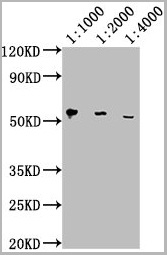
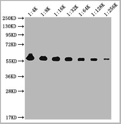
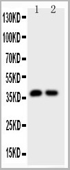
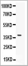
 WB (Western Blot)
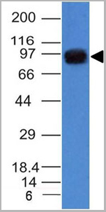
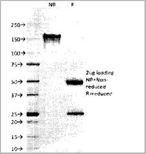
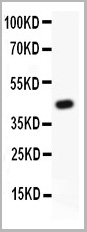
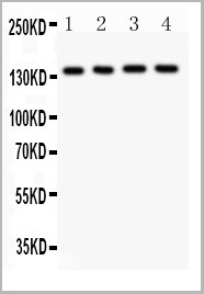
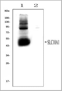
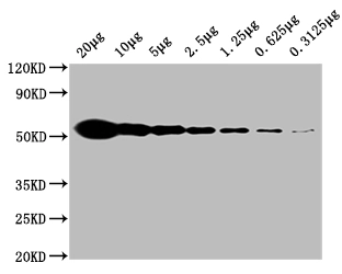
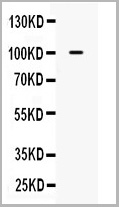
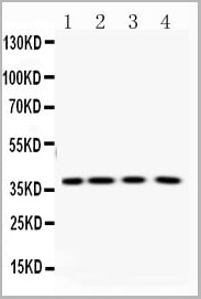
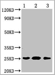
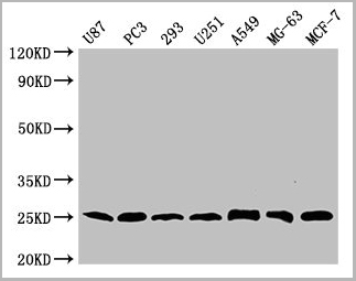
(Western blot analysis of SLC10A1 using anti- SLC10A1 antibody (AAA11653).Electrophoresis was performed on a 5-20% SDS-PAGE gel at 70V (Stacking gel) / 90V (Resolving gel) for 2-3 hours. The sample well of each lane was loaded with 50ug of sample under reducing conditions.Lane 1: rat liver tissue lysates (positive control), Lane 2: rat kidney tissue lysates, (negative control)After Electrophoresis, proteins were transferred to a Nitrocellulose membrane at 150mA for 50-90 minutes. Blocked the membrane with 5% Non-fat Milk/ TBS for 1.5 hour at RT. The membrane was incubated with rabbit anti-SLC10A1 antigen affinity purified polyclonal antibody (AAA11653) at 0.25ug/mL overnight at 4°C, then washed with TBS-0.1%Tween 3 times with 5 minutes each and probed with a goat anti-rabbit IgG-HRP secondary antibody at a dilution of 1:10000 for 1.5 hour at RT. The signal is developed using an Enhanced Chemiluminescent detection (ECL) kit with Tanon 5200 system. A specific band was detected for SLC10A1 at approximately 50KD. The expected band size for SLC10A1 is at 38KD.)
WB (Western Blot)
(Western blot analysis of SLC10A1 using anti- SLC10A1 antibody (AAA11653).Electrophoresis was performed on a 5-20% SDS-PAGE gel at 70V (Stacking gel) / 90V (Resolving gel) for 2-3 hours. The sample well of each lane was loaded with 50ug of sample under reducing conditions.Lane 1: rat liver tissue lysates (positive control), Lane 2: rat kidney tissue lysates, (negative control)After Electrophoresis, proteins were transferred to a Nitrocellulose membrane at 150mA for 50-90 minutes. Blocked the membrane with 5% Non-fat Milk/ TBS for 1.5 hour at RT. The membrane was incubated with rabbit anti-SLC10A1 antigen affinity purified polyclonal antibody (AAA11653) at 0.25ug/mL overnight at 4°C, then washed with TBS-0.1%Tween 3 times with 5 minutes each and probed with a goat anti-rabbit IgG-HRP secondary antibody at a dilution of 1:10000 for 1.5 hour at RT. The signal is developed using an Enhanced Chemiluminescent detection (ECL) kit with Tanon 5200 system. A specific band was detected for SLC10A1 at approximately 50KD. The expected band size for SLC10A1 is at 38KD.)
SLC10A1, Polyclonal Antibody (Cat# AAA11653)
Full Name
Anti-SLC10A1 Antibody
Gene Names
Slc10a1; Ntcp
Reactivity
Mouse, Rat
Applications
Flow Cytometry, Immunofluorescence, Immunohistochemistry, Immunohistochemistry, Immunocytochemistry, Western Blot
Purity
Immunogen affinity purified.
Pricing
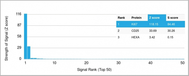 Application Data
(Analysis of Protein Array containing more than 19,000 full-length human proteins using Ki67 Mouse Monoclonal Antibody (MKI67/2466). Z- and S- Score: The Z-score represents the strength of a signal that a monoclonal antibody (MAb) (in combination with a fluorescently-tagged anti-IgG secondary antibody) produces when binding to a particular protein on the HuProtTM array. Z-scores are described in units of standard deviations (SD's) above the mean value of all signals generated on that array. If targets on HuProtTM are arranged in descending order of the Z-score, the S-score is the difference (also in units of SD's) between the Z-score. S-score therefore represents the relative target specificity of a MAb to its intended target. A MAb is considered to specific to its intended target, if the MAb has an S-score of at least 2.5. For example, if a MAb binds to protein X with a Z-score of 43 and to protein Y with a Z-score of 14, then the S-score for the binding of that MAb to protein X is equal to 29.)
Application Data
(Analysis of Protein Array containing more than 19,000 full-length human proteins using Ki67 Mouse Monoclonal Antibody (MKI67/2466). Z- and S- Score: The Z-score represents the strength of a signal that a monoclonal antibody (MAb) (in combination with a fluorescently-tagged anti-IgG secondary antibody) produces when binding to a particular protein on the HuProtTM array. Z-scores are described in units of standard deviations (SD's) above the mean value of all signals generated on that array. If targets on HuProtTM are arranged in descending order of the Z-score, the S-score is the difference (also in units of SD's) between the Z-score. S-score therefore represents the relative target specificity of a MAb to its intended target. A MAb is considered to specific to its intended target, if the MAb has an S-score of at least 2.5. For example, if a MAb binds to protein X with a Z-score of 43 and to protein Y with a Z-score of 14, then the S-score for the binding of that MAb to protein X is equal to 29.)
Ki-67, Monoclonal Antibody (Cat# AAA23909)
Full Name
Ki-67 (Proliferating Cell Marker)
Gene Names
MKI67; KIA; MIB-; MIB-1; PPP1R105
Reactivity
Human. Others not known.
Applications
Flow Cytometry, Immunofluorescence, Immunohistochemistry
Purity
Purified Ab with BSA and Azide at 200ug/ml OR Purified Ab WITHOUT BSA and Azide at 1.0mg/ml
Pricing
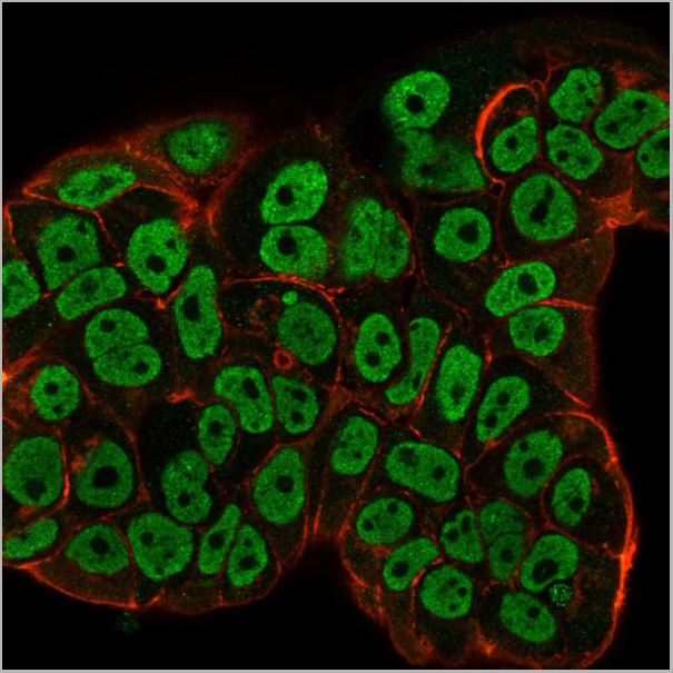
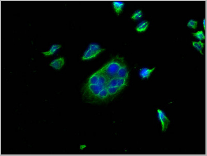
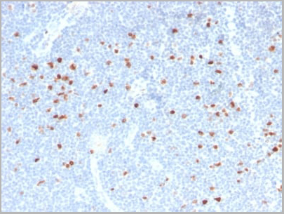
 IF (Immunofluorescence)
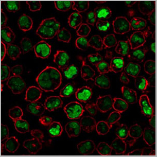
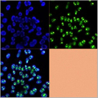
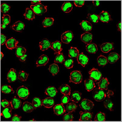
(Immunofluorescence Analysis of methanol-fixed human tonsil cryosection stained with CF488A CD3e clone RIV9 (green), mounted in EverBrite’ Mounting Medium with DAPI (nuclei, blue).)
IF (Immunofluorescence)
(Immunofluorescence Analysis of methanol-fixed human tonsil cryosection stained with CF488A CD3e clone RIV9 (green), mounted in EverBrite’ Mounting Medium with DAPI (nuclei, blue).)
CD3, Monoclonal Antibody (Cat# AAA13839)
Full Name
CD3 (T-Cell Marker) Mouse Monoclonal Antibody
Gene Names
CD3E; T3E; TCRE; IMD18
Reactivity
Human, Mouse, Rat
Applications
Functional Assay, Flow Cytometry, Immunofluorescence
Pricing
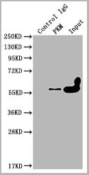
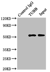 IP (Immunoprecipitation)
(Immunoprecipitating TUBB in Hela whole cell lysateLane 1: Mouse control IgG instead in Hela whole cell lysate.Lane 2: (2ug) + Hela whole cell lysate (500ug)Lane 3: Hela whole cell lysate (5ug)For western blotting, the blot was detected at 1:2000, and a HRP-conjugated Protein G antibody was used as the secondary antibody at 1:5000)
IP (Immunoprecipitation)
(Immunoprecipitating TUBB in Hela whole cell lysateLane 1: Mouse control IgG instead in Hela whole cell lysate.Lane 2: (2ug) + Hela whole cell lysate (500ug)Lane 3: Hela whole cell lysate (5ug)For western blotting, the blot was detected at 1:2000, and a HRP-conjugated Protein G antibody was used as the secondary antibody at 1:5000)
TUBB, Monoclonal Antibody (Cat# AAA28058)
Full Name
TUBB Monoclonal Antibody
Reactivity
Human, Rat, Rabbit, Mouse
Applications
Western Blot, Immunohistochemistry, Immunofluorescence, Flow Cytometry, Immunoprecipitation
Purity
>95%, Protein A purified
Pricing
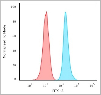
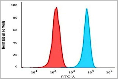
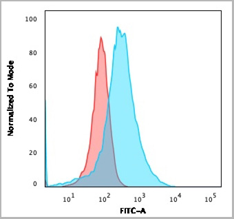
 FCM (Flow Cytometry)
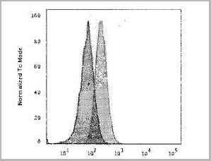
(Flow Cytometry detection of FOLR1 in MCF-7 human cell line using AAA14691. MCF-7 human breast cancer cell line was stained with AAA14691 (filled histogram) or isotype control antibody (open histogram), followed byPhycoerythrin-conjugated Anti-MouseIgG Secondary Antibody.)
FCM (Flow Cytometry)
(Flow Cytometry detection of FOLR1 in MCF-7 human cell line using AAA14691. MCF-7 human breast cancer cell line was stained with AAA14691 (filled histogram) or isotype control antibody (open histogram), followed byPhycoerythrin-conjugated Anti-MouseIgG Secondary Antibody.)
FOLR1, Monoclonal Antibody (Cat# AAA14691)
Full Name
FOLR1 (Folate receptor 1 (adult) Adult folate-binding protein, FBP, Folate receptor, adult, Folate receptor 1, Folate receptor alpha, FOLR, FR-alpha, KB cells FBP, MOv18, Ovarian tumor-associated antigen MOv18)
Gene Names
FOLR1; FBP; FOLR
Reactivity
Human
Applications
Flow Cytometry, Western Blot, Immunocytochemistry
Purity
Purified by Protein G affinity chromatography from hybridoma culture supernatant.
Pricing
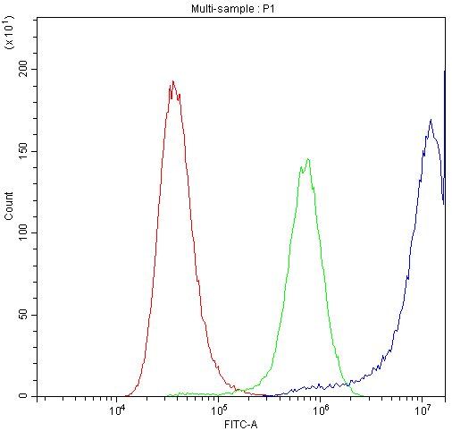
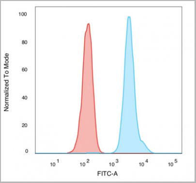
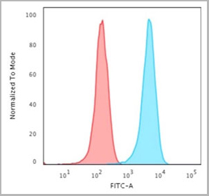
 FCM (Flow Cytometry)
(Flow Cytometric Analysis of Human Raji cells using HLA-DP (MHC II) Monoclonal Antibody (SPM421) followed by Goat anti-Mouse IgG-CF488 (Blue); Isotype control (Red).)
FCM (Flow Cytometry)
(Flow Cytometric Analysis of Human Raji cells using HLA-DP (MHC II) Monoclonal Antibody (SPM421) followed by Goat anti-Mouse IgG-CF488 (Blue); Isotype control (Red).)
HLA-DP, Antibody (Cat# AAA13830)
Full Name
HLA-DP (MHC II) Mouse Monoclonal Antibody
Gene Names
HLA-DPB1; DPB1; HLA-DP; HLA-DPB; HLA-DP1B
Reactivity
Human. Others not known.
Applications
Flow Cytometry, Immunofluorescence
Pricing
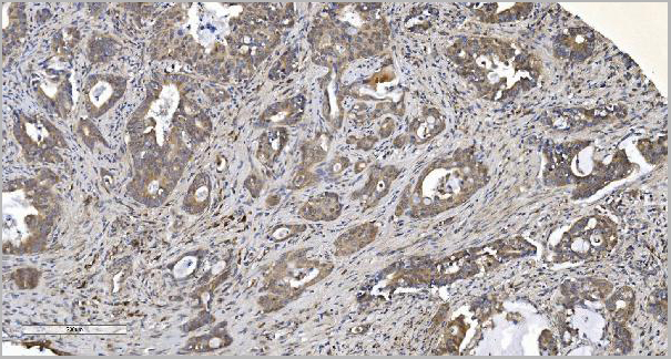
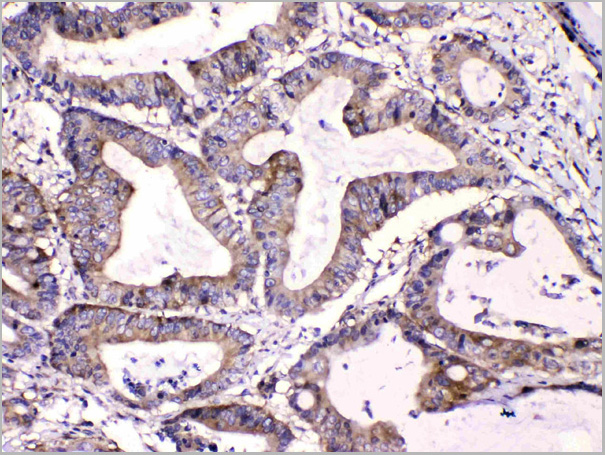
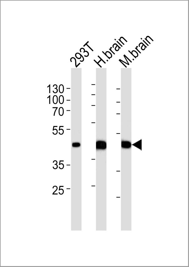 IHC (Immunohistochemistry)
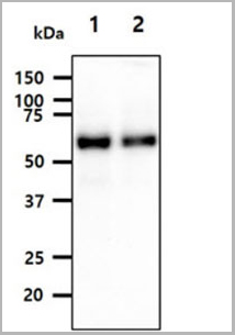
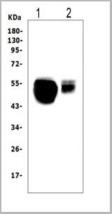
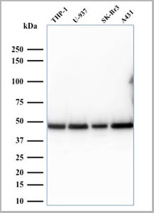
(Western blot analysis of lysates from 293T cell line human brain and mouse brain tissue lysate(from left to right), using STRADA Antibody (C-term). AAA28673 was diluted at 1:1000 at each lane. A goat anti-rabbit IgG H&L(HRP) at 1:5000 dilution was used as the secondary antibody. Lysates at 35ug per lane.)
IHC (Immunohistochemistry)
(Western blot analysis of lysates from 293T cell line human brain and mouse brain tissue lysate(from left to right), using STRADA Antibody (C-term). AAA28673 was diluted at 1:1000 at each lane. A goat anti-rabbit IgG H&L(HRP) at 1:5000 dilution was used as the secondary antibody. Lysates at 35ug per lane.)
STRADA, Polyclonal Antibody (Cat# AAA28673)
Full Name
STRADA Antibody (C-term)
Gene Names
STRADA; LYK5; PMSE; Stlk; STRAD; NY-BR-96
Reactivity
Human, mouse (Predicted Reactivity: Monkey)
Applications
Western Blot, Flow Cytometry, Immunofluorescence, Immunohistochemistry
Purity
Purified Rabbit Polyclonal Antibody (Pab)
Pricing
 FCM (Flow Cytometry)
(Figure 6. Flow Cytometry analysis of U20S cells using anti-Calpastatin antibody (AAA11679).Overlay histogram showing U20S cells stained with AAA11679 (Blue line).The cells were blocked with 10% normal goat serum. And then incubated with rabbit anti-Calpastatin Antibody (AAA11679,1ug/1x10^6 cells) for 30 min at 20 degree C. DyLight ®488 conjugated goat anti-rabbit IgG (5-10ug/1x10^6 cells) was used as secondary antibody for 30 minutes at 20 degree C. Isotype control antibody (Green line) was rabbit IgG (1ug/1x106) used under the same conditions. Unlabelled sample (Red line) was also used as a control.)
FCM (Flow Cytometry)
(Figure 6. Flow Cytometry analysis of U20S cells using anti-Calpastatin antibody (AAA11679).Overlay histogram showing U20S cells stained with AAA11679 (Blue line).The cells were blocked with 10% normal goat serum. And then incubated with rabbit anti-Calpastatin Antibody (AAA11679,1ug/1x10^6 cells) for 30 min at 20 degree C. DyLight ®488 conjugated goat anti-rabbit IgG (5-10ug/1x10^6 cells) was used as secondary antibody for 30 minutes at 20 degree C. Isotype control antibody (Green line) was rabbit IgG (1ug/1x106) used under the same conditions. Unlabelled sample (Red line) was also used as a control.)
Calpastatin, Polyclonal Antibody (Cat# AAA11679)
Full Name
Anti-Calpastatin Antibody
Gene Names
CAST; BS-17; PLACK
Reactivity
Human
Applications
Western Blot, Immunohistochemistry
Purity
Immunogen affinity purified.
Pricing

 FCM (Flow Cytometry)
(Figure 7. Flow Cytometry analysis of U251 cells using anti-PNP antibody (AAA11680).Overlay histogram showing U251 cells stained with AAA11680 (Blue line).The cells were blocked with 10% normal goat serum. And then incubated with rabbit anti-PNP Antibody (AAA11680,1ug/1x10^6 cells) for 30 min at 20 degree C. DyLight ®488 conjugated goat anti-rabbit IgG (5-10ug/1x10^6 cells) was used as secondary antibody for 30 minutes at 20 degree C. Isotype control antibody (Green line) was rabbit IgG (1ug/1x106) used under the same conditions. Unlabelled sample (Red line) was also used as a control.)
FCM (Flow Cytometry)
(Figure 7. Flow Cytometry analysis of U251 cells using anti-PNP antibody (AAA11680).Overlay histogram showing U251 cells stained with AAA11680 (Blue line).The cells were blocked with 10% normal goat serum. And then incubated with rabbit anti-PNP Antibody (AAA11680,1ug/1x10^6 cells) for 30 min at 20 degree C. DyLight ®488 conjugated goat anti-rabbit IgG (5-10ug/1x10^6 cells) was used as secondary antibody for 30 minutes at 20 degree C. Isotype control antibody (Green line) was rabbit IgG (1ug/1x106) used under the same conditions. Unlabelled sample (Red line) was also used as a control.)
PNP, Polyclonal Antibody (Cat# AAA11680)
Full Name
Anti-PNP Antibody
Gene Names
PNP; NP; PUNP; PRO1837
Reactivity
Human, Mouse, Rat
Applications
Western Blot, Immunohistochemistry
Purity
Immunogen affinity purified.
Pricing
 FCM (Flow Cytometry)
(Flow Cytometric Analysis of PFA-fixed Raji cells with Human Nuclear Antigen Mouse Monoclonal Antibody (235-1); followed by goat anti-mouse IgG-CF488 (Blue); Isotype Control (Red).)
FCM (Flow Cytometry)
(Flow Cytometric Analysis of PFA-fixed Raji cells with Human Nuclear Antigen Mouse Monoclonal Antibody (235-1); followed by goat anti-mouse IgG-CF488 (Blue); Isotype Control (Red).)
Human Nuclear Antigen, Monoclonal Antibody (Cat# AAA13804)
Full Name
Human Nuclear Antigen
Reactivity
Human and Non-human primates. Does not react with mouse, rat, and chicken. Others not known.
Applications
Flow Cytometry, Immunofluorescence, Immunoprecipitation, Immunocytochemistry, Immunohistochemistry
Pricing
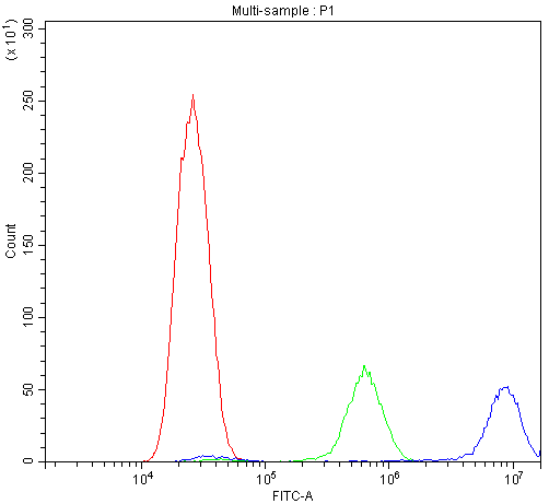
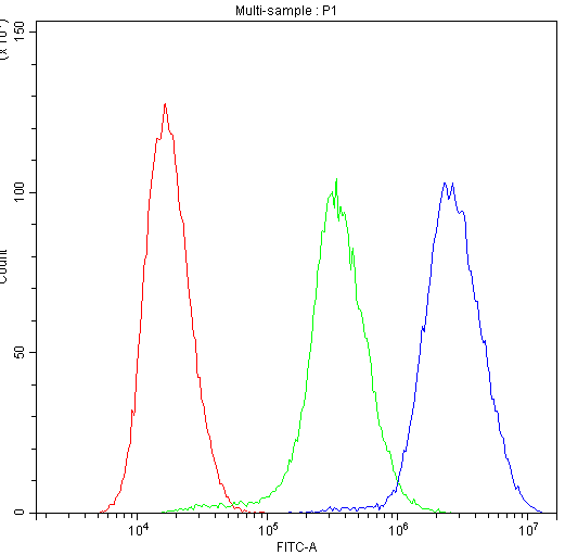
 FCM (Flow Cytometry)
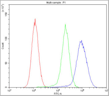
(Overlay histogram showing U87 cells stained with AAA28063 (red line) at 1:100. The cells were fixed in 4% formaldehyde and permeated by 0.2% TritonX-100. Then 10% normal goat serum was Incubated to block non-specific protein-protein interactions followed by the antibody (1ug/1*106cells) for 1 h at 4 degree C. The secondary antibody used was FITC-conjugated Goat Anti-Mouse IgG(H+L) at 1/100 dilution for 30min at 4 degree C. Isotype control antibody (green line) was mouse IgG2b (1ug/1*106cells) used under the same conditions. Acquisition of >10,000 events was performed.)
FCM (Flow Cytometry)
(Overlay histogram showing U87 cells stained with AAA28063 (red line) at 1:100. The cells were fixed in 4% formaldehyde and permeated by 0.2% TritonX-100. Then 10% normal goat serum was Incubated to block non-specific protein-protein interactions followed by the antibody (1ug/1*106cells) for 1 h at 4 degree C. The secondary antibody used was FITC-conjugated Goat Anti-Mouse IgG(H+L) at 1/100 dilution for 30min at 4 degree C. Isotype control antibody (green line) was mouse IgG2b (1ug/1*106cells) used under the same conditions. Acquisition of >10,000 events was performed.)
CD9, Monoclonal Antibody (Cat# AAA28063)
Full Name
CD9 Monoclonal Antibody
Gene Names
CD9; MIC3; MRP-1; BTCC-1; DRAP-27; TSPAN29; TSPAN-29
Reactivity
Human
Applications
Western Blot, Immunohistochemistry, Immunofluorescence, Flow Cytometry
Purity
>95%, Protein G purified
Pricing
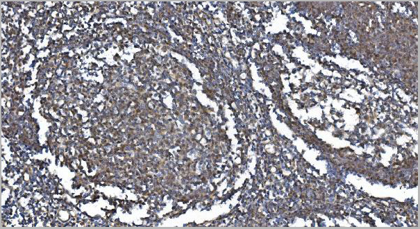
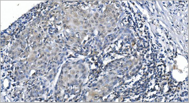
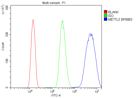 FCM (Flow Cytometry)
(Figure 6. Flow Cytometry analysis of HL-60 cells using anti-METTL3 antibody (AAA19247).Overlay histogram showing HL-60 cells stained with AAA19247 (Blue line). The cells were blocked with 10% normal goat serum. And then incubated with rabbit anti-METTL3 Antibody (AAA19247, 1μg/1x106 cells) for 30 min at 20 degree C. DyLight®488 conjugated goat anti-rabbit IgG (5-10μg/1x106 cells) was used as secondary antibody for 30 minutes at 20 degree C. Isotype control antibody (Green line) was rabbit IgG (1μg/1x106) used under the same conditions. Unlabelled sample (Red line) was also used as a control.)
FCM (Flow Cytometry)
(Figure 6. Flow Cytometry analysis of HL-60 cells using anti-METTL3 antibody (AAA19247).Overlay histogram showing HL-60 cells stained with AAA19247 (Blue line). The cells were blocked with 10% normal goat serum. And then incubated with rabbit anti-METTL3 Antibody (AAA19247, 1μg/1x106 cells) for 30 min at 20 degree C. DyLight®488 conjugated goat anti-rabbit IgG (5-10μg/1x106 cells) was used as secondary antibody for 30 minutes at 20 degree C. Isotype control antibody (Green line) was rabbit IgG (1μg/1x106) used under the same conditions. Unlabelled sample (Red line) was also used as a control.)
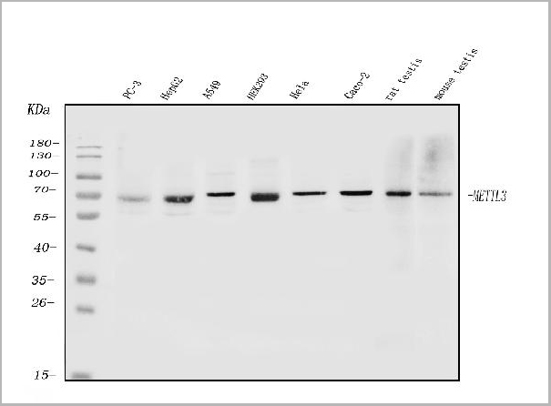
METTL3, Polyclonal Antibody (Cat# AAA19247)
Full Name
Anti-METTL3 Antibody
Gene Names
METTL3; M6A; IME4; Spo8; MT-A70
Reactivity
Human, Mouse, Rat
Applications
Western Blot, Immunohistochemistry, Immunocytochemistry, Immunofluorescence, Flow Cytometry, Direct ELISA
Purity
Immunogen affinity purified.
Pricing

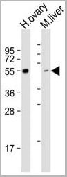 WB (Western Blot)
(All lanes : Anti-LAG3 Antibody (Center) at 1:1000 dilutionLane 1: human ovary lysateLane 2: mouse liver lysate Lysates/proteins at 20 ug per lane.Secondary Goat Anti-Rabbit IgG, (H+L), Peroxidase conjugated at 1/10000 dilution.Predicted band size : 57 kDaBlocking/Dilution buffer: 5% NFDM/TBST.)
WB (Western Blot)
(All lanes : Anti-LAG3 Antibody (Center) at 1:1000 dilutionLane 1: human ovary lysateLane 2: mouse liver lysate Lysates/proteins at 20 ug per lane.Secondary Goat Anti-Rabbit IgG, (H+L), Peroxidase conjugated at 1/10000 dilution.Predicted band size : 57 kDaBlocking/Dilution buffer: 5% NFDM/TBST.)
LAG3, Polyclonal Antibody (Cat# AAA28766)
Full Name
LAG3 Antibody (Center)
Gene Names
LAG3; CD223
Reactivity
Human, Mouse
Applications
Western Blot, Immunohistochemistry, Flow Cytometry
Purity
This antibody is purified through a protein A column, followed by peptide affinity purification.
Pricing
 FCM (Flow Cytometry)
(Figure 7. Flow Cytometry analysis of Hela cells using anti-Calbindin antibody (AAA11603).Overlay histogram showing Hela cells stained with AAA11603 (Blue line).The cells were blocked with 10% normal goat serum. And then incubated with rabbit anti-Calbindin Antibody (AAA11603,1ug/1x10^6 cells) for 30 min at 20 degree C. DyLight®488 conjugated goat anti-rabbit IgG (5-10ug/1x10^6 cells) was used as secondary antibody for 30 minutes at 20 degree C. Isotype control antibody (Green line) was rabbit IgG (1ug/1x106) used under the same conditions. Unlabelled sample (Red line) was also used as a control.)
FCM (Flow Cytometry)
(Figure 7. Flow Cytometry analysis of Hela cells using anti-Calbindin antibody (AAA11603).Overlay histogram showing Hela cells stained with AAA11603 (Blue line).The cells were blocked with 10% normal goat serum. And then incubated with rabbit anti-Calbindin Antibody (AAA11603,1ug/1x10^6 cells) for 30 min at 20 degree C. DyLight®488 conjugated goat anti-rabbit IgG (5-10ug/1x10^6 cells) was used as secondary antibody for 30 minutes at 20 degree C. Isotype control antibody (Green line) was rabbit IgG (1ug/1x106) used under the same conditions. Unlabelled sample (Red line) was also used as a control.)
Calbindin, Polyclonal Antibody (Cat# AAA11603)
Full Name
Anti-Calbindin Antibody
Gene Names
CALB1; CALB
Reactivity
Human, Mouse, Rat
Applications
Western Blot, Immunohistochemistry
Purity
Immunogen affinity purified.
Pricing
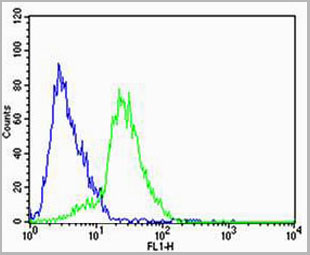
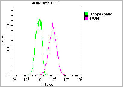
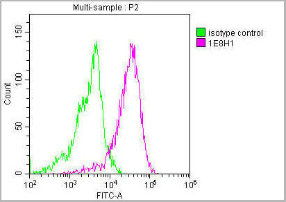
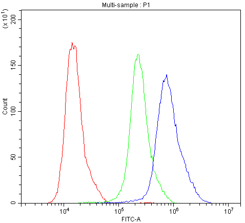
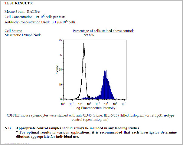 FCM (Flow Cytometry)
CD45, Monoclonal Antibody (Cat# AAA14270)
Full Name
Anti-Mouse CD45, Purified (Clone IBL-5/25) (rat IgG1)
Applications
Immunohistochemistry, Flow Cytometry, Immunoprecipitation
Purity
Purified from ascitic fluid via Protein G Chromatography
Pricing


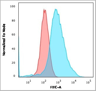
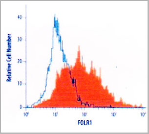
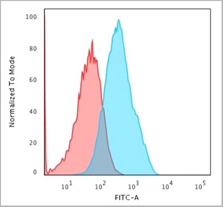
 FCM (Flow Cytometry)
(Flow Cytometry analyis of SSTR2 in human cell line. Human breast cancer cell line was stained with AAA14651 (filled histogram) or isotype control antibody (open histogram), followed by Allophycocyanin-conjugated Anti-Mouse IgG F(ab')2 secondary antibody. To facilitate intracellular staining, cells were fixed with paraformaldehyde and permeabilized with saponin.)
FCM (Flow Cytometry)
(Flow Cytometry analyis of SSTR2 in human cell line. Human breast cancer cell line was stained with AAA14651 (filled histogram) or isotype control antibody (open histogram), followed by Allophycocyanin-conjugated Anti-Mouse IgG F(ab')2 secondary antibody. To facilitate intracellular staining, cells were fixed with paraformaldehyde and permeabilized with saponin.)
Somatostatin Receptor Type 2, Monoclonal Antibody (Cat# AAA14651)
Full Name
Somatostatin Receptor Type 2 (SSTr2)
Gene Names
SSTR2; SSTR2
Reactivity
Human
Applications
Immunohistochemistry, Immunocytochemistry, Flow Cytometry
Purity
Purified by Protein A affinity chromatography from hybridoma culture supernatant.
Pricing

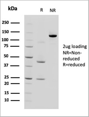
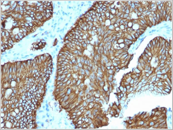
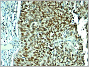
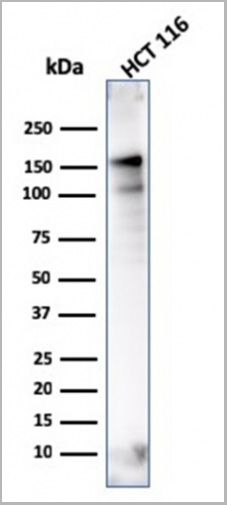
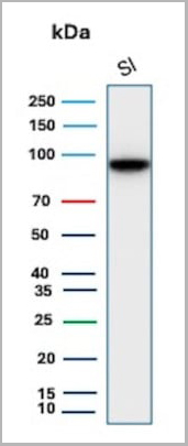 WB (Western Blot)
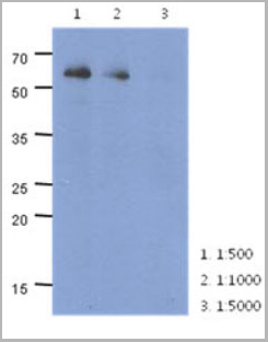
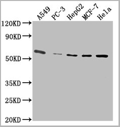
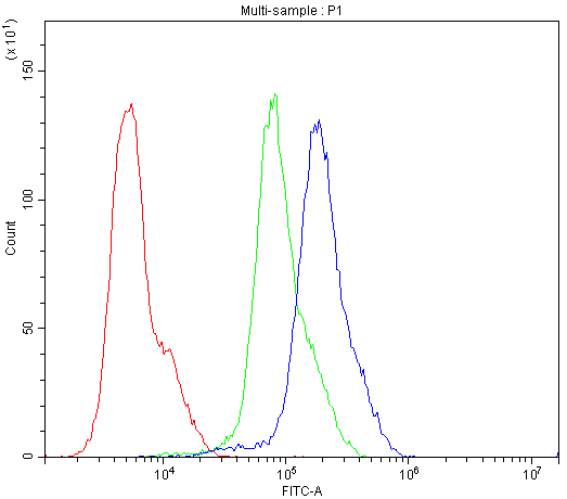
(Western Blot Analysis of human Small Intestine tissue lysate using CDH17 Mouse Monoclonal Antibody (CDH17/2618).)
WB (Western Blot)
(Western Blot Analysis of human Small Intestine tissue lysate using CDH17 Mouse Monoclonal Antibody (CDH17/2618).)
Cadherin 17/LI Cadherin, Monoclonal Antibody (Cat# AAA23911)
Full Name
Cadherin 17/LI Cadherin (Liver-Intestine Marker)
Gene Names
CDH17; HPT1; CDH16; HPT-1
Reactivity
Human
Applications
Flow Cytometry, Immunohistochemistry, Western Blot
Pricing
 FCM (Flow Cytometry)
(Figure 7. Flow Cytometry analysis of K562 cells using anti-CASP3 antibody (AAA11633).Overlay histogram showing K562 cells stained with AAA11633 (Blue line).The cells were blocked with 10% normal goat serum. And then incubated with rabbit anti-CASP3 Antibody (AAA11633,1ug/1x10^6 cells) for 30 min at 20 degree C. DyLight ®488 conjugated goat anti-rabbit IgG (5-10ug/1x10^6 cells) was used as secondary antibody for 30 minutes at 20 degree C. Isotype control antibody (Green line) was rabbit IgG (1ug/1x106) used under the same conditions. Unlabelled sample (Red line) was also used as a control.)
FCM (Flow Cytometry)
(Figure 7. Flow Cytometry analysis of K562 cells using anti-CASP3 antibody (AAA11633).Overlay histogram showing K562 cells stained with AAA11633 (Blue line).The cells were blocked with 10% normal goat serum. And then incubated with rabbit anti-CASP3 Antibody (AAA11633,1ug/1x10^6 cells) for 30 min at 20 degree C. DyLight ®488 conjugated goat anti-rabbit IgG (5-10ug/1x10^6 cells) was used as secondary antibody for 30 minutes at 20 degree C. Isotype control antibody (Green line) was rabbit IgG (1ug/1x106) used under the same conditions. Unlabelled sample (Red line) was also used as a control.)
Caspase-3, Polyclonal Antibody (Cat# AAA11633)
Full Name
Anti-Caspase-3 Antibody
Gene Names
CASP3; CPP32; SCA-1; CPP32B
Reactivity
Human, Mouse, Rat. No cross reactivity with other proteins.
Applications
Western Blot, Immunohistochemistry
Purity
Immunogen affinity purified.
Pricing
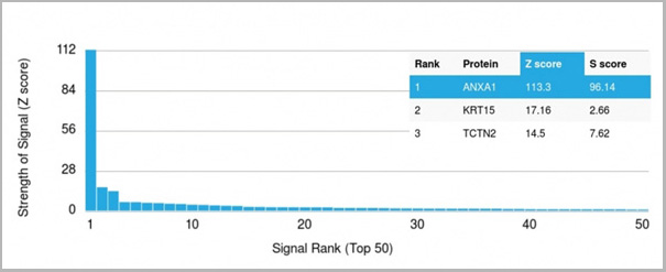 Application Data
(Analysis of Protein Array containing more than 19,000 full-length human proteins using Annexin A1 Mouse Monoclonal Antibody (ANXA1/3566). Z- and S- Score: The Z-score represents the strength of a signal that a monoclonal antibody (MAb) (in combination with a fluorescently-tagged anti-IgG secondary antibody) produces when binding to a particular protein on the HuProtTM array. Z-scores are described in units of standard deviations (SD’s) above the mean value of all signals generated on that array. If targets on HuProtTM are arranged in descending order of the Z-score, the S-score is the difference (also in units of SD’s) between the Z-score. S-score therefore represents the relative target specificity of a MAb to its intended target. A MAb is considered to specific to its intended target, if the MAb has an S-score of at least 2.5. For example, if a MAb binds to protein X with a Z-score of 43 and to protein Y with a Z-score of 14, then the S-score for the binding of that MAb to protein X is equal to 29.)
Application Data
(Analysis of Protein Array containing more than 19,000 full-length human proteins using Annexin A1 Mouse Monoclonal Antibody (ANXA1/3566). Z- and S- Score: The Z-score represents the strength of a signal that a monoclonal antibody (MAb) (in combination with a fluorescently-tagged anti-IgG secondary antibody) produces when binding to a particular protein on the HuProtTM array. Z-scores are described in units of standard deviations (SD’s) above the mean value of all signals generated on that array. If targets on HuProtTM are arranged in descending order of the Z-score, the S-score is the difference (also in units of SD’s) between the Z-score. S-score therefore represents the relative target specificity of a MAb to its intended target. A MAb is considered to specific to its intended target, if the MAb has an S-score of at least 2.5. For example, if a MAb binds to protein X with a Z-score of 43 and to protein Y with a Z-score of 14, then the S-score for the binding of that MAb to protein X is equal to 29.)
Annexin A1, Monoclonal Antibody (Cat# AAA23918)
Full Name
Annexin A1/(Hairy Cell Leukemia Marker)
Gene Names
ANXA1; ANX1; LPC1
Reactivity
Human
Applications
Flow Cytometry, Immunofluorescence, Immunohistochemistry
Purity
Purified Ab with BSA and Azide at 200ug/ml OR Purified Ab WITHOUT BSA and Azide at 1.0mg/ml
Pricing


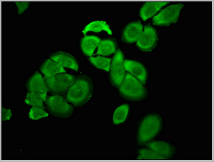
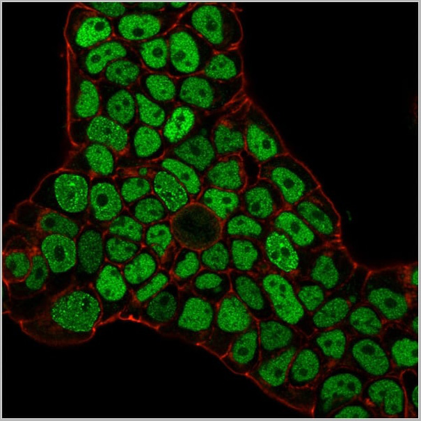
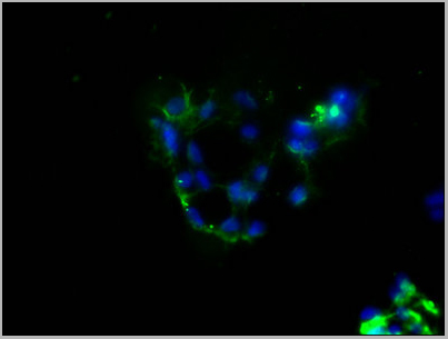
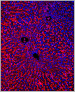
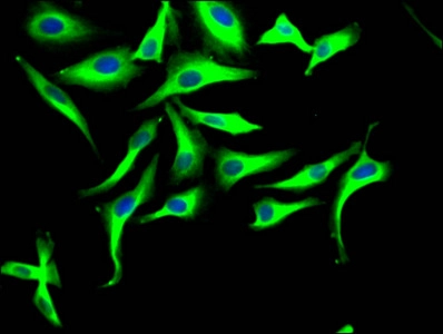
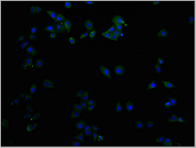
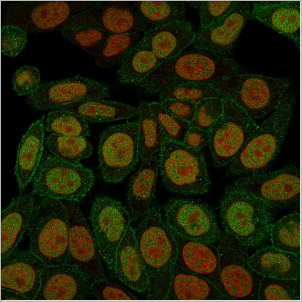
 IF (Immunofluorescence)
(Confocal immunofluorescence image of HeLa Cells using S100A4 Mouse Monoclonal Antibody (S100A4/1482) followed by goat anti-mouse IgG-CF488 (green). Reddot is used to label the nuclei Red.)
IF (Immunofluorescence)
(Confocal immunofluorescence image of HeLa Cells using S100A4 Mouse Monoclonal Antibody (S100A4/1482) followed by goat anti-mouse IgG-CF488 (green). Reddot is used to label the nuclei Red.)
S100A4, Monoclonal Antibody (Cat# AAA23892)
Full Name
S100A4 (Marker of Tumor Metastasis)
Gene Names
S100A4; 42A; 18A2; CAPL; FSP1; MTS1; P9KA; PEL98
Reactivity
Human, Mouse
Applications
Flow Cytometry, Immunofluorescence, Western Blot, Immunohistochemistry
Pricing
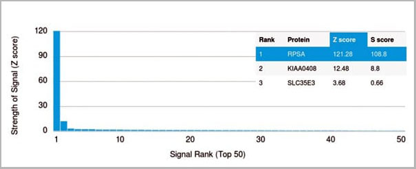 Application Data
(Analysis of Protein Array containing more than 19,000 full-length human proteins using Laminin Receptor Monospecific Mouse Monoclonal Antibody (RPSA/2699) Z- and S- Score: The Z-score represents the strength of a signal that a monoclonal antibody (MAb) (in combination with a fluorescently-tagged anti-IgG secondary antibody) produces when binding to a particular protein on the HuProtTM array. Z-scores are described in units of standard deviations (SD's) above the mean value of all signals generated on that array. If targets on HuProtTM are arranged in descending order of the Z-score, the S-score is the difference (also in units of SD's) between the Z-score. S-score therefore represents the relative target specificity of a MAb to its intended target. A MAb is considered to specific to its intended target, if the MAb has an S-score of at least 2.5. For example, if a MAb binds to protein X with a Z-score of 43 and to protein Y with a Z-score of 14, then the S-score for the binding of that MAb to protein X is equal to 29.)
Application Data
(Analysis of Protein Array containing more than 19,000 full-length human proteins using Laminin Receptor Monospecific Mouse Monoclonal Antibody (RPSA/2699) Z- and S- Score: The Z-score represents the strength of a signal that a monoclonal antibody (MAb) (in combination with a fluorescently-tagged anti-IgG secondary antibody) produces when binding to a particular protein on the HuProtTM array. Z-scores are described in units of standard deviations (SD's) above the mean value of all signals generated on that array. If targets on HuProtTM are arranged in descending order of the Z-score, the S-score is the difference (also in units of SD's) between the Z-score. S-score therefore represents the relative target specificity of a MAb to its intended target. A MAb is considered to specific to its intended target, if the MAb has an S-score of at least 2.5. For example, if a MAb binds to protein X with a Z-score of 43 and to protein Y with a Z-score of 14, then the S-score for the binding of that MAb to protein X is equal to 29.)
Laminin Receptor/RPSA, Monoclonal Antibody (Cat# AAA23908)
Full Name
Laminin Receptor/RPSA (Marker of Metastatic Potential)
Gene Names
RPSA; SA; LBP; LRP; p40; 67LR; ICAS; lamR; 37LRP; LAMBR; LAMR1; LRP/LR; LBP/p40; NEM/1CHD4
Reactivity
Human. Others not known.
Applications
Flow Cytometry, Immunofluorescence, Western Blot, Immunohistochemistry
Pricing
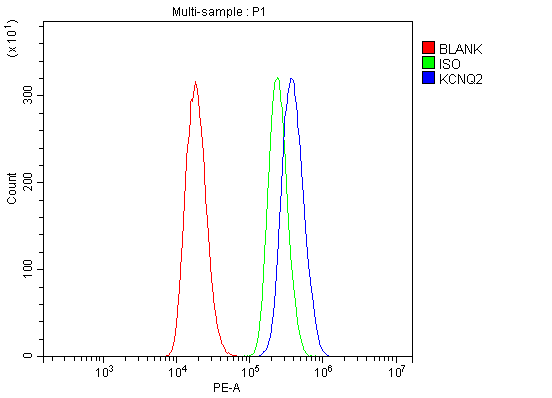
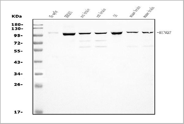
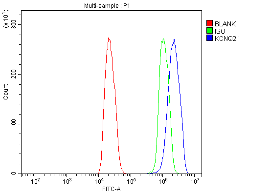 FCM (Flow Cytometry)
(Figure 7. Flow Cytometry analysis of C6 cells using anti-KCNQ2 antibody. Overlay histogram showing C6 cells stained with (Blue line). The cells were blocked with 10% normal goat serum. And then incubated with rabbit anti-KCNQ2 Antibody (1 ug/1x10^6 cells) for 30 min at 20°C. DyLight®488 conjugated goat anti-rabbit IgG (BA1127, 5-10 ug/1x10^6 cells) was used as secondary antibody for 30 minutes at 20°C. Isotype control antibody (Green line) was rabbit IgG (1 ug/1x10^6) used under the same conditions. Unlabelled sample (Red line) was also used as a control.)
FCM (Flow Cytometry)
(Figure 7. Flow Cytometry analysis of C6 cells using anti-KCNQ2 antibody. Overlay histogram showing C6 cells stained with (Blue line). The cells were blocked with 10% normal goat serum. And then incubated with rabbit anti-KCNQ2 Antibody (1 ug/1x10^6 cells) for 30 min at 20°C. DyLight®488 conjugated goat anti-rabbit IgG (BA1127, 5-10 ug/1x10^6 cells) was used as secondary antibody for 30 minutes at 20°C. Isotype control antibody (Green line) was rabbit IgG (1 ug/1x10^6) used under the same conditions. Unlabelled sample (Red line) was also used as a control.)
KCNQ2, Polyclonal Antibody (Cat# AAA19238)
Full Name
Anti-KCNQ2 Antibody
Gene Names
KCNQ2; EBN; BFNC; EBN1; ENB1; BFNS1; EIEE7; HNSPC; KV7.2; KCNA11; KVEBN1
Reactivity
Human, Mouse, Rat
Applications
Western Blot, Immunohistochemistry, Flow Cytometry
Purity
Immunogen affinity purified.
Pricing