Filters
Clonality
Type
Reactivity
Gene Name
Isotype
Host
Application
Clone
298 results for "Mouse IgG Isotype Control" - showing 200-250


 FCM (Flow Cytometry)
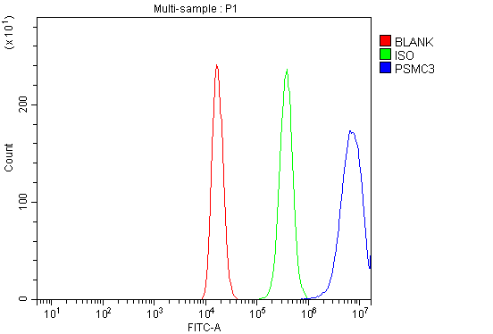
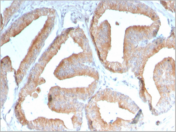
(Figure 7. Flow Cytometry analysis of 293T cells using anti-TBP-1/PSMC3 antibody (AAA19314).Overlay histogram showing 293T cells stained with AAA19314 (Blue line). The cells were blocked with 10% normal goat serum. And then incubated with rabbit anti-TBP-1/PSMC3 Antibody (AAA19314, 1μg/1x106 cells) for 30 min at 20 degree C. DyLight®488 conjugated goat anti-rabbit IgG (5-10μg/1x106 cells) was used as secondary antibody for 30 minutes at 20 degree C. Isotype control antibody (Green line) was rabbit IgG (1μg/1x106) used under the same conditions. Unlabelled sample (Red line) was also used as a control.)
FCM (Flow Cytometry)
(Figure 7. Flow Cytometry analysis of 293T cells using anti-TBP-1/PSMC3 antibody (AAA19314).Overlay histogram showing 293T cells stained with AAA19314 (Blue line). The cells were blocked with 10% normal goat serum. And then incubated with rabbit anti-TBP-1/PSMC3 Antibody (AAA19314, 1μg/1x106 cells) for 30 min at 20 degree C. DyLight®488 conjugated goat anti-rabbit IgG (5-10μg/1x106 cells) was used as secondary antibody for 30 minutes at 20 degree C. Isotype control antibody (Green line) was rabbit IgG (1μg/1x106) used under the same conditions. Unlabelled sample (Red line) was also used as a control.)
TBP-1/PSMC3, Polyclonal Antibody (Cat# AAA19314)
Full Name
Anti-TBP-1/PSMC3 Antibody
Gene Names
PSMC3; TBP1
Reactivity
Human, Mouse, Rat
Applications
WB, IHC-P, FC/FACS/FCM, EIA
Purity
Immunogen affinity purified.
Pricing


 FCM (Flow Cytometry)
(Figure 10. Flow Cytometry analysis of A549 cells using anti- U2AF65/U2AF2 antibody (AAA19375).Overlay histogram showing A549 cells stained with AAA19375 (Blue line). The cells were blocked with 10% normal goat serum. And then incubated with mouse anti-U2AF65/U2AF2 Antibody (AAA19375, 1μg/1x106 cells) for 30 min at 20 degree C. DyLight®488 conjugated goat anti-mouse IgG (BA1126, 5-10μg/1x106 cells) was used as secondary antibody for 30 minutes at 20 degree C. Isotype control antibody (Green line) was mouse IgG (1μg/1x106) used under the same conditions. Unlabelled sample (Red line) was also used as a control.)
FCM (Flow Cytometry)
(Figure 10. Flow Cytometry analysis of A549 cells using anti- U2AF65/U2AF2 antibody (AAA19375).Overlay histogram showing A549 cells stained with AAA19375 (Blue line). The cells were blocked with 10% normal goat serum. And then incubated with mouse anti-U2AF65/U2AF2 Antibody (AAA19375, 1μg/1x106 cells) for 30 min at 20 degree C. DyLight®488 conjugated goat anti-mouse IgG (BA1126, 5-10μg/1x106 cells) was used as secondary antibody for 30 minutes at 20 degree C. Isotype control antibody (Green line) was mouse IgG (1μg/1x106) used under the same conditions. Unlabelled sample (Red line) was also used as a control.)
U2AF65/U2AF2, Monoclonal Antibody (Cat# AAA19375)
Full Name
Anti-U2AF65/U2AF2 Antibody (monoclonal, 10F4)
Gene Names
U2AF2; U2AF65
Reactivity
Human, Mouse, Rat
Applications
WB, IHC-P, ICC, IF, FC/FACS/FCM
Purity
Immunogen affinity purified.
Pricing


 FCM (Flow Cytometry)
(Figure 7. Flow Cytometry analysis of HELA cells using anti-DNAJC10 antibody (AAA19320).Overlay histogram showing HELA cells stained with AAA19320 (Blue line). The cells were blocked with 10% normal goat serum. And then incubated with rabbit anti-DNAJC10 Antibody (AAA19320, 1μg/1x106 cells) for 30 min at 20 degree C. DyLight®488 conjugated goat anti-rabbit IgG (5-10μg/1x106 cells) was used as secondary antibody for 30 minutes at 20 degree C. Isotype control antibody (Green line) was rabbit IgG (1μg/1x106) used under the same conditions. Unlabelled sample (Red line) was also used as a control.)
FCM (Flow Cytometry)
(Figure 7. Flow Cytometry analysis of HELA cells using anti-DNAJC10 antibody (AAA19320).Overlay histogram showing HELA cells stained with AAA19320 (Blue line). The cells were blocked with 10% normal goat serum. And then incubated with rabbit anti-DNAJC10 Antibody (AAA19320, 1μg/1x106 cells) for 30 min at 20 degree C. DyLight®488 conjugated goat anti-rabbit IgG (5-10μg/1x106 cells) was used as secondary antibody for 30 minutes at 20 degree C. Isotype control antibody (Green line) was rabbit IgG (1μg/1x106) used under the same conditions. Unlabelled sample (Red line) was also used as a control.)
DNAJC10, Polyclonal Antibody (Cat# AAA19320)
Full Name
Anti-DNAJC10 Antibody
Gene Names
DNAJC10; JPDI; MTHr; ERdj5; PDIA19
Reactivity
Human, Mouse, Rat
Applications
WB, IHC-P, ICC, IF, FC/FACS/FCM, EIA
Purity
Immunogen affinity purified.
Pricing


 FCM (Flow Cytometry)
(Figure 6. Flow Cytometry analysis of K562 cells using anti-D2AP antibody (M01756). Overlay histogram showing K562 cells stained with M01756 (Blue line).The cells were blocked with 10% normal goat serum. And then incubated with mouse anti-D2AP Antibody (M01756, 1ug/1x106 cells) for 30 min at 20 degree C. DyLight488 conjugated goat anti-mouse IgG (BA1126, 5-10ug/1x106 cells) was used as secondary antibody for 30 minutes at 20 degree C. Isotype control antibody (Green line) was mouse IgG (1ug/1x106) used under the same conditions. Unlabelled sample (Red line) was also used as a control.)
FCM (Flow Cytometry)
(Figure 6. Flow Cytometry analysis of K562 cells using anti-D2AP antibody (M01756). Overlay histogram showing K562 cells stained with M01756 (Blue line).The cells were blocked with 10% normal goat serum. And then incubated with mouse anti-D2AP Antibody (M01756, 1ug/1x106 cells) for 30 min at 20 degree C. DyLight488 conjugated goat anti-mouse IgG (BA1126, 5-10ug/1x106 cells) was used as secondary antibody for 30 minutes at 20 degree C. Isotype control antibody (Green line) was mouse IgG (1ug/1x106) used under the same conditions. Unlabelled sample (Red line) was also used as a control.)
CD2AP, Monoclonal Antibody (Cat# AAA19185)
Full Name
Anti-CD2AP Antibody (monoclonal, 5F8)
Gene Names
CD2AP; CMS
Reactivity
Human, Mouse, Rat
Applications
WB, IHC, FC/FACS
Purity
Immunogen Affinity Purified
Pricing


 FCM (Flow Cytometry)
(Figure 8. Flow Cytometry analysis of A549 cells using anti-Transketolase/TKT antibody (AAA19368).Overlay histogram showing A549 cells stained with AAA19368 (Blue line). The cells were blocked with 10% normal goat serum. And then incubated with mouse anti- Transketolase/TKT Antibody (AAA19368, 1μg/1x106 cells) for 30 min at 20 degree C. DyLight®488 conjugated goat anti-mouse IgG (BA1126, 5-10μg/1x106 cells) was used as secondary antibody for 30 minutes at 20 degree C. Isotype control antibody (Green line) was mouse IgG (1μg/1x106) used under the same conditions. Unlabelled sample (Red line) was also used as a control.)
FCM (Flow Cytometry)
(Figure 8. Flow Cytometry analysis of A549 cells using anti-Transketolase/TKT antibody (AAA19368).Overlay histogram showing A549 cells stained with AAA19368 (Blue line). The cells were blocked with 10% normal goat serum. And then incubated with mouse anti- Transketolase/TKT Antibody (AAA19368, 1μg/1x106 cells) for 30 min at 20 degree C. DyLight®488 conjugated goat anti-mouse IgG (BA1126, 5-10μg/1x106 cells) was used as secondary antibody for 30 minutes at 20 degree C. Isotype control antibody (Green line) was mouse IgG (1μg/1x106) used under the same conditions. Unlabelled sample (Red line) was also used as a control.)
Transketolase/TKT, Monoclonal Antibody (Cat# AAA19368)
Full Name
Anti-Transketolase/TKT Antibody (monoclonal, 2I3)
Gene Names
TKT; TK; TKT1; HEL107
Reactivity
Human, Mouse, Rat
Applications
WB, IHC-P, ICC, IF, FC/FACS/FCM
Purity
Immunogen affinity purified.
Pricing


 FCM (Flow Cytometry)
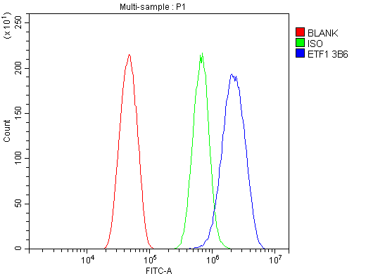
(Figure 11. Flow Cytometry analysis of RH35 cells using anti- eRF1/ETF1 antibody (AAA19382).Overlay histogram showing RH35 cells stained with AAA19382 (Blue line). The cells were blocked with 10% normal goat serum. And then incubated with mouse anti-eRF1/ETF1 Antibody (AAA19382, 1μg/1x106 cells) for 30 min at 20 degree C. DyLight®488 conjugated goat anti-mouse IgG (BA1126, 5-10μg/1x106 cells) was used as secondary antibody for 30 minutes at 20 degree C. Isotype control antibody (Green line) was mouse IgG (1μg/1x106) used under the same conditions. Unlabelled sample (Red line) was also used as a control.)
FCM (Flow Cytometry)
(Figure 11. Flow Cytometry analysis of RH35 cells using anti- eRF1/ETF1 antibody (AAA19382).Overlay histogram showing RH35 cells stained with AAA19382 (Blue line). The cells were blocked with 10% normal goat serum. And then incubated with mouse anti-eRF1/ETF1 Antibody (AAA19382, 1μg/1x106 cells) for 30 min at 20 degree C. DyLight®488 conjugated goat anti-mouse IgG (BA1126, 5-10μg/1x106 cells) was used as secondary antibody for 30 minutes at 20 degree C. Isotype control antibody (Green line) was mouse IgG (1μg/1x106) used under the same conditions. Unlabelled sample (Red line) was also used as a control.)
eRF1/ETF1, Monoclonal Antibody (Cat# AAA19382)
Full Name
Anti-eRF1/ETF1 Antibody (monoclonal, 3B6)
Gene Names
ETF1; ERF; RF1; ERF1; TB3-1; D5S1995; SUP45L1
Reactivity
Human, Mouse, Rat
Applications
WB, IHC-P, ICC, IF, FC/FACS/FCM
Purity
Immunogen affinity purified.
Pricing


 FCM (Flow Cytometry)
(Figure 8. Flow Cytometry analysis of THP-1 cells using anti-INPPL1 antibody (AAA19364).Overlay histogram showing THP-1 cells stained with AAA19364 (Blue line). The cells were blocked with 10% normal goat serum. And then incubated with mouse anti- INPPL1 Antibody (AAA19364, 1μg/1x106 cells) for 30 min at 20 degree C. DyLight®488 conjugated goat anti-mouse IgG (BA1126, 5-10μg/1x106 cells) was used as secondary antibody for 30 minutes at 20 degree C. Isotype control antibody (Green line) was mouse IgG (1μg/1x106) used under the same conditions. Unlabelled sample (Red line) was also used as a control.)
FCM (Flow Cytometry)
(Figure 8. Flow Cytometry analysis of THP-1 cells using anti-INPPL1 antibody (AAA19364).Overlay histogram showing THP-1 cells stained with AAA19364 (Blue line). The cells were blocked with 10% normal goat serum. And then incubated with mouse anti- INPPL1 Antibody (AAA19364, 1μg/1x106 cells) for 30 min at 20 degree C. DyLight®488 conjugated goat anti-mouse IgG (BA1126, 5-10μg/1x106 cells) was used as secondary antibody for 30 minutes at 20 degree C. Isotype control antibody (Green line) was mouse IgG (1μg/1x106) used under the same conditions. Unlabelled sample (Red line) was also used as a control.)
INPPL1, Monoclonal Antibody (Cat# AAA19364)
Full Name
Anti-INPPL1 Antibody (monoclonal, 8C13)
Gene Names
INPPL1; OPSMD; SHIP2
Reactivity
Human, Mouse, Rat
Applications
WB, IHC-P, ICC, IF, FC/FACS/FCM
Purity
Immunogen affinity purified.
Pricing


 FCM (Flow Cytometry)
(Figure 9. Flow Cytometry analysis of U251 cells using anti-RAB1B antibody (AAA19296).Overlay histogram showing U251 cells stained with AAA19296 (Blue line). The cells were blocked with 10% normal goat serum. And then incubated with rabbit anti-RAB1B Antibody (AAA19296, 1μg/1x106 cells) for 30 min at 20 degree C. DyLight®488 conjugated goat anti-rabbit IgG (5-10μg/1x106 cells) was used as secondary antibody for 30 minutes at 20 degree C. Isotype control antibody (Green line) was rabbit IgG (1μg/1x106) used under the same conditions. Unlabelled sample (Red line) was also used as a control.)
FCM (Flow Cytometry)
(Figure 9. Flow Cytometry analysis of U251 cells using anti-RAB1B antibody (AAA19296).Overlay histogram showing U251 cells stained with AAA19296 (Blue line). The cells were blocked with 10% normal goat serum. And then incubated with rabbit anti-RAB1B Antibody (AAA19296, 1μg/1x106 cells) for 30 min at 20 degree C. DyLight®488 conjugated goat anti-rabbit IgG (5-10μg/1x106 cells) was used as secondary antibody for 30 minutes at 20 degree C. Isotype control antibody (Green line) was rabbit IgG (1μg/1x106) used under the same conditions. Unlabelled sample (Red line) was also used as a control.)
RAB1B, Polyclonal Antibody (Cat# AAA19296)
Full Name
Anti-RAB1B Antibody
Reactivity
Human, Mouse, Rat
Applications
WB, IHC-P, ICC, IF, FC/FACS/FCM
Purity
Immunogen affinity purified.
Pricing


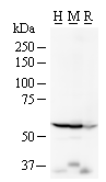
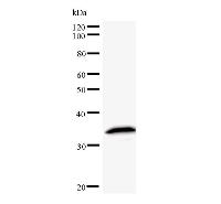
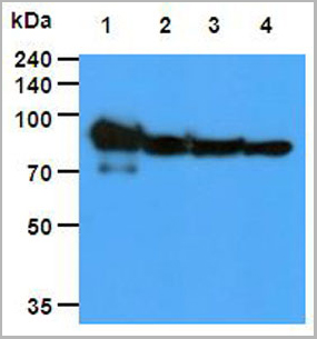
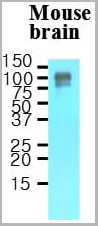
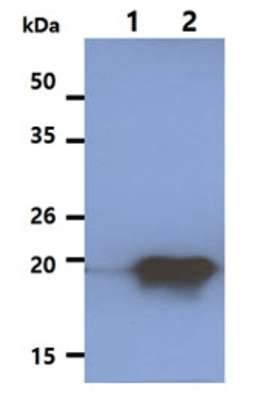
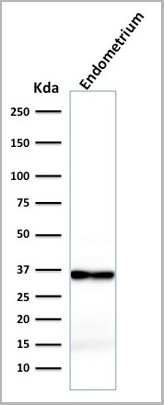
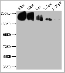
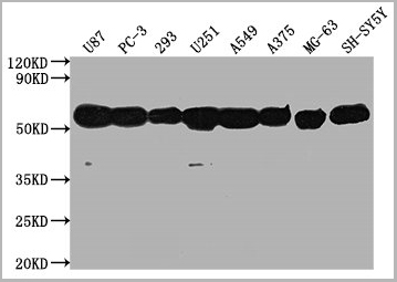
 WB (Western Blot)
(Western blot analysis of lysates from 293, Hela, mouse NIH/3T3, rat PC-12 cell line and rat brain tissue lysate(from left to right), using RPS6 Antibody (N-term). AAA28643 was diluted at 1:2000 at each lane. A goat anti-mouse IgG H&L(HRP) at 1:3000 dilution was used as the secondary antibody. Lysates at 35ug per lane.)
WB (Western Blot)
(Western blot analysis of lysates from 293, Hela, mouse NIH/3T3, rat PC-12 cell line and rat brain tissue lysate(from left to right), using RPS6 Antibody (N-term). AAA28643 was diluted at 1:2000 at each lane. A goat anti-mouse IgG H&L(HRP) at 1:3000 dilution was used as the secondary antibody. Lysates at 35ug per lane.)
RPS6, Monoclonal Antibody (Cat# AAA28643)
Full Name
RPS6 Antibody (N-term)
Gene Names
RPS6; S6
Reactivity
Human, rat (Predicted Reactivity: Monkey, Mouse)
Applications
EIA, IHC, WB, FC/FACS, IF
Purity
Purified Mouse Monoclonal Antibody (Mab)
Pricing


 FCM (Flow Cytometry)
(Figure 7. Flow Cytometry analysis of MCF-7 cells using anti-RGS6 antibody (AAA19308).Overlay histogram showing MCF-7 cells stained with AAA19308 (Blue line). The cells were blocked with 10% normal goat serum. And then incubated with rabbit anti-RGS6 Antibody (AAA19308, 1μg/1x106 cells) for 30 min at 20 degree C. DyLight®488 conjugated goat anti-rabbit IgG (5-10μg/1x106 cells) was used as secondary antibody for 30 minutes at 20 degree C. Isotype control antibody (Green line) was rabbit IgG (1μg/1x106) used under the same conditions. Unlabelled sample (Red line) was also used as a control.)
FCM (Flow Cytometry)
(Figure 7. Flow Cytometry analysis of MCF-7 cells using anti-RGS6 antibody (AAA19308).Overlay histogram showing MCF-7 cells stained with AAA19308 (Blue line). The cells were blocked with 10% normal goat serum. And then incubated with rabbit anti-RGS6 Antibody (AAA19308, 1μg/1x106 cells) for 30 min at 20 degree C. DyLight®488 conjugated goat anti-rabbit IgG (5-10μg/1x106 cells) was used as secondary antibody for 30 minutes at 20 degree C. Isotype control antibody (Green line) was rabbit IgG (1μg/1x106) used under the same conditions. Unlabelled sample (Red line) was also used as a control.)
RGS6, Polyclonal Antibody (Cat# AAA19308)
Full Name
Anti-RGS6 Antibody
Gene Names
RGS6; GAP
Reactivity
Human, Mouse, Rat
Applications
WB, IHC-P, FC/FACS/FCM, EIA
Purity
Immunogen affinity purified.
Pricing


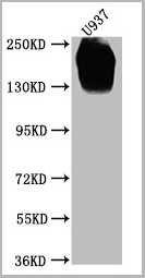
 FCM (Flow Cytometry)
(Figure 12. Flow Cytometry analysis of U937 cells using anti-DYNLL1/PIN antibody (AAA19276).Overlay histogram showing U937 cells stained with AAA19276 (Blue line). The cells were blocked with 10% normal goat serum. And then incubated with rabbit anti-DYNLL1/PIN Antibody (AAA19276, 1μg/1x106 cells) for 30 min at 20 degree C. DyLight®488 conjugated goat anti-rabbit IgG (5-10μg/1x106 cells) was used as secondary antibody for 30 minutes at 20 degree C. Isotype control antibody (Green line) was rabbit IgG (1μg/1x106) used under the same conditions. Unlabelled sample (Red line) was also used as a control.)
FCM (Flow Cytometry)
(Figure 12. Flow Cytometry analysis of U937 cells using anti-DYNLL1/PIN antibody (AAA19276).Overlay histogram showing U937 cells stained with AAA19276 (Blue line). The cells were blocked with 10% normal goat serum. And then incubated with rabbit anti-DYNLL1/PIN Antibody (AAA19276, 1μg/1x106 cells) for 30 min at 20 degree C. DyLight®488 conjugated goat anti-rabbit IgG (5-10μg/1x106 cells) was used as secondary antibody for 30 minutes at 20 degree C. Isotype control antibody (Green line) was rabbit IgG (1μg/1x106) used under the same conditions. Unlabelled sample (Red line) was also used as a control.)
DYNLL1/PIN, Polyclonal Antibody (Cat# AAA19276)
Full Name
Anti-DYNLL1/PIN Antibody
Gene Names
DYNLL1; LC8; PIN; DLC1; DLC8; LC8a; DNCL1; hdlc1; DNCLC1
Reactivity
Human, Mouse, Rat
Applications
WB, IHC-P, ICC, IF, FC/FACS/FCM, EIA
Purity
Immunogen affinity purified.
Pricing


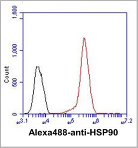
 FCM (Flow Cytometry)
(Figure 7. Flow Cytometry analysis of A549 cells using anti-Hsp90 alpha antibody (AAA19359).Overlay histogram showing A549 cells stained with AAA19359 (Blue line). The cells were blocked with 10% normal goat serum. And then incubated with mouse anti-Hsp90 alpha Antibody (AAA19359, 1μg/1x106 cells) for 30 min at 20 degree C. DyLight®488 conjugated goat anti-mouse IgG (BA1126, 5-10μg/1x106 cells) was used as secondary antibody for 30 minutes at 20 degree C. Isotype control antibody (Green line) was mouse IgG (1μg/1x106) used under the same conditions. Unlabelled sample (Red line) was also used as a control.)
FCM (Flow Cytometry)
(Figure 7. Flow Cytometry analysis of A549 cells using anti-Hsp90 alpha antibody (AAA19359).Overlay histogram showing A549 cells stained with AAA19359 (Blue line). The cells were blocked with 10% normal goat serum. And then incubated with mouse anti-Hsp90 alpha Antibody (AAA19359, 1μg/1x106 cells) for 30 min at 20 degree C. DyLight®488 conjugated goat anti-mouse IgG (BA1126, 5-10μg/1x106 cells) was used as secondary antibody for 30 minutes at 20 degree C. Isotype control antibody (Green line) was mouse IgG (1μg/1x106) used under the same conditions. Unlabelled sample (Red line) was also used as a control.)
Hsp90 alpha, Monoclonal Antibody (Cat# AAA19359)
Full Name
Anti-Hsp90 alpha Antibody (monoclonal, 6B5)
Gene Names
HSP90AA1; EL52; HSPN; LAP2; HSP86; HSPC1; HSPCA; Hsp89; Hsp90; HSP89A; HSP90A; HSP90N; HSPCAL1; HSPCAL4
Reactivity
Human, Mouse, Rat, Monkey
Applications
WB, IHC-P, ICC, IF, FC/FACS/FCM
Purity
Immunogen affinity purified.
Pricing


 FCM (Flow Cytometry)
(Figure 7. Flow Cytometry analysis of U20S cells using anti- ADA antibody (AAA19184). Overlay histogram showing U20S cells stained with AAA19184 (Blue line).The cells were blocked with 10% normal goat serum. And then incubated with mouse anti- ADA Antibody (AAA19184, 1ug/1x106 cells) for 30 min at 20 degree C. DyLight488 conjugated goat anti-mouse IgG (BA1126, 5-10ug/1x106 cells) was used as secondary antibody for 30 minutes at 20 degree C. Isotype control antibody (Green line) was mouse IgG (1ug/1x106) used under the same conditions. Unlabelled sample (Red line) was also used as a control.)
FCM (Flow Cytometry)
(Figure 7. Flow Cytometry analysis of U20S cells using anti- ADA antibody (AAA19184). Overlay histogram showing U20S cells stained with AAA19184 (Blue line).The cells were blocked with 10% normal goat serum. And then incubated with mouse anti- ADA Antibody (AAA19184, 1ug/1x106 cells) for 30 min at 20 degree C. DyLight488 conjugated goat anti-mouse IgG (BA1126, 5-10ug/1x106 cells) was used as secondary antibody for 30 minutes at 20 degree C. Isotype control antibody (Green line) was mouse IgG (1ug/1x106) used under the same conditions. Unlabelled sample (Red line) was also used as a control.)
ADA, Monoclonal Antibody (Cat# AAA19184)
Full Name
Anti-ADA Antibody (monoclonal, 6D4)
Reactivity
Human
Applications
WB, IHC, FC/FACS
Purity
Immunogen Affinity Purified
Pricing


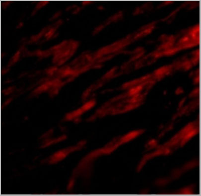
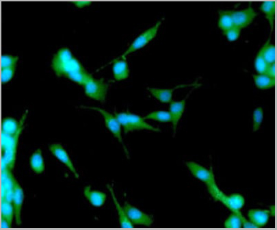
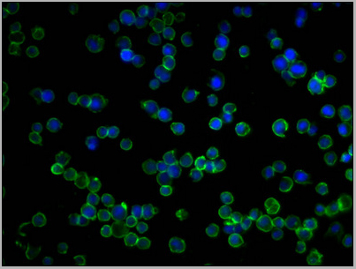
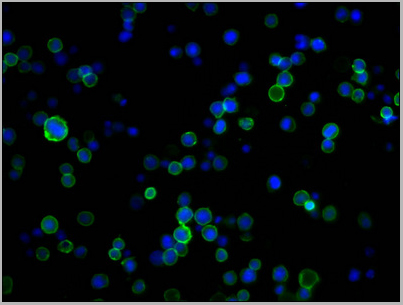
 IF (Immunofluorescence)
(Figure 11. IF analysis of Tubulin alpha using anti- Tubulin alpha antibody (AAA19381).Tubulin alpha was detected in immunocytochemical section of CACO-2 cells. Enzyme antigen retrieval was performed using IHC enzyme antigen retrieval reagent for 15 mins. The cells were blocked with 10% goat serum. And then incubated with 5μg/mL mouse anti- Tubulin alpha Antibody (AAA19381) overnight at 4 degree C. DyLight®594 Conjugated Goat Anti-Mouse IgG (BA1141) was used as secondary antibody at 1:100 dilution and incubated for 30 minutes at 37 degree C. The section was counterstained with DAPI. Visualize using a fluorescence microscope and filter sets appropriate for the label used.)
IF (Immunofluorescence)
(Figure 11. IF analysis of Tubulin alpha using anti- Tubulin alpha antibody (AAA19381).Tubulin alpha was detected in immunocytochemical section of CACO-2 cells. Enzyme antigen retrieval was performed using IHC enzyme antigen retrieval reagent for 15 mins. The cells were blocked with 10% goat serum. And then incubated with 5μg/mL mouse anti- Tubulin alpha Antibody (AAA19381) overnight at 4 degree C. DyLight®594 Conjugated Goat Anti-Mouse IgG (BA1141) was used as secondary antibody at 1:100 dilution and incubated for 30 minutes at 37 degree C. The section was counterstained with DAPI. Visualize using a fluorescence microscope and filter sets appropriate for the label used.)
Tubulin alpha, Monoclonal Antibody (Cat# AAA19381)
Full Name
Anti-Tubulin alpha Antibody (monoclonal, 7B12)
Gene Names
TUBA1A; LIS3; TUBA3; B-ALPHA-1
Reactivity
Human, Mouse, Rat
Applications
WB, IHC-P, ICC, IF, FC/FACS/FCM
Purity
Immunogen affinity purified.
Pricing


 FCM (Flow Cytometry)
(Figure 8. Flow Cytometry analysis of K562 cells using anti-PDIA6 antibody (AAA19282).Overlay histogram showing K562 cells stained with AAA19282 (Blue line). The cells were blocked with 10% normal goat serum. And then incubated with rabbit anti-PDIA6 Antibody (AAA19282, 1μg/1x106 cells) for 30 min at 20 degree C. DyLight®488 conjugated goat anti-rabbit IgG (5-10μg/1x106 cells) was used as secondary antibody for 30 minutes at 20 degree C. Isotype control antibody (Green line) was rabbit IgG (1μg/1x106) used under the same conditions. Unlabelled sample (Red line) was also used as a control.)
FCM (Flow Cytometry)
(Figure 8. Flow Cytometry analysis of K562 cells using anti-PDIA6 antibody (AAA19282).Overlay histogram showing K562 cells stained with AAA19282 (Blue line). The cells were blocked with 10% normal goat serum. And then incubated with rabbit anti-PDIA6 Antibody (AAA19282, 1μg/1x106 cells) for 30 min at 20 degree C. DyLight®488 conjugated goat anti-rabbit IgG (5-10μg/1x106 cells) was used as secondary antibody for 30 minutes at 20 degree C. Isotype control antibody (Green line) was rabbit IgG (1μg/1x106) used under the same conditions. Unlabelled sample (Red line) was also used as a control.)
PDIA6, Polyclonal Antibody (Cat# AAA19282)
Full Name
Anti-PDIA6 Antibody
Gene Names
PDIA6; P5; ERP5; TXNDC7
Reactivity
Human, Mouse, Rat
Applications
WB, IHC-P, ICC, IF, FC/FACS/FCM, EIA
Purity
Immunogen affinity purified.
Pricing


 IF (Immunofluorescence)
MSH2, Monoclonal Antibody (Cat# AAA23925)
Full Name
MSH2 (DNA Mismatch Repair Marker)
Gene Names
MSH2; FCC1; COCA1; HNPCC; LCFS2; hMSH2; HNPCC1
Reactivity
Human
Applications
FC, WB, IHC
Purity
Purified
Pricing


 FCM (Flow Cytometry)
(Figure 9. Flow Cytometry analysis of K562 cells using anti-MCM6 antibody (AAA19372).Overlay histogram showing K562 cells stained with AAA19372 (Blue line). The cells were blocked with 10% normal goat serum. And then incubated with mouse anti- MCM6 Antibody (AAA19372, 1μg/1x106 cells) for 30 min at 20 degree C. DyLight®488 conjugated goat anti-mouse IgG (BA1126, 5-10μg/1x106 cells) was used as secondary antibody for 30 minutes at 20 degree C. Isotype control antibody (Green line) was mouse IgG (1μg/1x106) used under the same conditions. Unlabelled sample (Red line) was also used as a control.)
FCM (Flow Cytometry)
(Figure 9. Flow Cytometry analysis of K562 cells using anti-MCM6 antibody (AAA19372).Overlay histogram showing K562 cells stained with AAA19372 (Blue line). The cells were blocked with 10% normal goat serum. And then incubated with mouse anti- MCM6 Antibody (AAA19372, 1μg/1x106 cells) for 30 min at 20 degree C. DyLight®488 conjugated goat anti-mouse IgG (BA1126, 5-10μg/1x106 cells) was used as secondary antibody for 30 minutes at 20 degree C. Isotype control antibody (Green line) was mouse IgG (1μg/1x106) used under the same conditions. Unlabelled sample (Red line) was also used as a control.)
MCM6, Monoclonal Antibody (Cat# AAA19372)
Full Name
Anti-MCM6 Antibody (monoclonal, 10I9)
Gene Names
MCM6; Mis5; P105MCM; MCG40308
Reactivity
Human
Applications
WB, IHC-P, ICC, IF, FC/FACS/FCM
Purity
Immunogen affinity purified.
Pricing


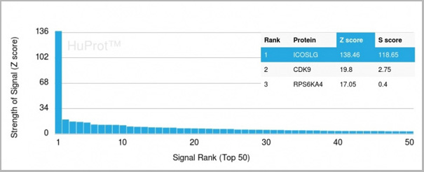
 Application Data
(Analysis of Protein Array containing more than 19,000 full-length human proteins using ROR-gamma / RORC Mouse Monoclonal Antibody (RORC/2941). Z- and S- Score: The Z-score represents the strength of a signal that a monoclonal antibody (MAb) (in combination with a fluorescently-tagged anti-IgG secondary antibody) produces when binding to a particular protein on the HuProtTM array. Z-scores are described in units of standard deviations (SD's) above the mean value of all signals generated on that array. If targets on HuProtTM are arranged in descending order of the Z-score, the S-score is the difference (also in units of SD's) between the Z-score. S-score therefore represents the relative target specificity of a MAb to its intended target. A MAb is considered to specific to its intended target, if the MAb has an S-score of at least 2.5. For example, if a MAb binds to protein X with a Z-score of 43 and to protein Y with a Z-score of 14, then the S-score for the binding of that MAb to protein X is equal to 29.)
Application Data
(Analysis of Protein Array containing more than 19,000 full-length human proteins using ROR-gamma / RORC Mouse Monoclonal Antibody (RORC/2941). Z- and S- Score: The Z-score represents the strength of a signal that a monoclonal antibody (MAb) (in combination with a fluorescently-tagged anti-IgG secondary antibody) produces when binding to a particular protein on the HuProtTM array. Z-scores are described in units of standard deviations (SD's) above the mean value of all signals generated on that array. If targets on HuProtTM are arranged in descending order of the Z-score, the S-score is the difference (also in units of SD's) between the Z-score. S-score therefore represents the relative target specificity of a MAb to its intended target. A MAb is considered to specific to its intended target, if the MAb has an S-score of at least 2.5. For example, if a MAb binds to protein X with a Z-score of 43 and to protein Y with a Z-score of 14, then the S-score for the binding of that MAb to protein X is equal to 29.)
ROR-gamma/RORC, Monoclonal Antibody (Cat# AAA23951)
Full Name
ROR-gamma/RORC (RAR-related Orphan Receptor C)
Gene Names
RORC; TOR; RORG; RZRG; IMD42; NR1F3; RZR-GAMMA
Reactivity
Human
Applications
FC, IHC
Purity
Purified Ab with BSA and Azide at 200ug/ml or Purified Ab with BSA and Azide at 200ug/ml or Purified Ab WITHOUT BSA and Azide at 1.0mg/ml
Pricing


 Application Data
(Analysis of Protein Array containing more than 19,000 full-length human proteins using Cytokeratin 15 Mouse Monoclonal Antibody (KRT15/2957). Z- and S- Score: The Z-score represents the strength of a signal that a monoclonal antibody (MAb) (in combination with a fluorescently-tagged anti-IgG secondary antibody) produces when binding to a particular protein on the HuProtTM array. Z-scores are described in units of standard deviations (SD's) above the mean value of all signals generated on that array. If targets on HuProtTM are arranged in descending order of the Z-score, the S-score is the difference (also in units of SD's) between the Z-score. S-score therefore represents the relative target specificity of a MAb to its intended target. A MAb is considered to specific to its intended target, if the MAb has an S-score of at least 2.5. For example, if a MAb binds to protein X with a Z-score of 43 and to protein Y with a Z-score of 14, then the S-score for the binding of that MAb to protein X is equal to 29.)
Application Data
(Analysis of Protein Array containing more than 19,000 full-length human proteins using Cytokeratin 15 Mouse Monoclonal Antibody (KRT15/2957). Z- and S- Score: The Z-score represents the strength of a signal that a monoclonal antibody (MAb) (in combination with a fluorescently-tagged anti-IgG secondary antibody) produces when binding to a particular protein on the HuProtTM array. Z-scores are described in units of standard deviations (SD's) above the mean value of all signals generated on that array. If targets on HuProtTM are arranged in descending order of the Z-score, the S-score is the difference (also in units of SD's) between the Z-score. S-score therefore represents the relative target specificity of a MAb to its intended target. A MAb is considered to specific to its intended target, if the MAb has an S-score of at least 2.5. For example, if a MAb binds to protein X with a Z-score of 43 and to protein Y with a Z-score of 14, then the S-score for the binding of that MAb to protein X is equal to 29.)
Cytokeratin 15, Monoclonal Antibody (Cat# AAA23923)
Full Name
Cytokeratin 15 (Esophageal Squamous Cell Carcinoma Marker)
Gene Names
KRT15; K15; CK15; K1CO
Reactivity
Human
Applications
FC/FACS, IF, WB, IHC
Purity
Purified Ab with BSA and Azide at 200ug/ml OR Purified Ab WITHOUT BSA and Azide at 1.0mg/ml
Pricing


 Application Data
(Analysis of Protein Array containing more than 19,000 full-length human proteins using CAIX-Monospecific Mouse Monoclonal Antibody (CA9/3406). Z- and S- Score: The Z-score represents the strength of a signal that a monoclonal antibody (MAb) (in combination with a fluorescently-tagged anti-IgG secondary antibody) produces when binding to a particular protein on the HuProtTM array. Z-scores are described in units of standard deviations (SD's) above the mean value of all signals generated on that array. If targets on HuProtTM are arranged in descending order of the Z-score, the S-score is the difference (also in units of SD's) between the Z-score. S-score therefore represents the relative target specificity of a MAb to its intended target. A MAb is considered to specific to its intended target, if the MAb has an S-score of at least 2.5. For example, if a MAb binds to protein X with a Z-score of 43 and to protein Y with a Z-score of 14, then the S-score for the binding of that MAb to protein X is equal to 29.)
Application Data
(Analysis of Protein Array containing more than 19,000 full-length human proteins using CAIX-Monospecific Mouse Monoclonal Antibody (CA9/3406). Z- and S- Score: The Z-score represents the strength of a signal that a monoclonal antibody (MAb) (in combination with a fluorescently-tagged anti-IgG secondary antibody) produces when binding to a particular protein on the HuProtTM array. Z-scores are described in units of standard deviations (SD's) above the mean value of all signals generated on that array. If targets on HuProtTM are arranged in descending order of the Z-score, the S-score is the difference (also in units of SD's) between the Z-score. S-score therefore represents the relative target specificity of a MAb to its intended target. A MAb is considered to specific to its intended target, if the MAb has an S-score of at least 2.5. For example, if a MAb binds to protein X with a Z-score of 43 and to protein Y with a Z-score of 14, then the S-score for the binding of that MAb to protein X is equal to 29.)
Renal Cell Carcinoma, Monoclonal Antibody (Cat# AAA23952)
Full Name
Renal Cell Carcinoma (Carbonic Anhydrase IX)
Gene Names
CA9; MN; CAIX
Reactivity
Human
Applications
FC/FACS, IHC, WB
Purity
Purified Ab with BSA and Azide at 200ug/ml or Purified Ab with BSA and Azide at 200ug/ml or Purified Ab WITHOUT BSA and Azide at 1.0mg/ml
Pricing


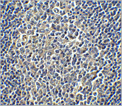
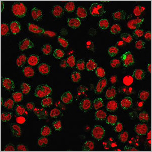
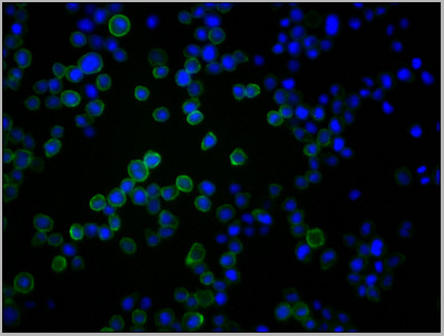
 ICC (Immunocytochemistry)
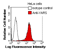
(Immunostaining analysis in HeLa cells. HeLa cells were fixed with 4% paraformaldehyde and permeabilized with 0.1% Triton X-100 in PBS. The cells were immunostained with anti-YARS mAb. [Lot No. YAR5H08-1])
ICC (Immunocytochemistry)
(Immunostaining analysis in HeLa cells. HeLa cells were fixed with 4% paraformaldehyde and permeabilized with 0.1% Triton X-100 in PBS. The cells were immunostained with anti-YARS mAb. [Lot No. YAR5H08-1])
YARS, Monoclonal Antibody (Cat# AAA10598)
Full Name
Mouse monoclonal antibody Anti-Human YARS
Gene Names
YARS; YRS; YTS; TYRRS; CMTDIC
Reactivity
Human
Applications
Immunocytochemistry, Immunoprecipitation, Western Blot, Flow Cytometry
Pricing
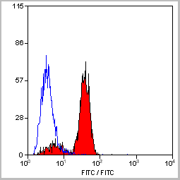
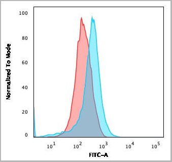
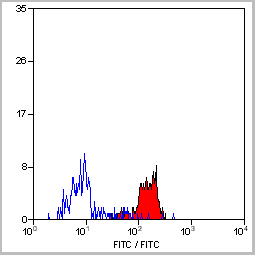
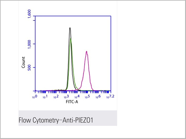 FCM (Flow Cytometry)
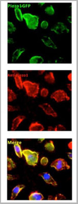
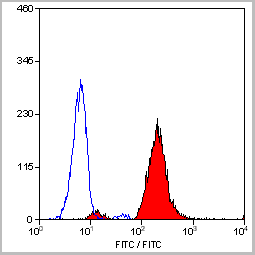
(Flow cytometric analysis of 2% paraformaldehyde-fixed THP1 (Human acute monocytic leukemia cell line) cells labeling PIEZO1 with AAA13800 at 1/200 dilution (red) compared with a mouse monoclonal IgG isotype control (black) and an unlabelled control (cells without incubation with primary antibody, green). Goat anti-mouse IgG (FITC) at 1/300 dilution was used as the secondary antibody.)
FCM (Flow Cytometry)
(Flow cytometric analysis of 2% paraformaldehyde-fixed THP1 (Human acute monocytic leukemia cell line) cells labeling PIEZO1 with AAA13800 at 1/200 dilution (red) compared with a mouse monoclonal IgG isotype control (black) and an unlabelled control (cells without incubation with primary antibody, green). Goat anti-mouse IgG (FITC) at 1/300 dilution was used as the secondary antibody.)
PIEZO1, Monoclonal Antibody (Cat# AAA13800)
Full Name
PIEZO1 Mouse mAb
Gene Names
PIEZO1; DHS; Mib; LMPH3; FAM38A
Reactivity
Homo Sapiens
Applications
Immunoprecipitation, Immunofluorescence, Flow Cytometry
Pricing
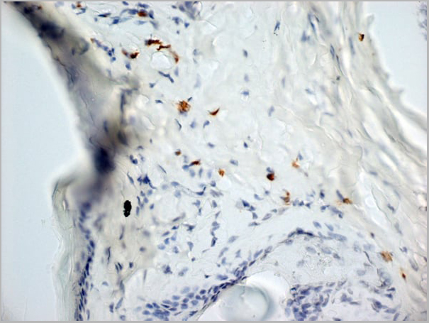
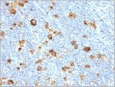
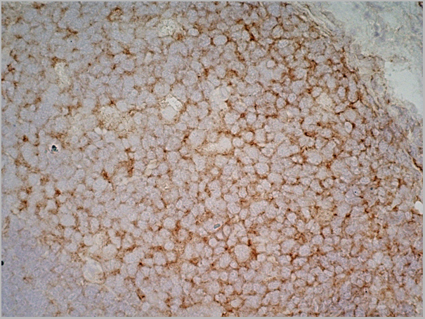
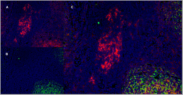
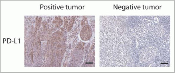 IF (Immunofluorescence)
(Figure 12 Immunohistochemistry Validation of PD-L1in Human Tumors (Gadiot et al., 2011)Immunohistochemical analysis of patient tumors labeling PD-L1 with anti-PD-L1 antibodies (AAA10941). Several anti-PD-L1 antibodies were tested for staining, “Only 1 antibody gave no background staining and was competitively blocked by the addition of PD-L1Fc protein (AAA10941)”.)
IF (Immunofluorescence)
(Figure 12 Immunohistochemistry Validation of PD-L1in Human Tumors (Gadiot et al., 2011)Immunohistochemical analysis of patient tumors labeling PD-L1 with anti-PD-L1 antibodies (AAA10941). Several anti-PD-L1 antibodies were tested for staining, “Only 1 antibody gave no background staining and was competitively blocked by the addition of PD-L1Fc protein (AAA10941)”.)
PDL-1, Polyclonal Antibody (Cat# AAA10941)
Full Name
PDL-1 Antibody
Gene Names
CD274; B7-H; B7H1; PDL1; PD-L1; PDCD1L1; PDCD1LG1
Applications
Western Blot, Immunohistochemistry, Immunofluorescence, Flow Cytometry
Purity
PD-L1 Antibody is affinity chromatography purified via peptide column.
Pricing
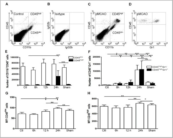
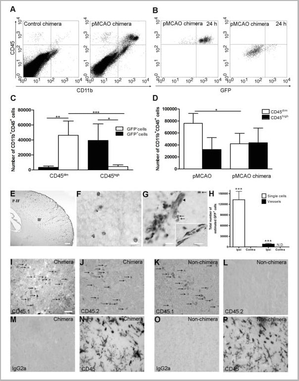
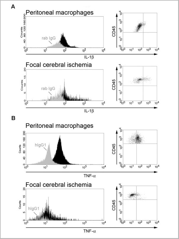
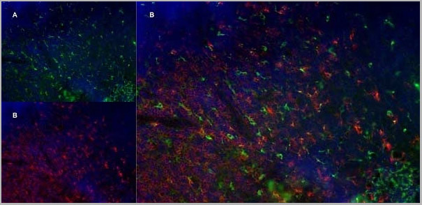
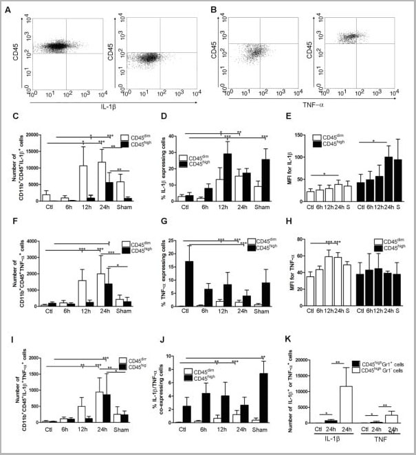 Application Data
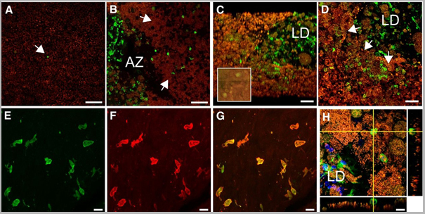
(Published customer image: Cytokine expression in segregated populations of cells following stroke. (A, B) Dot plots showing CD11b+CD45high macrophages/granulocytes (upper right quadrants) and CD11b+CD45dim microglia (bottom right quadrants) expressing IL-1beta (A) or TNF-a (B). (C-J) Bar graphs showing numbers and proportions of IL-1beta (C, D), TNF-a (F, G) and IL-1beta/TNF-a co-expressing (I, J) CD11b+CD45dim microglia and CD11b+CD45high macrophages/granulocytes in unmanipulated control mice (n = 10), in mice 6 (n = 7), 12 (n = 7), or 24 hours after pMCAO (n = 10), and in sham-operated mice 24 hours after pMCAO (n = 7). (E, H) Comparison of the MFI values for IL-1beta (E) and TNF-a (H) in viable CD11b+CD45dim microglia and CD11b+CD45high macrophages/granulocytes in unmanipulated mice, in mice 6, 12, or 24 hours after pMCAO, and in sham-operated mice 24 hours after pMCAO. Macrophages/granulocytes express significantly more IL-1beta than do microglial in unmanipulated mice, in mice 6, 12, or 24 hours after pMCAO, and in sham-operated mice 24 hours after pMCAO (E), whereas microglial cells express significantly higher levels of TNF-a than do macrophages/granulocytes at 12 h and 24 hours, and in sham-operated mice 24 hours after pMCAO (H). (K) CD11b+CD45highGr1- macrophages and not CD11b+CD45highGr1+ granulocytes are the main producers of IL-1beta and TNF-a 24 hours after pMCAO. *P < 0.05, **P < 0.01, and ***P < 0.001.From: http://www.jneuroinflammation.com/content/5/1/46.)
Application Data
(Published customer image: Cytokine expression in segregated populations of cells following stroke. (A, B) Dot plots showing CD11b+CD45high macrophages/granulocytes (upper right quadrants) and CD11b+CD45dim microglia (bottom right quadrants) expressing IL-1beta (A) or TNF-a (B). (C-J) Bar graphs showing numbers and proportions of IL-1beta (C, D), TNF-a (F, G) and IL-1beta/TNF-a co-expressing (I, J) CD11b+CD45dim microglia and CD11b+CD45high macrophages/granulocytes in unmanipulated control mice (n = 10), in mice 6 (n = 7), 12 (n = 7), or 24 hours after pMCAO (n = 10), and in sham-operated mice 24 hours after pMCAO (n = 7). (E, H) Comparison of the MFI values for IL-1beta (E) and TNF-a (H) in viable CD11b+CD45dim microglia and CD11b+CD45high macrophages/granulocytes in unmanipulated mice, in mice 6, 12, or 24 hours after pMCAO, and in sham-operated mice 24 hours after pMCAO. Macrophages/granulocytes express significantly more IL-1beta than do microglial in unmanipulated mice, in mice 6, 12, or 24 hours after pMCAO, and in sham-operated mice 24 hours after pMCAO (E), whereas microglial cells express significantly higher levels of TNF-a than do macrophages/granulocytes at 12 h and 24 hours, and in sham-operated mice 24 hours after pMCAO (H). (K) CD11b+CD45highGr1- macrophages and not CD11b+CD45highGr1+ granulocytes are the main producers of IL-1beta and TNF-a 24 hours after pMCAO. *P < 0.05, **P < 0.01, and ***P < 0.001.From: http://www.jneuroinflammation.com/content/5/1/46.)
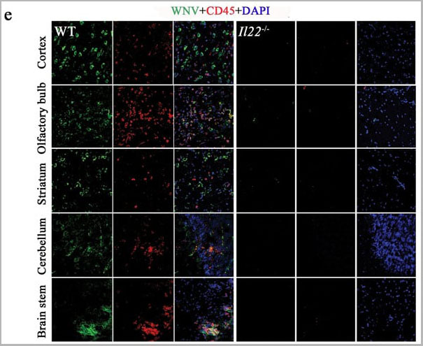
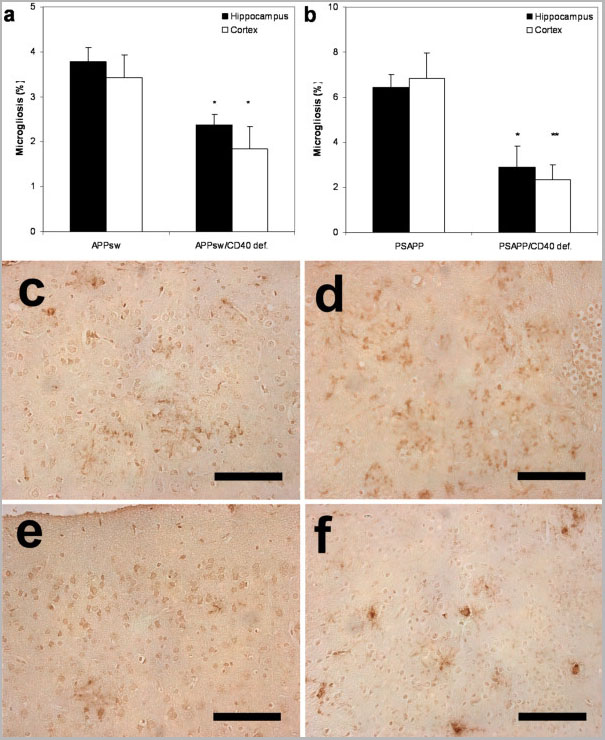
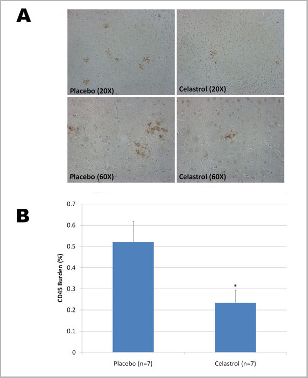
CD45, Monoclonal Antibody (Cat# AAA11896)
Full Name
RAT ANTI MOUSE CD45
Gene Names
Ptprc; loc; B220; Cd45; L-CA; Ly-5; T200; CD45R; Lyt-4
Applications
Immunohistochemistry, Flow Cytometry, Immunofluorescence, Immunoprecipitation
Pricing
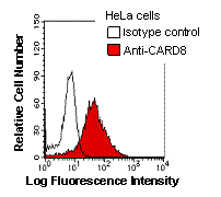


 ICC (Immunocytochemistry)
(Immunostaining analysis in HeLa cells. HeLa cells were fixed with 4% paraformaldehyde and permeabilized with 0.1% Triton X-100 in PBS. The cells were immunostained with anti-CARD8 mAb. [Lot No. 2108C2a-1])
ICC (Immunocytochemistry)
(Immunostaining analysis in HeLa cells. HeLa cells were fixed with 4% paraformaldehyde and permeabilized with 0.1% Triton X-100 in PBS. The cells were immunostained with anti-CARD8 mAb. [Lot No. 2108C2a-1])
CARD8, Monoclonal Antibody (Cat# AAA10604)
Full Name
Mouse monoclonal antibody Anti-Human CARD8
Gene Names
CARD8; NDPP; DACAR; DAKAR; NDPP1; TUCAN; CARDINAL
Reactivity
Human
Applications
Western Blot, Immunoprecipitation, Flow Cytometry, Immunocytochemistry
Purity
This antibody was purified using protein G column chromatography from culture supernatant of hybridoma cultured in a medium containing bovine IgG-depleted (approximately 95%) fetal bovine serum.
Pricing
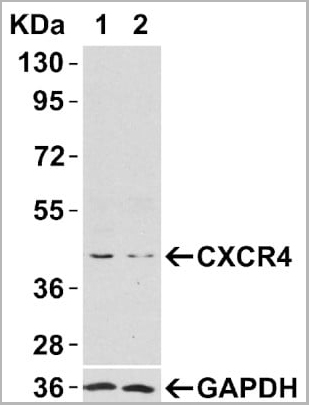
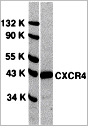
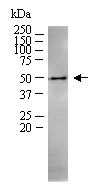
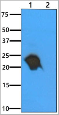
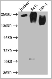
 WB (Western Blot)
(Figure 4 Animal Species ReactivityLoading: Lysates/proteins at 20 μg per lane. Antibodies: 1009 (2 μg/mL) or 1012 (2 μg/mL). 1 h incubation at RT in 5% NFDM/TBST. Secondary: Goat anti-rabbit IgG HRP conjugate at 1:10000 dilution.)
WB (Western Blot)
(Figure 4 Animal Species ReactivityLoading: Lysates/proteins at 20 μg per lane. Antibodies: 1009 (2 μg/mL) or 1012 (2 μg/mL). 1 h incubation at RT in 5% NFDM/TBST. Secondary: Goat anti-rabbit IgG HRP conjugate at 1:10000 dilution.)
CXCR4, Polyclonal Antibody (Cat# AAA10951)
Full Name
CXCR4 Antibody
Gene Names
CXCR4; FB22; HM89; LAP3; LCR1; NPYR; WHIM; CD184; LAP-3; LESTR; NPY3R; NPYRL; HSY3RR; NPYY3R; D2S201E
Reactivity
Human, Mouse, Rat
Applications
Western Blot, Immunocytochemistry, Immunoprecipitation, Immunofluorescence, Immunohistochemistry, Flow Cytometry
Purity
CXCR4 Antibody is affinity chromatography purified via peptide column.
Pricing
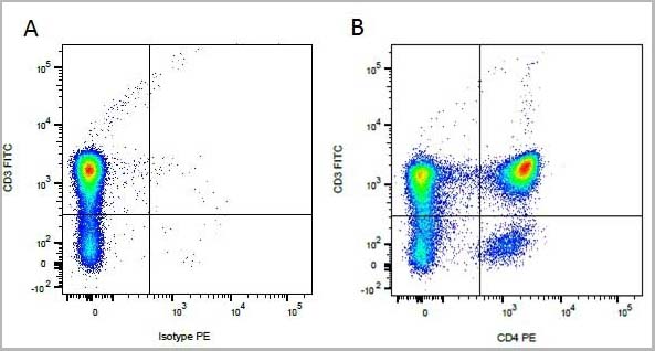
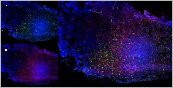
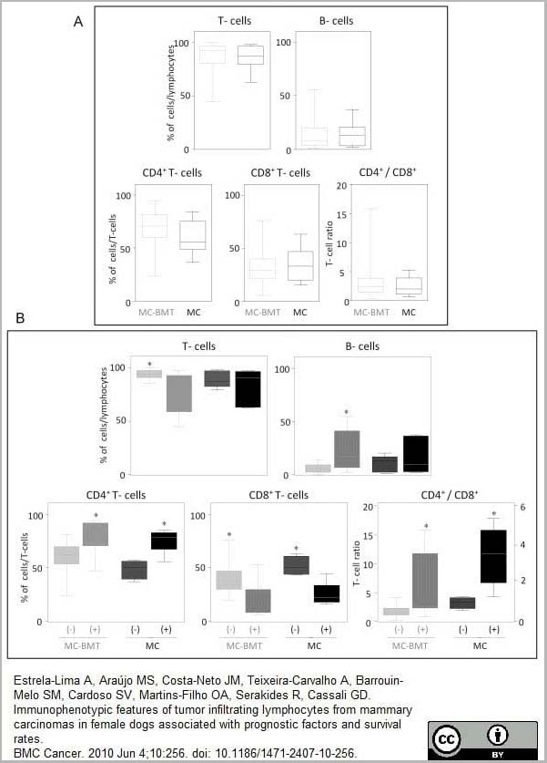 Application Data
(Published customer image:RPE conjugated Mouse anti Canine CD4 antibody, clone YKIC302.9used for the assessment of CD4 levels on canine cells by flow cytometry.Image caption:Immunophenotypic profile of tumor infiltrating lymphocyte in canine mammary carcinomas. Analysis of tumor infiltrating T-cells, B-lymphocytes and T-cell subsets from MC-BMT or MC (A), further subcategorized according to the absence (-) or presence (+) of lymph node metastasis (-) (B). Lymphocyte populations and subsets were identified by flow cytometric immunostaining as described in Material and Methods. Data were expressed as percentage of positive cells within gated lymphocytes and CD4+/CD8+ T-cell ratio. Significant differences at p < 0.05 are highlighted by asterisk.From: From: Estrela-Lima et al.BMC Cancer 2010 10:256.)
Application Data
(Published customer image:RPE conjugated Mouse anti Canine CD4 antibody, clone YKIC302.9used for the assessment of CD4 levels on canine cells by flow cytometry.Image caption:Immunophenotypic profile of tumor infiltrating lymphocyte in canine mammary carcinomas. Analysis of tumor infiltrating T-cells, B-lymphocytes and T-cell subsets from MC-BMT or MC (A), further subcategorized according to the absence (-) or presence (+) of lymph node metastasis (-) (B). Lymphocyte populations and subsets were identified by flow cytometric immunostaining as described in Material and Methods. Data were expressed as percentage of positive cells within gated lymphocytes and CD4+/CD8+ T-cell ratio. Significant differences at p < 0.05 are highlighted by asterisk.From: From: Estrela-Lima et al.BMC Cancer 2010 10:256.)
CD4, Monoclonal Antibody (Cat# AAA12279)
Full Name
RAT ANTI DOG CD4: RPE-Cy7
Applications
Flow Cytometry
Purity
Purified IgG prepared by affinity chromatography on Protein G from tissue culture supernatant
Pricing


 Application Data
(Staining of human peripheral blood granulocytes with CD18: Alexa Fluor 647)
Application Data
(Staining of human peripheral blood granulocytes with CD18: Alexa Fluor 647)
CD18, Monoclonal Antibody (Cat# AAA12051)
Full Name
RAT ANTI HUMAN CD18:RPE
Gene Names
ITGB2; LAD; CD18; MF17; MFI7; LCAMB; LFA-1; MAC-1
Applications
Flow Cytometry
Pricing


 Application Data
(Staining of human peripheral blood granulocytes with CD18: Alexa Fluor 647 (AAA11986A647))
Application Data
(Staining of human peripheral blood granulocytes with CD18: Alexa Fluor 647 (AAA11986A647))
CD18, Monoclonal Antibody (Cat# AAA11986)
Full Name
RAT ANTI HUMAN CD18
Gene Names
ITGB2; LAD; CD18; MF17; MFI7; LCAMB; LFA-1; MAC-1
Reactivity
Dog, Guinea Pig
Applications
Immunohistochemistry, Flow Cytometry, Immunoprecipitation
Pricing
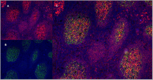
 Application Data
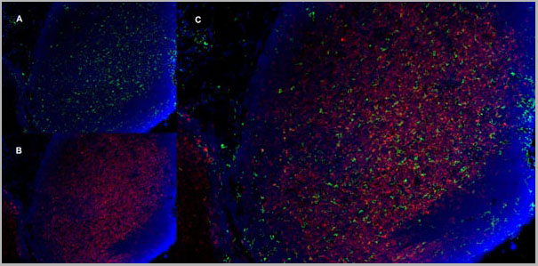
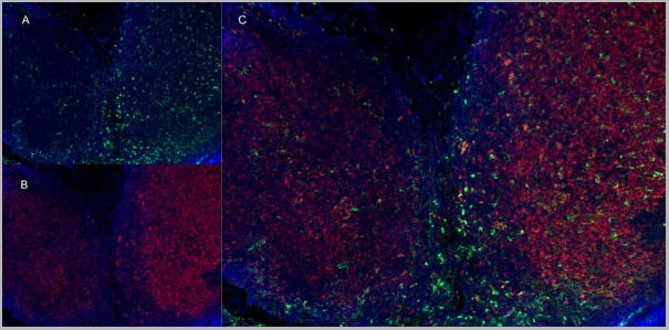
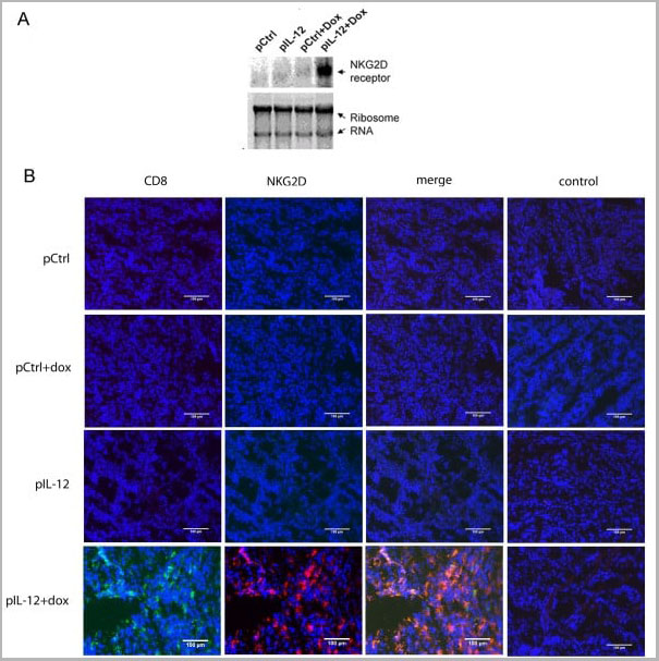
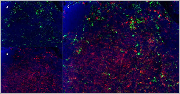
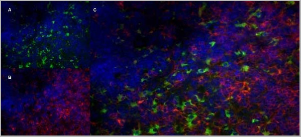
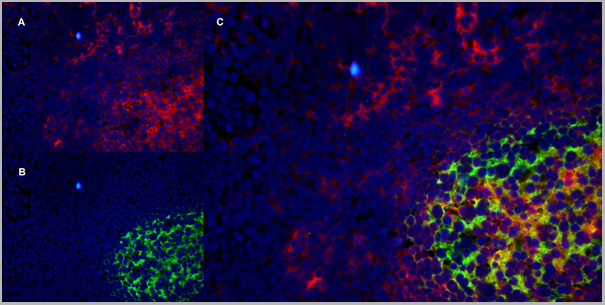
(Immunofluorescence staining of mouse lymph node cryosection with Rat anti Mouse CD11b, clone 5C6 , green in A and Rat anti Mouse CD8, clone YTS105.18 , red in B. C is the merged image with nuclei counterstained blue using DAPI. High power)
Application Data
(Immunofluorescence staining of mouse lymph node cryosection with Rat anti Mouse CD11b, clone 5C6 , green in A and Rat anti Mouse CD8, clone YTS105.18 , red in B. C is the merged image with nuclei counterstained blue using DAPI. High power)
CD8 ALPHA, Monoclonal Antibody (Cat# AAA11918)
Full Name
RAT ANTI MOUSE CD8
Gene Names
Cd8a; Ly-2; Ly-B; Ly-35; Lyt-2; BB154331
Applications
Immunohistochemistry, Flow Cytometry, Immunofluorescence
Pricing
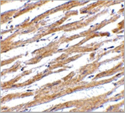
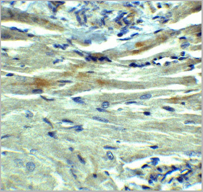
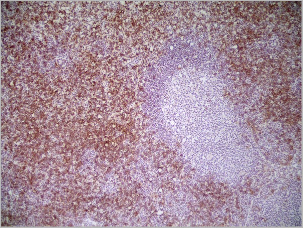
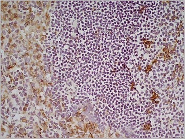
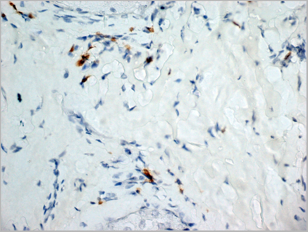
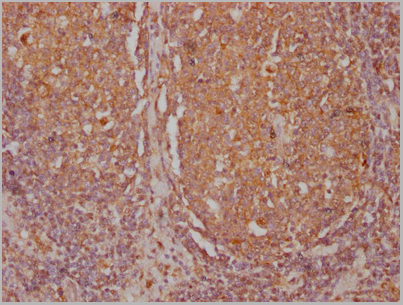
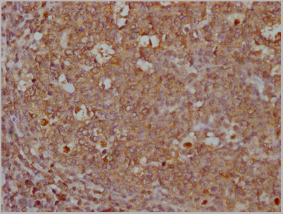
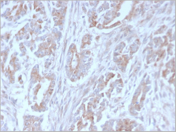
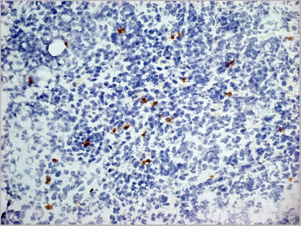
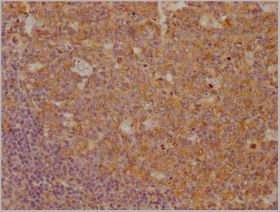
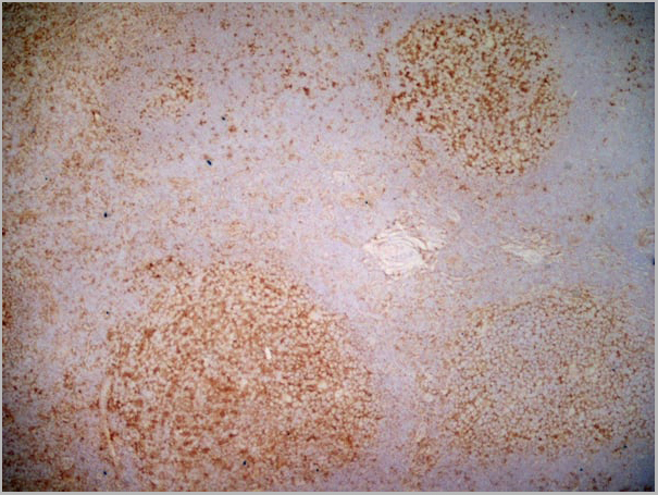
 IHC (Immunohistochemistry)
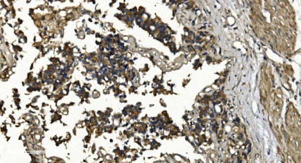
(Paraffin embedded sections of human urothelium were incubated with anti-human Hsp90 (1:100)for 2 hours at room temperature. Antigen retrieval was performed in 0.1M sodium citrate buffer and detectedusing Diaminobenzidine (DAB))
IHC (Immunohistochemistry)
(Paraffin embedded sections of human urothelium were incubated with anti-human Hsp90 (1:100)for 2 hours at room temperature. Antigen retrieval was performed in 0.1M sodium citrate buffer and detectedusing Diaminobenzidine (DAB))
Hsp90, Monoclonal Antibody (Cat# AAA11726)
Full Name
Hsp90 antibody
Gene Names
HSP90AA1; EL52; HSPN; LAP2; HSP86; HSPC1; HSPCA; Hsp89; Hsp90; LAP-2; HSP89A; HSP90A; HSP90N; Hsp103; HSPCAL1; HSPCAL4; HEL-S-65p
Reactivity
Human,
Applications
Western Blot, Immunohistochemistry
Purity
By protein-G affinity chromatography
Pricing
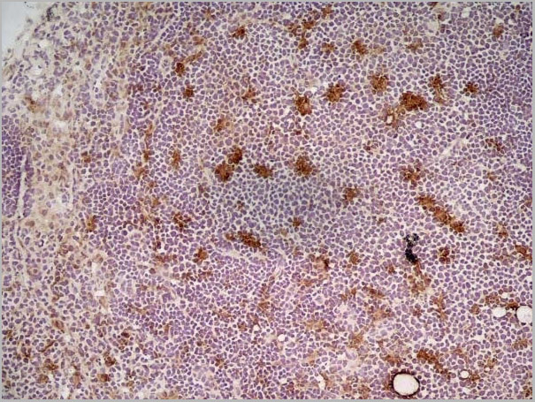
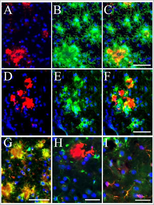
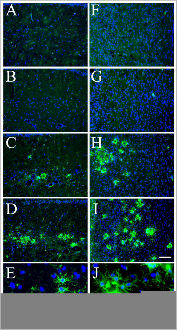
 Application Data
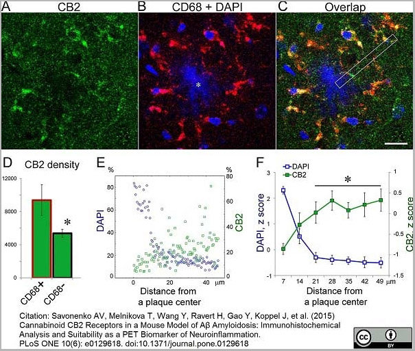
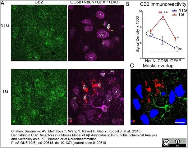
(Published customer image:Rat anti Mouse CD68 antibody, clone FA-11 used for the identification of microglia in mouse brain by immunofluorescence.Image caption:Comparison of CB2 immunoreactivity in neurons, activated microglia and astrocytes. ()
Application Data
(Published customer image:Rat anti Mouse CD68 antibody, clone FA-11 used for the identification of microglia in mouse brain by immunofluorescence.Image caption:Comparison of CB2 immunoreactivity in neurons, activated microglia and astrocytes. ()
CD68, Monoclonal Antibody (Cat# AAA12289)
Full Name
Rat anti Mouse CD68:Amethyst Orange
Gene Names
Cd68; Lamp4; gp110; Scard1
Reactivity
Mouse
Applications
Flow Cytometry
Purity
Purified IgG prepared by affinity chromatography on Protein G from tissue culture supernatant
Pricing
 IF (Immunofluorescence)
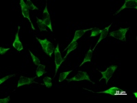
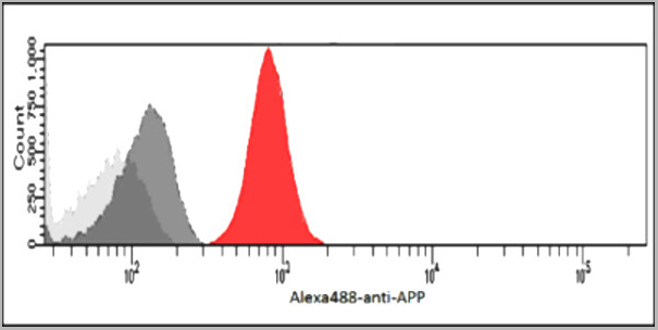
(ICC/IF analysis of APP/Protease Nexin II in U87MG cells. The cell was stained with AAA11729 (1:100). The secondary antibody (green) was used Alexa Fluor 488. DAPI was stained the cell nucleus (blue).)
IF (Immunofluorescence)
(ICC/IF analysis of APP/Protease Nexin II in U87MG cells. The cell was stained with AAA11729 (1:100). The secondary antibody (green) was used Alexa Fluor 488. DAPI was stained the cell nucleus (blue).)
APP, Monoclonal Antibody (Cat# AAA11729)
Full Name
APP antibody
Gene Names
APP; AAA; AD1; PN2; ABPP; APPI; CVAP; ABETA; PN-II; preA4; CTFgamma
Reactivity
Human
Applications
Western Blot, Flow Cytometry, Immunocytochemistry, Immunofluorescence
Purity
By protein-G affinity chromatography
Pricing
 Application Data
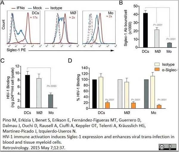
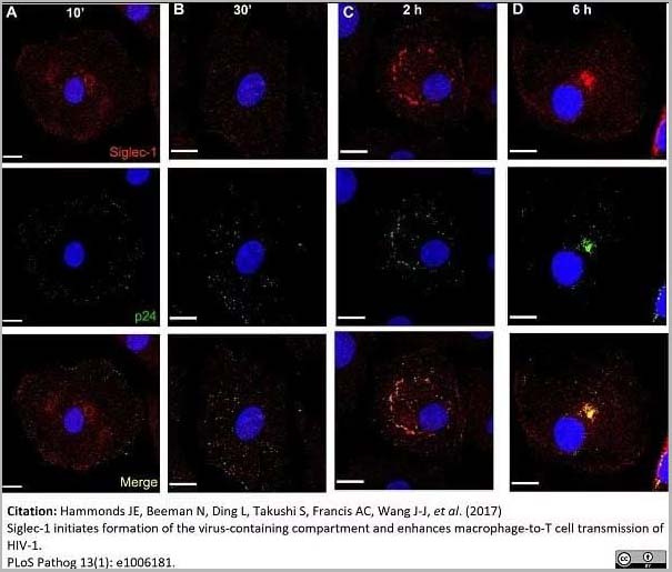
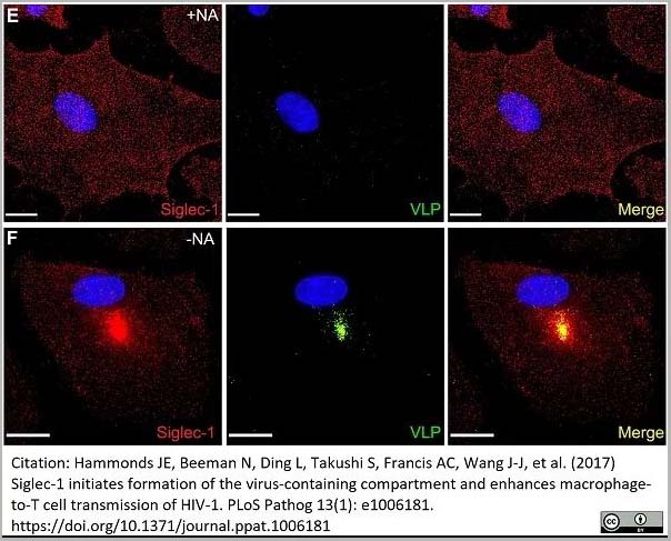
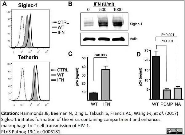
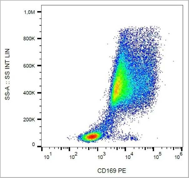
(PE conjugatedMouse anti Human CD169 antibody, clone 7-239 used to block CD169 function on myeloid cells.Image caption:Siglec-1 mediates HIV-1 uptake into a storage compartment and enhances HIV-1 trans-infection specially in IFN?-treated monocytes and DCs. A. Uptake of HIV-1NL4–3 by different myeloid cells exposed to IFN?. Cells were cultured with HIV-1 to measure p24Gag by ELISA. Mean values and SEM from four experiments include cells from 12 donors. B. Fold change in HIV-1NL4–3 uptake of cells treated with bafilomycin A1 compared to untreated cells. Mean values and SEM include cells from three donors. C. Relative uptake of HIV-1NL4–3 by IFN?-treated myeloid cells pre-incubated with the indicated mAbs. Values are normalized to the level of HIV-1 uptake by mock-treated cells (set at 100%). Mean values and SEM from two experiments include cells from six donors. D. Confocal microscopy analysis of different IFN?-treated myeloid cells pulsed with HIV-1Cherry and stained for Siglec-1 (Alexa 488), HLA-DR (Alexa 647) and DAPI. (Top) Representative viral pattern for each kind of myeloid cell analyzed, showing maximum fluorescence intensity of four channels. (Bottom) Percentage of myeloid cells with distinct viral patterns: random distribution, polarized accumulation, and sac-like compartment formation, as illustrated in the left drawing. Mean values of 50 cells from two different donors are shown. E. HIV-1 transmission from IFN?-treated myeloid cells to a luciferase reporter CD4+ cell line. HIV-1 infection was determined by induced luciferase activity in relative light units (RLUs). Mean values and SEM from four experiments include cells from 12 donors. F. Relative HIV-1 transmission from IFN?-treated myeloid cells pre-incubated with the indicated mAbs. Values are normalized to the level of HIV-1 trans-infected by mock-treated cells. Mean values and SEM from two experiments include cells from six donors. Statistical differences were assessed with a paired t test in A and E, and with a one sample t-test in B, C and F.From: Pino M, Erkizia I, Benet S, Erikson E, Fernández-Figueras MT, Guerrero D, Dalmau J, Ouchi D, Rausell A, Ciuffi A, Keppler OT, Telenti A, Kräusslich HG, Martinez-Picado J, Izquierdo-Useros N.HIV-1 immune activation induces Siglec-1 expression and enhances viral trans-infection in blood and tissue myeloid cells.Retrovirology. 2015 May 7;12:37.This image is from an open access article distributed under the terms of the Creative Commons Attribution License.)
Application Data
(PE conjugatedMouse anti Human CD169 antibody, clone 7-239 used to block CD169 function on myeloid cells.Image caption:Siglec-1 mediates HIV-1 uptake into a storage compartment and enhances HIV-1 trans-infection specially in IFN?-treated monocytes and DCs. A. Uptake of HIV-1NL4–3 by different myeloid cells exposed to IFN?. Cells were cultured with HIV-1 to measure p24Gag by ELISA. Mean values and SEM from four experiments include cells from 12 donors. B. Fold change in HIV-1NL4–3 uptake of cells treated with bafilomycin A1 compared to untreated cells. Mean values and SEM include cells from three donors. C. Relative uptake of HIV-1NL4–3 by IFN?-treated myeloid cells pre-incubated with the indicated mAbs. Values are normalized to the level of HIV-1 uptake by mock-treated cells (set at 100%). Mean values and SEM from two experiments include cells from six donors. D. Confocal microscopy analysis of different IFN?-treated myeloid cells pulsed with HIV-1Cherry and stained for Siglec-1 (Alexa 488), HLA-DR (Alexa 647) and DAPI. (Top) Representative viral pattern for each kind of myeloid cell analyzed, showing maximum fluorescence intensity of four channels. (Bottom) Percentage of myeloid cells with distinct viral patterns: random distribution, polarized accumulation, and sac-like compartment formation, as illustrated in the left drawing. Mean values of 50 cells from two different donors are shown. E. HIV-1 transmission from IFN?-treated myeloid cells to a luciferase reporter CD4+ cell line. HIV-1 infection was determined by induced luciferase activity in relative light units (RLUs). Mean values and SEM from four experiments include cells from 12 donors. F. Relative HIV-1 transmission from IFN?-treated myeloid cells pre-incubated with the indicated mAbs. Values are normalized to the level of HIV-1 trans-infected by mock-treated cells. Mean values and SEM from two experiments include cells from six donors. Statistical differences were assessed with a paired t test in A and E, and with a one sample t-test in B, C and F.From: Pino M, Erkizia I, Benet S, Erikson E, Fernández-Figueras MT, Guerrero D, Dalmau J, Ouchi D, Rausell A, Ciuffi A, Keppler OT, Telenti A, Kräusslich HG, Martinez-Picado J, Izquierdo-Useros N.HIV-1 immune activation induces Siglec-1 expression and enhances viral trans-infection in blood and tissue myeloid cells.Retrovirology. 2015 May 7;12:37.This image is from an open access article distributed under the terms of the Creative Commons Attribution License.)
CD169, Monoclonal Antibody (Cat# AAA12264)
Full Name
Mouse Anti Human CD169
Gene Names
SIGLEC1; SN; CD169; SIGLEC-1
Reactivity
Human
Applications
Immunohistochemistry, Flow Cytometry, Functional Assay, Immunoprecipitation, Western Blot
Purity
>95% by SDS PAGE
Purified IgG prepared by affinity chromatography on Protein A from tissue culture supernatant.
Purified IgG prepared by affinity chromatography on Protein A from tissue culture supernatant.
Pricing


 Application Data
(Staining of mouse peritoneal macrophages with Rat anti Mouse Beta-glucan Receptor: FITC)
Application Data
(Staining of mouse peritoneal macrophages with Rat anti Mouse Beta-glucan Receptor: FITC)
DECTIN-1, Monoclonal Antibody (Cat# AAA12129)
Full Name
RAT ANTI MOUSE DECTIN-1:Low Endotoxin
Gene Names
Clec7a; BGR; beta-GR; Clecsf12
Applications
Immunohistochemistry, Flow Cytometry, Functional Assay, Immunoprecipitation
Pricing


 Application Data
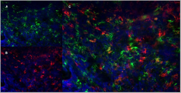
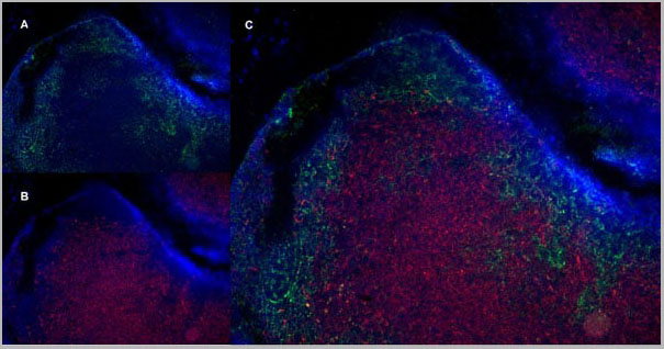
(Immunofluorescence staining of mouse lymph node cryosection with Rat anti Mouse CD11b, clone 5C6 , green in A and Rat anti Mouse CD8, clone YTS105.18 , red in B. C is the merged image with nuclei counterstained blue using DAPI. High power)
Application Data
(Immunofluorescence staining of mouse lymph node cryosection with Rat anti Mouse CD11b, clone 5C6 , green in A and Rat anti Mouse CD8, clone YTS105.18 , red in B. C is the merged image with nuclei counterstained blue using DAPI. High power)
CD8 ALPHA, Monoclonal Antibody (Cat# AAA12069)
Full Name
RAT ANTI MOUSE CD8
Gene Names
Cd8a; Ly-2; Ly-B; Ly-35; Lyt-2; BB154331
Reactivity
Mouse
Applications
Immunohistochemistry, Flow Cytometry, Immunofluorescence
Pricing


 Application Data
(Staining of mouse peritoneal macrophages with Rat anti Mouse Beta-glucan Receptor: FITC)
Application Data
(Staining of mouse peritoneal macrophages with Rat anti Mouse Beta-glucan Receptor: FITC)
DECTIN-1, Monoclonal Antibody (Cat# AAA12130)
Full Name
RAT ANTI MOUSE DECTIN-1:FITC
Gene Names
Clec7a; BGR; beta-GR; Clecsf12
Applications
Flow Cytometry
Pricing
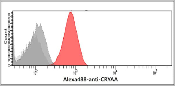 FCM (Flow Cytometry)
(Flow cytometry analysis of Crystallin alpha A in Balb/3T3 cells. The cell was stained with AAA11723 at 2-5ug for 1x10^6cells (red). A Goat anti mouse IgG (Alexa fluor 488) was used as the secondary antibody. Mouse monoclonal IgG was used as the isotype control (dark gray), cells without incubation with primary and secondary antibody was used as the negative control (light gray).)
FCM (Flow Cytometry)
(Flow cytometry analysis of Crystallin alpha A in Balb/3T3 cells. The cell was stained with AAA11723 at 2-5ug for 1x10^6cells (red). A Goat anti mouse IgG (Alexa fluor 488) was used as the secondary antibody. Mouse monoclonal IgG was used as the isotype control (dark gray), cells without incubation with primary and secondary antibody was used as the negative control (light gray).)
A crystallin A, Monoclonal Antibody (Cat# AAA11723)
Full Name
A crystallin A antibody
Gene Names
CRYAA; CRYA1; HSPB4; CTRCT9
Reactivity
Human
Applications
Western Blot, Flow Cytometry
Purity
By protein-G affinity chromatography
Pricing


 Application Data
(Staining of mouse peritoneal macrophages with Rat anti Mouse Beta-glucan Receptor: FITC)
Application Data
(Staining of mouse peritoneal macrophages with Rat anti Mouse Beta-glucan Receptor: FITC)
DECTIN-1, Monoclonal Antibody (Cat# AAA12136)
Full Name
RAT ANTI MOUSE DECTIN-1:RPE
Gene Names
Clec7a; BGR; beta-GR; Clecsf12
Applications
Flow Cytometry
Pricing
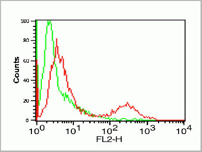
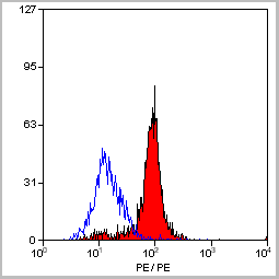
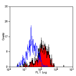
 FCM (Flow Cytometry)
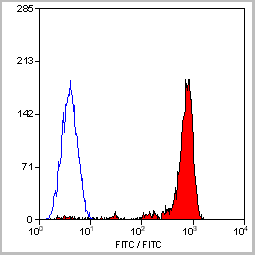
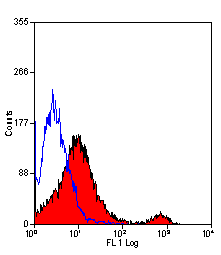
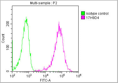
(Flow Cytometric Analysis of PFA-fixed K562 cells using Calponin-1 Recombinant Mouse Monoclonal Antibody (rCNN1/832) followed by Goat anti-Mouse IgG-CF488 (Blue); Isotype Control (Red).)
FCM (Flow Cytometry)
(Flow Cytometric Analysis of PFA-fixed K562 cells using Calponin-1 Recombinant Mouse Monoclonal Antibody (rCNN1/832) followed by Goat anti-Mouse IgG-CF488 (Blue); Isotype Control (Red).)
Calponin-1, Monoclonal Antibody (Cat# AAA13848)
Full Name
Calponin-1 (Smooth Muscle Marker)
Gene Names
CNN1; SMCC; Sm-Calp; HEL-S-14
Reactivity
Human, Rat
Applications
Flow Cytometry, Immunofluorescence, Immunohistochemistry
Pricing


 Application Data
(Staining of human peripheral blood granulocytes with CD18: Alexa Fluor 647)
Application Data
(Staining of human peripheral blood granulocytes with CD18: Alexa Fluor 647)
CD18, Monoclonal Antibody (Cat# AAA11881)
Full Name
RAT ANTI HUMAN CD18:FITC
Gene Names
ITGB2; LAD; CD18; MF17; MFI7; LCAMB; LFA-1; MAC-1
Applications
Flow Cytometry
Pricing


 Application Data
(Staining of human peripheral blood granulocytes with CD18: Alexa Fluor 647)
Application Data
(Staining of human peripheral blood granulocytes with CD18: Alexa Fluor 647)
CD18, Monoclonal Antibody (Cat# AAA11987)
Full Name
RAT ANTI HUMAN CD18
Gene Names
ITGB2; LAD; CD18; MF17; MFI7; LCAMB; LFA-1; MAC-1
Applications
Immunohistochemistry, Flow Cytometry, Immunoprecipitation
Pricing
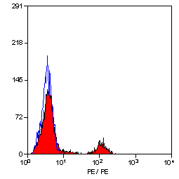
 FCM (Flow Cytometry)
(Flow Cytometric Analysis of U937 cells using CD15 Rabbit Recombinant Monoclonal Antibody (FUT4/1478R) followed by goat anti-rabbit IgG-CF488 (Blue); Isotype Control (Red).)
FCM (Flow Cytometry)
(Flow Cytometric Analysis of U937 cells using CD15 Rabbit Recombinant Monoclonal Antibody (FUT4/1478R) followed by goat anti-rabbit IgG-CF488 (Blue); Isotype Control (Red).)
CD15/FUT4, Monoclonal Antibody (Cat# AAA13849)
Full Name
CD15/FUT4 (Reed-Sternberg Cell Marker)
Gene Names
FUT4; LeX; CD15; ELFT; FCT3A; FUTIV; SSEA-1; FUC-TIV
Reactivity
Human, Mouse, Rat. Others not known.
Applications
Flow Cytometry, Immunofluorescence, Immunohistochemistry
Pricing
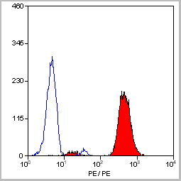
 FCM (Flow Cytometry)
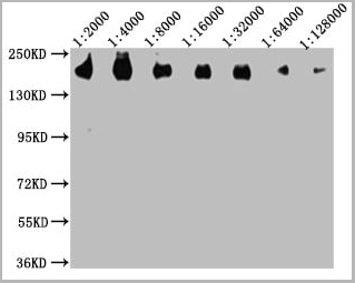
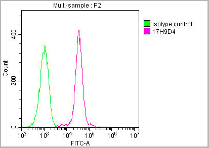
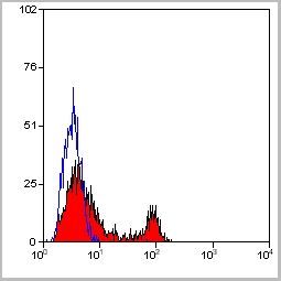
(Overlay histogram showing Raji cells stained with (red line) at 1:500. The cells were incubated in 10% normal goat serum to block non-specific protein-protein interactions followed by the antibody (1ug/1*106cells) for 1 h at 4 degree C. The secondary antibody used was FITC-conjugated Goat Anti-Mouse IgG(H+L) at 1/100 dilution for 30min at 4 degree C. Isotype control antibody (green line) was mouse IgG2b (1ug/1*106cells) used under the same conditions. Acquisition of >10,000 events was performed.)
FCM (Flow Cytometry)
(Overlay histogram showing Raji cells stained with (red line) at 1:500. The cells were incubated in 10% normal goat serum to block non-specific protein-protein interactions followed by the antibody (1ug/1*106cells) for 1 h at 4 degree C. The secondary antibody used was FITC-conjugated Goat Anti-Mouse IgG(H+L) at 1/100 dilution for 30min at 4 degree C. Isotype control antibody (green line) was mouse IgG2b (1ug/1*106cells) used under the same conditions. Acquisition of >10,000 events was performed.)
CD45, Monoclonal Antibody (Cat# AAA27049)
Full Name
CD45 Monoclonal Antibody
Gene Names
PTPRC; LCA; LY5; B220; CD45; L-CA; T200; CD45R; GP180
Reactivity
Human
Applications
Western Blot, Immunohistochemistry, Immunofluorescence, Flow Cytometry
Purity
>95%, Protein A purified
Pricing
 Application Data
(Analysis of Protein Array containing more than 19,000 full-length human proteins using ICOS-L Mouse Monoclonal Antibody (ICOSL/3111). Z- and S- Score: The Z-score represents the strength of a signal that a monoclonal antibody (MAb) (in combination with a fluorescently-tagged anti-IgG secondary antibody) produces when binding to a particular protein on the HuProtTM array. Z-scores are described in units of standard deviations (SD’s) above the mean value of all signals generated on that array. If targets on HuProtTM are arranged in descending order of the Z-score, the S-score is the difference (also in units of SD’s) between the Z-score. S-score therefore represents the relative target specificity of a MAb to its intended target. A MAb is considered to specific to its intended target, if the MAb has an S-score of at least 2.5. For example, if a MAb binds to protein X with a Z-score of 43 and to protein Y with a Z-score of 14, then the S-score for the binding of that MAb to protein X is equal to 29.)
Application Data
(Analysis of Protein Array containing more than 19,000 full-length human proteins using ICOS-L Mouse Monoclonal Antibody (ICOSL/3111). Z- and S- Score: The Z-score represents the strength of a signal that a monoclonal antibody (MAb) (in combination with a fluorescently-tagged anti-IgG secondary antibody) produces when binding to a particular protein on the HuProtTM array. Z-scores are described in units of standard deviations (SD’s) above the mean value of all signals generated on that array. If targets on HuProtTM are arranged in descending order of the Z-score, the S-score is the difference (also in units of SD’s) between the Z-score. S-score therefore represents the relative target specificity of a MAb to its intended target. A MAb is considered to specific to its intended target, if the MAb has an S-score of at least 2.5. For example, if a MAb binds to protein X with a Z-score of 43 and to protein Y with a Z-score of 14, then the S-score for the binding of that MAb to protein X is equal to 29.)
ICOS-L/ICOS Ligand/B7RP-1 (Immuno-Oncology Target), Monoclonal Antibody (Cat# AAA23916)
Full Name
ICOS-L/ICOS Ligand/B7RP-1 (Immuno-Oncology Target)
Gene Names
ICOSLG; B7h; B7H2; GL50; B7-H2; B7RP1; CD275; ICOSL; LICOS; B7RP-1; ICOS-L
Reactivity
Human
Applications
Flow Cytometry, Immunofluorescence, Immunohistochemistry
Purity
Purified Ab with BSA and Azide at 200ug/ml
Pricing


 Application Data
(Staining of mouse peritoneal macrophages with Rat anti Mouse Beta-glucan Receptor: FITC)
Application Data
(Staining of mouse peritoneal macrophages with Rat anti Mouse Beta-glucan Receptor: FITC)
DECTIN-1, Monoclonal Antibody (Cat# AAA12131)
Full Name
RAT ANTI MOUSE DECTIN-1:FITC
Gene Names
Clec7a; BGR; beta-GR; Clecsf12
Applications
Flow Cytometry
Pricing


 Application Data
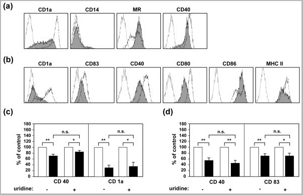
(Staining of human peripheral blood lymphocytes with Mouse anti Human CD40:RPE)
Application Data
(Staining of human peripheral blood lymphocytes with Mouse anti Human CD40:RPE)
CD40, Monoclonal Antibody (Cat# AAA12090)
Full Name
MOUSE ANTI HUMAN CD40
Gene Names
CD40; p50; Bp50; CDW40; TNFRSF5
Applications
Immunohistochemistry, Flow Cytometry, Immunoprecipitation, Immunohistochemistry
Pricing
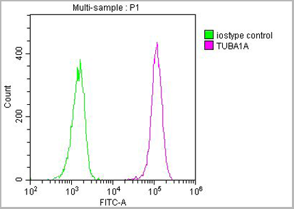
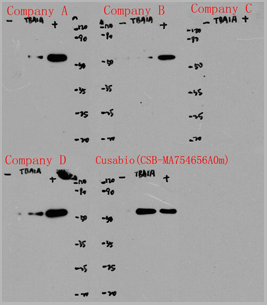
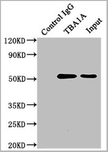
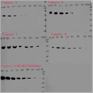
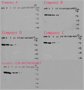
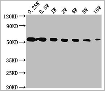
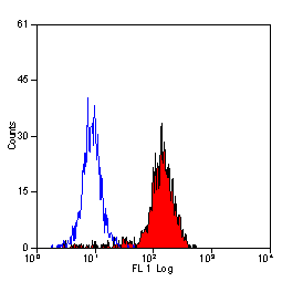
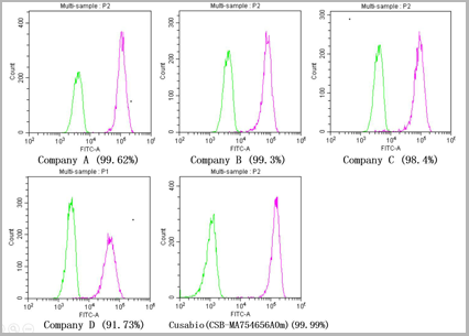 FCM (Flow Cytometry)
(Overlay histogram showing Hela cells stainedwith Company A (5ug), Company B (5ug), Company C (5ug), Company D (5ug), AAA27031 (5ug) (red line) at 1:150. The cells were incubated in 1x PBS /10% normal goat serum to block non-specific protein-protein interactions followed by primary antibody for 1 hat 4°C. The secondary antibody used was FITC goat anti-mouse IgG(H+L) at 1/200 dilution for 1 h at 4°C. Isotype control antibody (green line) was used under the same conditions. Acquisition of >10,000 events was performed.)
FCM (Flow Cytometry)
(Overlay histogram showing Hela cells stainedwith Company A (5ug), Company B (5ug), Company C (5ug), Company D (5ug), AAA27031 (5ug) (red line) at 1:150. The cells were incubated in 1x PBS /10% normal goat serum to block non-specific protein-protein interactions followed by primary antibody for 1 hat 4°C. The secondary antibody used was FITC goat anti-mouse IgG(H+L) at 1/200 dilution for 1 h at 4°C. Isotype control antibody (green line) was used under the same conditions. Acquisition of >10,000 events was performed.)
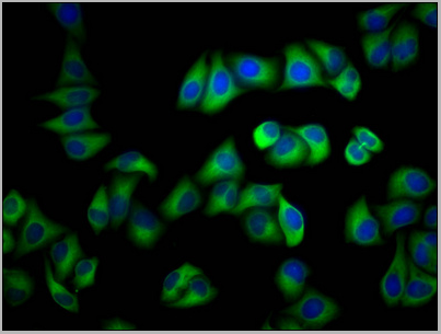
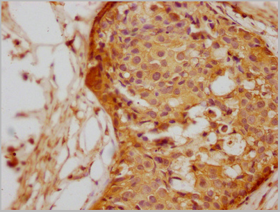
TUBA1A, Monoclonal Antibody (Cat# AAA27031)
Full Name
TUBA1A
Gene Names
Tuba1a; Tuba1; Tuba-1
Reactivity
Human, Rabbit, Rat, Mouse
Applications
Western Blot, Immunohistochemistry, Immunofluorescence, Flow Cytometry, Immunoprecipitation
Purity
>95%, Protein A purified
Pricing


 Application Data
(Staining of human peripheral blood lymphocytes with Mouse anti Human CD40:RPE)
Application Data
(Staining of human peripheral blood lymphocytes with Mouse anti Human CD40:RPE)
CD40, Monoclonal Antibody (Cat# AAA11867)
Full Name
MOUSE ANTI HUMAN CD40:FITC
Gene Names
CD40; p50; Bp50; CDW40; TNFRSF5
Applications
Flow Cytometry
Pricing


 Application Data
(Staining of human peripheral blood lymphocytes with Mouse anti Human CD40:RPE)
Application Data
(Staining of human peripheral blood lymphocytes with Mouse anti Human CD40:RPE)
CD40, Monoclonal Antibody (Cat# AAA11936)
Full Name
MOUSE ANTI HUMAN CD40
Gene Names
CD40; p50; Bp50; CDW40; TNFRSF5
Reactivity
Dog
Applications
Immunohistochemistry, Flow Cytometry, Immunoprecipitation, Immunohistochemistry
Pricing