Filters
Clonality
Type
Reactivity
Gene Name
Isotype
Host
Application
Clone
233 results for " Complement" - showing 150-200
 Standard Curve (Sample)
family with sequence similarity 132, member B, ELISA Kit (Cat# AAA18350)
Full Name
Mouse Protein FAM132B, FAM132B ELISA Kit
Gene Names
Fam132b; myonectin; 4832406C22
Reactivity
Mouse
Pricing


 WB (Western Blot)
(Western Blot;Sample: Mouse Serum.)
WB (Western Blot)
(Western Blot;Sample: Mouse Serum.)
Complement Component 9, Polyclonal Antibody (Cat# AAA20913)
Full Name
Polyclonal Antibody to Complement Component 9 (C9)
Reactivity
Mouse
Applications
WB, IHC, ICC, IP
Purity
Antigen-specific affinity chromatography followed by Protein A affinity chromatography
Pricing
 Standard Curve (Sample)
Complement 3a (C3a), ELISA Kit (Cat# AAA12467)
Full Name
Rat Complement 3a (C3a) ELISA Kit
Gene Names
C3AR1; AZ3B; C3AR; HNFAG09
Reactivity
Rat
Pricing
 Standard Curve (Sample)
complement component 1, q subcomponent, A chain, ELISA Kit (Cat# AAA18156)
Full Name
Human Complement C1q subcomponent subunit A, C1QA ELISA Kit
Reactivity
Human
Pricing
 Standard Curve (Sample)
Complement fragment 3a, C3a, ELISA Kit (Cat# AAA15529)
Full Name
Mouse Complement fragment 3a, C3a ELISA Kit
Reactivity
Mouse
Pricing
 SDS-PAGE
Complement C1q tumor necrosis factor-related protein 3 (C1qtnf3), Recombinant Protein (Cat# AAA18757)
Full Name
Recombinant Mouse Complement C1q tumor necrosis factor-related protein 3 (C1qtnf3)
Gene Names
C1qtnf3; Cors; CTRP3; Corcs; CORS26; CORS-26; 2310005P21Rik
Purity
Greater or equal to 85% purity as determined by SDS-PAGE.
Pricing
 Standard Curve (Sample)
Anti-Complement 1q Antibody, ELISA Kit (Cat# AAA16578)
Full Name
Mouse Anti-Complement 1q Antibody ELISA Kit
Gene Names
C1QTNF5; CTRP5
Reactivity
Mouse
Pricing
 Standard Curve (Sample)
Complement Fragment 3d, C3d, ELISA Kit (Cat# AAA27221)
Full Name
Monkey Complement Fragment 3d, C3d ELISA Kit
Reactivity
Monkey
Pricing
 Standard Curve (Sample)
adiponectin, C1Q and collagen domain containing (ADIPOQ), ELISA Kit (Cat# AAA15850)
Full Name
Sheep adiponectin, ADIPOQ ELISA Kit
Gene Names
ADIPOQ; APM1; ACRP30
Reactivity
Sheep
Pricing
 Standard Curve (Sample)
FAM132B, ELISA Kit (Cat# AAA27499)
Full Name
Human FAM132B (Protein FAM132B) ELISA Kit
Gene Names
ERFE; CTRP15; FAM132B; C1QTNF15
Reactivity
Human
Pricing
 Standard Curve (Sample)
Membrane Cofactor Protein, ELISA Kit (Cat# AAA17484)
Full Name
Human Membrane Cofactor Protein ELISA Kit
Gene Names
CD46; MCP; TLX; AHUS2; MIC10; TRA2.10
Reactivity
Human
Pricing
 Standard Curve (Sample)
Complement 1q, ELISA Kit (Cat# AAA16766)
Full Name
Canine Complement 1q ELISA Kit
Gene Names
C1QTNF5; CTRP5
Reactivity
Canine
Pricing
 Standard Curve (Sample)
Adiponectin, ELISA Kit (Cat# AAA17476)
Full Name
Human Adiponectin ELISA Kit
Gene Names
ADIPOQ; ACDC; ADPN; APM1; APM-1; GBP28; ACRP30; ADIPQTL1
Reactivity
Human
Pricing
 Standard Curve (Sample)
Complement C3, ELISA Kit (Cat# AAA17531)
Full Name
Mouse Complement C3 ELISA Kit
Gene Names
C3; ASP; C3a; C3b; AHUS5; ARMD9; CPAMD1; HEL-S-62p
Reactivity
Mouse
Pricing
 Standard Curve (Sample)
Complement component C6 (C6), ELISA Kit (Cat# AAA27249)
Full Name
Mouse Complement component C6 (C6) ELISA Kit
Reactivity
Mouse
Pricing
 Standard Curve (Sample)
Complement Component 3, ELISA Kit (Cat# AAA17333)
Full Name
Rat Complement Component 3 ELISA Kit
Gene Names
C3; ASP; C3a; C3b; AHUS5; ARMD9; CPAMD1; HEL-S-62p
Reactivity
Rat
Pricing
 Standard Curve (Sample)
Erythroferrone, ELISA Kit (Cat# AAA27427)
Full Name
Mouse Erythroferrone ELISA Kit
Gene Names
ERFE; CTRP15; FAM132B; C1QTNF15
Reactivity
Mouse
Pricing
 Standard Curve (Sample)
Complement 1q, ELISA Kit (Cat# AAA16380)
Full Name
Mouse Complement 1q ELISA Kit
Gene Names
C1QTNF5; CTRP5
Reactivity
Mouse
Pricing
 Standard Curve (Sample)
CR1, ELISA Kit (Cat# AAA21908)
Full Name
Human CR1 (Complement Receptor type 1) ELISA Kit
Gene Names
CR1; KN; C3BR; C4BR; CD35
Reactivity
Human
Pricing
 Standard Curve (Sample)
Complement 3a (C3a), ELISA Kit (Cat# AAA13039)
Full Name
Mouse Complement 3a (C3a) ELISA Kit
Gene Names
C3AR1; AZ3B; C3AR; HNFAG09
Reactivity
Mouse
Pricing
 Standard Curve (Sample)
complement C5 (C5), ELISA Kit (Cat# AAA15858)
Full Name
Monkey complement C5 (C5) ELISA Kit
Reactivity
Monkey
Pricing
 Standard Curve (Sample)
vitronectin, ELISA Kit (Cat# AAA15489)
Full Name
Human vitronectin, VTN ELISA Kit
Gene Names
VTN; VN; V75; VNT
Reactivity
Human
Pricing
 Standard Curve (Sample)
complement factor D (adipsin), ELISA Kit (Cat# AAA15247)
Full Name
Human Adipsin ELISA Kit
Gene Names
CFD; DF; ADN; PFD; ADIPSIN
Reactivity
Human
Pricing
 Standard Curve (Sample)
Erythroferrone, ELISA Kit (Cat# AAA27448)
Full Name
Bovine Erythroferrone ELISA Kit
Gene Names
ERFE; CTRP15; FAM132B; C1QTNF15
Reactivity
Bovine
Pricing
 Standard Curve (Sample)
Complement 3a (C3a), ELISA Kit (Cat# AAA13185)
Full Name
Rabbit Complement 3a (C3a) ELISA Kit
Gene Names
C3AR1; AZ3B; C3AR; HNFAG09
Reactivity
Rabbit
Pricing

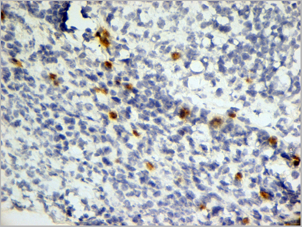
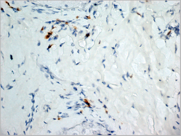
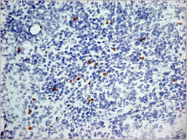
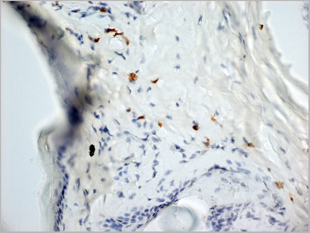
 IHC (Immunohistochemistry)
(Immunohistochemical analysis of paraffin-embedded Human thyroid cancer tissue using at dilution 1/50.)
IHC (Immunohistochemistry)
(Immunohistochemical analysis of paraffin-embedded Human thyroid cancer tissue using at dilution 1/50.)
C3AR1, Polyclonal Antibody (Cat# AAA29676)
Full Name
C3AR1 Antibody
Gene Names
C3AR1; AZ3B; C3AR; HNFAG09
Reactivity
Human
Applications
WB
Purity
Antigen affinity purification.
Pricing
 Standard Curve (Sample)
Adipsin, ELISA Kit (Cat# AAA17866)
Full Name
Human Adipsin ELISA Kit
Gene Names
CFD; DF; ADN; PFD; ADIPSIN
Reactivity
Human
Applications
SE
Pricing
 Standard Curve (Sample)
Complement C3 Convertase (C3c), ELISA Kit (Cat# AAA12831)
Full Name
Porcine Complement C3 Convertase (C3c) ELISA Kit
Gene Names
C3; ASP; C3a; C3b; AHUS5; ARMD9; CPAMD1; HEL-S-62p
Reactivity
Porcine
Pricing
 SDS-PAGE
Complement C3, Recombinant Protein (Cat# AAA18546)
Full Name
Recombinant Mouse Complement C3 (C3), partial
Gene Names
C3; ASP; Plp; HSE-MSF; AI255234
Purity
Greater or equal to 85% purity as determined by SDS-PAGE.
Pricing
 Standard Curve (Sample)
Complement Fragment 3d, C3d, ELISA Kit (Cat# AAA27321)
Full Name
Rat Complement Fragment 3d, C3d ELISA Kit
Reactivity
Rat
Pricing
 Standard Curve (Sample)
Complement 1q, ELISA Kit (Cat# AAA16817)
Full Name
Porcine Complement 1q ELISA Kit
Gene Names
C1QTNF5; CTRP5
Reactivity
Porcine
Pricing
 Standard Curve (Sample)
factor B, BF, ELISA Kit (Cat# AAA15205)
Full Name
Rat factor B, BF ELISA Kit
Gene Names
CFB; BF; FB; BFD; GBG; CFAB; CFBD; PBF2; AHUS4; FBI12; H2-Bf; ARMD14
Reactivity
Rat
Pricing
 Standard Curve (Sample)
C1q and tumor necrosis factor related protein 2, ELISA Kit (Cat# AAA18344)
Full Name
Human Complement C1q tumor necrosis factor-related protein 2, C1QTNF2 ELISA Kit
Gene Names
C1QTNF2; CTRP2; zacrp2
Reactivity
Human
Pricing
 Standard Curve (Sample)
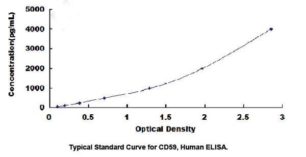
Protectin (CD59), ELISA Kit (Cat# AAA20560)
Full Name
Human Protectin (CD59) ELISA Kit
Reactivity
Human
Pricing
 Standard Curve (Sample)
CXCL15, ELISA Kit (Cat# AAA27879)
Full Name
Mouse CXCL15 ELISA Kit
Gene Names
Cxcl15; Il8; weche; Scyb15; lungkine
Reactivity
Mouse
Pricing
 Standard Curve (Sample)
clusterin, ELISA Kit (Cat# AAA18097)
Full Name
Horse Clusterin, CLU ELISA Kit
Reactivity
Horse
Pricing
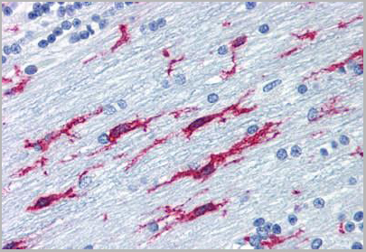 IHC (Immunohistochemistry)
(Anti-ITGB2 / CD18 antibody IHC of human brain, cerebellum. Immunohistochemistry of formalin-fixed, paraffin-embedded tissue after heat-induced antigen retrieval. Antibody concentration 10 ug/ml.)
IHC (Immunohistochemistry)
(Anti-ITGB2 / CD18 antibody IHC of human brain, cerebellum. Immunohistochemistry of formalin-fixed, paraffin-embedded tissue after heat-induced antigen retrieval. Antibody concentration 10 ug/ml.)
ITGB2 / MAC-1 / CD18, Monoclonal Antibody (Cat# AAA12341)
Full Name
Mouse Monoclonal [clone MEM-48] (IgG1) to Human ITGB2 / MAC-1 / CD18
Gene Names
ITGB2; LAD; CD18; MF17; MFI7; LCAMB; LFA-1; MAC-1
Reactivity
Human
Applications
Immunohistochemistry, Western Blot, Immunoprecipitation, Flow Cytometry, Functional Assay
Purity
Purified by Protein A affinity chromatography.
>95% by SDS-PAGE.
>95% by SDS-PAGE.
Pricing


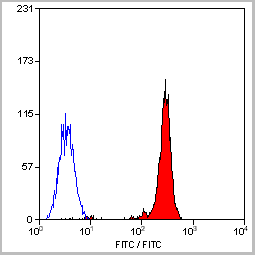
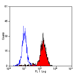
 Application Data
(Staining of human peripheral blood granulocytes with CD18: Alexa Fluor 647)
Application Data
(Staining of human peripheral blood granulocytes with CD18: Alexa Fluor 647)
CD18, Monoclonal Antibody (Cat# AAA12051)
Full Name
RAT ANTI HUMAN CD18:RPE
Gene Names
ITGB2; LAD; CD18; MF17; MFI7; LCAMB; LFA-1; MAC-1
Applications
Flow Cytometry
Pricing


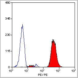
 Application Data
(Staining of human peripheral blood granulocytes with CD18: Alexa Fluor 647 (AAA11986A647))
Application Data
(Staining of human peripheral blood granulocytes with CD18: Alexa Fluor 647 (AAA11986A647))
CD18, Monoclonal Antibody (Cat# AAA11986)
Full Name
RAT ANTI HUMAN CD18
Gene Names
ITGB2; LAD; CD18; MF17; MFI7; LCAMB; LFA-1; MAC-1
Reactivity
Dog, Guinea Pig
Applications
Immunohistochemistry, Flow Cytometry, Immunoprecipitation
Pricing
Complement C9, Active Protein (Cat# AAA14378)
Full Name
Complement C9 protein
Gene Names
C9; C9D; ARMD15
Purity
> 95% pure
Pricing
 SDS-PAGE
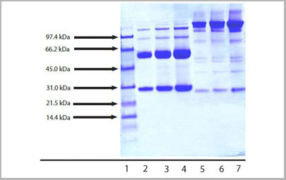
(4-12% Bis-Tris NuPAGE gel showing the following lanes:1- MW standard;2- IgG3 (5 ug reduced/heated);3- IgG3 (10 ug reduced/heated);4- IgG3 (20 ug reduced/heated);5- IgG3 (5 ug non-reduced/no heat);6- IgG3 (10 ug non-reduced/no heat);7- IgG3 (20 ug non-reduced/no heat))
SDS-PAGE
(4-12% Bis-Tris NuPAGE gel showing the following lanes:1- MW standard;2- IgG3 (5 ug reduced/heated);3- IgG3 (10 ug reduced/heated);4- IgG3 (20 ug reduced/heated);5- IgG3 (5 ug non-reduced/no heat);6- IgG3 (10 ug non-reduced/no heat);7- IgG3 (20 ug non-reduced/no heat))
Human IgG3 protein, Immunoglobulin (Cat# AAA14399)
Full Name
Human IgG3 protein
Purity
> 95% pure by SDS-PAGE
Pricing


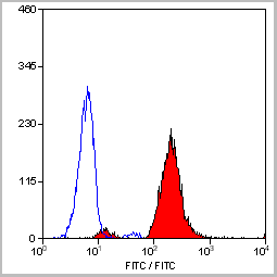
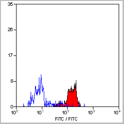
 Application Data
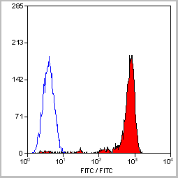
(Staining of mouse peritoneal macrophages with Rat anti Mouse Beta-glucan Receptor: FITC)
Application Data
(Staining of mouse peritoneal macrophages with Rat anti Mouse Beta-glucan Receptor: FITC)
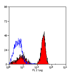
DECTIN-1, Monoclonal Antibody (Cat# AAA12129)
Full Name
RAT ANTI MOUSE DECTIN-1:Low Endotoxin
Gene Names
Clec7a; BGR; beta-GR; Clecsf12
Applications
Immunohistochemistry, Flow Cytometry, Functional Assay, Immunoprecipitation
Pricing


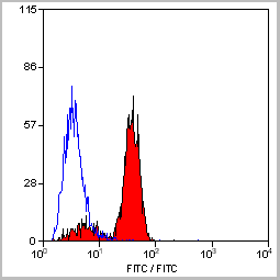


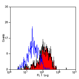
 Application Data
(Staining of mouse peritoneal macrophages with Rat anti Mouse Beta-glucan Receptor: FITC)
Application Data
(Staining of mouse peritoneal macrophages with Rat anti Mouse Beta-glucan Receptor: FITC)
DECTIN-1, Monoclonal Antibody (Cat# AAA12130)
Full Name
RAT ANTI MOUSE DECTIN-1:FITC
Gene Names
Clec7a; BGR; beta-GR; Clecsf12
Applications
Flow Cytometry
Pricing


 Application Data
(Staining of mouse peritoneal macrophages with Rat anti Mouse Beta-glucan Receptor: FITC)
Application Data
(Staining of mouse peritoneal macrophages with Rat anti Mouse Beta-glucan Receptor: FITC)
DECTIN-1, Monoclonal Antibody (Cat# AAA12136)
Full Name
RAT ANTI MOUSE DECTIN-1:RPE
Gene Names
Clec7a; BGR; beta-GR; Clecsf12
Applications
Flow Cytometry
Pricing
Complement Component C1q, Native Protein (Cat# AAA10909)
Full Name
Complement Component C1q Human
Purity
Greater than 95.0% as determined by SDS-PAGE.
Pricing


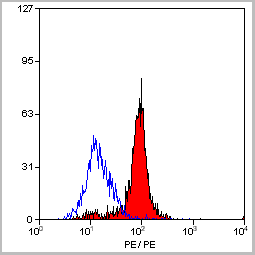
 Application Data
(Staining of human peripheral blood granulocytes with CD18: Alexa Fluor 647)
Application Data
(Staining of human peripheral blood granulocytes with CD18: Alexa Fluor 647)
CD18, Monoclonal Antibody (Cat# AAA11881)
Full Name
RAT ANTI HUMAN CD18:FITC
Gene Names
ITGB2; LAD; CD18; MF17; MFI7; LCAMB; LFA-1; MAC-1
Applications
Flow Cytometry
Pricing


 Application Data
(Staining of human peripheral blood granulocytes with CD18: Alexa Fluor 647)
Application Data
(Staining of human peripheral blood granulocytes with CD18: Alexa Fluor 647)
CD18, Monoclonal Antibody (Cat# AAA11987)
Full Name
RAT ANTI HUMAN CD18
Gene Names
ITGB2; LAD; CD18; MF17; MFI7; LCAMB; LFA-1; MAC-1
Applications
Immunohistochemistry, Flow Cytometry, Immunoprecipitation
Pricing


 Application Data
(Staining of mouse peritoneal macrophages with Rat anti Mouse Beta-glucan Receptor: FITC)
Application Data
(Staining of mouse peritoneal macrophages with Rat anti Mouse Beta-glucan Receptor: FITC)
DECTIN-1, Monoclonal Antibody (Cat# AAA12131)
Full Name
RAT ANTI MOUSE DECTIN-1:FITC
Gene Names
Clec7a; BGR; beta-GR; Clecsf12
Applications
Flow Cytometry
Pricing
 Application Data
(Staining of New Zealand Black mouse peripheral blood granulocytes with Rat anti Mouse Ly-6B.2 conjugated to FITC Data)
Application Data
(Staining of New Zealand Black mouse peripheral blood granulocytes with Rat anti Mouse Ly-6B.2 conjugated to FITC Data)
Ly-6B.2 ALLOANTIGEN, Monoclonal Antibody (Cat# AAA12197)
Full Name
RAT ANTI MOUSE Ly-6B.2 ALLOANTIGEN:RPE
Applications
Flow Cytometry
Pricing


 Application Data
(Staining of mouse peritoneal macrophages with Rat anti Mouse Beta-glucan Receptor: FITC)
Application Data
(Staining of mouse peritoneal macrophages with Rat anti Mouse Beta-glucan Receptor: FITC)
DECTIN-1, Monoclonal Antibody (Cat# AAA12133)
Full Name
RAT ANTI MOUSE DECTIN-1:FITC
Gene Names
Clec7a; BGR; beta-GR; Clecsf12
Applications
Flow Cytometry
Pricing