Filters
Clonality
Type
Reactivity
Gene Name
Isotype
Host
Application
Clone
298 results for " Actin" - showing 150-200


 IHC (Immunohistchemistry)
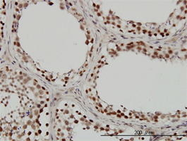
(Immunohistochemistry of paraffin-embedded human liver cancer using SMARCE1 Antibody at dilution of 1:200 (40x lens).)
IHC (Immunohistchemistry)
(Immunohistochemistry of paraffin-embedded human liver cancer using SMARCE1 Antibody at dilution of 1:200 (40x lens).)
SMARCE1, Polyclonal Antibody (Cat# AAA28107)
Full Name
SMARCE1 Polyclonal Antibody
Gene Names
SMARCE1; BAF57
Reactivity
Human, Mouse
Applications
Western Blot, Immunohistochemistry, Immunofluorescence, Immunoprecipitation, Chromatin Immunoprecipitation
Purity
Affinity Purification
Pricing


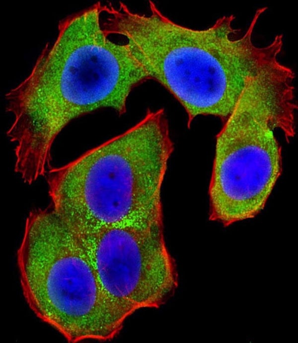
 IF (Immunofluorescence)
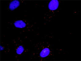
(Immunofluorescent analysis of 4% paraformaldehyde-fixed, 0.1% Triton X-100 permeabilized MCF-7 (human breast cancer cell line) cells labeling Pdx1 with at 1:25 dilution, followed by DyLight 488-conjugated IgG goat anti-rabbit secondary antibody at 1:200 dilution (green). Immunofluorescence image showing cytoplasm staining on MCF-7 cell line. Cytoplasmic actin is detected with DyLight 554 Phalloidin (PD18466410) at 1:100 dilution (red). The nuclear counter stain is DAPI (blue).)
IF (Immunofluorescence)
(Immunofluorescent analysis of 4% paraformaldehyde-fixed, 0.1% Triton X-100 permeabilized MCF-7 (human breast cancer cell line) cells labeling Pdx1 with at 1:25 dilution, followed by DyLight 488-conjugated IgG goat anti-rabbit secondary antibody at 1:200 dilution (green). Immunofluorescence image showing cytoplasm staining on MCF-7 cell line. Cytoplasmic actin is detected with DyLight 554 Phalloidin (PD18466410) at 1:100 dilution (red). The nuclear counter stain is DAPI (blue).)
OPN-a/b, Polyclonal Antibody (Cat# AAA26861)
Full Name
OPN-a/b, NT (SPP1, BNSP, OPN, Osteopontin, Bone sialoprotein 1, Nephropontin, Secreted phosphoprotein 1, Urinary stone protein, Uropontin) (MaxLight 490)
Gene Names
SPP1; OPN; BNSP; BSPI; ETA-1
Reactivity
Human
Applications
WB, IHC, IF
Purity
Purified by Protein A and Peptide Affinity Chromatography.
Pricing
 Standard Curve (Sample)
Filamin A Alpha (FLNa), Antibody Pair Kit (Cat# AAA21089)
Full Name
Human Filamin A Alpha (FLNa) Antibody Pair Kit (with Standard)
Gene Names
FLNA; FLN; FMD; MNS; OPD; ABPX; CSBS; CVD1; FLN1; NHBP; OPD1; OPD2; XLVD; XMVD; FLN-A; ABP-280
Reactivity
Human
Applications
EIA, CLIA
Pricing
 Standard Curve (Sample)
Anti-smooth muscle antibody (ASMA), ELISA Kit (Cat# AAA12870)
Full Name
Human Anti-smooth muscle antibody (ASMA) ELISA Kit
Gene Names
ARPC3; ARC21; p21-Arc
Reactivity
Human
Pricing
 SDS-PAGE
Probable global transcription activator SNF2L2, Recombinant Protein (Cat# AAA18437)
Full Name
Recombinant Human Probable global transcription activator SNF2L2
Purity
Greater or equal to 85% purity as determined by SDS-PAGE.
Pricing


 IF (Immunofluorescence)
(Immunofluorescence analysis of PC-12 cells using FLNA Polyclonal Antibody at dilution of 1:100 (40x lens). Blue: DAPI for nuclear staining.)
IF (Immunofluorescence)
(Immunofluorescence analysis of PC-12 cells using FLNA Polyclonal Antibody at dilution of 1:100 (40x lens). Blue: DAPI for nuclear staining.)
FLNA, Polyclonal Antibody (Cat# AAA22195)
Full Name
FLNA Polyclonal Antibody
Gene Names
FLNA; FLN; FMD; MNS; OPD; ABPX; CSBS; CVD1; FLN1; NHBP; OPD1; OPD2; XLVD; XMVD; FLN-A; ABP-280
Reactivity
Human, Mouse, Rat
Applications
IHC, IF
Purity
Affinity purification
Pricing


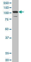
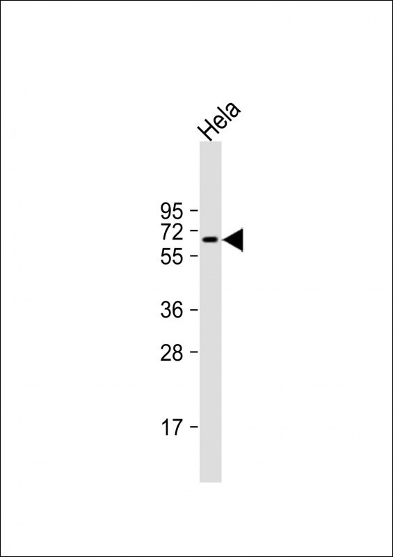
 WB (Western Blot)
(The anti-SUMO2/3 C-term Pab is used in Western blot to detect SUMO2/3 in HeLa cell lysate.)
WB (Western Blot)
(The anti-SUMO2/3 C-term Pab is used in Western blot to detect SUMO2/3 in HeLa cell lysate.)
SUMO2/3, Polyclonal Antibody (Cat# AAA28663)
Full Name
SUMO2/3 Antibody (C-term)
Gene Names
SUMO3; SMT3A; Smt3B; SMT3H1; SUMO-3
Reactivity
Human, mouse (Predicted Reactivity: Xenopus, Zebrafish, Bovine, Chicken, Hamster, Monkey, Pig, Rat)
Applications
IF, EIA, WB, IHC
Purity
Purified Rabbit Polyclonal Antibody (Pab)
Pricing
 SDS-PAGE
Actin, alpha skeletal muscle (ACTA1), Recombinant Protein (Cat# AAA18527)
Full Name
Recombinant Human Actin, alpha skeletal muscle (ACTA1)
Gene Names
ACTA1; ACTA; ASMA; CFTD; MPFD; NEM1; NEM2; NEM3; SHPM; CFTD1; CFTDM
Purity
Greater or equal to 85% purity as determined by SDS-PAGE.
Pricing
 Standard Curve (Sample)
alpha-Smooth muscle actin (alpha-SMA), ELISA Kit (Cat# AAA12991)
Full Name
Mouse alpha-Smooth muscle actin (alpha-SMA) ELISA Kit
Gene Names
ACTG2; ACT; ACTE; VSCM; ACTA3; ACTL3; ACTSG
Reactivity
Mouse
Pricing


 WB (Western Blot)
(CFL1 monoclonal antibody. Western Blot analysis of CFL1 expression in PC-12.)
WB (Western Blot)
(CFL1 monoclonal antibody. Western Blot analysis of CFL1 expression in PC-12.)
CFL1, Monoclonal Antibody (Cat# AAA24759)
Full Name
CFL1 (Cofilin-1, Cofilin, Non-muscle Isoform, 18kD Phosphoprotein, p18, CFL) (Biotin)
Gene Names
CFL1; CFL; cofilin; HEL-S-15
Reactivity
Human, Mouse
Applications
EIA, IF, IHC, WB
Purity
Purified by Protein A Affinity Chromatography.
Pricing


 WB (Western Blot)
(CFL1 monoclonal antibody. Western Blot analysis of CFL1 expression in PC-12.)
WB (Western Blot)
(CFL1 monoclonal antibody. Western Blot analysis of CFL1 expression in PC-12.)
CFL1, Monoclonal Antibody (Cat# AAA25645)
Full Name
CFL1 (Cofilin-1, Cofilin, Non-muscle Isoform, 18kD Phosphoprotein, p18, CFL) (PE)
Gene Names
CFL1; CFL; cofilin; HEL-S-15
Reactivity
Human, Mouse
Applications
Immunofluorescence, Immunohistochemistry, Western Blot
Purity
Purified by Protein A Affinity Chromatography.
Pricing


 IF (Immunofluorescence)
(Immunofluorescent analysis of 4% paraformaldehyde-fixed, 0.1% Triton X-100 permeabilized MCF-7 (human breast cancer cell line) cells labeling Pdx1 with at 1:25 dilution, followed by DyLight 488-conjugated IgG goat anti-rabbit secondary antibody at 1:200 dilution (green). Immunofluorescence image showing cytoplasm staining on MCF-7 cell line. Cytoplasmic actin is detected with DyLight 554 Phalloidin (PD18466410) at 1:100 dilution (red). The nuclear counter stain is DAPI (blue).)
IF (Immunofluorescence)
(Immunofluorescent analysis of 4% paraformaldehyde-fixed, 0.1% Triton X-100 permeabilized MCF-7 (human breast cancer cell line) cells labeling Pdx1 with at 1:25 dilution, followed by DyLight 488-conjugated IgG goat anti-rabbit secondary antibody at 1:200 dilution (green). Immunofluorescence image showing cytoplasm staining on MCF-7 cell line. Cytoplasmic actin is detected with DyLight 554 Phalloidin (PD18466410) at 1:100 dilution (red). The nuclear counter stain is DAPI (blue).)
OPN-a/b, Polyclonal Antibody (Cat# AAA26863)
Full Name
OPN-a/b, NT (SPP1, BNSP, OPN, Osteopontin, Bone sialoprotein 1, Nephropontin, Secreted phosphoprotein 1, Urinary stone protein, Uropontin) (MaxLight 650)
Gene Names
SPP1; OPN; BNSP; BSPI; ETA-1
Reactivity
Human
Applications
WB, IHC, IF
Purity
Purified by Protein A and Peptide Affinity Chromatography.
Pricing
 Standard Curve (Sample)
actin, alpha 2, smooth muscle, aorta, ELISA Kit (Cat# AAA15183)
Full Name
Human alpha-Smooth muscle actin, alpha-SMA ELISA Kit
Gene Names
ACTA2; AAT6; ACTSA; MYMY5
Reactivity
Human
Pricing
 Standard Curve (Sample)
Transcription activator BRG1, SMARCA4, ELISA Kit (Cat# AAA19085)
Full Name
Human Transcription activator BRG1, SMARCA4 ELISA Kit
Gene Names
SMARCA4; BRG1; CSS4; SNF2; SWI2; MRD16; RTPS2; BAF190; SNF2L4; SNF2LB; hSNF2b; BAF190A
Reactivity
Human
Pricing


 WB (Western Blot)
(CFL1 monoclonal antibody. Western Blot analysis of CFL1 expression in PC-12.)
WB (Western Blot)
(CFL1 monoclonal antibody. Western Blot analysis of CFL1 expression in PC-12.)
CFL1, Monoclonal Antibody (Cat# AAA25056)
Full Name
CFL1 (Cofilin-1, Cofilin, Non-muscle Isoform, 18kD Phosphoprotein, p18, CFL) (FITC)
Gene Names
CFL1; CFL; cofilin; HEL-S-15
Reactivity
Human, Mouse
Applications
EIA, IF, IHC, WB
Purity
Purified by Protein A Affinity Chromatography.
Pricing


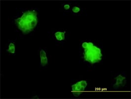
 IF (Immunofluorescence)
(Immunofluorescence of monoclonal antibody to TESK2 on HeLa cell. [antibody concentration 10 ug/ml])
IF (Immunofluorescence)
(Immunofluorescence of monoclonal antibody to TESK2 on HeLa cell. [antibody concentration 10 ug/ml])
TESK2, Monoclonal Antibody (Cat# AAA26049)
Full Name
TESK2 (Testis-Specific Kinase 2) (APC)
Applications
IF, IHC, WB
Purity
Purified
Pricing


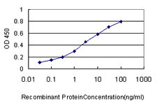 Application Data
(Detection limit for recombinant GST tagged SMARCB1 is ~0.03ng/ml as a capture antibody.)
Application Data
(Detection limit for recombinant GST tagged SMARCB1 is ~0.03ng/ml as a capture antibody.)
SMARCB1, Monoclonal Antibody (Cat# AAA25253)
Full Name
SMARCB1 (SWI/SNF-related Matrix-associated Actin-dependent Regulator of Chromatin Subfamily B Member 1, BRG1-associated Factor 47, BAF47, Integrase Interactor 1 Protein, SNF5 Homolog, hSNF5, BAF47, INI1, SNF5L1) (FITC)
Gene Names
SMARCB1; RDT; CSS3; INI1; SNF5; Snr1; BAF47; MRD15; RTPS1; Sfh1p; hSNFS; SNF5L1; SWNTS1; PPP1R144
Reactivity
Human, Mouse, Rat
Applications
EIA, IHC, WB
Purity
Purified by Protein A Affinity Chromatography.
Pricing


 IF (Immunofluorescence)
(Immunofluorescent analysis of 4% paraformaldehyde-fixed, 0.1% Triton X-100 permeabilized MCF-7 (human breast cancer cell line) cells labeling Pdx1 with at 1:25 dilution, followed by DyLight 488-conjugated IgG goat anti-rabbit secondary antibody at 1:200 dilution (green). Immunofluorescence image showing cytoplasm staining on MCF-7 cell line. Cytoplasmic actin is detected with DyLight 554 Phalloidin (PD18466410) at 1:100 dilution (red). The nuclear counter stain is DAPI (blue).)
IF (Immunofluorescence)
(Immunofluorescent analysis of 4% paraformaldehyde-fixed, 0.1% Triton X-100 permeabilized MCF-7 (human breast cancer cell line) cells labeling Pdx1 with at 1:25 dilution, followed by DyLight 488-conjugated IgG goat anti-rabbit secondary antibody at 1:200 dilution (green). Immunofluorescence image showing cytoplasm staining on MCF-7 cell line. Cytoplasmic actin is detected with DyLight 554 Phalloidin (PD18466410) at 1:100 dilution (red). The nuclear counter stain is DAPI (blue).)
OPN-a/b, Polyclonal Antibody (Cat# AAA26864)
Full Name
OPN-a/b, NT (SPP1, BNSP, OPN, Osteopontin, Bone sialoprotein 1, Nephropontin, Secreted phosphoprotein 1, Urinary stone protein, Uropontin) (MaxLight 750)
Gene Names
SPP1; OPN; BNSP; BSPI; ETA-1
Reactivity
Human
Applications
WB, IHC, IF
Purity
Purified by Protein A and Peptide Affinity Chromatography.
Pricing


 IF (Immunofluorescence)
(Immunofluorescent analysis of 4% paraformaldehyde-fixed, 0.1% Triton X-100 permeabilized MCF-7 (human breast cancer cell line) cells labeling Pdx1 with at 1:25 dilution, followed by DyLight 488-conjugated IgG goat anti-rabbit secondary antibody at 1:200 dilution (green). Immunofluorescence image showing cytoplasm staining on MCF-7 cell line. Cytoplasmic actin is detected with DyLight 554 Phalloidin (PD18466410) at 1:100 dilution (red). The nuclear counter stain is DAPI (blue).)
IF (Immunofluorescence)
(Immunofluorescent analysis of 4% paraformaldehyde-fixed, 0.1% Triton X-100 permeabilized MCF-7 (human breast cancer cell line) cells labeling Pdx1 with at 1:25 dilution, followed by DyLight 488-conjugated IgG goat anti-rabbit secondary antibody at 1:200 dilution (green). Immunofluorescence image showing cytoplasm staining on MCF-7 cell line. Cytoplasmic actin is detected with DyLight 554 Phalloidin (PD18466410) at 1:100 dilution (red). The nuclear counter stain is DAPI (blue).)
OPN-a/b, Polyclonal Antibody (Cat# AAA26862)
Full Name
OPN-a/b, NT (SPP1, BNSP, OPN, Osteopontin, Bone sialoprotein 1, Nephropontin, Secreted phosphoprotein 1, Urinary stone protein, Uropontin) (MaxLight 550)
Gene Names
SPP1; OPN; BNSP; BSPI; ETA-1
Reactivity
Human
Applications
WB, IHC, IF
Purity
Purified by Protein A and Peptide Affinity Chromatography.
Pricing


 FCM (Flow Cytometry)
(GTF2I Antibody (C-term) flow cytometric analysis of k562 cells (bottom histogram) compared to a negative control cell (top histogram).FITC-conjugated goat-anti-rabbit secondary antibodies were used for the analysis.)
FCM (Flow Cytometry)
(GTF2I Antibody (C-term) flow cytometric analysis of k562 cells (bottom histogram) compared to a negative control cell (top histogram).FITC-conjugated goat-anti-rabbit secondary antibodies were used for the analysis.)
GTF2I, Polyclonal Antibody (Cat# AAA28707)
Full Name
GTF2I Antibody (C-term)
Gene Names
GTF2I; WBS; DIWS; SPIN; IB291; BAP135; BTKAP1; TFII-I; WBSCR6; GTFII-I
Reactivity
Human (Predicted Reactivity: Rat)
Applications
Immunofluorescence, Immunohistochemistry, Flow Cytometry, Western Blot
Purity
Peptide Affinity Purified Rabbit Polyclonal Antibody (Pab)
Pricing


 IF (Immunofluorescence)
(Immunofluorescent analysis of 4% paraformaldehyde-fixed, 0.1% Triton X-100 permeabilized MCF-7 (human breast cancer cell line) cells labeling Pdx1 with at 1:25 dilution, followed by DyLight 488-conjugated IgG goat anti-rabbit secondary antibody at 1:200 dilution (green). Immunofluorescence image showing cytoplasm staining on MCF-7 cell line. Cytoplasmic actin is detected with DyLight 554 Phalloidin (PD18466410) at 1:100 dilution (red). The nuclear counter stain is DAPI (blue).)
IF (Immunofluorescence)
(Immunofluorescent analysis of 4% paraformaldehyde-fixed, 0.1% Triton X-100 permeabilized MCF-7 (human breast cancer cell line) cells labeling Pdx1 with at 1:25 dilution, followed by DyLight 488-conjugated IgG goat anti-rabbit secondary antibody at 1:200 dilution (green). Immunofluorescence image showing cytoplasm staining on MCF-7 cell line. Cytoplasmic actin is detected with DyLight 554 Phalloidin (PD18466410) at 1:100 dilution (red). The nuclear counter stain is DAPI (blue).)
OPN-a/b, Polyclonal Antibody (Cat# AAA26859)
Full Name
OPN-a/b, NT (SPP1, BNSP, OPN, Osteopontin, Bone sialoprotein 1, Nephropontin, Secreted phosphoprotein 1, Urinary stone protein, Uropontin) (FITC)
Gene Names
SPP1; OPN; BNSP; BSPI; ETA-1
Reactivity
Human
Applications
WB, IHC, IF
Purity
Purified by Protein A and Peptide Affinity Chromatography.
Pricing


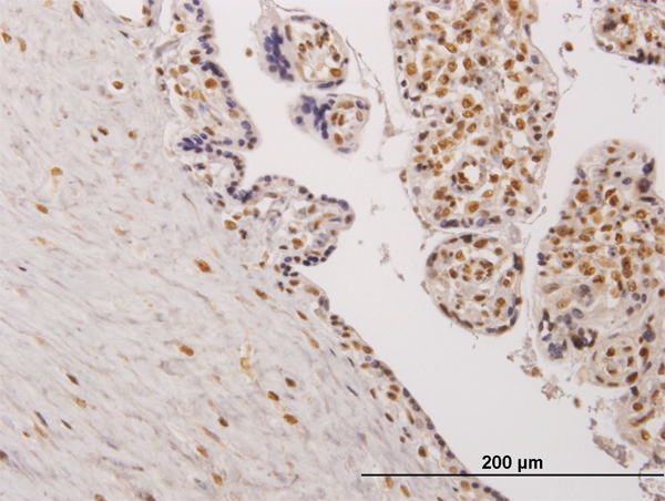
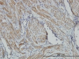
 IHC (Immunohistochemistry)
(Figure 8. IHC analysis of TAGLN/Transgelin using anti-TAGLN/Transgelin antibody (AAA19286).TAGLN/Transgelin was detected in paraffin-embedded section of rat testis tissue. Heat mediated antigen retrieval was performed in EDTA buffer (pH8. 0, epitope retrieval solution). The tissue section was blocked with 10% goat serum. The tissue section was then incubated with 2μg/ml rabbit anti-TAGLN/Transgelin Antibody (AAA19286) overnight at 4 degree C. Biotinylated goat anti-rabbit IgG was used as secondary antibody and incubated for 30 minutes at 37 degree C. The tissue section was developed using Strepavidin-Biotin-Complex (SABC) (Catalog # with DAB as the chromogen.)
IHC (Immunohistochemistry)
(Figure 8. IHC analysis of TAGLN/Transgelin using anti-TAGLN/Transgelin antibody (AAA19286).TAGLN/Transgelin was detected in paraffin-embedded section of rat testis tissue. Heat mediated antigen retrieval was performed in EDTA buffer (pH8. 0, epitope retrieval solution). The tissue section was blocked with 10% goat serum. The tissue section was then incubated with 2μg/ml rabbit anti-TAGLN/Transgelin Antibody (AAA19286) overnight at 4 degree C. Biotinylated goat anti-rabbit IgG was used as secondary antibody and incubated for 30 minutes at 37 degree C. The tissue section was developed using Strepavidin-Biotin-Complex (SABC) (Catalog # with DAB as the chromogen.)
TAGLN/Transgelin, Polyclonal Antibody (Cat# AAA19286)
Full Name
Anti-TAGLN/Transgelin Antibody
Gene Names
TAGLN; SM22; SMCC; TAGLN1; WS3-10
Reactivity
Human, Mouse, Rat
Applications
WB, IHC-P, ICC, IF, FC/FACS/FCM, EIA
Purity
Immunogen affinity purified.
Pricing
 Standard Curve (Sample)
gelsolin (amyloidosis, Finnish type), ELISA Kit (Cat# AAA15199)
Full Name
Human Gelsolin ELISA Kit
Gene Names
GSN; ADF; AGEL
Reactivity
Human
Pricing


 WB (Western Blot)
(DAAM1 monoclonal antibody Western Blot analysis of DAAM1 expression in A-431.)
WB (Western Blot)
(DAAM1 monoclonal antibody Western Blot analysis of DAAM1 expression in A-431.)
DAAM1, Monoclonal Antibody (Cat# AAA24775)
Full Name
DAAM1 (Disheveled-associated Activator Of Morphogenesis 1, KIAA0666) (Biotin)
Reactivity
Human, Rat
Applications
EIA, WB
Purity
Purified by Protein A Affinity Chromatography.
Pricing


 IF (Immunofluorescence)
(Immunofluorescent analysis of 4% paraformaldehyde-fixed, 0.1% Triton X-100 permeabilized MCF-7 (human breast cancer cell line) cells labeling Pdx1 with at 1:25 dilution, followed by DyLight 488-conjugated IgG goat anti-rabbit secondary antibody at 1:200 dilution (green). Immunofluorescence image showing cytoplasm staining on MCF-7 cell line. Cytoplasmic actin is detected with DyLight 554 Phalloidin (PD18466410) at 1:100 dilution (red). The nuclear counter stain is DAPI (blue).)
IF (Immunofluorescence)
(Immunofluorescent analysis of 4% paraformaldehyde-fixed, 0.1% Triton X-100 permeabilized MCF-7 (human breast cancer cell line) cells labeling Pdx1 with at 1:25 dilution, followed by DyLight 488-conjugated IgG goat anti-rabbit secondary antibody at 1:200 dilution (green). Immunofluorescence image showing cytoplasm staining on MCF-7 cell line. Cytoplasmic actin is detected with DyLight 554 Phalloidin (PD18466410) at 1:100 dilution (red). The nuclear counter stain is DAPI (blue).)
OPN-a/b, Polyclonal Antibody (Cat# AAA26860)
Full Name
OPN-a/b, NT (SPP1, BNSP, OPN, Osteopontin, Bone sialoprotein 1, Nephropontin, Secreted phosphoprotein 1, Urinary stone protein, Uropontin) (MaxLight 405)
Gene Names
SPP1; OPN; BNSP; BSPI; ETA-1
Reactivity
Human
Applications
WB, IHC, IF
Purity
Purified by Protein A and Peptide Affinity Chromatography.
Pricing


 WB (Western Blot)
(DAAM1 monoclonal antibody Western Blot analysis of DAAM1 expression in A-431.)
WB (Western Blot)
(DAAM1 monoclonal antibody Western Blot analysis of DAAM1 expression in A-431.)
DAAM1, Monoclonal Antibody (Cat# AAA25072)
Full Name
DAAM1 (Disheveled-associated Activator Of Morphogenesis 1, KIAA0666) (FITC)
Reactivity
Human, Rat
Applications
EIA, WB
Purity
Purified by Protein A Affinity Chromatography.
Pricing


 WB (Western Blot)
(DAAM1 monoclonal antibody Western Blot analysis of DAAM1 expression in A-431.)
WB (Western Blot)
(DAAM1 monoclonal antibody Western Blot analysis of DAAM1 expression in A-431.)
DAAM1, Monoclonal Antibody (Cat# AAA24480)
Full Name
DAAM1 (Disheveled-associated Activator Of Morphogenesis 1, KIAA0666) APC
Reactivity
Human, Rat
Applications
EIA, WB
Purity
Purified by Protein A Affinity Chromatography.
Pricing
 Standard Curve (Sample)
beta-actin (Beta actin), ELISA Kit (Cat# AAA17761)
Full Name
Human beta-actin (Beta actin)
Reactivity
Human
Pricing
 Standard Curve (Sample)
alpha Smooth Muscle Actin, ELISA Kit (Cat# AAA16647)
Full Name
Mouse alpha Smooth Muscle Actin ELISA Kit
Gene Names
ACTA2; AAT6; ACTSA; MYMY5
Reactivity
Mouse
Pricing


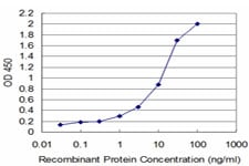
 Application Data
(Detection limit for recombinant GST tagged SMARCB1 is ~0.03ng/ml as a capture antibody.)
Application Data
(Detection limit for recombinant GST tagged SMARCB1 is ~0.03ng/ml as a capture antibody.)
SMARCB1, Monoclonal Antibody (Cat# AAA25843)
Full Name
SMARCB1 (SWI/SNF-related Matrix-associated Actin-dependent Regulator of Chromatin Subfamily B Member 1, BRG1-associated Factor 47, BAF47, Integrase Interactor 1 Protein, SNF5 Homolog, hSNF5, BAF47, INI1, SNF5L1) (PE)
Gene Names
SMARCB1; RDT; CSS3; INI1; SNF5; Snr1; BAF47; MRD15; RTPS1; Sfh1p; hSNFS; SNF5L1; SWNTS1; PPP1R144
Reactivity
Human, Mouse, Rat
Applications
EIA, IHC, WB
Purity
Purified by Protein A Affinity Chromatography.
Pricing


 IF (Immunofluorescence)
(Immunofluorescence of monoclonal antibody to TESK2 on HeLa cell. [antibody concentration 10 ug/ml])
IF (Immunofluorescence)
(Immunofluorescence of monoclonal antibody to TESK2 on HeLa cell. [antibody concentration 10 ug/ml])
TESK2, Monoclonal Antibody (Cat# AAA26581)
Full Name
TESK2 (Testis-Specific Kinase 2) (PE)
Applications
IF, IHC, WB
Purity
Purified
Pricing


 IF (Immunofluorescence)
(Immunofluorescent analysis of 4% paraformaldehyde-fixed, 0.1% Triton X-100 permeabilized U-2 OS (human osteosarcoma cell line) cells labeling DLL3 with (1:25), followed by Dylight 488- conjugated goat anti- rabbit IgG secondary antibody at (1:200) (green). Immunofluorescence image showing nucleus and weak cytoplasm staining on U-2 OScell line. Cytoplasmic actin is detected with Dylight 554 Phalloidin (1:100) (red).)
IF (Immunofluorescence)
(Immunofluorescent analysis of 4% paraformaldehyde-fixed, 0.1% Triton X-100 permeabilized U-2 OS (human osteosarcoma cell line) cells labeling DLL3 with (1:25), followed by Dylight 488- conjugated goat anti- rabbit IgG secondary antibody at (1:200) (green). Immunofluorescence image showing nucleus and weak cytoplasm staining on U-2 OScell line. Cytoplasmic actin is detected with Dylight 554 Phalloidin (1:100) (red).)
DLL3, Polyclonal Antibody (Cat# AAA26850)
Full Name
DLL3, CT (DLL3, Delta-like protein 3, Drosophila Delta homolog 3) (APC)
Gene Names
DLL3; SCDO1
Reactivity
Human
Applications
IF, IHC, WB
Purity
Purified by Protein A Affinity Chromatography.
Pricing


 IF (Immunofluorescence)
(Immunofluorescence of monoclonal antibody to TESK2 on HeLa cell. [antibody concentration 10 ug/ml])
IF (Immunofluorescence)
(Immunofluorescence of monoclonal antibody to TESK2 on HeLa cell. [antibody concentration 10 ug/ml])
TESK2, Monoclonal Antibody (Cat# AAA26314)
Full Name
TESK2 (Testis-Specific Kinase 2) (FITC)
Applications
IF, IHC, WB
Purity
Purified
Pricing
 Standard Curve (Sample)
alpha Smooth Muscle Actin, ELISA Kit (Cat# AAA15956)
Full Name
Rat alpha Smooth Muscle Actin ELISA Kit
Gene Names
ACTA2; AAT6; ACTSA; MYMY5
Reactivity
Rat
Pricing
 Standard Curve (Sample)
actin, alpha 2, smooth muscle, aorta, ELISA Kit (Cat# AAA15073)
Full Name
Rat alpha-Smooth muscle actin, alpha-SMA ELISA Kit
Reactivity
Rat
Pricing


 Application Data
(Ref. 3: Isolation, culture and immunocytochemical characterization of HUCPC. The dissection under sterile condition of foetal and full-term cords was performed to expose the WJ, the vein and arteries (A). After the in vitro expansion of foetal HUCPC (B) the cells ..)
Application Data
(Ref. 3: Isolation, culture and immunocytochemical characterization of HUCPC. The dissection under sterile condition of foetal and full-term cords was performed to expose the WJ, the vein and arteries (A). After the in vitro expansion of foetal HUCPC (B) the cells ..)
Chick Embryo Extract, Ultrafiltrate, Reagent (Cat# AAA14749)
Full Name
Chick Embryo Extract, Ultrafiltrate (CEE)
Purity
Molecular Biology Grade
Pricing


 IF (Immunofluorescence)
(Immunofluorescent analysis of 4% paraformaldehyde-fixed, 0.1% Triton X-100 permeabilized MCF-7 (human breast cancer cell line) cells labeling Pdx1 with AAA14796 at 1:25 dilution, followed by Dylight® 488-conjugated IgG goat anti-rabbit secondary antibody at 1:200 dilution (green). Immunofluorescence image showing cytoplasm staining on MCF-7 cell line. Cytoplasmic actin is detected with Dylight® 554 Phalloidin (PD18466410) at 1:100 dilution (red). The nuclear counter stain is DAPI (blue).)
IF (Immunofluorescence)
(Immunofluorescent analysis of 4% paraformaldehyde-fixed, 0.1% Triton X-100 permeabilized MCF-7 (human breast cancer cell line) cells labeling Pdx1 with AAA14796 at 1:25 dilution, followed by Dylight® 488-conjugated IgG goat anti-rabbit secondary antibody at 1:200 dilution (green). Immunofluorescence image showing cytoplasm staining on MCF-7 cell line. Cytoplasmic actin is detected with Dylight® 554 Phalloidin (PD18466410) at 1:100 dilution (red). The nuclear counter stain is DAPI (blue).)
OPN-a/b, Polyclonal Antibody (Cat# AAA14796)
Full Name
OPN-a/b, NT (SPP1, BNSP, OPN, Osteopontin, Bone sialoprotein 1, Nephropontin, Secreted phosphoprotein 1, Urinary stone protein, Uropontin)
Reactivity
Human
Applications
EL/EIA, WB, IHC, IF
Purity
Affinity Purified
Purified by Protein A affinity chromatography.
Purified by Protein A affinity chromatography.
Pricing


 WB (Western Blot)
(Host: RabbitTarget Name: ACTBSample Type: JurkatAntibody Dilution: 1.0ug/mlACTB is strongly supported by BioGPS gene expression data to be expressed in Jurkat)
WB (Western Blot)
(Host: RabbitTarget Name: ACTBSample Type: JurkatAntibody Dilution: 1.0ug/mlACTB is strongly supported by BioGPS gene expression data to be expressed in Jurkat)
ACTB, Polyclonal Antibody (Cat# AAA23467)
Full Name
ACTB Antibody - middle region
Gene Names
ACTB; BRWS1; PS1TP5BP1
Reactivity
Cow, Goat, Horse, Human, Mouse, Pig, Rat, Sheep, Zebrafish
Applications
IHC, WB
Purity
Affinity Purified
Pricing


 WB (Western Blot)
(SSH3 monoclonal antibody, Western Blot analysis of SSH3 expression in A-431.)
WB (Western Blot)
(SSH3 monoclonal antibody, Western Blot analysis of SSH3 expression in A-431.)
SSH3, Monoclonal Antibody (Cat# AAA25854)
Full Name
SSH3 (Protein Phosphatase Slingshot Homolog 3, SSH-like Protein 3, SSH3L, SSH-3L, hSSH-3L) (PE)
Gene Names
SSH3; SSH3L
Reactivity
Human
Applications
EIA, IF, IHC, WB
Purity
Purified by Protein A Affinity Chromatography.
Pricing
 Standard Curve (Sample)
alpha-Smooth muscle actin (alpha-SMA), ELISA Kit (Cat# AAA12916)
Full Name
Rat alpha-Smooth muscle actin (alpha-SMA) ELISA Kit
Gene Names
ACTG2; ACT; ACTE; VSCM; ACTA3; ACTL3; ACTSG
Reactivity
Rat
Pricing
 Standard Curve (Sample)
G Actin (GACT), ELISA Kit (Cat# AAA17293)
Full Name
Human G Actin (GACT) Elisa Kit
Reactivity
Human
Pricing
 Standard Curve (Sample)
Gelsolin, GS, ELISA Kit (Cat# AAA11297)
Full Name
Human Gelsolin, GS ELISA Kit
Gene Names
GSN; ADF
Reactivity
Human
Pricing


 FCM (Flow Cytometry)
(TYSY Antibody (C-term) flow cytometric analysis of Hela cells (right histogram) compared to a negative control cell (left histogram).FITC-conjugated goat-anti-rabbit secondary antibodies were used for the analysis.)
FCM (Flow Cytometry)
(TYSY Antibody (C-term) flow cytometric analysis of Hela cells (right histogram) compared to a negative control cell (left histogram).FITC-conjugated goat-anti-rabbit secondary antibodies were used for the analysis.)
TYSY, Polyclonal Antibody (Cat# AAA28647)
Full Name
TYSY Antibody (C-term)
Gene Names
TYMS; TS; TMS; HST422
Reactivity
Human
Applications
WB, EIA, IHC, IF, FC/FACS
Purity
Peptide Affinity Purified Rabbit Polyclonal Antibody (Pab)
Pricing
 Standard Curve (Sample)
 Standard Curve (Sample)
Fascin, ELISA Kit (Cat# AAA17663)
Full Name
Human Fascin ELISA Kit
Gene Names
FSCN1; HSN; SNL; p55; FAN1
Reactivity
Human
Pricing


 IF (Immunofluorescence)
(Immunofluorescence of monoclonal antibody to TESK2 on HeLa cell. [antibody concentration 10 ug/ml])
IF (Immunofluorescence)
(Immunofluorescence of monoclonal antibody to TESK2 on HeLa cell. [antibody concentration 10 ug/ml])
TESK2, Monoclonal Antibody (Cat# AAA26580)
Full Name
TESK2 (Testis-Specific Kinase 2) (PE)
Applications
IF, IHC, WB
Purity
Purified
Pricing


 IF (Immunofluorescence)
(Immunofluorescent analysis of 4% paraformaldehyde-fixed, 0.1% Triton X-100 permeabilized MCF-7 (human breast cancer cell line) cells labeling Pdx1 with at 1:25 dilution, followed by DyLight 488-conjugated IgG goat anti-rabbit secondary antibody at 1:200 dilution (green). Immunofluorescence image showing cytoplasm staining on MCF-7 cell line. Cytoplasmic actin is detected with DyLight 554 Phalloidin (PD18466410) at 1:100 dilution (red). The nuclear counter stain is DAPI (blue).)
IF (Immunofluorescence)
(Immunofluorescent analysis of 4% paraformaldehyde-fixed, 0.1% Triton X-100 permeabilized MCF-7 (human breast cancer cell line) cells labeling Pdx1 with at 1:25 dilution, followed by DyLight 488-conjugated IgG goat anti-rabbit secondary antibody at 1:200 dilution (green). Immunofluorescence image showing cytoplasm staining on MCF-7 cell line. Cytoplasmic actin is detected with DyLight 554 Phalloidin (PD18466410) at 1:100 dilution (red). The nuclear counter stain is DAPI (blue).)
OPN-a/b, Polyclonal Antibody (Cat# AAA26858)
Full Name
OPN-a/b, NT (SPP1, BNSP, OPN, Osteopontin, Bone sialoprotein 1, Nephropontin, Secreted phosphoprotein 1, Urinary stone protein, Uropontin) (Biotin)
Gene Names
SPP1; OPN; BNSP; BSPI; ETA-1
Reactivity
Human
Applications
WB, IHC, IF, EIA
Purity
Purified by Protein A and Peptide Affinity Chromatography.
Pricing


 WB (Western Blot)
(SSH3 monoclonal antibody, Western Blot analysis of SSH3 expression in A-431.)
WB (Western Blot)
(SSH3 monoclonal antibody, Western Blot analysis of SSH3 expression in A-431.)
SSH3, Monoclonal Antibody (Cat# AAA25558)
Full Name
SSH3 (Protein Phosphatase Slingshot Homolog 3, SSH-like Protein 3, SSH3L, SSH-3L, hSSH-3L) (HRP)
Gene Names
SSH3; SSH3L
Reactivity
Human
Applications
EIA, IHC, WB
Purity
Purified by Protein A Affinity Chromatography.
Pricing


 Application Data
(Detection limit for recombinant GST tagged SMARCB1 is ~0.03ng/ml as a capture antibody.)
Application Data
(Detection limit for recombinant GST tagged SMARCB1 is ~0.03ng/ml as a capture antibody.)
SMARCB1, Monoclonal Antibody (Cat# AAA25547)
Full Name
SMARCB1 (SWI/SNF-related Matrix-associated Actin-dependent Regulator of Chromatin Subfamily B Member 1, BRG1-associated Factor 47, BAF47, Integrase Interactor 1 Protein, SNF5 Homolog, hSNF5, BAF47, INI1, SNF5L1) (HRP)
Gene Names
SMARCB1; RDT; CSS3; INI1; SNF5; Snr1; BAF47; MRD15; RTPS1; Sfh1p; hSNFS; SNF5L1; SWNTS1; PPP1R144
Reactivity
Human, Mouse, Rat
Applications
EIA, IHC, WB
Purity
Purified by Protein A Affinity Chromatography.
Pricing


 WB (Western Blot)
(ACTB monoclonal antibody Western Blot analysis of ACTB expression in K-562.)
WB (Western Blot)
(ACTB monoclonal antibody Western Blot analysis of ACTB expression in K-562.)
ACTB, Monoclonal Antibody (Cat# AAA25597)
Full Name
ACTB (Actin, Cytoplasmic 1, Beta-actin) (PE)
Gene Names
ACTB; BRWS1; PS1TP5BP1
Reactivity
Human, Mouse
Applications
EIA, IHC, WB
Purity
Purified by Protein A Affinity Chromatography.
Pricing