Filters
Clonality
Type
Reactivity
Gene Name
Isotype
Host
Application
Clone
1718 results for " Cell Tissue Lysates" - showing 1450-1500
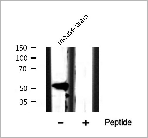
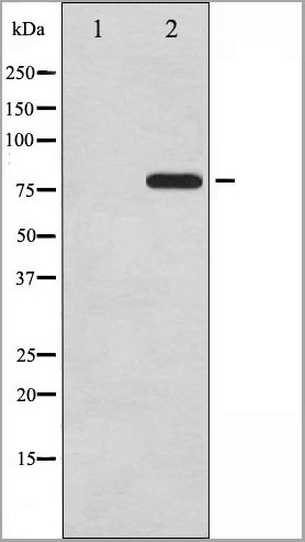
 WB (Western Blot)
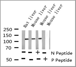
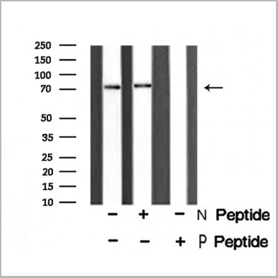
(Western blot analysis of EGFR phosphorylation expression in 293 whole cell lysates, The lane on the left is treated with the antigen-specific peptide.)
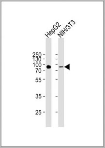
WB (Western Blot)
(Western blot analysis of EGFR phosphorylation expression in 293 whole cell lysates, The lane on the left is treated with the antigen-specific peptide.)
EGFR, Polyclonal Antibody (Cat# AAA30962)
Full Name
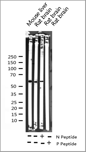
Phospho-EGFR (Ser695) Antibody
Gene Names
EGFR; ERBB; HER1; mENA; ERBB1; PIG61; NISBD2
Reactivity
Human, Mouse, Rat
Applications
Western Blot, Immunohistochemistry, Immunofluorescence, Immunocytochemistry
Purity
From purified rabbit serum by affinity purification via sequential chromatography on phospho-and non-phospho-peptide affinity columns.
Pricing
 Standard Curve (Sample)
Clara Cell Protein 16 (CC16), ELISA Kit (Cat# AAA23117)
Full Name
Mouse Clara Cell Protein 16 (CC16) Mini Samples ELISA Kit
Gene Names
Scgb1a1; UG; UGB; Utg; CC10; CC16; CCSP; PCB-BP
Reactivity
Mouse
Applications
ELISA
Pricing
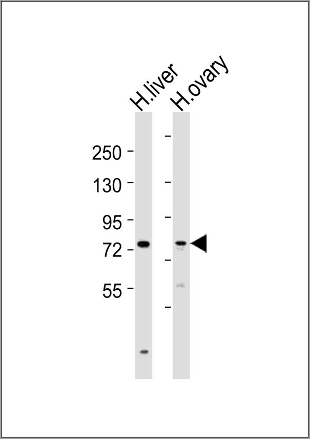
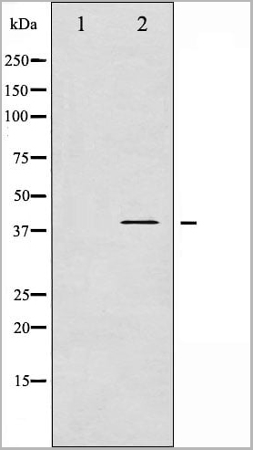
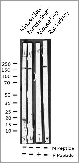
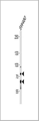
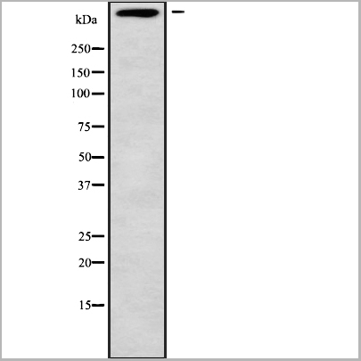
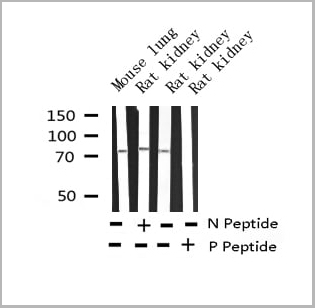
 WB (Western Blot)
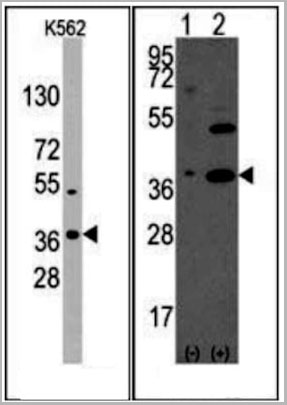
(Western blot analysis of RPL22/L1 using K562 whole cell lysates)
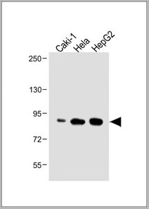
WB (Western Blot)
(Western blot analysis of RPL22/L1 using K562 whole cell lysates)
RPL22/L1, Polyclonal Antibody (Cat# AAA31142)
Full Name
RPL22/L1 Antibody
Reactivity
Human, Mouse, Rat
Applications
Western Blot, Immunohistochemistry
Purity
The antiserum was purified by peptide affinity chromatography using SulfoLink™ Coupling Resin (Thermo Fisher Scientific).
Pricing
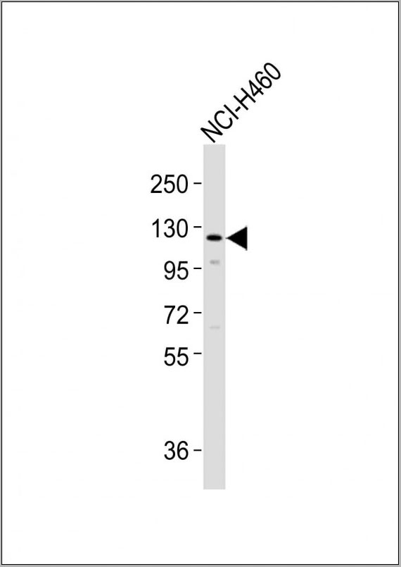
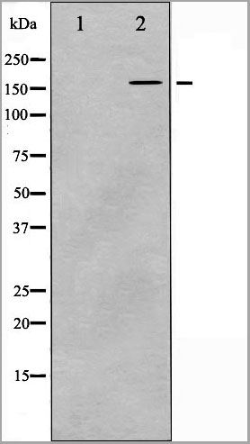
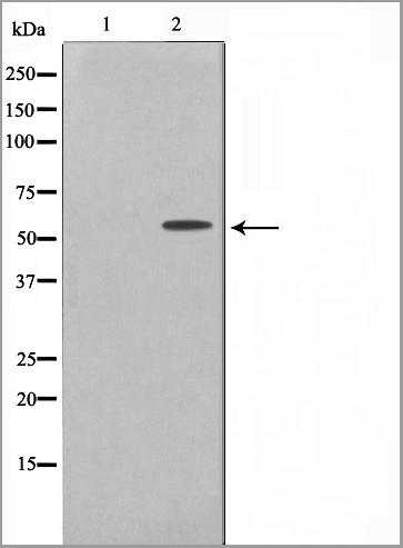
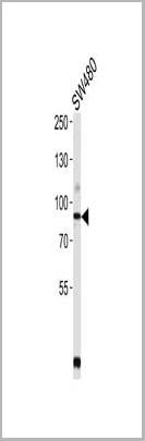
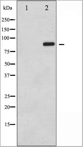
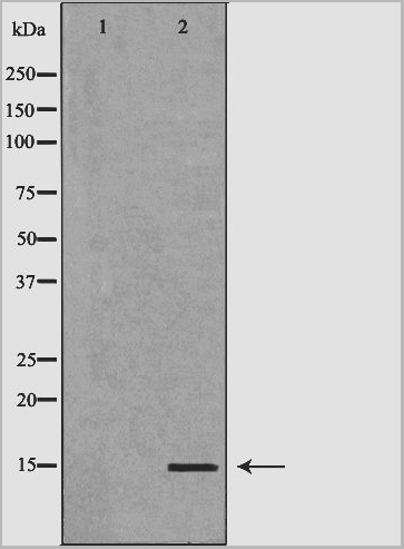
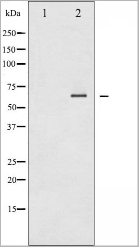
 WB (Western Blot)
(SLCO1B3 Antibody (C-term) western blot analysis in NCI-H460 cell line lysates (35ug/lane).This demonstrates the SLCO1B3 antibody detected the SLCO1B3 protein (arrow).)
WB (Western Blot)
(SLCO1B3 Antibody (C-term) western blot analysis in NCI-H460 cell line lysates (35ug/lane).This demonstrates the SLCO1B3 antibody detected the SLCO1B3 protein (arrow).)
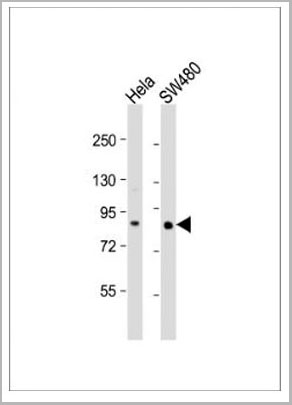
SLCO1B3, Polyclonal Antibody (Cat# AAA28785)
Full Name
SLCO1B3 Antibody (C-term)
Gene Names
SLCO1B3; LST3; HBLRR; LST-2; OATP8; OATP-8; OATP1B3; SLC21A8; LST-3TM13
Reactivity
Human
Applications
Western Blot
Purity
Peptide Affinity Purified Rabbit Polyclonal Antibody (Pab)
Pricing
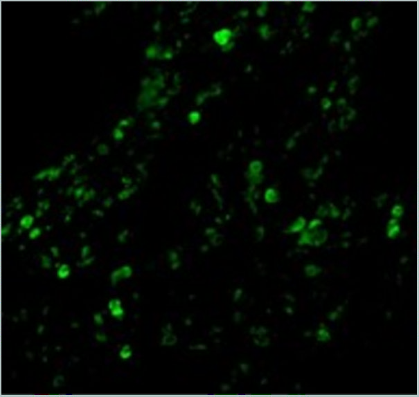
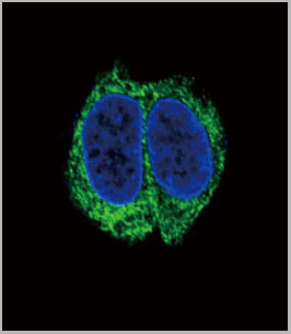
 IF (Immunofluorescence)
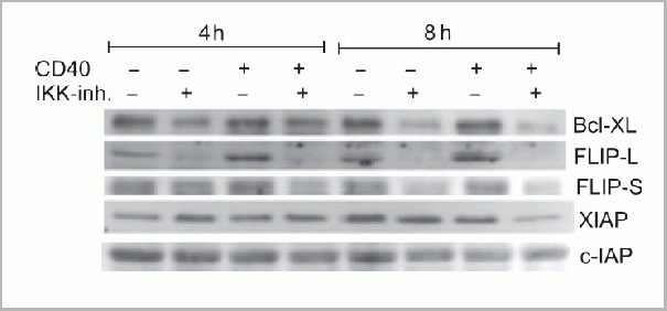
(Immunofluorescence Validation of cIAP in Human Lung Tissue Immunofluorescent analysis of 4% paraformaldehydefixed human lung tissue abeling cIAP with 3325 at 20 ug/mL, followed by goat anti-rabbit IgG secondary antibody at 1/500 dilution (green).)
IF (Immunofluorescence)
(Immunofluorescence Validation of cIAP in Human Lung Tissue Immunofluorescent analysis of 4% paraformaldehydefixed human lung tissue abeling cIAP with 3325 at 20 ug/mL, followed by goat anti-rabbit IgG secondary antibody at 1/500 dilution (green).)
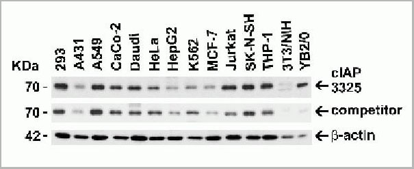
cIAP, Polyclonal Antibody (Cat# AAA10954)
Full Name
cIAP Antibody
Gene Names
BIRC2; API1; MIHB; HIAP2; RNF48; cIAP1; Hiap-2; c-IAP1
Reactivity
Human, Mouse
Applications
Western Blot, Immunohistochemistry, Immunofluorescence
Purity
cIAP Antibody is affinity chromatography purified via peptide column.
Pricing
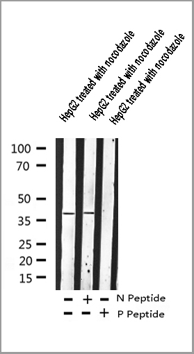
 WB (Western Blot)
(Western blot analysis of Adrenergic Receptor beta2 phosphorylation expression in nocodazole treated HepG2 whole cell lysates, The lane on the left is treated with the antigen-specific peptide.)
WB (Western Blot)
(Western blot analysis of Adrenergic Receptor beta2 phosphorylation expression in nocodazole treated HepG2 whole cell lysates, The lane on the left is treated with the antigen-specific peptide.)
Adrenergic Receptor beta2, Polyclonal Antibody (Cat# AAA30989)
Full Name
Phospho-Adrenergic Receptor beta2 (Ser346) Antibody
Gene Names
ADRB2; BAR; B2AR; ADRBR; ADRB2R; BETA2AR
Reactivity
Human, Mouse, Rat
Applications
Western Blot, Immunohistochemistry, Immunofluorescence, Immunocytochemistry
Purity
Peptide affinity purification
Pricing
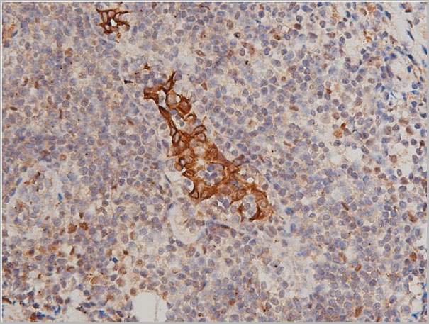
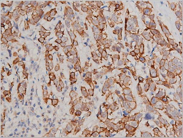
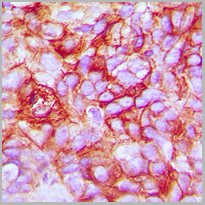
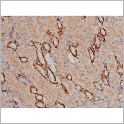
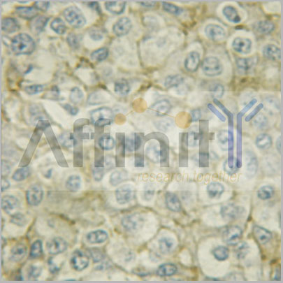
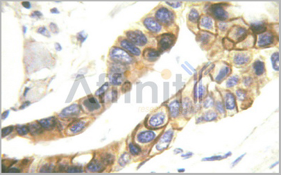
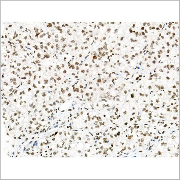
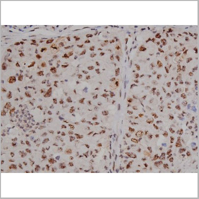
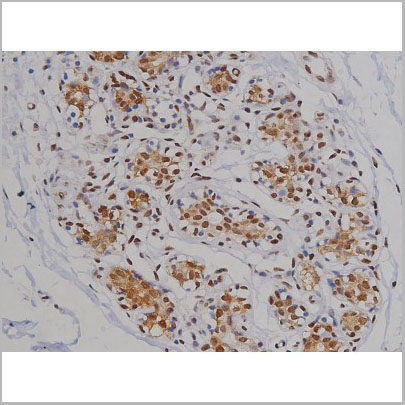
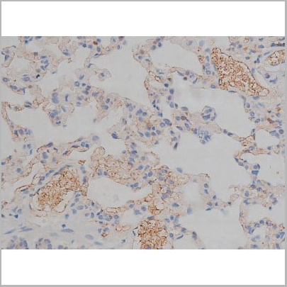
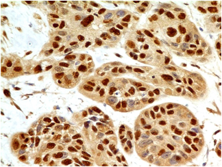
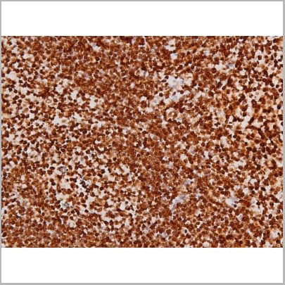
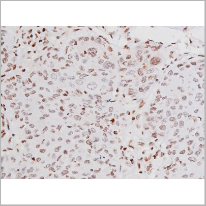
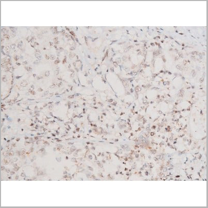
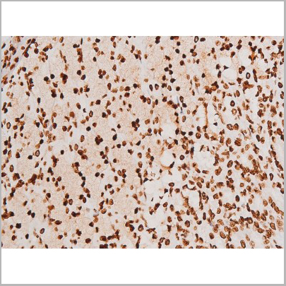
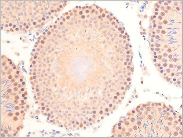
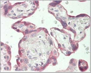
 IHC (Immunohistochemistry)
(AAA30985 at 1/50 staining human breast cancer tissue sections by IHC-P. The tissue was formaldehyde fixed and a heat mediated antigen retrieval step in citrate buffer was performed. The tissue was then blocked and incubated with the antibody for 1.5 hours at 22 degree C. An HRP conjugated goat anti-rabbit antibody was used as the secondary.)
IHC (Immunohistochemistry)
(AAA30985 at 1/50 staining human breast cancer tissue sections by IHC-P. The tissue was formaldehyde fixed and a heat mediated antigen retrieval step in citrate buffer was performed. The tissue was then blocked and incubated with the antibody for 1.5 hours at 22 degree C. An HRP conjugated goat anti-rabbit antibody was used as the secondary.)
Keratin 8, Polyclonal Antibody (Cat# AAA30985)
Full Name
Phospho-Keratin 8 (Ser432) Antibody
Gene Names
KRT8; K8; KO; CK8; CK-8; CYK8; K2C8; CARD2
Reactivity
Human, Mouse, Rat
Applications
Western Blot, Immunohistochemistry, Immunofluorescence, Immunocytochemistry
Purity
From purified rabbit serum by affinity purification via sequential chromatography on phospho-and non-phospho-peptide affinity columns.
Pricing

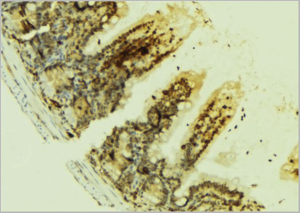

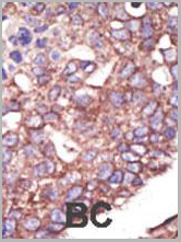
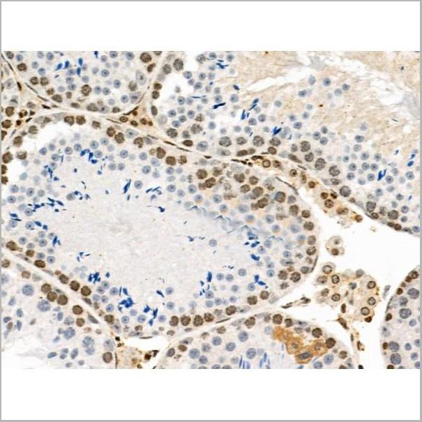
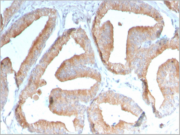
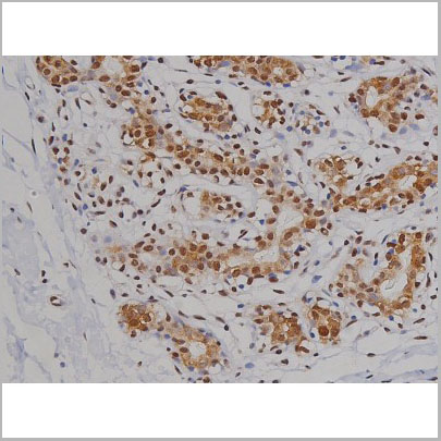
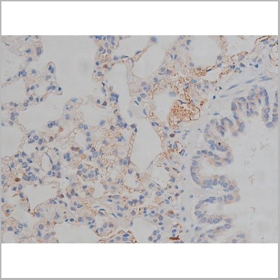
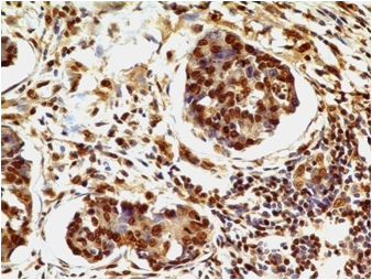
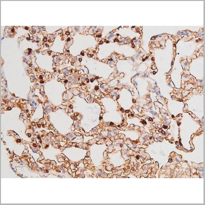
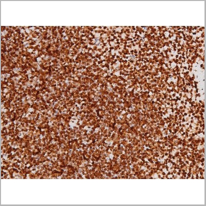
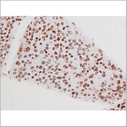
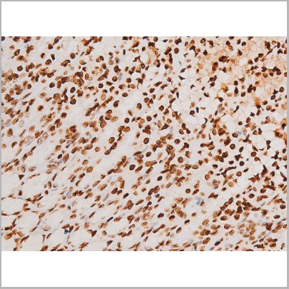
 IHC (Immunohistochemistry)
(AAA31083 at 1/100 staining human breast tissues sections by IHC-P. The tissue was formaldehyde fixed and a heat mediated antigen retrieval step in citrate buffer was performed. The tissue was then blocked and incubated with the antibody for 1.5 hours at 92)
IHC (Immunohistochemistry)
(AAA31083 at 1/100 staining human breast tissues sections by IHC-P. The tissue was formaldehyde fixed and a heat mediated antigen retrieval step in citrate buffer was performed. The tissue was then blocked and incubated with the antibody for 1.5 hours at 92)
EGFR, Polyclonal Antibody (Cat# AAA31083)
Full Name
EGFR Antibody
Gene Names
EGFR; ERBB; HER1; mENA; ERBB1; PIG61; NISBD2
Reactivity
Human, Mouse, Rat
Applications
Western Blot, Immunohistochemistry, Immunofluorescence, Immunocytochemistry
Purity
The antiserum was purified by peptide affinity chromatography using SulfoLink Coupling Resin.
Pricing
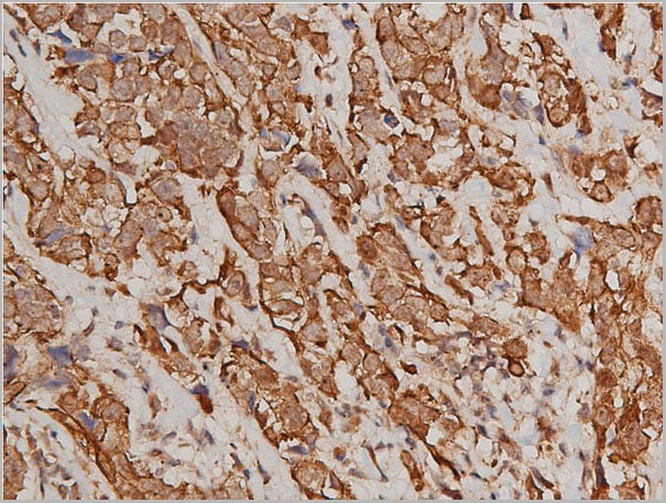
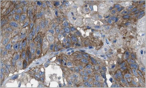
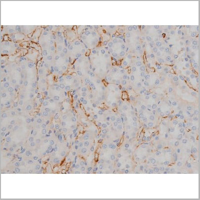
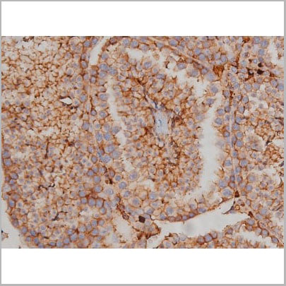
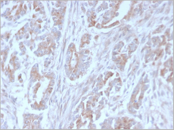
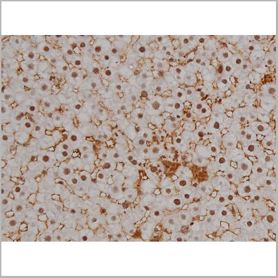
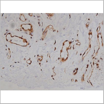
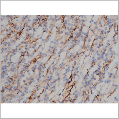
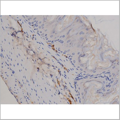
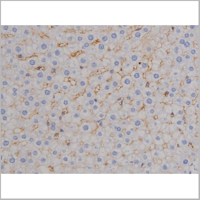
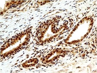
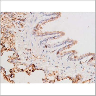
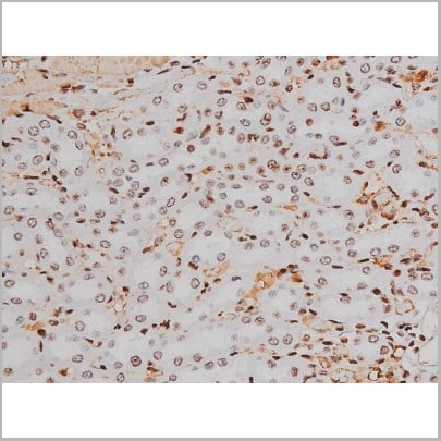
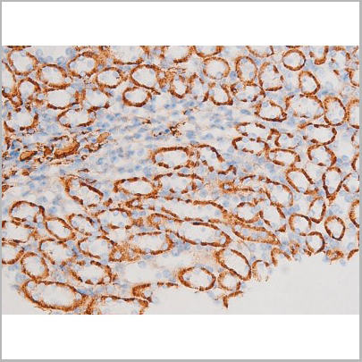
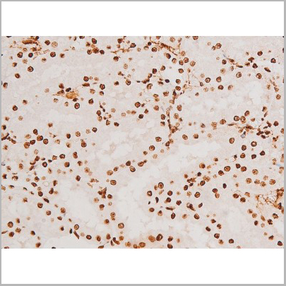
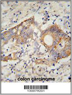
 IHC (Immunohistochemistry)
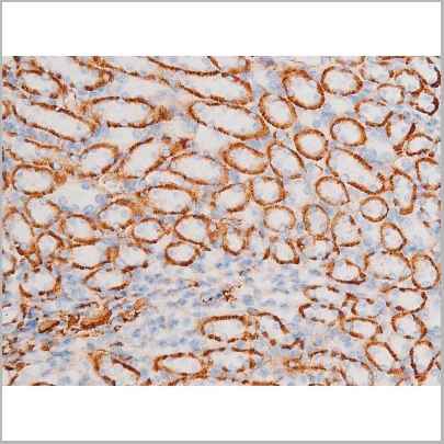
(Immunohistochemical analysis of Taurine Transporter staining in human breast cancer formalin fixed paraffin embedded tissue section. The section was pre-treated using heat mediated antigen retrieval with sodium citrate buffer (pH 6.0). The section was then incubated with the antibody at room temperature and detected using an HRP conjugated compact polymer system. DAB was used as the chromogen. The section was then counterstained with haematoxylin and mounted with DPX.)
IHC (Immunohistochemistry)
(Immunohistochemical analysis of Taurine Transporter staining in human breast cancer formalin fixed paraffin embedded tissue section. The section was pre-treated using heat mediated antigen retrieval with sodium citrate buffer (pH 6.0). The section was then incubated with the antibody at room temperature and detected using an HRP conjugated compact polymer system. DAB was used as the chromogen. The section was then counterstained with haematoxylin and mounted with DPX.)
Taurine Transporter, Polyclonal Antibody (Cat# AAA27812)
Full Name
Anti-Taurine Transporter Antibody
Gene Names
SLC6A6; TAUT
Reactivity
Human, Mouse, Rat, Dog
Applications
Western Blot, Immunohistochemistry
Purity
The antibody was purified by affinity chromatography.
Pricing
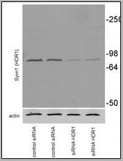
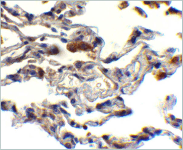
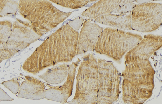
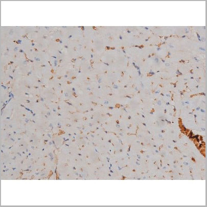
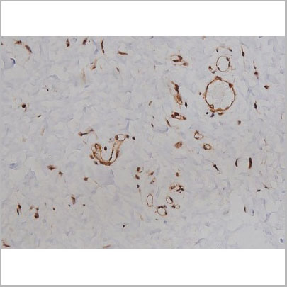
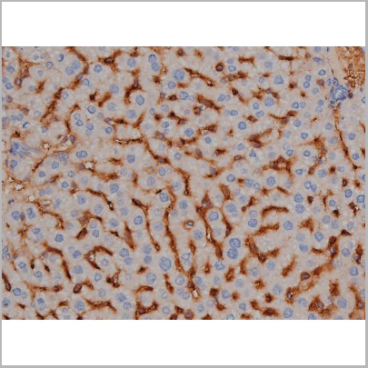
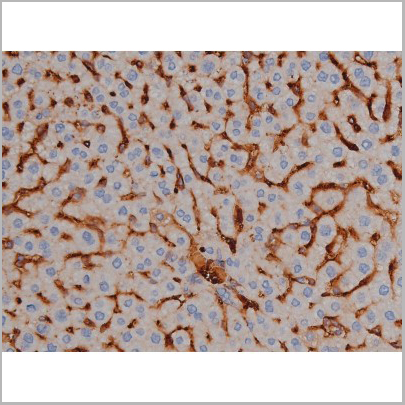
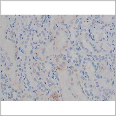
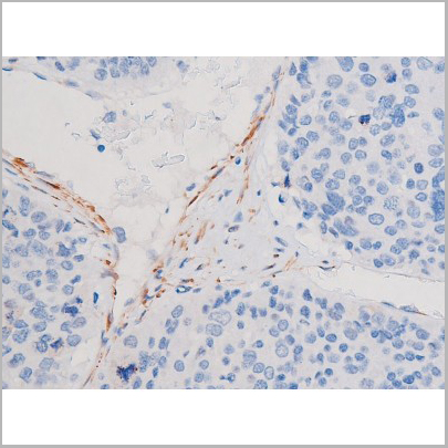
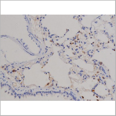
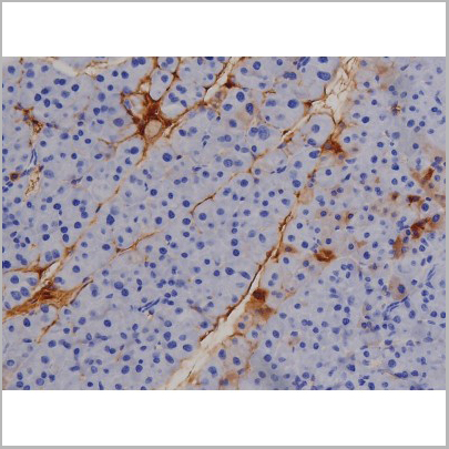
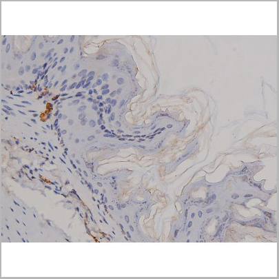
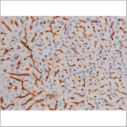
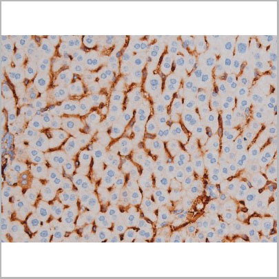
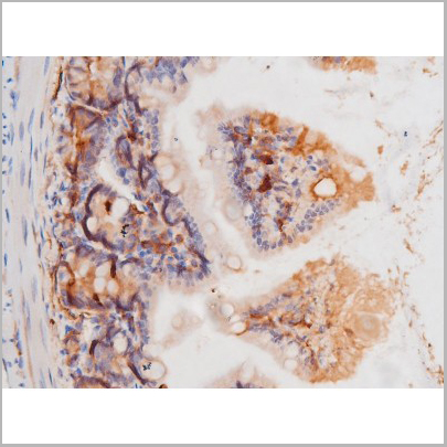
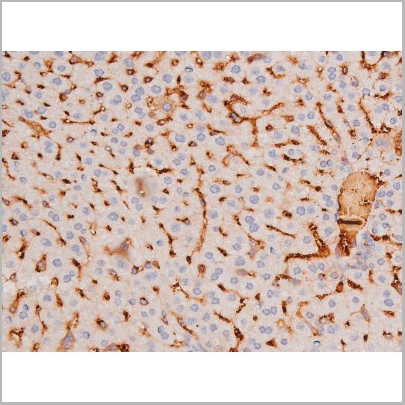
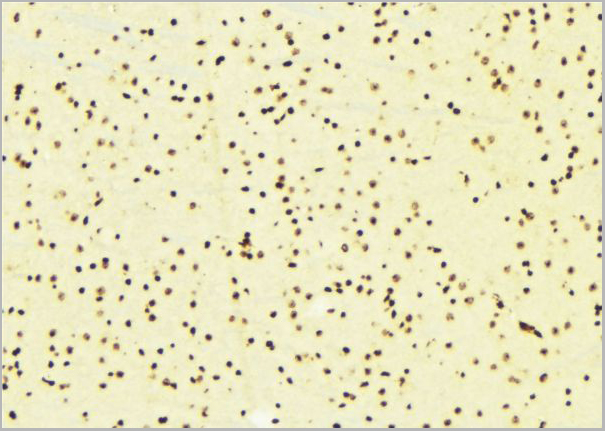
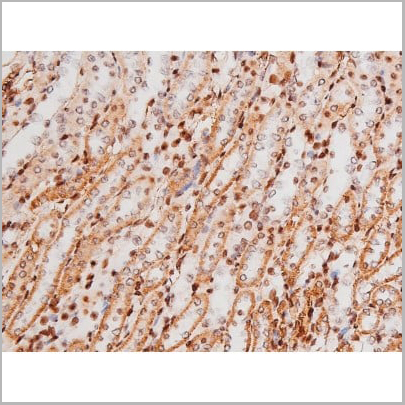
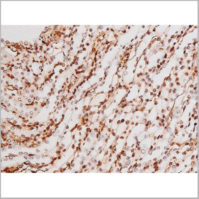
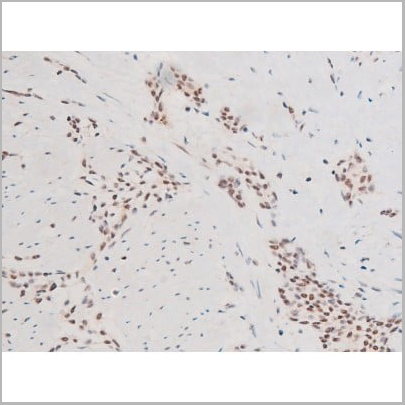
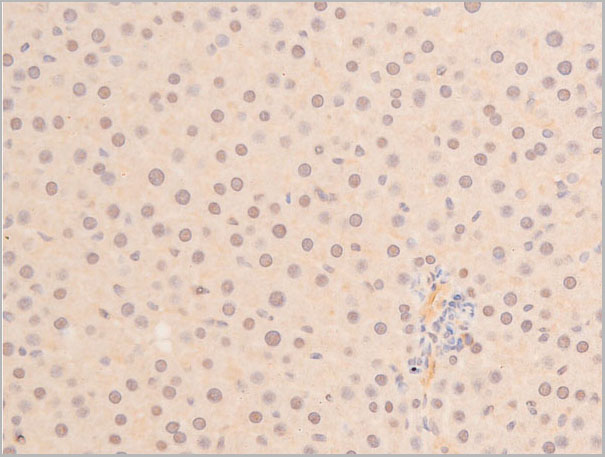
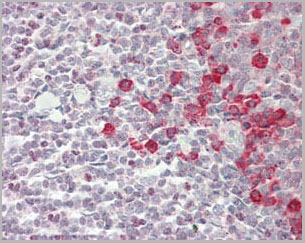
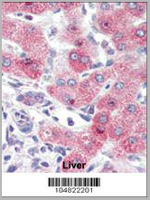 IHC (Immunohistchemistry)
(Formalin-fixed and paraffin-embedded human Liver tissue reacted with SYVN1 (HRD1) Antibody (C-term) , which was peroxidase-conjugated to the secondary antibody, followed by AEC staining. This data demonstrates the use of this antibody for immunohistochemistry; clinical relevance has not been evaluated.)
IHC (Immunohistchemistry)
(Formalin-fixed and paraffin-embedded human Liver tissue reacted with SYVN1 (HRD1) Antibody (C-term) , which was peroxidase-conjugated to the secondary antibody, followed by AEC staining. This data demonstrates the use of this antibody for immunohistochemistry; clinical relevance has not been evaluated.)
SYVN1 (HRD1), Polyclonal Antibody (Cat# AAA28764)
Full Name
SYVN1 (HRD1) Antibody (C-term)
Gene Names
SYVN1; DER3; HRD1
Reactivity
Human, mouse
Applications
Immunofluorescence, Immunohistochemistry, Western Blot
Purity
Purified Rabbit Polyclonal Antibody (Pab)
Pricing
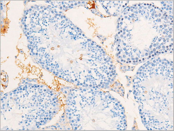
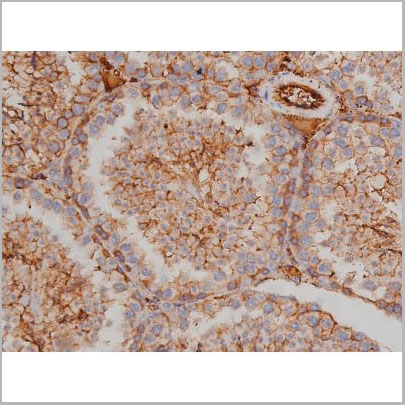
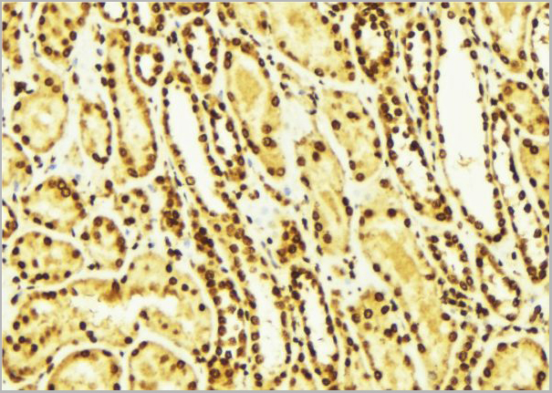
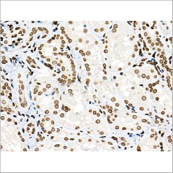
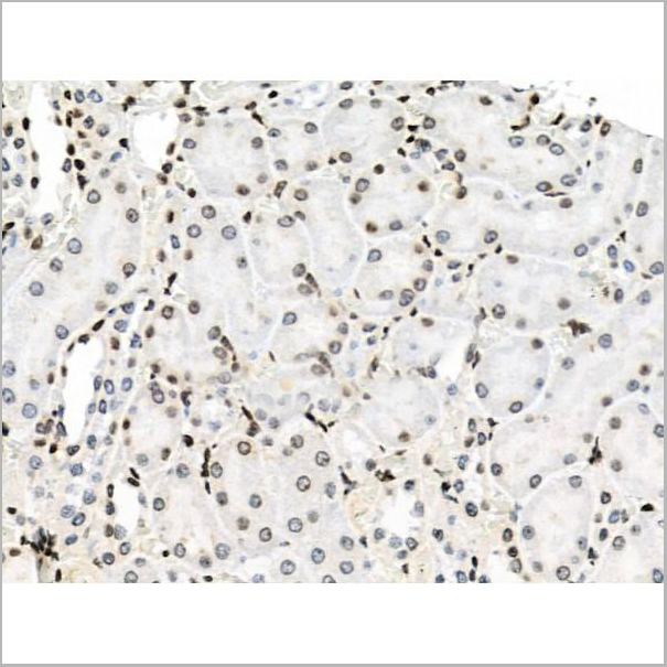
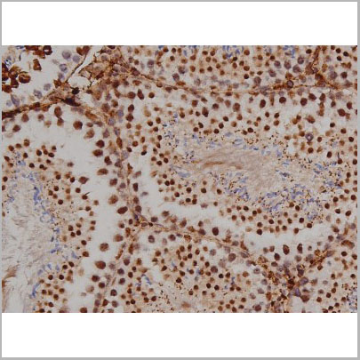
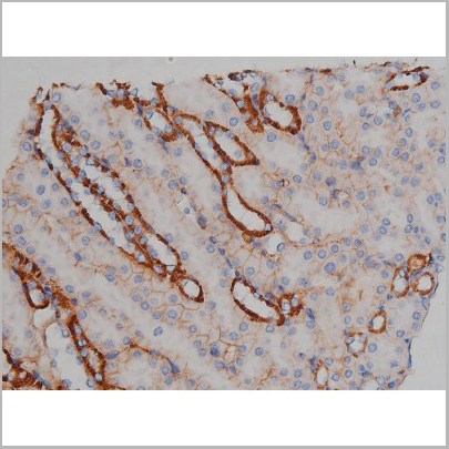
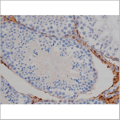
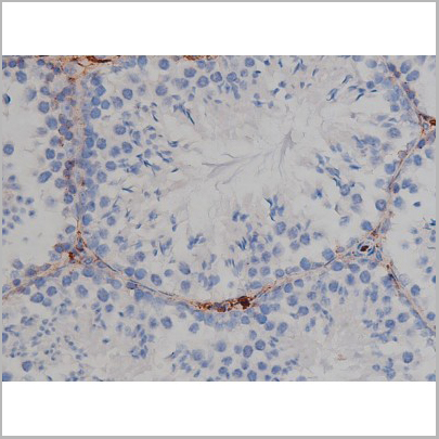
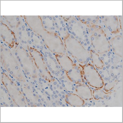
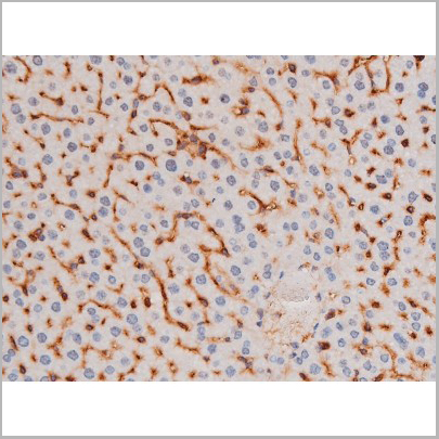
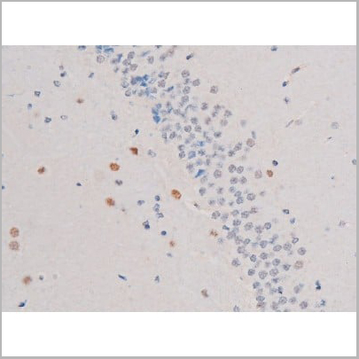
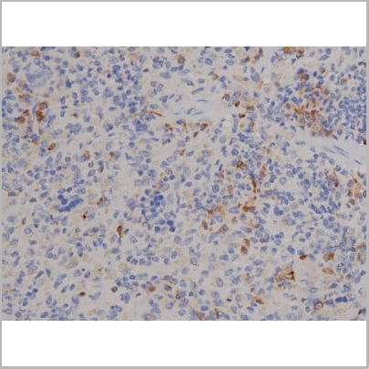
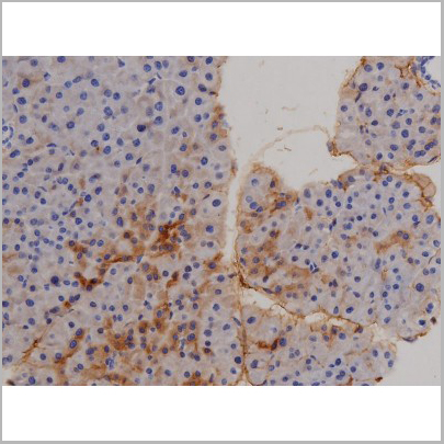
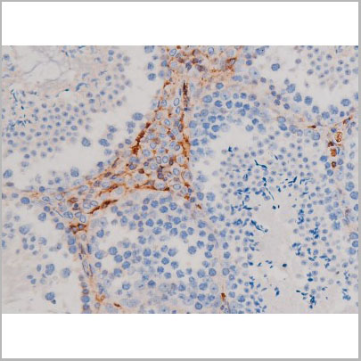
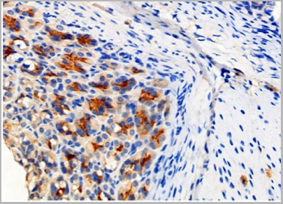
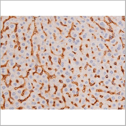
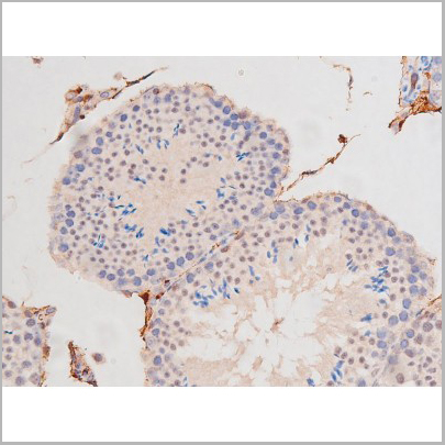
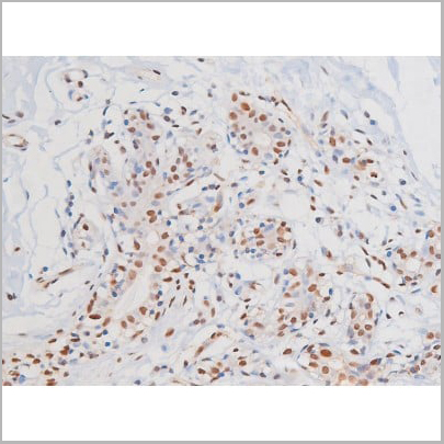
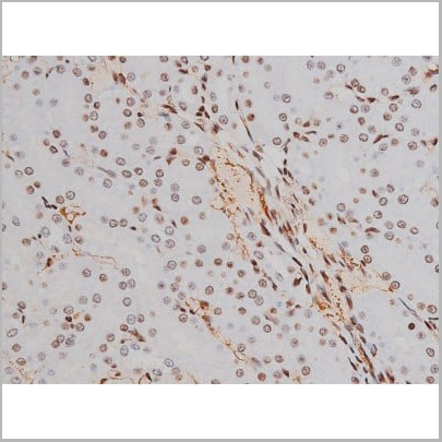
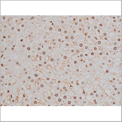
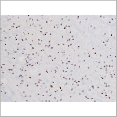
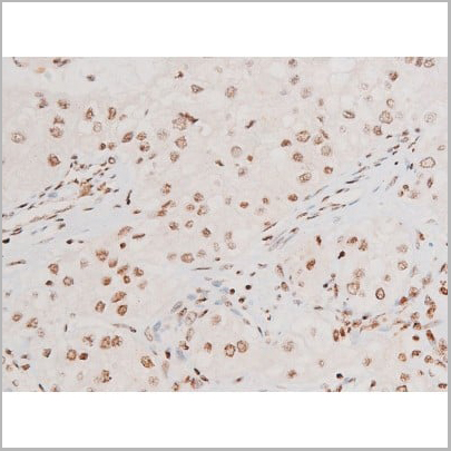
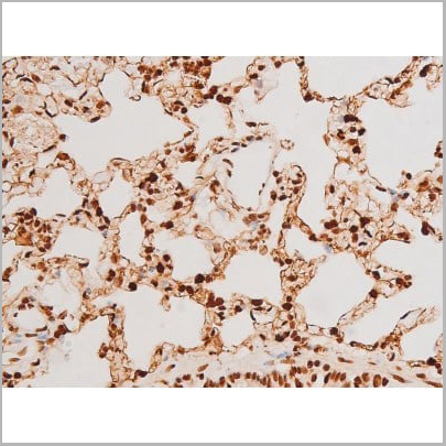
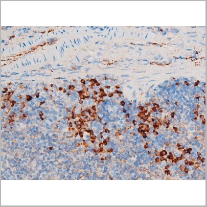
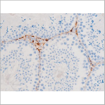
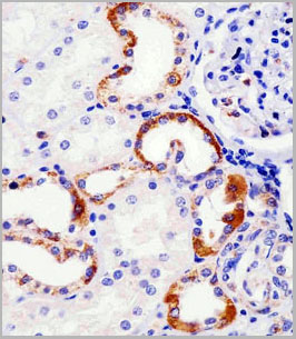
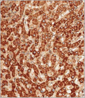
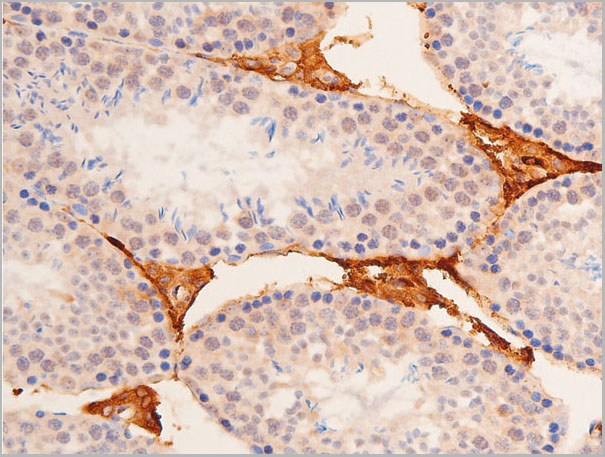
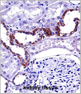 IHC (Immunohistochemistry)
(MCOLN1 Antibody (C-term) (AAA28690)immunohistochemistry analysis in formalin fixed and paraffin embedded human kidney tissue followed by peroxidase conjugation of the secondary antibody and DAB staining.This data demonstrates the use of MCOLN1 Antibody (C-term) for immunohistochemistry. Clinical relevance has not been evaluated.)
IHC (Immunohistochemistry)
(MCOLN1 Antibody (C-term) (AAA28690)immunohistochemistry analysis in formalin fixed and paraffin embedded human kidney tissue followed by peroxidase conjugation of the secondary antibody and DAB staining.This data demonstrates the use of MCOLN1 Antibody (C-term) for immunohistochemistry. Clinical relevance has not been evaluated.)
MCOLN1, Polyclonal Antibody (Cat# AAA28690)
Full Name
MCOLN1 Antibody (C-term)
Gene Names
MCOLN1; ML4; MG-2; MLIV; MST080; TRPML1; MSTP080; TRP-ML1; TRPM-L1
Reactivity
Human
Predicted Reactivity: Monkey, Mouse
Predicted Reactivity: Monkey, Mouse
Applications
Western Blot, Immunohistochemistry
Purity
Purified through a protein A column, followed by peptide affinity purification.
Pricing
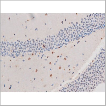
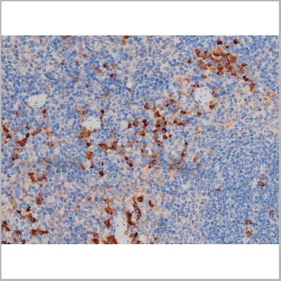
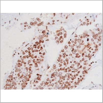
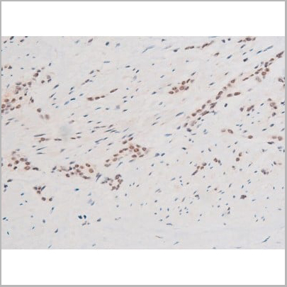
 IHC (Immunohistochemistry)
(AAA30986 at 1/200 staining Rat spleen tissue sections by IHC-P. The tissue was formaldehyde fixed and a heat mediated antigen retrieval step in citrate buffer was performed. The tissue was then blocked and incubated with the antibody for 1.5 hours at 22 degree C. An HRP conjugated goat anti-rabbit antibody was used as the secondary.)
IHC (Immunohistochemistry)
(AAA30986 at 1/200 staining Rat spleen tissue sections by IHC-P. The tissue was formaldehyde fixed and a heat mediated antigen retrieval step in citrate buffer was performed. The tissue was then blocked and incubated with the antibody for 1.5 hours at 22 degree C. An HRP conjugated goat anti-rabbit antibody was used as the secondary.)
Fyn, Polyclonal Antibody (Cat# AAA30986)
Full Name
Phospho-Fyn (Tyr530) Antibody
Gene Names
FYN; SLK; SYN; p59-FYN
Reactivity
Human, Mouse, Rat
Applications
Western Blot, Immunohistochemistry, Immunofluorescence, Immunocytochemistry
Purity
From purified rabbit serum by affinity purification via sequential chromatography on phospho-and non-phospho-peptide affinity columns.
Pricing
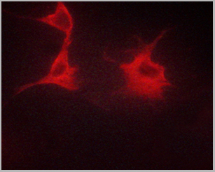
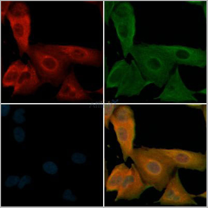
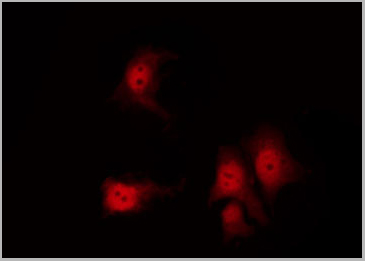
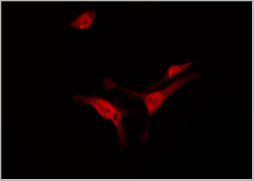
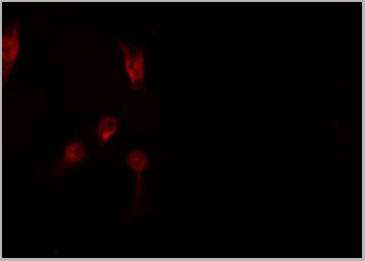
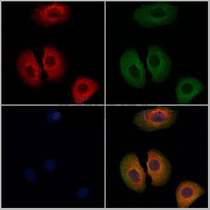
 IF (Immunofluorescence)
(AAA31071 staining HuvEc by IF/ICC. The sample were fixed with PFA and permeabilized in 0.1% Triton X-100, then blocked in 10% serum for 45 minutes at 25 degree C. The primary antibody was diluted at 1/200 and incubated with the sample for 1 hour at 37 degree C. An Alexa Fluor 594 conjugated goat anti-rabbit IgG (H+L) Ab, diluted at 1/600, was used as the secondary antibody.)
IF (Immunofluorescence)
(AAA31071 staining HuvEc by IF/ICC. The sample were fixed with PFA and permeabilized in 0.1% Triton X-100, then blocked in 10% serum for 45 minutes at 25 degree C. The primary antibody was diluted at 1/200 and incubated with the sample for 1 hour at 37 degree C. An Alexa Fluor 594 conjugated goat anti-rabbit IgG (H+L) Ab, diluted at 1/600, was used as the secondary antibody.)
HER4, Polyclonal Antibody (Cat# AAA31071)
Full Name
Phospho-HER4 (Tyr1284) Antibody
Gene Names
ERBB4; HER4; ALS19; p180erbB4
Reactivity
Human, Mouse, Rat
Applications
Western Blot, Immunohistochemistry, Immunofluorescence, Immunocytochemistry
Purity
From purified rabbit serum by affinity purification via sequential chromatography on phospho-and non-phospho-peptide affinity columns.
Pricing
 Application Data
(At 25 degree C. Samples were then incubated with primary Ab(At 37 degree C. An AlexaFluor594 conjugated goat anti-rabbit IgG(H+L) Ab(Red) and an AlexaFluor488 conjugated goat anti-mouse IgG(H+L) Ab(Green) were used as the secondary antibody.The nuclear counter stain is DAPI(blue).)
Application Data
(At 25 degree C. Samples were then incubated with primary Ab(At 37 degree C. An AlexaFluor594 conjugated goat anti-rabbit IgG(H+L) Ab(Red) and an AlexaFluor488 conjugated goat anti-mouse IgG(H+L) Ab(Green) were used as the secondary antibody.The nuclear counter stain is DAPI(blue).)
PRC1, Polyclonal Antibody (Cat# AAA31450)
Full Name
Phospho-PRC1 (Thr481) Antibody
Gene Names
PRC1; ASE1
Reactivity
Human, Mouse, Rat
Predicted Reactivity: Pig (80%), Horse (90%)
Predicted Reactivity: Pig (80%), Horse (90%)
Applications
Western Blot, Immunohistochemistry, Immunofluorescence, Immunocytochemistry, Peptide ELISA
Purity
The antibody is from purified rabbit serum by affinity purification via sequential chromatography on phospho-peptide and non-phospho-peptide affinity columns.
Pricing
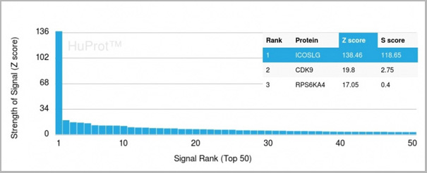 Application Data
(Analysis of Protein Array containing more than 19,000 full-length human proteins using ICOS-L Mouse Monoclonal Antibody (ICOSL/3111). Z- and S- Score: The Z-score represents the strength of a signal that a monoclonal antibody (MAb) (in combination with a fluorescently-tagged anti-IgG secondary antibody) produces when binding to a particular protein on the HuProtTM array. Z-scores are described in units of standard deviations (SD’s) above the mean value of all signals generated on that array. If targets on HuProtTM are arranged in descending order of the Z-score, the S-score is the difference (also in units of SD’s) between the Z-score. S-score therefore represents the relative target specificity of a MAb to its intended target. A MAb is considered to specific to its intended target, if the MAb has an S-score of at least 2.5. For example, if a MAb binds to protein X with a Z-score of 43 and to protein Y with a Z-score of 14, then the S-score for the binding of that MAb to protein X is equal to 29.)
Application Data
(Analysis of Protein Array containing more than 19,000 full-length human proteins using ICOS-L Mouse Monoclonal Antibody (ICOSL/3111). Z- and S- Score: The Z-score represents the strength of a signal that a monoclonal antibody (MAb) (in combination with a fluorescently-tagged anti-IgG secondary antibody) produces when binding to a particular protein on the HuProtTM array. Z-scores are described in units of standard deviations (SD’s) above the mean value of all signals generated on that array. If targets on HuProtTM are arranged in descending order of the Z-score, the S-score is the difference (also in units of SD’s) between the Z-score. S-score therefore represents the relative target specificity of a MAb to its intended target. A MAb is considered to specific to its intended target, if the MAb has an S-score of at least 2.5. For example, if a MAb binds to protein X with a Z-score of 43 and to protein Y with a Z-score of 14, then the S-score for the binding of that MAb to protein X is equal to 29.)
ICOS-L/ICOS Ligand/B7RP-1 (Immuno-Oncology Target), Monoclonal Antibody (Cat# AAA23916)
Full Name
ICOS-L/ICOS Ligand/B7RP-1 (Immuno-Oncology Target)
Gene Names
ICOSLG; B7h; B7H2; GL50; B7-H2; B7RP1; CD275; ICOSL; LICOS; B7RP-1; ICOS-L
Reactivity
Human
Applications
Flow Cytometry, Immunofluorescence, Immunohistochemistry
Purity
Purified Ab with BSA and Azide at 200ug/ml
Pricing
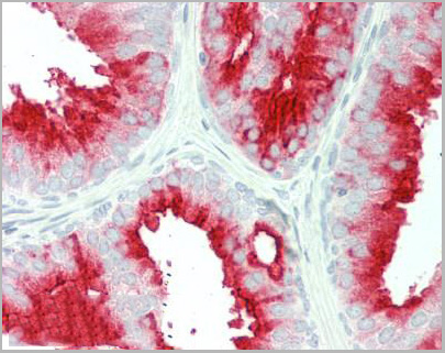
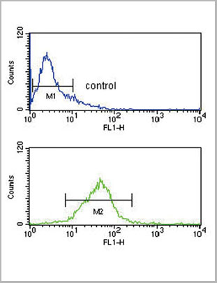
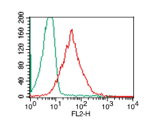
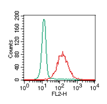
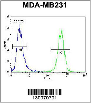
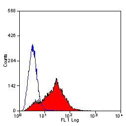
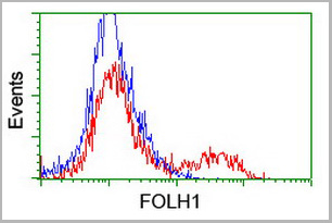 FCM (Flow Cytometry)
(HEK293T cells transfected with either overexpress plasmid (Red) or empty vector control plasmid (Blue) were immunostained by anti-FOLH1 antibody, and then analyzed by flow cytometry.)
FCM (Flow Cytometry)
(HEK293T cells transfected with either overexpress plasmid (Red) or empty vector control plasmid (Blue) were immunostained by anti-FOLH1 antibody, and then analyzed by flow cytometry.)
FOLH1 / PSMA, Monoclonal Antibody (Cat# AAA12376)
Full Name
Anti-FOLH1 / PSMA Antibody (clone 3H5) IHC-plus
Gene Names
FOLH1; PSM; FGCP; FOLH; GCP2; PSMA; mGCP; GCPII; NAALAD1; NAALAdase
Reactivity
Mouse, Human
Applications
Immunohistochemistry, Immunofluorescence, Western Blot, Flow Cytometry
Purity
Protein A/G purified
Pricing
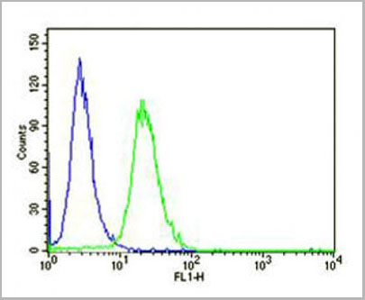
 FCM (Flow Cytometry)
(WB - MCSF Receptor (CSF1R) Antibody (C-term) AAA28757 detail IHC-P - MCSF Receptor (CSF1R) Antibody (C-term) AAA28757 detail IHC-P - MCSF Receptor (CSF1R) Antibody (C-term) AAA28757 detail FC - MCSF Receptor (CSF1R) Antibody (C-term) AAA28757 detail Overlay histogram showing HepG2 cells stained with AAA28757(green line). The cells were fixed with 2% paraformaldehyde (10 min) and then permeabilized with 90% methanol for 10 min. The cells were then icubated in 2% bovine serum albumin to block non-specific protein-protein interactions followed by the antibody (AAA28757, 1:25 dilution) for 60 min at 37ºC. The secondary antibody used was Goat-Anti-Rabbit IgG, DyLight® 488 Conjugated Highly Cross-Adsorbed(1583138) at 1/200 dilution for 40 min at 37ºC. Isotype control antibody (blue line) was rabbit IgG1 (1ug/1x10^6 cells) used under the same conditions. Acquisition of >10, 000 events was performed.)
FCM (Flow Cytometry)
(WB - MCSF Receptor (CSF1R) Antibody (C-term) AAA28757 detail IHC-P - MCSF Receptor (CSF1R) Antibody (C-term) AAA28757 detail IHC-P - MCSF Receptor (CSF1R) Antibody (C-term) AAA28757 detail FC - MCSF Receptor (CSF1R) Antibody (C-term) AAA28757 detail Overlay histogram showing HepG2 cells stained with AAA28757(green line). The cells were fixed with 2% paraformaldehyde (10 min) and then permeabilized with 90% methanol for 10 min. The cells were then icubated in 2% bovine serum albumin to block non-specific protein-protein interactions followed by the antibody (AAA28757, 1:25 dilution) for 60 min at 37ºC. The secondary antibody used was Goat-Anti-Rabbit IgG, DyLight® 488 Conjugated Highly Cross-Adsorbed(1583138) at 1/200 dilution for 40 min at 37ºC. Isotype control antibody (blue line) was rabbit IgG1 (1ug/1x10^6 cells) used under the same conditions. Acquisition of >10, 000 events was performed.)
MCSF Receptor (CSF1R), Polyclonal Antibody (Cat# AAA28757)
Full Name
MCSF Receptor (CSF1R) Antibody (C-term)
Gene Names
CSF1R; FMS; CSFR; FIM2; HDLS; C-FMS; CD115; CSF-1R; M-CSF-R
Reactivity
Human
Applications
Western Blot, Flow Cytometry, Immunohistochemistry
Purity
Purified Rabbit Polyclonal Antibody (Pab)
Pricing
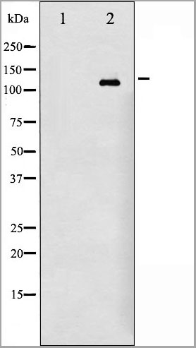
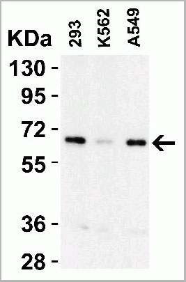
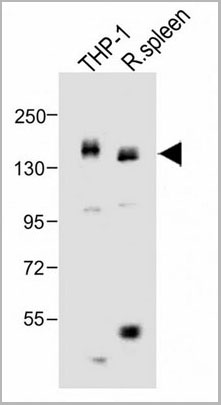
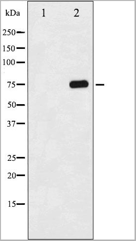
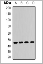
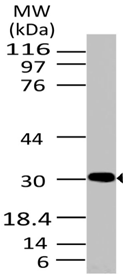
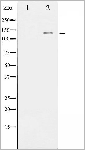
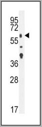
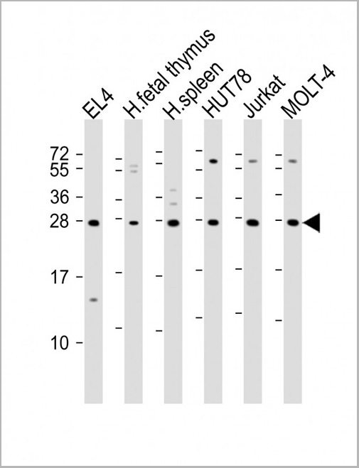 WB (Western Blot)
(All lanes : Anti-TIGIT Antibody at 1:2000 dilutionLane 1: EL4 whole cell lysateLane 2: human fetal thymus lysateLane 3: human spleen lysateLane 4: HUT78 whole cell lysateLane 5: Jurkat whole cell lysateLane 6: MOLT-4 whole cell lysateLysates/proteins at 20 ug per lane. SecondaryGoat Anti-Rabbit IgG, (H+L), Peroxidase conjugated at 1/10000 dilution. Predicted band size : 26 kDaBlocking/Dilution buffer: 5% NFDM/TBST.)
WB (Western Blot)
(All lanes : Anti-TIGIT Antibody at 1:2000 dilutionLane 1: EL4 whole cell lysateLane 2: human fetal thymus lysateLane 3: human spleen lysateLane 4: HUT78 whole cell lysateLane 5: Jurkat whole cell lysateLane 6: MOLT-4 whole cell lysateLysates/proteins at 20 ug per lane. SecondaryGoat Anti-Rabbit IgG, (H+L), Peroxidase conjugated at 1/10000 dilution. Predicted band size : 26 kDaBlocking/Dilution buffer: 5% NFDM/TBST.)
TIGIT, Polyclonal Antibody (Cat# AAA28803)
Full Name
TIGIT Antibody
Gene Names
TIGIT; VSIG9; VSTM3; WUCAM
Reactivity
Human, Mouse
Applications
Western Blot
Purity
This antibody is purified through a protein A column, followed by peptide affinity purification.
Pricing
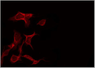
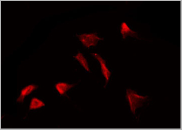
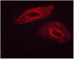
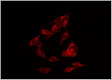
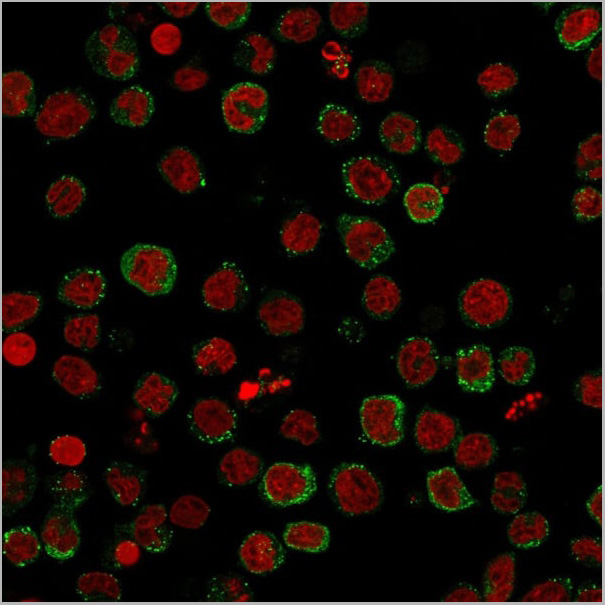
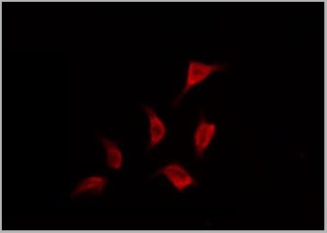
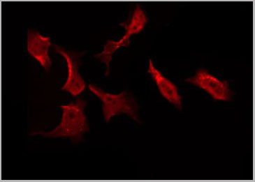
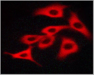
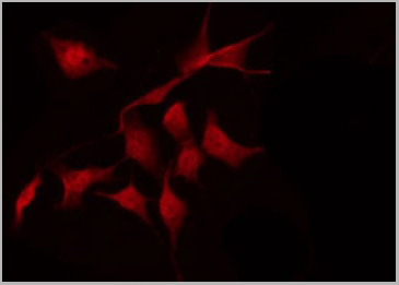
 IF (Immunofluorescence)
(AAA31047 staining RAW264.7 by IF/ICC. The sample were fixed with PFA and permeabilized in 0.1% Triton X-100, then blocked in 10% serum for 45 minutes at 25 degree C. The primary antibody was diluted at 1/200 and incubated with the sample for 1 hour at 37 degree C. An Alexa Fluor 594 conjugated goat anti-rabbit IgG (H+L) Ab, diluted at 1/600, was used as the secondary antibody.)
IF (Immunofluorescence)
(AAA31047 staining RAW264.7 by IF/ICC. The sample were fixed with PFA and permeabilized in 0.1% Triton X-100, then blocked in 10% serum for 45 minutes at 25 degree C. The primary antibody was diluted at 1/200 and incubated with the sample for 1 hour at 37 degree C. An Alexa Fluor 594 conjugated goat anti-rabbit IgG (H+L) Ab, diluted at 1/600, was used as the secondary antibody.)
STAT5A/B, Polyclonal Antibody (Cat# AAA31047)
Full Name
Phospho-STAT5A/B (Ser725/730) Antibody
Gene Names
STAT5A; MGF; STAT5
Reactivity
Human, Mouse, Rat
Applications
Western Blot, Immunohistochemistry, Immunofluorescence, Immunocytochemistry
Purity
From purified rabbit serum by affinity purification via sequential chromatography on phospho-and non-phospho-peptide affinity columns.
Pricing
 WB (Western Blot)
(Western blot analysis of Phospho-Tau (Ser396) expression in various lysates)
WB (Western Blot)
(Western blot analysis of Phospho-Tau (Ser396) expression in various lysates)
Tau, Polyclonal Antibody (Cat# AAA31003)
Full Name
Phospho-Tau (Ser396) Antibody
Gene Names
MAPT; TAU; MSTD; PPND; DDPAC; MAPTL; MTBT1; MTBT2; FTDP-17; PPP1R103
Reactivity
Human, Mouse, Rat
Applications
Western Blot, Immunohistochemistry
Purity
From purified rabbit serum by affinity purification via sequential chromatography on phospho-and non-phospho-peptide affinity columns.
Pricing
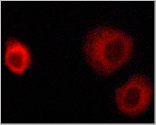
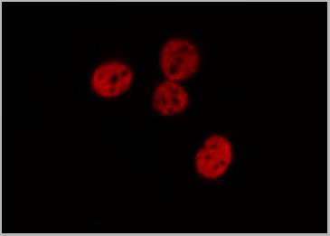
 IF (Immunofluorescence)
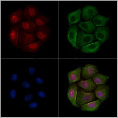
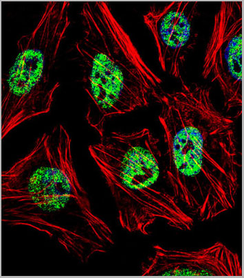
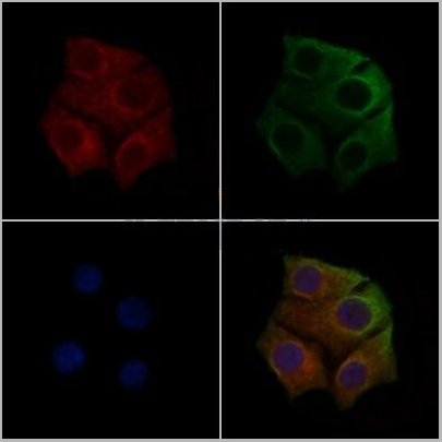
(Fluorescent confocal image of Hela cell stained with NANOG Antibody (N-term). Hela cells were fixed with 4% PFA (20 min), permeabilized with Triton X-100 (0.1%, 10 min), then incubated with NANOG primary antibody (1:25, 1 h at 37 degree). For secondary antibody, Alexa Fluor 488 conjugated donkey anti-rabbit antibody (green) was used (1:400, 50 min at 37 degree).Cytoplasmic actin was counterstained with Alexa Fluor 555 (red) conjugated Phalloidin (7units/ml, 1 h at 37 degree). Nuclei were counterstained with DAPI (blue) (10 ug/ml, 10 min). NANOG immunoreactivity is localized to Nucleus significantly.)
IF (Immunofluorescence)
(Fluorescent confocal image of Hela cell stained with NANOG Antibody (N-term). Hela cells were fixed with 4% PFA (20 min), permeabilized with Triton X-100 (0.1%, 10 min), then incubated with NANOG primary antibody (1:25, 1 h at 37 degree). For secondary antibody, Alexa Fluor 488 conjugated donkey anti-rabbit antibody (green) was used (1:400, 50 min at 37 degree).Cytoplasmic actin was counterstained with Alexa Fluor 555 (red) conjugated Phalloidin (7units/ml, 1 h at 37 degree). Nuclei were counterstained with DAPI (blue) (10 ug/ml, 10 min). NANOG immunoreactivity is localized to Nucleus significantly.)
NANOG, Polyclonal Antibody (Cat# AAA28706)
Full Name
NANOG Antibody (N-term)
Reactivity
Human (Predicted Reactivity: Monkey)
Applications
Western Blot, Immunofluorescence, Immunohistochemistry, Flow Cytometry
Purity
Purified Rabbit Polyclonal Antibody (Pab)
Pricing
 IF (Immunofluorescence)
(AAA30998 staining HepG2 by IF/ICC. The sample were fixed with PFA and permeabilized in 0.1% Triton X-100, then blocked in 10% serum for 45 minutes at 25 degree C. The primary antibody was diluted at 1/200 and incubated with the sample for 1 hour at 37 degree C. An Alexa Fluor 594 conjugated goat anti-rabbit IgG (H+L) Ab, diluted at 1/600, was used as the secondary antibody.)
IF (Immunofluorescence)
(AAA30998 staining HepG2 by IF/ICC. The sample were fixed with PFA and permeabilized in 0.1% Triton X-100, then blocked in 10% serum for 45 minutes at 25 degree C. The primary antibody was diluted at 1/200 and incubated with the sample for 1 hour at 37 degree C. An Alexa Fluor 594 conjugated goat anti-rabbit IgG (H+L) Ab, diluted at 1/600, was used as the secondary antibody.)
Tau, Polyclonal Antibody (Cat# AAA30998)
Full Name
Phospho-Tau (Ser356) Antibody
Gene Names
MAPT; TAU; MSTD; PPND; DDPAC; MAPTL; MTBT1; MTBT2; FTDP-17; PPP1R103
Reactivity
Human, Mouse, Rat
Applications
Western Blot, Immunohistochemistry, Immunofluorescence, Immunocytochemistry
Purity
From purified rabbit serum by affinity purification via sequential chromatography on phospho-and non-phospho-peptide affinity columns.
Pricing
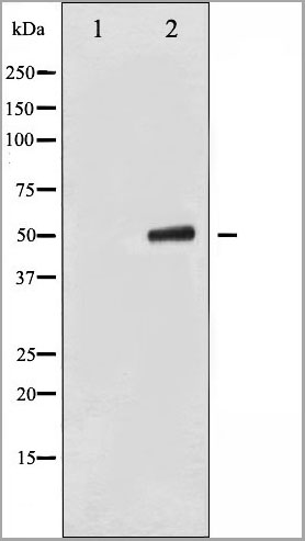
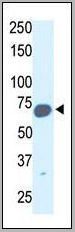
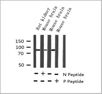
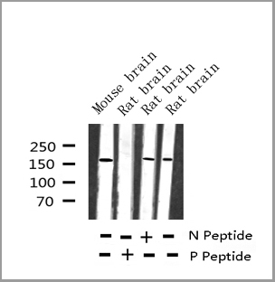
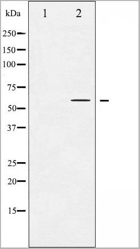
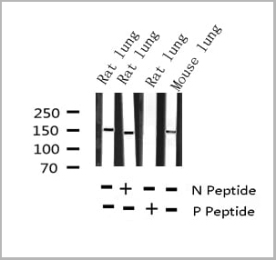
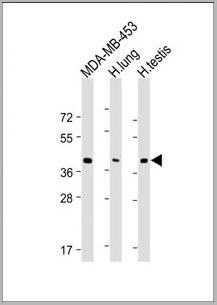
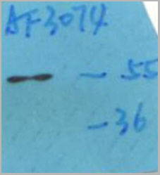
 WB (Western Blot)
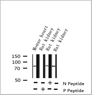
(Host: RabbitTarget Name: GBA2Sample Tissue: Mouse Thymus lysatesAntibody Dilution: 1ug/ml)
WB (Western Blot)
(Host: RabbitTarget Name: GBA2Sample Tissue: Mouse Thymus lysatesAntibody Dilution: 1ug/ml)
GBA2, Polyclonal Antibody (Cat# AAA23602)
Full Name
GBA2 Antibody - middle region
Gene Names
Gba2; F630034E04
Reactivity
Tested Species/Predicted species Reactivity: Mouse
Applications
Western Blot
Purity
Affinity purified
Pricing
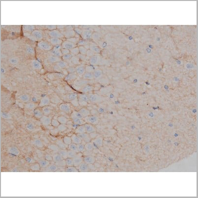
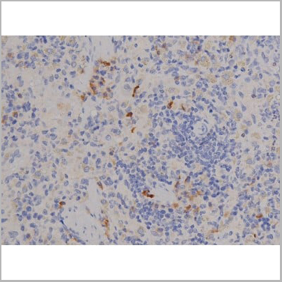
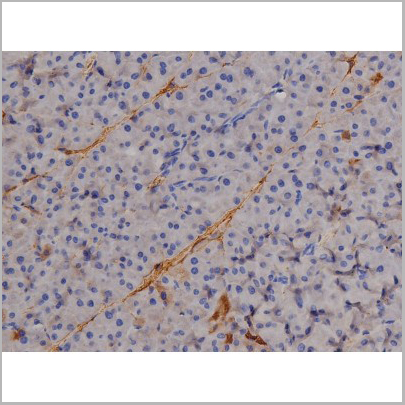
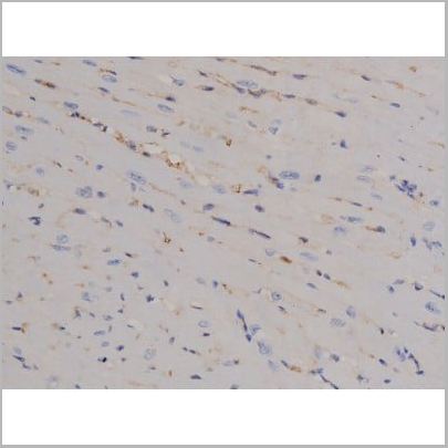
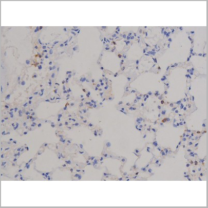
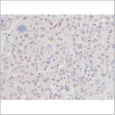
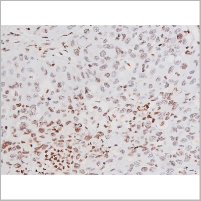
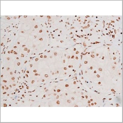
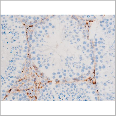
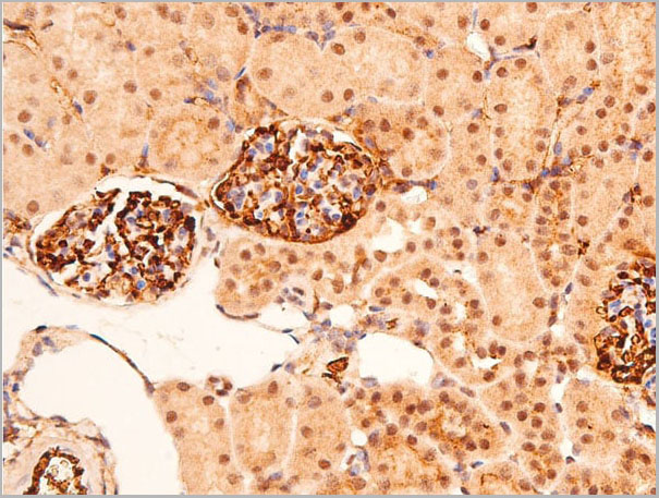
 IHC (Immunohistochemistry)
(FOXD1 Antibody (N-term) (AAA28675) immunohistochemistry analysis in formalin fixed and paraffin embedded human kidney tissue followed by peroxidase conjugation of the secondary antibody and DAB staining.This data demonstrates the use of FOXD1 Antibody (N-term) for immunohistochemistry. Clinical relevance has not been evaluated.)
IHC (Immunohistochemistry)
(FOXD1 Antibody (N-term) (AAA28675) immunohistochemistry analysis in formalin fixed and paraffin embedded human kidney tissue followed by peroxidase conjugation of the secondary antibody and DAB staining.This data demonstrates the use of FOXD1 Antibody (N-term) for immunohistochemistry. Clinical relevance has not been evaluated.)
FOXD1, Polyclonal Antibody (Cat# AAA28675)
Full Name
FOXD1 Antibody (N-term)
Gene Names
FOXD1; FKHL8; FREAC4; FREAC-4
Reactivity
Human, Mouse; Predicted: Mouse
Applications
Western Blot, Immunohistochemistry, Immunofluorescence
Purity
This antibody is purified through a protein A column, followed by peptide affinity purification.
Pricing
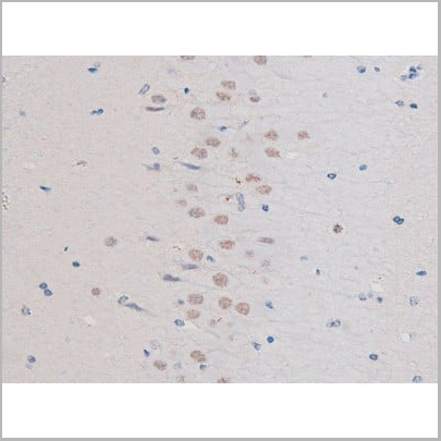
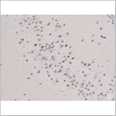
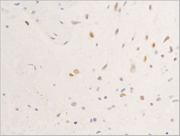
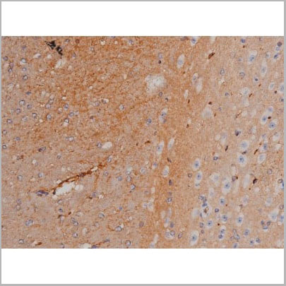
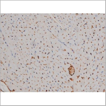
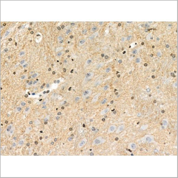
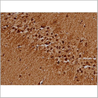
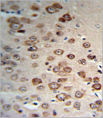
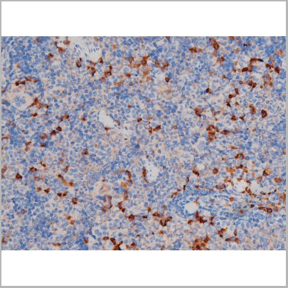
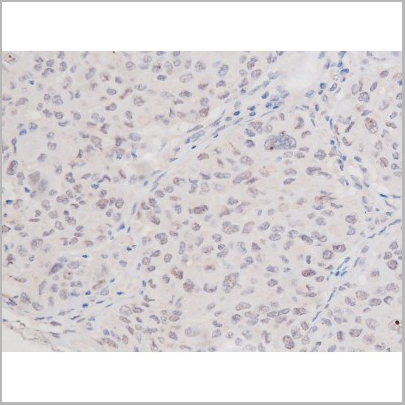
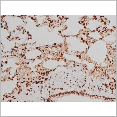
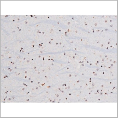
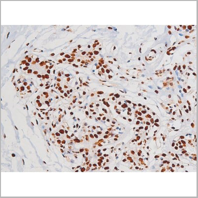
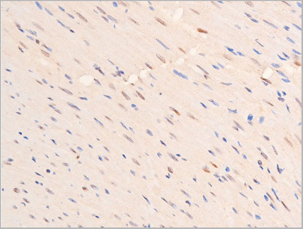
 IHC (Immunohistochemistry)
(AAA31037 at 1/200 staining Rat brain tissue sections by IHC-P. The tissue was formaldehyde fixed and a heat mediated antigen retrieval step in citrate buffer was performed. The tissue was then blocked and incubated with the antibody for 1.5 hours at 22 degree C. An HRP conjugated goat anti-rabbit antibody was used as the secondary.)
IHC (Immunohistochemistry)
(AAA31037 at 1/200 staining Rat brain tissue sections by IHC-P. The tissue was formaldehyde fixed and a heat mediated antigen retrieval step in citrate buffer was performed. The tissue was then blocked and incubated with the antibody for 1.5 hours at 22 degree C. An HRP conjugated goat anti-rabbit antibody was used as the secondary.)
IRS-1, Polyclonal Antibody (Cat# AAA31037)
Full Name
Phospho-IRS-1 (Ser636) Antibody
Gene Names
IRS1; HIRS-1
Reactivity
Human, Mouse, Rat, Monkey
Applications
Western Blot, Immunohistochemistry, Immunofluorescence, Immunocytochemistry
Purity
From purified rabbit serum by affinity purification via sequential chromatography on phospho-and non-phospho-peptide affinity columns.
Pricing
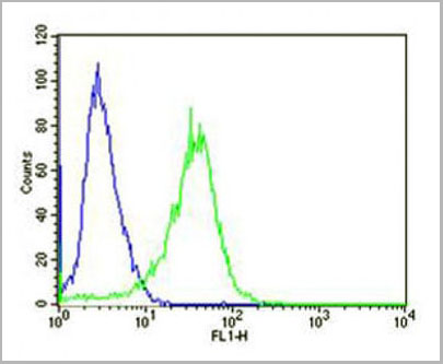
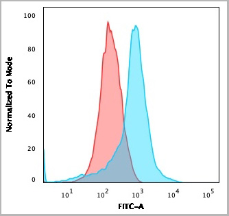
 FCM (Flow Cytometry)
(PCSK9 Antibody (N-term) (Cat. #AAA28774) flow cytometry analysis of Jurkat cells (bottom histogram) compared to a negative control cell(top histogram).FITC-conjugated goat-anti-rabbit secondary antibodies were used for the analysis.)
FCM (Flow Cytometry)
(PCSK9 Antibody (N-term) (Cat. #AAA28774) flow cytometry analysis of Jurkat cells (bottom histogram) compared to a negative control cell(top histogram).FITC-conjugated goat-anti-rabbit secondary antibodies were used for the analysis.)
PCSK9, Polyclonal Antibody (Cat# AAA28774)
Full Name
PCSK9 Antibody (N-term)
Gene Names
PCSK9; FH3; PC9; NARC1; LDLCQ1; NARC-1; HCHOLA3
Reactivity
Human
Applications
Western Blot, Immunohistochemistry, Flow Cytometry
Pricing
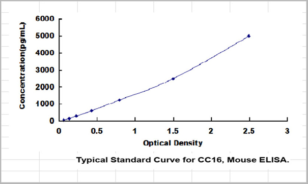
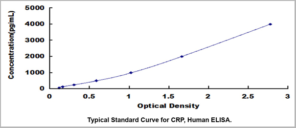 Standard Curve (Sample)
C Reactive Protein (CRP), ELISA Kit (Cat# AAA23109)
Full Name
Human C Reactive Protein (CRP) High Sensitive ELISA Kit
Gene Names
CRP; PTX1
Reactivity
Human
Applications
ELISA
Pricing
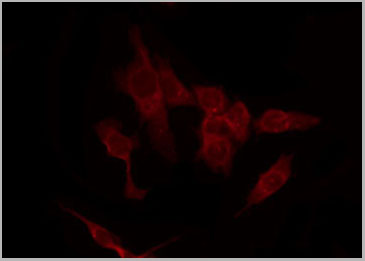
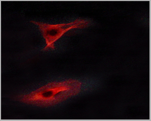
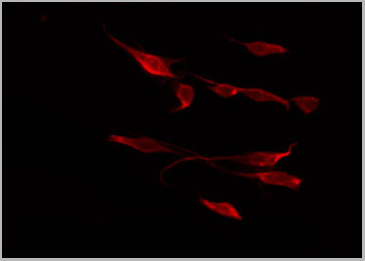
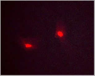
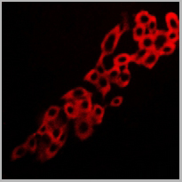 IF (Immunofluorescence)
(Immunofluorescent analysis of Adenosine A2a Receptor staining in LOVO cells. Formalin-fixed cells were permeabilized with 0.1% Triton X-100 in TBS for 5-10 minutes and blocked with 3% BSA-PBS for 30 minutes at room temperature. Cells were probed with the primary antibody in 3% BSA-PBS and incubated overnight at 4 degree C in a humidified chamber. Cells were washed with PBST and incubated with a DyLight 594-conjugated secondary antibody (red) in PBS at room temperature in the dark.)
IF (Immunofluorescence)
(Immunofluorescent analysis of Adenosine A2a Receptor staining in LOVO cells. Formalin-fixed cells were permeabilized with 0.1% Triton X-100 in TBS for 5-10 minutes and blocked with 3% BSA-PBS for 30 minutes at room temperature. Cells were probed with the primary antibody in 3% BSA-PBS and incubated overnight at 4 degree C in a humidified chamber. Cells were washed with PBST and incubated with a DyLight 594-conjugated secondary antibody (red) in PBS at room temperature in the dark.)
Adenosine A2a Receptor, Polyclonal Antibody (Cat# AAA27805)
Full Name
Anti-Adenosine A2a Receptor Antibody
Gene Names
ADORA2A; A2aR; RDC8; ADORA2
Reactivity
Human, Mouse, Rat, Dog
Applications
Western Blot, Immunohistochemistry, Immunofluorescence, Immunocytochemistry
Purity
The antibody was purified by immunogen affinity chromatography.
Pricing
 WB (Western Blot)
(Western blot analysis of Phospho-STAT6 (Tyr641) expression in various lysates)
WB (Western Blot)
(Western blot analysis of Phospho-STAT6 (Tyr641) expression in various lysates)
STAT6, Polyclonal Antibody (Cat# AAA31045)
Full Name
Phospho-STAT6 (Tyr641) Antibody
Gene Names
STAT6; STAT6B; STAT6C; D12S1644; IL-4-STAT
Reactivity
Human, Mouse, Rat
Applications
Western Blot, Immunohistochemistry, Immunofluorescence, Immunocytochemistry
Purity
From purified rabbit serum by affinity purification via sequential chromatography on phospho-and non-phospho-peptide affinity columns.
Pricing
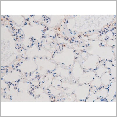
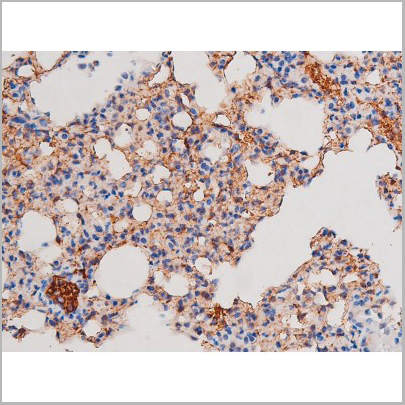
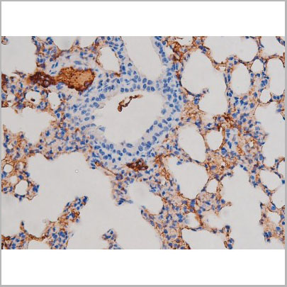
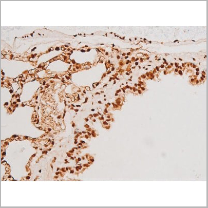
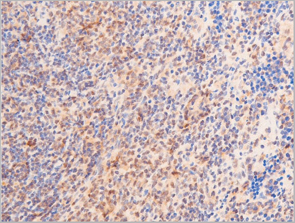
 IHC (Immunohistchemistry)
(AAA31044 at 1/200 staining Rat lung tissue sections by IHC-P. The tissue was formaldehyde fixed and a heat mediated antigen retrieval step in citrate buffer was performed. The tissue was then blocked and incubated with the antibody for 1.5 hours at 22 degree C. An HRP conjugated goat anti-rabbit antibody was used as the secondary.)
IHC (Immunohistchemistry)
(AAA31044 at 1/200 staining Rat lung tissue sections by IHC-P. The tissue was formaldehyde fixed and a heat mediated antigen retrieval step in citrate buffer was performed. The tissue was then blocked and incubated with the antibody for 1.5 hours at 22 degree C. An HRP conjugated goat anti-rabbit antibody was used as the secondary.)
STAT1, Polyclonal Antibody (Cat# AAA31044)
Full Name
Phospho-STAT1 (Tyr701) Antibody
Gene Names
STAT1; CANDF7; IMD31A; IMD31B; IMD31C; ISGF-3; STAT91
Reactivity
Human, Mouse, Rat
Applications
Western Blot, Immunohistochemistry, Immunofluorescence, Immunoprecipitation
Purity
From purified rabbit serum by affinity purification via sequential chromatography on phospho-and non-phospho-peptide affinity columns.
Pricing
thyroxine, T4, ELISA Kit (Cat# AAA29737)
Full Name
Rat thyroxine, T4 ELISA Kit
Reactivity
Rat
Pricing
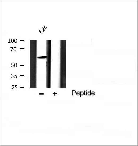 WB (Western Blot)
(Western blot analysis of Akt phosphorylation expression in B2C Cell line lysates, The lane on the right is treated with the antigen-specific peptide.)
WB (Western Blot)
(Western blot analysis of Akt phosphorylation expression in B2C Cell line lysates, The lane on the right is treated with the antigen-specific peptide.)
Akt, Polyclonal Antibody (Cat# AAA31035)
Full Name
Phospho-Akt (Ser124) Antibody
Gene Names
AKT1; AKT; PKB; RAC; CWS6; PRKBA; PKB-ALPHA; RAC-ALPHA
Reactivity
Human, Mouse, Rat
Applications
Western Blot, Immunohistochemistry, Immunofluorescence, Immunocytochemistry
Purity
From purified rabbit serum by affinity purification via sequential chromatography on phospho-and non-phospho-peptide affinity columns.
Pricing
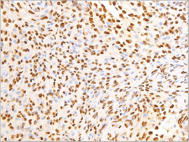
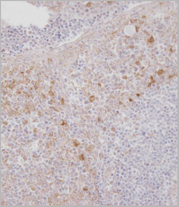
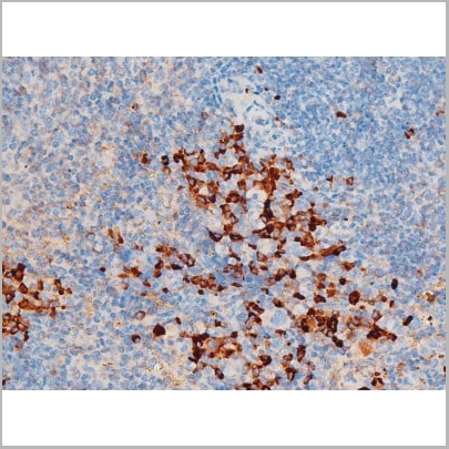
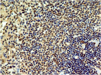 IHC (Immunohistochemistry)
(Immunohistochemical analysis of HMGB1 in normal human spleen tissue using HMGB1 antibody (Clone: ABM24D3) at 1 ug/ml.)
IHC (Immunohistochemistry)
(Immunohistochemical analysis of HMGB1 in normal human spleen tissue using HMGB1 antibody (Clone: ABM24D3) at 1 ug/ml.)
HMGB1, Monoclonal Antibody (Cat# AAA14866)
Full Name
HMGB1 Monoclonal Antibody
Gene Names
HMGB1; HMG1; HMG3; SBP-1
Reactivity
Human (Predicted Reactivity: Mouse
Applications
Western Blot, Immunohistochemistry, Flow Cytometry
Pricing
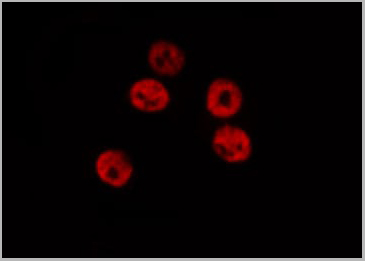
 IF (Immunofluorescence)
(AAA31141 staining Hela cells by IF/ICC. The samples were fixed with PFA and permeabilized in 0.1% Triton X-100,then blocked in 10% serum for 45 minutes at 25°C. Samples were then incubated with primary Ab(AAA31141 1:200) and mouse anti-beta tubulin Ab( 1:200) for 1 hour at 37°C. An AlexaFluor594 conjugated goat anti-rabbit IgG(H+L) Ab(Red) and an AlexaFluor488 conjugated goat anti-mouse IgG(H+L) Ab(Green) were used as the secondary antibody.)
IF (Immunofluorescence)
(AAA31141 staining Hela cells by IF/ICC. The samples were fixed with PFA and permeabilized in 0.1% Triton X-100,then blocked in 10% serum for 45 minutes at 25°C. Samples were then incubated with primary Ab(AAA31141 1:200) and mouse anti-beta tubulin Ab( 1:200) for 1 hour at 37°C. An AlexaFluor594 conjugated goat anti-rabbit IgG(H+L) Ab(Red) and an AlexaFluor488 conjugated goat anti-mouse IgG(H+L) Ab(Green) were used as the secondary antibody.)
MUC5B, Polyclonal Antibody (Cat# AAA31141)
Full Name
MUC5B Antibody
Gene Names
MUC5B; MG1; MUC5; MUC9; MUC-5B
Reactivity
Human
Applications
Western Blot, Immunofluorescence, Immunocytochemistry
Purity
Peptide affinity purification
Pricing


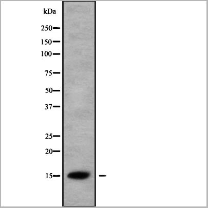
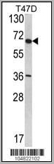
 WB (Western Blot)
(Western blot analysis of Phospho-KIT (Tyr703) expression in various lysates)
WB (Western Blot)
(Western blot analysis of Phospho-KIT (Tyr703) expression in various lysates)
KIT, Polyclonal Antibody (Cat# AAA31006)
Full Name
Phospho-KIT (Tyr703) Antibody
Gene Names
KIT; PBT; SCFR; C-Kit; CD117; MASTC
Reactivity
Human, Mouse, Rat
Applications
Western Blot, Immunohistochemistry, Immunofluorescence, Immunocytochemistry
Purity
From purified rabbit serum by affinity purification via sequential chromatography on phospho-and non-phospho-peptide affinity columns.
Pricing


 Application Data
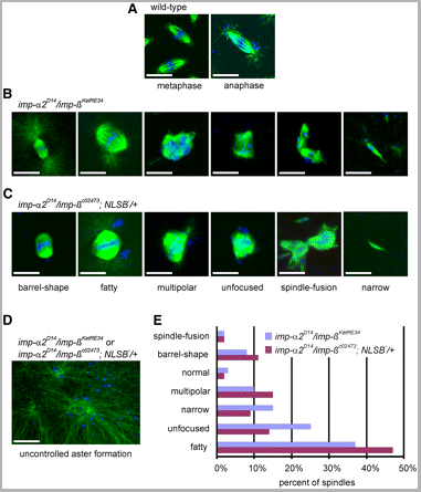
(Published customer image: Spindle abnormalities in embryos derived from imp-a2D14/imp-betaKetRE34 and imp-a2D14/imp-betac02473; NLSB-/+ females. (A -D) Wild-type and mutant embryos stained for a-tubulin (green) and DNA (blue). (A) Mitotic spindles in wild-type embryos at metaphase and anaphase. (B, C) Categories of spindle abnormalities found in embryos derived from (B) imp-a2D14/imp-betaKetRE34 and (C) imp-a2D14/imp-betac02743; NLSB-/+ females. (D) Formation of aster networks found in both genotypes. Scale bar: 10 um. (E) Frequency of spindle defects in embryos from both types of mutant females. Female genotypes are displayed at the upper right corner. At least 200 spindles were scored for both genotypes.From: Specific Cooperation Between Imp-a2 and Imp-beta/Ketel in Spindle Assembly During Drosophila Early Nuclear Divisions Erika Vir¡gh, M¡ty¡s Gorj¡n¡cz, Istv¡n T¶r¶k, Tolga Eichhorn, Sowjanya Kallakuri, Tam¡s Szlanka, Istv¡n Kiss, and Bernard M. Mechler G3 January 2012 2:1-14.)
Application Data
(Published customer image: Spindle abnormalities in embryos derived from imp-a2D14/imp-betaKetRE34 and imp-a2D14/imp-betac02473; NLSB-/+ females. (A -D) Wild-type and mutant embryos stained for a-tubulin (green) and DNA (blue). (A) Mitotic spindles in wild-type embryos at metaphase and anaphase. (B, C) Categories of spindle abnormalities found in embryos derived from (B) imp-a2D14/imp-betaKetRE34 and (C) imp-a2D14/imp-betac02743; NLSB-/+ females. (D) Formation of aster networks found in both genotypes. Scale bar: 10 um. (E) Frequency of spindle defects in embryos from both types of mutant females. Female genotypes are displayed at the upper right corner. At least 200 spindles were scored for both genotypes.From: Specific Cooperation Between Imp-a2 and Imp-beta/Ketel in Spindle Assembly During Drosophila Early Nuclear Divisions Erika Vir¡gh, M¡ty¡s Gorj¡n¡cz, Istv¡n T¶r¶k, Tolga Eichhorn, Sowjanya Kallakuri, Tam¡s Szlanka, Istv¡n Kiss, and Bernard M. Mechler G3 January 2012 2:1-14.)
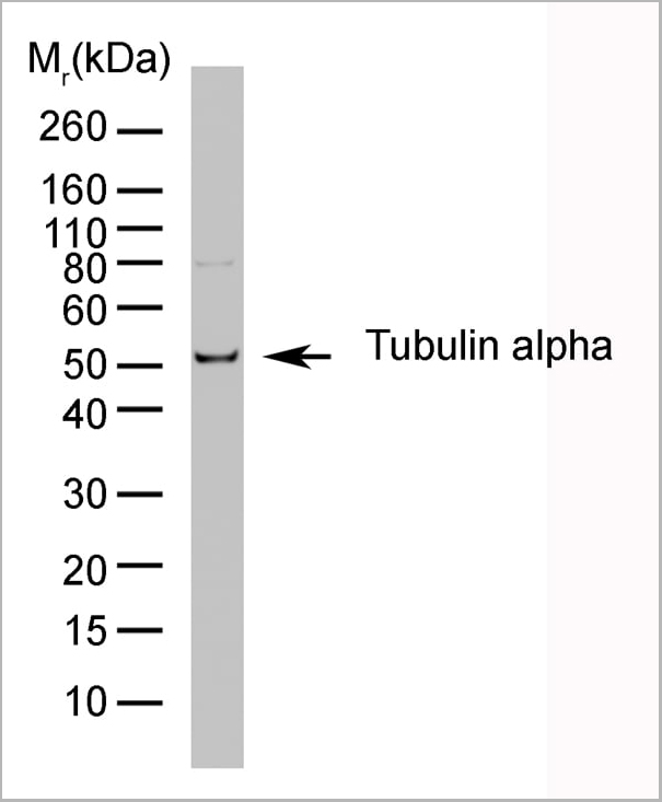
TUBULIN ALPHA, Monoclonal Antibody (Cat# AAA12232)
Full Name
RAT ANTI TUBULIN ALPHA:HRP
Applications
Immunohistochemistry, Western Blot
Pricing


 Application Data
(Published customer image: Spindle abnormalities in embryos derived from imp-a2D14/imp-betaKetRE34 and imp-a2D14/imp-betac02473; NLSB-/+ females. (A -D) Wild-type and mutant embryos stained for a-tubulin (green) and DNA (blue). (A) Mitotic spindles in wild-type embryos at metaphase and anaphase. (B, C) Categories of spindle abnormalities found in embryos derived from (B) imp-a2D14/imp-betaKetRE34 and (C) imp-a2D14/imp-betac02743; NLSB-/+ females. (D) Formation of aster networks found in both genotypes. Scale bar: 10 um. (E) Frequency of spindle defects in embryos from both types of mutant females. Female genotypes are displayed at the upper right corner. At least 200 spindles were scored for both genotypes.From: Specific Cooperation Between Imp-a2 and Imp-beta/Ketel in Spindle Assembly During Drosophila Early Nuclear Divisions Erika Vir¡gh, M¡ty¡s Gorj¡n¡cz, Istv¡n T¶r¶k, Tolga Eichhorn, Sowjanya Kallakuri, Tam¡s Szlanka, Istv¡n Kiss, and Bernard M. Mechler G3 January 2012 2:1-14.)
Application Data
(Published customer image: Spindle abnormalities in embryos derived from imp-a2D14/imp-betaKetRE34 and imp-a2D14/imp-betac02473; NLSB-/+ females. (A -D) Wild-type and mutant embryos stained for a-tubulin (green) and DNA (blue). (A) Mitotic spindles in wild-type embryos at metaphase and anaphase. (B, C) Categories of spindle abnormalities found in embryos derived from (B) imp-a2D14/imp-betaKetRE34 and (C) imp-a2D14/imp-betac02743; NLSB-/+ females. (D) Formation of aster networks found in both genotypes. Scale bar: 10 um. (E) Frequency of spindle defects in embryos from both types of mutant females. Female genotypes are displayed at the upper right corner. At least 200 spindles were scored for both genotypes.From: Specific Cooperation Between Imp-a2 and Imp-beta/Ketel in Spindle Assembly During Drosophila Early Nuclear Divisions Erika Vir¡gh, M¡ty¡s Gorj¡n¡cz, Istv¡n T¶r¶k, Tolga Eichhorn, Sowjanya Kallakuri, Tam¡s Szlanka, Istv¡n Kiss, and Bernard M. Mechler G3 January 2012 2:1-14.)
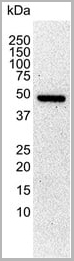
TUBULIN ALPHA, Monoclonal Antibody (Cat# AAA12009)
Full Name
RAT ANTI TUBULIN ALPHA
Applications
Immunohistochemistry, Immunofluorescence, Immunoprecipitation, Radioimmunoassay, Western Blot
Pricing


 Application Data
(Published customer image: Mouse anti V5 tag antibody, clone SV5-Pk1 used for the detection of V5 tagged WEEV_nsP3 protein by western blotting and immunofluorescenceImage caption: WEEV nsP3 interaction with host IKKbeta. A) U87MGs were transfected in a 6-well plate with 5 ug of pUC19 and WEEV_nsP3_HA for 24 hours. Cell lysates were resolved using SDS-PAGE and subsequently immunoblotted with V5 antibody and beta-actin served as a loading control. B) U87MGs were transfected with WEEV_nsP3_V5; cells were fixed after 24 hours and stained with antibodies against the endogenous IKKbeta and the V5 tag. Cells were incubated with appropriate secondary Alexa Fluor antibodies and the nuclei stained with DAPI. Co-localization of IKKbeta with WEEV_nsP3_V5 (yellow) was observed as shown by the arrows. B) Panels E -H serve as an example of transfected cells in a given field of view that show co-localization of IKKbeta and WEEV_nsP3_V5 24 hours post transfection. Panels I-L represent magnified images of other cells showing co-localization of IKKbeta and WEEV_nsP3_V5. Panel M is a magnified image of panel L. The co-localization was confirmed by Z-stack analysis. Co-localization was calculated to be approximately in 61% of cells (163 cells were counted of which 44% demonstrated expression of nsP3. Of those cells that expressed nsP3, 61% showed co-localization of both proteins). Images were taken using Nikon Eclipse TE2000-U at 60x magnification and are representative of 2 independent experiments.From: Amaya M, Voss K, Sampey G, Senina S, de la Fuente C, et al. (2014) The Role of IKKbeta in Venezuelan Equine Encephalitis Virus Infection. PLoS ONE 9(2): e86745.)
Application Data
(Published customer image: Mouse anti V5 tag antibody, clone SV5-Pk1 used for the detection of V5 tagged WEEV_nsP3 protein by western blotting and immunofluorescenceImage caption: WEEV nsP3 interaction with host IKKbeta. A) U87MGs were transfected in a 6-well plate with 5 ug of pUC19 and WEEV_nsP3_HA for 24 hours. Cell lysates were resolved using SDS-PAGE and subsequently immunoblotted with V5 antibody and beta-actin served as a loading control. B) U87MGs were transfected with WEEV_nsP3_V5; cells were fixed after 24 hours and stained with antibodies against the endogenous IKKbeta and the V5 tag. Cells were incubated with appropriate secondary Alexa Fluor antibodies and the nuclei stained with DAPI. Co-localization of IKKbeta with WEEV_nsP3_V5 (yellow) was observed as shown by the arrows. B) Panels E -H serve as an example of transfected cells in a given field of view that show co-localization of IKKbeta and WEEV_nsP3_V5 24 hours post transfection. Panels I-L represent magnified images of other cells showing co-localization of IKKbeta and WEEV_nsP3_V5. Panel M is a magnified image of panel L. The co-localization was confirmed by Z-stack analysis. Co-localization was calculated to be approximately in 61% of cells (163 cells were counted of which 44% demonstrated expression of nsP3. Of those cells that expressed nsP3, 61% showed co-localization of both proteins). Images were taken using Nikon Eclipse TE2000-U at 60x magnification and are representative of 2 independent experiments.From: Amaya M, Voss K, Sampey G, Senina S, de la Fuente C, et al. (2014) The Role of IKKbeta in Venezuelan Equine Encephalitis Virus Infection. PLoS ONE 9(2): e86745.)
V5-TAG, Monoclonal Antibody (Cat# AAA12081)
Full Name
MOUSE ANTI V5-TAG:HRP
Applications
Western Blot
Pricing
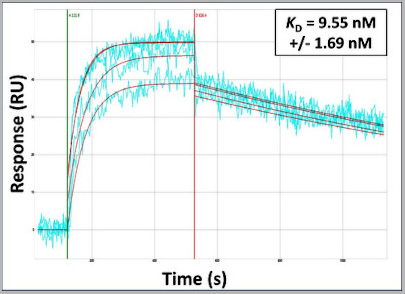 Application Data
(Surface Plasmon Resonance Kinetic Characterization of Polyclonal Antibody Affinity. Purified polyclonal antibodies were immobilized on a Protein A/G coated Carterra LSA sensor chip (PAGH200M) at concentrations of 5, and 50 ug/mL in duplicate. Antibodies on the surface were exposed to interaction with peptides sequentially via microfluidic controlled flow at 333nM peptide concentration for kinetic characterization of the binders for affinity and specificity, followed by curve fitting using the Kinetics software. Kd determinations for both concentrations were averaged and results and standard deviation are shown.)
Application Data
(Surface Plasmon Resonance Kinetic Characterization of Polyclonal Antibody Affinity. Purified polyclonal antibodies were immobilized on a Protein A/G coated Carterra LSA sensor chip (PAGH200M) at concentrations of 5, and 50 ug/mL in duplicate. Antibodies on the surface were exposed to interaction with peptides sequentially via microfluidic controlled flow at 333nM peptide concentration for kinetic characterization of the binders for affinity and specificity, followed by curve fitting using the Kinetics software. Kd determinations for both concentrations were averaged and results and standard deviation are shown.)
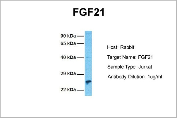
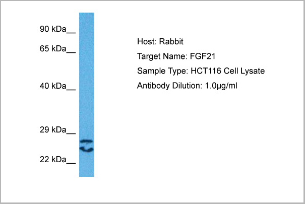
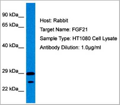
FGF21, Polyclonal Antibody (Cat# AAA23563)
Full Name
FGF21 antibody - N-terminal region
Reactivity
Tested Species Reactivity: Human
Predicted Species Reactivity: Human, Mouse, Rat, Cow, Dog, Guinea Pig, Horse, Rabbit
Predicted Species Reactivity: Human, Mouse, Rat, Cow, Dog, Guinea Pig, Horse, Rabbit
Applications
Immunohistochemistry, Western Blot
Purity
Affinity Purified
Pricing
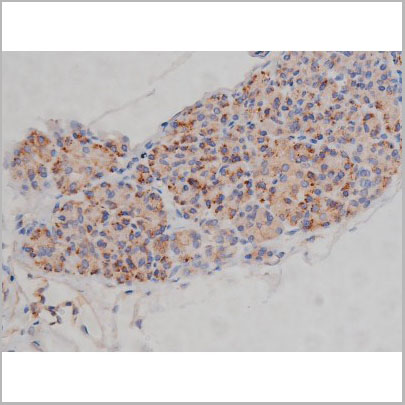
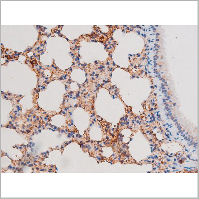
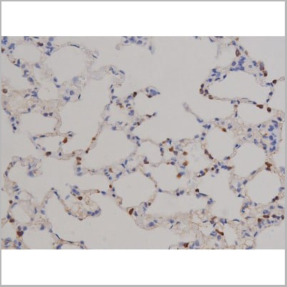
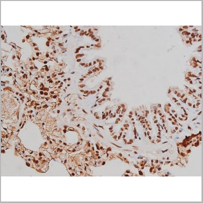
 IHC (Immunohistochemistry)
(AAA31121 at 1/100 staining Mouse brain tissue by IHC-P. The sample was formaldehyde fixed and a heat mediated antigen retrieval step in citrate buffer was performed. The sample was then blocked and incubated with the antibody for 1.5 hours at 22 degree C. An HRP conjugated goat anti-rabbit antibody was used as the secondary.)
IHC (Immunohistochemistry)
(AAA31121 at 1/100 staining Mouse brain tissue by IHC-P. The sample was formaldehyde fixed and a heat mediated antigen retrieval step in citrate buffer was performed. The sample was then blocked and incubated with the antibody for 1.5 hours at 22 degree C. An HRP conjugated goat anti-rabbit antibody was used as the secondary.)
FABP4, Polyclonal Antibody (Cat# AAA31121)
Full Name
FABP4 Antibody
Gene Names
FABP4; aP2; ALBP; AFABP; A-FABP; HEL-S-104
Reactivity
Human, Mouse, Rat
Applications
Western Blot, Immunohistochemistry
Purity
The antiserum was purified by peptide affinity chromatography using SulfoLink Coupling Resin.
Pricing
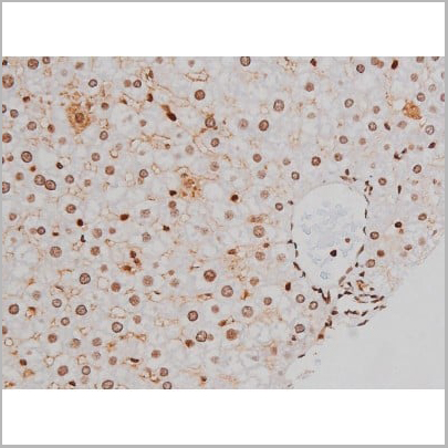
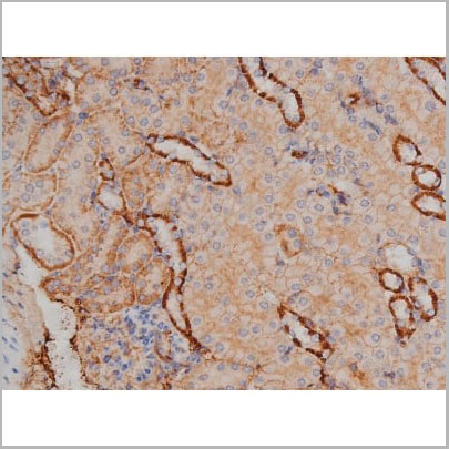
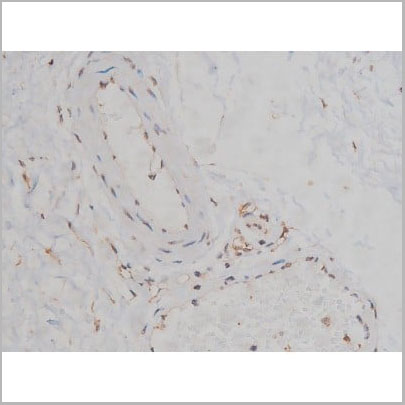
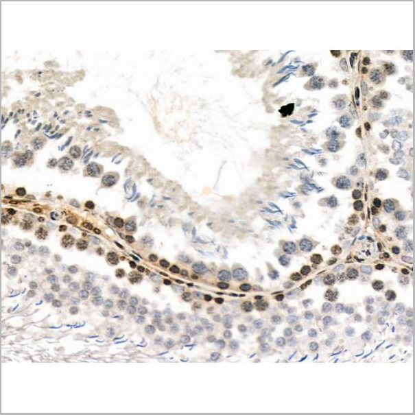
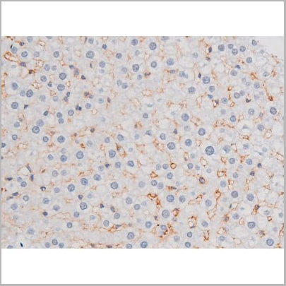
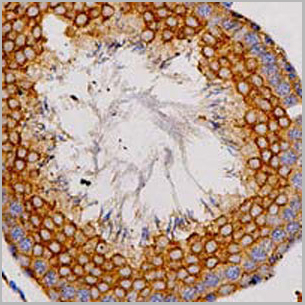
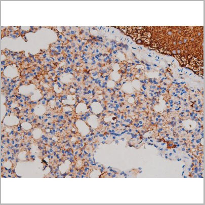
 IHC-P (Immunohistochemistry-Paraffin)
(At 1/200 staining Rat ganstric tissue sections by IHC-P. The tissue was formaldehyde fixed and a heat mediatedantigen retrieval step in citrate buffer was performed. Thetissue was then blocked and incubated with the antibody for1.5 hours at 22°C. An HRP conjugated goat anti-rabbitantibody was used as the secondary antibody.)
IHC-P (Immunohistochemistry-Paraffin)
(At 1/200 staining Rat ganstric tissue sections by IHC-P. The tissue was formaldehyde fixed and a heat mediatedantigen retrieval step in citrate buffer was performed. Thetissue was then blocked and incubated with the antibody for1.5 hours at 22°C. An HRP conjugated goat anti-rabbitantibody was used as the secondary antibody.)
c-Kit, Polyclonal Antibody (Cat# AAA31007)
Full Name
Phospho-c-Kit (Tyr721) Antibody
Gene Names
KIT; PBT; SCFR; C-Kit; CD117; MASTC
Reactivity
Human, Mouse, Rat
Applications
Western Blot, Immunohistochemistry, Immunofluorescence, Immunocytochemistry
Purity
Peptide affinity purification
Pricing
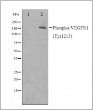
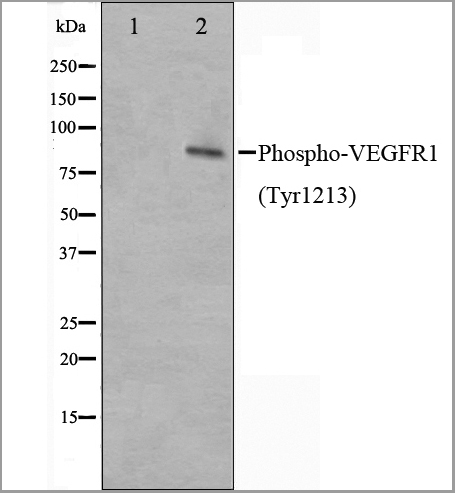
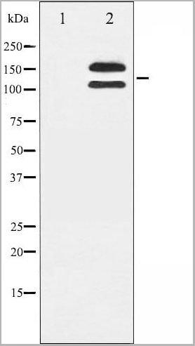
 Application Data
(AAA31020 staining HepG2 cells(30min of 4uM Forskolin treatment) by IF/ICC. The samples were fixed with PFA and permeabilized in 0.1% Triton X-100,then blocked in 10% serum for 45 minutes at 25°C. Samples were then incubated with primary Ab(AAA31020) and mouse anti-beta tubulin Ab for 1 hour at 37°C. An AlexaFluor594 conjugated goat anti-rabbit IgG(H+L) Ab(Red) and an AlexaFluor488 conjugated goat anti-mouse IgG(H+L) Ab(Green) were used as the secondary Ab. The nuclear counter stain is DAPI(blue))
Application Data
(AAA31020 staining HepG2 cells(30min of 4uM Forskolin treatment) by IF/ICC. The samples were fixed with PFA and permeabilized in 0.1% Triton X-100,then blocked in 10% serum for 45 minutes at 25°C. Samples were then incubated with primary Ab(AAA31020) and mouse anti-beta tubulin Ab for 1 hour at 37°C. An AlexaFluor594 conjugated goat anti-rabbit IgG(H+L) Ab(Red) and an AlexaFluor488 conjugated goat anti-mouse IgG(H+L) Ab(Green) were used as the secondary Ab. The nuclear counter stain is DAPI(blue))
VEGFR1, Polyclonal Antibody (Cat# AAA31020)
Full Name
Phospho-VEGFR1 (Tyr1213) Antibody
Gene Names
FLT1; FLT; FLT-1; VEGFR1; VEGFR-1
Reactivity
Human, Mouse, Rat
Applications
Western Blot, Immunofluorescence, Immunocytochemistry, Immunohistochemistry
Purity
The Ab is from purified rabbit serum by affinity purification via sequential chromatography on phospho-peptide and non-phospho-peptide affinity columns.
Pricing
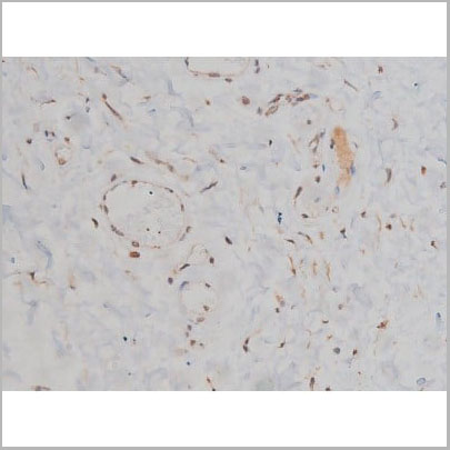
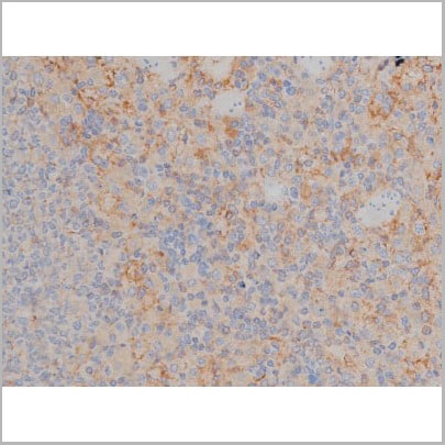
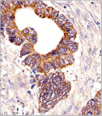
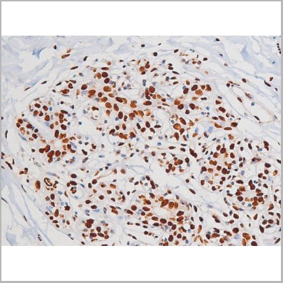
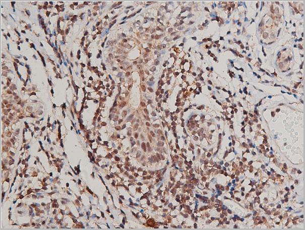
 IHC (Immunohistochemistry)
(AAA31051 at 1/200 staining Human bladder cancer tissue sections by IHC-P. The tissue was formaldehyde fixed and a heat mediated antigen retrieval step in citrate buffer was performed. The tissue was then blocked and incubated with the antibody for 1.5 hours at 22 degree C. An HRP conjugated goat anti-rabbit antibody was used as the secondary.)
IHC (Immunohistochemistry)
(AAA31051 at 1/200 staining Human bladder cancer tissue sections by IHC-P. The tissue was formaldehyde fixed and a heat mediated antigen retrieval step in citrate buffer was performed. The tissue was then blocked and incubated with the antibody for 1.5 hours at 22 degree C. An HRP conjugated goat anti-rabbit antibody was used as the secondary.)
SYK, Polyclonal Antibody (Cat# AAA31051)
Full Name
Phospho-SYK (Tyr348) Antibody
Gene Names
SYK; p72-Syk
Reactivity
Human, Mouse, Rat, Monkey
Applications
Western Blot, Immunohistochemistry, Immunofluorescence, Immunocytochemistry
Purity
From purified rabbit serum by affinity purification via sequential chromatography on phospho-and non-phospho-peptide affinity columns.
Pricing
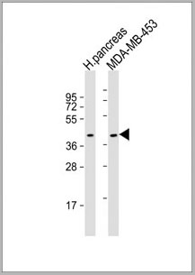
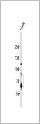
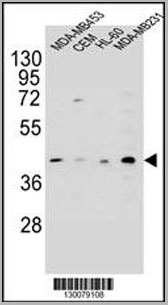 WB (Western Blot)
(HHLA2 Antibody (N-term) western blot analysis in MDA-MB453,CEM,HL-60,MDA-MB231 cell line lysates (35ug/lane).This demonstrates the HHLA2 antibody detected the HHLA2 protein (arrow).)
WB (Western Blot)
(HHLA2 Antibody (N-term) western blot analysis in MDA-MB453,CEM,HL-60,MDA-MB231 cell line lysates (35ug/lane).This demonstrates the HHLA2 antibody detected the HHLA2 protein (arrow).)
HHLA2, Polyclonal Antibody (Cat# AAA28722)
Full Name
HHLA2 Antibody (N-term)
Gene Names
HHLA2; B7H7; B7-H7
Reactivity
Human
Applications
Western Blot, Immunohistochemistry, Flow Cytometry
Purity
Peptide Affinity Purified Rabbit Polyclonal Antibody (Pab)
Pricing
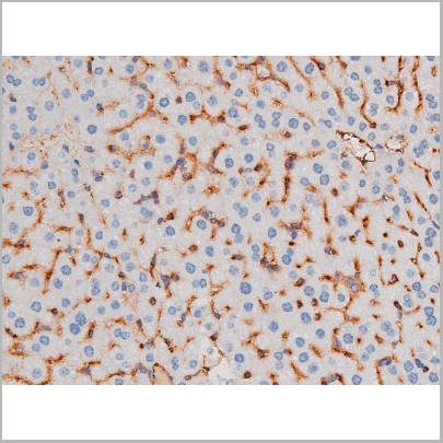
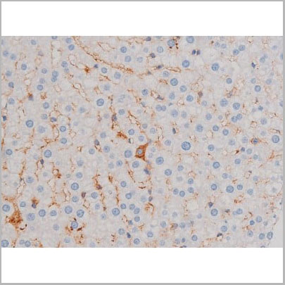
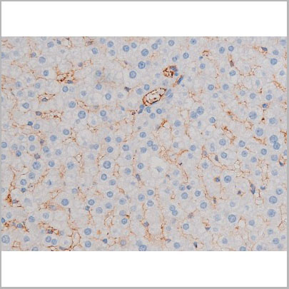
 IHC (Immunohistchemistry)
(Immunohistochemical analysis of paraffin-embedded human liver tissue using AAA28677 performed on the Leica® BOND RXm. Tissue was fixed with formaldehyde at room temperature; antigen retrieval was by heat mediation with a EDTA buffer (pH9. 0). Samples were incubated with primary antibody(1:1000) for 1 hours at room temperature. A undiluted biotinylated CRF Anti-Polyvalent HRP Polymer antibody was used as the secondary antibody.)
IHC (Immunohistchemistry)
(Immunohistochemical analysis of paraffin-embedded human liver tissue using AAA28677 performed on the Leica® BOND RXm. Tissue was fixed with formaldehyde at room temperature; antigen retrieval was by heat mediation with a EDTA buffer (pH9. 0). Samples were incubated with primary antibody(1:1000) for 1 hours at room temperature. A undiluted biotinylated CRF Anti-Polyvalent HRP Polymer antibody was used as the secondary antibody.)
ATG7, Polyclonal Antibody (Cat# AAA28677)
Full Name
ATG7 Antibody (C-term)
Gene Names
ATG7; GSA7; APG7L; APG7-LIKE
Reactivity
Human, Mouse
Predicted reactivity: Chicken, Rat
Predicted reactivity: Chicken, Rat
Applications
Western Blot, Immunohistochemistry, Immunofluorescence
Purity
Purified Rabbit Polyclonal Antibody (Pab)
Pricing

 WB (Western Blot)
(Western blot analysis of p53 phosphorylation expression in Etoposide treated 293 whole cell lysates, The lane on the left is treated with the antigen-specific peptide.)
WB (Western Blot)
(Western blot analysis of p53 phosphorylation expression in Etoposide treated 293 whole cell lysates, The lane on the left is treated with the antigen-specific peptide.)
p53, Polyclonal Antibody (Cat# AAA30976)
Full Name
Phospho-p53 (Ser392) Antibody
Gene Names
TP53; P53; BCC7; LFS1; TRP53
Reactivity
Human, Mouse, Rat
Applications
Western Blot, Immunohistochemistry, Immunofluorescence, Immunocytochemistry
Purity
From purified rabbit serum by affinity purification via sequential chromatography on phospho-and non-phospho-peptide affinity columns.
Pricing
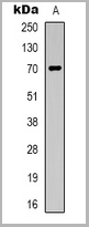
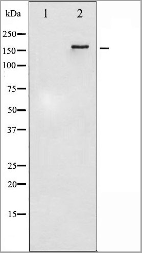
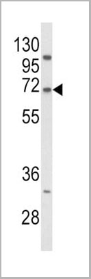
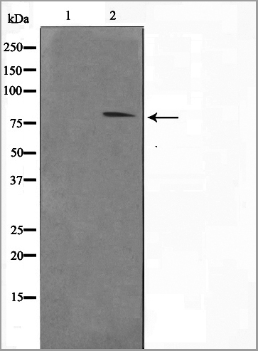
 WB (Western Blot)
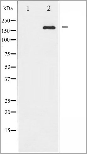
(TGFBR2 Antibody western blot of mouse lung tissue lysates (35 ug/lane). The TGFBR2 antibody detected the TGFBR2 protein (arrow).)
WB (Western Blot)
(TGFBR2 Antibody western blot of mouse lung tissue lysates (35 ug/lane). The TGFBR2 antibody detected the TGFBR2 protein (arrow).)
TGFBR2, Polyclonal Antibody (Cat# AAA21269)
Full Name
Anti-TGFBR2 Antibody (aa13-40) IHC-plus
Gene Names
TGFBR2; AAT3; FAA3; LDS2; MFS2; RIIC; LDS1B; LDS2B; TAAD2; TGFR-2; TGFbeta-RII
Reactivity
Mouse, Human
Applications
Immunohistochemistry, Immunofluorescence, Western Blot
Purity
Protein A purified
Pricing


 Application Data
(Staining of mouse spleen with Hamster anti Mouse CD81: Alexa Fluor 488)
Application Data
(Staining of mouse spleen with Hamster anti Mouse CD81: Alexa Fluor 488)
CD81, Monoclonal Antibody (Cat# AAA11869)
Full Name
HAMSTER ANTI MOUSE CD81:FITC
Gene Names
Cd81; Tapa1; Tapa-1; Tspan28
Applications
Flow Cytometry
Pricing


 Application Data
(PE conjugatedMouse anti Human CD169 antibody, clone 7-239 used to block CD169 function on myeloid cells.Image caption:Siglec-1 mediates HIV-1 uptake into a storage compartment and enhances HIV-1 trans-infection specially in IFN?-treated monocytes and DCs. A. Uptake of HIV-1NL4–3 by different myeloid cells exposed to IFN?. Cells were cultured with HIV-1 to measure p24Gag by ELISA. Mean values and SEM from four experiments include cells from 12 donors. B. Fold change in HIV-1NL4–3 uptake of cells treated with bafilomycin A1 compared to untreated cells. Mean values and SEM include cells from three donors. C. Relative uptake of HIV-1NL4–3 by IFN?-treated myeloid cells pre-incubated with the indicated mAbs. Values are normalized to the level of HIV-1 uptake by mock-treated cells (set at 100%). Mean values and SEM from two experiments include cells from six donors. D. Confocal microscopy analysis of different IFN?-treated myeloid cells pulsed with HIV-1Cherry and stained for Siglec-1 (Alexa 488), HLA-DR (Alexa 647) and DAPI. (Top) Representative viral pattern for each kind of myeloid cell analyzed, showing maximum fluorescence intensity of four channels. (Bottom) Percentage of myeloid cells with distinct viral patterns: random distribution, polarized accumulation, and sac-like compartment formation, as illustrated in the left drawing. Mean values of 50 cells from two different donors are shown. E. HIV-1 transmission from IFN?-treated myeloid cells to a luciferase reporter CD4+ cell line. HIV-1 infection was determined by induced luciferase activity in relative light units (RLUs). Mean values and SEM from four experiments include cells from 12 donors. F. Relative HIV-1 transmission from IFN?-treated myeloid cells pre-incubated with the indicated mAbs. Values are normalized to the level of HIV-1 trans-infected by mock-treated cells. Mean values and SEM from two experiments include cells from six donors. Statistical differences were assessed with a paired t test in A and E, and with a one sample t-test in B, C and F.From: Pino M, Erkizia I, Benet S, Erikson E, Fernández-Figueras MT, Guerrero D, Dalmau J, Ouchi D, Rausell A, Ciuffi A, Keppler OT, Telenti A, Kräusslich HG, Martinez-Picado J, Izquierdo-Useros N.HIV-1 immune activation induces Siglec-1 expression and enhances viral trans-infection in blood and tissue myeloid cells.Retrovirology. 2015 May 7;12:37.This image is from an open access article distributed under the terms of the Creative Commons Attribution License.)
Application Data
(PE conjugatedMouse anti Human CD169 antibody, clone 7-239 used to block CD169 function on myeloid cells.Image caption:Siglec-1 mediates HIV-1 uptake into a storage compartment and enhances HIV-1 trans-infection specially in IFN?-treated monocytes and DCs. A. Uptake of HIV-1NL4–3 by different myeloid cells exposed to IFN?. Cells were cultured with HIV-1 to measure p24Gag by ELISA. Mean values and SEM from four experiments include cells from 12 donors. B. Fold change in HIV-1NL4–3 uptake of cells treated with bafilomycin A1 compared to untreated cells. Mean values and SEM include cells from three donors. C. Relative uptake of HIV-1NL4–3 by IFN?-treated myeloid cells pre-incubated with the indicated mAbs. Values are normalized to the level of HIV-1 uptake by mock-treated cells (set at 100%). Mean values and SEM from two experiments include cells from six donors. D. Confocal microscopy analysis of different IFN?-treated myeloid cells pulsed with HIV-1Cherry and stained for Siglec-1 (Alexa 488), HLA-DR (Alexa 647) and DAPI. (Top) Representative viral pattern for each kind of myeloid cell analyzed, showing maximum fluorescence intensity of four channels. (Bottom) Percentage of myeloid cells with distinct viral patterns: random distribution, polarized accumulation, and sac-like compartment formation, as illustrated in the left drawing. Mean values of 50 cells from two different donors are shown. E. HIV-1 transmission from IFN?-treated myeloid cells to a luciferase reporter CD4+ cell line. HIV-1 infection was determined by induced luciferase activity in relative light units (RLUs). Mean values and SEM from four experiments include cells from 12 donors. F. Relative HIV-1 transmission from IFN?-treated myeloid cells pre-incubated with the indicated mAbs. Values are normalized to the level of HIV-1 trans-infected by mock-treated cells. Mean values and SEM from two experiments include cells from six donors. Statistical differences were assessed with a paired t test in A and E, and with a one sample t-test in B, C and F.From: Pino M, Erkizia I, Benet S, Erikson E, Fernández-Figueras MT, Guerrero D, Dalmau J, Ouchi D, Rausell A, Ciuffi A, Keppler OT, Telenti A, Kräusslich HG, Martinez-Picado J, Izquierdo-Useros N.HIV-1 immune activation induces Siglec-1 expression and enhances viral trans-infection in blood and tissue myeloid cells.Retrovirology. 2015 May 7;12:37.This image is from an open access article distributed under the terms of the Creative Commons Attribution License.)
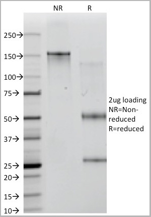
CD169, Monoclonal Antibody (Cat# AAA12265)
Full Name
Mouse Anti Human CD169: RPE
Gene Names
SIGLEC1; SN; CD169; SIGLEC-1
Reactivity
Human
Applications
Flow Cytometry
Purity
>95% by SDS PAGE
Purified IgG prepared by affinity chromatography on Protein A from tissue culture supernatant.
Purified IgG prepared by affinity chromatography on Protein A from tissue culture supernatant.
Pricing


 Application Data
(Staining of mouse spleen with Hamster anti Mouse CD81: Alexa Fluor 488)
Application Data
(Staining of mouse spleen with Hamster anti Mouse CD81: Alexa Fluor 488)
CD81, Monoclonal Antibody (Cat# AAA12033)
Full Name
HAMSTER ANTI MOUSE CD81:RPE
Gene Names
Cd81; Tapa1; Tapa-1; Tspan28
Applications
Flow Cytometry
Pricing