Filters
Clonality
Type
Reactivity
Gene Name
Isotype
Host
Application
Clone
149 results for " 14 3 3" - showing 100-149
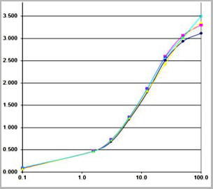
 Standard Curve (Sample)
Pentraxin-3 (PTX-3), ELISA Kit (Cat# AAA22403)
Full Name
Human Pentraxin-3 (PTX-3) ELISA Kit
Gene Names
PTX3; TSG-14; TNFAIP5
Reactivity
Human
Pricing
 Standard Curve (Sample)
tyrosine hydroxylase (TH), ELISA Kit (Cat# AAA18229)
Full Name
Mouse tyrosine hydroxylase, TH ELISA Kit
Reactivity
Mouse
Pricing


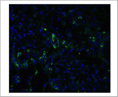
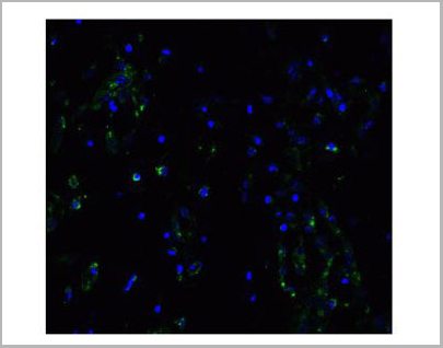
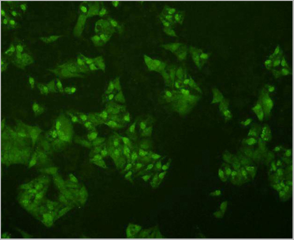
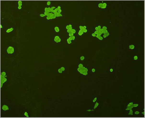
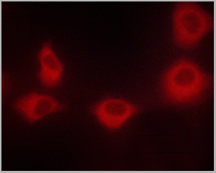
 IF (Immunofluorescence)
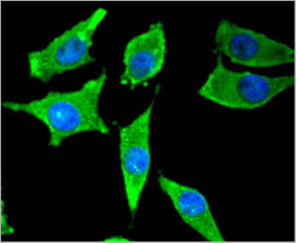
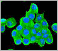
(Immunofluorescence analysis of A549 cells using PEX14 antibody. Blue: DAPI for nuclear staining.)
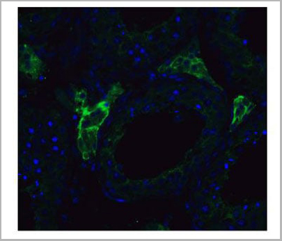
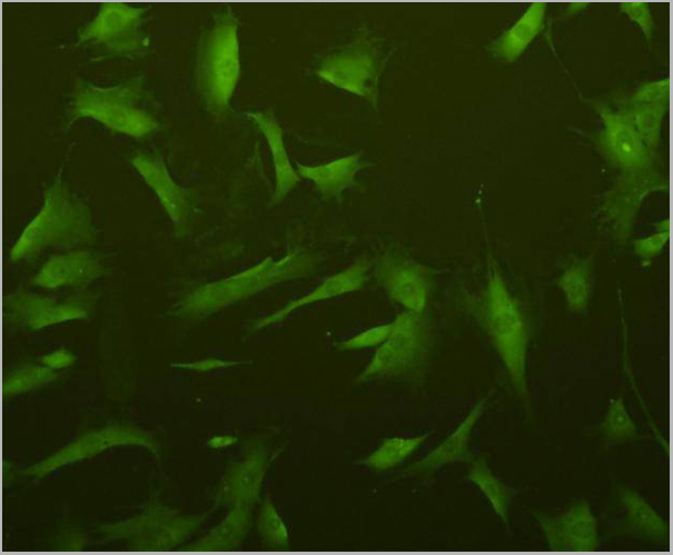
IF (Immunofluorescence)
(Immunofluorescence analysis of A549 cells using PEX14 antibody. Blue: DAPI for nuclear staining.)
PEX14, Polyclonal Antibody (Cat# AAA28157)
Full Name
PEX14
Gene Names
PEX14; NAPP2; PBD13A; Pex14p; dJ734G22.2
Reactivity
Human, Mouse, Rat
Applications
WB, IHC
Pricing


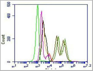
 FCM (Flow Cytometry)
(Figure 8. Flow Cytometry analysis of K562 cells using anti-PDIA6 antibody (AAA19282).Overlay histogram showing K562 cells stained with AAA19282 (Blue line). The cells were blocked with 10% normal goat serum. And then incubated with rabbit anti-PDIA6 Antibody (AAA19282, 1μg/1x106 cells) for 30 min at 20 degree C. DyLight®488 conjugated goat anti-rabbit IgG (5-10μg/1x106 cells) was used as secondary antibody for 30 minutes at 20 degree C. Isotype control antibody (Green line) was rabbit IgG (1μg/1x106) used under the same conditions. Unlabelled sample (Red line) was also used as a control.)
FCM (Flow Cytometry)
(Figure 8. Flow Cytometry analysis of K562 cells using anti-PDIA6 antibody (AAA19282).Overlay histogram showing K562 cells stained with AAA19282 (Blue line). The cells were blocked with 10% normal goat serum. And then incubated with rabbit anti-PDIA6 Antibody (AAA19282, 1μg/1x106 cells) for 30 min at 20 degree C. DyLight®488 conjugated goat anti-rabbit IgG (5-10μg/1x106 cells) was used as secondary antibody for 30 minutes at 20 degree C. Isotype control antibody (Green line) was rabbit IgG (1μg/1x106) used under the same conditions. Unlabelled sample (Red line) was also used as a control.)
PDIA6, Polyclonal Antibody (Cat# AAA19282)
Full Name
Anti-PDIA6 Antibody
Gene Names
PDIA6; P5; ERP5; TXNDC7
Reactivity
Human, Mouse, Rat
Applications
WB, IHC-P, ICC, IF, FC/FACS/FCM, EIA
Purity
Immunogen affinity purified.
Pricing


 Application Data
(Analysis of Protein Array containing more than 19,000 full-length human proteins using ROR-gamma / RORC Mouse Monoclonal Antibody (RORC/2941). Z- and S- Score: The Z-score represents the strength of a signal that a monoclonal antibody (MAb) (in combination with a fluorescently-tagged anti-IgG secondary antibody) produces when binding to a particular protein on the HuProtTM array. Z-scores are described in units of standard deviations (SD's) above the mean value of all signals generated on that array. If targets on HuProtTM are arranged in descending order of the Z-score, the S-score is the difference (also in units of SD's) between the Z-score. S-score therefore represents the relative target specificity of a MAb to its intended target. A MAb is considered to specific to its intended target, if the MAb has an S-score of at least 2.5. For example, if a MAb binds to protein X with a Z-score of 43 and to protein Y with a Z-score of 14, then the S-score for the binding of that MAb to protein X is equal to 29.)
Application Data
(Analysis of Protein Array containing more than 19,000 full-length human proteins using ROR-gamma / RORC Mouse Monoclonal Antibody (RORC/2941). Z- and S- Score: The Z-score represents the strength of a signal that a monoclonal antibody (MAb) (in combination with a fluorescently-tagged anti-IgG secondary antibody) produces when binding to a particular protein on the HuProtTM array. Z-scores are described in units of standard deviations (SD's) above the mean value of all signals generated on that array. If targets on HuProtTM are arranged in descending order of the Z-score, the S-score is the difference (also in units of SD's) between the Z-score. S-score therefore represents the relative target specificity of a MAb to its intended target. A MAb is considered to specific to its intended target, if the MAb has an S-score of at least 2.5. For example, if a MAb binds to protein X with a Z-score of 43 and to protein Y with a Z-score of 14, then the S-score for the binding of that MAb to protein X is equal to 29.)
ROR-gamma/RORC, Monoclonal Antibody (Cat# AAA23951)
Full Name
ROR-gamma/RORC (RAR-related Orphan Receptor C)
Gene Names
RORC; TOR; RORG; RZRG; IMD42; NR1F3; RZR-GAMMA
Reactivity
Human
Applications
FC, IHC
Purity
Purified Ab with BSA and Azide at 200ug/ml or Purified Ab with BSA and Azide at 200ug/ml or Purified Ab WITHOUT BSA and Azide at 1.0mg/ml
Pricing
 SDS-PAGE
Laminin Alpha 4 (LAMa4), Recombinant Protein (Cat# AAA20284)
Full Name
Recombinant Laminin Alpha 4 (LAMa4)
Gene Names
LAMA4; LAMA3; CMD1JJ; LAMA4*-1
Reactivity
Homo sapiens (Human)
Applications
Western Blot
Purity
>95%
Pricing
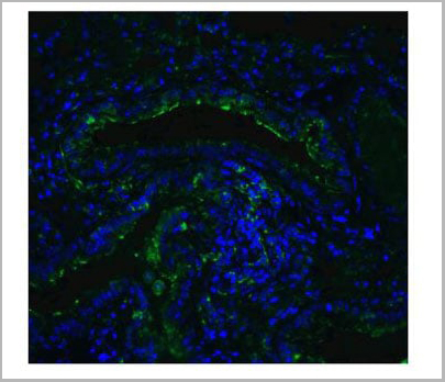
 IF (Immunofluorescence)
(Figure 11 Immunofluorescence Validation of ACE2 In Caco2 CellsImmunofluorescent analysis of 4% paraformaldehyde-fixed Caco2 cells labeling ACE2 with AAA10945 at 20 ug/mL, followed by goat anti-rabbit IgG secondary antibody at 1/500 dilution (green) and DAPI staining (blue). Imageshowing membrane staining on Caco2 cells.)
IF (Immunofluorescence)
(Figure 11 Immunofluorescence Validation of ACE2 In Caco2 CellsImmunofluorescent analysis of 4% paraformaldehyde-fixed Caco2 cells labeling ACE2 with AAA10945 at 20 ug/mL, followed by goat anti-rabbit IgG secondary antibody at 1/500 dilution (green) and DAPI staining (blue). Imageshowing membrane staining on Caco2 cells.)
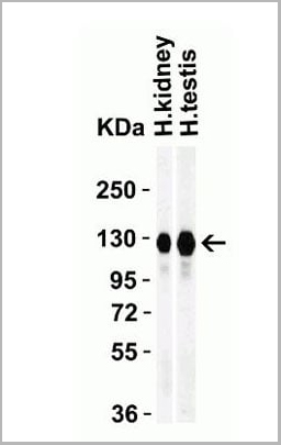
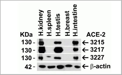
ACE2, Polyclonal Antibody (Cat# AAA10945)
Full Name
ACE2 Antibody
Gene Names
ACE2; ACEH
Reactivity
Human, Mouse, Rat
Applications
Western Blot, Immunohistochemistry, Immunofluorescence
Purity
ACE2 Antibody is affinity chromatography purified via peptide column.
Pricing
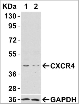
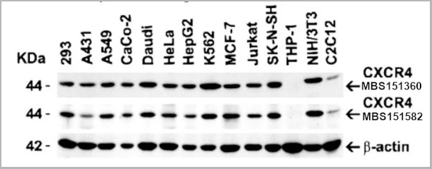
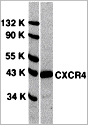
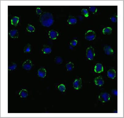
 WB (Western Blot)
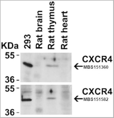
(Figure 4 Animal Species ReactivityLoading: Lysates/proteins at 20 μg per lane. Antibodies: 1009 (2 μg/mL) or 1012 (2 μg/mL). 1 h incubation at RT in 5% NFDM/TBST. Secondary: Goat anti-rabbit IgG HRP conjugate at 1:10000 dilution.)
WB (Western Blot)
(Figure 4 Animal Species ReactivityLoading: Lysates/proteins at 20 μg per lane. Antibodies: 1009 (2 μg/mL) or 1012 (2 μg/mL). 1 h incubation at RT in 5% NFDM/TBST. Secondary: Goat anti-rabbit IgG HRP conjugate at 1:10000 dilution.)
CXCR4, Polyclonal Antibody (Cat# AAA10951)
Full Name
CXCR4 Antibody
Gene Names
CXCR4; FB22; HM89; LAP3; LCR1; NPYR; WHIM; CD184; LAP-3; LESTR; NPY3R; NPYRL; HSY3RR; NPYY3R; D2S201E
Reactivity
Human, Mouse, Rat
Applications
Western Blot, Immunocytochemistry, Immunoprecipitation, Immunofluorescence, Immunohistochemistry, Flow Cytometry
Purity
CXCR4 Antibody is affinity chromatography purified via peptide column.
Pricing
 Application Data
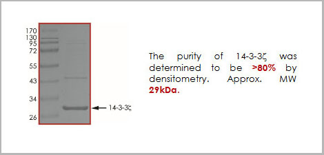
14-3-3 zeta, Recombinant Protein (Cat# AAA14245)
Full Name
14-3-3 zeta recombinant protein
Gene Names
YWHAZ; YWHAD; KCIP-1; 14-3-3-zeta
Applications
Western Blot
Purity
> 80%
Pricing
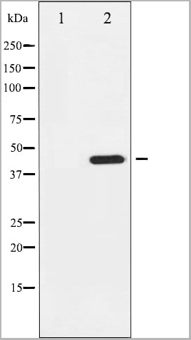
 WB (Western Blot)
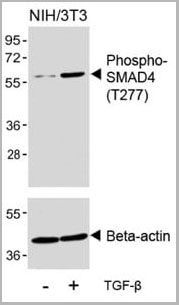
(Western blot analysis of lysates from NIH/3T3 cell line, untreated or treated with TGF-beta (100ng/ml, 30 min), using Phospho-SMAD4 Antibody(upper) or Beta-actin (lower).)
WB (Western Blot)
(Western blot analysis of lysates from NIH/3T3 cell line, untreated or treated with TGF-beta (100ng/ml, 30 min), using Phospho-SMAD4 Antibody(upper) or Beta-actin (lower).)
Phospho-SMAD4 (T277), Polyclonal Antibody (Cat# AAA28715)
Full Name
Phospho-SMAD4 (T277) Antibody
Gene Names
SMAD4; JIP; DPC4; MADH4; MYHRS
Reactivity
Human, Mouse
Predicted Species Reactivity: Rat
Predicted Species Reactivity: Rat
Applications
Western Blot
Purity
This antibody is purified through a protein A column, followed by peptide affinity purification.
Pricing
Tyrosine Hydroxylase, Antibody (Cat# AAA13664)
Full Name
Goat anti-Tyrosine Hydroxylase Antibody
Gene Names
TH; TYH; DYT14; DYT5b
Reactivity
Tested: Human; Expected from sequence similarity: Human, Rat, Dog
Applications
Peptide ELISA, Immunohistochemistry
Purity
Purified from goat serum by ammonium sulphate precipitation followed by antigen affinity chromatography using the immunizing peptide.
Pricing


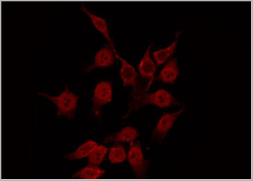
 IF (Immunofluorescence)
(Figure 8 Immunofluorescence Validation of JMJD3 in K562 Cells Immunofluorescent analysis of 4% paraformaldehyde fixed K562 cells labeling JMJD3 with AAA10925 at 20 ug/mL, followed by goat anti-rabbit IgG secondary antibody at 1/500 dilution (red).)
IF (Immunofluorescence)
(Figure 8 Immunofluorescence Validation of JMJD3 in K562 Cells Immunofluorescent analysis of 4% paraformaldehyde fixed K562 cells labeling JMJD3 with AAA10925 at 20 ug/mL, followed by goat anti-rabbit IgG secondary antibody at 1/500 dilution (red).)
JMJD3, Antibody (Cat# AAA10925)
Full Name
JMJD3 Antibody
Gene Names
KDM6B; JMJD3
Reactivity
Human, Mouse, Rat
Applications
Immunocytochemistry, Immunofluorescence, Western Blot
Purity
JMJD3 Antibody is affinity chromatography purified via peptide column.
Pricing

 Application Data
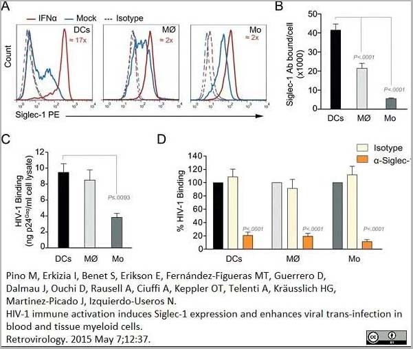
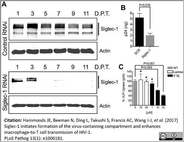
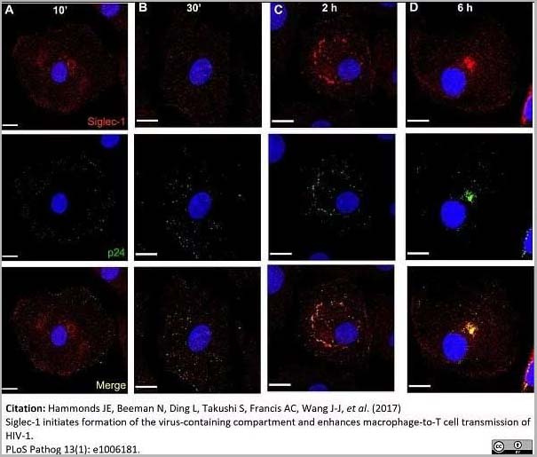
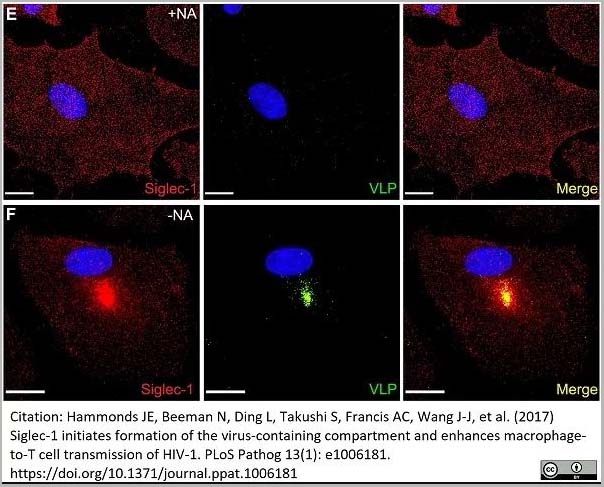
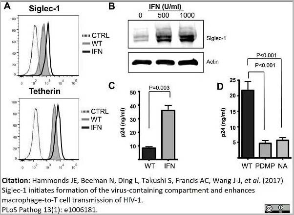
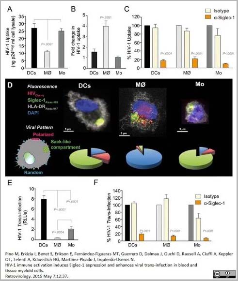
(PE conjugatedMouse anti Human CD169 antibody, clone 7-239 used to block CD169 function on myeloid cells.Image caption:Siglec-1 mediates HIV-1 uptake into a storage compartment and enhances HIV-1 trans-infection specially in IFN?-treated monocytes and DCs. A. Uptake of HIV-1NL4–3 by different myeloid cells exposed to IFN?. Cells were cultured with HIV-1 to measure p24Gag by ELISA. Mean values and SEM from four experiments include cells from 12 donors. B. Fold change in HIV-1NL4–3 uptake of cells treated with bafilomycin A1 compared to untreated cells. Mean values and SEM include cells from three donors. C. Relative uptake of HIV-1NL4–3 by IFN?-treated myeloid cells pre-incubated with the indicated mAbs. Values are normalized to the level of HIV-1 uptake by mock-treated cells (set at 100%). Mean values and SEM from two experiments include cells from six donors. D. Confocal microscopy analysis of different IFN?-treated myeloid cells pulsed with HIV-1Cherry and stained for Siglec-1 (Alexa 488), HLA-DR (Alexa 647) and DAPI. (Top) Representative viral pattern for each kind of myeloid cell analyzed, showing maximum fluorescence intensity of four channels. (Bottom) Percentage of myeloid cells with distinct viral patterns: random distribution, polarized accumulation, and sac-like compartment formation, as illustrated in the left drawing. Mean values of 50 cells from two different donors are shown. E. HIV-1 transmission from IFN?-treated myeloid cells to a luciferase reporter CD4+ cell line. HIV-1 infection was determined by induced luciferase activity in relative light units (RLUs). Mean values and SEM from four experiments include cells from 12 donors. F. Relative HIV-1 transmission from IFN?-treated myeloid cells pre-incubated with the indicated mAbs. Values are normalized to the level of HIV-1 trans-infected by mock-treated cells. Mean values and SEM from two experiments include cells from six donors. Statistical differences were assessed with a paired t test in A and E, and with a one sample t-test in B, C and F.From: Pino M, Erkizia I, Benet S, Erikson E, Fernández-Figueras MT, Guerrero D, Dalmau J, Ouchi D, Rausell A, Ciuffi A, Keppler OT, Telenti A, Kräusslich HG, Martinez-Picado J, Izquierdo-Useros N.HIV-1 immune activation induces Siglec-1 expression and enhances viral trans-infection in blood and tissue myeloid cells.Retrovirology. 2015 May 7;12:37.This image is from an open access article distributed under the terms of the Creative Commons Attribution License.)
Application Data
(PE conjugatedMouse anti Human CD169 antibody, clone 7-239 used to block CD169 function on myeloid cells.Image caption:Siglec-1 mediates HIV-1 uptake into a storage compartment and enhances HIV-1 trans-infection specially in IFN?-treated monocytes and DCs. A. Uptake of HIV-1NL4–3 by different myeloid cells exposed to IFN?. Cells were cultured with HIV-1 to measure p24Gag by ELISA. Mean values and SEM from four experiments include cells from 12 donors. B. Fold change in HIV-1NL4–3 uptake of cells treated with bafilomycin A1 compared to untreated cells. Mean values and SEM include cells from three donors. C. Relative uptake of HIV-1NL4–3 by IFN?-treated myeloid cells pre-incubated with the indicated mAbs. Values are normalized to the level of HIV-1 uptake by mock-treated cells (set at 100%). Mean values and SEM from two experiments include cells from six donors. D. Confocal microscopy analysis of different IFN?-treated myeloid cells pulsed with HIV-1Cherry and stained for Siglec-1 (Alexa 488), HLA-DR (Alexa 647) and DAPI. (Top) Representative viral pattern for each kind of myeloid cell analyzed, showing maximum fluorescence intensity of four channels. (Bottom) Percentage of myeloid cells with distinct viral patterns: random distribution, polarized accumulation, and sac-like compartment formation, as illustrated in the left drawing. Mean values of 50 cells from two different donors are shown. E. HIV-1 transmission from IFN?-treated myeloid cells to a luciferase reporter CD4+ cell line. HIV-1 infection was determined by induced luciferase activity in relative light units (RLUs). Mean values and SEM from four experiments include cells from 12 donors. F. Relative HIV-1 transmission from IFN?-treated myeloid cells pre-incubated with the indicated mAbs. Values are normalized to the level of HIV-1 trans-infected by mock-treated cells. Mean values and SEM from two experiments include cells from six donors. Statistical differences were assessed with a paired t test in A and E, and with a one sample t-test in B, C and F.From: Pino M, Erkizia I, Benet S, Erikson E, Fernández-Figueras MT, Guerrero D, Dalmau J, Ouchi D, Rausell A, Ciuffi A, Keppler OT, Telenti A, Kräusslich HG, Martinez-Picado J, Izquierdo-Useros N.HIV-1 immune activation induces Siglec-1 expression and enhances viral trans-infection in blood and tissue myeloid cells.Retrovirology. 2015 May 7;12:37.This image is from an open access article distributed under the terms of the Creative Commons Attribution License.)
CD169, Monoclonal Antibody (Cat# AAA12264)
Full Name
Mouse Anti Human CD169
Gene Names
SIGLEC1; SN; CD169; SIGLEC-1
Reactivity
Human
Applications
Immunohistochemistry, Flow Cytometry, Functional Assay, Immunoprecipitation, Western Blot
Purity
>95% by SDS PAGE
Purified IgG prepared by affinity chromatography on Protein A from tissue culture supernatant.
Purified IgG prepared by affinity chromatography on Protein A from tissue culture supernatant.
Pricing
 Application Data
(The cancer cell lines were stably expressing DYKDDDDK-tagged RIP3 and RIP expression was detected by anti-RIP3 antibodies (AAA10922) in RIP3-overexpressed cells.)
Application Data
(The cancer cell lines were stably expressing DYKDDDDK-tagged RIP3 and RIP expression was detected by anti-RIP3 antibodies (AAA10922) in RIP3-overexpressed cells.)
RIP3, Polyclonal Antibody (Cat# AAA10922)
Full Name
RIP3 Antibody
Gene Names
Ripk3; Rip3; AW107945; 2610528K09Rik
Reactivity
Human, Mouse, Rat
Applications
Immunofluorescence, Immunohistochemistry, Immunoprecipitation, Western Blot
Purity
RIP3 Antibody is affinity chromatography purified via peptide column.
Pricing
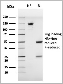
super leptin antagonist, Active Protein (Cat# AAA13623)
Full Name
Mouse super leptin antagonist (mutant D23/L39A/D40A/F41A)
Purity
The purity of mouse super-active leptin antagonist is greater than 98.0% as determined by:
(a) Gel filtration analysis.
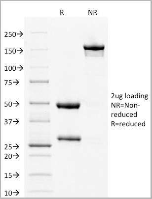
(b) Analysis by reducing and non-reducing SDS-PAGE gel.
(a) Gel filtration analysis.
(b) Analysis by reducing and non-reducing SDS-PAGE gel.
 WB (Western Blot)
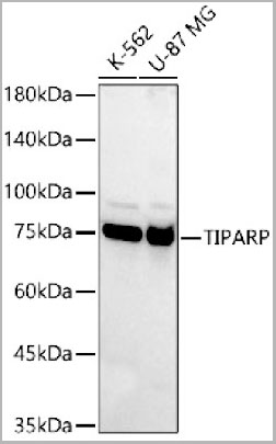
(Western blot analysis of various lysates, using TIPARP Rabbit pAb (AAA28273) at 1:400 dilution.Secondary antibody: HRP Goat Anti-Rabbit IgG (H+L) at 1:10000 dilution.Lysates/proteins: 25ug per lane.Blocking buffer: 3% nonfat dry milk in TBST.Detection: ECL Basic Kit.Exposure time: 10s.)
WB (Western Blot)
(Western blot analysis of various lysates, using TIPARP Rabbit pAb (AAA28273) at 1:400 dilution.Secondary antibody: HRP Goat Anti-Rabbit IgG (H+L) at 1:10000 dilution.Lysates/proteins: 25ug per lane.Blocking buffer: 3% nonfat dry milk in TBST.Detection: ECL Basic Kit.Exposure time: 10s.)
TIPARP, Polyclonal Antibody (Cat# AAA28273)
Full Name
TIPARP Polyclonal Antibody
Gene Names
TIPARP; PARP7; ARTD14; pART14
Applications
Western Blot
Purity
Affinity Purification
Pricing
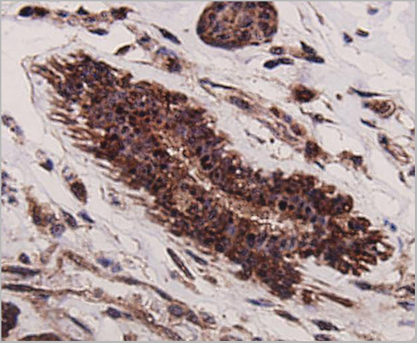
 IHC (Immunohistochemistry)
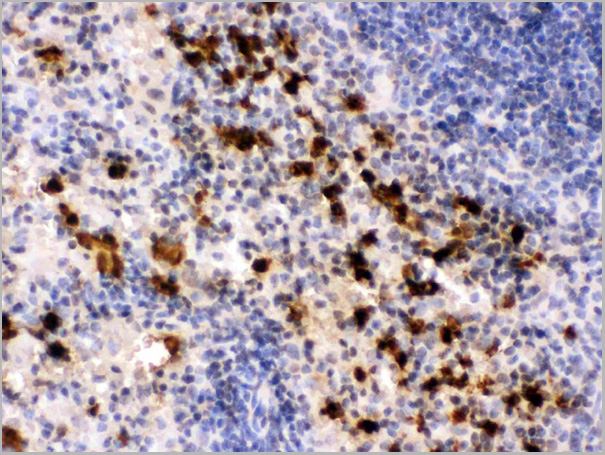
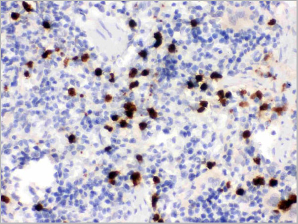
(Anti- S100A9 Picoband antibody, AAA11645, IHC(P)IHC(P): Rat Spleen Tissue)
IHC (Immunohistochemistry)
(Anti- S100A9 Picoband antibody, AAA11645, IHC(P)IHC(P): Rat Spleen Tissue)
S100A9, Polyclonal Antibody (Cat# AAA11645)
Full Name
Anti-S100A9 Antibody
Gene Names
S100a9; p14; Cagb; GAGB; L1Ag; BEE22; MRP14; 60B8Ag; AW546964
Reactivity
Mouse, Rat
Applications
Western Blot, Immunohistochemistry, Immunocytochemistry, Immunofluorescence, Flow Cytometry
Purity
Immunogen Affinity Purified
Pricing
 FCM (Flow Cytometry)
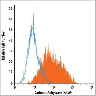
(Detection of Carbonic Anhydrase IX/CA9 in U‑87 MG Human Cell Line by Flow Cytometry using AAA14680.)
FCM (Flow Cytometry)
(Detection of Carbonic Anhydrase IX/CA9 in U‑87 MG Human Cell Line by Flow Cytometry using AAA14680.)
Carbonic Anhydrase IX, Monoclonal Antibody (Cat# AAA14680)
Full Name
Carbonic Anhydrase IX (CA9) (PE)
Gene Names
CA9; MN; CAIX
Applications
Flow Cytometry
Purity
Affinity Purified
Purified by Protein A affinity chromatography from hybridoma culture supernant.
Purified by Protein A affinity chromatography from hybridoma culture supernant.
Pricing
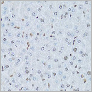
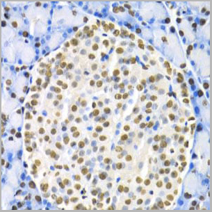
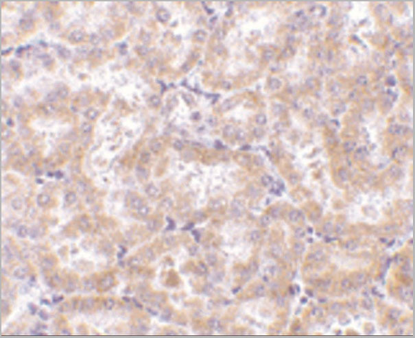
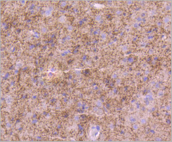
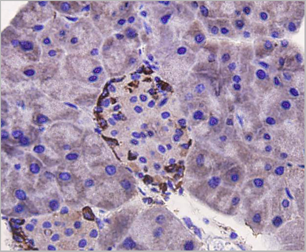
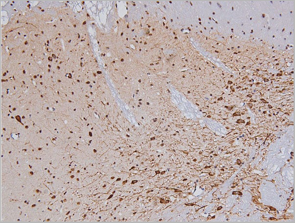
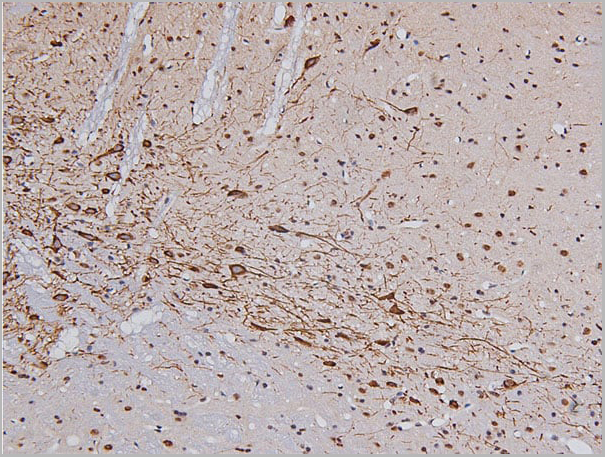
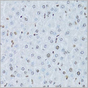 IHC (Immunohistchemistry)
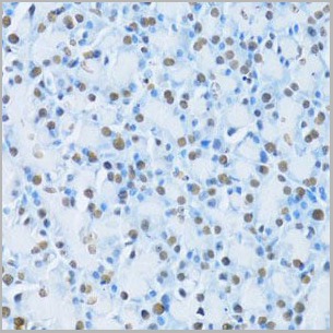
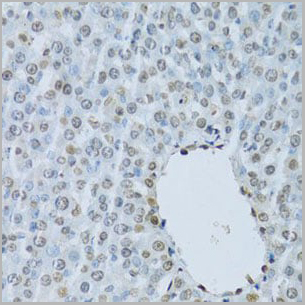
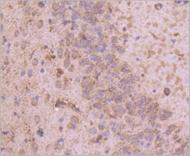
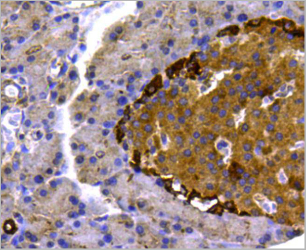
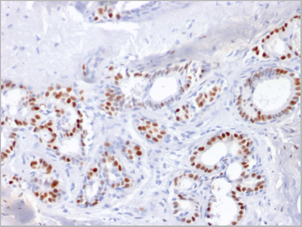
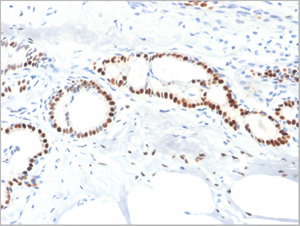
(Immunohistochemistry of paraffin-embedded mouse liver using HNF4A Antibody at dilution of 1:100 (40x lens).)
IHC (Immunohistchemistry)
(Immunohistochemistry of paraffin-embedded mouse liver using HNF4A Antibody at dilution of 1:100 (40x lens).)
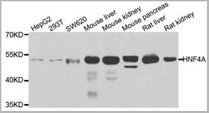
HNF4A, Polyclonal Antibody (Cat# AAA10685)
Full Name
HNF4A Polyclonal Antibody
Gene Names
HNF4A; TCF; HNF4; MODY; FRTS4; MODY1; NR2A1; TCF14; HNF4a7; HNF4a8; HNF4a9; NR2A21; HNF4alpha
Reactivity
Human, Mouse, Rat
Applications
Western Blot, Immunohistochemistry
Purity
Affinity Purification
Pricing
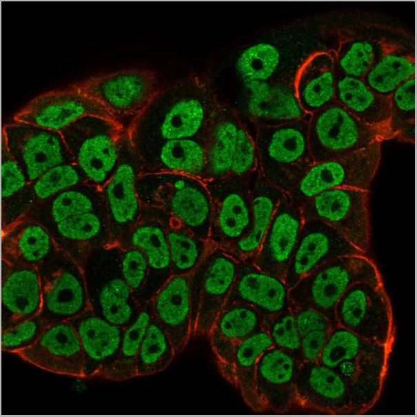
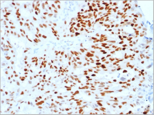
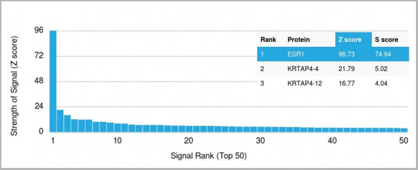
 Application Data
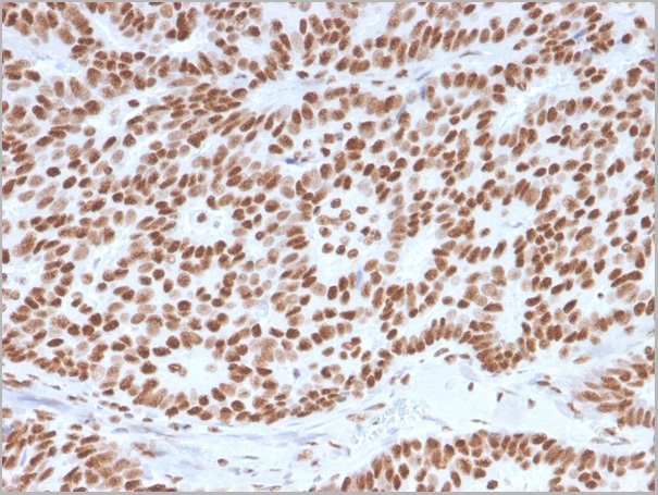
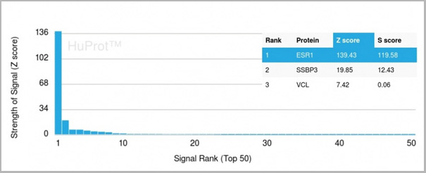
(Analysis of Protein Array containing more than 19,000 full-length human proteins using Estrogen Receptor alpha Mouse Monoclonal Antibody (ESR1/3557) Z- and S- Score: The Z-score represents the strength of a signal that a monoclonal antibody (MAb) (in combination with a fluorescently-tagged anti-IgG secondary antibody) produces when binding to a particular protein on the HuProtTM array. Z-scores are described in units of standard deviations (SD’s) above the mean value of all signals generated on that array. If targets on HuProtTM are arranged in descending order of the Z-score, the S-score is the difference (also in units of SD’s) between the Z-score. S-score therefore represents the relative target specificity of a MAb to its intended target. A MAb is considered to specific to its intended target, if the MAb has an S-score of at least 2.5. For example, if a MAb binds to protein X with a Z-score of 43 and to protein Y with a Z-score of 14, then the S-score for the binding of that MAb to protein X is equal to 29.)
Application Data
(Analysis of Protein Array containing more than 19,000 full-length human proteins using Estrogen Receptor alpha Mouse Monoclonal Antibody (ESR1/3557) Z- and S- Score: The Z-score represents the strength of a signal that a monoclonal antibody (MAb) (in combination with a fluorescently-tagged anti-IgG secondary antibody) produces when binding to a particular protein on the HuProtTM array. Z-scores are described in units of standard deviations (SD’s) above the mean value of all signals generated on that array. If targets on HuProtTM are arranged in descending order of the Z-score, the S-score is the difference (also in units of SD’s) between the Z-score. S-score therefore represents the relative target specificity of a MAb to its intended target. A MAb is considered to specific to its intended target, if the MAb has an S-score of at least 2.5. For example, if a MAb binds to protein X with a Z-score of 43 and to protein Y with a Z-score of 14, then the S-score for the binding of that MAb to protein X is equal to 29.)
Estrogen Receptor, alpha, Monoclonal Antibody (Cat# AAA23915)
Full Name
Estrogen Receptor, alpha (Marker of Estrogen Dependence)
Gene Names
ESR1; ER; ESR; Era; ESRA; ESTRR; NR3A1
Reactivity
Human
Applications
Immunofluorescence, Flow Cytometry, Immunohistochemistry
Purity
Purified Ab with BSA and Azide at 200ug/ml OR Purified Ab WITHOUT BSA and Azide at 1.0mg/ml
Pricing


 Application Data
(Published customer image: Mouse anti V5 tag antibody, clone SV5-Pk1 used for the detection of V5 tagged WEEV_nsP3 protein by western blotting and immunofluorescenceImage caption: WEEV nsP3 interaction with host IKKbeta. A) U87MGs were transfected in a 6-well plate with 5 ug of pUC19 and WEEV_nsP3_HA for 24 hours. Cell lysates were resolved using SDS-PAGE and subsequently immunoblotted with V5 antibody and beta-actin served as a loading control. B) U87MGs were transfected with WEEV_nsP3_V5; cells were fixed after 24 hours and stained with antibodies against the endogenous IKKbeta and the V5 tag. Cells were incubated with appropriate secondary Alexa Fluor antibodies and the nuclei stained with DAPI. Co-localization of IKKbeta with WEEV_nsP3_V5 (yellow) was observed as shown by the arrows. B) Panels E -H serve as an example of transfected cells in a given field of view that show co-localization of IKKbeta and WEEV_nsP3_V5 24 hours post transfection. Panels I-L represent magnified images of other cells showing co-localization of IKKbeta and WEEV_nsP3_V5. Panel M is a magnified image of panel L. The co-localization was confirmed by Z-stack analysis. Co-localization was calculated to be approximately in 61% of cells (163 cells were counted of which 44% demonstrated expression of nsP3. Of those cells that expressed nsP3, 61% showed co-localization of both proteins). Images were taken using Nikon Eclipse TE2000-U at 60x magnification and are representative of 2 independent experiments.From: Amaya M, Voss K, Sampey G, Senina S, de la Fuente C, et al. (2014) The Role of IKKbeta in Venezuelan Equine Encephalitis Virus Infection. PLoS ONE 9(2): e86745.)
Application Data
(Published customer image: Mouse anti V5 tag antibody, clone SV5-Pk1 used for the detection of V5 tagged WEEV_nsP3 protein by western blotting and immunofluorescenceImage caption: WEEV nsP3 interaction with host IKKbeta. A) U87MGs were transfected in a 6-well plate with 5 ug of pUC19 and WEEV_nsP3_HA for 24 hours. Cell lysates were resolved using SDS-PAGE and subsequently immunoblotted with V5 antibody and beta-actin served as a loading control. B) U87MGs were transfected with WEEV_nsP3_V5; cells were fixed after 24 hours and stained with antibodies against the endogenous IKKbeta and the V5 tag. Cells were incubated with appropriate secondary Alexa Fluor antibodies and the nuclei stained with DAPI. Co-localization of IKKbeta with WEEV_nsP3_V5 (yellow) was observed as shown by the arrows. B) Panels E -H serve as an example of transfected cells in a given field of view that show co-localization of IKKbeta and WEEV_nsP3_V5 24 hours post transfection. Panels I-L represent magnified images of other cells showing co-localization of IKKbeta and WEEV_nsP3_V5. Panel M is a magnified image of panel L. The co-localization was confirmed by Z-stack analysis. Co-localization was calculated to be approximately in 61% of cells (163 cells were counted of which 44% demonstrated expression of nsP3. Of those cells that expressed nsP3, 61% showed co-localization of both proteins). Images were taken using Nikon Eclipse TE2000-U at 60x magnification and are representative of 2 independent experiments.From: Amaya M, Voss K, Sampey G, Senina S, de la Fuente C, et al. (2014) The Role of IKKbeta in Venezuelan Equine Encephalitis Virus Infection. PLoS ONE 9(2): e86745.)
V5-TAG, Monoclonal Antibody (Cat# AAA12081)
Full Name
MOUSE ANTI V5-TAG:HRP
Applications
Western Blot
Pricing
Elafin, Polyclonal Antibody (Cat# AAA27749)
Full Name
Anti-Elafin Antibody, Rabbit Polyclonal
Gene Names
PI3; ESI; WAP3; SKALP; WFDC14; cementoin
Applications
ELISA
Purity
Protein A affinity chromatography
Pricing
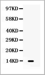
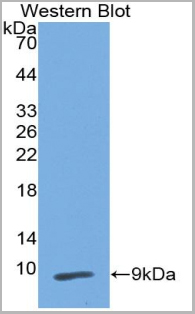 WB (Western Blot)
(Western Blot:Sample: Recombinant PI3, Human.)
WB (Western Blot)
(Western Blot:Sample: Recombinant PI3, Human.)
Peptidase Inhibitor 3, Monoclonal Antibody (Cat# AAA20831)
Full Name
Monoclonal Antibody to Peptidase Inhibitor 3, Skin Derived (PI3)
Gene Names
PI3; ESI; WAP3; SKALP; WFDC14; cementoin
Reactivity
Human, Pig
Applications
WB, IHC, ICC, IP
Purity
Purification: Protein A + Protein G affinity chromatography
Pricing
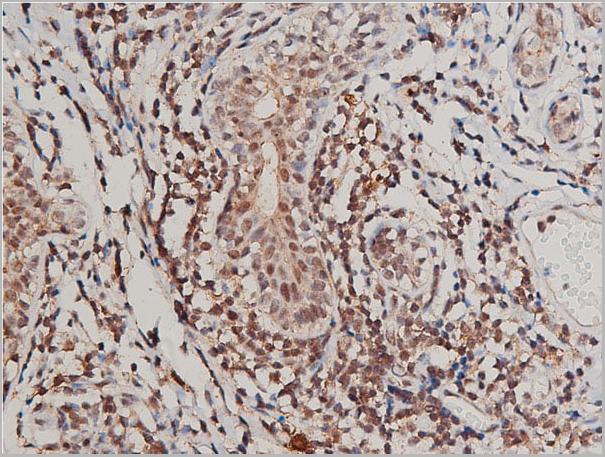
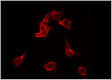
 Application Data
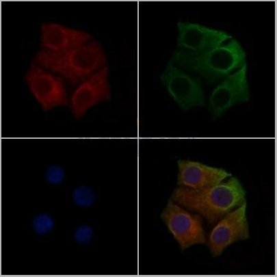
(AAA31020 staining HepG2 cells(30min of 4uM Forskolin treatment) by IF/ICC. The samples were fixed with PFA and permeabilized in 0.1% Triton X-100,then blocked in 10% serum for 45 minutes at 25°C. Samples were then incubated with primary Ab(AAA31020) and mouse anti-beta tubulin Ab for 1 hour at 37°C. An AlexaFluor594 conjugated goat anti-rabbit IgG(H+L) Ab(Red) and an AlexaFluor488 conjugated goat anti-mouse IgG(H+L) Ab(Green) were used as the secondary Ab. The nuclear counter stain is DAPI(blue))
Application Data
(AAA31020 staining HepG2 cells(30min of 4uM Forskolin treatment) by IF/ICC. The samples were fixed with PFA and permeabilized in 0.1% Triton X-100,then blocked in 10% serum for 45 minutes at 25°C. Samples were then incubated with primary Ab(AAA31020) and mouse anti-beta tubulin Ab for 1 hour at 37°C. An AlexaFluor594 conjugated goat anti-rabbit IgG(H+L) Ab(Red) and an AlexaFluor488 conjugated goat anti-mouse IgG(H+L) Ab(Green) were used as the secondary Ab. The nuclear counter stain is DAPI(blue))
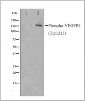
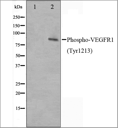
VEGFR1, Polyclonal Antibody (Cat# AAA31020)
Full Name
Phospho-VEGFR1 (Tyr1213) Antibody
Gene Names
FLT1; FLT; FLT-1; VEGFR1; VEGFR-1
Reactivity
Human, Mouse, Rat
Applications
Western Blot, Immunofluorescence, Immunocytochemistry, Immunohistochemistry
Purity
The Ab is from purified rabbit serum by affinity purification via sequential chromatography on phospho-peptide and non-phospho-peptide affinity columns.
Pricing


 Application Data
(PE conjugatedMouse anti Human CD169 antibody, clone 7-239 used to block CD169 function on myeloid cells.Image caption:Siglec-1 mediates HIV-1 uptake into a storage compartment and enhances HIV-1 trans-infection specially in IFN?-treated monocytes and DCs. A. Uptake of HIV-1NL4–3 by different myeloid cells exposed to IFN?. Cells were cultured with HIV-1 to measure p24Gag by ELISA. Mean values and SEM from four experiments include cells from 12 donors. B. Fold change in HIV-1NL4–3 uptake of cells treated with bafilomycin A1 compared to untreated cells. Mean values and SEM include cells from three donors. C. Relative uptake of HIV-1NL4–3 by IFN?-treated myeloid cells pre-incubated with the indicated mAbs. Values are normalized to the level of HIV-1 uptake by mock-treated cells (set at 100%). Mean values and SEM from two experiments include cells from six donors. D. Confocal microscopy analysis of different IFN?-treated myeloid cells pulsed with HIV-1Cherry and stained for Siglec-1 (Alexa 488), HLA-DR (Alexa 647) and DAPI. (Top) Representative viral pattern for each kind of myeloid cell analyzed, showing maximum fluorescence intensity of four channels. (Bottom) Percentage of myeloid cells with distinct viral patterns: random distribution, polarized accumulation, and sac-like compartment formation, as illustrated in the left drawing. Mean values of 50 cells from two different donors are shown. E. HIV-1 transmission from IFN?-treated myeloid cells to a luciferase reporter CD4+ cell line. HIV-1 infection was determined by induced luciferase activity in relative light units (RLUs). Mean values and SEM from four experiments include cells from 12 donors. F. Relative HIV-1 transmission from IFN?-treated myeloid cells pre-incubated with the indicated mAbs. Values are normalized to the level of HIV-1 trans-infected by mock-treated cells. Mean values and SEM from two experiments include cells from six donors. Statistical differences were assessed with a paired t test in A and E, and with a one sample t-test in B, C and F.From: Pino M, Erkizia I, Benet S, Erikson E, Fernández-Figueras MT, Guerrero D, Dalmau J, Ouchi D, Rausell A, Ciuffi A, Keppler OT, Telenti A, Kräusslich HG, Martinez-Picado J, Izquierdo-Useros N.HIV-1 immune activation induces Siglec-1 expression and enhances viral trans-infection in blood and tissue myeloid cells.Retrovirology. 2015 May 7;12:37.This image is from an open access article distributed under the terms of the Creative Commons Attribution License.)
Application Data
(PE conjugatedMouse anti Human CD169 antibody, clone 7-239 used to block CD169 function on myeloid cells.Image caption:Siglec-1 mediates HIV-1 uptake into a storage compartment and enhances HIV-1 trans-infection specially in IFN?-treated monocytes and DCs. A. Uptake of HIV-1NL4–3 by different myeloid cells exposed to IFN?. Cells were cultured with HIV-1 to measure p24Gag by ELISA. Mean values and SEM from four experiments include cells from 12 donors. B. Fold change in HIV-1NL4–3 uptake of cells treated with bafilomycin A1 compared to untreated cells. Mean values and SEM include cells from three donors. C. Relative uptake of HIV-1NL4–3 by IFN?-treated myeloid cells pre-incubated with the indicated mAbs. Values are normalized to the level of HIV-1 uptake by mock-treated cells (set at 100%). Mean values and SEM from two experiments include cells from six donors. D. Confocal microscopy analysis of different IFN?-treated myeloid cells pulsed with HIV-1Cherry and stained for Siglec-1 (Alexa 488), HLA-DR (Alexa 647) and DAPI. (Top) Representative viral pattern for each kind of myeloid cell analyzed, showing maximum fluorescence intensity of four channels. (Bottom) Percentage of myeloid cells with distinct viral patterns: random distribution, polarized accumulation, and sac-like compartment formation, as illustrated in the left drawing. Mean values of 50 cells from two different donors are shown. E. HIV-1 transmission from IFN?-treated myeloid cells to a luciferase reporter CD4+ cell line. HIV-1 infection was determined by induced luciferase activity in relative light units (RLUs). Mean values and SEM from four experiments include cells from 12 donors. F. Relative HIV-1 transmission from IFN?-treated myeloid cells pre-incubated with the indicated mAbs. Values are normalized to the level of HIV-1 trans-infected by mock-treated cells. Mean values and SEM from two experiments include cells from six donors. Statistical differences were assessed with a paired t test in A and E, and with a one sample t-test in B, C and F.From: Pino M, Erkizia I, Benet S, Erikson E, Fernández-Figueras MT, Guerrero D, Dalmau J, Ouchi D, Rausell A, Ciuffi A, Keppler OT, Telenti A, Kräusslich HG, Martinez-Picado J, Izquierdo-Useros N.HIV-1 immune activation induces Siglec-1 expression and enhances viral trans-infection in blood and tissue myeloid cells.Retrovirology. 2015 May 7;12:37.This image is from an open access article distributed under the terms of the Creative Commons Attribution License.)
CD169, Monoclonal Antibody (Cat# AAA12265)
Full Name
Mouse Anti Human CD169: RPE
Gene Names
SIGLEC1; SN; CD169; SIGLEC-1
Reactivity
Human
Applications
Flow Cytometry
Purity
>95% by SDS PAGE
Purified IgG prepared by affinity chromatography on Protein A from tissue culture supernatant.
Purified IgG prepared by affinity chromatography on Protein A from tissue culture supernatant.
Pricing
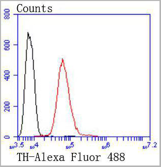 FCM (Flow Cytometry)
(Flow cytometric analysis of SH-SY-5Y cells with Tyrosine Hydroxylase antibody at 1/50 dilution (red) compared with an unlabelled control (cells without incubation with primary antibody; black). Alexa Fluor 488-conjugated goat anti rabbit IgG was use as the secondary antibody)
FCM (Flow Cytometry)
(Flow cytometric analysis of SH-SY-5Y cells with Tyrosine Hydroxylase antibody at 1/50 dilution (red) compared with an unlabelled control (cells without incubation with primary antibody; black). Alexa Fluor 488-conjugated goat anti rabbit IgG was use as the secondary antibody)
Tyrosine Hydroxylase, Monoclonal Antibody (Cat# AAA30209)
Full Name
Tyrosine Hydroxylase Antibody
Gene Names
TH; TYH; DYT14; DYT5b
Reactivity
Human, Mouse, Rat
Applications
Western Blot, Immunocytochemistry, Immunohistochemistry, Flow Cytometry
Purity
ProA affinity purified
Pricing


 Application Data
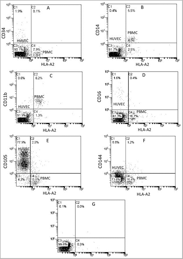
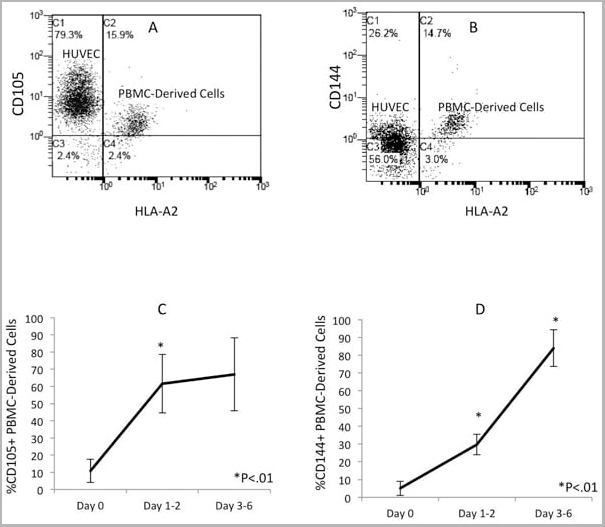
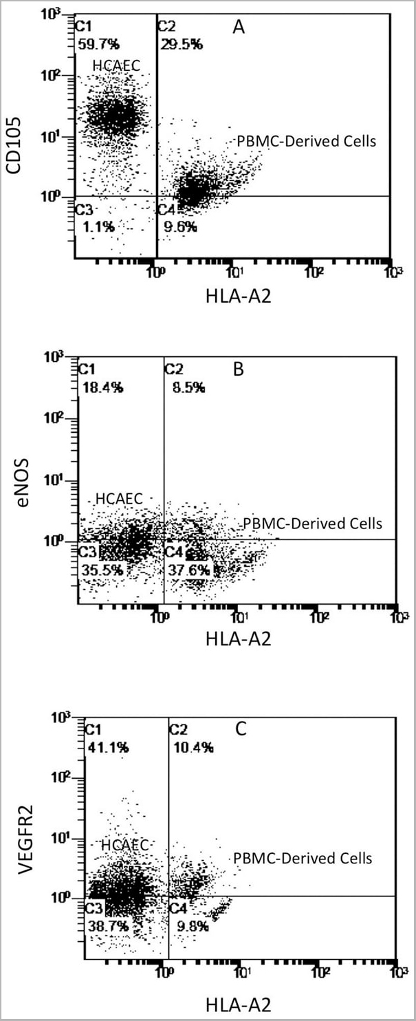
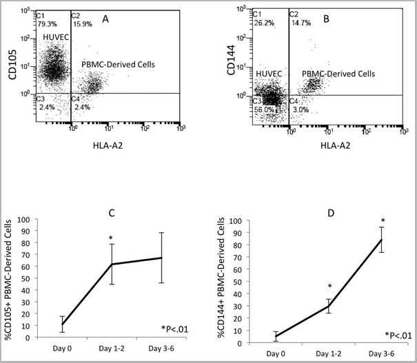
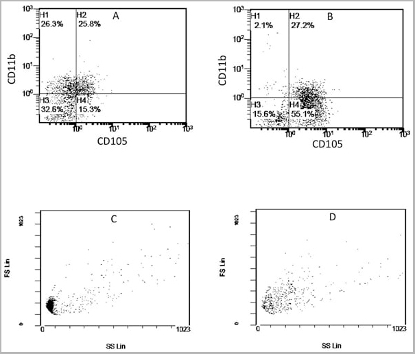
(Published customer image: Mouse anti Human CD105 antibody, clone SN6 used for flow cytometry.Image caption:Phenotype change from HLA-A2+/CD11b+/CD105- to HLA-A2+/CD11b-/CD105+ on endothelium-adherent blood monocyte-derived cells with increase in size and granularity during co-culture. HLA-A2+ PBMCs (1x10⁶ cells/well) were incubated for 2 h (Day 0) with HLA-A2- HUVECs, after which the non-adherent cells were removed by washing. The cell layers were analysed by three-colour flow cytometry staining for HLA-A2, CD11b and CD105 on (A) Day 1 and (B) Day 2. These plots were gated for HLA-A2+ cells. Forward scatter/side scatter dot plots gated for HLA-A2+ cells on Day 0 (C) and Day 2 (D) was shown. These are representative of 2 individual experiments.From: Tso C, Rye K-A, Barter P (2012) Phenotypic and Functional Changes in Blood Monocytes Following Adherence to Endothelium. PLoS ONE 7(5): e37091.)
Application Data
(Published customer image: Mouse anti Human CD105 antibody, clone SN6 used for flow cytometry.Image caption:Phenotype change from HLA-A2+/CD11b+/CD105- to HLA-A2+/CD11b-/CD105+ on endothelium-adherent blood monocyte-derived cells with increase in size and granularity during co-culture. HLA-A2+ PBMCs (1x10⁶ cells/well) were incubated for 2 h (Day 0) with HLA-A2- HUVECs, after which the non-adherent cells were removed by washing. The cell layers were analysed by three-colour flow cytometry staining for HLA-A2, CD11b and CD105 on (A) Day 1 and (B) Day 2. These plots were gated for HLA-A2+ cells. Forward scatter/side scatter dot plots gated for HLA-A2+ cells on Day 0 (C) and Day 2 (D) was shown. These are representative of 2 individual experiments.From: Tso C, Rye K-A, Barter P (2012) Phenotypic and Functional Changes in Blood Monocytes Following Adherence to Endothelium. PLoS ONE 7(5): e37091.)
CD105, Monoclonal Antibody (Cat# AAA12084)
Full Name
MOUSE ANTI HUMAN CD105:RPE
Gene Names
ENG; END; HHT1; ORW1
Applications
Flow Cytometry
Pricing
Double-stranded RNA (dsRNA), Monoclonal ELISA Kit (Cat# AAA14576)
Full Name
Double-stranded RNA (dsRNA) ELISA kit (J2 based)
Reactivity
General
Applications
ELISA
Pricing


 Application Data
(Published customer image: Mouse anti Human CD105 antibody, clone SN6 used for flow cytometry.Image caption:Phenotype change from HLA-A2+/CD11b+/CD105- to HLA-A2+/CD11b-/CD105+ on endothelium-adherent blood monocyte-derived cells with increase in size and granularity during co-culture. HLA-A2+ PBMCs (1x10⁶ cells/well) were incubated for 2 h (Day 0) with HLA-A2- HUVECs, after which the non-adherent cells were removed by washing. The cell layers were analysed by three-colour flow cytometry staining for HLA-A2, CD11b and CD105 on (A) Day 1 and (B) Day 2. These plots were gated for HLA-A2+ cells. Forward scatter/side scatter dot plots gated for HLA-A2+ cells on Day 0 (C) and Day 2 (D) was shown. These are representative of 2 individual experiments.From: Tso C, Rye K-A, Barter P (2012) Phenotypic and Functional Changes in Blood Monocytes Following Adherence to Endothelium. PLoS ONE 7(5): e37091.)
Application Data
(Published customer image: Mouse anti Human CD105 antibody, clone SN6 used for flow cytometry.Image caption:Phenotype change from HLA-A2+/CD11b+/CD105- to HLA-A2+/CD11b-/CD105+ on endothelium-adherent blood monocyte-derived cells with increase in size and granularity during co-culture. HLA-A2+ PBMCs (1x10⁶ cells/well) were incubated for 2 h (Day 0) with HLA-A2- HUVECs, after which the non-adherent cells were removed by washing. The cell layers were analysed by three-colour flow cytometry staining for HLA-A2, CD11b and CD105 on (A) Day 1 and (B) Day 2. These plots were gated for HLA-A2+ cells. Forward scatter/side scatter dot plots gated for HLA-A2+ cells on Day 0 (C) and Day 2 (D) was shown. These are representative of 2 individual experiments.From: Tso C, Rye K-A, Barter P (2012) Phenotypic and Functional Changes in Blood Monocytes Following Adherence to Endothelium. PLoS ONE 7(5): e37091.)
CD105, Monoclonal Antibody (Cat# AAA11933)
Full Name
MOUSE ANTI HUMAN CD105
Gene Names
ENG; END; HHT1; ORW1
Reactivity
Cynomolgus monkey, Horse, Monkey, Primate, Rhesus Monkey
Applications
Immunohistochemistry, Flow Cytometry, Immunoprecipitation, Western Blot
Pricing
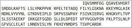
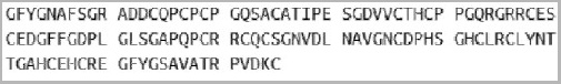 Sequence
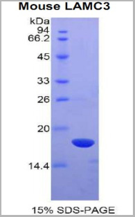
Laminin Gamma 3 (LAMC3), Recombinant Protein (Cat# AAA20229)
Full Name
Recombinant Laminin Gamma 3 (LAMC3)
Gene Names
Lamc3; AI562206; AW240805
Applications
Western Blot
Purity
> 95%
Pricing
Rhinovirus outer capsid Protein 3, Monoclonal Antibody (Cat# AAA13450)
Full Name
Rhinovirus outer capsid Protein 3 (VP3)
Applications
Immunofluorescence
Purity
> 90% pure. Protein A Chromatography.
Pricing


 Application Data
(Published customer image: Mouse anti Human CD105 antibody, clone SN6 used for flow cytometry.Image caption:Phenotype change from HLA-A2+/CD11b+/CD105- to HLA-A2+/CD11b-/CD105+ on endothelium-adherent blood monocyte-derived cells with increase in size and granularity during co-culture. HLA-A2+ PBMCs (1x10⁶ cells/well) were incubated for 2 h (Day 0) with HLA-A2- HUVECs, after which the non-adherent cells were removed by washing. The cell layers were analysed by three-colour flow cytometry staining for HLA-A2, CD11b and CD105 on (A) Day 1 and (B) Day 2. These plots were gated for HLA-A2+ cells. Forward scatter/side scatter dot plots gated for HLA-A2+ cells on Day 0 (C) and Day 2 (D) was shown. These are representative of 2 individual experiments.From: Tso C, Rye K-A, Barter P (2012) Phenotypic and Functional Changes in Blood Monocytes Following Adherence to Endothelium. PLoS ONE 7(5): e37091.)
Application Data
(Published customer image: Mouse anti Human CD105 antibody, clone SN6 used for flow cytometry.Image caption:Phenotype change from HLA-A2+/CD11b+/CD105- to HLA-A2+/CD11b-/CD105+ on endothelium-adherent blood monocyte-derived cells with increase in size and granularity during co-culture. HLA-A2+ PBMCs (1x10⁶ cells/well) were incubated for 2 h (Day 0) with HLA-A2- HUVECs, after which the non-adherent cells were removed by washing. The cell layers were analysed by three-colour flow cytometry staining for HLA-A2, CD11b and CD105 on (A) Day 1 and (B) Day 2. These plots were gated for HLA-A2+ cells. Forward scatter/side scatter dot plots gated for HLA-A2+ cells on Day 0 (C) and Day 2 (D) was shown. These are representative of 2 individual experiments.From: Tso C, Rye K-A, Barter P (2012) Phenotypic and Functional Changes in Blood Monocytes Following Adherence to Endothelium. PLoS ONE 7(5): e37091.)
CD105, Monoclonal Antibody (Cat# AAA12085)
Full Name
MOUSE ANTI HUMAN CD105
Gene Names
ENG; END; HHT1; ORW1
Applications
Immunohistochemistry, Flow Cytometry, Immunoprecipitation, Western Blot
Pricing


 Application Data
(Published customer image: Mouse anti V5 tag antibody, clone SV5-Pk1 used for the detection of V5 tagged WEEV_nsP3 protein by western blotting and immunofluorescenceImage caption: WEEV nsP3 interaction with host IKKbeta. A) U87MGs were transfected in a 6-well plate with 5 ug of pUC19 and WEEV_nsP3_HA for 24 hours. Cell lysates were resolved using SDS-PAGE and subsequently immunoblotted with V5 antibody and beta-actin served as a loading control. B) U87MGs were transfected with WEEV_nsP3_V5; cells were fixed after 24 hours and stained with antibodies against the endogenous IKKbeta and the V5 tag. Cells were incubated with appropriate secondary Alexa Fluor antibodies and the nuclei stained with DAPI. Co-localization of IKKbeta with WEEV_nsP3_V5 (yellow) was observed as shown by the arrows. B) Panels E -H serve as an example of transfected cells in a given field of view that show co-localization of IKKbeta and WEEV_nsP3_V5 24 hours post transfection. Panels I-L represent magnified images of other cells showing co-localization of IKKbeta and WEEV_nsP3_V5. Panel M is a magnified image of panel L. The co-localization was confirmed by Z-stack analysis. Co-localization was calculated to be approximately in 61% of cells (163 cells were counted of which 44% demonstrated expression of nsP3. Of those cells that expressed nsP3, 61% showed co-localization of both proteins). Images were taken using Nikon Eclipse TE2000-U at 60x magnification and are representative of 2 independent experiments.From: Amaya M, Voss K, Sampey G, Senina S, de la Fuente C, et al. (2014) The Role of IKKbeta in Venezuelan Equine Encephalitis Virus Infection. PLoS ONE 9(2): e86745.)
Application Data
(Published customer image: Mouse anti V5 tag antibody, clone SV5-Pk1 used for the detection of V5 tagged WEEV_nsP3 protein by western blotting and immunofluorescenceImage caption: WEEV nsP3 interaction with host IKKbeta. A) U87MGs were transfected in a 6-well plate with 5 ug of pUC19 and WEEV_nsP3_HA for 24 hours. Cell lysates were resolved using SDS-PAGE and subsequently immunoblotted with V5 antibody and beta-actin served as a loading control. B) U87MGs were transfected with WEEV_nsP3_V5; cells were fixed after 24 hours and stained with antibodies against the endogenous IKKbeta and the V5 tag. Cells were incubated with appropriate secondary Alexa Fluor antibodies and the nuclei stained with DAPI. Co-localization of IKKbeta with WEEV_nsP3_V5 (yellow) was observed as shown by the arrows. B) Panels E -H serve as an example of transfected cells in a given field of view that show co-localization of IKKbeta and WEEV_nsP3_V5 24 hours post transfection. Panels I-L represent magnified images of other cells showing co-localization of IKKbeta and WEEV_nsP3_V5. Panel M is a magnified image of panel L. The co-localization was confirmed by Z-stack analysis. Co-localization was calculated to be approximately in 61% of cells (163 cells were counted of which 44% demonstrated expression of nsP3. Of those cells that expressed nsP3, 61% showed co-localization of both proteins). Images were taken using Nikon Eclipse TE2000-U at 60x magnification and are representative of 2 independent experiments.From: Amaya M, Voss K, Sampey G, Senina S, de la Fuente C, et al. (2014) The Role of IKKbeta in Venezuelan Equine Encephalitis Virus Infection. PLoS ONE 9(2): e86745.)
V5-TAG, Monoclonal Antibody (Cat# AAA11930)
Full Name
MOUSE ANTI V5-TAG
Applications
Immunohistochemistry, Flow Cytometry, Immunofluorescence, Immunoprecipitation, Western Blot, Radioimmunoassay
Pricing


 Application Data
(Published customer image: Mouse anti V5 tag antibody, clone SV5-Pk1 used for the detection of V5 tagged WEEV_nsP3 protein by western blotting and immunofluorescenceImage caption: WEEV nsP3 interaction with host IKKbeta. A) U87MGs were transfected in a 6-well plate with 5 ug of pUC19 and WEEV_nsP3_HA for 24 hours. Cell lysates were resolved using SDS-PAGE and subsequently immunoblotted with V5 antibody and beta-actin served as a loading control. B) U87MGs were transfected with WEEV_nsP3_V5; cells were fixed after 24 hours and stained with antibodies against the endogenous IKKbeta and the V5 tag. Cells were incubated with appropriate secondary Alexa Fluor antibodies and the nuclei stained with DAPI. Co-localization of IKKbeta with WEEV_nsP3_V5 (yellow) was observed as shown by the arrows. B) Panels E -H serve as an example of transfected cells in a given field of view that show co-localization of IKKbeta and WEEV_nsP3_V5 24 hours post transfection. Panels I-L represent magnified images of other cells showing co-localization of IKKbeta and WEEV_nsP3_V5. Panel M is a magnified image of panel L. The co-localization was confirmed by Z-stack analysis. Co-localization was calculated to be approximately in 61% of cells (163 cells were counted of which 44% demonstrated expression of nsP3. Of those cells that expressed nsP3, 61% showed co-localization of both proteins). Images were taken using Nikon Eclipse TE2000-U at 60x magnification and are representative of 2 independent experiments.From: Amaya M, Voss K, Sampey G, Senina S, de la Fuente C, et al. (2014) The Role of IKKbeta in Venezuelan Equine Encephalitis Virus Infection. PLoS ONE 9(2): e86745.)
Application Data
(Published customer image: Mouse anti V5 tag antibody, clone SV5-Pk1 used for the detection of V5 tagged WEEV_nsP3 protein by western blotting and immunofluorescenceImage caption: WEEV nsP3 interaction with host IKKbeta. A) U87MGs were transfected in a 6-well plate with 5 ug of pUC19 and WEEV_nsP3_HA for 24 hours. Cell lysates were resolved using SDS-PAGE and subsequently immunoblotted with V5 antibody and beta-actin served as a loading control. B) U87MGs were transfected with WEEV_nsP3_V5; cells were fixed after 24 hours and stained with antibodies against the endogenous IKKbeta and the V5 tag. Cells were incubated with appropriate secondary Alexa Fluor antibodies and the nuclei stained with DAPI. Co-localization of IKKbeta with WEEV_nsP3_V5 (yellow) was observed as shown by the arrows. B) Panels E -H serve as an example of transfected cells in a given field of view that show co-localization of IKKbeta and WEEV_nsP3_V5 24 hours post transfection. Panels I-L represent magnified images of other cells showing co-localization of IKKbeta and WEEV_nsP3_V5. Panel M is a magnified image of panel L. The co-localization was confirmed by Z-stack analysis. Co-localization was calculated to be approximately in 61% of cells (163 cells were counted of which 44% demonstrated expression of nsP3. Of those cells that expressed nsP3, 61% showed co-localization of both proteins). Images were taken using Nikon Eclipse TE2000-U at 60x magnification and are representative of 2 independent experiments.From: Amaya M, Voss K, Sampey G, Senina S, de la Fuente C, et al. (2014) The Role of IKKbeta in Venezuelan Equine Encephalitis Virus Infection. PLoS ONE 9(2): e86745.)
V5-TAG, Monoclonal Antibody (Cat# AAA11864)
Full Name
MOUSE ANTI V5-TAG:FITC
Applications
Immunofluorescence
Pricing
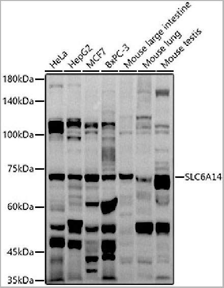 WB (Western Blot)
(Western blot analysis of extracts of various cell lines, using SLC6A14 antibody (AAA28230) at 1:1000 dilution.Secondary antibody: HRP Goat Anti-Rabbit IgG (H+L) at 1:10000 dilution. Lysates/proteins: 25ug per lane. Blocking buffer: 3% nonfat dry milk in TBST. Detection: ECL Basic Kit. Exposure time: 90s)
WB (Western Blot)
(Western blot analysis of extracts of various cell lines, using SLC6A14 antibody (AAA28230) at 1:1000 dilution.Secondary antibody: HRP Goat Anti-Rabbit IgG (H+L) at 1:10000 dilution. Lysates/proteins: 25ug per lane. Blocking buffer: 3% nonfat dry milk in TBST. Detection: ECL Basic Kit. Exposure time: 90s)
SLC6A14, Polyclonal Antibody (Cat# AAA28230)
Full Name
SLC6A14 Polyclonal Antibody
Gene Names
SLC6A14; BMIQ11
Reactivity
Human, Mouse
Applications
Western Blot
Purity
Affinity Purification
Pricing


 Application Data
(Published customer image: Mouse anti Human CD105 antibody, clone SN6 used for flow cytometry.Image caption:Phenotype change from HLA-A2+/CD11b+/CD105- to HLA-A2+/CD11b-/CD105+ on endothelium-adherent blood monocyte-derived cells with increase in size and granularity during co-culture. HLA-A2+ PBMCs (1x10⁶ cells/well) were incubated for 2 h (Day 0) with HLA-A2- HUVECs, after which the non-adherent cells were removed by washing. The cell layers were analysed by three-colour flow cytometry staining for HLA-A2, CD11b and CD105 on (A) Day 1 and (B) Day 2. These plots were gated for HLA-A2+ cells. Forward scatter/side scatter dot plots gated for HLA-A2+ cells on Day 0 (C) and Day 2 (D) was shown. These are representative of 2 individual experiments.From: Tso C, Rye K-A, Barter P (2012) Phenotypic and Functional Changes in Blood Monocytes Following Adherence to Endothelium. PLoS ONE 7(5): e37091.)
Application Data
(Published customer image: Mouse anti Human CD105 antibody, clone SN6 used for flow cytometry.Image caption:Phenotype change from HLA-A2+/CD11b+/CD105- to HLA-A2+/CD11b-/CD105+ on endothelium-adherent blood monocyte-derived cells with increase in size and granularity during co-culture. HLA-A2+ PBMCs (1x10⁶ cells/well) were incubated for 2 h (Day 0) with HLA-A2- HUVECs, after which the non-adherent cells were removed by washing. The cell layers were analysed by three-colour flow cytometry staining for HLA-A2, CD11b and CD105 on (A) Day 1 and (B) Day 2. These plots were gated for HLA-A2+ cells. Forward scatter/side scatter dot plots gated for HLA-A2+ cells on Day 0 (C) and Day 2 (D) was shown. These are representative of 2 individual experiments.From: Tso C, Rye K-A, Barter P (2012) Phenotypic and Functional Changes in Blood Monocytes Following Adherence to Endothelium. PLoS ONE 7(5): e37091.)
CD105, Monoclonal Antibody (Cat# AAA12029)
Full Name
MOUSE ANTI HUMAN CD105:RPE
Gene Names
ENG; END; HHT1; ORW1
Applications
Flow Cytometry
Pricing


 Application Data
(Published customer image: Mouse anti Human CD105 antibody, clone SN6 used for flow cytometry.Image caption:Phenotype change from HLA-A2+/CD11b+/CD105- to HLA-A2+/CD11b-/CD105+ on endothelium-adherent blood monocyte-derived cells with increase in size and granularity during co-culture. HLA-A2+ PBMCs (1x10⁶ cells/well) were incubated for 2 h (Day 0) with HLA-A2- HUVECs, after which the non-adherent cells were removed by washing. The cell layers were analysed by three-colour flow cytometry staining for HLA-A2, CD11b and CD105 on (A) Day 1 and (B) Day 2. These plots were gated for HLA-A2+ cells. Forward scatter/side scatter dot plots gated for HLA-A2+ cells on Day 0 (C) and Day 2 (D) was shown. These are representative of 2 individual experiments.From: Tso C, Rye K-A, Barter P (2012) Phenotypic and Functional Changes in Blood Monocytes Following Adherence to Endothelium. PLoS ONE 7(5): e37091.)
Application Data
(Published customer image: Mouse anti Human CD105 antibody, clone SN6 used for flow cytometry.Image caption:Phenotype change from HLA-A2+/CD11b+/CD105- to HLA-A2+/CD11b-/CD105+ on endothelium-adherent blood monocyte-derived cells with increase in size and granularity during co-culture. HLA-A2+ PBMCs (1x10⁶ cells/well) were incubated for 2 h (Day 0) with HLA-A2- HUVECs, after which the non-adherent cells were removed by washing. The cell layers were analysed by three-colour flow cytometry staining for HLA-A2, CD11b and CD105 on (A) Day 1 and (B) Day 2. These plots were gated for HLA-A2+ cells. Forward scatter/side scatter dot plots gated for HLA-A2+ cells on Day 0 (C) and Day 2 (D) was shown. These are representative of 2 individual experiments.From: Tso C, Rye K-A, Barter P (2012) Phenotypic and Functional Changes in Blood Monocytes Following Adherence to Endothelium. PLoS ONE 7(5): e37091.)
CD105, Monoclonal Antibody (Cat# AAA12083)
Full Name
MOUSE ANTI HUMAN CD105:FITC
Gene Names
ENG; END; HHT1; ORW1
Applications
Flow Cytometry
Pricing


 Application Data
(Published customer image: Mouse anti Human CD105 antibody, clone SN6 used for flow cytometry.Image caption:Phenotype change from HLA-A2+/CD11b+/CD105- to HLA-A2+/CD11b-/CD105+ on endothelium-adherent blood monocyte-derived cells with increase in size and granularity during co-culture. HLA-A2+ PBMCs (1x10⁶ cells/well) were incubated for 2 h (Day 0) with HLA-A2- HUVECs, after which the non-adherent cells were removed by washing. The cell layers were analysed by three-colour flow cytometry staining for HLA-A2, CD11b and CD105 on (A) Day 1 and (B) Day 2. These plots were gated for HLA-A2+ cells. Forward scatter/side scatter dot plots gated for HLA-A2+ cells on Day 0 (C) and Day 2 (D) was shown. These are representative of 2 individual experiments.From: Tso C, Rye K-A, Barter P (2012) Phenotypic and Functional Changes in Blood Monocytes Following Adherence to Endothelium. PLoS ONE 7(5): e37091.)
Application Data
(Published customer image: Mouse anti Human CD105 antibody, clone SN6 used for flow cytometry.Image caption:Phenotype change from HLA-A2+/CD11b+/CD105- to HLA-A2+/CD11b-/CD105+ on endothelium-adherent blood monocyte-derived cells with increase in size and granularity during co-culture. HLA-A2+ PBMCs (1x10⁶ cells/well) were incubated for 2 h (Day 0) with HLA-A2- HUVECs, after which the non-adherent cells were removed by washing. The cell layers were analysed by three-colour flow cytometry staining for HLA-A2, CD11b and CD105 on (A) Day 1 and (B) Day 2. These plots were gated for HLA-A2+ cells. Forward scatter/side scatter dot plots gated for HLA-A2+ cells on Day 0 (C) and Day 2 (D) was shown. These are representative of 2 individual experiments.From: Tso C, Rye K-A, Barter P (2012) Phenotypic and Functional Changes in Blood Monocytes Following Adherence to Endothelium. PLoS ONE 7(5): e37091.)
CD105, Monoclonal Antibody (Cat# AAA11865)
Full Name
MOUSE ANTI HUMAN CD105:FITC
Gene Names
ENG; END; HHT1; ORW1
Applications
Flow Cytometry
Pricing


 Application Data
(Published customer image: Mouse anti V5 tag antibody, clone SV5-Pk1 used for the detection of V5 tagged WEEV_nsP3 protein by western blotting and immunofluorescenceImage caption: WEEV nsP3 interaction with host IKKbeta. A) U87MGs were transfected in a 6-well plate with 5 ug of pUC19 and WEEV_nsP3_HA for 24 hours. Cell lysates were resolved using SDS-PAGE and subsequently immunoblotted with V5 antibody and beta-actin served as a loading control. B) U87MGs were transfected with WEEV_nsP3_V5; cells were fixed after 24 hours and stained with antibodies against the endogenous IKKbeta and the V5 tag. Cells were incubated with appropriate secondary Alexa Fluor antibodies and the nuclei stained with DAPI. Co-localization of IKKbeta with WEEV_nsP3_V5 (yellow) was observed as shown by the arrows. B) Panels E -H serve as an example of transfected cells in a given field of view that show co-localization of IKKbeta and WEEV_nsP3_V5 24 hours post transfection. Panels I-L represent magnified images of other cells showing co-localization of IKKbeta and WEEV_nsP3_V5. Panel M is a magnified image of panel L. The co-localization was confirmed by Z-stack analysis. Co-localization was calculated to be approximately in 61% of cells (163 cells were counted of which 44% demonstrated expression of nsP3. Of those cells that expressed nsP3, 61% showed co-localization of both proteins). Images were taken using Nikon Eclipse TE2000-U at 60x magnification and are representative of 2 independent experiments.From: Amaya M, Voss K, Sampey G, Senina S, de la Fuente C, et al. (2014) The Role of IKKbeta in Venezuelan Equine Encephalitis Virus Infection. PLoS ONE 9(2): e86745.)
Application Data
(Published customer image: Mouse anti V5 tag antibody, clone SV5-Pk1 used for the detection of V5 tagged WEEV_nsP3 protein by western blotting and immunofluorescenceImage caption: WEEV nsP3 interaction with host IKKbeta. A) U87MGs were transfected in a 6-well plate with 5 ug of pUC19 and WEEV_nsP3_HA for 24 hours. Cell lysates were resolved using SDS-PAGE and subsequently immunoblotted with V5 antibody and beta-actin served as a loading control. B) U87MGs were transfected with WEEV_nsP3_V5; cells were fixed after 24 hours and stained with antibodies against the endogenous IKKbeta and the V5 tag. Cells were incubated with appropriate secondary Alexa Fluor antibodies and the nuclei stained with DAPI. Co-localization of IKKbeta with WEEV_nsP3_V5 (yellow) was observed as shown by the arrows. B) Panels E -H serve as an example of transfected cells in a given field of view that show co-localization of IKKbeta and WEEV_nsP3_V5 24 hours post transfection. Panels I-L represent magnified images of other cells showing co-localization of IKKbeta and WEEV_nsP3_V5. Panel M is a magnified image of panel L. The co-localization was confirmed by Z-stack analysis. Co-localization was calculated to be approximately in 61% of cells (163 cells were counted of which 44% demonstrated expression of nsP3. Of those cells that expressed nsP3, 61% showed co-localization of both proteins). Images were taken using Nikon Eclipse TE2000-U at 60x magnification and are representative of 2 independent experiments.From: Amaya M, Voss K, Sampey G, Senina S, de la Fuente C, et al. (2014) The Role of IKKbeta in Venezuelan Equine Encephalitis Virus Infection. PLoS ONE 9(2): e86745.)
V5-TAG, Monoclonal Antibody (Cat# AAA12211)
Full Name
MOUSE ANTI V5-TAG
Applications
Immunohistochemistry, Flow Cytometry, Immunofluorescence, Immunoprecipitation, Western Blot, Radioimmunoassay
Pricing
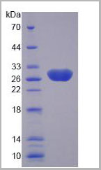
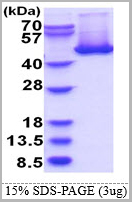
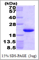 SDS-PAGE
(3 ug by SDS-PAGE under reducing condition and visualized by coomassie blue stain.)
SDS-PAGE
(3 ug by SDS-PAGE under reducing condition and visualized by coomassie blue stain.)
HMGN1, Recombinant Protein (Cat# AAA11776)
Full Name
HMGN1, 1-100aa, Human, His tag, E Coli
Gene Names
HMGN1; HMG14
Applications
SDS-PAGE
Purity
> 95% by SDS-PAGE
Pricing
Rhinovirus outer capsid protein 3, Monoclonal Antibody (Cat# AAA13449)
Full Name
Rhinovirus outer capsid protein 3 (VP3)
Applications
Immunofluorescence
Purity
> 90% pure. Protein A Chromatography
Pricing
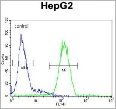
 FCM (Flow Cytometry)
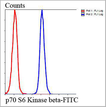
(Flow cytometric analysis of HepG2 cells with p70 S6 Kinase beta antibody at 1/100 dilution (blue) compared with an unlabelled control (cells without incubation with primary antibody; red). Goat anti rabbit IgG (FITC) was used as the secondary antibody.)
FCM (Flow Cytometry)
(Flow cytometric analysis of HepG2 cells with p70 S6 Kinase beta antibody at 1/100 dilution (blue) compared with an unlabelled control (cells without incubation with primary antibody; red). Goat anti rabbit IgG (FITC) was used as the secondary antibody.)
p70S6 Kinase beta, Polyclonal Antibody (Cat# AAA29952)
Full Name
p70 S6 Kinase beta Antibody
Gene Names
RPS6KB2; KLS; SRK; S6K2; STK14B; p70S6Kb; P70-beta; S6K-beta2; P70-beta-1; P70-beta-2; p70(S6K)-beta
Reactivity
Human, Mouse, Rat
Applications
Western Blot, Immunocytochemistry, Immunohistochemistry, Flow Cytometry
Purity
Peptide affinity purified
Pricing


 Application Data
(Published customer image: Mouse anti V5 tag antibody, clone SV5-Pk1 used for the detection of V5 tagged WEEV_nsP3 protein by western blotting and immunofluorescenceImage caption: WEEV nsP3 interaction with host IKKbeta. A) U87MGs were transfected in a 6-well plate with 5 ug of pUC19 and WEEV_nsP3_HA for 24 hours. Cell lysates were resolved using SDS-PAGE and subsequently immunoblotted with V5 antibody and beta-actin served as a loading control. B) U87MGs were transfected with WEEV_nsP3_V5; cells were fixed after 24 hours and stained with antibodies against the endogenous IKKbeta and the V5 tag. Cells were incubated with appropriate secondary Alexa Fluor antibodies and the nuclei stained with DAPI. Co-localization of IKKbeta with WEEV_nsP3_V5 (yellow) was observed as shown by the arrows. B) Panels E -H serve as an example of transfected cells in a given field of view that show co-localization of IKKbeta and WEEV_nsP3_V5 24 hours post transfection. Panels I-L represent magnified images of other cells showing co-localization of IKKbeta and WEEV_nsP3_V5. Panel M is a magnified image of panel L. The co-localization was confirmed by Z-stack analysis. Co-localization was calculated to be approximately in 61% of cells (163 cells were counted of which 44% demonstrated expression of nsP3. Of those cells that expressed nsP3, 61% showed co-localization of both proteins). Images were taken using Nikon Eclipse TE2000-U at 60x magnification and are representative of 2 independent experiments.From: Amaya M, Voss K, Sampey G, Senina S, de la Fuente C, et al. (2014) The Role of IKKbeta in Venezuelan Equine Encephalitis Virus Infection. PLoS ONE 9(2): e86745.)
Application Data
(Published customer image: Mouse anti V5 tag antibody, clone SV5-Pk1 used for the detection of V5 tagged WEEV_nsP3 protein by western blotting and immunofluorescenceImage caption: WEEV nsP3 interaction with host IKKbeta. A) U87MGs were transfected in a 6-well plate with 5 ug of pUC19 and WEEV_nsP3_HA for 24 hours. Cell lysates were resolved using SDS-PAGE and subsequently immunoblotted with V5 antibody and beta-actin served as a loading control. B) U87MGs were transfected with WEEV_nsP3_V5; cells were fixed after 24 hours and stained with antibodies against the endogenous IKKbeta and the V5 tag. Cells were incubated with appropriate secondary Alexa Fluor antibodies and the nuclei stained with DAPI. Co-localization of IKKbeta with WEEV_nsP3_V5 (yellow) was observed as shown by the arrows. B) Panels E -H serve as an example of transfected cells in a given field of view that show co-localization of IKKbeta and WEEV_nsP3_V5 24 hours post transfection. Panels I-L represent magnified images of other cells showing co-localization of IKKbeta and WEEV_nsP3_V5. Panel M is a magnified image of panel L. The co-localization was confirmed by Z-stack analysis. Co-localization was calculated to be approximately in 61% of cells (163 cells were counted of which 44% demonstrated expression of nsP3. Of those cells that expressed nsP3, 61% showed co-localization of both proteins). Images were taken using Nikon Eclipse TE2000-U at 60x magnification and are representative of 2 independent experiments.From: Amaya M, Voss K, Sampey G, Senina S, de la Fuente C, et al. (2014) The Role of IKKbeta in Venezuelan Equine Encephalitis Virus Infection. PLoS ONE 9(2): e86745.)
V5-TAG, Monoclonal Antibody (Cat# AAA11850)
Full Name
MOUSE ANTI V5-TAG:Biotin
Applications
Immunohistochemistry, Western Blot
Pricing
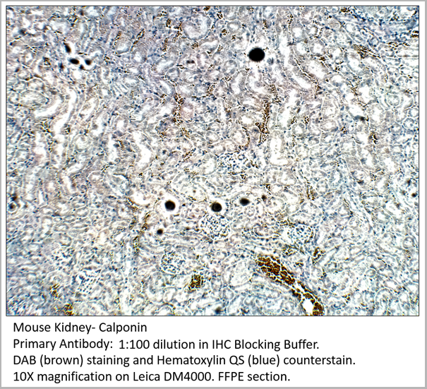
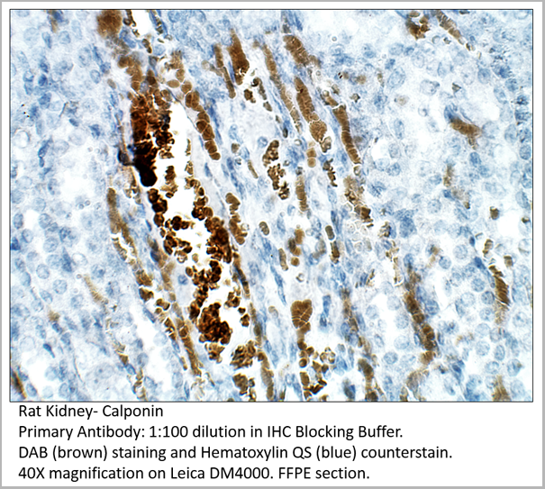
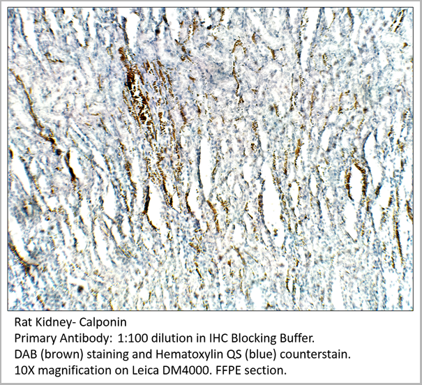
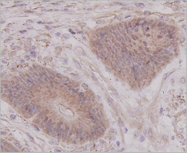
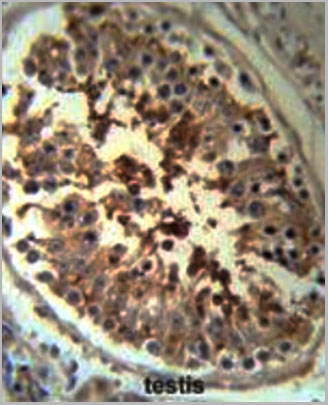
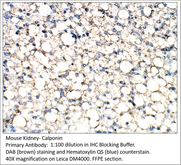 IHC (Immunohistochemistry)
Calponin, Polyclonal Antibody (Cat# AAA14404)
Full Name
Calponin Antibody
Gene Names
CNN1; SMCC; Sm-Calp; HEL-S-14
Reactivity
Human, Mouse, Rat
Applications
Immunohistochemistry, Immunoprecipitation, Western Blot
Pricing
 FCM (Flow Cytometry)
(SERPINH1 Antibody (Center) (Cat. #AAA28730) flow cytometric analysis of HepG2 cells (right histogram) compared to a negative control cell (left histogram).FITC-conjugated goat-anti-rabbit secondary antibodies were used for the analysis.)
FCM (Flow Cytometry)
(SERPINH1 Antibody (Center) (Cat. #AAA28730) flow cytometric analysis of HepG2 cells (right histogram) compared to a negative control cell (left histogram).FITC-conjugated goat-anti-rabbit secondary antibodies were used for the analysis.)
SERPINH1, Polyclonal Antibody (Cat# AAA28730)
Full Name
SERPINH1 Antibody (Center)
Gene Names
SERPINH1; CBP1; CBP2; OI10; gp46; AsTP3; HSP47; PIG14; PPROM; RA-A47; SERPINH2
Reactivity
Human (Predicted Reactivity: Bovine, Mouse, Rat)
Applications
Western Blot, Immunohistochemistry, Flow Cytometry
Purity
Purified Rabbit Polyclonal Antibody (Pab)
Pricing
 SDS-PAGE
(3ug by SDS-PAGE under reducing condition and visualized by coomassie blue stain.)
SDS-PAGE
(3ug by SDS-PAGE under reducing condition and visualized by coomassie blue stain.)
HSP47, Recombinant Protein (Cat# AAA11757)
Full Name
HSP47 (Colligin), 18-418aa, Human, His tag, E Coli
Gene Names
SERPINH1; CBP1; CBP2; OI10; gp46; AsTP3; HSP47; PIG14; PPROM; RA-A47; SERPINH2
Applications
SDS-PAGE
Purity
> 90% by SDS-PAGE
Pricing
 IA (Immuno Assay)
(used as capture antibody. Different concentrations were tested (0.5-2 ug/ml).)
IA (Immuno Assay)
(used as capture antibody. Different concentrations were tested (0.5-2 ug/ml).)
Calprotectin, Monoclonal Antibody (Cat# AAA14609)
Full Name
Calprotectin, Human, mAb 27E10, Biotin
Reactivity
Mouse: No; Rhesus Monkey: Yes (subpopulations of macrophages)
Applications
Immunohistochemistry, Immunofluorescence, Flow Cytometry, Immunoassay, Immunoprecipitation, Western Blot
Purity
Protein G
Pricing
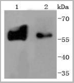
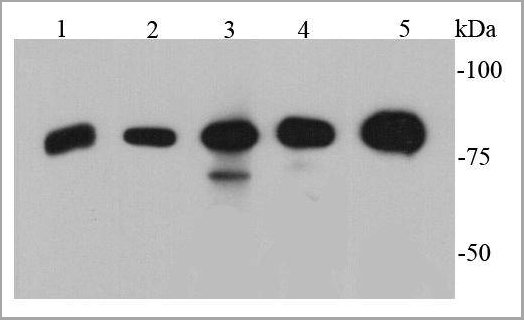
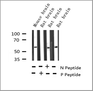 WB (Western Blot)
(Western blot analysis of Phospho-Tyrosine Hydroxylase (Ser19) expression in various lysates)
WB (Western Blot)
(Western blot analysis of Phospho-Tyrosine Hydroxylase (Ser19) expression in various lysates)
Tyrosine Hydroxylase, Polyclonal Antibody (Cat# AAA30988)
Full Name
Phospho-Tyrosine Hydroxylase (Ser19) Antibody
Gene Names
TH; TYH; DYT14; DYT5b
Reactivity
Human, Mouse, Rat
Applications
Western Blot, Immunohistochemistry, Immunofluorescence, Immunocytochemistry
Purity
From purified rabbit serum by affinity purification via sequential chromatography on phospho-and non-phospho-peptide affinity columns.
Pricing
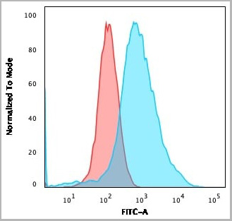
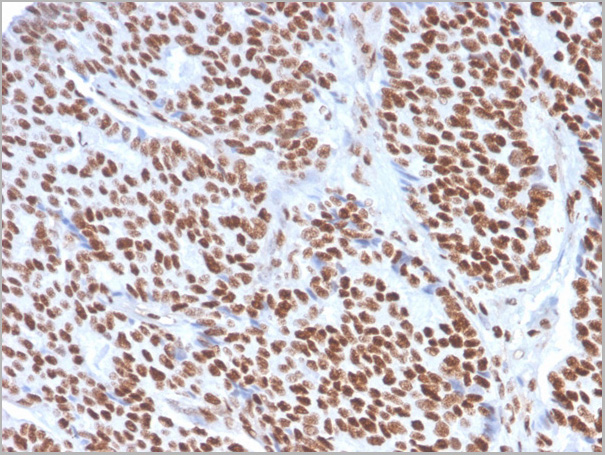
 Application Data
(Analysis of Protein Array containing more than 19,000 full-length human proteins using Estrogen Receptor alpha Mouse Monoclonal Antibody (ESR1/1904) Z- and S- Score: The Z-score represents the strength of a signal that a monoclonal antibody (MAb) (in combination with a fluorescently-tagged anti-IgG secondary antibody) produces when binding to a particular protein on the HuProtTM array. Z-scores are described in units of standard deviations (SD’s) above the mean value of all signals generated on that array. If targets on HuProtTM are arranged in descending order of the Z-score, the S-score is the difference (also in units of SD’s) between the Z-score. S-score therefore represents the relative target specificity of a MAb to its intended target. A MAb is considered to specific to its intended target, if the MAb has an S-score of at least 2.5. For example, if a MAb binds to protein X with a Z-score of 43 and to protein Y with a Z-score of 14, then the S-score for the binding of that MAb to protein X is equal to 29.)
Application Data
(Analysis of Protein Array containing more than 19,000 full-length human proteins using Estrogen Receptor alpha Mouse Monoclonal Antibody (ESR1/1904) Z- and S- Score: The Z-score represents the strength of a signal that a monoclonal antibody (MAb) (in combination with a fluorescently-tagged anti-IgG secondary antibody) produces when binding to a particular protein on the HuProtTM array. Z-scores are described in units of standard deviations (SD’s) above the mean value of all signals generated on that array. If targets on HuProtTM are arranged in descending order of the Z-score, the S-score is the difference (also in units of SD’s) between the Z-score. S-score therefore represents the relative target specificity of a MAb to its intended target. A MAb is considered to specific to its intended target, if the MAb has an S-score of at least 2.5. For example, if a MAb binds to protein X with a Z-score of 43 and to protein Y with a Z-score of 14, then the S-score for the binding of that MAb to protein X is equal to 29.)
Estrogen Receptor, alpha, Monoclonal Antibody (Cat# AAA23914)
Full Name
Estrogen Receptor, alpha (Marker of Estrogen Dependence)
Gene Names
ESR1; ER; ESR; Era; ESRA; ESTRR; NR3A1
Reactivity
Human
Applications
Western Blot, Immunohistochemistry
Purity
Purified Ab with BSA and Azide at 200ug/ml OR Purified Ab WITHOUT BSA and Azide at 1.0mg/ml
Pricing