Filters
Clonality
Type
Reactivity
Gene Name
Isotype
Host
Application
Clone
149 results for " 14 3 3" - showing 50-100


 IF (Immunofluorescence)
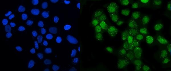
(Immunofluorescence analysis of U2OS cells using p70 S6 Kinase 1 1 antibody at dilution of 1:100. Blue: DAPI for nuclear staining.)
IF (Immunofluorescence)
(Immunofluorescence analysis of U2OS cells using p70 S6 Kinase 1 1 antibody at dilution of 1:100. Blue: DAPI for nuclear staining.)
p70 S6 Kinase 1, Polyclonal Antibody (Cat# AAA28344)
Full Name
p70 S6 Kinase 1 Rabbit pAb
Gene Names
RPS6KB1; S6K; PS6K; S6K1; STK14A; p70-S6K; p70 S6KA; p70-alpha; S6K-beta-1; p70(S6K)-alpha
Reactivity
Human, Mouse, Rat
Applications
Western Blot, Immunohistochemistry, Immunofluorescence
Purity
Affinity purification
Pricing


 IHC (Immunohistchemistry)
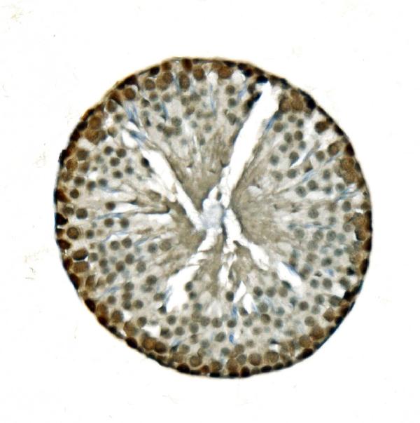
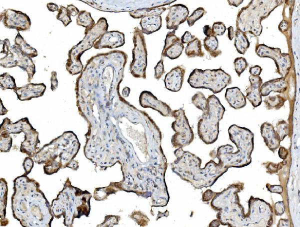
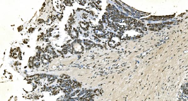
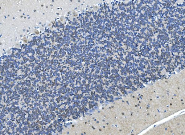
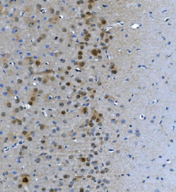
(Immunohistochemistry Analysis: Representative lot data. (Fig. 1 and 2) Paraffin-embedded mouse and human brain tissue was prepared using heat-induced epitope retrieval in citrate buffer, pH 6.0. Immunostaining was performed using a 1:100 dilution. Reactivity was detected using the IHC-Select Detection Kit. Staining pattern appears as cytoplasmic. (Fig. 3 and 4) Paraffin-embedded mouse and mouse olfactory lobe and cerebellum brain tissue was prepared using heat-induced epitope retrieval in citrate buffer, pH 6.0. Immunostaining was performed using a Chicken IgY Antibody 1:100 dilution of Cat. No. AB15894, anti-Tbr2. Reactivity was detected using the IHC-Select Detection Kit. Immunoreactivity seen here is mostly nuclear.)
IHC (Immunohistchemistry)
(Immunohistochemistry Analysis: Representative lot data. (Fig. 1 and 2) Paraffin-embedded mouse and human brain tissue was prepared using heat-induced epitope retrieval in citrate buffer, pH 6.0. Immunostaining was performed using a 1:100 dilution. Reactivity was detected using the IHC-Select Detection Kit. Staining pattern appears as cytoplasmic. (Fig. 3 and 4) Paraffin-embedded mouse and mouse olfactory lobe and cerebellum brain tissue was prepared using heat-induced epitope retrieval in citrate buffer, pH 6.0. Immunostaining was performed using a Chicken IgY Antibody 1:100 dilution of Cat. No. AB15894, anti-Tbr2. Reactivity was detected using the IHC-Select Detection Kit. Immunoreactivity seen here is mostly nuclear.)
EOMES, Polyclonal Antibody (Cat# AAA26891)
Full Name
EOMES (Eomesodermin Homolog, T-box Brain Protein 2, TBR2, T-brain-2, TBR-2)
Gene Names
Eomes; Tbr2; TBR-2; C77258
Reactivity
Mouse, Human, Rat
Applications
Immunohistochemistry, Western Blot
Purity
Purified by affinity chromatography.
Pricing


 FCM (Flow Cytometry)
(Flow cytometric analysis of THP-1 cells with eEF1A1 antibody at 1/100 dilution (red) compared with an unlabelled control (cells without incubation with primary antibody; black). Alexa Fluor 488-conjugated goat anti-rabbit IgG was used as the secondary antibody.)
FCM (Flow Cytometry)
(Flow cytometric analysis of THP-1 cells with eEF1A1 antibody at 1/100 dilution (red) compared with an unlabelled control (cells without incubation with primary antibody; black). Alexa Fluor 488-conjugated goat anti-rabbit IgG was used as the secondary antibody.)
EEF1A1, Monoclonal Antibody (Cat# AAA30467)
Full Name
EEF1A1 Antibody
Gene Names
EEF1A1; CCS3; EF1A; PTI1; CCS-3; EE1A1; EEF-1; EEF1A; EF-Tu; LENG7; eEF1A-1; GRAF-1EF; HNGC:16303
Reactivity
Human, Mouse, Rat
Applications
Western Blot, Immunocytochemistry, Immunofluorescence, Immunohistochemistry, Flow Cytometry, Immunoprecipitation
Purity
ProA affinity purified
Pricing


 IHC (Immunohistchemistry)
(Immunohistochemistry Analysis: Representative lot data. (Fig. 1 and 2) Paraffin-embedded mouse and human brain tissue was prepared using heat-induced epitope retrieval in citrate buffer, pH 6.0. Immunostaining was performed using a 1:100 dilution. Reactivity was detected using the IHC-Select Detection Kit. Staining pattern appears as cytoplasmic. (Fig. 3 and 4) Paraffin-embedded mouse and mouse olfactory lobe and cerebellum brain tissue was prepared using heat-induced epitope retrieval in citrate buffer, pH 6.0. Immunostaining was performed using a Chicken IgY Antibody 1:100 dilution of Cat. No. AB15894, anti-Tbr2. Reactivity was detected using the IHC-Select Detection Kit. Immunoreactivity seen here is mostly nuclear.)
IHC (Immunohistchemistry)
(Immunohistochemistry Analysis: Representative lot data. (Fig. 1 and 2) Paraffin-embedded mouse and human brain tissue was prepared using heat-induced epitope retrieval in citrate buffer, pH 6.0. Immunostaining was performed using a 1:100 dilution. Reactivity was detected using the IHC-Select Detection Kit. Staining pattern appears as cytoplasmic. (Fig. 3 and 4) Paraffin-embedded mouse and mouse olfactory lobe and cerebellum brain tissue was prepared using heat-induced epitope retrieval in citrate buffer, pH 6.0. Immunostaining was performed using a Chicken IgY Antibody 1:100 dilution of Cat. No. AB15894, anti-Tbr2. Reactivity was detected using the IHC-Select Detection Kit. Immunoreactivity seen here is mostly nuclear.)
EOMES, Polyclonal Antibody (Cat# AAA26892)
Full Name
EOMES (Eomesodermin Homolog, T-box Brain Protein 2, TBR2, T-brain-2, TBR-2)
Gene Names
Eomes; Tbr2; TBR-2; C77258
Reactivity
Mouse, Human, Rat
Applications
Immunohistochemistry, Western Blot
Purity
Purified by affinity chromatography.
Pricing
 Standard Curve (Sample)
14 3 3 protein eta (YWHAH), ELISA Kit (Cat# AAA27323)
Full Name
Human 14 3 3 protein eta (YWHAH) ELISA Kit
Gene Names
YWHAH; YWHA1
Reactivity
Human
Pricing


 FCM (Flow Cytometry)
(Dual staining of pig peripheral blood lymphocytes with Mouse anti Pig CD335 detected with Goat anti Mouse IgG (H/L):FITC (STAR117F), and Mouse anti Pig wCD8a:RPE)
FCM (Flow Cytometry)
(Dual staining of pig peripheral blood lymphocytes with Mouse anti Pig CD335 detected with Goat anti Mouse IgG (H/L):FITC (STAR117F), and Mouse anti Pig wCD8a:RPE)
CD335, Monoclonal Antibody (Cat# AAA12253)
Full Name
MOUSE ANTI PIG CD335
Reactivity
Pig
Applications
Flow Cytometry, Immunofluorescence
Pricing


 IHC (Immunohistchemistry)
(Immunohistochemistry Analysis: Representative lot data. (Fig. 1 and 2) Paraffin-embedded mouse and human brain tissue was prepared using heat-induced epitope retrieval in citrate buffer, pH 6.0. Immunostaining was performed using a 1:100 dilution. Reactivity was detected using the IHC-Select Detection Kit. Staining pattern appears as cytoplasmic. (Fig. 3 and 4) Paraffin-embedded mouse and mouse olfactory lobe and cerebellum brain tissue was prepared using heat-induced epitope retrieval in citrate buffer, pH 6.0. Immunostaining was performed using a Chicken IgY Antibody 1:100 dilution of Cat. No. AB15894, anti-Tbr2. Reactivity was detected using the IHC-Select Detection Kit. Immunoreactivity seen here is mostly nuclear.)
IHC (Immunohistchemistry)
(Immunohistochemistry Analysis: Representative lot data. (Fig. 1 and 2) Paraffin-embedded mouse and human brain tissue was prepared using heat-induced epitope retrieval in citrate buffer, pH 6.0. Immunostaining was performed using a 1:100 dilution. Reactivity was detected using the IHC-Select Detection Kit. Staining pattern appears as cytoplasmic. (Fig. 3 and 4) Paraffin-embedded mouse and mouse olfactory lobe and cerebellum brain tissue was prepared using heat-induced epitope retrieval in citrate buffer, pH 6.0. Immunostaining was performed using a Chicken IgY Antibody 1:100 dilution of Cat. No. AB15894, anti-Tbr2. Reactivity was detected using the IHC-Select Detection Kit. Immunoreactivity seen here is mostly nuclear.)
EOMES, Polyclonal Antibody (Cat# AAA26886)
Full Name
EOMES (Eomesodermin Homolog, T-box Brain Protein 2, TBR2, T-brain-2, TBR-2)
Gene Names
Eomes; Tbr2; TBR-2; C77258
Reactivity
Mouse, Human, Rat
Applications
Immunohistochemistry, Western Blot
Purity
Purified by affinity chromatography.
Pricing


 IF (Immunofluorescence)
(Immunofluorescent analysis of 4% paraformaldehyde-fixed, 0.1% Triton X-100 permeabilized MCF-7 (human breast cancer cell line) cells labeling Pdx1 with at 1:25 dilution, followed by DyLight 488-conjugated IgG goat anti-rabbit secondary antibody at 1:200 dilution (green). Immunofluorescence image showing cytoplasm staining on MCF-7 cell line. Cytoplasmic actin is detected with DyLight 554 Phalloidin (PD18466410) at 1:100 dilution (red). The nuclear counter stain is DAPI (blue).)
IF (Immunofluorescence)
(Immunofluorescent analysis of 4% paraformaldehyde-fixed, 0.1% Triton X-100 permeabilized MCF-7 (human breast cancer cell line) cells labeling Pdx1 with at 1:25 dilution, followed by DyLight 488-conjugated IgG goat anti-rabbit secondary antibody at 1:200 dilution (green). Immunofluorescence image showing cytoplasm staining on MCF-7 cell line. Cytoplasmic actin is detected with DyLight 554 Phalloidin (PD18466410) at 1:100 dilution (red). The nuclear counter stain is DAPI (blue).)
OPN-a/b, Polyclonal Antibody (Cat# AAA26865)
Full Name
OPN-a/b, NT (SPP1, BNSP, OPN, Osteopontin, Bone sialoprotein 1, Nephropontin, Secreted phosphoprotein 1, Urinary stone protein, Uropontin) (PE)
Gene Names
SPP1; OPN; BNSP; BSPI; ETA-1
Reactivity
Human
Applications
WB, IHC, IF
Purity
Purified by Protein A and Peptide Affinity Chromatography.
Pricing


 Application Data
(Analysis of Protein Array containing more than 19,000 full-length human proteins using HER-2 Mouse Monoclonal Antibody (ERBB2/3080). Z- and S- Score: The Z-score represents the strength of a signal that a monoclonal antibody (MAb) (in combination with a fluorescently-tagged anti-IgG secondary antibody) produces when binding to a particular protein on the HuProtTM array. Z-scores are described in units of standard deviations (SD's) above the mean value of all signals generated on that array. If targets on HuProtTM are arranged in descending order of the Z-score, the S-score is the difference (also in units of SD's) between the Z-score. S-score therefore represents the relative target specificity of a MAb to its intended target. A MAb is considered to specific to its intended target, if the MAb has an S-score of at least 2.5. For example, if a MAb binds to protein X with a Z-score of 43 and to protein Y with a Z-score of 14, then the S-score for the binding of that MAb to protein X is equal to 29.)
Application Data
(Analysis of Protein Array containing more than 19,000 full-length human proteins using HER-2 Mouse Monoclonal Antibody (ERBB2/3080). Z- and S- Score: The Z-score represents the strength of a signal that a monoclonal antibody (MAb) (in combination with a fluorescently-tagged anti-IgG secondary antibody) produces when binding to a particular protein on the HuProtTM array. Z-scores are described in units of standard deviations (SD's) above the mean value of all signals generated on that array. If targets on HuProtTM are arranged in descending order of the Z-score, the S-score is the difference (also in units of SD's) between the Z-score. S-score therefore represents the relative target specificity of a MAb to its intended target. A MAb is considered to specific to its intended target, if the MAb has an S-score of at least 2.5. For example, if a MAb binds to protein X with a Z-score of 43 and to protein Y with a Z-score of 14, then the S-score for the binding of that MAb to protein X is equal to 29.)
HER-2/c-erbB-2/neu/CD340, Monoclonal Antibody (Cat# AAA23912)
Full Name
HER-2/c-erbB-2/neu/CD340
Gene Names
ERBB2; NEU; NGL; HER2; TKR1; CD340; HER-2; MLN 19; HER-2/neu
Reactivity
Human
Applications
Immunohistochemistry
Purity
Purified Ab with BSA and Azide at 200ug/ml OR Purified Ab WITHOUT BSA and Azide at 1.0mg/ml
Pricing


 IHC (Immunohistchemistry)
(Immunohistochemistry Analysis: Representative lot data. (Fig. 1 and 2) Paraffin-embedded mouse and human brain tissue was prepared using heat-induced epitope retrieval in citrate buffer, pH 6.0. Immunostaining was performed using a 1:100 dilution of AAA14728. Reactivity was detected using the IHC-Select Detection Kit. Staining pattern appears as cytoplasmic. (Fig. 3 and 4) Paraffin-embedded mouse and mouse olfactory lobe and cerebellum brain tissue was prepared using heat-induced epitope retrieval in citrate buffer, pH 6.0. Immunostaining was performed using a Chicken IgY Antibody 1:100 dilution of Cat. No. AB15894, anti-Tbr2. Reactivity was detected using the IHC-Select Detection Kit . Immunoreactivity seen here is mostly nuclear.)
IHC (Immunohistchemistry)
(Immunohistochemistry Analysis: Representative lot data. (Fig. 1 and 2) Paraffin-embedded mouse and human brain tissue was prepared using heat-induced epitope retrieval in citrate buffer, pH 6.0. Immunostaining was performed using a 1:100 dilution of AAA14728. Reactivity was detected using the IHC-Select Detection Kit. Staining pattern appears as cytoplasmic. (Fig. 3 and 4) Paraffin-embedded mouse and mouse olfactory lobe and cerebellum brain tissue was prepared using heat-induced epitope retrieval in citrate buffer, pH 6.0. Immunostaining was performed using a Chicken IgY Antibody 1:100 dilution of Cat. No. AB15894, anti-Tbr2. Reactivity was detected using the IHC-Select Detection Kit . Immunoreactivity seen here is mostly nuclear.)
EOMES, Polyclonal Antibody (Cat# AAA14728)
Full Name
EOMES, NT (Eomesodermin Homolog, T-box Brain Protein 2, TBR2, T-brain-2, TBR-2)
Gene Names
EOMES; TBR2
Reactivity
Human, Mouse, Rat
Applications
Western Blot, Immunohistochemistry
Purity
Affinity Purified
Purified by affinity chromatography.
Purified by affinity chromatography.
Pricing


 Application Data
(Published customer image: Mouse anti Human CD49d antibody, clone HP2/1 used for binding efficiency determinationImage caption:Binding efficiencies (BE) of different a4beta7 molecules composed of distinct a4 mutants to monoclonal antibodies against a4, beta7 or the a4beta7 heterodimer. Binding efficiency is determined by the ratio between the mean fluorescence of antibody binding to each a4 molecule and of the binding in a mock-transfected cell culture (see Materials and Methods for details). Dark gray bars represent binding to the human (wild type) a4 clone, whereas light gray bars are those of binding to the different a4 mutants (as shown in the x-axis). a4 mutants which included substitutions at codon 201 are boxed. A, binding of anti-a4 2b4 antibody. B, binding of anti-a4 HP2/1 antibody. C, BE of different anti-a4 and beta7 antibodies to the human a4 and the quintuple a4 mutant (5 aa mut). Bars represent the range of standard errors deduced from triplicate experiments. p-values of Student's t tests are shown above each comparison. NS, non-significant (> 0.05).From:Darc M, Hait SH, Soares EA, Cicala C, Seuanez HN, et al. (2011) Polymorphisms in the a4 Integrin of Neotropical Primates: Insights for Binding of Natural Ligands and HIV-1 gp120 to the Human a4beta7. PLoS ONE 6(9): e24461.)
Application Data
(Published customer image: Mouse anti Human CD49d antibody, clone HP2/1 used for binding efficiency determinationImage caption:Binding efficiencies (BE) of different a4beta7 molecules composed of distinct a4 mutants to monoclonal antibodies against a4, beta7 or the a4beta7 heterodimer. Binding efficiency is determined by the ratio between the mean fluorescence of antibody binding to each a4 molecule and of the binding in a mock-transfected cell culture (see Materials and Methods for details). Dark gray bars represent binding to the human (wild type) a4 clone, whereas light gray bars are those of binding to the different a4 mutants (as shown in the x-axis). a4 mutants which included substitutions at codon 201 are boxed. A, binding of anti-a4 2b4 antibody. B, binding of anti-a4 HP2/1 antibody. C, BE of different anti-a4 and beta7 antibodies to the human a4 and the quintuple a4 mutant (5 aa mut). Bars represent the range of standard errors deduced from triplicate experiments. p-values of Student's t tests are shown above each comparison. NS, non-significant (> 0.05).From:Darc M, Hait SH, Soares EA, Cicala C, Seuanez HN, et al. (2011) Polymorphisms in the a4 Integrin of Neotropical Primates: Insights for Binding of Natural Ligands and HIV-1 gp120 to the Human a4beta7. PLoS ONE 6(9): e24461.)
CD49d, Monoclonal Antibody (Cat# AAA12003)
Full Name
MOUSE ANTI HUMAN CD49d
Gene Names
ITGA4; IA4; CD49D
Applications
Immunohistochemistry, Flow Cytometry, Functional Assay, Immunoprecipitation
Pricing


 IHC (Immunohistochemistry)
(Immunohistochemical analysis of paraffin-embedded cerebellum tissues using ZEB1 mouse mAb with DAB staining.)
IHC (Immunohistochemistry)
(Immunohistochemical analysis of paraffin-embedded cerebellum tissues using ZEB1 mouse mAb with DAB staining.)
CD22, Monoclonal Antibody (Cat# AAA14141)
Full Name
Anti-CD22 Mouse mAb
Gene Names
CD22; SIGLEC2; SIGLEC-2
Reactivity
Human
Applications
Western Blot, Immunohistochemistry, Immunocytochemistry, Flow Cytometry
Pricing


 FCM (Flow Cytometry)
(Dual staining of pig peripheral blood lymphocytes with Mouse anti Pig CD335 detected with Goat anti Mouse IgG (H/L):FITC (STAR117F), and Mouse anti Pig wCD8a:RPE)
FCM (Flow Cytometry)
(Dual staining of pig peripheral blood lymphocytes with Mouse anti Pig CD335 detected with Goat anti Mouse IgG (H/L):FITC (STAR117F), and Mouse anti Pig wCD8a:RPE)
CD335, Monoclonal Antibody (Cat# AAA12251)
Full Name
MOUSE ANTI PIG CD335: APC
Reactivity
Pig
Applications
Flow Cytometry
Pricing
 Standard Curve (Sample)
17-Beta-Hydroxysteroid Dehydrogenase Type 14 (HSD17b14), ELISA Kit (Cat# AAA20568)
Full Name
Human 17-Beta-Hydroxysteroid Dehydrogenase Type 14 (HSD17b14) ELISA Kit
Gene Names
HSD17B14; DHRS10; SDR47C1; retSDR3
Reactivity
Human
Pricing

 WB (Western Blot)
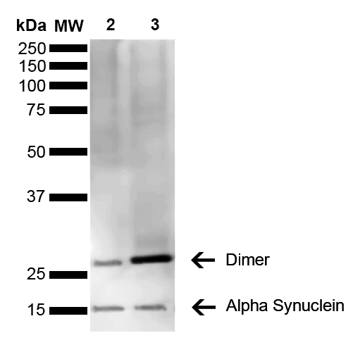
(Western Blot analysis of Mouse, Rat Brain showing detection of 14 kDa Alpha Synuclein protein using Mouse Anti-Alpha Synuclein Monoclonal Antibody, Clone 3C11. Lane 1: Molecular Weight Ladder (MW). Lane 2: Mouse brain cell lysate. Lane 3: Rat brain cell lysate. Load: 15 ug. Block: 5% Skim Milk in 1X TBST. Primary Antibody: Mouse Anti-Alpha Synuclein Monoclonal Antibody at 1:1000 for 2 hours at RT. Secondary Antibody: Goat Anti-Mouse HRP:IgG at 1:3000 for 1 hour at RT. Color Development: ECL solution (Super Signal West Pico) for 5 min in RT. Predicted/Observed Size: 14 kDa. Other Band(s): ~30 kDa (dimer).)
WB (Western Blot)
(Western Blot analysis of Mouse, Rat Brain showing detection of 14 kDa Alpha Synuclein protein using Mouse Anti-Alpha Synuclein Monoclonal Antibody, Clone 3C11. Lane 1: Molecular Weight Ladder (MW). Lane 2: Mouse brain cell lysate. Lane 3: Rat brain cell lysate. Load: 15 ug. Block: 5% Skim Milk in 1X TBST. Primary Antibody: Mouse Anti-Alpha Synuclein Monoclonal Antibody at 1:1000 for 2 hours at RT. Secondary Antibody: Goat Anti-Mouse HRP:IgG at 1:3000 for 1 hour at RT. Color Development: ECL solution (Super Signal West Pico) for 5 min in RT. Predicted/Observed Size: 14 kDa. Other Band(s): ~30 kDa (dimer).)
Alpha Synuclein, Monoclonal Antibody (Cat# AAA17810)
Full Name
Alpha Synuclein Antibody, Clone 3C11
Gene Names
SNCA; PD1; NACP; PARK1; PARK4
Reactivity
Human; Mouse; Rat
Applications
WB, DB, ICC, IF, EIA
Purity
Protein G Purified
Pricing


 FCM (Flow Cytometry)
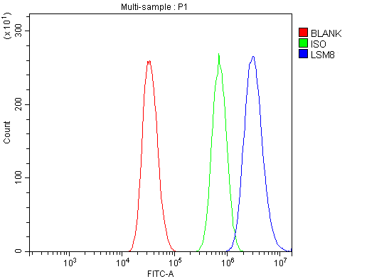
(Figure 13. Flow Cytometry analysis of A431 cells using anti-LSM8 antibody (AAA19337).Overlay histogram showing A431 cells stained with AAA19337 (Blue line). The cells were blocked with 10% normal goat serum. And then incubated with rabbit anti-LSM8 Antibody (AAA19337, 1μg/1x106 cells) for 30 min at 20 degree C. DyLight®488 conjugated goat anti-rabbit IgG (5-10μg/1x106 cells) was used as secondary antibody for 30 minutes at 20 degree C. Isotype control antibody (Green line) was rabbit IgG (1μg/1x106) used under the same conditions. Unlabelled sample (Red line) was also used as a control.)
FCM (Flow Cytometry)
(Figure 13. Flow Cytometry analysis of A431 cells using anti-LSM8 antibody (AAA19337).Overlay histogram showing A431 cells stained with AAA19337 (Blue line). The cells were blocked with 10% normal goat serum. And then incubated with rabbit anti-LSM8 Antibody (AAA19337, 1μg/1x106 cells) for 30 min at 20 degree C. DyLight®488 conjugated goat anti-rabbit IgG (5-10μg/1x106 cells) was used as secondary antibody for 30 minutes at 20 degree C. Isotype control antibody (Green line) was rabbit IgG (1μg/1x106) used under the same conditions. Unlabelled sample (Red line) was also used as a control.)
LSM8, Polyclonal Antibody (Cat# AAA19337)
Full Name
Anti-LSM8 Antibody
Reactivity
Human, Mouse, Rat
Applications
WB, IHC-P, ICC, IF, FC/FACS/FCM, EIA
Purity
Immunogen affinity purified.
Pricing
 Standard Curve (Sample)
Keratin 14 (KRT14), RTU ELISA Kit (Cat# AAA23965)
Full Name
Human Keratin 14 (KRT14) ELISA Kit
Gene Names
KRT14; K14; NFJ; CK14; EBS3; EBS4
Reactivity
Human
Pricing


 IF (Immunofluorescence)
(Immunofluorescent analysis of 4% paraformaldehyde-fixed, 0.1% Triton X-100 permeabilized MCF-7 (human breast cancer cell line) cells labeling Pdx1 with at 1:25 dilution, followed by DyLight 488-conjugated IgG goat anti-rabbit secondary antibody at 1:200 dilution (green). Immunofluorescence image showing cytoplasm staining on MCF-7 cell line. Cytoplasmic actin is detected with DyLight 554 Phalloidin (PD18466410) at 1:100 dilution (red). The nuclear counter stain is DAPI (blue).)
IF (Immunofluorescence)
(Immunofluorescent analysis of 4% paraformaldehyde-fixed, 0.1% Triton X-100 permeabilized MCF-7 (human breast cancer cell line) cells labeling Pdx1 with at 1:25 dilution, followed by DyLight 488-conjugated IgG goat anti-rabbit secondary antibody at 1:200 dilution (green). Immunofluorescence image showing cytoplasm staining on MCF-7 cell line. Cytoplasmic actin is detected with DyLight 554 Phalloidin (PD18466410) at 1:100 dilution (red). The nuclear counter stain is DAPI (blue).)
OPN-a/b, Polyclonal Antibody (Cat# AAA26857)
Full Name
OPN-a/b, NT (SPP1, BNSP, OPN, Osteopontin, Bone sialoprotein 1, Nephropontin, Secreted phosphoprotein 1, Urinary stone protein, Uropontin) (APC)
Gene Names
SPP1; OPN; BNSP; BSPI; ETA-1
Reactivity
Human
Applications
WB, IHC, IF
Purity
Purified by Protein A and Peptide Affinity Chromatography.
Pricing
 Standard Curve (Sample)
tyrosine 3-monooxygenase/tryptophan 5-monooxygenase activation protein, beta polypeptide, ELISA Kit (Cat# AAA15240)
Full Name
Human 14-3-3 protein beta/alpha, YWHAB ELISA Kit
Gene Names
YWHAB; HS1; GW128; YWHAA; KCIP-1; HEL-S-1
Reactivity
Human
Pricing


 IF (Immunofluorescence)
(Immunofluorescent analysis of 4% paraformaldehyde-fixed, 0.1% Triton X-100 permeabilized MCF-7 (human breast cancer cell line) cells labeling Pdx1 with at 1:25 dilution, followed by DyLight 488-conjugated IgG goat anti-rabbit secondary antibody at 1:200 dilution (green). Immunofluorescence image showing cytoplasm staining on MCF-7 cell line. Cytoplasmic actin is detected with DyLight 554 Phalloidin (PD18466410) at 1:100 dilution (red). The nuclear counter stain is DAPI (blue).)
IF (Immunofluorescence)
(Immunofluorescent analysis of 4% paraformaldehyde-fixed, 0.1% Triton X-100 permeabilized MCF-7 (human breast cancer cell line) cells labeling Pdx1 with at 1:25 dilution, followed by DyLight 488-conjugated IgG goat anti-rabbit secondary antibody at 1:200 dilution (green). Immunofluorescence image showing cytoplasm staining on MCF-7 cell line. Cytoplasmic actin is detected with DyLight 554 Phalloidin (PD18466410) at 1:100 dilution (red). The nuclear counter stain is DAPI (blue).)
OPN-a/b, Polyclonal Antibody (Cat# AAA26861)
Full Name
OPN-a/b, NT (SPP1, BNSP, OPN, Osteopontin, Bone sialoprotein 1, Nephropontin, Secreted phosphoprotein 1, Urinary stone protein, Uropontin) (MaxLight 490)
Gene Names
SPP1; OPN; BNSP; BSPI; ETA-1
Reactivity
Human
Applications
WB, IHC, IF
Purity
Purified by Protein A and Peptide Affinity Chromatography.
Pricing
 Standard Curve (Sample)
tyrosine 3-monooxygenase/tryptophan 5-monooxygenase activation protein, gamma polypeptide, ELISA Kit (Cat# AAA18247)
Full Name
Human 14-3-3 protein gamma, YWHAG ELISA Kit
Gene Names
YWHAG; PPP1R170; 14-3-3GAMMA
Reactivity
Human
Pricing


 FCM (Flow Cytometry)
(Figure 14. Flow Cytometry analysis of A431 cells using anti-SAMDH1 antibody (AAA19356).Overlay histogram showing A431 cells stained with AAA19356 (Blue line). The cells were blocked with 10% normal goat serum. And then incubated with mouse anti- SAMDH1 Antibody (AAA19356, 1μg/1x106 cells) for 30 min at 20 degree C. DyLight®488 conjugated goat anti-mouse IgG (BA1126, 5-10μg/1x106 cells) was used as secondary antibody for 30 minutes at 20 degree C. Isotype control antibody (Green line) was mouse IgG (1μg/1x106) used under the same conditions. Unlabelled sample (Red line) was also used as a control.)
FCM (Flow Cytometry)
(Figure 14. Flow Cytometry analysis of A431 cells using anti-SAMDH1 antibody (AAA19356).Overlay histogram showing A431 cells stained with AAA19356 (Blue line). The cells were blocked with 10% normal goat serum. And then incubated with mouse anti- SAMDH1 Antibody (AAA19356, 1μg/1x106 cells) for 30 min at 20 degree C. DyLight®488 conjugated goat anti-mouse IgG (BA1126, 5-10μg/1x106 cells) was used as secondary antibody for 30 minutes at 20 degree C. Isotype control antibody (Green line) was mouse IgG (1μg/1x106) used under the same conditions. Unlabelled sample (Red line) was also used as a control.)
SAMHD1, Monoclonal Antibody (Cat# AAA19356)
Full Name
Anti-SAMHD1 Antibody (monoclonal, 10H8)
Gene Names
SAMHD1; DCIP; CHBL2; HDDC1; MOP-5; SBBI88
Reactivity
Human
Applications
WB, IHC-P, ICC, IF, FC/FACS/FCM
Purity
Immunogen affinity purified.
Pricing


 IF (Immunofluorescence)
(Immunofluorescent analysis of 4% paraformaldehyde-fixed, 0.1% Triton X-100 permeabilized MCF-7 (human breast cancer cell line) cells labeling Pdx1 with at 1:25 dilution, followed by DyLight 488-conjugated IgG goat anti-rabbit secondary antibody at 1:200 dilution (green). Immunofluorescence image showing cytoplasm staining on MCF-7 cell line. Cytoplasmic actin is detected with DyLight 554 Phalloidin (PD18466410) at 1:100 dilution (red). The nuclear counter stain is DAPI (blue).)
IF (Immunofluorescence)
(Immunofluorescent analysis of 4% paraformaldehyde-fixed, 0.1% Triton X-100 permeabilized MCF-7 (human breast cancer cell line) cells labeling Pdx1 with at 1:25 dilution, followed by DyLight 488-conjugated IgG goat anti-rabbit secondary antibody at 1:200 dilution (green). Immunofluorescence image showing cytoplasm staining on MCF-7 cell line. Cytoplasmic actin is detected with DyLight 554 Phalloidin (PD18466410) at 1:100 dilution (red). The nuclear counter stain is DAPI (blue).)
OPN-a/b, Polyclonal Antibody (Cat# AAA26863)
Full Name
OPN-a/b, NT (SPP1, BNSP, OPN, Osteopontin, Bone sialoprotein 1, Nephropontin, Secreted phosphoprotein 1, Urinary stone protein, Uropontin) (MaxLight 650)
Gene Names
SPP1; OPN; BNSP; BSPI; ETA-1
Reactivity
Human
Applications
WB, IHC, IF
Purity
Purified by Protein A and Peptide Affinity Chromatography.
Pricing
 Standard Curve (Sample)
14-3-3 Protein (14-3-3 Pro), ELISA Kit (Cat# AAA13145)
Full Name
Human 14-3-3 Protein (14-3-3 Pro) ELISA Kit
Reactivity
Human
Pricing


 IF (Immunofluorescence)
(Immunofluorescent analysis of 4% paraformaldehyde-fixed, 0.1% Triton X-100 permeabilized MCF-7 (human breast cancer cell line) cells labeling Pdx1 with at 1:25 dilution, followed by DyLight 488-conjugated IgG goat anti-rabbit secondary antibody at 1:200 dilution (green). Immunofluorescence image showing cytoplasm staining on MCF-7 cell line. Cytoplasmic actin is detected with DyLight 554 Phalloidin (PD18466410) at 1:100 dilution (red). The nuclear counter stain is DAPI (blue).)
IF (Immunofluorescence)
(Immunofluorescent analysis of 4% paraformaldehyde-fixed, 0.1% Triton X-100 permeabilized MCF-7 (human breast cancer cell line) cells labeling Pdx1 with at 1:25 dilution, followed by DyLight 488-conjugated IgG goat anti-rabbit secondary antibody at 1:200 dilution (green). Immunofluorescence image showing cytoplasm staining on MCF-7 cell line. Cytoplasmic actin is detected with DyLight 554 Phalloidin (PD18466410) at 1:100 dilution (red). The nuclear counter stain is DAPI (blue).)
OPN-a/b, Polyclonal Antibody (Cat# AAA26864)
Full Name
OPN-a/b, NT (SPP1, BNSP, OPN, Osteopontin, Bone sialoprotein 1, Nephropontin, Secreted phosphoprotein 1, Urinary stone protein, Uropontin) (MaxLight 750)
Gene Names
SPP1; OPN; BNSP; BSPI; ETA-1
Reactivity
Human
Applications
WB, IHC, IF
Purity
Purified by Protein A and Peptide Affinity Chromatography.
Pricing
 Standard Curve (Sample)
Lysyl Oxidase Like Protein 2 (LOXL2), ELISA Kit (Cat# AAA20504)
Full Name
Mouse Lysyl Oxidase Like Protein 2 (LOXL2) ELISA Kit
Gene Names
LOXL2; LOR2; WS9-14
Reactivity
Mouse
Pricing


 IF (Immunofluorescence)
(Immunofluorescent analysis of 4% paraformaldehyde-fixed, 0.1% Triton X-100 permeabilized MCF-7 (human breast cancer cell line) cells labeling Pdx1 with at 1:25 dilution, followed by DyLight 488-conjugated IgG goat anti-rabbit secondary antibody at 1:200 dilution (green). Immunofluorescence image showing cytoplasm staining on MCF-7 cell line. Cytoplasmic actin is detected with DyLight 554 Phalloidin (PD18466410) at 1:100 dilution (red). The nuclear counter stain is DAPI (blue).)
IF (Immunofluorescence)
(Immunofluorescent analysis of 4% paraformaldehyde-fixed, 0.1% Triton X-100 permeabilized MCF-7 (human breast cancer cell line) cells labeling Pdx1 with at 1:25 dilution, followed by DyLight 488-conjugated IgG goat anti-rabbit secondary antibody at 1:200 dilution (green). Immunofluorescence image showing cytoplasm staining on MCF-7 cell line. Cytoplasmic actin is detected with DyLight 554 Phalloidin (PD18466410) at 1:100 dilution (red). The nuclear counter stain is DAPI (blue).)
OPN-a/b, Polyclonal Antibody (Cat# AAA26862)
Full Name
OPN-a/b, NT (SPP1, BNSP, OPN, Osteopontin, Bone sialoprotein 1, Nephropontin, Secreted phosphoprotein 1, Urinary stone protein, Uropontin) (MaxLight 550)
Gene Names
SPP1; OPN; BNSP; BSPI; ETA-1
Reactivity
Human
Applications
WB, IHC, IF
Purity
Purified by Protein A and Peptide Affinity Chromatography.
Pricing
 Standard Curve (Sample)
tyrosine hydroxylase, ELISA Kit (Cat# AAA15200)
Full Name
Human tyrosine hydroxylase, TH ELISA Kit
Gene Names
TH; TYH; DYT14; DYT5b
Reactivity
Human
Pricing


 IHC (Immunohistochemistry)
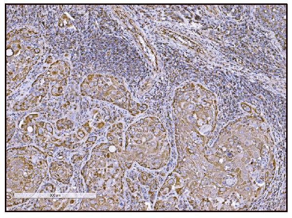
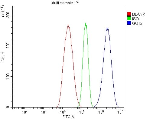
(Figure 14. IHC analysis of FABP-1/GOT2 using anti-FABP-1/GOT2 antibody (AAA19309).FABP-1/GOT2 was detected in paraffin-embedded section of rat lymph node tissue. Heat mediated antigen retrieval was performed in EDTA buffer (pH8. 0, epitope retrieval solution). The tissue section was blocked with 10% goat serum. The tissue section was then incubated with 2μg/ml rabbit anti-FABP-1/GOT2 Antibody (AAA19309) overnight at 4 degree C. Biotinylated goat anti-rabbit IgG was used as secondary antibody and incubated for 30 minutes at 37 degree C. The tissue section was developed using Strepavidin-Biotin-Complex (SABC) (Catalog # with DAB as the chromogen.)
IHC (Immunohistochemistry)
(Figure 14. IHC analysis of FABP-1/GOT2 using anti-FABP-1/GOT2 antibody (AAA19309).FABP-1/GOT2 was detected in paraffin-embedded section of rat lymph node tissue. Heat mediated antigen retrieval was performed in EDTA buffer (pH8. 0, epitope retrieval solution). The tissue section was blocked with 10% goat serum. The tissue section was then incubated with 2μg/ml rabbit anti-FABP-1/GOT2 Antibody (AAA19309) overnight at 4 degree C. Biotinylated goat anti-rabbit IgG was used as secondary antibody and incubated for 30 minutes at 37 degree C. The tissue section was developed using Strepavidin-Biotin-Complex (SABC) (Catalog # with DAB as the chromogen.)
FABP-1/GOT2, Polyclonal Antibody (Cat# AAA19309)
Full Name
Anti-FABP-1/GOT2 Antibody
Gene Names
GOT2; KAT4; KATIV; mitAAT
Reactivity
Human, Mouse, Rat
Applications
WB, IHC-P, ICC, IF, FC/FACS/FCM, EIA
Purity
Immunogen affinity purified.
Pricing
 Standard Curve (Sample)
Pentraxin-3 (PTX-3), ELISA Kit (Cat# AAA22480)
Full Name
Rabbit Pentraxin-3 (PTX-3) ELISA Kit
Gene Names
PTX3; TSG-14; TNFAIP5
Reactivity
Rabbit
Pricing
 Standard Curve (Sample)
Tyrosine 3/Tryptophan 5 Monooxygenase Activation Protein Zeta (YWHAz), ELISA Kit (Cat# AAA20812)
Full Name
Multi-species Tyrosine 3/Tryptophan 5 Monooxygenase Activation Protein Zeta (YWHAz) ELISA Kit
Reactivity
Human, Mouse, Rat
Pricing


 ICC (Immunocytochemistry)
(ICC staining VCP in SW480 cells (green). Cells were fixed in paraformaldehyde, permeabilised with 0.25% Triton X100/PBS.)
ICC (Immunocytochemistry)
(ICC staining VCP in SW480 cells (green). Cells were fixed in paraformaldehyde, permeabilised with 0.25% Triton X100/PBS.)
VCP, Polyclonal Antibody (Cat# AAA29948)
Full Name
VCP Antibody
Gene Names
VCP; p97; TERA; ALS14; IBMPFD
Reactivity
Human, Mouse, Zebrafish
Applications
WB, ICC, IHC
Purity
Peptide affinity purified
Pricing


 WB (Western Blot)
(WB Suggested Anti-HNRPL Antibody Titration: 0.2-1 ug/mlELISA Titer: 1:62500Positive Control: Jurkat cell lysateHNRNPL is strongly supported by BioGPS gene expression data to be expressed in Human Jurkat cells)
WB (Western Blot)
(WB Suggested Anti-HNRPL Antibody Titration: 0.2-1 ug/mlELISA Titer: 1:62500Positive Control: Jurkat cell lysateHNRNPL is strongly supported by BioGPS gene expression data to be expressed in Human Jurkat cells)
HNRPL, Polyclonal Antibody (Cat# AAA23474)
Full Name
HNRPL antibody - N-terminal region
Gene Names
HNRNPL; HNRPL; hnRNP-L; P/OKcl.14
Reactivity
Cow, Dog, Guinea Pig, Horse, Human, Mouse, Rabbit, Rat
Applications
IF, IP, IHC, WB
Purity
Affinity Purified
Pricing


 IF (Immunofluorescence)
(Immunofluorescent analysis of 4% paraformaldehyde-fixed, 0.1% Triton X-100 permeabilized MCF-7 (human breast cancer cell line) cells labeling Pdx1 with at 1:25 dilution, followed by DyLight 488-conjugated IgG goat anti-rabbit secondary antibody at 1:200 dilution (green). Immunofluorescence image showing cytoplasm staining on MCF-7 cell line. Cytoplasmic actin is detected with DyLight 554 Phalloidin (PD18466410) at 1:100 dilution (red). The nuclear counter stain is DAPI (blue).)
IF (Immunofluorescence)
(Immunofluorescent analysis of 4% paraformaldehyde-fixed, 0.1% Triton X-100 permeabilized MCF-7 (human breast cancer cell line) cells labeling Pdx1 with at 1:25 dilution, followed by DyLight 488-conjugated IgG goat anti-rabbit secondary antibody at 1:200 dilution (green). Immunofluorescence image showing cytoplasm staining on MCF-7 cell line. Cytoplasmic actin is detected with DyLight 554 Phalloidin (PD18466410) at 1:100 dilution (red). The nuclear counter stain is DAPI (blue).)
OPN-a/b, Polyclonal Antibody (Cat# AAA26859)
Full Name
OPN-a/b, NT (SPP1, BNSP, OPN, Osteopontin, Bone sialoprotein 1, Nephropontin, Secreted phosphoprotein 1, Urinary stone protein, Uropontin) (FITC)
Gene Names
SPP1; OPN; BNSP; BSPI; ETA-1
Reactivity
Human
Applications
WB, IHC, IF
Purity
Purified by Protein A and Peptide Affinity Chromatography.
Pricing


 Application Data
(Published customer image: Mouse anti Human CD49d antibody, clone HP2/1 used for binding efficiency determinationImage caption:Binding efficiencies (BE) of different a4beta7 molecules composed of distinct a4 mutants to monoclonal antibodies against a4, beta7 or the a4beta7 heterodimer. Binding efficiency is determined by the ratio between the mean fluorescence of antibody binding to each a4 molecule and of the binding in a mock-transfected cell culture (see Materials and Methods for details). Dark gray bars represent binding to the human (wild type) a4 clone, whereas light gray bars are those of binding to the different a4 mutants (as shown in the x-axis). a4 mutants which included substitutions at codon 201 are boxed. A, binding of anti-a4 2b4 antibody. B, binding of anti-a4 HP2/1 antibody. C, BE of different anti-a4 and beta7 antibodies to the human a4 and the quintuple a4 mutant (5 aa mut). Bars represent the range of standard errors deduced from triplicate experiments. p-values of Student's t tests are shown above each comparison. NS, non-significant (> 0.05).From:Darc M, Hait SH, Soares EA, Cicala C, Seuanez HN, et al. (2011) Polymorphisms in the a4 Integrin of Neotropical Primates: Insights for Binding of Natural Ligands and HIV-1 gp120 to the Human a4beta7. PLoS ONE 6(9): e24461.)
Application Data
(Published customer image: Mouse anti Human CD49d antibody, clone HP2/1 used for binding efficiency determinationImage caption:Binding efficiencies (BE) of different a4beta7 molecules composed of distinct a4 mutants to monoclonal antibodies against a4, beta7 or the a4beta7 heterodimer. Binding efficiency is determined by the ratio between the mean fluorescence of antibody binding to each a4 molecule and of the binding in a mock-transfected cell culture (see Materials and Methods for details). Dark gray bars represent binding to the human (wild type) a4 clone, whereas light gray bars are those of binding to the different a4 mutants (as shown in the x-axis). a4 mutants which included substitutions at codon 201 are boxed. A, binding of anti-a4 2b4 antibody. B, binding of anti-a4 HP2/1 antibody. C, BE of different anti-a4 and beta7 antibodies to the human a4 and the quintuple a4 mutant (5 aa mut). Bars represent the range of standard errors deduced from triplicate experiments. p-values of Student's t tests are shown above each comparison. NS, non-significant (> 0.05).From:Darc M, Hait SH, Soares EA, Cicala C, Seuanez HN, et al. (2011) Polymorphisms in the a4 Integrin of Neotropical Primates: Insights for Binding of Natural Ligands and HIV-1 gp120 to the Human a4beta7. PLoS ONE 6(9): e24461.)
CD49d, Monoclonal Antibody (Cat# AAA12002)
Full Name
MOUSE ANTI HUMAN CD49d
Gene Names
ITGA4; IA4; CD49D
Reactivity
Bovine, Cat, Cynomolgus monkey, Goat, Horse, Llama, Mink, Mustelid, Pig, Rabbit, Rat, Rhesus Monkey
Applications
Immunohistochemistry, Flow Cytometry, Functional Assay, Immunoprecipitation
Pricing


 IF (Immunofluorescence)
(Figure 9. IF analysis of ApoER2/LRP8 using anti- ApoER2/LRP8 antibody (AAA19275).ApoER2/LRP8 was detected in immunocytochemical section of HepG2 cells. Enzyme antigen retrieval was performed using IHC enzyme antigen retrieval reagent for 15 mins. The cells were blocked with 10% goat serum. And then incubated with 4μg/mL rabbit anti- ApoER2/LRP8 Antibody (AAA19275) overnight at 4 degree C. DyLight®488 Conjugated Goat Anti-Rabbit IgG was used as secondary antibody at 1:100 dilution and incubated for 30 minutes at 37 degree C. The section was counterstained with DAPI. Visualize using a fluorescence microscope and filter sets appropriate for the label used.)
IF (Immunofluorescence)
(Figure 9. IF analysis of ApoER2/LRP8 using anti- ApoER2/LRP8 antibody (AAA19275).ApoER2/LRP8 was detected in immunocytochemical section of HepG2 cells. Enzyme antigen retrieval was performed using IHC enzyme antigen retrieval reagent for 15 mins. The cells were blocked with 10% goat serum. And then incubated with 4μg/mL rabbit anti- ApoER2/LRP8 Antibody (AAA19275) overnight at 4 degree C. DyLight®488 Conjugated Goat Anti-Rabbit IgG was used as secondary antibody at 1:100 dilution and incubated for 30 minutes at 37 degree C. The section was counterstained with DAPI. Visualize using a fluorescence microscope and filter sets appropriate for the label used.)
ApoER2/LRP8, Polyclonal Antibody (Cat# AAA19275)
Full Name
Anti-ApoER2/LRP8 Antibody
Gene Names
LRP8; MCI1; LRP-8; APOER2; HSZ75190
Reactivity
Human, Mouse, Rat
Applications
WB, IHC-P, ICC, IF, FC/FACS/FCM, EIA
Purity
Immunogen affinity purified.
Pricing


 IF (Immunofluorescence)
(Figure 15. IF analysis of LSM2 using anti- LSM2 antibody (AAA19316).LSM2 was detected in immunocytochemical section of MCF-7 cells. Enzyme antigen retrieval was performed using IHC enzyme antigen retrieval reagent for 15 mins. The cells were blocked with 10% goat serum. And then incubated with 5μg/mL rabbit anti- LSM2 Antibody (AAA19316) overnight at 4 degree C. DyLight®488 Conjugated Goat Anti-Rabbit IgG was used as secondary antibody at 1:100 dilution and incubated for 30 minutes at 37 degree C. The section was counterstained with DAPI. Visualize using a fluorescence microscope and filter sets appropriate for the label used.)
IF (Immunofluorescence)
(Figure 15. IF analysis of LSM2 using anti- LSM2 antibody (AAA19316).LSM2 was detected in immunocytochemical section of MCF-7 cells. Enzyme antigen retrieval was performed using IHC enzyme antigen retrieval reagent for 15 mins. The cells were blocked with 10% goat serum. And then incubated with 5μg/mL rabbit anti- LSM2 Antibody (AAA19316) overnight at 4 degree C. DyLight®488 Conjugated Goat Anti-Rabbit IgG was used as secondary antibody at 1:100 dilution and incubated for 30 minutes at 37 degree C. The section was counterstained with DAPI. Visualize using a fluorescence microscope and filter sets appropriate for the label used.)
LSM2, Polyclonal Antibody (Cat# AAA19316)
Full Name
Anti-LSM2 Antibody
Gene Names
LSM2; G7B; snRNP; C6orf28; YBL026W
Reactivity
Human, Mouse, Rat, Monkey
Applications
WB, IHC-P, ICC, IF, FC/FACS/FCM
Purity
Immunogen affinity purified.
Pricing


 IF (Immunofluorescence)
(Immunofluorescent analysis of 4% paraformaldehyde-fixed, 0.1% Triton X-100 permeabilized MCF-7 (human breast cancer cell line) cells labeling Pdx1 with at 1:25 dilution, followed by DyLight 488-conjugated IgG goat anti-rabbit secondary antibody at 1:200 dilution (green). Immunofluorescence image showing cytoplasm staining on MCF-7 cell line. Cytoplasmic actin is detected with DyLight 554 Phalloidin (PD18466410) at 1:100 dilution (red). The nuclear counter stain is DAPI (blue).)
IF (Immunofluorescence)
(Immunofluorescent analysis of 4% paraformaldehyde-fixed, 0.1% Triton X-100 permeabilized MCF-7 (human breast cancer cell line) cells labeling Pdx1 with at 1:25 dilution, followed by DyLight 488-conjugated IgG goat anti-rabbit secondary antibody at 1:200 dilution (green). Immunofluorescence image showing cytoplasm staining on MCF-7 cell line. Cytoplasmic actin is detected with DyLight 554 Phalloidin (PD18466410) at 1:100 dilution (red). The nuclear counter stain is DAPI (blue).)
OPN-a/b, Polyclonal Antibody (Cat# AAA26860)
Full Name
OPN-a/b, NT (SPP1, BNSP, OPN, Osteopontin, Bone sialoprotein 1, Nephropontin, Secreted phosphoprotein 1, Urinary stone protein, Uropontin) (MaxLight 405)
Gene Names
SPP1; OPN; BNSP; BSPI; ETA-1
Reactivity
Human
Applications
WB, IHC, IF
Purity
Purified by Protein A and Peptide Affinity Chromatography.
Pricing


 Application Data
(Analysis of Protein Array containing more than 19,000 full-length human proteins using AMACR/p504S Mouse Monoclonal Antibody (AMACR/1864). Z- and S- Score: The Z-score represents the strength of a signal that a monoclonal antibody (MAb) (in combination with a fluorescently-tagged anti-IgG secondary antibody) produces when binding to a particular protein on the HuProtTM array. Z-scores are described in units of standard deviations (SD's) above the mean value of all signals generated on that array. If targets on HuProtTM are arranged in descending order of the Z-score, the S-score is the difference (also in units of SD's) between the Z-score. S-score therefore represents the relative target specificity of a MAb to its intended target. A MAb is considered to specific to its intended target, if the MAb has an S-score of at least 2.5. For example, if a MAb binds to protein X with a Z-score of 43 and to protein Y with a Z-score of 14, then the S-score for the binding of that MAb to protein X is equal to 29.)
Application Data
(Analysis of Protein Array containing more than 19,000 full-length human proteins using AMACR/p504S Mouse Monoclonal Antibody (AMACR/1864). Z- and S- Score: The Z-score represents the strength of a signal that a monoclonal antibody (MAb) (in combination with a fluorescently-tagged anti-IgG secondary antibody) produces when binding to a particular protein on the HuProtTM array. Z-scores are described in units of standard deviations (SD's) above the mean value of all signals generated on that array. If targets on HuProtTM are arranged in descending order of the Z-score, the S-score is the difference (also in units of SD's) between the Z-score. S-score therefore represents the relative target specificity of a MAb to its intended target. A MAb is considered to specific to its intended target, if the MAb has an S-score of at least 2.5. For example, if a MAb binds to protein X with a Z-score of 43 and to protein Y with a Z-score of 14, then the S-score for the binding of that MAb to protein X is equal to 29.)
AMACR/p504S, Monoclonal Antibody (Cat# AAA23906)
Full Name
AMACR/p504S (Prostate Cancer Marker)
Gene Names
AMACR; RM; RACE; CBAS4; P504S; AMACRD
Reactivity
Human. Others not known.
Applications
Western Blot, Immunohistochemistry
Purity
Purified Ab with BSA and Azide at 200ug/ml OR Purified Ab WITHOUT BSA and Azide at 1.0mg/ml
Pricing
 SDS-PAGE
Glutamate receptor 3 (GRIA3), Recombinant Protein (Cat# AAA18426)
Full Name
Recombinant Human Glutamate receptor 3 (GRIA3), partial
Gene Names
GRIA3; GLUR3; GLURC; GluA3; MRX94; GLUR-C; GLUR-K3
Purity
Greater or equal to 85% purity as determined by SDS-PAGE.
Pricing
 Standard Curve (Sample)
14-3-3 protein sigma, ELISA Kit (Cat# AAA17647)
Full Name
Human 14-3-3 protein sigma ELISA Kit
Gene Names
SFN; YWHAS
Reactivity
Human
Pricing
 Standard Curve (Sample)
PTX3/Pentraxin 3, ELISA Kit (Cat# AAA11592)
Full Name
Human PTX3/Pentraxin 3 PicoKine ELISA Kit
Gene Names
PTX3; TSG-14; TNFAIP5
Reactivity
Human
Pricing
 Standard Curve (Sample)
Tyrosine Hydroxylase (TH), ELISA Kit (Cat# AAA20403)
Full Name
Mouse Tyrosine Hydroxylase (TH) ELISA Kit
Gene Names
TH; TYH; DYT14; DYT5b
Reactivity
Mouse
Pricing


 FCM (Flow Cytometry)
(Figure 10. Flow Cytometry analysis of U20S cells using anti-PDIA5 antibody (AAA19331).Overlay histogram showing U20S cells stained with AAA19331 (Blue line). The cells were blocked with 10% normal goat serum. And then incubated with rabbit anti-PDIA5 Antibody (AAA19331, 1μg/1x106 cells) for 30 min at 20 degree C. DyLight®488 conjugated goat anti-rabbit IgG (5-10μg/1x106 cells) was used as secondary antibody for 30 minutes at 20 degree C. Isotype control antibody (Green line) was rabbit IgG (1μg/1x106) used under the same conditions. Unlabelled sample (Red line) was also used as a control.)
FCM (Flow Cytometry)
(Figure 10. Flow Cytometry analysis of U20S cells using anti-PDIA5 antibody (AAA19331).Overlay histogram showing U20S cells stained with AAA19331 (Blue line). The cells were blocked with 10% normal goat serum. And then incubated with rabbit anti-PDIA5 Antibody (AAA19331, 1μg/1x106 cells) for 30 min at 20 degree C. DyLight®488 conjugated goat anti-rabbit IgG (5-10μg/1x106 cells) was used as secondary antibody for 30 minutes at 20 degree C. Isotype control antibody (Green line) was rabbit IgG (1μg/1x106) used under the same conditions. Unlabelled sample (Red line) was also used as a control.)
PDIA5, Polyclonal Antibody (Cat# AAA19331)
Full Name
Anti-PDIA5 Antibody
Gene Names
PDIA5; PDIR
Reactivity
Human, Mouse, Rat
Applications
WB, IHC-P, ICC, IF, FC/FACS/FCM, EIA
Purity
Immunogen affinity purified.
Pricing
 Standard Curve (Sample)
tyrosine hydroxylase, ELISA Kit (Cat# AAA15167)
Full Name
Rat Tyrosine Hydroxylase, TH ELISA Kit
Gene Names
Th; The
Reactivity
Rat
Pricing


 Application Data
(Ref. 3: Isolation, culture and immunocytochemical characterization of HUCPC. The dissection under sterile condition of foetal and full-term cords was performed to expose the WJ, the vein and arteries (A). After the in vitro expansion of foetal HUCPC (B) the cells ..)
Application Data
(Ref. 3: Isolation, culture and immunocytochemical characterization of HUCPC. The dissection under sterile condition of foetal and full-term cords was performed to expose the WJ, the vein and arteries (A). After the in vitro expansion of foetal HUCPC (B) the cells ..)
Chick Embryo Extract, Ultrafiltrate, Reagent (Cat# AAA14749)
Full Name
Chick Embryo Extract, Ultrafiltrate (CEE)
Purity
Molecular Biology Grade
Pricing


 FCM (Flow Cytometry)
(Figure 6. Flow Cytometry analysis of CACO-2 cells using anti-Galectin 2/LGALS2 antibody (AAA19287).Overlay histogram showing CACO-2 cells stained with AAA19287 (Blue line). The cells were blocked with 10% normal goat serum. And then incubated with rabbit anti-Galectin 2/LGALS2 Antibody (AAA19287, 1μg/1x106 cells) for 30 min at 20 degree C. DyLight®488 conjugated goat anti-rabbit IgG (5-10μg/1x106 cells) was used as secondary antibody for 30 minutes at 20 degree C. Isotype control antibody (Green line) was rabbit IgG (1μg/1x106) used under the same conditions. Unlabelled sample (Red line) was also used as a control.)
FCM (Flow Cytometry)
(Figure 6. Flow Cytometry analysis of CACO-2 cells using anti-Galectin 2/LGALS2 antibody (AAA19287).Overlay histogram showing CACO-2 cells stained with AAA19287 (Blue line). The cells were blocked with 10% normal goat serum. And then incubated with rabbit anti-Galectin 2/LGALS2 Antibody (AAA19287, 1μg/1x106 cells) for 30 min at 20 degree C. DyLight®488 conjugated goat anti-rabbit IgG (5-10μg/1x106 cells) was used as secondary antibody for 30 minutes at 20 degree C. Isotype control antibody (Green line) was rabbit IgG (1μg/1x106) used under the same conditions. Unlabelled sample (Red line) was also used as a control.)
Galectin 2/LGALS2, Polyclonal Antibody (Cat# AAA19287)
Full Name
Anti-Galectin 2/LGALS2 Antibody
Gene Names
LGALS2; HL14
Reactivity
Human
Applications
WB, IHC-P, ICC, IF, FC/FACS/FCM, EIA
Purity
Immunogen affinity purified.
Pricing


 FCM (Flow Cytometry)
(Figure 18. Flow Cytometry analysis of A431 cells using anti-LSM7 antibody (AAA19327).Overlay histogram showing A431 cells stained with AAA19327 (Blue line). The cells were blocked with 10% normal goat serum. And then incubated with rabbit anti-LSM7 Antibody (AAA19327, 1μg/1x106 cells) for 30 min at 20 degree C. DyLight®488 conjugated goat anti-rabbit IgG (5-10μg/1x106 cells) was used as secondary antibody for 30 minutes at 20 degree C. Isotype control antibody (Green line) was rabbit IgG (1μg/1x106) used under the same conditions. Unlabelled sample (Red line) was also used as a control.)
FCM (Flow Cytometry)
(Figure 18. Flow Cytometry analysis of A431 cells using anti-LSM7 antibody (AAA19327).Overlay histogram showing A431 cells stained with AAA19327 (Blue line). The cells were blocked with 10% normal goat serum. And then incubated with rabbit anti-LSM7 Antibody (AAA19327, 1μg/1x106 cells) for 30 min at 20 degree C. DyLight®488 conjugated goat anti-rabbit IgG (5-10μg/1x106 cells) was used as secondary antibody for 30 minutes at 20 degree C. Isotype control antibody (Green line) was rabbit IgG (1μg/1x106) used under the same conditions. Unlabelled sample (Red line) was also used as a control.)
LSM7, Polyclonal Antibody (Cat# AAA19327)
Full Name
Anti-LSM7 Antibody
Gene Names
LSM7; YNL147W
Reactivity
Human, Mouse, Rat
Applications
WB, IHC-P, ICC, IF, FC/FACS/FCM, EIA
Purity
Immunogen affinity purified.
Pricing


 IF (Immunofluorescence)
(Immunofluorescent analysis of 4% paraformaldehyde-fixed, 0.1% Triton X-100 permeabilized MCF-7 (human breast cancer cell line) cells labeling Pdx1 with at 1:25 dilution, followed by DyLight 488-conjugated IgG goat anti-rabbit secondary antibody at 1:200 dilution (green). Immunofluorescence image showing cytoplasm staining on MCF-7 cell line. Cytoplasmic actin is detected with DyLight 554 Phalloidin (PD18466410) at 1:100 dilution (red). The nuclear counter stain is DAPI (blue).)
IF (Immunofluorescence)
(Immunofluorescent analysis of 4% paraformaldehyde-fixed, 0.1% Triton X-100 permeabilized MCF-7 (human breast cancer cell line) cells labeling Pdx1 with at 1:25 dilution, followed by DyLight 488-conjugated IgG goat anti-rabbit secondary antibody at 1:200 dilution (green). Immunofluorescence image showing cytoplasm staining on MCF-7 cell line. Cytoplasmic actin is detected with DyLight 554 Phalloidin (PD18466410) at 1:100 dilution (red). The nuclear counter stain is DAPI (blue).)
OPN-a/b, Polyclonal Antibody (Cat# AAA26858)
Full Name
OPN-a/b, NT (SPP1, BNSP, OPN, Osteopontin, Bone sialoprotein 1, Nephropontin, Secreted phosphoprotein 1, Urinary stone protein, Uropontin) (Biotin)
Gene Names
SPP1; OPN; BNSP; BSPI; ETA-1
Reactivity
Human
Applications
WB, IHC, IF, EIA
Purity
Purified by Protein A and Peptide Affinity Chromatography.
Pricing


 FCM (Flow Cytometry)
(Figure 10. Flow Cytometry analysis of SiHa cells using anti-PRDM14 antibody (AAA19283).Overlay histogram showing SiHa cells stained with AAA19283 (Blue line). The cells were blocked with 10% normal goat serum. And then incubated with rabbit anti-PRDM14 Antibody (AAA19283, 1μg/1x106 cells) for 30 min at 20 degree C. DyLight®488 conjugated goat anti-rabbit IgG (5-10μg/1x106 cells) was used as secondary antibody for 30 minutes at 20 degree C. Isotype control antibody (Green line) was rabbit IgG (1μg/1x106) used under the same conditions. Unlabelled sample (Red line) was also used as a control.)
FCM (Flow Cytometry)
(Figure 10. Flow Cytometry analysis of SiHa cells using anti-PRDM14 antibody (AAA19283).Overlay histogram showing SiHa cells stained with AAA19283 (Blue line). The cells were blocked with 10% normal goat serum. And then incubated with rabbit anti-PRDM14 Antibody (AAA19283, 1μg/1x106 cells) for 30 min at 20 degree C. DyLight®488 conjugated goat anti-rabbit IgG (5-10μg/1x106 cells) was used as secondary antibody for 30 minutes at 20 degree C. Isotype control antibody (Green line) was rabbit IgG (1μg/1x106) used under the same conditions. Unlabelled sample (Red line) was also used as a control.)
PRDM14, Polyclonal Antibody (Cat# AAA19283)
Full Name
Anti-PRDM14 Antibody
Gene Names
PRDM14; PFM11
Reactivity
Human, Mouse, Rat
Applications
WB, IHC-P, ICC, IF, FC/FACS/FCM
Purity
Immunogen affinity purified.
Pricing