Filters
Clonality
Type
Reactivity
Gene Name
Isotype
Host
Application
Clone
1488 results for " E" - showing 1350-1400
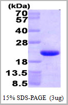
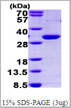
 SDS-PAGE
(3ug by SDS-PAGE under reducing condition and visualized by coomasie blue stain)
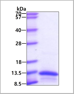
SDS-PAGE
(3ug by SDS-PAGE under reducing condition and visualized by coomasie blue stain)
PTP-1B (Protein Tyrosine Phosphatase1B), Active Protein (Cat# AAA11740)
Full Name
PTP-1B (Protein Tyrosine Phosphatase1B), 1-321aa, Human, E Coli (Bioactivity Validated)
Gene Names
PTPN1; PTP1B
Applications
Enzyme Activity, SDS-PAGE
Purity
> 95% by SDS-PAGE
Pricing
 WB (Western Blot)
(Western BlotPositive WB detected in: recombinant proteinAll lanes: csgA Antibody at 1:1000SecondaryGoat polyclonal to rabbit IgG at 1/50000 dilutionPredicted band size: 17 kDaObserved band size: 17 kDa)
WB (Western Blot)
(Western BlotPositive WB detected in: recombinant proteinAll lanes: csgA Antibody at 1:1000SecondaryGoat polyclonal to rabbit IgG at 1/50000 dilutionPredicted band size: 17 kDaObserved band size: 17 kDa)
csgA, Polyclonal Antibody (Cat# AAA27056)
Full Name
csgA Antibody
Gene Names
csgA; agfA; ECK1028; JW1025
Reactivity
E Coli (strain K12)
Applications
Western Blot
Purity
Affinity-chromatography
Pricing
CEA, Monoclonal Antibody (Cat# AAA14296)
Full Name
CEA antibody (in PBS)
Gene Names
CEACAM6; NCA; CEAL; CD66c
Applications
ELISA
Pricing


 Application Data
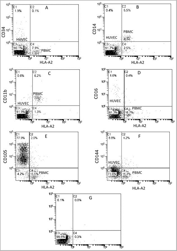
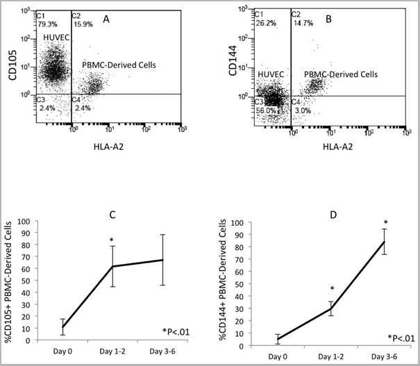
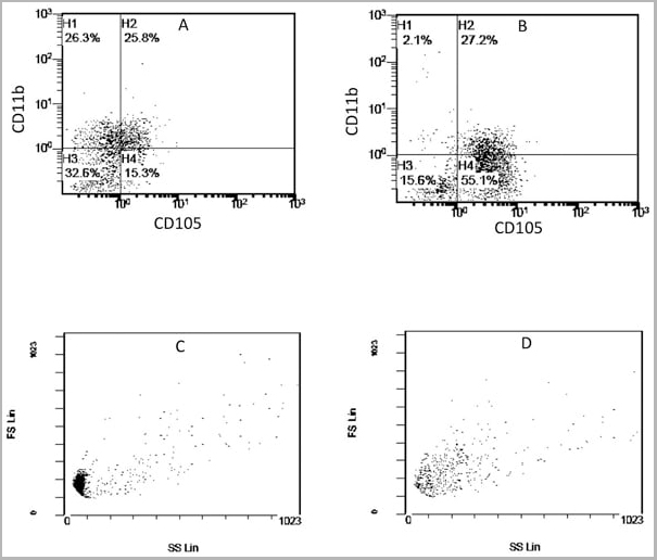
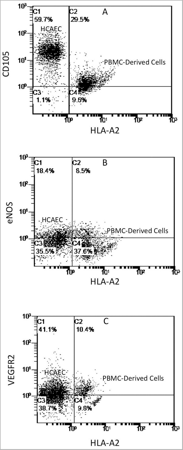
(Published customer image: Mouse anti Human CD105 antibody, clone SN6 used for flow cytometry.Image caption:Phenotype change from HLA-A2+/CD11b+/CD105- to HLA-A2+/CD11b-/CD105+ on endothelium-adherent blood monocyte-derived cells with increase in size and granularity during co-culture. HLA-A2+ PBMCs (1x10⁶ cells/well) were incubated for 2 h (Day 0) with HLA-A2- HUVECs, after which the non-adherent cells were removed by washing. The cell layers were analysed by three-colour flow cytometry staining for HLA-A2, CD11b and CD105 on (A) Day 1 and (B) Day 2. These plots were gated for HLA-A2+ cells. Forward scatter/side scatter dot plots gated for HLA-A2+ cells on Day 0 (C) and Day 2 (D) was shown. These are representative of 2 individual experiments.From: Tso C, Rye K-A, Barter P (2012) Phenotypic and Functional Changes in Blood Monocytes Following Adherence to Endothelium. PLoS ONE 7(5): e37091.)
Application Data
(Published customer image: Mouse anti Human CD105 antibody, clone SN6 used for flow cytometry.Image caption:Phenotype change from HLA-A2+/CD11b+/CD105- to HLA-A2+/CD11b-/CD105+ on endothelium-adherent blood monocyte-derived cells with increase in size and granularity during co-culture. HLA-A2+ PBMCs (1x10⁶ cells/well) were incubated for 2 h (Day 0) with HLA-A2- HUVECs, after which the non-adherent cells were removed by washing. The cell layers were analysed by three-colour flow cytometry staining for HLA-A2, CD11b and CD105 on (A) Day 1 and (B) Day 2. These plots were gated for HLA-A2+ cells. Forward scatter/side scatter dot plots gated for HLA-A2+ cells on Day 0 (C) and Day 2 (D) was shown. These are representative of 2 individual experiments.From: Tso C, Rye K-A, Barter P (2012) Phenotypic and Functional Changes in Blood Monocytes Following Adherence to Endothelium. PLoS ONE 7(5): e37091.)
CD105, Monoclonal Antibody (Cat# AAA12083)
Full Name
MOUSE ANTI HUMAN CD105:FITC
Gene Names
ENG; END; HHT1; ORW1
Applications
Flow Cytometry
Pricing


 Application Data
(Published customer image: Mouse anti Human CD105 antibody, clone SN6 used for flow cytometry.Image caption:Phenotype change from HLA-A2+/CD11b+/CD105- to HLA-A2+/CD11b-/CD105+ on endothelium-adherent blood monocyte-derived cells with increase in size and granularity during co-culture. HLA-A2+ PBMCs (1x10⁶ cells/well) were incubated for 2 h (Day 0) with HLA-A2- HUVECs, after which the non-adherent cells were removed by washing. The cell layers were analysed by three-colour flow cytometry staining for HLA-A2, CD11b and CD105 on (A) Day 1 and (B) Day 2. These plots were gated for HLA-A2+ cells. Forward scatter/side scatter dot plots gated for HLA-A2+ cells on Day 0 (C) and Day 2 (D) was shown. These are representative of 2 individual experiments.From: Tso C, Rye K-A, Barter P (2012) Phenotypic and Functional Changes in Blood Monocytes Following Adherence to Endothelium. PLoS ONE 7(5): e37091.)
Application Data
(Published customer image: Mouse anti Human CD105 antibody, clone SN6 used for flow cytometry.Image caption:Phenotype change from HLA-A2+/CD11b+/CD105- to HLA-A2+/CD11b-/CD105+ on endothelium-adherent blood monocyte-derived cells with increase in size and granularity during co-culture. HLA-A2+ PBMCs (1x10⁶ cells/well) were incubated for 2 h (Day 0) with HLA-A2- HUVECs, after which the non-adherent cells were removed by washing. The cell layers were analysed by three-colour flow cytometry staining for HLA-A2, CD11b and CD105 on (A) Day 1 and (B) Day 2. These plots were gated for HLA-A2+ cells. Forward scatter/side scatter dot plots gated for HLA-A2+ cells on Day 0 (C) and Day 2 (D) was shown. These are representative of 2 individual experiments.From: Tso C, Rye K-A, Barter P (2012) Phenotypic and Functional Changes in Blood Monocytes Following Adherence to Endothelium. PLoS ONE 7(5): e37091.)
CD105, Monoclonal Antibody (Cat# AAA11865)
Full Name
MOUSE ANTI HUMAN CD105:FITC
Gene Names
ENG; END; HHT1; ORW1
Applications
Flow Cytometry
Pricing


 Application Data
(Published customer image: Mouse anti Human CD105 antibody, clone SN6 used for flow cytometry.Image caption:Phenotype change from HLA-A2+/CD11b+/CD105- to HLA-A2+/CD11b-/CD105+ on endothelium-adherent blood monocyte-derived cells with increase in size and granularity during co-culture. HLA-A2+ PBMCs (1x10⁶ cells/well) were incubated for 2 h (Day 0) with HLA-A2- HUVECs, after which the non-adherent cells were removed by washing. The cell layers were analysed by three-colour flow cytometry staining for HLA-A2, CD11b and CD105 on (A) Day 1 and (B) Day 2. These plots were gated for HLA-A2+ cells. Forward scatter/side scatter dot plots gated for HLA-A2+ cells on Day 0 (C) and Day 2 (D) was shown. These are representative of 2 individual experiments.From: Tso C, Rye K-A, Barter P (2012) Phenotypic and Functional Changes in Blood Monocytes Following Adherence to Endothelium. PLoS ONE 7(5): e37091.)
Application Data
(Published customer image: Mouse anti Human CD105 antibody, clone SN6 used for flow cytometry.Image caption:Phenotype change from HLA-A2+/CD11b+/CD105- to HLA-A2+/CD11b-/CD105+ on endothelium-adherent blood monocyte-derived cells with increase in size and granularity during co-culture. HLA-A2+ PBMCs (1x10⁶ cells/well) were incubated for 2 h (Day 0) with HLA-A2- HUVECs, after which the non-adherent cells were removed by washing. The cell layers were analysed by three-colour flow cytometry staining for HLA-A2, CD11b and CD105 on (A) Day 1 and (B) Day 2. These plots were gated for HLA-A2+ cells. Forward scatter/side scatter dot plots gated for HLA-A2+ cells on Day 0 (C) and Day 2 (D) was shown. These are representative of 2 individual experiments.From: Tso C, Rye K-A, Barter P (2012) Phenotypic and Functional Changes in Blood Monocytes Following Adherence to Endothelium. PLoS ONE 7(5): e37091.)
CD105, Monoclonal Antibody (Cat# AAA12029)
Full Name
MOUSE ANTI HUMAN CD105:RPE
Gene Names
ENG; END; HHT1; ORW1
Applications
Flow Cytometry
Pricing
 Application Data
(Staining of mouse spleen with Hamster anti Mouse CD81: Alexa Fluor 488)
Application Data
(Staining of mouse spleen with Hamster anti Mouse CD81: Alexa Fluor 488)
CD81, Monoclonal Antibody (Cat# AAA11942)
Full Name
HAMSTER ANTI MOUSE CD81
Gene Names
Cd81; Tapa1; Tapa-1; Tspan28
Reactivity
Rat
Applications
Immunohistochemistry, Flow Cytometry, Immunoprecipitation, Western Blot
Pricing


 Application Data
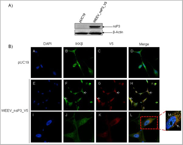
(Published customer image: Mouse anti V5 tag antibody, clone SV5-Pk1 used for the detection of V5 tagged WEEV_nsP3 protein by western blotting and immunofluorescenceImage caption: WEEV nsP3 interaction with host IKKbeta. A) U87MGs were transfected in a 6-well plate with 5 ug of pUC19 and WEEV_nsP3_HA for 24 hours. Cell lysates were resolved using SDS-PAGE and subsequently immunoblotted with V5 antibody and beta-actin served as a loading control. B) U87MGs were transfected with WEEV_nsP3_V5; cells were fixed after 24 hours and stained with antibodies against the endogenous IKKbeta and the V5 tag. Cells were incubated with appropriate secondary Alexa Fluor antibodies and the nuclei stained with DAPI. Co-localization of IKKbeta with WEEV_nsP3_V5 (yellow) was observed as shown by the arrows. B) Panels E -H serve as an example of transfected cells in a given field of view that show co-localization of IKKbeta and WEEV_nsP3_V5 24 hours post transfection. Panels I-L represent magnified images of other cells showing co-localization of IKKbeta and WEEV_nsP3_V5. Panel M is a magnified image of panel L. The co-localization was confirmed by Z-stack analysis. Co-localization was calculated to be approximately in 61% of cells (163 cells were counted of which 44% demonstrated expression of nsP3. Of those cells that expressed nsP3, 61% showed co-localization of both proteins). Images were taken using Nikon Eclipse TE2000-U at 60x magnification and are representative of 2 independent experiments.From: Amaya M, Voss K, Sampey G, Senina S, de la Fuente C, et al. (2014) The Role of IKKbeta in Venezuelan Equine Encephalitis Virus Infection. PLoS ONE 9(2): e86745.)
Application Data
(Published customer image: Mouse anti V5 tag antibody, clone SV5-Pk1 used for the detection of V5 tagged WEEV_nsP3 protein by western blotting and immunofluorescenceImage caption: WEEV nsP3 interaction with host IKKbeta. A) U87MGs were transfected in a 6-well plate with 5 ug of pUC19 and WEEV_nsP3_HA for 24 hours. Cell lysates were resolved using SDS-PAGE and subsequently immunoblotted with V5 antibody and beta-actin served as a loading control. B) U87MGs were transfected with WEEV_nsP3_V5; cells were fixed after 24 hours and stained with antibodies against the endogenous IKKbeta and the V5 tag. Cells were incubated with appropriate secondary Alexa Fluor antibodies and the nuclei stained with DAPI. Co-localization of IKKbeta with WEEV_nsP3_V5 (yellow) was observed as shown by the arrows. B) Panels E -H serve as an example of transfected cells in a given field of view that show co-localization of IKKbeta and WEEV_nsP3_V5 24 hours post transfection. Panels I-L represent magnified images of other cells showing co-localization of IKKbeta and WEEV_nsP3_V5. Panel M is a magnified image of panel L. The co-localization was confirmed by Z-stack analysis. Co-localization was calculated to be approximately in 61% of cells (163 cells were counted of which 44% demonstrated expression of nsP3. Of those cells that expressed nsP3, 61% showed co-localization of both proteins). Images were taken using Nikon Eclipse TE2000-U at 60x magnification and are representative of 2 independent experiments.From: Amaya M, Voss K, Sampey G, Senina S, de la Fuente C, et al. (2014) The Role of IKKbeta in Venezuelan Equine Encephalitis Virus Infection. PLoS ONE 9(2): e86745.)
V5-TAG, Monoclonal Antibody (Cat# AAA12211)
Full Name
MOUSE ANTI V5-TAG
Applications
Immunohistochemistry, Flow Cytometry, Immunofluorescence, Immunoprecipitation, Western Blot, Radioimmunoassay
Pricing
Growth Hormone, Recombinant, Recombinant Protein (Cat# AAA13447)
Full Name
hGH, Recombinant
Gene Names
GH1; GH; GHN; GH-N; hGH-N; IGHD1B
Purity
>98% pure (RP-HPLC and SDS-PAGE). Purified by chromatographic techniques. Product is sterile filtered.
Pricing
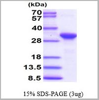
 SDS-PAGE
(3ug by SDS-PAGE under reducing condition and visualized by coomassie blue stain)
SDS-PAGE
(3ug by SDS-PAGE under reducing condition and visualized by coomassie blue stain)
CLPP, Recombinant Protein (Cat# AAA11805)
Full Name
CLPP, 57-277aa, Human, E Coli
Gene Names
CLPP; DFNB81; PRLTS3
Applications
SDS-PAGE
Purity
> 95% by SDS-PAGE
Pricing
 SDS-PAGE
(3 ug by SDS-PAGE under reducing condition and visualized by coomassie blue stain.)
SDS-PAGE
(3 ug by SDS-PAGE under reducing condition and visualized by coomassie blue stain.)
HMGN1, Recombinant Protein (Cat# AAA11776)
Full Name
HMGN1, 1-100aa, Human, His tag, E Coli
Gene Names
HMGN1; HMG14
Applications
SDS-PAGE
Purity
> 95% by SDS-PAGE
Pricing
GDF11, Active Protein (Cat# AAA14381)
Full Name
GDF11 protein
Gene Names
GDF11; BMP11; BMP-11
Purity
> 98% by SDS-PAGE analysis and HPLC
Pricing
Flap Structure-Specific Endonuclease 1, Recombinant Protein (Cat# AAA10862)
Full Name
Recombinant Human Flap Structure-Specific Endonuclease 1
Gene Names
FEN1; MF1; RAD2; FEN-1
Purity
Greater than 90.0% as determined by SDS-PAGE.
Pricing
 SDS-PAGE
(3ug by SDS-PAGE under reducing condition and visualized by coomassie blue stain.)
SDS-PAGE
(3ug by SDS-PAGE under reducing condition and visualized by coomassie blue stain.)
CCL19, Recombinant Protein (Cat# AAA11793)
Full Name
CCL19, 22-98aa, Human, T7 tag, E Coli
Gene Names
CCL19; ELC; CKb11; MIP3B; MIP-3b; SCYA19
Applications
SDS-PAGE
Purity
> 95% by SDS-PAGE
Pricing
 SDS-PAGE
(3ug by SDS-PAGE under reducing condition and visualized by coomassie blue stain)
SDS-PAGE
(3ug by SDS-PAGE under reducing condition and visualized by coomassie blue stain)
C1QBP, Recombinant Protein (Cat# AAA11759)
Full Name
C1QBP,74-282aa, Human, E Coli
Applications
SDS-PAGE
Purity
≥ 95% by SDS-PAGE
Pricing
 WB (Western Blot)
(Positive WB detected in Recombinant proteinAll lanes: L1R antibody at 1:2000Secondary Goat polyclonal to rabbit IgG at 1/50000 dilutionPredicted band size: 26.8 kDabserved band size: 29 kDa)
WB (Western Blot)
(Positive WB detected in Recombinant proteinAll lanes: L1R antibody at 1:2000Secondary Goat polyclonal to rabbit IgG at 1/50000 dilutionPredicted band size: 26.8 kDabserved band size: 29 kDa)
L1R, Polyclonal Antibody (Cat# AAA27054)
Full Name
Rabbit anti-Vaccinia virus (strain Copenhagen)(VACV) L1R Polyclonal Antibody
Reactivity
Vaccinia virus
Applications
Western Blot
Purity
Protein G
Pricing
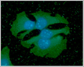
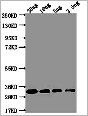
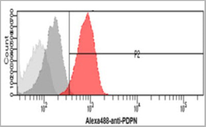 FCM (Flow Cytometry)
(Flow cytometry analysis of Podoplanin in HeLa cells. The cell was stained with (AAA11728) at 2-5ug for 1x10^6cells (red). A Goat anti mouse IgG (Alexa fluor 488) was used as the secondary antibody. Mouse monoclonal IgG was used as the isotype control (dark gray), cells without incubation with primary and secondary antibody was used as the negative control (light gray).)
FCM (Flow Cytometry)
(Flow cytometry analysis of Podoplanin in HeLa cells. The cell was stained with (AAA11728) at 2-5ug for 1x10^6cells (red). A Goat anti mouse IgG (Alexa fluor 488) was used as the secondary antibody. Mouse monoclonal IgG was used as the isotype control (dark gray), cells without incubation with primary and secondary antibody was used as the negative control (light gray).)
PDPN, Monoclonal Antibody (Cat# AAA11728)
Full Name
PDPN antibody
Gene Names
PDPN; T1A; GP36; GP40; Gp38; OTS8; T1A2; TI1A; T1A-2; AGGRUS; HT1A-1; PA2.26
Reactivity
Human
Applications
Flow Cytometry, Immunocytochemistry, Immunofluorescence
Purity
By protein-G affinity chromatography
Pricing
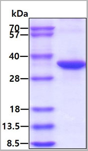 SDS-PAGE
(3ug by SDS-PAGE under reducing condition and visualized by coomassie blue stain.)
SDS-PAGE
(3ug by SDS-PAGE under reducing condition and visualized by coomassie blue stain.)
NQO1, Active Protein (Cat# AAA11735)
Full Name
Recombinant Human NQO1 Protein
Gene Names
NQO1; DTD; QR1; DHQU; DIA4; NMOR1; NMORI
Applications
SDS-PAGE, ELISA
Purity
>95% by SDS-PAGE
Pricing
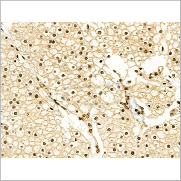
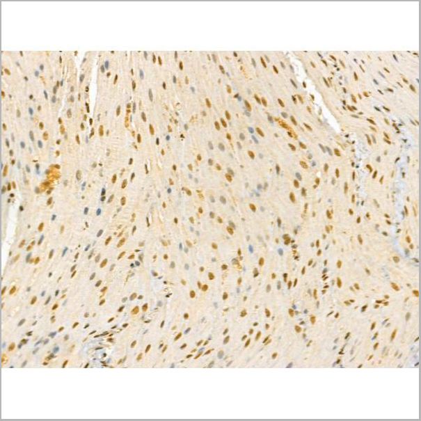
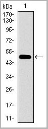
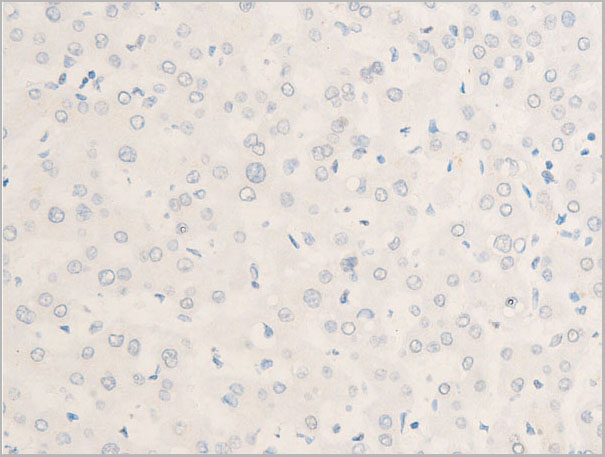
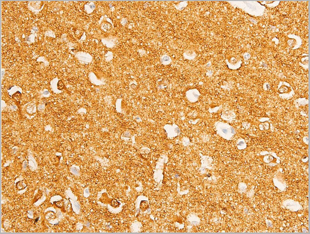
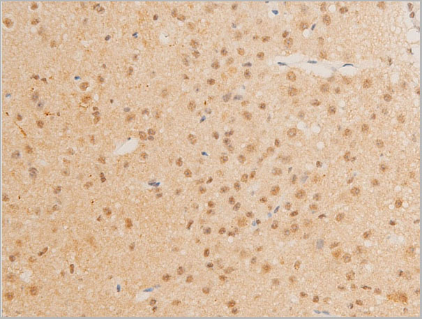
 IHC (Immunohistochemistry)
(Immunohistochemical analysis of SHARP1 staining in human brain formalin fixed paraffin embedded tissue section. The section was pre-treated using heat mediated antigen retrieval with sodium citrate buffer (pH 6.0). The section was then incubated with the antibody at room temperature and detected using an HRP conjugated compact polymer system. DAB was used as the chromogen. The section was then counterstained with haematoxylin and mounted with DPX.)
IHC (Immunohistochemistry)
(Immunohistochemical analysis of SHARP1 staining in human brain formalin fixed paraffin embedded tissue section. The section was pre-treated using heat mediated antigen retrieval with sodium citrate buffer (pH 6.0). The section was then incubated with the antibody at room temperature and detected using an HRP conjugated compact polymer system. DAB was used as the chromogen. The section was then counterstained with haematoxylin and mounted with DPX.)
SHARP1, Polyclonal Antibody (Cat# AAA27795)
Full Name
Anti-SHARP1 Antibody
Gene Names
BHLHE41; DEC2; hDEC2; BHLHB3; SHARP1
Reactivity
Human, Mouse, Rat
Applications
Western Blot, Immunohistochemistry
Purity
The antibody was purified by immunogen affinity chromatography.
Pricing
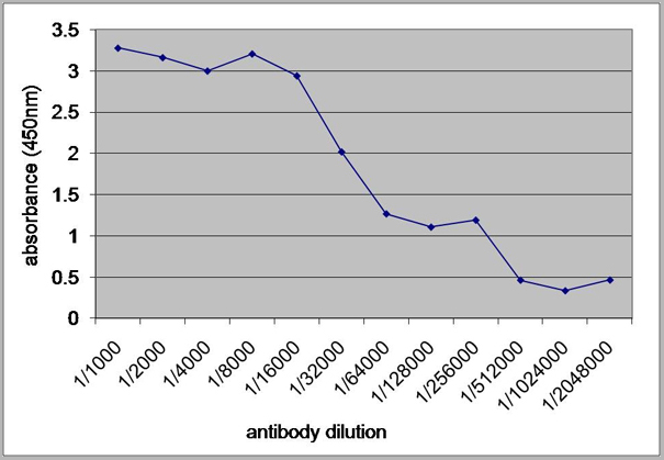
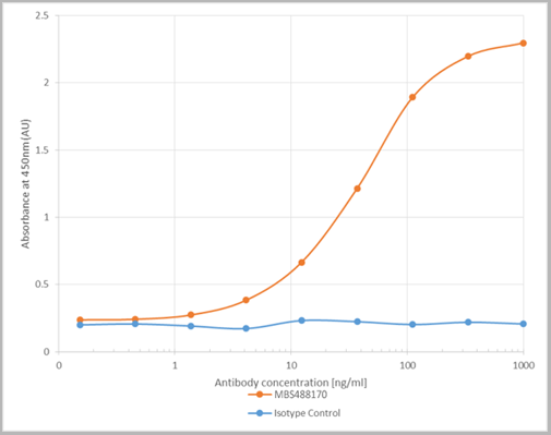 ELISA
(ELISA on Denge Virus-Like Particles using AAA14182 Dengue Virus Serotype 2 VLPs were coated onto a plate at 5µg/ml. Anti-flavivirus antibody (AAA14182) added to plate in a 3-fold serial dilution starting at 1000 ng/ml. Detection performed using HRP labelled goat anti-mouse IgG.)
ELISA
(ELISA on Denge Virus-Like Particles using AAA14182 Dengue Virus Serotype 2 VLPs were coated onto a plate at 5µg/ml. Anti-flavivirus antibody (AAA14182) added to plate in a 3-fold serial dilution starting at 1000 ng/ml. Detection performed using HRP labelled goat anti-mouse IgG.)
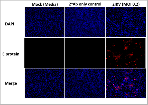
Flavivirus group antigen, Recombinant Antibody (Cat# AAA14182)
Full Name
Anti-Flavivirus group antigen [D1-4G2-4-15 (4G2)]
Reactivity
Dengue Virus, Zika Virus, West Nile Virus, Flaviviridae, Yellow Fever Virus
Applications
Immunofluorescence, Western Blot, Neutralization Assay, Flow Cytometry, Immunohistochemistry
Purity
Protein A Affinity Purified
Pricing
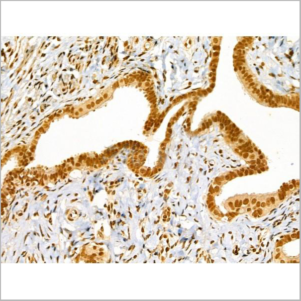
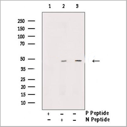
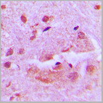
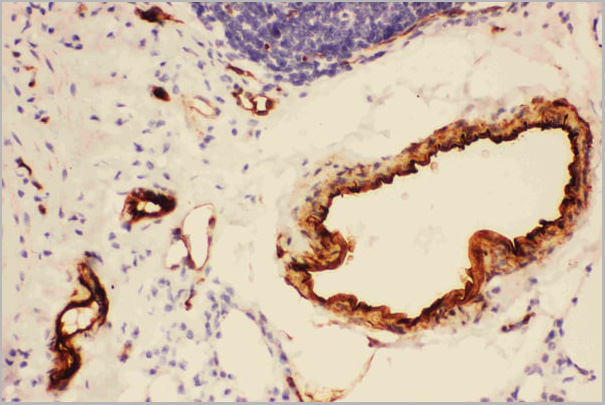
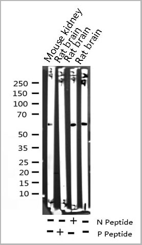
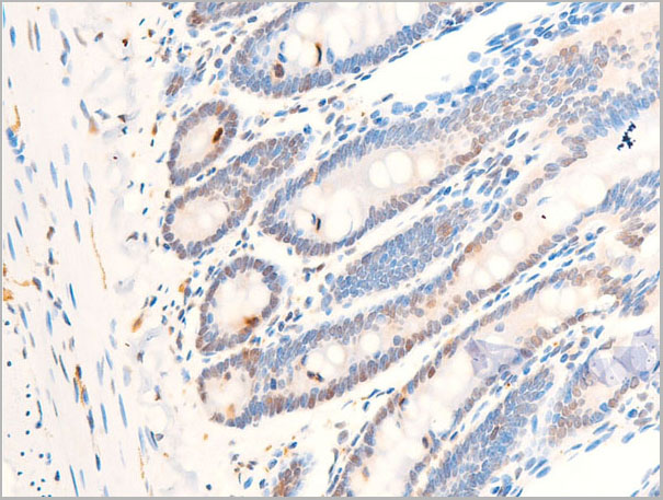
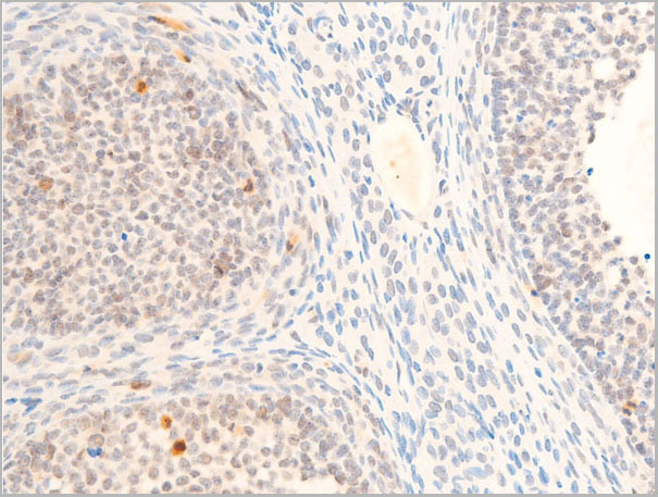
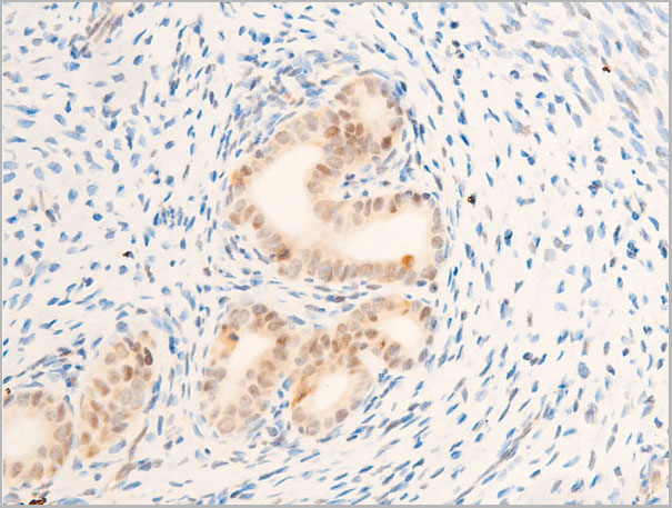
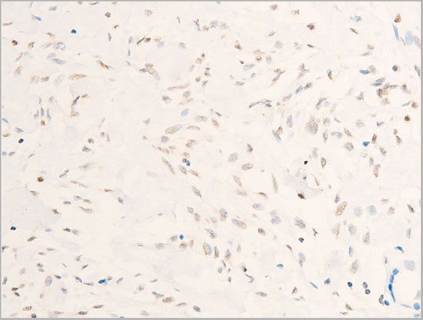
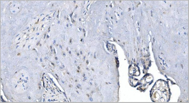
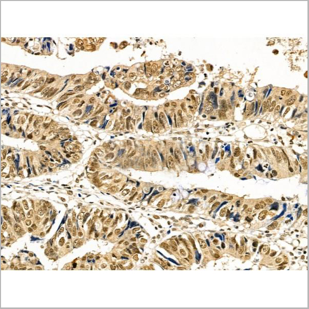 IHC (Immunohistochemistry)
(At 1/100 staining Human colorectal cancer by IHC-P. The sample was formaldehyde fixed and a heat mediated antigen retrieval step in citrate buffer was performed. The sample was then blocked and incubated with the primary antibody at 4 degree C overnight. An HRP conjugated anti-Rabbit antibody was used as the secondary antibody.)
IHC (Immunohistochemistry)
(At 1/100 staining Human colorectal cancer by IHC-P. The sample was formaldehyde fixed and a heat mediated antigen retrieval step in citrate buffer was performed. The sample was then blocked and incubated with the primary antibody at 4 degree C overnight. An HRP conjugated anti-Rabbit antibody was used as the secondary antibody.)
Cyclin E1, Polyclonal Antibody (Cat# AAA31362)
Full Name
Phospho-Cyclin E1 (Ser73) Antibody
Gene Names
CCNE1; CCNE
Reactivity
Human, Mouse, Rat
Applications
Western Blot, Immunohistochemistry, Peptide ELISA
Purity
Peptide affinity purification
Pricing
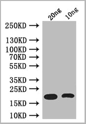
 WB (Western Blot)
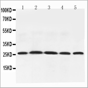
(Figure 6. Western blot analysis of Adiponectin using anti-Adiponectin antibody (AAA11602).Electrophoresis was performed on a 5-20% SDS-PAGE gel at 70V (Stacking gel) / 90V (Resolving gel) for 2-3 hours. The sample well of each lane was loaded with 50ug of sample under reducing conditions.Lane 1: Rat Testis Tissue Lysate,Lane 2: Rat Spleen Tissue Lysate,Lane 3: Rat Skeletal Muscle Tissue Lysate,Lane 4: Rat Cardiac Muscle Tissue Lysate,Lane 5: Rat Liver Tissue Lysate,After Electrophoresis, proteins were transferred to a Nitrocellulose membrane at 150mA for 50-90 minutes. Blocked the membrane with 5% Non-fat Milk/ TBS for 1.5 hour at RT. The membrane was incubated with rabbit anti-Adiponectin antigen affinity purified polyclonal antibody at 0.5ug/mL overnight at 4 degree C, then washed with TBS-0.1%Tween 3 times with 5 minutes each and probed with a goat anti-rabbit IgG-HRP secondary antibody at a dilution of 1:10000 for 1.5 hour at RT. The signal is developed using an Enhanced Chemiluminescent detection (ECL) kit with Tanon 5200 system. A specific band was detected for Adiponectin at approximately 26KD. The expected band size for Adiponectin is at 26KD.)
WB (Western Blot)
(Figure 6. Western blot analysis of Adiponectin using anti-Adiponectin antibody (AAA11602).Electrophoresis was performed on a 5-20% SDS-PAGE gel at 70V (Stacking gel) / 90V (Resolving gel) for 2-3 hours. The sample well of each lane was loaded with 50ug of sample under reducing conditions.Lane 1: Rat Testis Tissue Lysate,Lane 2: Rat Spleen Tissue Lysate,Lane 3: Rat Skeletal Muscle Tissue Lysate,Lane 4: Rat Cardiac Muscle Tissue Lysate,Lane 5: Rat Liver Tissue Lysate,After Electrophoresis, proteins were transferred to a Nitrocellulose membrane at 150mA for 50-90 minutes. Blocked the membrane with 5% Non-fat Milk/ TBS for 1.5 hour at RT. The membrane was incubated with rabbit anti-Adiponectin antigen affinity purified polyclonal antibody at 0.5ug/mL overnight at 4 degree C, then washed with TBS-0.1%Tween 3 times with 5 minutes each and probed with a goat anti-rabbit IgG-HRP secondary antibody at a dilution of 1:10000 for 1.5 hour at RT. The signal is developed using an Enhanced Chemiluminescent detection (ECL) kit with Tanon 5200 system. A specific band was detected for Adiponectin at approximately 26KD. The expected band size for Adiponectin is at 26KD.)
Adiponectin, Polyclonal Antibody (Cat# AAA11602)
Full Name
Anti-Adiponectin antibody
Gene Names
Adipoq; Acdc; Acrp30
Reactivity
Mouse, Rat
Applications
Western Blot, Immunohistochemistry
Purity
Immunogen affinity purified.
Pricing
 Application Data
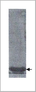
(Arrow: Yellow fever virus envelope protein (Lot# 10-2018))
Application Data
(Arrow: Yellow fever virus envelope protein (Lot# 10-2018))
Yellow fever virus envelope, Recombinant Protein (Cat# AAA14646)
Full Name
Yellow fever virus envelope protein
Applications
Lateral Flow
Purity
>95% pure (10 % SDS-PAGE Coomassie blue staining).
Pricing
 SDS-PAGE
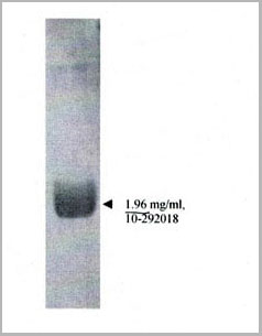
(Arrow: 1.96 mg/ml)
SDS-PAGE
(Arrow: 1.96 mg/ml)
Zika virus envelope, Recombinant Protein (Cat# AAA14630)
Full Name
Recombinant Zika envelope protein
Applications
Immunoassay
Purity
>95% pure (10% SDS-PAGE, Coomassie blue stain).
Pricing
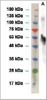
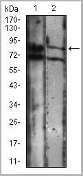
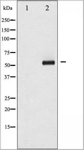
 WB (Western Blot)
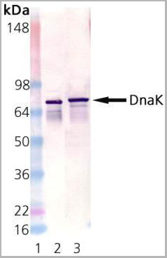
(Western Blot Analysis using AAA14655: Lane 1: MW Marker; Lane 2: DnaK active recombinant protein, Lane 3: E. coli cell lysate.)
WB (Western Blot)
(Western Blot Analysis using AAA14655: Lane 1: MW Marker; Lane 2: DnaK active recombinant protein, Lane 3: E. coli cell lysate.)
DnaK, Monoclonal Antibody (Cat# AAA14655)
Full Name
DnaK
Reactivity
E Coli
Applications
Western Blot
Purity
Affinity Purified
Purified by Protein G affinity chromatography.
Purified by Protein G affinity chromatography.
Pricing
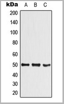
 WB (Western Blot)
(Western blot: primary antibody, anti-envelope (Zika virus) rabbit polyclonal antibody (1:2000 dilution); secondary antibody, HRP-conjugated goat anti-rabbit IgG (1:4000)A, full length envelope protein of Zika virus)
WB (Western Blot)
(Western blot: primary antibody, anti-envelope (Zika virus) rabbit polyclonal antibody (1:2000 dilution); secondary antibody, HRP-conjugated goat anti-rabbit IgG (1:4000)A, full length envelope protein of Zika virus)
Envelope (E) (Zika Virus), Antibody (Cat# AAA13695)
Full Name
Anti-Envelope (E) (Zika Virus) Polyclonal Antibody, Rabbit Serum
Applications
Western Blot
Pricing
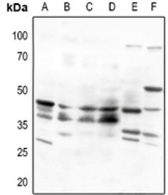 WB (Western Blot)
(Western blot analysis of Beta-3 Adrenergic Receptor expression in HEK293T (A), Hela (B), HepG2 (C), A2780 (D), mouse kidney (E), rat liver (F) whole cell lysates.)
WB (Western Blot)
(Western blot analysis of Beta-3 Adrenergic Receptor expression in HEK293T (A), Hela (B), HepG2 (C), A2780 (D), mouse kidney (E), rat liver (F) whole cell lysates.)
Beta-3 Adrenergic Receptor, Polyclonal Antibody (Cat# AAA27792)
Full Name
Anti-Beta-3 Adrenergic Receptor Antibody
Gene Names
ADRB3; BETA3AR
Reactivity
Human, Mouse, Rat
Applications
Western Blot
Purity
The antibody was purified by immunogen affinity chromatography.
Pricing
E Coli, Polyclonal Antibody (Cat# AAA14318)
Full Name
E Coli antibody
Applications
ELISA
Purity
> 95% pure
Pricing
Retinoic Acid Receptor Responder 2, Recombinant Protein (Cat# AAA10868)
Full Name
Recombinant Human Retinoic Acid Receptor Responder 2
Gene Names
RARRES2; TIG2; HP10433
Purity
Greater than 98.0% as determined by: (a) Analysis by RP-HPLC. (b) Analysis by SDS-PAGE.
Pricing
 SDS-PAGE
(3ug by SDS-PAGE under reducing condition and visualized by coomassie blue stain)
SDS-PAGE
(3ug by SDS-PAGE under reducing condition and visualized by coomassie blue stain)
PPARG, Recombinant Protein (Cat# AAA11778)
Full Name
PPARG, 209-477aa, Human, His tag, E Coli
Gene Names
PPARG; GLM1; CIMT1; NR1C3; PPARG1; PPARG2; PPARgamma
Applications
SDS-PAGE
Purity
> 95% by SDS-PAGE
Pricing
CEA, Native Protein (Cat# AAA14396)
Full Name
CEA Ag(Preservative-free)
Purity
>95% by SDS-PAGE and electrophoresis
Pricing
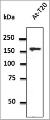
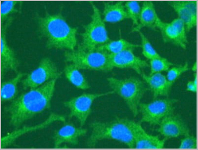 IF (Immunofluorescence)
(anti-CLTC Ab – Membrane Vesicle Marker in Hepa1-6 cells at 1/50 dilution; cells were fixed with methanol)
IF (Immunofluorescence)
(anti-CLTC Ab – Membrane Vesicle Marker in Hepa1-6 cells at 1/50 dilution; cells were fixed with methanol)
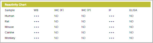
Clathrin HC, Polyclonal Antibody (Cat# AAA13865)
Full Name
Clathrin HC Polyclonal Antibody
Gene Names
CLTC; Hc; CHC; CHC17; CLH-17; CLTCL2
Reactivity
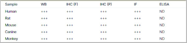
Human, Mouse, Rat, Canine, Monkey
Applications
Western Blot, Immunofluorescence, Immunohistochemistry, Immunohistochemistry
Purity
This antibody is epitope-affinity purified from goat antiserum.
Pricing
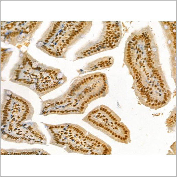
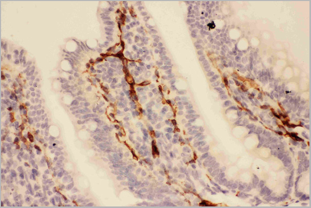
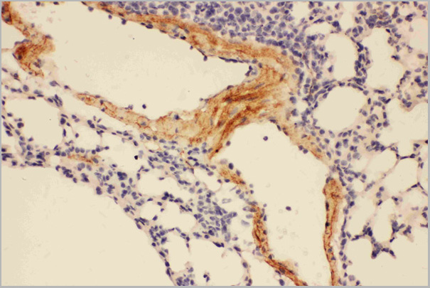
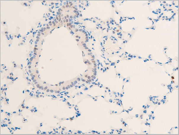
 IHC (Immunohistochemistry)
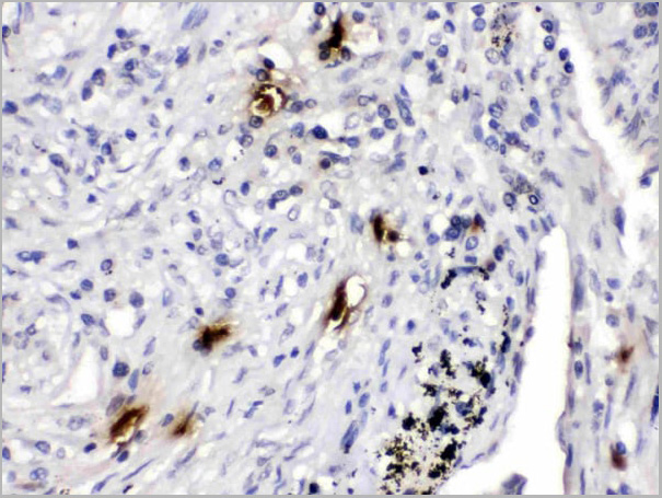
(Mast Cell Tryptase was detected in paraffin-embedded sections of human lung cancer tissues using rabbit anti- Mast Cell Tryptase Antigen Affinity purified polyclonal antibody at 1ug/mL. The immunohistochemical section was developed using SABC method.)
IHC (Immunohistochemistry)
(Mast Cell Tryptase was detected in paraffin-embedded sections of human lung cancer tissues using rabbit anti- Mast Cell Tryptase Antigen Affinity purified polyclonal antibody at 1ug/mL. The immunohistochemical section was developed using SABC method.)
Mast Cell Tryptase, Polyclonal Antibody (Cat# AAA11674)
Full Name
Anti-Mast Cell Tryptase Antibody
Gene Names
TPSAB1; TPS1; TPS2; TPSB1
Reactivity
Human
Applications
Western Blot, Immunohistochemistry
Purity
Immunogen affinity purified.
Pricing


 Application Data
(Published customer image: Mouse anti V5 tag antibody, clone SV5-Pk1 used for the detection of V5 tagged WEEV_nsP3 protein by western blotting and immunofluorescenceImage caption: WEEV nsP3 interaction with host IKKbeta. A) U87MGs were transfected in a 6-well plate with 5 ug of pUC19 and WEEV_nsP3_HA for 24 hours. Cell lysates were resolved using SDS-PAGE and subsequently immunoblotted with V5 antibody and beta-actin served as a loading control. B) U87MGs were transfected with WEEV_nsP3_V5; cells were fixed after 24 hours and stained with antibodies against the endogenous IKKbeta and the V5 tag. Cells were incubated with appropriate secondary Alexa Fluor antibodies and the nuclei stained with DAPI. Co-localization of IKKbeta with WEEV_nsP3_V5 (yellow) was observed as shown by the arrows. B) Panels E -H serve as an example of transfected cells in a given field of view that show co-localization of IKKbeta and WEEV_nsP3_V5 24 hours post transfection. Panels I-L represent magnified images of other cells showing co-localization of IKKbeta and WEEV_nsP3_V5. Panel M is a magnified image of panel L. The co-localization was confirmed by Z-stack analysis. Co-localization was calculated to be approximately in 61% of cells (163 cells were counted of which 44% demonstrated expression of nsP3. Of those cells that expressed nsP3, 61% showed co-localization of both proteins). Images were taken using Nikon Eclipse TE2000-U at 60x magnification and are representative of 2 independent experiments.From: Amaya M, Voss K, Sampey G, Senina S, de la Fuente C, et al. (2014) The Role of IKKbeta in Venezuelan Equine Encephalitis Virus Infection. PLoS ONE 9(2): e86745.)
Application Data
(Published customer image: Mouse anti V5 tag antibody, clone SV5-Pk1 used for the detection of V5 tagged WEEV_nsP3 protein by western blotting and immunofluorescenceImage caption: WEEV nsP3 interaction with host IKKbeta. A) U87MGs were transfected in a 6-well plate with 5 ug of pUC19 and WEEV_nsP3_HA for 24 hours. Cell lysates were resolved using SDS-PAGE and subsequently immunoblotted with V5 antibody and beta-actin served as a loading control. B) U87MGs were transfected with WEEV_nsP3_V5; cells were fixed after 24 hours and stained with antibodies against the endogenous IKKbeta and the V5 tag. Cells were incubated with appropriate secondary Alexa Fluor antibodies and the nuclei stained with DAPI. Co-localization of IKKbeta with WEEV_nsP3_V5 (yellow) was observed as shown by the arrows. B) Panels E -H serve as an example of transfected cells in a given field of view that show co-localization of IKKbeta and WEEV_nsP3_V5 24 hours post transfection. Panels I-L represent magnified images of other cells showing co-localization of IKKbeta and WEEV_nsP3_V5. Panel M is a magnified image of panel L. The co-localization was confirmed by Z-stack analysis. Co-localization was calculated to be approximately in 61% of cells (163 cells were counted of which 44% demonstrated expression of nsP3. Of those cells that expressed nsP3, 61% showed co-localization of both proteins). Images were taken using Nikon Eclipse TE2000-U at 60x magnification and are representative of 2 independent experiments.From: Amaya M, Voss K, Sampey G, Senina S, de la Fuente C, et al. (2014) The Role of IKKbeta in Venezuelan Equine Encephalitis Virus Infection. PLoS ONE 9(2): e86745.)
V5-TAG, Monoclonal Antibody (Cat# AAA11850)
Full Name
MOUSE ANTI V5-TAG:Biotin
Applications
Immunohistochemistry, Western Blot
Pricing
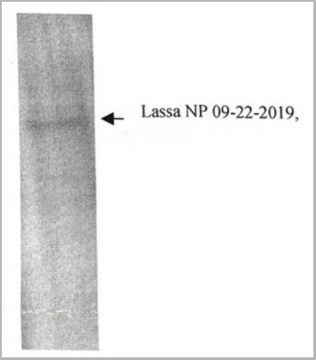 Application Data
Lassa virus full length nucleoprotein, Recombinant Protein (Cat# AAA14634)
Full Name
Lassa virus full length nucleoprotein
Applications
Immunoassay
Purity
>90% pure (10% SDS-PAGE, Coomassie blue stain).
Pricing
Epididymis protein 4, HE4, Recombinant Protein (Cat# AAA13529)
Full Name
Human Epididymis protein 4, HE4 Protein (Active)
Applications
ELISA, SDS-PAGE
Purity
>95% by SDS-PAGE
Pricing
Troponin-C2, Recombinant Protein (Cat# AAA10806)
Full Name
Recombinant Human Troponin-C2
Purity
Greater than 95.0% as determined by SDS-PAGE.
Pricing
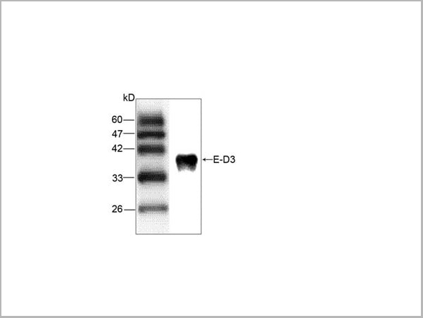 Application Data
JEV E-D3, Monoclonal Antibody (Cat# AAA13645)
Full Name
Anti-JEV E-D3 Antibody
Applications
Western Blot
Purity
Affinity purified
Pricing
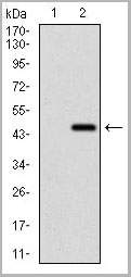
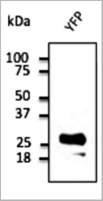 WB (Western Blot)
(Anti-YFP Ab at 1/2,000 dilution; 293HEK cells transduced with YFP Ad; lysates at 50 µg per lane; rabbit polyclonal to goat IgG (HRP) at 1/10,000 dilution;)
WB (Western Blot)
(Anti-YFP Ab at 1/2,000 dilution; 293HEK cells transduced with YFP Ad; lysates at 50 µg per lane; rabbit polyclonal to goat IgG (HRP) at 1/10,000 dilution;)
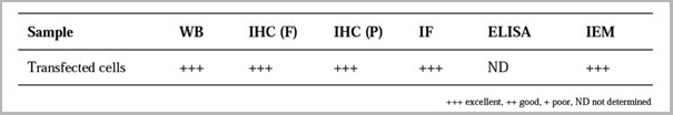
YFP, Polyclonal Antibody (Cat# AAA13878)
Full Name
YFP Polyclonal Antibody
Reactivity
Transfected cells proteins
Applications
Western Blot, Immunofluorescence, Immunohistochemistry
Purity
This antibody is epitope-affinity purified from goat antiserum.
Pricing

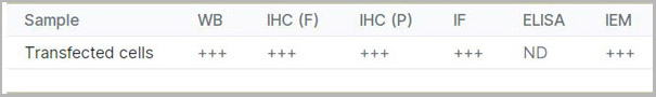
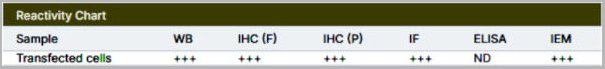
 Reactivity Chart (RC)
TagRFP, Polyclonal Antibody (Cat# AAA13884)
Full Name
anti-TagRFP
Reactivity
Transfected cells proteins
Applications
Western Blot, Immunofluorescence, Immunohistochemistry, Immunohistochemistry, Immunoelectron Microscopy
Purity
This antibody is epitope-affinity purified from goat antiserum
Pricing
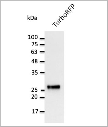 Application Data
(Anti-TurboRFP Ab at 1/2,500 dilution using HEK293 transfected cell lysates at 50 ug per lane; rabbit polyclonal to goat IgG (HRP) at 1/10,000 dilution;)
Application Data
(Anti-TurboRFP Ab at 1/2,500 dilution using HEK293 transfected cell lysates at 50 ug per lane; rabbit polyclonal to goat IgG (HRP) at 1/10,000 dilution;)
TurboRFP, Polyclonal Antibody (Cat# AAA13881)
Full Name
anti-TurboRFP
Reactivity
Transfected cells proteins
Applications
Western Blot, Immunofluorescence, Immunohistochemistry, Immunohistochemistry, Immunoelectron Microscopy
Purity
This antibody is epitope-affinity purified from goat antiserum.
Pricing
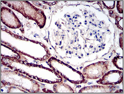 IHC (Immunohistchemistry)
(Immunohistochemical analysis of paraffin-embedded kidney tissues using CDH16 mouse mAb with DAB staining.)
IHC (Immunohistchemistry)
(Immunohistochemical analysis of paraffin-embedded kidney tissues using CDH16 mouse mAb with DAB staining.)
CDH16, Monoclonal Antibody (Cat# AAA14153)
Full Name
Anti-CDH16 Mouse mAb
Reactivity
Human
Applications
Western Blot, Immunohistochemistry, Immunocytochemistry, Flow Cytometry
Purity
Unpurification
Pricing
 SDS-PAGE
(3ug by SDS-PAGE under reducing condition and visualized by coomassie blue stain.)
SDS-PAGE
(3ug by SDS-PAGE under reducing condition and visualized by coomassie blue stain.)
Midkine, Recombinant Protein (Cat# AAA11769)
Full Name
Midkine, 21-143aa, Human, His tag, E Coli
Gene Names
MDK; MK; ARAP; NEGF2
Applications
SDS-PAGE
Purity
> 90% by SDS-PAGE
Pricing
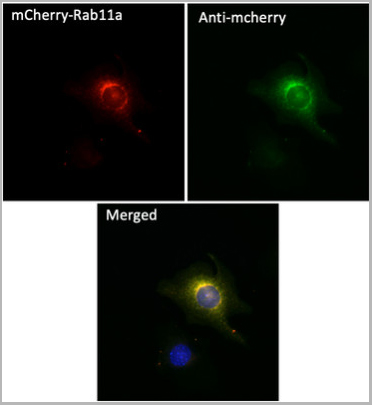
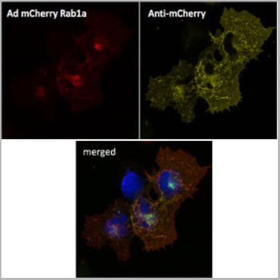
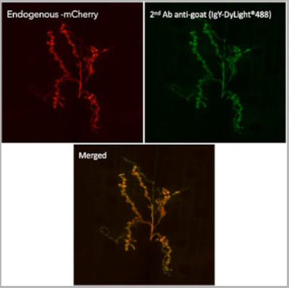
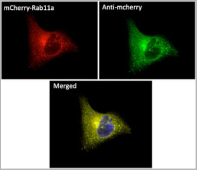
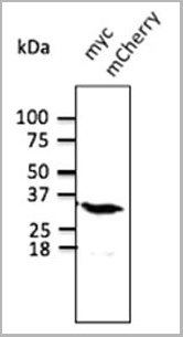
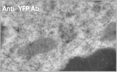
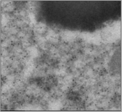 IEM (Immunogold Electron Microscopy)
(Immunogold labeling of RPE, in vivo injected with mCherry expressing vector)
IEM (Immunogold Electron Microscopy)
(Immunogold labeling of RPE, in vivo injected with mCherry expressing vector)
mCherry, Polyclonal Antibody (Cat# AAA13867)
Full Name
mCherry Polyclonal Antibody
Reactivity
Transfected cells proteins
Applications
Immunofluorescence, Western Blot, Immunohistochemistry, Immunohistochemistry, Immunoelectron Microscopy
Pricing
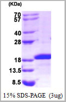
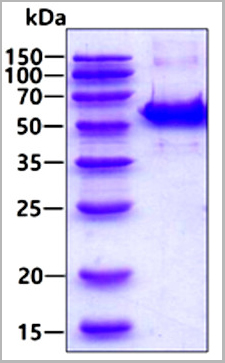 SDS-PAGE
(3ug by SDS-PAGE under reducing condition and visualized by coomassie blue stain.)
SDS-PAGE
(3ug by SDS-PAGE under reducing condition and visualized by coomassie blue stain.)
SEPSECS, Recombinant Protein (Cat# AAA11804)
Full Name
SEPSECS, 1-501aa, Human, His tag, E Coli
Gene Names
SEPSECS; LP; SLA; PCH2D; SLA/LP
Applications
SDS-PAGE
Purity
> 90% by SDS-PAGE
Pricing
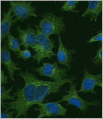
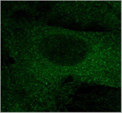
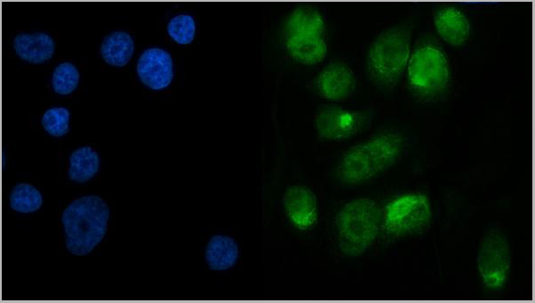
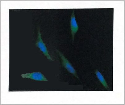 IF (Immunofluorescence)
(Immunofluorescence (IFC) analysis of PC3 cells using AAA14697 (1:100). The secondary antibody (green) was used Alexa Fluor 488. DAPI was stained the cell nucleus (blue).)
IF (Immunofluorescence)
(Immunofluorescence (IFC) analysis of PC3 cells using AAA14697 (1:100). The secondary antibody (green) was used Alexa Fluor 488. DAPI was stained the cell nucleus (blue).)
Ribonuclease inhibitor 1, Monoclonal Antibody (Cat# AAA14697)
Full Name
Ribonuclease inhibitor 1 (RNH1)
Gene Names
RNH1; RAI; RNH; MGC4569; MGC18200; MGC54054
Applications
Immunofluorescence, Western Blot
Purity
Affinity Purified
Purified by Protein G affinity chromatography.
Purified by Protein G affinity chromatography.
Pricing
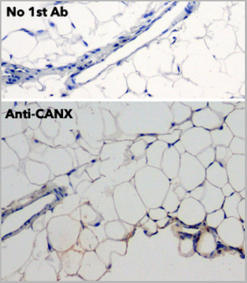
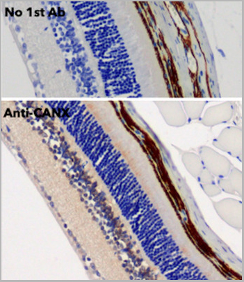
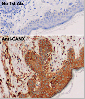
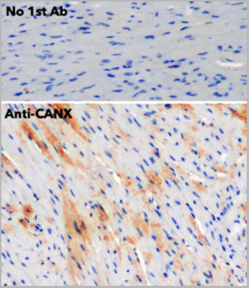
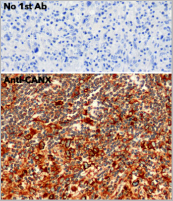
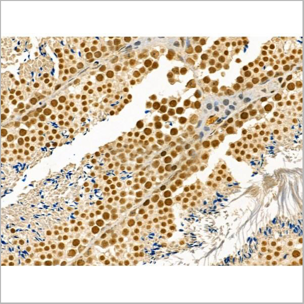
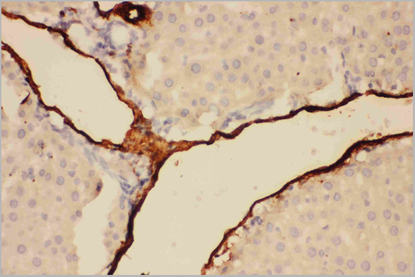
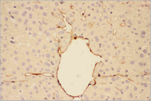
 IHC (Immunohistochemistry)
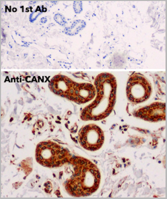
(IHC of human adipose tissue anti-CANX antibody and FFPE tissue after heat-induced antigen retrieval. Anti-CANX Ab at 1:750/DAB detection.)
IHC (Immunohistochemistry)
(IHC of human adipose tissue anti-CANX antibody and FFPE tissue after heat-induced antigen retrieval. Anti-CANX Ab at 1:750/DAB detection.)
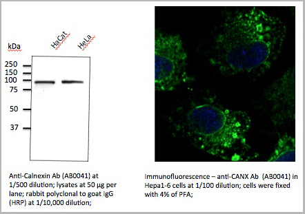
CANX, Polyclonal Antibody (Cat# AAA13872)
Full Name
Calnexin Polyclonal Antibody
Gene Names
canx; zgc:63524; wu:fe06b12
Reactivity
Human, Rat, Mouse, Monkey, Canine proteins
Applications
Immunofluorescence, Western Blot, Immunohistochemistry, Immunohistochemistry
Pricing
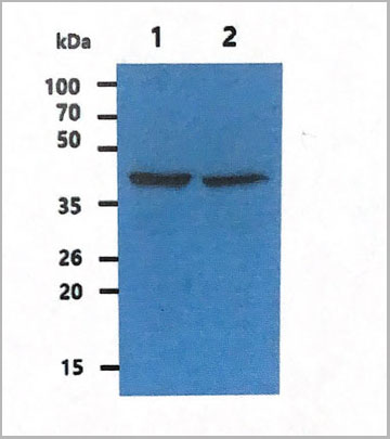
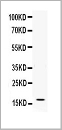 WB (Western Blot)
(Figure. Western blot analysis of IFN gamma using anti- IFN gamma antibody (AAA11517).Electrophoresis was performed on a 5-20% SDS-PAGE gel at 70V (Stacking gel) / 90V (Resolving gel) for 2-3 hours. The sample well of each lane was loaded with 50ug of sample under reducing conditions.Lane: Recombinant Human IFN gamma Protein 0.5ngAfter Electrophoresis, proteins were transferred to a Nitrocellulose membrane at 150mA for 50-90 minutes. Blocked the membrane with 5% Non-fat Milk/ TBS for 1.5 hour at RT. The membrane was incubated with rabbit anti- IFN gamma antigen affinity purified polyclonal antibody at 0.5ug/mL overnight at 4 degree C, then washed with TBS-0.1%Tween 3 times with 5 minutes each and probed with a goat anti-rabbit IgG-HRP secondary antibody at a dilution of 1:10000 for 1.5 hour at RT. The signal is developed using an Enhanced Chemiluminescent detection (ECL) kit with Tanon 5200 system. A specific band was detected for IFN gamma at approximately 17KD. The expected band size for IFN gamma is at 17KD.)
WB (Western Blot)
(Figure. Western blot analysis of IFN gamma using anti- IFN gamma antibody (AAA11517).Electrophoresis was performed on a 5-20% SDS-PAGE gel at 70V (Stacking gel) / 90V (Resolving gel) for 2-3 hours. The sample well of each lane was loaded with 50ug of sample under reducing conditions.Lane: Recombinant Human IFN gamma Protein 0.5ngAfter Electrophoresis, proteins were transferred to a Nitrocellulose membrane at 150mA for 50-90 minutes. Blocked the membrane with 5% Non-fat Milk/ TBS for 1.5 hour at RT. The membrane was incubated with rabbit anti- IFN gamma antigen affinity purified polyclonal antibody at 0.5ug/mL overnight at 4 degree C, then washed with TBS-0.1%Tween 3 times with 5 minutes each and probed with a goat anti-rabbit IgG-HRP secondary antibody at a dilution of 1:10000 for 1.5 hour at RT. The signal is developed using an Enhanced Chemiluminescent detection (ECL) kit with Tanon 5200 system. A specific band was detected for IFN gamma at approximately 17KD. The expected band size for IFN gamma is at 17KD.)
Interferon gamma, Polyclonal Antibody (Cat# AAA11517)
Full Name
Anti-human Interferon gamma antibody
Gene Names
IFNG; IFG; IFI
Reactivity
Human
Applications
Western Blot, Immunohistochemistry, Neutralization Assay, Immunoprecipitation
Purity
Immunogen affinity purified.
Pricing
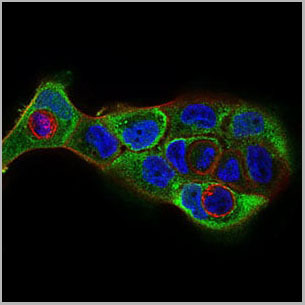
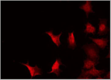 IF (Immunofluorescence)
(AAA30966 staining 293 by IF/ICC. The sample were fixed with PFA and permeabilized in 0.1% Triton X-100, then blocked in 10% serum for 45 minutes at 25 degree C. The primary antibody was diluted at 1/200 and incubated with the sample for 1 hour at 37 degree C. An Alexa Fluor 594 conjugated goat anti-rabbit IgG (H+L) Ab, diluted at 1/600, was used as the secondary antibody.)
IF (Immunofluorescence)
(AAA30966 staining 293 by IF/ICC. The sample were fixed with PFA and permeabilized in 0.1% Triton X-100, then blocked in 10% serum for 45 minutes at 25 degree C. The primary antibody was diluted at 1/200 and incubated with the sample for 1 hour at 37 degree C. An Alexa Fluor 594 conjugated goat anti-rabbit IgG (H+L) Ab, diluted at 1/600, was used as the secondary antibody.)
Myc, Polyclonal Antibody (Cat# AAA30966)
Full Name
Phospho-Myc (Thr58) Antibody
Gene Names
MYC; MRTL; MYCC; c-Myc; bHLHe39
Reactivity
Human, Mouse, Rat
Applications
Western Blot, Immunohistochemistry, Immunofluorescence, Immunocytochemistry, Immunoprecipitation
Purity
From purified rabbit serum by affinity purification via sequential chromatography on phospho-and non-phospho-peptide affinity columns.
Pricing
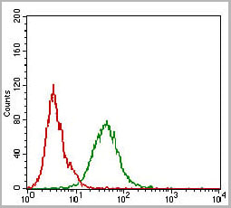
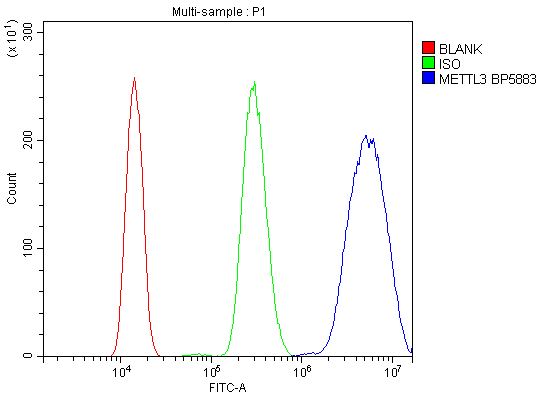 FCM (Flow Cytometry)
(Figure 6. Flow Cytometry analysis of HL-60 cells using anti-METTL3 antibody (AAA19247).Overlay histogram showing HL-60 cells stained with AAA19247 (Blue line). The cells were blocked with 10% normal goat serum. And then incubated with rabbit anti-METTL3 Antibody (AAA19247, 1μg/1x106 cells) for 30 min at 20 degree C. DyLight®488 conjugated goat anti-rabbit IgG (5-10μg/1x106 cells) was used as secondary antibody for 30 minutes at 20 degree C. Isotype control antibody (Green line) was rabbit IgG (1μg/1x106) used under the same conditions. Unlabelled sample (Red line) was also used as a control.)
FCM (Flow Cytometry)
(Figure 6. Flow Cytometry analysis of HL-60 cells using anti-METTL3 antibody (AAA19247).Overlay histogram showing HL-60 cells stained with AAA19247 (Blue line). The cells were blocked with 10% normal goat serum. And then incubated with rabbit anti-METTL3 Antibody (AAA19247, 1μg/1x106 cells) for 30 min at 20 degree C. DyLight®488 conjugated goat anti-rabbit IgG (5-10μg/1x106 cells) was used as secondary antibody for 30 minutes at 20 degree C. Isotype control antibody (Green line) was rabbit IgG (1μg/1x106) used under the same conditions. Unlabelled sample (Red line) was also used as a control.)
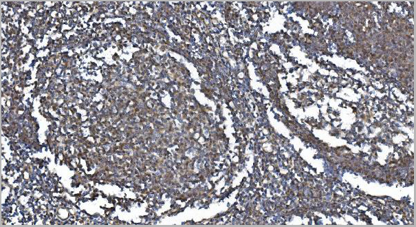
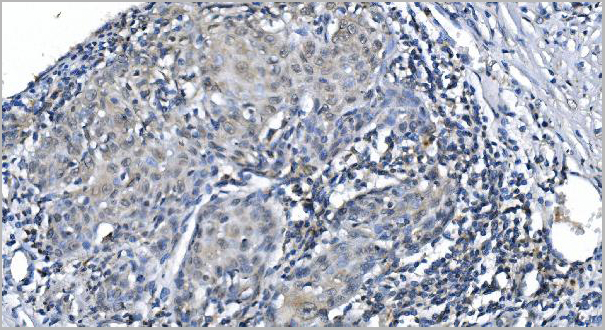
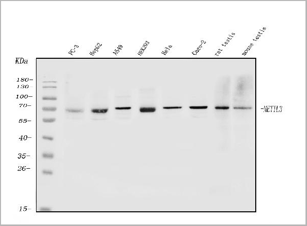
METTL3, Polyclonal Antibody (Cat# AAA19247)
Full Name
Anti-METTL3 Antibody
Gene Names
METTL3; M6A; IME4; Spo8; MT-A70
Reactivity
Human, Mouse, Rat
Applications
Western Blot, Immunohistochemistry, Immunocytochemistry, Immunofluorescence, Flow Cytometry, Direct ELISA
Purity
Immunogen affinity purified.
Pricing