Filters
Clonality
Type
Reactivity
Gene Name
Isotype
Host
Application
Clone
2304 results for " secondary" - showing 1200-1250


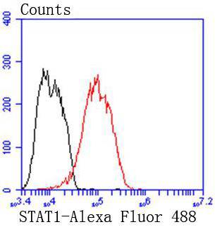 FCM (Flow Cytometry)
(Flow cytometric analysis of MCF-7 cells with STAT1 alpha antibody at 1/50 dilution (red) compared with an unlabelled control (cells without incubation with primary antibody; black). Alexa Fluor 488-conjugated goat anti rabbit IgG was used as the secondary antibody)
FCM (Flow Cytometry)
(Flow cytometric analysis of MCF-7 cells with STAT1 alpha antibody at 1/50 dilution (red) compared with an unlabelled control (cells without incubation with primary antibody; black). Alexa Fluor 488-conjugated goat anti rabbit IgG was used as the secondary antibody)
STAT1 alpha, Monoclonal Antibody (Cat# AAA30190)
Full Name
STAT1 alpha Antibody
Gene Names
STAT1; CANDF7; ISGF-3; STAT91
Reactivity
Human
Applications
WB, ICC, IF, IHC, IP, FC/FACS
Purity
ProA affinity purified
Pricing


 IHC (Immunohistochemistry)
(Figure 7. IHC analysis of PARK7 / DJ1 using anti-PARK7 / DJ1 antibody (AAA19139).PARK7 / DJ1 was detected in paraffin-embedded section of rat testis tissue. Heat mediated antigen retrieval was performed in citrate buffer (pH6, epitope retrieval solution) for 20 mins. The tissue section was blocked with 10% goat serum. The tissue section was then incubated with 1ug/ml rabbit anti-PARK7 / DJ1 Antibody (AAA19139) overnight at 4 degree C. Biotinylated goat anti-rabbit IgG was used as secondary antibody and incubated for 30 minutes at 37 degree C. The tissue section was developed using Strepavidin-Biotin-Complex (SABC) with DAB as the chromogen.)
IHC (Immunohistochemistry)
(Figure 7. IHC analysis of PARK7 / DJ1 using anti-PARK7 / DJ1 antibody (AAA19139).PARK7 / DJ1 was detected in paraffin-embedded section of rat testis tissue. Heat mediated antigen retrieval was performed in citrate buffer (pH6, epitope retrieval solution) for 20 mins. The tissue section was blocked with 10% goat serum. The tissue section was then incubated with 1ug/ml rabbit anti-PARK7 / DJ1 Antibody (AAA19139) overnight at 4 degree C. Biotinylated goat anti-rabbit IgG was used as secondary antibody and incubated for 30 minutes at 37 degree C. The tissue section was developed using Strepavidin-Biotin-Complex (SABC) with DAB as the chromogen.)
PARK7/DJ1, Polyclonal Antibody (Cat# AAA19139)
Full Name
Anti-PARK7/DJ1 Picoband Antibody
Gene Names
Park7; Dj1; CAP1; DJ-1; SP22
Reactivity
Mouse, Rat
No cross reactivity with other proteins.
No cross reactivity with other proteins.
Applications
EIA, IHC, WB
Purity
Immunogen affinity purified
Pricing


 IF (Immunofluorescence)
(Immunofluorescent analysis of 4% paraformaldehyde-fixed, 0.1% Triton X-100 permeabilized MCF-7 (human breast cancer cell line) cells labeling Pdx1 with at 1:25 dilution, followed by DyLight 488-conjugated IgG goat anti-rabbit secondary antibody at 1:200 dilution (green). Immunofluorescence image showing cytoplasm staining on MCF-7 cell line. Cytoplasmic actin is detected with DyLight 554 Phalloidin (PD18466410) at 1:100 dilution (red). The nuclear counter stain is DAPI (blue).)
IF (Immunofluorescence)
(Immunofluorescent analysis of 4% paraformaldehyde-fixed, 0.1% Triton X-100 permeabilized MCF-7 (human breast cancer cell line) cells labeling Pdx1 with at 1:25 dilution, followed by DyLight 488-conjugated IgG goat anti-rabbit secondary antibody at 1:200 dilution (green). Immunofluorescence image showing cytoplasm staining on MCF-7 cell line. Cytoplasmic actin is detected with DyLight 554 Phalloidin (PD18466410) at 1:100 dilution (red). The nuclear counter stain is DAPI (blue).)
OPN-a/b, Polyclonal Antibody (Cat# AAA26862)
Full Name
OPN-a/b, NT (SPP1, BNSP, OPN, Osteopontin, Bone sialoprotein 1, Nephropontin, Secreted phosphoprotein 1, Urinary stone protein, Uropontin) (MaxLight 550)
Gene Names
SPP1; OPN; BNSP; BSPI; ETA-1
Reactivity
Human
Applications
WB, IHC, IF
Purity
Purified by Protein A and Peptide Affinity Chromatography.
Pricing


 IF (Immunofluorescence)
(Figure 6. IF analysis of SCG10/STMN2 using anti- SCG10/STMN2 antibody (AAA19299).SCG10/STMN2 was detected in immunocytochemical section of U20S cells. Enzyme antigen retrieval was performed using IHC enzyme antigen retrieval reagent for 15 mins. The cells were blocked with 10% goat serum. And then incubated with 5μg/mL rabbit anti- SCG10/STMN2 Antibody (AAA19299) overnight at 4 degree C. DyLight®488 Conjugated Goat Anti-Rabbit IgG was used as secondary antibody at 1:100 dilution and incubated for 30 minutes at 37 degree C. The section was counterstained with DAPI. Visualize using a fluorescence microscope and filter sets appropriate for the label used.)
IF (Immunofluorescence)
(Figure 6. IF analysis of SCG10/STMN2 using anti- SCG10/STMN2 antibody (AAA19299).SCG10/STMN2 was detected in immunocytochemical section of U20S cells. Enzyme antigen retrieval was performed using IHC enzyme antigen retrieval reagent for 15 mins. The cells were blocked with 10% goat serum. And then incubated with 5μg/mL rabbit anti- SCG10/STMN2 Antibody (AAA19299) overnight at 4 degree C. DyLight®488 Conjugated Goat Anti-Rabbit IgG was used as secondary antibody at 1:100 dilution and incubated for 30 minutes at 37 degree C. The section was counterstained with DAPI. Visualize using a fluorescence microscope and filter sets appropriate for the label used.)
SCG10/STMN2, Polyclonal Antibody (Cat# AAA19299)
Full Name
Anti-SCG10/STMN2 Antibody
Gene Names
STMN2; SCG10; SCGN10
Reactivity
Human, Mouse, Rat
Applications
WB, IHC-P, ICC, IF, FC/FACS/FCM, EIA
Purity
Immunogen affinity purified.
Pricing


 FCM (Flow Cytometry)
(Figure 6. Flow Cytometry analysis of HEPA1-6 cells using anti-Rad9/Rad9a antibody (AAA19288).Overlay histogram showing HEPA1-6 cells stained with AAA19288 (Blue line). The cells were blocked with 10% normal goat serum. And then incubated with rabbit anti-Rad9/Rad9a Antibody (AAA19288,1μg/1x106 cells) for 30 min at 20 degree C. DyLight®488 conjugated goat anti-rabbit IgG (5-10μg/1x106 cells) was used as secondary antibody for 30 minutes at 20 degree C. Isotype control antibody (Green line) was rabbit IgG (1μg/1x106) used under the same conditions. Unlabelled sample (Red line) was also used as a control.)
FCM (Flow Cytometry)
(Figure 6. Flow Cytometry analysis of HEPA1-6 cells using anti-Rad9/Rad9a antibody (AAA19288).Overlay histogram showing HEPA1-6 cells stained with AAA19288 (Blue line). The cells were blocked with 10% normal goat serum. And then incubated with rabbit anti-Rad9/Rad9a Antibody (AAA19288,1μg/1x106 cells) for 30 min at 20 degree C. DyLight®488 conjugated goat anti-rabbit IgG (5-10μg/1x106 cells) was used as secondary antibody for 30 minutes at 20 degree C. Isotype control antibody (Green line) was rabbit IgG (1μg/1x106) used under the same conditions. Unlabelled sample (Red line) was also used as a control.)
Rad9/Rad9a, Polyclonal Antibody (Cat# AAA19288)
Full Name
Anti-Rad9/Rad9a Antibody
Gene Names
Rad9a; Rad9
Reactivity
Mouse, Rat
Applications
WB, IHC-P, FC/FACS/FCM, EIA
Purity
Immunogen affinity purified.
Pricing


 WB (Western Blot)
(WB Suggested Anti-KRT8 Antibody Titration: 0.2-1 ug/mlPositive Control: HepG2 cell lysateKRT8 is supported by BioGPS gene expression data to be expressed in HepG2)
WB (Western Blot)
(WB Suggested Anti-KRT8 Antibody Titration: 0.2-1 ug/mlPositive Control: HepG2 cell lysateKRT8 is supported by BioGPS gene expression data to be expressed in HepG2)
KRT8, Polyclonal Antibody (Cat# AAA23499)
Full Name
KRT8 antibody - middle region
Gene Names
KRT8; K8; KO; CK8; CK-8; CYK8; K2C8; CARD2
Reactivity
Cow, Dog, Guinea Pig, Horse, Human, Mouse, Pig, Rabbit, Rat
Applications
WB, IHC
Purity
Affinity Purified
Pricing


 FCM (Flow Cytometry)
(Flow cytometric analysis of SiHa cells with PHF8 antibody at 1/100 dilution (fuchsia) compared with an unlabelled control (cells without incubation with primary antibody; yellow). Alexa Fluor 488-conjugated goat anti-rabbit IgG was used as the secondary antibody.)
FCM (Flow Cytometry)
(Flow cytometric analysis of SiHa cells with PHF8 antibody at 1/100 dilution (fuchsia) compared with an unlabelled control (cells without incubation with primary antibody; yellow). Alexa Fluor 488-conjugated goat anti-rabbit IgG was used as the secondary antibody.)
PHF8, Polyclonal Antibody (Cat# AAA29947)
Full Name
PHF8 Antibody
Gene Names
PHF8; KDM7B; JHDM1F; MRXSSD; ZNF422
Reactivity
Human, Mouse, Rat
Applications
WB, ICC, IHC, FC/FACS
Purity
Protein affinity purified
Pricing


 IP (Immunoprecipitation)
(Hspb7 was immunoprecipitated using: Lane A:0.5 mg NIH-3T3 Whole Cell Lysate Lane B:0.5 mg A549 Whole Cell Lysate 4 uL anti-Hspb7 rabbit polyclonal antibody and 15 ul of 50 % Protein G agarose. Primary antibody: Anti-Hspb7 rabbit polyclonal antibody,at 1:100 dilution Secondary antibody: Dylight 800-labeled antibody to rabbit IgG (H+L), at 1:5000 dilution Developed using the odssey technique. Performed under reducing conditions. Predicted band size: 19 kDa Observed band size: 22 kDa)
IP (Immunoprecipitation)
(Hspb7 was immunoprecipitated using: Lane A:0.5 mg NIH-3T3 Whole Cell Lysate Lane B:0.5 mg A549 Whole Cell Lysate 4 uL anti-Hspb7 rabbit polyclonal antibody and 15 ul of 50 % Protein G agarose. Primary antibody: Anti-Hspb7 rabbit polyclonal antibody,at 1:100 dilution Secondary antibody: Dylight 800-labeled antibody to rabbit IgG (H+L), at 1:5000 dilution Developed using the odssey technique. Performed under reducing conditions. Predicted band size: 19 kDa Observed band size: 22 kDa)
HSPB7, Polyclonal Antibody (Cat# AAA27732)
Full Name
Anti-HSPB7 Antibody, Rabbit Polyclonal
Gene Names
Hspb7; 27kDa; cvHsp; Hsp25-2
Reactivity
Mouse, Rat
Applications
Western Blot, Immunohistochemistry, Immunoprecipitation
Purity
Protein A & Antigen Affinity
Pricing


 Application Data
(C:FGFR2/isolectinB4 (C) and FGFR1/isolectinB4 (D) staining of apparent mesenchymal cells and the subpopulation of endothelial cells. Virtually all other dispersed apparent mesenchymal cells express FGFR1 and FGFR2 (merged image in E). F: FGFR2 (F) and FGFR1 (G) staining in clustered cells of epithelial origin (inferred by morphology here) demonstrating that epithelial cells express both FGFR1 and FGFR2 (merged image with DAPI staining in H).)
Application Data
(C:FGFR2/isolectinB4 (C) and FGFR1/isolectinB4 (D) staining of apparent mesenchymal cells and the subpopulation of endothelial cells. Virtually all other dispersed apparent mesenchymal cells express FGFR1 and FGFR2 (merged image in E). F: FGFR2 (F) and FGFR1 (G) staining in clustered cells of epithelial origin (inferred by morphology here) demonstrating that epithelial cells express both FGFR1 and FGFR2 (merged image with DAPI staining in H).)
FGFR2, Polyclonal Antibody (Cat# AAA26854)
Full Name
FGFR2, NT (FGFR2, BEK, KGFR, KSAM, Fibroblast growth factor receptor 2, K-sam, Keratinocyte growth factor receptor, CD332) (FITC)
Gene Names
FGFR2; BEK; JWS; BBDS; CEK3; CFD1; ECT1; KGFR; TK14; TK25; BFR-1; CD332; K-SAM
Reactivity
Human, Monkey, Mouse, Rat
Applications
WB, IHC, IF, FC/FACS
Purity
Purified by Protein G Affinity Chromatography.
Pricing


 WB (Western Blot)
(Formalin-fixed and paraffin-embedded human cancer tissue reacted with the primary antibody, which was peroxidase-conjugated to the secondary antibody, followed by AEC staining. This data demonstrates the use of this antibody for immunohistochemistry; clinical relevance has not been evaluated. BC = breast carcinoma; HC = hepatocarcinoma.)
WB (Western Blot)
(Formalin-fixed and paraffin-embedded human cancer tissue reacted with the primary antibody, which was peroxidase-conjugated to the secondary antibody, followed by AEC staining. This data demonstrates the use of this antibody for immunohistochemistry; clinical relevance has not been evaluated. BC = breast carcinoma; HC = hepatocarcinoma.)
SIRT3, Polyclonal Antibody (Cat# AAA28669)
Full Name
SIRT3 Antibody (C-term)
Gene Names
SIRT3; SIR2L3
Reactivity
Human, mouse
Applications
WB, EIA, IHC
Purity
Purified Rabbit Polyclonal Antibody (Pab)
Pricing
 Standard Curve (Sample)
Ampicillin (ABPC), ELISA Kit (Cat# AAA21175)
Full Name
General species Ampicillin (ABPC)(Competitive EIA) ELISA Kit
Reactivity
General species
Applications
EIA
Pricing


 FCM (Flow Cytometry)
(GTF2I Antibody (C-term) flow cytometric analysis of k562 cells (bottom histogram) compared to a negative control cell (top histogram).FITC-conjugated goat-anti-rabbit secondary antibodies were used for the analysis.)
FCM (Flow Cytometry)
(GTF2I Antibody (C-term) flow cytometric analysis of k562 cells (bottom histogram) compared to a negative control cell (top histogram).FITC-conjugated goat-anti-rabbit secondary antibodies were used for the analysis.)
GTF2I, Polyclonal Antibody (Cat# AAA28707)
Full Name
GTF2I Antibody (C-term)
Gene Names
GTF2I; WBS; DIWS; SPIN; IB291; BAP135; BTKAP1; TFII-I; WBSCR6; GTFII-I
Reactivity
Human (Predicted Reactivity: Rat)
Applications
Immunofluorescence, Immunohistochemistry, Flow Cytometry, Western Blot
Purity
Peptide Affinity Purified Rabbit Polyclonal Antibody (Pab)
Pricing


 IHC (Immunohistchemistry)
(Figure 6. IHC analysis of Annexin VI using anti-Annexin VI antibody (AAA19170).Annexin VI was detected in paraffin-embedded section of rat spleen tissue. Heat mediated antigen retrieval was performed in citrate buffer (pH6, epitope retrieval solution) for 20 mins. The tissue section was blocked with 10% goat serum. The tissue section was then incubated with 2ug/ml rabbit anti-Annexin VI Antibody (AAA19170) overnight at 4 degree C. Biotinylated goat anti-rabbit IgG was used as secondary antibody and incubated for 30 minutes at 37 degree C. The tissue section was developed using Strepavidin-Biotin-Complex (SABC) with DAB as the chromogen.)
IHC (Immunohistchemistry)
(Figure 6. IHC analysis of Annexin VI using anti-Annexin VI antibody (AAA19170).Annexin VI was detected in paraffin-embedded section of rat spleen tissue. Heat mediated antigen retrieval was performed in citrate buffer (pH6, epitope retrieval solution) for 20 mins. The tissue section was blocked with 10% goat serum. The tissue section was then incubated with 2ug/ml rabbit anti-Annexin VI Antibody (AAA19170) overnight at 4 degree C. Biotinylated goat anti-rabbit IgG was used as secondary antibody and incubated for 30 minutes at 37 degree C. The tissue section was developed using Strepavidin-Biotin-Complex (SABC) with DAB as the chromogen.)
Annexin VI, Polyclonal Antibody (Cat# AAA19170)
Full Name
Anti-Annexin VI Picoband Antibody
Gene Names
ANXA6; ANX6; CBP68
Reactivity
Human, Mouse, Rat
No cross reactivity with other proteins.
No cross reactivity with other proteins.
Applications
EIA, IHC, WB
Pricing


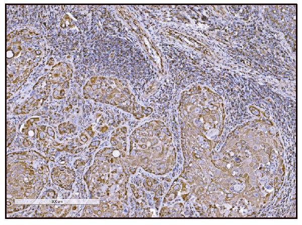


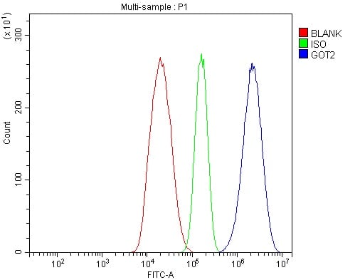


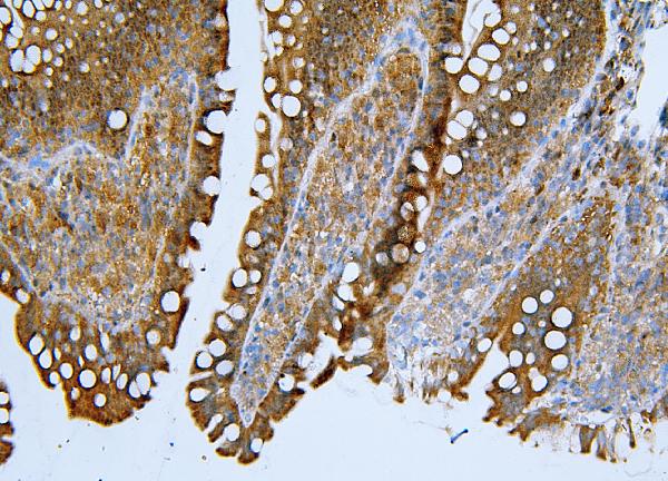
 IHC (Immunohistochemistry)
(Figure 14. IHC analysis of FABP-1/GOT2 using anti-FABP-1/GOT2 antibody (AAA19309).FABP-1/GOT2 was detected in paraffin-embedded section of rat lymph node tissue. Heat mediated antigen retrieval was performed in EDTA buffer (pH8. 0, epitope retrieval solution). The tissue section was blocked with 10% goat serum. The tissue section was then incubated with 2μg/ml rabbit anti-FABP-1/GOT2 Antibody (AAA19309) overnight at 4 degree C. Biotinylated goat anti-rabbit IgG was used as secondary antibody and incubated for 30 minutes at 37 degree C. The tissue section was developed using Strepavidin-Biotin-Complex (SABC) (Catalog # with DAB as the chromogen.)
IHC (Immunohistochemistry)
(Figure 14. IHC analysis of FABP-1/GOT2 using anti-FABP-1/GOT2 antibody (AAA19309).FABP-1/GOT2 was detected in paraffin-embedded section of rat lymph node tissue. Heat mediated antigen retrieval was performed in EDTA buffer (pH8. 0, epitope retrieval solution). The tissue section was blocked with 10% goat serum. The tissue section was then incubated with 2μg/ml rabbit anti-FABP-1/GOT2 Antibody (AAA19309) overnight at 4 degree C. Biotinylated goat anti-rabbit IgG was used as secondary antibody and incubated for 30 minutes at 37 degree C. The tissue section was developed using Strepavidin-Biotin-Complex (SABC) (Catalog # with DAB as the chromogen.)
FABP-1/GOT2, Polyclonal Antibody (Cat# AAA19309)
Full Name
Anti-FABP-1/GOT2 Antibody
Gene Names
GOT2; KAT4; KATIV; mitAAT
Reactivity
Human, Mouse, Rat
Applications
WB, IHC-P, ICC, IF, FC/FACS/FCM, EIA
Purity
Immunogen affinity purified.
Pricing

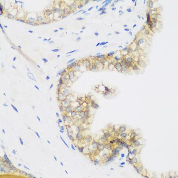


 IHC (Immunohistchemistry)
(Immunohistochemistry of paraffin-embedded mouse liver using KIAA1456 antibody at dilution of 1:200 (40x lens).)
IHC (Immunohistchemistry)
(Immunohistochemistry of paraffin-embedded mouse liver using KIAA1456 antibody at dilution of 1:200 (40x lens).)
KIAA1456, Polyclonal Antibody (Cat# AAA28126)
Full Name
KIAA1456 Polyclonal Antibody
Gene Names
KIAA1456; TRM9L; C8orf79
Reactivity
Human
Applications
WB, IHC, IF
Purity
Affinity Purification
Pricing


 IF (Immunofluorescence)
(Immunofluorescence analysis of HeLa cells using CSNK1E antibody. Blue: DAPI for nuclear staining.)
IF (Immunofluorescence)
(Immunofluorescence analysis of HeLa cells using CSNK1E antibody. Blue: DAPI for nuclear staining.)
CSNK1E, Polyclonal Antibody (Cat# AAA28143)
Full Name
CSNK1E Polyclonal Antibody
Gene Names
CSNK1E; HCKIE; CKIepsilon
Reactivity
Human, Mouse, Rat
Applications
WB, IHC, IF
Purity
Affinity Purification
Pricing


 IHC (Immunohistchemistry)
(Immunohistochemistry of paraffin-embedded mouse brain using JMJD6 Antibody at dilution of 1:100 (40x lens).)
IHC (Immunohistchemistry)
(Immunohistochemistry of paraffin-embedded mouse brain using JMJD6 Antibody at dilution of 1:100 (40x lens).)
JMJD6, Polyclonal Antibody (Cat# AAA28145)
Full Name
JMJD6 Polyclonal Antibody
Gene Names
JMJD6; PSR; PTDSR; PTDSR1
Reactivity
Human, Mouse, Rat
Applications
WB, IHC, IF, IP
Purity
Affinity Purification
Pricing


 DB (Dot Blot)
(Dot blot analysis using Rabbit Anti-Amyloid Fibrils (OC) Polyclonal Antibody (SPC-507). Tissue: Cell lysates. Species: Human. Primary Antibody: Rabbit Anti-Amyloid Fibrils (OC) Polyclonal Antibody (SPC-507) at 1:500, 1:5000. Beta Amyloid HEPES-NaCl aggregation, showing 1:500 (L) and 1:5000 (R) time lapse dot blot.)
DB (Dot Blot)
(Dot blot analysis using Rabbit Anti-Amyloid Fibrils (OC) Polyclonal Antibody (SPC-507). Tissue: Cell lysates. Species: Human. Primary Antibody: Rabbit Anti-Amyloid Fibrils (OC) Polyclonal Antibody (SPC-507) at 1:500, 1:5000. Beta Amyloid HEPES-NaCl aggregation, showing 1:500 (L) and 1:5000 (R) time lapse dot blot.)
Amyloid Fibrils (OC), Polyclonal Antibody (Cat# AAA17797)
Full Name
Amyloid Fibrils (OC) Antibody: FITC
Reactivity
Human. Potentially mouse and rat based on species homology.
Applications
IP, ICC, IHC, EIA, WB, DB
Pricing


 DB (Dot Blot)
(Dot blot analysis using Rabbit Anti-Amyloid Fibrils (OC) Polyclonal Antibody (SPC-507). Tissue: Cell lysates. Species: Human. Primary Antibody: Rabbit Anti-Amyloid Fibrils (OC) Polyclonal Antibody (SPC-507) at 1:500, 1:5000. Beta Amyloid HEPES-NaCl aggregation, showing 1:500 (L) and 1:5000 (R) time lapse dot blot.)
DB (Dot Blot)
(Dot blot analysis using Rabbit Anti-Amyloid Fibrils (OC) Polyclonal Antibody (SPC-507). Tissue: Cell lysates. Species: Human. Primary Antibody: Rabbit Anti-Amyloid Fibrils (OC) Polyclonal Antibody (SPC-507) at 1:500, 1:5000. Beta Amyloid HEPES-NaCl aggregation, showing 1:500 (L) and 1:5000 (R) time lapse dot blot.)
Amyloid Fibrils (OC), Polyclonal Antibody (Cat# AAA17801)
Full Name
Amyloid Fibrils (OC) Antibody: ATTO 594
Reactivity
Human. Potentially mouse and rat based on species homology.
Applications
IP, ICC, IHC, EIA, WB, DB
Pricing


 WB (Western Blot)
(Western blot analysis of lysate from HepG2 cell line using PPT1 Antibody. AAA28640 was diluted at 1:1000 at each lane. A goat anti-mouse IgG H&L(HRP) at 1:3000 dilution was used as the secondary antibody. Lysate at 35ug per lane.)
WB (Western Blot)
(Western blot analysis of lysate from HepG2 cell line using PPT1 Antibody. AAA28640 was diluted at 1:1000 at each lane. A goat anti-mouse IgG H&L(HRP) at 1:3000 dilution was used as the secondary antibody. Lysate at 35ug per lane.)
PPT1, Monoclonal Antibody (Cat# AAA28640)
Full Name
PPT1 Antibody
Gene Names
PPT1; PPT; CLN1; INCL
Reactivity
Human
Applications
WB, EIA, FC/FACS, IHC
Purity
Purified Mouse Monoclonal Antibody (Mab)
Pricing


 IHC (Immunohistochemistry)
(At 1/100 staining Mouse heart tissue by IHC-P. The sample was formaldehyde fixed and a heat mediated antigen retrieval step in citrate buffer was performed. The sample was then blocked and incubated with the primary antibody at 4 degree C overnight. An HRP conjugated anti-Rabbit antibody was used as the secondary antibody.)
IHC (Immunohistochemistry)
(At 1/100 staining Mouse heart tissue by IHC-P. The sample was formaldehyde fixed and a heat mediated antigen retrieval step in citrate buffer was performed. The sample was then blocked and incubated with the primary antibody at 4 degree C overnight. An HRP conjugated anti-Rabbit antibody was used as the secondary antibody.)
ID2, Polyclonal Antibody (Cat# AAA31291)
Full Name
Phospho-ID2 (Thr27) Antibody
Gene Names
ID2; GIG8; ID2A; ID2H; bHLHb26
Reactivity
Human, Mouse, Rat
Applications
Immunohistochemistry, Peptide ELISA
Purity
The antibody is from purified rabbit serum by affinity purification via sequential chromatography on phospho-peptide and non-phospho-peptide affinity columns.
Pricing


 FCM (Flow Cytometry)
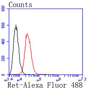
(Flow cytometric analysis of SW480 cells with Ret antibody at 1/50 dilution (red) compared with an unlabelled control (cells without incubation with primary antibody; black). Alexa Fluor 488-conjugated goat anti rabbit IgG was used as the secondary antibody.)
FCM (Flow Cytometry)
(Flow cytometric analysis of SW480 cells with Ret antibody at 1/50 dilution (red) compared with an unlabelled control (cells without incubation with primary antibody; black). Alexa Fluor 488-conjugated goat anti rabbit IgG was used as the secondary antibody.)
Ret, Monoclonal Antibody (Cat# AAA30185)
Full Name
Ret Antibody
Gene Names
RET; PTC; MTC1; HSCR1; MEN2A; MEN2B; RET51; CDHF12; CDHR16; RET-ELE1
Reactivity
Human, Mouse, Rat
Applications
WB, ICC, IF, IHC, IP, FC/FACS
Purity
ProA affinity purified
Pricing


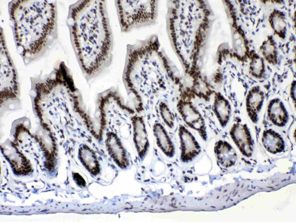
 IHC (Immunohistochemistry)
(Figure 8. IHC analysis of PRDM1/Blimp1 using anti-PRDM1/Blimp1 antibody (AAA19133).PRDM1/Blimp1 was detected in paraffin-embedded section of rat small intestine tissue. Heat mediated antigen retrieval was performed in citrate buffer (pH6, epitope retrieval solution) for 20 mins. The tissue section was blocked with 10% goat serum. The tissue section was then incubated with 1ug/ml rabbit anti-PRDM1/Blimp1 Antibody (AAA19133) overnight at 4 degree C. Biotinylated goat anti-rabbit IgG was used as secondary antibody and incubated for 30 minutes at 37 degree C. The tissue section was developed using Strepavidin-Biotin-Complex (SABC) with DAB as the chromogen.)
IHC (Immunohistochemistry)
(Figure 8. IHC analysis of PRDM1/Blimp1 using anti-PRDM1/Blimp1 antibody (AAA19133).PRDM1/Blimp1 was detected in paraffin-embedded section of rat small intestine tissue. Heat mediated antigen retrieval was performed in citrate buffer (pH6, epitope retrieval solution) for 20 mins. The tissue section was blocked with 10% goat serum. The tissue section was then incubated with 1ug/ml rabbit anti-PRDM1/Blimp1 Antibody (AAA19133) overnight at 4 degree C. Biotinylated goat anti-rabbit IgG was used as secondary antibody and incubated for 30 minutes at 37 degree C. The tissue section was developed using Strepavidin-Biotin-Complex (SABC) with DAB as the chromogen.)
PRDM1/Blimp1, Polyclonal Antibody (Cat# AAA19133)
Full Name
Anti-PRDM1/Blimp1 Picoband antibody
Gene Names
PRDM1; BLIMP1; PRDI-BF1
Reactivity
Human, Mouse, Rat
No cross reactivity with other proteins.
No cross reactivity with other proteins.
Applications
EIA, IHC, WB
Pricing


 Application Data
(C:FGFR2/isolectinB4 (C) and FGFR1/isolectinB4 (D) staining of apparent mesenchymal cells and the subpopulation of endothelial cells. Virtually all other dispersed apparent mesenchymal cells express FGFR1 and FGFR2 (merged image in E). F: FGFR2 (F) and FGFR1 (G) staining in clustered cells of epithelial origin (inferred by morphology here) demonstrating that epithelial cells express both FGFR1 and FGFR2 (merged image with DAPI staining in H).)
Application Data
(C:FGFR2/isolectinB4 (C) and FGFR1/isolectinB4 (D) staining of apparent mesenchymal cells and the subpopulation of endothelial cells. Virtually all other dispersed apparent mesenchymal cells express FGFR1 and FGFR2 (merged image in E). F: FGFR2 (F) and FGFR1 (G) staining in clustered cells of epithelial origin (inferred by morphology here) demonstrating that epithelial cells express both FGFR1 and FGFR2 (merged image with DAPI staining in H).)
FGFR2, Polyclonal Antibody (Cat# AAA26851)
Full Name
FGFR2, NT (FGFR2, BEK, KGFR, KSAM, Fibroblast growth factor receptor 2, K-sam, Keratinocyte growth factor receptor, CD332) (AP)
Gene Names
FGFR2; BEK; JWS; BBDS; CEK3; CFD1; ECT1; KGFR; TK14; TK25; BFR-1; CD332; K-SAM
Reactivity
Human, Monkey, Mouse, Rat
Applications
IF, EIA, IHC, WB
Purity
Purified by Protein G Affinity Chromatography.
Pricing


 IHC (Immunohistchemistry)
(Figure 6. IHC analysis of CPI17 alpha using anti- CPI17 alpha antibody (AAA19176).CPI17 alpha was detected in paraffin-embedded section of human placenta tissues. Heat mediated antigen retrieval was performed in citrate buffer (pH6, epitope retrieval solution) for 20 mins. The tissue section was blocked with 10% goat serum. The tissue section was then incubated with 1ug/ml rabbit anti- CPI17 alpha Antibody (AAA19176) overnight at 4 degree C. Biotinylated goat anti-rabbit IgG was used as secondary antibody and incubated for 30 minutes at 37 degree C. The tissue section was developed using Strepavidin-Biotin-Complex (SABC) with DAB as the chromogen.)
IHC (Immunohistchemistry)
(Figure 6. IHC analysis of CPI17 alpha using anti- CPI17 alpha antibody (AAA19176).CPI17 alpha was detected in paraffin-embedded section of human placenta tissues. Heat mediated antigen retrieval was performed in citrate buffer (pH6, epitope retrieval solution) for 20 mins. The tissue section was blocked with 10% goat serum. The tissue section was then incubated with 1ug/ml rabbit anti- CPI17 alpha Antibody (AAA19176) overnight at 4 degree C. Biotinylated goat anti-rabbit IgG was used as secondary antibody and incubated for 30 minutes at 37 degree C. The tissue section was developed using Strepavidin-Biotin-Complex (SABC) with DAB as the chromogen.)
CPI17 alpha, Polyclonal Antibody (Cat# AAA19176)
Full Name
Anti-CPI17 alpha Picoband Antibody
Gene Names
PPP1R14A; CPI17; CPI-17; PPP1INL
Reactivity
Human, Mouse, Rat
No cross reactivity with other proteins
No cross reactivity with other proteins
Applications
IHC, WB
Purity
Immunogen affinity purified
Pricing


 FCM (Flow Cytometry)
(Figure 7. Flow Cytometry analysis of 293T cells using anti-OTULIN antibody (AAA19319).Overlay histogram showing 293T cells stained with AAA19319 (Blue line). The cells were blocked with 10% normal goat serum. And then incubated with rabbit anti-OTULIN Antibody (AAA19319, 1μg/1x106 cells) for 30 min at 20 degree C. DyLight®488 conjugated goat anti-rabbit IgG (5-10μg/1x106 cells) was used as secondary antibody for 30 minutes at 20 degree C. Isotype control antibody (Green line) was rabbit IgG (1μg/1x106) used under the same conditions. Unlabelled sample (Red line) was also used as a control.)
FCM (Flow Cytometry)
(Figure 7. Flow Cytometry analysis of 293T cells using anti-OTULIN antibody (AAA19319).Overlay histogram showing 293T cells stained with AAA19319 (Blue line). The cells were blocked with 10% normal goat serum. And then incubated with rabbit anti-OTULIN Antibody (AAA19319, 1μg/1x106 cells) for 30 min at 20 degree C. DyLight®488 conjugated goat anti-rabbit IgG (5-10μg/1x106 cells) was used as secondary antibody for 30 minutes at 20 degree C. Isotype control antibody (Green line) was rabbit IgG (1μg/1x106) used under the same conditions. Unlabelled sample (Red line) was also used as a control.)
OTULIN, Polyclonal Antibody (Cat# AAA19319)
Full Name
Anti-OTULIN Antibody
Gene Names
OTULIN; GUM; FAM105B
Reactivity
Human, Mouse, Rat
Applications
WB, IHC-P, ICC, IF, FC/FACS/FCM, EIA
Purity
Immunogen affinity purified.
Pricing


 FCM (Flow Cytometry)
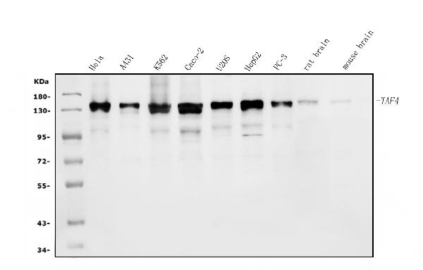
(Figure 13. Flow Cytometry analysis of Hela cells using anti-TAF4 antibody (AAA19307).Overlay histogram showing Hela cells stained with AAA19307 (Blue line). The cells were blocked with 10% normal goat serum. And then incubated with rabbit anti-TAF4 Antibody (AAA19307, 1μg/1x106 cells) for 30 min at 20 degree C. DyLight®488 conjugated goat anti-rabbit IgG (5-10μg/1x106 cells) was used as secondary antibody for 30 minutes at 20 degree C. Isotype control antibody (Green line) was rabbit IgG (1μg/1x106) used under the same conditions. Unlabelled sample (Red line) was also used as a control.)
FCM (Flow Cytometry)
(Figure 13. Flow Cytometry analysis of Hela cells using anti-TAF4 antibody (AAA19307).Overlay histogram showing Hela cells stained with AAA19307 (Blue line). The cells were blocked with 10% normal goat serum. And then incubated with rabbit anti-TAF4 Antibody (AAA19307, 1μg/1x106 cells) for 30 min at 20 degree C. DyLight®488 conjugated goat anti-rabbit IgG (5-10μg/1x106 cells) was used as secondary antibody for 30 minutes at 20 degree C. Isotype control antibody (Green line) was rabbit IgG (1μg/1x106) used under the same conditions. Unlabelled sample (Red line) was also used as a control.)
TAF4, Polyclonal Antibody (Cat# AAA19307)
Full Name
Anti-TAF4 Antibody
Gene Names
TAF4; TAF2C; TAF4A; TAF2C1; TAFII130; TAFII135
Reactivity
Human, Mouse, Rat
Applications
WB, IHC-P, ICC, IF, EIA
Purity
Immunogen affinity purified.
Pricing


 IHC (Immunohistchemistry)
(Immunohistochemistry of paraffin-embedded mouse brain using IRAK4 antibody at dilution of 1:100 (40x lens).)
IHC (Immunohistchemistry)
(Immunohistochemistry of paraffin-embedded mouse brain using IRAK4 antibody at dilution of 1:100 (40x lens).)
IRAK4, Polyclonal Antibody (Cat# AAA28120)
Full Name
IRAK4 Polyclonal Antibody
Gene Names
IRAK4; IPD1; REN64; IRAK-4; NY-REN-64
Reactivity
Human, Mouse, Rat
Applications
WB, IHC
Purity
Affinity Purification
Pricing


 WB (Western Blot)
(WB Suggested Anti-CHEK1 Antibody Titration: 0.2-1 ug/mlELISA Titer: 1:312500Positive Control: Human Small Intestine)
WB (Western Blot)
(WB Suggested Anti-CHEK1 Antibody Titration: 0.2-1 ug/mlELISA Titer: 1:312500Positive Control: Human Small Intestine)
CHEK1, Polyclonal Antibody (Cat# AAA23414)
Full Name
CHEK1 antibody - middle region
Gene Names
CHEK1; CHK1
Reactivity
Cow, Dog, Guinea Pig, Horse, Human, Mouse, Rabbit, Rat
Applications
IHC, WB
Purity
Affinity Purified
Pricing


 FCM (Flow Cytometry)
(Flow cytometric analysis of Daudi cells with Tle6 antibody at 1/100 dilution (red) compared with an unlabelled control (cells without incubation with primary antibody; black). Alexa Fluor 488-conjugated goat anti-rabbit IgG was used as the secondary antibody.)
FCM (Flow Cytometry)
(Flow cytometric analysis of Daudi cells with Tle6 antibody at 1/100 dilution (red) compared with an unlabelled control (cells without incubation with primary antibody; black). Alexa Fluor 488-conjugated goat anti-rabbit IgG was used as the secondary antibody.)
Tle6, Polyclonal Antibody (Cat# AAA29935)
Full Name
Tle6 Antibody
Gene Names
TLE6; GRG6
Reactivity
Human, Mouse
Applications
WB, ICC, IHC, FC/FACS
Purity
Peptide affinity purified.
Pricing


 WB (Western Blot)
(WB Suggested Anti-HNRPL Antibody Titration: 0.2-1 ug/mlELISA Titer: 1:62500Positive Control: Jurkat cell lysateHNRNPL is strongly supported by BioGPS gene expression data to be expressed in Human Jurkat cells)
WB (Western Blot)
(WB Suggested Anti-HNRPL Antibody Titration: 0.2-1 ug/mlELISA Titer: 1:62500Positive Control: Jurkat cell lysateHNRNPL is strongly supported by BioGPS gene expression data to be expressed in Human Jurkat cells)
HNRPL, Polyclonal Antibody (Cat# AAA23474)
Full Name
HNRPL antibody - N-terminal region
Gene Names
HNRNPL; HNRPL; hnRNP-L; P/OKcl.14
Reactivity
Cow, Dog, Guinea Pig, Horse, Human, Mouse, Rabbit, Rat
Applications
IF, IP, IHC, WB
Purity
Affinity Purified
Pricing


 IHC (Immunohistchemistry)
(Figure 6. IHC analysis of Cdc20 using anti-Cdc20 antibody (AAA19132).Cdc20 was detected in paraffin-embedded section of human mammary cancer tissue. Heat mediated antigen retrieval was performed in citrate buffer (pH6, epitope retrieval solution) for 20 mins. The tissue section was blocked with 10% goat serum. The tissue section was then incubated with 1ug/ml rabbit anti-Cdc20 Antibody (AAA19132) overnight at 4 degree C. Biotinylated goat anti-rabbit IgG was used as secondary antibody and incubated for 30 minutes at 37 degree C. The tissue section was developed using Strepavidin-Biotin-Complex (SABC) with DAB as the chromogen.)
IHC (Immunohistchemistry)
(Figure 6. IHC analysis of Cdc20 using anti-Cdc20 antibody (AAA19132).Cdc20 was detected in paraffin-embedded section of human mammary cancer tissue. Heat mediated antigen retrieval was performed in citrate buffer (pH6, epitope retrieval solution) for 20 mins. The tissue section was blocked with 10% goat serum. The tissue section was then incubated with 1ug/ml rabbit anti-Cdc20 Antibody (AAA19132) overnight at 4 degree C. Biotinylated goat anti-rabbit IgG was used as secondary antibody and incubated for 30 minutes at 37 degree C. The tissue section was developed using Strepavidin-Biotin-Complex (SABC) with DAB as the chromogen.)
Cdc20/P55 Cdc, Polyclonal Antibody (Cat# AAA19132)
Full Name
Anti-Cdc20/P55 Cdc Picoband Antibody
Gene Names
CDC20; CDC20A; p55CDC; bA276H19.3
Reactivity
Human, Mouse, Rat
No cross reactivity with other proteins.
No cross reactivity with other proteins.
Applications
IHC, WB
Purity
Immunogen affinity purified
Pricing


 IF (Immunofluorescence)
(Immunofluorescent analysis of 4% paraformaldehyde-fixed, 0.1% Triton X-100 permeabilized MCF-7 (human breast cancer cell line) cells labeling Pdx1 with at 1:25 dilution, followed by DyLight 488-conjugated IgG goat anti-rabbit secondary antibody at 1:200 dilution (green). Immunofluorescence image showing cytoplasm staining on MCF-7 cell line. Cytoplasmic actin is detected with DyLight 554 Phalloidin (PD18466410) at 1:100 dilution (red). The nuclear counter stain is DAPI (blue).)
IF (Immunofluorescence)
(Immunofluorescent analysis of 4% paraformaldehyde-fixed, 0.1% Triton X-100 permeabilized MCF-7 (human breast cancer cell line) cells labeling Pdx1 with at 1:25 dilution, followed by DyLight 488-conjugated IgG goat anti-rabbit secondary antibody at 1:200 dilution (green). Immunofluorescence image showing cytoplasm staining on MCF-7 cell line. Cytoplasmic actin is detected with DyLight 554 Phalloidin (PD18466410) at 1:100 dilution (red). The nuclear counter stain is DAPI (blue).)
OPN-a/b, Polyclonal Antibody (Cat# AAA26859)
Full Name
OPN-a/b, NT (SPP1, BNSP, OPN, Osteopontin, Bone sialoprotein 1, Nephropontin, Secreted phosphoprotein 1, Urinary stone protein, Uropontin) (FITC)
Gene Names
SPP1; OPN; BNSP; BSPI; ETA-1
Reactivity
Human
Applications
WB, IHC, IF
Purity
Purified by Protein A and Peptide Affinity Chromatography.
Pricing


 FCM (Flow Cytometry)
(Figure 11. Flow Cytometry analysis of A431 cells using anti-SAMHD1 antibody (AAA19355).Overlay histogram showing A431 cells stained with AAA19355 (Blue line). The cells were blocked with 10% normal goat serum. And then incubated with mouse anti- SAMHD1 Antibody (AAA19355, 1μg/1x106 cells) for 30 min at 20 degree C. DyLight®488 conjugated goat anti-mouse IgG (BA1126, 5-10μg/1x106 cells) was used as secondary antibody for 30 minutes at 20 degree C. Isotype control antibody (Green line) was mouse IgG (1μg/1x106) used under the same conditions. Unlabelled sample (Red line) was also used as a control.)
FCM (Flow Cytometry)
(Figure 11. Flow Cytometry analysis of A431 cells using anti-SAMHD1 antibody (AAA19355).Overlay histogram showing A431 cells stained with AAA19355 (Blue line). The cells were blocked with 10% normal goat serum. And then incubated with mouse anti- SAMHD1 Antibody (AAA19355, 1μg/1x106 cells) for 30 min at 20 degree C. DyLight®488 conjugated goat anti-mouse IgG (BA1126, 5-10μg/1x106 cells) was used as secondary antibody for 30 minutes at 20 degree C. Isotype control antibody (Green line) was mouse IgG (1μg/1x106) used under the same conditions. Unlabelled sample (Red line) was also used as a control.)
SAMHD1, Monoclonal Antibody (Cat# AAA19355)
Full Name
Anti-SAMHD1 Antibody (monoclonal, 3B9)
Gene Names
SAMHD1; DCIP; CHBL2; HDDC1; MOP-5; SBBI88
Reactivity
Human
Applications
WB, IHC-P, ICC, IF, FC/FACS/FCM
Purity
Immunogen affinity purified.
Pricing


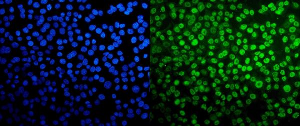
 IF (Immunofluorescence)
(Immunofluorescence analysis of F9 cells using TET2 Rabbit pAb (AAA28132) at dilution of 1:50 (40x lens). Blue: DAPI for nuclear staining.)
IF (Immunofluorescence)
(Immunofluorescence analysis of F9 cells using TET2 Rabbit pAb (AAA28132) at dilution of 1:50 (40x lens). Blue: DAPI for nuclear staining.)
TET2, Polyclonal Antibody (Cat# AAA28132)
Full Name
TET2 Polyclonal Antibody
Gene Names
TET2; MDS; KIAA1546
Applications
WB, IF, IP
Purity
Affinity Purification
Pricing


 DB (Dot Blot)
(Dot blot analysis using Rabbit Anti-Amyloid Fibrils (OC) Polyclonal Antibody (SPC-507). Tissue: Cell lysates. Species: Human. Primary Antibody: Rabbit Anti-Amyloid Fibrils (OC) Polyclonal Antibody (SPC-507) at 1:500, 1:5000. Beta Amyloid HEPES-NaCl aggregation, showing 1:500 (L) and 1:5000 (R) time lapse dot blot.)
DB (Dot Blot)
(Dot blot analysis using Rabbit Anti-Amyloid Fibrils (OC) Polyclonal Antibody (SPC-507). Tissue: Cell lysates. Species: Human. Primary Antibody: Rabbit Anti-Amyloid Fibrils (OC) Polyclonal Antibody (SPC-507) at 1:500, 1:5000. Beta Amyloid HEPES-NaCl aggregation, showing 1:500 (L) and 1:5000 (R) time lapse dot blot.)
Amyloid Fibrils (OC), Polyclonal Antibody (Cat# AAA17798)
Full Name
Amyloid Fibrils (OC) Antibody: ATTO 390
Reactivity
Human. Potentially mouse and rat based on species homology.
Applications
IP, ICC, IHC, EIA, WB, DB
Pricing


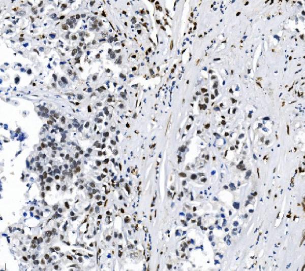
 IHC (Immunohistochemistry)
(Immunohistochemistry of paraffin-embedded human colon cancer using AAA18859 at dilution of 1:100)
IHC (Immunohistochemistry)
(Immunohistochemistry of paraffin-embedded human colon cancer using AAA18859 at dilution of 1:100)
Cellular tumor antigen p53, Polyclonal Antibody (Cat# AAA18859)
Full Name
Rabbit anti-human Cellular tumor antigen p53 polyclonal Antibody
Gene Names
TP53; P53; BCC7; LFS1; TRP53
Reactivity
Human
Applications
EIA, WB, IHC
Purity
Caprylic Acid Ammonium Sulfate Precipitation Purified
Pricing


 IHC (Immunohistchemistry)
(Immunohistochemistry analysis using Mouse Anti-Hsp70 Monoclonal Antibody, Clone BB70. Tissue: hepatocytes. Species: Rat. Fixation: Paraffin Embedded. Primary Antibody: Mouse Anti-Hsp70 Monoclonal Antibody at 1:200. Liver sections were paraffin embedded. First pictures in series show two hours after exposure to stress, the second shows the control. Courtesy of: G. Matic, University of Belgrade, Serbia.)
IHC (Immunohistchemistry)
(Immunohistochemistry analysis using Mouse Anti-Hsp70 Monoclonal Antibody, Clone BB70. Tissue: hepatocytes. Species: Rat. Fixation: Paraffin Embedded. Primary Antibody: Mouse Anti-Hsp70 Monoclonal Antibody at 1:200. Liver sections were paraffin embedded. First pictures in series show two hours after exposure to stress, the second shows the control. Courtesy of: G. Matic, University of Belgrade, Serbia.)
Hsp70/Hsc70, Monoclonal Antibody (Cat# AAA17780)
Full Name
Hsp70/Hsc70 Antibody: Biotin
Gene Names
HSPA2; HSP70
Reactivity
Human, Mouse, Rat, Sheep, Dog, Beluga, Bovine, Fish, Guinea pig, Scallop pig, Hamster, Rabbit, Chicken, Xenopus, Drosophila, Yeast
Applications
WB, IP, IHC
Pricing


 WB (Western Blot)
(Western blot analysis of Human Lysates showing detection of Hsp90 protein using Mouse Anti-Hsp90 Monoclonal Antibody, Clone H9010. Primary Antibody: Mouse Anti-Hsp90 Monoclonal Antibody at 1:1000. Comparison of clone H9010 behavior with Hsp90 human beta (1) and Hsp90 human alpha (2). Courtesy of: David Toft, Mayo Clinic.)
WB (Western Blot)
(Western blot analysis of Human Lysates showing detection of Hsp90 protein using Mouse Anti-Hsp90 Monoclonal Antibody, Clone H9010. Primary Antibody: Mouse Anti-Hsp90 Monoclonal Antibody at 1:1000. Comparison of clone H9010 behavior with Hsp90 human beta (1) and Hsp90 human alpha (2). Courtesy of: David Toft, Mayo Clinic.)
Hsp90, Monoclonal Antibody (Cat# AAA17787)
Full Name
Hsp90 Antibody: RPE
Gene Names
HSP90AB1; HSP84; HSPC2; HSPCB; D6S182; HSP90B
Reactivity
Human (beta-specific), Rabbit (Beta Specific), Chicken (Alpha/Beta), Rat, Canine
Applications
WB, IP, EIA, IHC, IF
Pricing


 IHC (Immunohistchemistry)
(Figure 6. IHC analysis of Musashi 1/Msi1 using anti- Musashi 1/Msi1 antibody (AAA19172).Musashi 1/Msi1 was detected in paraffin-embedded section of human mammary cancer tissues. Heat mediated antigen retrieval was performed in citrate buffer (pH6, epitope retrieval solution) for 20 mins. The tissue section was blocked with 10% goat serum. The tissue section was then incubated with 1ug/ml rabbit anti- Musashi 1/Msi1 Antibody (AAA19172) overnight at 4 degree C. Biotinylated goat anti-rabbit IgG was used as secondary antibody and incubated for 30 minutes at 37 degree C. The tissue section was developed using Strepavidin-Biotin-Complex (SABC) with DAB as the chromogen.)
IHC (Immunohistchemistry)
(Figure 6. IHC analysis of Musashi 1/Msi1 using anti- Musashi 1/Msi1 antibody (AAA19172).Musashi 1/Msi1 was detected in paraffin-embedded section of human mammary cancer tissues. Heat mediated antigen retrieval was performed in citrate buffer (pH6, epitope retrieval solution) for 20 mins. The tissue section was blocked with 10% goat serum. The tissue section was then incubated with 1ug/ml rabbit anti- Musashi 1/Msi1 Antibody (AAA19172) overnight at 4 degree C. Biotinylated goat anti-rabbit IgG was used as secondary antibody and incubated for 30 minutes at 37 degree C. The tissue section was developed using Strepavidin-Biotin-Complex (SABC) with DAB as the chromogen.)
Musashi 1/Msi1, Polyclonal Antibody (Cat# AAA19172)
Full Name
Anti-Musashi 1/Msi1 Picoband Antibody
Reactivity
Human, Mouse, Rat
No cross reactivity with other proteins
No cross reactivity with other proteins
Applications
IHC, WB
Purity
Immunogen affinity purified
Pricing


 FCM (Flow Cytometry)
(Figure 6. Flow Cytometry analysis of HEPA1-6 cells using anti-PAR4/Pawr antibody (AAA19278).Overlay histogram showing HEPA1-6 cells stained with AAA19278 (Blue line). The cells were blocked with 10% normal goat serum. And then incubated with rabbit anti-PAR4/Pawr Antibody (AAA19278, 1μg/1x106 cells) for 30 min at 20 degree C. DyLight®488 conjugated goat anti-rabbit IgG (5-10μg/1x106 cells) was used as secondary antibody for 30 minutes at 20 degree C. Isotype control antibody (Green line) was rabbit IgG (1μg/1x106) used under the same conditions. Unlabelled sample (Red line) was also used as a control.)
FCM (Flow Cytometry)
(Figure 6. Flow Cytometry analysis of HEPA1-6 cells using anti-PAR4/Pawr antibody (AAA19278).Overlay histogram showing HEPA1-6 cells stained with AAA19278 (Blue line). The cells were blocked with 10% normal goat serum. And then incubated with rabbit anti-PAR4/Pawr Antibody (AAA19278, 1μg/1x106 cells) for 30 min at 20 degree C. DyLight®488 conjugated goat anti-rabbit IgG (5-10μg/1x106 cells) was used as secondary antibody for 30 minutes at 20 degree C. Isotype control antibody (Green line) was rabbit IgG (1μg/1x106) used under the same conditions. Unlabelled sample (Red line) was also used as a control.)
PAR4/Pawr, Polyclonal Antibody (Cat# AAA19278)
Full Name
Anti-PAR4/Pawr Antibody
Gene Names
Pawr; PAR4; Par-4; 2310001G03Rik
Reactivity
Mouse, Rat
Applications
WB, IHC-P, ICC, IF, FC/FACS/FCM, EIA
Purity
Immunogen affinity purified.
Pricing


 IHC (Immunohistochemistry)
(Figure 8. IHC analysis of TAGLN/Transgelin using anti-TAGLN/Transgelin antibody (AAA19286).TAGLN/Transgelin was detected in paraffin-embedded section of rat testis tissue. Heat mediated antigen retrieval was performed in EDTA buffer (pH8. 0, epitope retrieval solution). The tissue section was blocked with 10% goat serum. The tissue section was then incubated with 2μg/ml rabbit anti-TAGLN/Transgelin Antibody (AAA19286) overnight at 4 degree C. Biotinylated goat anti-rabbit IgG was used as secondary antibody and incubated for 30 minutes at 37 degree C. The tissue section was developed using Strepavidin-Biotin-Complex (SABC) (Catalog # with DAB as the chromogen.)
IHC (Immunohistochemistry)
(Figure 8. IHC analysis of TAGLN/Transgelin using anti-TAGLN/Transgelin antibody (AAA19286).TAGLN/Transgelin was detected in paraffin-embedded section of rat testis tissue. Heat mediated antigen retrieval was performed in EDTA buffer (pH8. 0, epitope retrieval solution). The tissue section was blocked with 10% goat serum. The tissue section was then incubated with 2μg/ml rabbit anti-TAGLN/Transgelin Antibody (AAA19286) overnight at 4 degree C. Biotinylated goat anti-rabbit IgG was used as secondary antibody and incubated for 30 minutes at 37 degree C. The tissue section was developed using Strepavidin-Biotin-Complex (SABC) (Catalog # with DAB as the chromogen.)
TAGLN/Transgelin, Polyclonal Antibody (Cat# AAA19286)
Full Name
Anti-TAGLN/Transgelin Antibody
Gene Names
TAGLN; SM22; SMCC; TAGLN1; WS3-10
Reactivity
Human, Mouse, Rat
Applications
WB, IHC-P, ICC, IF, FC/FACS/FCM, EIA
Purity
Immunogen affinity purified.
Pricing


 FCM (Flow Cytometry)
(Figure 10. Flow Cytometry analysis of A431 cells using anti-CDC27 antibody (AAA19285).Overlay histogram showing A431 cells stained with AAA19285 (Blue line). The cells were blocked with 10% normal goat serum. And then incubated with rabbit anti-CDC27 Antibody (AAA19285, 1μg/1x106 cells) for 30 min at 20 degree C. DyLight®488 conjugated goat anti-rabbit IgG (5-10μg/1x106 cells) was used as secondary antibody for 30 minutes at 20 degree C. Isotype control antibody (Green line) was rabbit IgG (1μg/1x106) used under the same conditions. Unlabelled sample (Red line) was also used as a control.)
FCM (Flow Cytometry)
(Figure 10. Flow Cytometry analysis of A431 cells using anti-CDC27 antibody (AAA19285).Overlay histogram showing A431 cells stained with AAA19285 (Blue line). The cells were blocked with 10% normal goat serum. And then incubated with rabbit anti-CDC27 Antibody (AAA19285, 1μg/1x106 cells) for 30 min at 20 degree C. DyLight®488 conjugated goat anti-rabbit IgG (5-10μg/1x106 cells) was used as secondary antibody for 30 minutes at 20 degree C. Isotype control antibody (Green line) was rabbit IgG (1μg/1x106) used under the same conditions. Unlabelled sample (Red line) was also used as a control.)
CDC27, Polyclonal Antibody (Cat# AAA19285)
Full Name
Anti-CDC27 Antibody
Gene Names
CDC27; APC3; HNUC; NUC2; ANAPC3; CDC27Hs; D0S1430E; D17S978E
Reactivity
Human, Mouse, Rat
Applications
WB, IHC-P, ICC, IF, FC/FACS/FCM, EIA
Purity
Immunogen affinity purified.
Pricing


 FCM (Flow Cytometry)
(PROX-1-S514 Antibody flow cytometric analysis of 293 cells (right histogram) compared to a negative control cell (left histogram).FITC-conjugated goat-anti-rabbit secondary antibodies were used for the analysis.)
FCM (Flow Cytometry)
(PROX-1-S514 Antibody flow cytometric analysis of 293 cells (right histogram) compared to a negative control cell (left histogram).FITC-conjugated goat-anti-rabbit secondary antibodies were used for the analysis.)
PROX-1-S514, Polyclonal Antibody (Cat# AAA28650)
Full Name
PROX-1-S514 Antibody
Reactivity
Human, mouse
Applications
WB, EIA, IHC, FC/FACS
Purity
Peptide Affinity Purified Rabbit Polyclonal Antibody (Pab)
Pricing


 IHC (Immunohistchemistry)
(Immunohistochemistry analysis using Mouse Anti-Hsp70 Monoclonal Antibody, Clone BB70. Tissue: hepatocytes. Species: Rat. Fixation: Paraffin Embedded. Primary Antibody: Mouse Anti-Hsp70 Monoclonal Antibody at 1:200. Liver sections were paraffin embedded. First pictures in series show two hours after exposure to stress, the second shows the control. Courtesy of: G. Matic, University of Belgrade, Serbia.)
IHC (Immunohistchemistry)
(Immunohistochemistry analysis using Mouse Anti-Hsp70 Monoclonal Antibody, Clone BB70. Tissue: hepatocytes. Species: Rat. Fixation: Paraffin Embedded. Primary Antibody: Mouse Anti-Hsp70 Monoclonal Antibody at 1:200. Liver sections were paraffin embedded. First pictures in series show two hours after exposure to stress, the second shows the control. Courtesy of: G. Matic, University of Belgrade, Serbia.)
Hsp70/Hsc70, Monoclonal Antibody (Cat# AAA17784)
Full Name
Hsp70/Hsc70 Antibody: ATTO 390
Gene Names
HSPA2; HSP70
Reactivity
Human, Mouse, Rat, Sheep, Dog, Beluga, Bovine, Fish, Guinea pig, Scallop pig, Hamster, Rabbit, Chicken, Xenopus, Drosophila, Yeast
Applications
WB, IP, IHC
Pricing


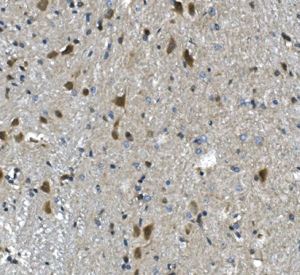
 IHC (Immunohistchemistry)
(Figure 6. IHC analysis of PP2A-alpha/PPP2CA using anti-PP2A-alpha/PPP2CA antibody (AAA19366).PP2A-alpha/PPP2CA was detected in paraffin-embedded section of rat brain tissue. Heat mediated antigen retrieval was performed in EDTA buffer (pH8. 0, epitope retrieval solution). The tissue section was blocked with 10% goat serum. The tissue section was then incubated with 1μg/ml mouse anti-PP2A-alpha/PPP2CA Antibody (AAA19366) overnight at 4 degree C. Biotinylated goat anti-mouse IgG was used as secondary antibody and incubated for 30 minutes at 37 degree C. The tissue section was developed using Strepavidin-Biotin-Complex (SABC) (Catalog # with DAB as the chromogen.)
IHC (Immunohistchemistry)
(Figure 6. IHC analysis of PP2A-alpha/PPP2CA using anti-PP2A-alpha/PPP2CA antibody (AAA19366).PP2A-alpha/PPP2CA was detected in paraffin-embedded section of rat brain tissue. Heat mediated antigen retrieval was performed in EDTA buffer (pH8. 0, epitope retrieval solution). The tissue section was blocked with 10% goat serum. The tissue section was then incubated with 1μg/ml mouse anti-PP2A-alpha/PPP2CA Antibody (AAA19366) overnight at 4 degree C. Biotinylated goat anti-mouse IgG was used as secondary antibody and incubated for 30 minutes at 37 degree C. The tissue section was developed using Strepavidin-Biotin-Complex (SABC) (Catalog # with DAB as the chromogen.)
PP2A-alpha/PPP2CA, Monoclonal Antibody (Cat# AAA19366)
Full Name
Anti-PP2A-alpha/PPP2CA Antibody (monoclonal, 3B6)
Gene Names
PPP2CA; RP-C; PP2Ac; PP2CA; PP2Calpha
Reactivity
Human, Monkey, Mouse, Rat
Applications
WB, IHC-P, ICC, IF, FC/FACS/FCM
Purity
Immunogen affinity purified.
Pricing


 IF (Immunofluorescence)
(Figure 9. IF analysis of ApoER2/LRP8 using anti- ApoER2/LRP8 antibody (AAA19275).ApoER2/LRP8 was detected in immunocytochemical section of HepG2 cells. Enzyme antigen retrieval was performed using IHC enzyme antigen retrieval reagent for 15 mins. The cells were blocked with 10% goat serum. And then incubated with 4μg/mL rabbit anti- ApoER2/LRP8 Antibody (AAA19275) overnight at 4 degree C. DyLight®488 Conjugated Goat Anti-Rabbit IgG was used as secondary antibody at 1:100 dilution and incubated for 30 minutes at 37 degree C. The section was counterstained with DAPI. Visualize using a fluorescence microscope and filter sets appropriate for the label used.)
IF (Immunofluorescence)
(Figure 9. IF analysis of ApoER2/LRP8 using anti- ApoER2/LRP8 antibody (AAA19275).ApoER2/LRP8 was detected in immunocytochemical section of HepG2 cells. Enzyme antigen retrieval was performed using IHC enzyme antigen retrieval reagent for 15 mins. The cells were blocked with 10% goat serum. And then incubated with 4μg/mL rabbit anti- ApoER2/LRP8 Antibody (AAA19275) overnight at 4 degree C. DyLight®488 Conjugated Goat Anti-Rabbit IgG was used as secondary antibody at 1:100 dilution and incubated for 30 minutes at 37 degree C. The section was counterstained with DAPI. Visualize using a fluorescence microscope and filter sets appropriate for the label used.)
ApoER2/LRP8, Polyclonal Antibody (Cat# AAA19275)
Full Name
Anti-ApoER2/LRP8 Antibody
Gene Names
LRP8; MCI1; LRP-8; APOER2; HSZ75190
Reactivity
Human, Mouse, Rat
Applications
WB, IHC-P, ICC, IF, FC/FACS/FCM, EIA
Purity
Immunogen affinity purified.
Pricing


 IF (Immunofluorescence)
(Figure 15. IF analysis of LSM2 using anti- LSM2 antibody (AAA19316).LSM2 was detected in immunocytochemical section of MCF-7 cells. Enzyme antigen retrieval was performed using IHC enzyme antigen retrieval reagent for 15 mins. The cells were blocked with 10% goat serum. And then incubated with 5μg/mL rabbit anti- LSM2 Antibody (AAA19316) overnight at 4 degree C. DyLight®488 Conjugated Goat Anti-Rabbit IgG was used as secondary antibody at 1:100 dilution and incubated for 30 minutes at 37 degree C. The section was counterstained with DAPI. Visualize using a fluorescence microscope and filter sets appropriate for the label used.)
IF (Immunofluorescence)
(Figure 15. IF analysis of LSM2 using anti- LSM2 antibody (AAA19316).LSM2 was detected in immunocytochemical section of MCF-7 cells. Enzyme antigen retrieval was performed using IHC enzyme antigen retrieval reagent for 15 mins. The cells were blocked with 10% goat serum. And then incubated with 5μg/mL rabbit anti- LSM2 Antibody (AAA19316) overnight at 4 degree C. DyLight®488 Conjugated Goat Anti-Rabbit IgG was used as secondary antibody at 1:100 dilution and incubated for 30 minutes at 37 degree C. The section was counterstained with DAPI. Visualize using a fluorescence microscope and filter sets appropriate for the label used.)
LSM2, Polyclonal Antibody (Cat# AAA19316)
Full Name
Anti-LSM2 Antibody
Gene Names
LSM2; G7B; snRNP; C6orf28; YBL026W
Reactivity
Human, Mouse, Rat, Monkey
Applications
WB, IHC-P, ICC, IF, FC/FACS/FCM
Purity
Immunogen affinity purified.
Pricing


 IHC (Immunohistochemistry)
(Immunohistochemical analysis of paraffin-embedded Human pancreas. 1, Antibody was diluted at 1:200(4 degree ,overnight). 2, High-pressure and temperature EDTA, pH8.0 was used for antigen retrieval. 3,Secondary antibody was diluted at 1:200(room temperature, 30min).)
IHC (Immunohistochemistry)
(Immunohistochemical analysis of paraffin-embedded Human pancreas. 1, Antibody was diluted at 1:200(4 degree ,overnight). 2, High-pressure and temperature EDTA, pH8.0 was used for antigen retrieval. 3,Secondary antibody was diluted at 1:200(room temperature, 30min).)
CD4, Monoclonal Antibody (Cat# AAA23995)
Full Name
CD4 (7H9) Mouse mAb
Gene Names
CD4; CD4mut
Reactivity
Human, Rat, Mouse
Applications
IHC-P
Purity
Affinity purified
Pricing
 WB (Western Blot)
(Western BlotPositive WB detected in: HepG2 whole cell lysate, Mouse liver tissue, Mouse kidney tissueAll lanes: NAPRT antibody at 4ug/mlSecondaryGoat polyclonal to rabbit IgG at 1/10000 dilutionPredicted band size: 58,61,57 kDaObserved band size: 58 kDa)
WB (Western Blot)
(Western BlotPositive WB detected in: HepG2 whole cell lysate, Mouse liver tissue, Mouse kidney tissueAll lanes: NAPRT antibody at 4ug/mlSecondaryGoat polyclonal to rabbit IgG at 1/10000 dilutionPredicted band size: 58,61,57 kDaObserved band size: 58 kDa)
Nicotinate phosphoribosyltransferase, Polyclonal Antibody (Cat# AAA18824)
Full Name
Rabbit anti-human Nicotinate phosphoribosyltransferase polyclonal Antibody
Gene Names
NAPRT; NAPRT1; PP3856
Reactivity
Human, Mouse
Applications
EIA, WB
Purity
>95%, Protein G Purified
Pricing