Filters
Clonality
Type
Reactivity
Gene Name
Isotype
Host
Application
Clone
2304 results for " secondary" - showing 1100-1150


 FCM (Flow Cytometry)
(Figure 6. Flow Cytometry analysis of RAW264. 7 cells using anti-TRIM25/EFP antibody (AAA19272).Overlay histogram showing RAW264. 7 cells stained with AAA19272 (Blue line). The cells were blocked with 10% normal goat serum. And then incubated with rabbit anti-TRIM25/EFP Antibody (AAA19272, 1μg/1x106 cells) for 30 min at 20 degree C. DyLight®488 conjugated goat anti-rabbit IgG (5-10μg/1x106 cells) was used as secondary antibody for 30 minutes at 20 degree C. Isotype control antibody (Green line) was rabbit IgG (1μg/1x106) used under the same conditions. Unlabelled sample (Red line) was also used as a control.)
FCM (Flow Cytometry)
(Figure 6. Flow Cytometry analysis of RAW264. 7 cells using anti-TRIM25/EFP antibody (AAA19272).Overlay histogram showing RAW264. 7 cells stained with AAA19272 (Blue line). The cells were blocked with 10% normal goat serum. And then incubated with rabbit anti-TRIM25/EFP Antibody (AAA19272, 1μg/1x106 cells) for 30 min at 20 degree C. DyLight®488 conjugated goat anti-rabbit IgG (5-10μg/1x106 cells) was used as secondary antibody for 30 minutes at 20 degree C. Isotype control antibody (Green line) was rabbit IgG (1μg/1x106) used under the same conditions. Unlabelled sample (Red line) was also used as a control.)
TRIM25/EFP, Polyclonal Antibody (Cat# AAA19272)
Full Name
Anti-TRIM25/EFP Antibody
Gene Names
Trim25; EFP; Zfp147; AA960166; AL022677
Reactivity
Mouse, Rat
Applications
WB, IHC-P, ICC, IF, FC/FACS/FCM, EIA
Purity
Immunogen affinity purified.
Pricing
 WB (Western Blot)
(Western blotAll lanes: Beta-defensin 14 antibody at 12ug/ml+ Rat liver tissueSecondaryGoat polyclonal to rabbit IgG at 1/10000 dilutionPredicted band size: 8 kDaObserved band size: 8 kDa)
WB (Western Blot)
(Western blotAll lanes: Beta-defensin 14 antibody at 12ug/ml+ Rat liver tissueSecondaryGoat polyclonal to rabbit IgG at 1/10000 dilutionPredicted band size: 8 kDaObserved band size: 8 kDa)
Beta-defensin 14, Polyclonal Antibody (Cat# AAA18814)
Full Name
Rabbit anti-mouse Beta-defensin 14 polyclonal Antibody
Gene Names
Defb14; BD-14
Reactivity
Mouse, Rat
Applications
EIA, WB
Purity
>95%, Protein G purified
Pricing


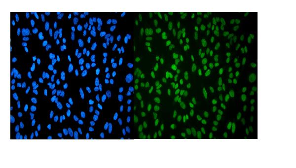
 IF (Immunofluorescence)
(Immunofluorescence analysis of U2OS cells using UHRF1 antibody. Blue: DAPI for nuclear staining.)
IF (Immunofluorescence)
(Immunofluorescence analysis of U2OS cells using UHRF1 antibody. Blue: DAPI for nuclear staining.)
UHRF1, Polyclonal Antibody (Cat# AAA10713)
Full Name
UHRF1 Polyclonal Antibody
Gene Names
UHRF1; Np95; hNP95; ICBP90; RNF106; hUHRF1; huNp95
Reactivity
Human
Applications
Western Blot, Immunohistochemistry, Immunofluorescence, Immunoprecipitation, Chromatin Immunoprecipitation
Purity
Affinity Purification
Pricing


 FCM (Flow Cytometry)
(Flow cytometric analysis of MCF-7 cells with Fragilis antibody at 1/100 dilution (red) compared with an unlabelled control (cells without incubation with primary antibody; black). Alexa Fluor 488-conjugated goat anti rabbit IgG was used as the secondary antibody.)
FCM (Flow Cytometry)
(Flow cytometric analysis of MCF-7 cells with Fragilis antibody at 1/100 dilution (red) compared with an unlabelled control (cells without incubation with primary antibody; black). Alexa Fluor 488-conjugated goat anti rabbit IgG was used as the secondary antibody.)
Fragilis, Monoclonal Antibody (Cat# AAA30394)
Full Name
Fragilis Antibody
Gene Names
IFITM3; 1-8U; IP15; DSPA2b
Reactivity
Human
Applications
Western Blot, Immunocytochemistry, Immunofluorescence, Immunohistochemistry, Flow Cytometry
Purity
ProA affinity purified
Pricing


 DB (Dot Blot)
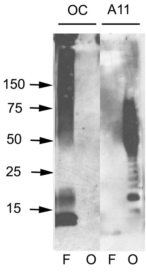
(Dot blot analysis using Rabbit Anti-Amyloid Fibrils (OC) Polyclonal Antibody (SPC-507). Tissue: Cell lysates. Species: Human. Primary Antibody: Rabbit Anti-Amyloid Fibrils (OC) Polyclonal Antibody (SPC-507) at 1:500, 1:5000. Beta Amyloid HEPES-NaCl aggregation, showing 1:500 (L) and 1:5000 (R) time lapse dot blot.)
DB (Dot Blot)
(Dot blot analysis using Rabbit Anti-Amyloid Fibrils (OC) Polyclonal Antibody (SPC-507). Tissue: Cell lysates. Species: Human. Primary Antibody: Rabbit Anti-Amyloid Fibrils (OC) Polyclonal Antibody (SPC-507) at 1:500, 1:5000. Beta Amyloid HEPES-NaCl aggregation, showing 1:500 (L) and 1:5000 (R) time lapse dot blot.)
Amyloid Fibrils (OC), Polyclonal Antibody (Cat# AAA17802)
Full Name
Amyloid Fibrils (OC) Antibody: RPE
Reactivity
Human. Potentially mouse and rat based on species homology.
Applications
IP, ICC, IHC, EIA, WB, DB
Pricing


 FCM (Flow Cytometry)
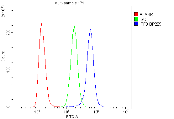
(Figure 6. Flow Cytometry analysis of K562 cells using anti-IRF3 antibody (AAA19216).Overlay histogram showing K562 cells stained with AAA19216 (Blue line). The cells were blocked with 10% normal goat serum. And then incubated with rabbit anti-IRF3 Antibody (AAA19216, 1μg/1x106 cells) for 30 min at 20 degree C. DyLight®488 conjugated goat anti-rabbit IgG (5-10μg/1x106 cells) was used as secondary antibody for 30 minutes at 20 degree C. Isotype control antibody (Green line) was rabbit IgG (1μg/1x106) used under the same conditions. Unlabelled sample (Red line) was also used as a control.)
FCM (Flow Cytometry)
(Figure 6. Flow Cytometry analysis of K562 cells using anti-IRF3 antibody (AAA19216).Overlay histogram showing K562 cells stained with AAA19216 (Blue line). The cells were blocked with 10% normal goat serum. And then incubated with rabbit anti-IRF3 Antibody (AAA19216, 1μg/1x106 cells) for 30 min at 20 degree C. DyLight®488 conjugated goat anti-rabbit IgG (5-10μg/1x106 cells) was used as secondary antibody for 30 minutes at 20 degree C. Isotype control antibody (Green line) was rabbit IgG (1μg/1x106) used under the same conditions. Unlabelled sample (Red line) was also used as a control.)
IRF3, Polyclonal Antibody (Cat# AAA19216)
Full Name
Anti-IRF3 Antibody
Reactivity
Human
Applications
Western Blot, Immunohistochemistry, Flow Cytometry, Direct ELISA
Purity
Immunogen affinity purified.
Pricing


 FCM (Flow Cytometry)
(Figure 7. Flow Cytometry analysis of SiHa cells using anti-Aconitase 1/ACO1 antibody (AAA19267).Overlay histogram showing SiHa cells stained with AAA19267 (Blue line). The cells were blocked with 10% normal goat serum. And then incubated with rabbit anti-Aconitase 1/ACO1 Antibody (AAA19267,1μg/1x106 cells) for 30 min at 20 degree C. DyLight®488 conjugated goat anti-rabbit IgG (5-10μg/1x106 cells) was used as secondary antibody for 30 minutes at 20 degree C. Isotype control antibody (Green line) was rabbit IgG (1μg/1x106) used under the same conditions. Unlabelled sample (Red line) was also used as a control.)
FCM (Flow Cytometry)
(Figure 7. Flow Cytometry analysis of SiHa cells using anti-Aconitase 1/ACO1 antibody (AAA19267).Overlay histogram showing SiHa cells stained with AAA19267 (Blue line). The cells were blocked with 10% normal goat serum. And then incubated with rabbit anti-Aconitase 1/ACO1 Antibody (AAA19267,1μg/1x106 cells) for 30 min at 20 degree C. DyLight®488 conjugated goat anti-rabbit IgG (5-10μg/1x106 cells) was used as secondary antibody for 30 minutes at 20 degree C. Isotype control antibody (Green line) was rabbit IgG (1μg/1x106) used under the same conditions. Unlabelled sample (Red line) was also used as a control.)
Aconitase 1/ACO1, Polyclonal Antibody (Cat# AAA19267)
Full Name
Anti-Aconitase 1/ACO1 Antibody
Gene Names
ACO1; IRP1; ACONS; IREB1; IREBP; IREBP1
Reactivity
Human, Mouse, Monkey, Rat
Applications
WB, IHC-P, ICC, IF, FC/FACS/FCM, EIA
Purity
Immunogen affinity purified.
Pricing


 FCM (Flow Cytometry)
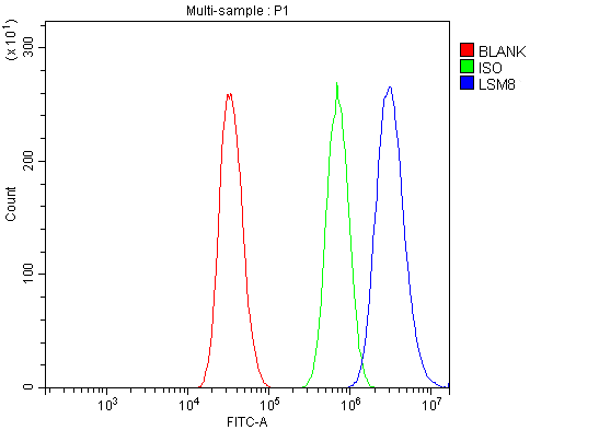
(Figure 13. Flow Cytometry analysis of A431 cells using anti-LSM8 antibody (AAA19337).Overlay histogram showing A431 cells stained with AAA19337 (Blue line). The cells were blocked with 10% normal goat serum. And then incubated with rabbit anti-LSM8 Antibody (AAA19337, 1μg/1x106 cells) for 30 min at 20 degree C. DyLight®488 conjugated goat anti-rabbit IgG (5-10μg/1x106 cells) was used as secondary antibody for 30 minutes at 20 degree C. Isotype control antibody (Green line) was rabbit IgG (1μg/1x106) used under the same conditions. Unlabelled sample (Red line) was also used as a control.)
FCM (Flow Cytometry)
(Figure 13. Flow Cytometry analysis of A431 cells using anti-LSM8 antibody (AAA19337).Overlay histogram showing A431 cells stained with AAA19337 (Blue line). The cells were blocked with 10% normal goat serum. And then incubated with rabbit anti-LSM8 Antibody (AAA19337, 1μg/1x106 cells) for 30 min at 20 degree C. DyLight®488 conjugated goat anti-rabbit IgG (5-10μg/1x106 cells) was used as secondary antibody for 30 minutes at 20 degree C. Isotype control antibody (Green line) was rabbit IgG (1μg/1x106) used under the same conditions. Unlabelled sample (Red line) was also used as a control.)
LSM8, Polyclonal Antibody (Cat# AAA19337)
Full Name
Anti-LSM8 Antibody
Reactivity
Human, Mouse, Rat
Applications
WB, IHC-P, ICC, IF, FC/FACS/FCM, EIA
Purity
Immunogen affinity purified.
Pricing

 IP (Immunoprecipitation)
(IP:Result of anti-GFP-tag Monoclonal antibodyLine 1:Control IgGLine 2:Precipitateing GFP-tagged fusion proteinLine 3:GFP Transfected HEK-293 cells lysateSecondaryGoat polyclonal to Mouse IgG at 1/5000 dilutionPredicted band size: 30kdObserved band size: 30kd)
IP (Immunoprecipitation)
(IP:Result of anti-GFP-tag Monoclonal antibodyLine 1:Control IgGLine 2:Precipitateing GFP-tagged fusion proteinLine 3:GFP Transfected HEK-293 cells lysateSecondaryGoat polyclonal to Mouse IgG at 1/5000 dilutionPredicted band size: 30kdObserved band size: 30kd)
GFP, Monoclonal Antibody (Cat# AAA18855)
Full Name
Mouse Anti-GFP monoclonal Antibody
Applications
EIA, WB, IP
Purity
>95%, Protein G Purified
Pricing


 IHC (Immunohistchemistry)
(Immunohistochemistry of paraffin-embedded mouse stomach using TRMT2A antibody at dilution of 1:100 (40x lens).)
IHC (Immunohistchemistry)
(Immunohistochemistry of paraffin-embedded mouse stomach using TRMT2A antibody at dilution of 1:100 (40x lens).)
TRMT2A, Polyclonal Antibody (Cat# AAA28127)
Full Name
TRMT2A Polyclonal Antibody
Gene Names
TRMT2A; HTF9C
Reactivity
Human, Mouse, Rat
Applications
WB, IHC
Purity
Affinity Purification
Pricing


 IF (Immunofluorescence)
(Immunofluorescence analysis of HeLa cells using RPS6KA3 antibody. Blue: DAPI for nuclear staining.)
IF (Immunofluorescence)
(Immunofluorescence analysis of HeLa cells using RPS6KA3 antibody. Blue: DAPI for nuclear staining.)
RPS6KA3, Polyclonal Antibody (Cat# AAA28081)
Full Name
RPS6KA3 Polyclonal Antibody
Gene Names
RPS6KA3; CLS; RSK; HU-3; RSK2; MRX19; ISPK-1; p90-RSK2; pp90RSK2; MAPKAPK1B; S6K-alpha3
Reactivity
Human, Mouse, Rat
Applications
Western Blot, Immunohistochemistry, Immunofluorescence
Purity
Affinity Purification
Pricing


 IF (Immunofluorescence)
(Immunofluorescence analysis of U2OS cells usingTFEB Rabbit pAb (AAA28118) at dilution of 1:50 (40xlens).Secondary antibody: Cy3 Goat Anti-Rabbit IgG(H+L) at 1:500 dilution. Blue: DAPI for nuclear staining.)
IF (Immunofluorescence)
(Immunofluorescence analysis of U2OS cells usingTFEB Rabbit pAb (AAA28118) at dilution of 1:50 (40xlens).Secondary antibody: Cy3 Goat Anti-Rabbit IgG(H+L) at 1:500 dilution. Blue: DAPI for nuclear staining.)
TFEB, Polyclonal Antibody (Cat# AAA28118)
Full Name
TFEB Polyclonal Antibody
Gene Names
TFEB; TCFEB; BHLHE35; ALPHATFEB
Applications
WB, IHC-p, IF, ICC, EIA
Purity
Affinity Purification
Pricing


 IP (Immunoprecipitation)
(Immunoprecipitation analysis of 150ug extracts of MCF7 cells using 3ug HIST3H3 antibody. Western blot was performed from the immunoprecipitate using HIST3H3 antibody at a dilition of 1:500.)
IP (Immunoprecipitation)
(Immunoprecipitation analysis of 150ug extracts of MCF7 cells using 3ug HIST3H3 antibody. Western blot was performed from the immunoprecipitate using HIST3H3 antibody at a dilition of 1:500.)
HIST3H3, Polyclonal Antibody (Cat# AAA28284)
Full Name
HIST3H3 Polyclonal Antibody
Gene Names
HIST3H3; H3t; H3.4; H3/g; H3FT
Reactivity
Human, Mouse, Rat, Other (Wide Range)
Applications
Western Blot, Immunohistochemistry, Immunoprecipitation
Purity
Affinity Purification
Pricing


 WB (Western Blot)
(Western blot analysis of lysates from 293T, Hela cell line (from left to right), using GLS Antibody (C-term). AAA28657 was diluted at 1:1000 at each lane. A goat anti-rabbit IgG H&L(HRP) at 1:5000 dilution was used as the secondary antibody. Lysates at 35ug per lane.)
WB (Western Blot)
(Western blot analysis of lysates from 293T, Hela cell line (from left to right), using GLS Antibody (C-term). AAA28657 was diluted at 1:1000 at each lane. A goat anti-rabbit IgG H&L(HRP) at 1:5000 dilution was used as the secondary antibody. Lysates at 35ug per lane.)
GLS, Polyclonal Antibody (Cat# AAA28657)
Full Name
GLS Antibody (C-term)
Gene Names
GLS; GAC; GAM; KGA; GLS1; AAD20
Reactivity
Human, mouse (Predicted Reactivity: Rat)
Applications
WB, EIA, IHC, IF, FC/FACS
Purity
Peptide Affinity Purified Rabbit Polyclonal Antibody (Pab)
Pricing


 IP (Immunoprecipitation)
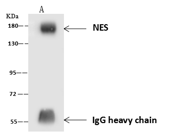
(NES was immunoprecipitated using: Lane A:0.5 mg U251MG Whole Cell Lysate 4 uL anti-NES mouse monoclonal antibody and 60 ug of Immunomagnetic beads Protein A/G. Primary antibody: Anti-NES mouse monoclonal antibody,at 1:100 dilution Secondary antibody: Rabbit Anti-Mouse IgG F(ab)2/HRP at 1/10000 dilution Developed using the ECL technique. Performed under reducing conditions. Predicted band size: 177 kDa Observed band size :177 kDa)
IP (Immunoprecipitation)
(NES was immunoprecipitated using: Lane A:0.5 mg U251MG Whole Cell Lysate 4 uL anti-NES mouse monoclonal antibody and 60 ug of Immunomagnetic beads Protein A/G. Primary antibody: Anti-NES mouse monoclonal antibody,at 1:100 dilution Secondary antibody: Rabbit Anti-Mouse IgG F(ab)2/HRP at 1/10000 dilution Developed using the ECL technique. Performed under reducing conditions. Predicted band size: 177 kDa Observed band size :177 kDa)
Nestin, Monoclonal Antibody (Cat# AAA27730)
Full Name
Anti-Nestin Antibody, Mouse Monoclonal
Gene Names
NES; Nbla00170
Reactivity
Human
Applications
Western Blot, Immunohistochemistry, Flow Cytometry, Immunoprecipitation
Purity
Protein A
Pricing


 IF (Immunofluorescence)
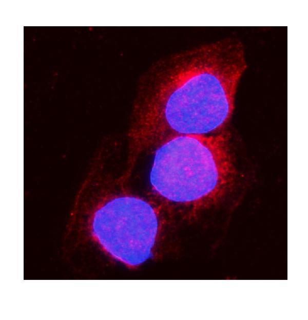
(Immunofluorescent analysis of 4% paraformaldehyde-fixed, 0.1% Triton X-100 permeabilized MCF-7 (human breast cancer cell line) cells labeling Pdx1 with at 1:25 dilution, followed by DyLight 488-conjugated IgG goat anti-rabbit secondary antibody at 1:200 dilution (green). Immunofluorescence image showing cytoplasm staining on MCF-7 cell line. Cytoplasmic actin is detected with DyLight 554 Phalloidin (PD18466410) at 1:100 dilution (red). The nuclear counter stain is DAPI (blue).)
IF (Immunofluorescence)
(Immunofluorescent analysis of 4% paraformaldehyde-fixed, 0.1% Triton X-100 permeabilized MCF-7 (human breast cancer cell line) cells labeling Pdx1 with at 1:25 dilution, followed by DyLight 488-conjugated IgG goat anti-rabbit secondary antibody at 1:200 dilution (green). Immunofluorescence image showing cytoplasm staining on MCF-7 cell line. Cytoplasmic actin is detected with DyLight 554 Phalloidin (PD18466410) at 1:100 dilution (red). The nuclear counter stain is DAPI (blue).)
OPN-a/b, Polyclonal Antibody (Cat# AAA26857)
Full Name
OPN-a/b, NT (SPP1, BNSP, OPN, Osteopontin, Bone sialoprotein 1, Nephropontin, Secreted phosphoprotein 1, Urinary stone protein, Uropontin) (APC)
Gene Names
SPP1; OPN; BNSP; BSPI; ETA-1
Reactivity
Human
Applications
WB, IHC, IF
Purity
Purified by Protein A and Peptide Affinity Chromatography.
Pricing


 FCM (Flow Cytometry)
(Figure 6. Flow Cytometry analysis of PC-3 cells using anti-MitoNEET/CISD1 antibody (AAA19293).Overlay histogram showing PC-3 cells stained with AAA19293 (Blue line). The cells were blocked with 10% normal goat serum. And then incubated with rabbit anti-MitoNEET/CISD1 Antibody (AAA19293,1μg/1x106 cells) for 30 min at 20 degree C. DyLight®488 conjugated goat anti-rabbit IgG (5-10μg/1x106 cells) was used as secondary antibody for 30 minutes at 20 degree C. Isotype control antibody (Green line) was rabbit IgG (1μg/1x106) used under the same conditions. Unlabelled sample (Red line) was also used as a control.)
FCM (Flow Cytometry)
(Figure 6. Flow Cytometry analysis of PC-3 cells using anti-MitoNEET/CISD1 antibody (AAA19293).Overlay histogram showing PC-3 cells stained with AAA19293 (Blue line). The cells were blocked with 10% normal goat serum. And then incubated with rabbit anti-MitoNEET/CISD1 Antibody (AAA19293,1μg/1x106 cells) for 30 min at 20 degree C. DyLight®488 conjugated goat anti-rabbit IgG (5-10μg/1x106 cells) was used as secondary antibody for 30 minutes at 20 degree C. Isotype control antibody (Green line) was rabbit IgG (1μg/1x106) used under the same conditions. Unlabelled sample (Red line) was also used as a control.)
MitoNEET/CISD1, Polyclonal Antibody (Cat# AAA19293)
Full Name
Anti-MitoNEET/CISD1 Antibody
Gene Names
CISD1; ZCD1; MDS029; C10orf70; mitoNEET
Reactivity
Human, Mouse, Rat, Monkey
Applications
WB, IHC-P, FC/FACS/FCM, EIA
Purity
Immunogen affinity purified.
Pricing


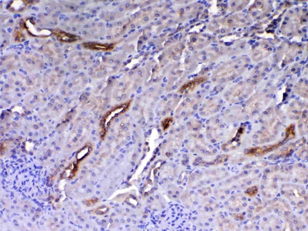
 IHC (Immunohistchemistry)
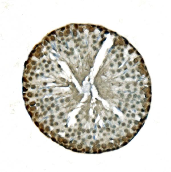
(Figure 6. IHC analysis of Flt3 / CD135 using anti-Flt3 / CD135 antibody (AAA19130).Flt3 / CD135 was detected in paraffin-embedded section of mouse kidney tissue. Heat mediated antigen retrieval was performed in citrate buffer (pH6, epitope retrieval solution) for 20 mins. The tissue section was blocked with 10% goat serum. The tissue section was then incubated with 1ug/ml rabbit anti-Flt3 / CD135 Antibody (AAA19130) overnight at 4 degree C. Biotinylated goat anti-rabbit IgG was used as secondary antibody and incubated for 30 minutes at 37 degree C. The tissue section was developed using Strepavidin-Biotin-Complex (SABC) with DAB as the chromogen.)
IHC (Immunohistchemistry)
(Figure 6. IHC analysis of Flt3 / CD135 using anti-Flt3 / CD135 antibody (AAA19130).Flt3 / CD135 was detected in paraffin-embedded section of mouse kidney tissue. Heat mediated antigen retrieval was performed in citrate buffer (pH6, epitope retrieval solution) for 20 mins. The tissue section was blocked with 10% goat serum. The tissue section was then incubated with 1ug/ml rabbit anti-Flt3 / CD135 Antibody (AAA19130) overnight at 4 degree C. Biotinylated goat anti-rabbit IgG was used as secondary antibody and incubated for 30 minutes at 37 degree C. The tissue section was developed using Strepavidin-Biotin-Complex (SABC) with DAB as the chromogen.)
Flt3/CD135, Polyclonal Antibody (Cat# AAA19130)
Full Name
Anti-Flt3/CD135 Picoband Antibody
Reactivity
Mouse, Rat
No cross reactivity with other proteins.
No cross reactivity with other proteins.
Applications
EIA, IHC, WB
Purity
Immunogen affinity purified
Pricing


 FCM (Flow Cytometry)
(Flow cytometric analysis of SH-SY-5Y cells with Huntingtin antibody at 1/100 dilution (red) compared with an unlabelled control (cells without incubation with primary antibody; black). Alexa Fluor 488-conjugated goat anti rabbit IgG was used as the secondary antibody.)
FCM (Flow Cytometry)
(Flow cytometric analysis of SH-SY-5Y cells with Huntingtin antibody at 1/100 dilution (red) compared with an unlabelled control (cells without incubation with primary antibody; black). Alexa Fluor 488-conjugated goat anti rabbit IgG was used as the secondary antibody.)
Huntingtin, Monoclonal Antibody (Cat# AAA30460)
Full Name
Huntingtin Antibody
Gene Names
HTT; HD; IT15
Reactivity
Human, Mouse, Rat
Applications
WB, IHC, FC/FACS, ICC
Purity
ProA affinity purified
Pricing


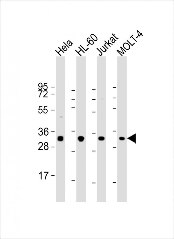
 WB (Western Blot)
(WB Suggested Anti-IGF2BP1 Antibody Titration: 0.2-1 ug/mlELISA Titer: 1:312500Positive Control: Jurkat cell lysate)
WB (Western Blot)
(WB Suggested Anti-IGF2BP1 Antibody Titration: 0.2-1 ug/mlELISA Titer: 1:312500Positive Control: Jurkat cell lysate)
IGF2BP1, Polyclonal Antibody (Cat# AAA23578)
Full Name
IGF2BP1 antibody - N-terminal region
Gene Names
IGF2BP1; IMP1; ZBP1; CRDBP; IMP-1; CRD-BP; VICKZ1
Reactivity
Cow, Dog, Guinea Pig, Horse, Human, Mouse, Rabbit, Rat, Sheep, Zebrafish
Applications
Immunohistochemistry, Western Blot
Purity
Affinity Purified
Pricing


 FCM (Flow Cytometry)
(SYP Antibody (C-term) flow cytometric analysis of Neuro-2a cells (right histogram) compared to a negative control cell (left histogram).FITC-conjugated goat-anti-rabbit secondary antibodies were used for the analysis.)
FCM (Flow Cytometry)
(SYP Antibody (C-term) flow cytometric analysis of Neuro-2a cells (right histogram) compared to a negative control cell (left histogram).FITC-conjugated goat-anti-rabbit secondary antibodies were used for the analysis.)
SYP, Polyclonal Antibody (Cat# AAA28723)
Full Name
SYP Antibody (C-term)
Gene Names
SYP; MRX96; MRXSYP
Reactivity
Human, mouse (Predicted Reactivity: Rat)
Applications
Flow Cytometry, Immunofluorescence, Immunohistochemistry, Western Blot
Purity
Peptide Affinity Purified Rabbit Polyclonal Antibody (Pab)
Pricing


 IHC (Immunohistchemistry)
(Immunohistochemistry analysis using Mouse Anti-Hsp70 Monoclonal Antibody, Clone BB70. Tissue: hepatocytes. Species: Rat. Fixation: Paraffin Embedded. Primary Antibody: Mouse Anti-Hsp70 Monoclonal Antibody at 1:200. Liver sections were paraffin embedded. First pictures in series show two hours after exposure to stress, the second shows the control. Courtesy of: G. Matic, University of Belgrade, Serbia.)
IHC (Immunohistchemistry)
(Immunohistochemistry analysis using Mouse Anti-Hsp70 Monoclonal Antibody, Clone BB70. Tissue: hepatocytes. Species: Rat. Fixation: Paraffin Embedded. Primary Antibody: Mouse Anti-Hsp70 Monoclonal Antibody at 1:200. Liver sections were paraffin embedded. First pictures in series show two hours after exposure to stress, the second shows the control. Courtesy of: G. Matic, University of Belgrade, Serbia.)
Hsp70/Hsc70, Monoclonal Antibody (Cat# AAA17791)
Full Name
Hsp70/Hsc70 Antibody: PerCP
Gene Names
HSPA2; HSP70
Reactivity
Human, Mouse, Rat, Sheep, Dog, Beluga, Bovine, Fish, Guinea pig, Scallop pig, Hamster, Rabbit, Chicken, Xenopus, Drosophila, Yeast
Applications
WB, IP, IHC
Pricing


 IHC (Immunohistochemistry)
(Figure 7. IHC analysis of Periostin using anti-Periostin antibody (AAA19149).Periostin was detected in paraffin-embedded section of rat small intestine tissue. Heat mediated antigen retrieval was performed in citrate buffer (pH6, epitope retrieval solution) for 20 mins. The tissue section was blocked with 10% goat serum. The tissue section was then incubated with 1ug/ml rabbit anti-Periostin Antibody (AAA19149) overnight at 4 degree C. Biotinylated goat anti-rabbit IgG was used as secondary antibody and incubated for 30 minutes at 37 degree C. The tissue section was developed using Strepavidin-Biotin-Complex (SABC) with DAB as the chromogen.)
IHC (Immunohistochemistry)
(Figure 7. IHC analysis of Periostin using anti-Periostin antibody (AAA19149).Periostin was detected in paraffin-embedded section of rat small intestine tissue. Heat mediated antigen retrieval was performed in citrate buffer (pH6, epitope retrieval solution) for 20 mins. The tissue section was blocked with 10% goat serum. The tissue section was then incubated with 1ug/ml rabbit anti-Periostin Antibody (AAA19149) overnight at 4 degree C. Biotinylated goat anti-rabbit IgG was used as secondary antibody and incubated for 30 minutes at 37 degree C. The tissue section was developed using Strepavidin-Biotin-Complex (SABC) with DAB as the chromogen.)
Periostin, Polyclonal Antibody (Cat# AAA19149)
Full Name
Anti-Periostin Picoband Antibody
Gene Names
Postn; Plf
Reactivity
Mouse, Rat
No cross reactivity with other proteins.
No cross reactivity with other proteins.
Applications
EIA, IHC, WB
Purity
Immunogen affinity purified
Pricing


 IHC (Immunohistchemistry)
(Figure 6. IHC analysis of HE4 using anti-HE4 antibody (AAA19161).HE4 was detected in paraffin-embedded section of rat small intestine tissue. Heat mediated antigen retrieval was performed in citrate buffer (pH6, epitope retrieval solution) for 20 mins. The tissue section was blocked with 10% goat serum. The tissue section was then incubated with 1ug/ml rabbit anti-HE4 Antibody (AAA19161) overnight at 4 degree C. Biotinylated goat anti-rabbit IgG was used as secondary antibody and incubated for 30 minutes at 37 degree C. The tissue section was developed using Strepavidin-Biotin-Complex (SABC) with DAB as the chromogen.)
IHC (Immunohistchemistry)
(Figure 6. IHC analysis of HE4 using anti-HE4 antibody (AAA19161).HE4 was detected in paraffin-embedded section of rat small intestine tissue. Heat mediated antigen retrieval was performed in citrate buffer (pH6, epitope retrieval solution) for 20 mins. The tissue section was blocked with 10% goat serum. The tissue section was then incubated with 1ug/ml rabbit anti-HE4 Antibody (AAA19161) overnight at 4 degree C. Biotinylated goat anti-rabbit IgG was used as secondary antibody and incubated for 30 minutes at 37 degree C. The tissue section was developed using Strepavidin-Biotin-Complex (SABC) with DAB as the chromogen.)
HE4, Polyclonal Antibody (Cat# AAA19161)
Full Name
Anti-HE4 Picoband Antibody
Gene Names
Wfdc2; re4
Reactivity
Mouse, Rat
No cross reactivity with other proteins.
No cross reactivity with other proteins.
Applications
IHC, WB
Purity
Immunogen affinity purified
Pricing


 IHC (Immunohistchemistry)
(Immunohistochemistry of paraffin-embedded human liver cancer using SMARCE1 Antibody at dilution of 1:200 (40x lens).)
IHC (Immunohistchemistry)
(Immunohistochemistry of paraffin-embedded human liver cancer using SMARCE1 Antibody at dilution of 1:200 (40x lens).)
SMARCE1, Polyclonal Antibody (Cat# AAA28107)
Full Name
SMARCE1 Polyclonal Antibody
Gene Names
SMARCE1; BAF57
Reactivity
Human, Mouse
Applications
Western Blot, Immunohistochemistry, Immunofluorescence, Immunoprecipitation, Chromatin Immunoprecipitation
Purity
Affinity Purification
Pricing


 WB (Western Blot)
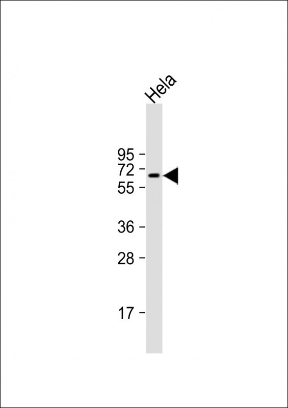
(WB Suggested Anti-MTUS1 Antibody Titration: 0.2-1 ug/mlELISA Titer: 1:1562500Positive Control: Hela cell lysateMTUS1 is strongly supported by BioGPS gene expression data to be expressed in Human HeLa cells)
WB (Western Blot)
(WB Suggested Anti-MTUS1 Antibody Titration: 0.2-1 ug/mlELISA Titer: 1:1562500Positive Control: Hela cell lysateMTUS1 is strongly supported by BioGPS gene expression data to be expressed in Human HeLa cells)
MTUS1, Polyclonal Antibody (Cat# AAA23518)
Full Name
MTUS1 antibody - middle region
Gene Names
MTUS1; ATBP; ATIP; ICIS; MP44; ATIP3; MTSG1
Reactivity
Cow, Dog, Guinea Pig, Human, Mouse, Rabbit, Rat
Applications
IHC, WB
Purity
Affinity Purified
Pricing


 IHC (Immunohistchemistry)
(Figure 6. IHC analysis of HP1 gamma using anti-HP1 gamma antibody (AAA19145).HP1 gamma was detected in paraffin-embedded section of rat small intestine tissue. Heat mediated antigen retrieval was performed in citrate buffer (pH6, epitope retrieval solution) for 20 mins. The tissue section was blocked with 10% goat serum. The tissue section was then incubated with 1ug/ml rabbit anti-HP1 gamma Antibody (AAA19145) overnight at 4 degree C. Biotinylated goat anti-rabbit IgG was used as secondary antibody and incubated for 30 minutes at 37 degree C. The tissue section was developed using Strepavidin-Biotin-Complex (SABC) with DAB as the chromogen.)
IHC (Immunohistchemistry)
(Figure 6. IHC analysis of HP1 gamma using anti-HP1 gamma antibody (AAA19145).HP1 gamma was detected in paraffin-embedded section of rat small intestine tissue. Heat mediated antigen retrieval was performed in citrate buffer (pH6, epitope retrieval solution) for 20 mins. The tissue section was blocked with 10% goat serum. The tissue section was then incubated with 1ug/ml rabbit anti-HP1 gamma Antibody (AAA19145) overnight at 4 degree C. Biotinylated goat anti-rabbit IgG was used as secondary antibody and incubated for 30 minutes at 37 degree C. The tissue section was developed using Strepavidin-Biotin-Complex (SABC) with DAB as the chromogen.)
HP1 gamma, Polyclonal Antibody (Cat# AAA19145)
Full Name
Anti-HP1 gamma Picoband antibody
Gene Names
CBX3; HECH; HP1-GAMMA; HP1Hs-gamma
Reactivity
Human, Mouse, Rat
No cross reactivity with other proteins.
No cross reactivity with other proteins.
Applications
EIA, IHC, WB
Pricing


 IF (Immunofluorescence)
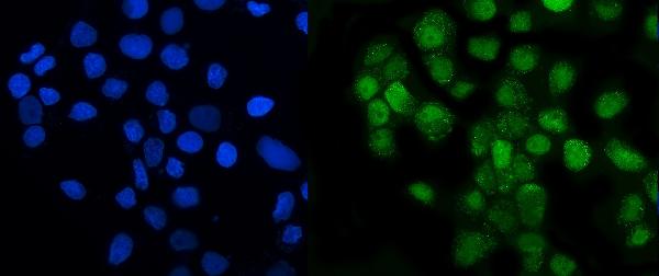
(Figure 6. IF analysis of GRSF1 using anti- GRSF1 antibody (AAA19315).GRSF1 was detected in immunocytochemical section of MCF-7 cells. Enzyme antigen retrieval was performed using IHC enzyme antigen retrieval reagent for 15 mins. The cells were blocked with 10% goat serum. And then incubated with 5μg/mL rabbit anti- GRSF1 Antibody (AAA19315) overnight at 4 degree C. DyLight®488 Conjugated Goat Anti-Rabbit IgG was used as secondary antibody at 1:100 dilution and incubated for 30 minutes at 37 degree C. The section was counterstained with DAPI. Visualize using a fluorescence microscope and filter sets appropriate for the label used.)
IF (Immunofluorescence)
(Figure 6. IF analysis of GRSF1 using anti- GRSF1 antibody (AAA19315).GRSF1 was detected in immunocytochemical section of MCF-7 cells. Enzyme antigen retrieval was performed using IHC enzyme antigen retrieval reagent for 15 mins. The cells were blocked with 10% goat serum. And then incubated with 5μg/mL rabbit anti- GRSF1 Antibody (AAA19315) overnight at 4 degree C. DyLight®488 Conjugated Goat Anti-Rabbit IgG was used as secondary antibody at 1:100 dilution and incubated for 30 minutes at 37 degree C. The section was counterstained with DAPI. Visualize using a fluorescence microscope and filter sets appropriate for the label used.)
GRSF1, Polyclonal Antibody (Cat# AAA19315)
Full Name
Anti-GRSF1 Antibody
Reactivity
Human
Applications
WB, IHC-P, ICC, IF, FC/FACS/FCM, EIA
Purity
Immunogen affinity purified.
Pricing


 FCM (Flow Cytometry)
(Figure 8. Flow Cytometry analysis of K562 cells using anti-HP1 alpha/CBX5 antibody (AAA19266).Overlay histogram showing K562 cells stained with AAA19266 (Blue line). The cells were blocked with 10% normal goat serum. And then incubated with rabbit anti-HP1 alpha/CBX5 Antibody (AAA19266,1μg/1x106 cells) for 30 min at 20 degree C. DyLight®488 conjugated goat anti-rabbit IgG (5-10μg/1x106 cells) was used as secondary antibody for 30 minutes at 20 degree C. Isotype control antibody (Green line) was rabbit IgG (1μg/1x106) used under the same conditions. Unlabelled sample (Red line) was also used as a control.)
FCM (Flow Cytometry)
(Figure 8. Flow Cytometry analysis of K562 cells using anti-HP1 alpha/CBX5 antibody (AAA19266).Overlay histogram showing K562 cells stained with AAA19266 (Blue line). The cells were blocked with 10% normal goat serum. And then incubated with rabbit anti-HP1 alpha/CBX5 Antibody (AAA19266,1μg/1x106 cells) for 30 min at 20 degree C. DyLight®488 conjugated goat anti-rabbit IgG (5-10μg/1x106 cells) was used as secondary antibody for 30 minutes at 20 degree C. Isotype control antibody (Green line) was rabbit IgG (1μg/1x106) used under the same conditions. Unlabelled sample (Red line) was also used as a control.)
HP1 alpha/CBX5, Polyclonal Antibody (Cat# AAA19266)
Full Name
Anti-HP1 alpha/CBX5 Antibody
Gene Names
CBX5; HP1; HP1A
Reactivity
Human, Mouse, Rat
Applications
WB, IHC-P, ICC, IF, FC/FACS/FCM, EIA
Purity
Immunogen affinity purified.
Pricing


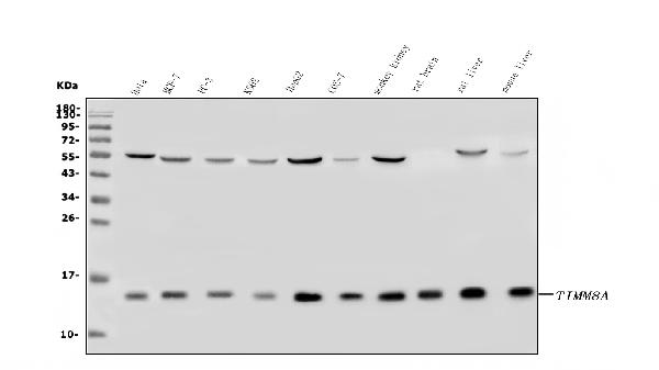
 FCM (Flow Cytometry)
(Figure 10. Flow Cytometry analysis of A431 cells using anti-TIMM8A/DDP antibody (AAA19317).Overlay histogram showing A431 cells stained with AAA19317 (Blue line). The cells were blocked with 10% normal goat serum. And then incubated with rabbit anti-TIMM8A/DDP Antibody (AAA19317, 1μg/1x106 cells) for 30 min at 20 degree C. DyLight®488 conjugated goat anti-rabbit IgG (5-10μg/1x106 cells) was used as secondary antibody for 30 minutes at 20 degree C. Isotype control antibody (Green line) was rabbit IgG (1μg/1x106) used under the same conditions. Unlabelled sample (Red line) was also used as a control.)
FCM (Flow Cytometry)
(Figure 10. Flow Cytometry analysis of A431 cells using anti-TIMM8A/DDP antibody (AAA19317).Overlay histogram showing A431 cells stained with AAA19317 (Blue line). The cells were blocked with 10% normal goat serum. And then incubated with rabbit anti-TIMM8A/DDP Antibody (AAA19317, 1μg/1x106 cells) for 30 min at 20 degree C. DyLight®488 conjugated goat anti-rabbit IgG (5-10μg/1x106 cells) was used as secondary antibody for 30 minutes at 20 degree C. Isotype control antibody (Green line) was rabbit IgG (1μg/1x106) used under the same conditions. Unlabelled sample (Red line) was also used as a control.)
TIMM8A/DDP, Polyclonal Antibody (Cat# AAA19317)
Full Name
Anti-TIMM8A/DDP Antibody
Gene Names
TIMM8A; DDP; MTS; DDP1; DFN1; TIM8
Reactivity
Human, Mouse, Rat, Monkey
Applications
WB, IHC-P, ICC, IF, EIA
Purity
Immunogen affinity purified.
Pricing


 FCM (Flow Cytometry)
(Figure 9. Flow Cytometry analysis of U251 cells using anti-VPRBP/DCAF1 antibody (AAA19343).Overlay histogram showing U251 cells stained with AAA19343 (Blue line). The cells were blocked with 10% normal goat serum. And then incubated with rabbit anti-VPRBP/DCAF1 Antibody (AAA19343, 1μg/1x106 cells) for 30 min at 20 degree C. DyLight®488 conjugated goat anti-rabbit IgG (5-10μg/1x106 cells) was used as secondary antibody for 30 minutes at 20 degree C. Isotype control antibody (Green line) was rabbit IgG (1μg/1x106) used under the same conditions. Unlabelled sample (Red line) was also used as a control.)
FCM (Flow Cytometry)
(Figure 9. Flow Cytometry analysis of U251 cells using anti-VPRBP/DCAF1 antibody (AAA19343).Overlay histogram showing U251 cells stained with AAA19343 (Blue line). The cells were blocked with 10% normal goat serum. And then incubated with rabbit anti-VPRBP/DCAF1 Antibody (AAA19343, 1μg/1x106 cells) for 30 min at 20 degree C. DyLight®488 conjugated goat anti-rabbit IgG (5-10μg/1x106 cells) was used as secondary antibody for 30 minutes at 20 degree C. Isotype control antibody (Green line) was rabbit IgG (1μg/1x106) used under the same conditions. Unlabelled sample (Red line) was also used as a control.)
VPRBP/DCAF1, Polyclonal Antibody (Cat# AAA19343)
Full Name
Anti-VPRBP/DCAF1 Antibody
Gene Names
DCAF1; RIP; VPRBP
Reactivity
Human, Mouse, Rat
Applications
WB, IHC-P, ICC, IF, FC/FACS/FCM, EIA
Purity
Immunogen affinity purified.
Pricing


 FCM (Flow Cytometry)
(Flow cytometry analysis of GOT1 in HeLa cell line, staining at 2-5ug for 1x10⁶cells (red line). The secondary antibody used goat anti-mouse IgG Alexa fluor 488 conjugate. Isotype control antibody was mouse IgG (black line).)
FCM (Flow Cytometry)
(Flow cytometry analysis of GOT1 in HeLa cell line, staining at 2-5ug for 1x10⁶cells (red line). The secondary antibody used goat anti-mouse IgG Alexa fluor 488 conjugate. Isotype control antibody was mouse IgG (black line).)
GOT1, Monoclonal Antibody (Cat# AAA11733)
Full Name
GOT1 antibody
Gene Names
GOT1; AST1; cCAT; GIG18; cAspAT; ASTQTL1
Reactivity
Human
Applications
Western Blot, Flow Cytometry, Immunocytochemistry, Immunofluorescence
Purity
By protein-A affinity chromatography
Pricing


 IF (Immunofluorescence)
(Immunofluorescent analysis of 4% paraformaldehyde-fixed, 0.1% Triton X-100 permeabilized MCF-7 (human breast cancer cell line) cells labeling Pdx1 with at 1:25 dilution, followed by DyLight 488-conjugated IgG goat anti-rabbit secondary antibody at 1:200 dilution (green). Immunofluorescence image showing cytoplasm staining on MCF-7 cell line. Cytoplasmic actin is detected with DyLight 554 Phalloidin (PD18466410) at 1:100 dilution (red). The nuclear counter stain is DAPI (blue).)
IF (Immunofluorescence)
(Immunofluorescent analysis of 4% paraformaldehyde-fixed, 0.1% Triton X-100 permeabilized MCF-7 (human breast cancer cell line) cells labeling Pdx1 with at 1:25 dilution, followed by DyLight 488-conjugated IgG goat anti-rabbit secondary antibody at 1:200 dilution (green). Immunofluorescence image showing cytoplasm staining on MCF-7 cell line. Cytoplasmic actin is detected with DyLight 554 Phalloidin (PD18466410) at 1:100 dilution (red). The nuclear counter stain is DAPI (blue).)
OPN-a/b, Polyclonal Antibody (Cat# AAA26861)
Full Name
OPN-a/b, NT (SPP1, BNSP, OPN, Osteopontin, Bone sialoprotein 1, Nephropontin, Secreted phosphoprotein 1, Urinary stone protein, Uropontin) (MaxLight 490)
Gene Names
SPP1; OPN; BNSP; BSPI; ETA-1
Reactivity
Human
Applications
WB, IHC, IF
Purity
Purified by Protein A and Peptide Affinity Chromatography.
Pricing


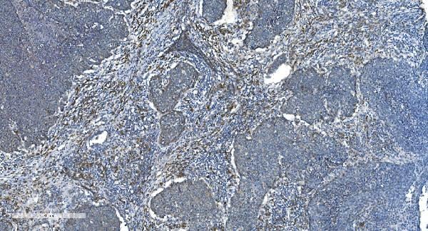
 IHC (Immunohistchemistry)
(Figure 6. IHC analysis of Cytoglobin using anti- Cytoglobin antibody (AAA11681).Cytoglobin was detected in paraffin-embedded section of human lung cancer tissues. Heat mediated antigen retrieval was performed in citrate buffer (pH6, epitope retrieval solution) for 20 mins. The tissue section was blocked with 10% goat serum. The tissue section was then incubated with 1ug/ml rabbit anti- Cytoglobin Antibody (AAA11681) overnight at 4 degree C. Biotinylated goat anti-rabbit IgG was used as secondary antibody and incubated for 30 minutes at 37 degree C. The tissue section was developed using Strepavidin-Biotin-Complex (SABC) with DAB as the chromogen.)
IHC (Immunohistchemistry)
(Figure 6. IHC analysis of Cytoglobin using anti- Cytoglobin antibody (AAA11681).Cytoglobin was detected in paraffin-embedded section of human lung cancer tissues. Heat mediated antigen retrieval was performed in citrate buffer (pH6, epitope retrieval solution) for 20 mins. The tissue section was blocked with 10% goat serum. The tissue section was then incubated with 1ug/ml rabbit anti- Cytoglobin Antibody (AAA11681) overnight at 4 degree C. Biotinylated goat anti-rabbit IgG was used as secondary antibody and incubated for 30 minutes at 37 degree C. The tissue section was developed using Strepavidin-Biotin-Complex (SABC) with DAB as the chromogen.)
Cytoglobin, Polyclonal Antibody (Cat# AAA11681)
Full Name
Anti-Cytoglobin Antibody
Gene Names
CYGB; HGB; STAP
Reactivity
Human, Mouse, Rat
Applications
Western Blot, Immunohistochemistry
Purity
Immunogen affinity purified.
Pricing


 FCM (Flow Cytometry)
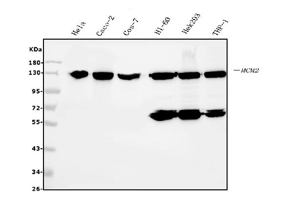
(Figure 6. Flow Cytometry analysis of HL-60 cells using anti- MCM2 antibody (AAA19351).Overlay histogram showing HL-60 cells stained with AAA19351 (Blue line). The cells were blocked with 10% normal goat serum. And then incubated with mouse anti-MCM2 Antibody (AAA19351, 1μg/1x106 cells) for 30 min at 20 degree C. DyLight®488 conjugated goat anti-mouse IgG (BA1126, 5-10μg/1x106 cells) was used as secondary antibody for 30 minutes at 20 degree C. Isotype control antibody (Green line) was mouse IgG (1μg/1x106) used under the same conditions. Unlabelled sample (Red line) was also used as a control.)
FCM (Flow Cytometry)
(Figure 6. Flow Cytometry analysis of HL-60 cells using anti- MCM2 antibody (AAA19351).Overlay histogram showing HL-60 cells stained with AAA19351 (Blue line). The cells were blocked with 10% normal goat serum. And then incubated with mouse anti-MCM2 Antibody (AAA19351, 1μg/1x106 cells) for 30 min at 20 degree C. DyLight®488 conjugated goat anti-mouse IgG (BA1126, 5-10μg/1x106 cells) was used as secondary antibody for 30 minutes at 20 degree C. Isotype control antibody (Green line) was mouse IgG (1μg/1x106) used under the same conditions. Unlabelled sample (Red line) was also used as a control.)
MCM2, Monoclonal Antibody (Cat# AAA19351)
Full Name
Anti-MCM2 Antibody (monoclonal, 11C4)
Gene Names
MCM2; BM28; CCNL1; CDCL1; cdc19; D3S3194; MITOTIN
Reactivity
Human, Monkey
Applications
WB, IHC-P, ICC, IF, FC/FACS/FCM
Purity
Immunogen affinity purified.
Pricing


 WB (Western Blot)
(WB Suggested Anti-ELF2 Antibody Titration: 0.2-1 ug/mlELISA Titer: 1:12500Positive Control: Jurkat cell lysate)
WB (Western Blot)
(WB Suggested Anti-ELF2 Antibody Titration: 0.2-1 ug/mlELISA Titer: 1:12500Positive Control: Jurkat cell lysate)
ELF2, Polyclonal Antibody (Cat# AAA23388)
Full Name
ELF2 antibody - N-terminal region
Gene Names
ELF2; EU32; NERF; NERF-2; NERF-1A; NERF-1B; NERF-1a,b
Reactivity
Cow, Dog, Guinea Pig, Horse, Human, Mouse, Rabbit, Rat
Applications
Western Blot
Purity
Affinity Purified
Pricing


 FCM (Flow Cytometry)
(Flow cytometric analysis of HL-60 cells with CD71 antibody at 1/100 dilution (red) compared with an unlabelled control (cells without incubation with primary antibody; black). Alexa Fluor 488-conjugated Goat anti rabbit IgG was used as the secondary antibody.)
FCM (Flow Cytometry)
(Flow cytometric analysis of HL-60 cells with CD71 antibody at 1/100 dilution (red) compared with an unlabelled control (cells without incubation with primary antibody; black). Alexa Fluor 488-conjugated Goat anti rabbit IgG was used as the secondary antibody.)
CD71, Polyclonal Antibody (Cat# AAA29924)
Full Name
CD71 Antibody
Gene Names
TFRC; T9; TR; TFR; p90; CD71; TFR1; TRFR
Reactivity
Human, Mouse
Applications
ICC, IHC, FC/FACS
Purity
ProA affinity purified
Pricing


 FCM (Flow Cytometry)
(Flow cytometric analysis of Hela cells with KAP1 antibody at 1/50 dilution (red) compared with an unlabelled control (cells without incubation with primary antibody; black). Alexa Fluor 488-conjugated goat anti rabbit IgG was used as the secondary antibody.)
FCM (Flow Cytometry)
(Flow cytometric analysis of Hela cells with KAP1 antibody at 1/50 dilution (red) compared with an unlabelled control (cells without incubation with primary antibody; black). Alexa Fluor 488-conjugated goat anti rabbit IgG was used as the secondary antibody.)
KAP1, Monoclonal Antibody (Cat# AAA30205)
Full Name
KAP1 Antibody
Gene Names
TRIM28; KAP1; TF1B; RNF96; TIF1B; PPP1R157
Reactivity
Human, Mouse, Rat
Applications
WB, ICC, IF, IHC, FC/FACS
Purity
ProA affinity purified
Pricing


 Application Data
(C:FGFR2/isolectinB4 (C) and FGFR1/isolectinB4 (D) staining of apparent mesenchymal cells and the subpopulation of endothelial cells. Virtually all other dispersed apparent mesenchymal cells express FGFR1 and FGFR2 (merged image in E). F: FGFR2 (F) and FGFR1 (G) staining in clustered cells of epithelial origin (inferred by morphology here) demonstrating that epithelial cells express both FGFR1 and FGFR2 (merged image with DAPI staining in H).)
Application Data
(C:FGFR2/isolectinB4 (C) and FGFR1/isolectinB4 (D) staining of apparent mesenchymal cells and the subpopulation of endothelial cells. Virtually all other dispersed apparent mesenchymal cells express FGFR1 and FGFR2 (merged image in E). F: FGFR2 (F) and FGFR1 (G) staining in clustered cells of epithelial origin (inferred by morphology here) demonstrating that epithelial cells express both FGFR1 and FGFR2 (merged image with DAPI staining in H).)
FGFR2, Polyclonal Antibody (Cat# AAA14790)
Full Name
FGFR2, NT (FGFR2, BEK, KGFR, KSAM, Fibroblast growth factor receptor 2, K-sam, Keratinocyte growth factor receptor, CD332)
Gene Names
FGFR2; BEK; JWS; BBDS; CEK3; CFD1; ECT1; KGFR; TK14; TK25; BFR-1; CD332; K-SAM
Reactivity
Human, Monkey, Mouse, Rat
Applications
EL/EIA, WB, IHC, FC/FACS, IF
Purity
Affinity Purified
Purified by Protein A affinity chromatography.
Purified by Protein A affinity chromatography.
Pricing


 FCM (Flow Cytometry)
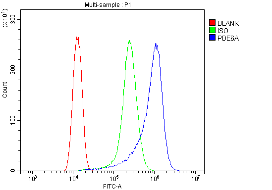
(Figure 6. Flow Cytometry analysis of HELA cells using anti-PDE6 alpha/PDE6A antibody (AAA19318).Overlay histogram showing HELA cells stained with AAA19318 (Blue line). The cells were blocked with 10% normal goat serum. And then incubated with rabbit anti-PDE6 alpha/PDE6A Antibody (AAA19318, 1μg/1x106 cells) for 30 min at 20 degree C. DyLight®488 conjugated goat anti-rabbit IgG (5-10μg/1x106 cells) was used as secondary antibody for 30 minutes at 20 degree C. Isotype control antibody (Green line) was rabbit IgG (1μg/1x106) used under the same conditions. Unlabelled sample (Red line) was also used as a control.)
FCM (Flow Cytometry)
(Figure 6. Flow Cytometry analysis of HELA cells using anti-PDE6 alpha/PDE6A antibody (AAA19318).Overlay histogram showing HELA cells stained with AAA19318 (Blue line). The cells were blocked with 10% normal goat serum. And then incubated with rabbit anti-PDE6 alpha/PDE6A Antibody (AAA19318, 1μg/1x106 cells) for 30 min at 20 degree C. DyLight®488 conjugated goat anti-rabbit IgG (5-10μg/1x106 cells) was used as secondary antibody for 30 minutes at 20 degree C. Isotype control antibody (Green line) was rabbit IgG (1μg/1x106) used under the same conditions. Unlabelled sample (Red line) was also used as a control.)
PDE6 alpha/PDE6A, Polyclonal Antibody (Cat# AAA19318)
Full Name
Anti-PDE6 alpha/PDE6A Antibody
Gene Names
PDE6A; PDEA; RP43; CGPR-A; PDE6A
Reactivity
Human, Mouse, Rat
Applications
WB, IHC-P, ICC, IF, FC/FACS/FCM, EIA
Purity
Immunogen affinity purified.
Pricing


 WB (Western Blot)
(WB Suggested Anti-TRAK1 Antibody Titration: 0.2-1 ug/mlELISA Titer: 1:1562500Positive Control: HT1080 cell lysateThere is BioGPS gene expression data showing that TRAK1 is expressed in HT1080)
WB (Western Blot)
(WB Suggested Anti-TRAK1 Antibody Titration: 0.2-1 ug/mlELISA Titer: 1:1562500Positive Control: HT1080 cell lysateThere is BioGPS gene expression data showing that TRAK1 is expressed in HT1080)
TRAK1, Polyclonal Antibody (Cat# AAA23554)
Full Name
TRAK1 antibody - middle region
Gene Names
TRAK1; MILT1; EIEE68; OIP106
Reactivity
Cow, Dog, Horse, Human, Pig, Rabbit, Rat
Applications
Western Blot
Purity
Affinity Purified
Pricing


 FCM (Flow Cytometry)
(Flow cytometric analysis of HepG2 cells with Glutathione Synthetase antibody at 1/100 dilution (red) compared with an unlabelled control (cells without incubation with primary antibody; black). Alexa Fluor 488-conjugated goat anti rabbit IgG was used as the secondary antibody.)
FCM (Flow Cytometry)
(Flow cytometric analysis of HepG2 cells with Glutathione Synthetase antibody at 1/100 dilution (red) compared with an unlabelled control (cells without incubation with primary antibody; black). Alexa Fluor 488-conjugated goat anti rabbit IgG was used as the secondary antibody.)
Glutathione Synthetase, Monoclonal Antibody (Cat# AAA30462)
Full Name
Glutathione Synthetase Antibody
Gene Names
GSS; GSHS; HEL-S-64p; HEL-S-88n
Reactivity
Human, Mouse, Rat
Applications
WB, ICC, IF, IHC, FC/FACS
Purity
ProA affinity purified
Pricing


 IHC (Immunohistchemistry)
(Figure 6. IHC analysis of GALE using anti-GALE antibody (AAA19135).GALE was detected in paraffin-embedded section of rat kidney tissue. Heat mediated antigen retrieval was performed in citrate buffer (pH6, epitope retrieval solution) for 20 mins. The tissue section was blocked with 10% goat serum. The tissue section was then incubated with 1ug/ml rabbit anti-GALE Antibody (AAA19135) overnight at 4 degree C. Biotinylated goat anti-rabbit IgG was used as secondary antibody and incubated for 30 minutes at 37 degree C. The tissue section was developed using Strepavidin-Biotin-Complex (SABC) with DAB as the chromogen.)
IHC (Immunohistchemistry)
(Figure 6. IHC analysis of GALE using anti-GALE antibody (AAA19135).GALE was detected in paraffin-embedded section of rat kidney tissue. Heat mediated antigen retrieval was performed in citrate buffer (pH6, epitope retrieval solution) for 20 mins. The tissue section was blocked with 10% goat serum. The tissue section was then incubated with 1ug/ml rabbit anti-GALE Antibody (AAA19135) overnight at 4 degree C. Biotinylated goat anti-rabbit IgG was used as secondary antibody and incubated for 30 minutes at 37 degree C. The tissue section was developed using Strepavidin-Biotin-Complex (SABC) with DAB as the chromogen.)
GALE, Polyclonal Antibody (Cat# AAA19135)
Full Name
Anti-GALE Picoband antibody
Gene Names
GALE; SDR1E1
Reactivity
Human, Mouse, Rat
No cross reactivity with other proteins.
No cross reactivity with other proteins.
Applications
EIA, IHC, WB
Pricing


 FCM (Flow Cytometry)
(Figure 8. Flow Cytometry analysis of C6 cells using anti-Clathrin heavy chain/CLTC antibody (AAA19270).Overlay histogram showing C6 cells stained with AAA19270 (Blue line). The cells were blocked with 10% normal goat serum. And then incubated with rabbit anti-Clathrin heavy chain/CLTC Antibody (AAA19270,1μg/1x106 cells) for 30 min at 20 degree C. DyLight®488 conjugated goat anti-rabbit IgG (5-10μg/1x106 cells) was used as secondary antibody for 30 minutes at 20 degree C. Isotype control antibody (Green line) was rabbit IgG (1μg/1x106) used under the same conditions. Unlabelled sample (Red line) was also used as a control.)
FCM (Flow Cytometry)
(Figure 8. Flow Cytometry analysis of C6 cells using anti-Clathrin heavy chain/CLTC antibody (AAA19270).Overlay histogram showing C6 cells stained with AAA19270 (Blue line). The cells were blocked with 10% normal goat serum. And then incubated with rabbit anti-Clathrin heavy chain/CLTC Antibody (AAA19270,1μg/1x106 cells) for 30 min at 20 degree C. DyLight®488 conjugated goat anti-rabbit IgG (5-10μg/1x106 cells) was used as secondary antibody for 30 minutes at 20 degree C. Isotype control antibody (Green line) was rabbit IgG (1μg/1x106) used under the same conditions. Unlabelled sample (Red line) was also used as a control.)
Clathrin heavy chain/CLTC, Polyclonal Antibody (Cat# AAA19270)
Full Name
Anti-Clathrin heavy chain/CLTC Antibody
Gene Names
CLTC; Hc; CHC; CHC17; CLH-17; CLTCL2
Reactivity
Human, Mouse, Rat
Applications
WB, IHC-P, ICC, IF, FC/FACS/FCM, EIA
Purity
Immunogen affinity purified.
Pricing


 FCM (Flow Cytometry)
(Figure 7. Flow Cytometry analysis of PC-3 cells using anti-PVRL1/NECTIN1 antibody (AAA19303).Overlay histogram showing PC-3 cells stained with AAA19303 (Blue line). The cells were blocked with 10% normal goat serum. And then incubated with rabbit anti-PVRL1/NECTIN1 Antibody (AAA19303, 1μg/1x106 cells) for 30 min at 20 degree C. DyLight®488 conjugated goat anti-rabbit IgG (5-10μg/1x106 cells) was used as secondary antibody for 30 minutes at 20 degree C. Isotype control antibody (Green line) was rabbit IgG (1μg/1x106) used under the same conditions. Unlabelled sample (Red line) was also used as a control.)
FCM (Flow Cytometry)
(Figure 7. Flow Cytometry analysis of PC-3 cells using anti-PVRL1/NECTIN1 antibody (AAA19303).Overlay histogram showing PC-3 cells stained with AAA19303 (Blue line). The cells were blocked with 10% normal goat serum. And then incubated with rabbit anti-PVRL1/NECTIN1 Antibody (AAA19303, 1μg/1x106 cells) for 30 min at 20 degree C. DyLight®488 conjugated goat anti-rabbit IgG (5-10μg/1x106 cells) was used as secondary antibody for 30 minutes at 20 degree C. Isotype control antibody (Green line) was rabbit IgG (1μg/1x106) used under the same conditions. Unlabelled sample (Red line) was also used as a control.)
PVRL1/NECTIN1, Polyclonal Antibody (Cat# AAA19303)
Full Name
Anti-PVRL1/NECTIN1 Antibody
Gene Names
PVRL1; ED4; PRR; HIgR; HVEC; OFC7; PRR1; PVRR; CD111; PVRR1; SK-12; CLPED1; nectin-1
Reactivity
Human, Mouse, Rat
Applications
WB, IHC-P, FC/FACS/FCM, EIA
Purity
Immunogen affinity purified.
Pricing


 FCM (Flow Cytometry)
(Figure 9. Flow Cytometry analysis of THP-1 cells using anti-CHCHD10 antibody (AAA19310).Overlay histogram showing THP-1 cells stained with AAA19310 (Blue line). The cells were blocked with 10% normal goat serum. And then incubated with rabbit anti-CHCHD10 Antibody (AAA19310, 1μg/1x106 cells) for 30 min at 20 degree C. DyLight®488 conjugated goat anti-rabbit IgG (5-10μg/1x106 cells) was used as secondary antibody for 30 minutes at 20 degree C. Isotype control antibody (Green line) was rabbit IgG (1μg/1x106) used under the same conditions. Unlabelled sample (Red line) was also used as a control.)
FCM (Flow Cytometry)
(Figure 9. Flow Cytometry analysis of THP-1 cells using anti-CHCHD10 antibody (AAA19310).Overlay histogram showing THP-1 cells stained with AAA19310 (Blue line). The cells were blocked with 10% normal goat serum. And then incubated with rabbit anti-CHCHD10 Antibody (AAA19310, 1μg/1x106 cells) for 30 min at 20 degree C. DyLight®488 conjugated goat anti-rabbit IgG (5-10μg/1x106 cells) was used as secondary antibody for 30 minutes at 20 degree C. Isotype control antibody (Green line) was rabbit IgG (1μg/1x106) used under the same conditions. Unlabelled sample (Red line) was also used as a control.)
CHCHD10, Polyclonal Antibody (Cat# AAA19310)
Full Name
Anti-CHCHD10 Antibody
Gene Names
CHCHD10; FTDALS2; N27C7-4; C22orf16
Reactivity
Human, Mouse, Rat, Monkey
Applications
WB, IHC-P, ICC, IF, FC/FACS/FCM, EIA
Purity
Immunogen affinity purified.
Pricing


 FCM (Flow Cytometry)
(Flow cytometric analysis of Hela cells with VAMP8 antibody at 1/50 dilution (red) compared with an unlabelled control (cells without incubation with primary antibody; black). Alexa Fluor 488-conjugated goat anti rabbit IgG was used as the secondary antibody.)
FCM (Flow Cytometry)
(Flow cytometric analysis of Hela cells with VAMP8 antibody at 1/50 dilution (red) compared with an unlabelled control (cells without incubation with primary antibody; black). Alexa Fluor 488-conjugated goat anti rabbit IgG was used as the secondary antibody.)
VAMP8, Monoclonal Antibody (Cat# AAA30266)
Full Name
VAMP8 Antibody
Gene Names
VAMP8; EDB; VAMP-8
Reactivity
Human, Mouse, Rat
Applications
Western Blot, Immunocytochemistry, Immunofluorescence, Immunohistochemistry, Immunoprecipitation, Flow Cytometry
Purity
ProA affinity purified
Pricing


 FCM (Flow Cytometry)
(Flow cytometric analysis of Jurkat cells with NCK1 antibody at 1/50 dilution (red) compared with an unlabelled control (cells without incubation with primary antibody; black). Alexa Fluor 488-conjugated goat anti rabbit IgG was used as the secondary antibody.)
FCM (Flow Cytometry)
(Flow cytometric analysis of Jurkat cells with NCK1 antibody at 1/50 dilution (red) compared with an unlabelled control (cells without incubation with primary antibody; black). Alexa Fluor 488-conjugated goat anti rabbit IgG was used as the secondary antibody.)
NCK1, Monoclonal Antibody (Cat# AAA30418)
Full Name
NCK1 Antibody
Gene Names
NCK1; NCK; nck-1; NCKalpha
Reactivity
Human, Mouse, Rat
Applications
Western Blot, Immunocytochemistry, Immunofluorescence, Immunohistochemistry, Immunoprecipitation, Flow Cytometry
Purity
ProA affinity purified
Pricing


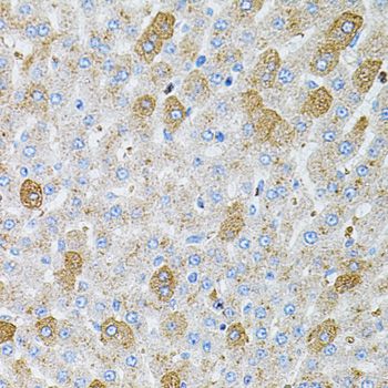
 IHC (Immunohistochemistry)
(Figure 7. IHC analysis of IDH2 using anti-IDH2 antibody (AAA11640).IDH2 was detected in frozen section of rat small intestine tissue. Heat mediated antigen retrieval was performed in citrate buffer (pH6, epitope retrieval solution) for 20 mins. The tissue section was blocked with 10% goat serum. The tissue section was then incubated with 1ug/ml rabbit anti-IDH2 Antibody (AAA11640) overnight at 4 degree C. Biotinylated goat anti-rabbit IgG was used as secondary antibody and incubated for 30 minutes at 37 degree C. The tissue section was developed using Strepavidin-Biotin-Complex (SABC) with DAB as the chromogen.)
IHC (Immunohistochemistry)
(Figure 7. IHC analysis of IDH2 using anti-IDH2 antibody (AAA11640).IDH2 was detected in frozen section of rat small intestine tissue. Heat mediated antigen retrieval was performed in citrate buffer (pH6, epitope retrieval solution) for 20 mins. The tissue section was blocked with 10% goat serum. The tissue section was then incubated with 1ug/ml rabbit anti-IDH2 Antibody (AAA11640) overnight at 4 degree C. Biotinylated goat anti-rabbit IgG was used as secondary antibody and incubated for 30 minutes at 37 degree C. The tissue section was developed using Strepavidin-Biotin-Complex (SABC) with DAB as the chromogen.)
IDH2, Polyclonal Antibody (Cat# AAA11640)
Full Name
Anti-IDH2 Antibody
Gene Names
IDH2; IDH; IDP; IDHM; IDPM; ICD-M; D2HGA2; mNADP-IDH
Reactivity
Human, Mouse, Rat
Applications
Western Blot, Immunohistochemistry
Purity
Immunogen Affinity Purified
Pricing


 FCM (Flow Cytometry)
(Figure 7. Flow Cytometry analysis of CACO-2 cells using anti-Glycine decarboxylase/GLDC antibody (AAA19300).Overlay histogram showing CACO-2 cells stained with AAA19300 (Blue line). The cells were blocked with 10% normal goat serum. And then incubated with rabbit anti-Glycine decarboxylase/GLDC Antibody (AAA19300, 1μg/1x106 cells) for 30 min at 20 degree C. DyLight®488 conjugated goat anti-rabbit IgG (5-10μg/1x106 cells) was used as secondary antibody for 30 minutes at 20 degree C. Isotype control antibody (Green line) was rabbit IgG (1μg/1x106) used under the same conditions. Unlabelled sample (Red line) was also used as a control.)
FCM (Flow Cytometry)
(Figure 7. Flow Cytometry analysis of CACO-2 cells using anti-Glycine decarboxylase/GLDC antibody (AAA19300).Overlay histogram showing CACO-2 cells stained with AAA19300 (Blue line). The cells were blocked with 10% normal goat serum. And then incubated with rabbit anti-Glycine decarboxylase/GLDC Antibody (AAA19300, 1μg/1x106 cells) for 30 min at 20 degree C. DyLight®488 conjugated goat anti-rabbit IgG (5-10μg/1x106 cells) was used as secondary antibody for 30 minutes at 20 degree C. Isotype control antibody (Green line) was rabbit IgG (1μg/1x106) used under the same conditions. Unlabelled sample (Red line) was also used as a control.)
Glycine decarboxylase/GLDC, Polyclonal Antibody (Cat# AAA19300)
Full Name
Anti-Glycine decarboxylase/GLDC Antibody
Gene Names
GLDC; GCE; GCSP; HYGN1
Reactivity
Human, Mouse, Rat
Applications
WB, IHC-P, FC/FACS/FCM, EIA
Purity
Immunogen affinity purified.
Pricing