Filters
Clonality
Type
Reactivity
Gene Name
Isotype
Host
Application
Clone
1050 results for " Signal " - showing 950-1000
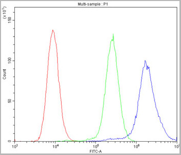
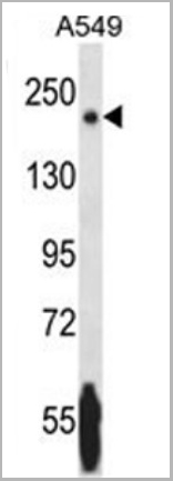
 FCM (Flow Cytometry)
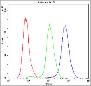
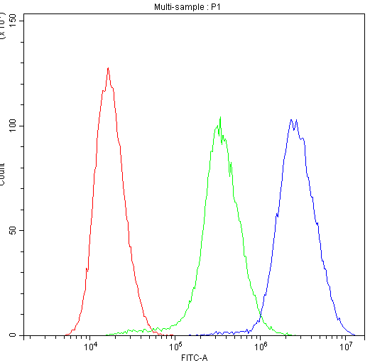
(Figure 6. Flow Cytometry analysis of A549 cells using anti-MDR3 antibody (AAA11652).Overlay histogram showing A549 cells stained with AAA11652 (Blue line).The cells were blocked with 10% normal goat serum. And then incubated with rabbit anti-MDR3 Antibody (AAA11652,1ug/1x106 cells) for 30 min at 20°C. DyLight®488 conjugated goat anti-rabbit IgG (5-10ug/1x106 cells) was used as secondary antibody for 30 minutes at 20°C. Isotype control antibody (Green line) was rabbit IgG (1ug/1x106) used under the same conditions. Unlabelled sample (Red line) was also used as a control.)
FCM (Flow Cytometry)
(Figure 6. Flow Cytometry analysis of A549 cells using anti-MDR3 antibody (AAA11652).Overlay histogram showing A549 cells stained with AAA11652 (Blue line).The cells were blocked with 10% normal goat serum. And then incubated with rabbit anti-MDR3 Antibody (AAA11652,1ug/1x106 cells) for 30 min at 20°C. DyLight®488 conjugated goat anti-rabbit IgG (5-10ug/1x106 cells) was used as secondary antibody for 30 minutes at 20°C. Isotype control antibody (Green line) was rabbit IgG (1ug/1x106) used under the same conditions. Unlabelled sample (Red line) was also used as a control.)
ABCB4, Polyclonal Antibody (Cat# AAA11652)
Full Name
Anti-ABCB4 Antibody
Gene Names
ABCB4; GBD1; ICP3; MDR2; MDR3; PGY3; ABC21; MDR2/3; PFIC-3
Reactivity
Human, Mouse, Rat
Applications
Western Blot, Immunohistochemistry, Immunofluorescence, Flow Cytometry
Purity
Immunogen Affinity Purified
Pricing
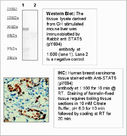
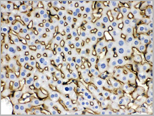
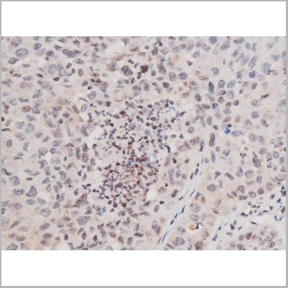
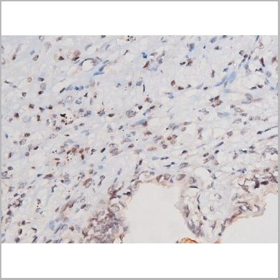
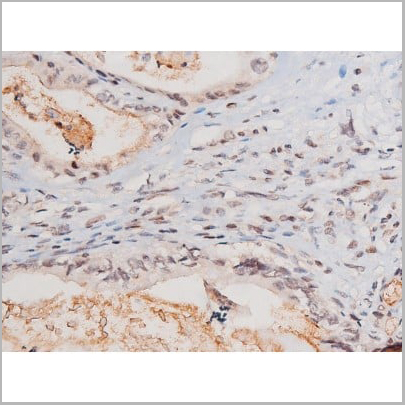
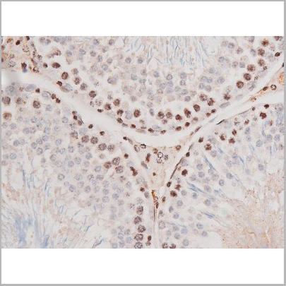
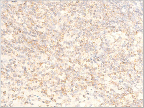
 IHC (Immunohistochemistry)
(Immunohistochemistry: Human breast carcinoma tissue (FFPE) stained with Anti-STAT5 (pY694) (Cat# AAA14089) antibody at 1:100 for 10 min @ RT. Staining of formalin-fixed tissue requires boiling tissue sections in 10 mM Citrate Buffer, pH 6.0 for 10 min followed by cooling at RT for 20 min.)
IHC (Immunohistochemistry)
(Immunohistochemistry: Human breast carcinoma tissue (FFPE) stained with Anti-STAT5 (pY694) (Cat# AAA14089) antibody at 1:100 for 10 min @ RT. Staining of formalin-fixed tissue requires boiling tissue sections in 10 mM Citrate Buffer, pH 6.0 for 10 min followed by cooling at RT for 20 min.)
STAT5 (pY694), Antibody (Cat# AAA14089)
Full Name
Rabbit anti Phospho- STAT5 (pTyr 694)
Reactivity
Human, Rat, Mouse
Applications
Western Blot, Immunohistochemistry, Immunoprecipitation
Purity
The Rabbit IgG is purified by site-modified Epitope Affinity Purification.
Pricing
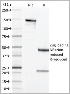
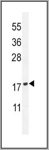
 SDS-PAGE
(3 ug by SDS-PAGE under reducing condition and visualized by coomasie blue stain)
SDS-PAGE
(3 ug by SDS-PAGE under reducing condition and visualized by coomasie blue stain)
CLC, Recombinant Protein (Cat# AAA11808)
Full Name
CLC, 1-142aa, Human, His tag, E Coli
Gene Names
CLC; GAL10; Gal-10; LGALS10; LGALS10A; LPPL_HUMAN
Applications
SDS-PAGE
Purity
> 90% by SDS-PAGE
Pricing
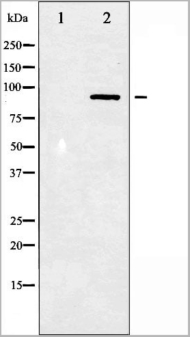
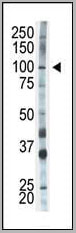
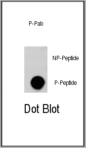
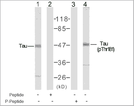
 WB (Western Blot)
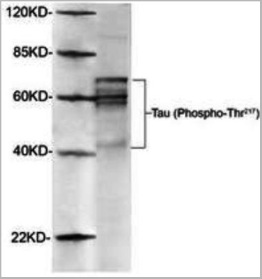
(Western Blot of Rat brain using AAA14727 (1ug/mL). The signal was developed with IRDyeTM800 conjugated giat anti-rabbit IgG)
WB (Western Blot)
(Western Blot of Rat brain using AAA14727 (1ug/mL). The signal was developed with IRDyeTM800 conjugated giat anti-rabbit IgG)
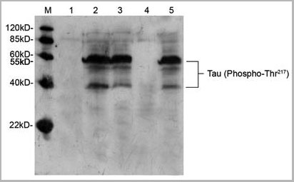
Tau, Polyclonal Antibody (Cat# AAA14727)
Full Name
Tau, phosphorylated (Thr217) (Tau Protein, Microtubule-associated Protein, MAP)
Gene Names
tau; BcDNA:RE16764; CG12881; CG31057; CG5606; DmelCG31057; dtau
Reactivity
Human
Applications
Western Blot
Purity
Purified by Affinity chromatography.
Pricing
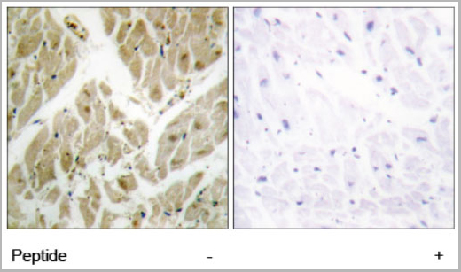
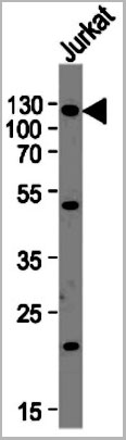
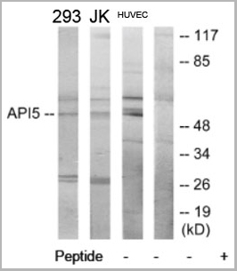 WB (Western Blot)
(Western blot analysis of extracts from 293 cells, Jurkat cells and HUVEC cells, using API-5 antibody.)
WB (Western Blot)
(Western blot analysis of extracts from 293 cells, Jurkat cells and HUVEC cells, using API-5 antibody.)
API-5, Antibody (Cat# AAA17963)
Full Name
API-5 Antibody
Gene Names
API5; AAC11; AAC-11
Reactivity
Human, Mouse
Applications
Western Blot, Immunohistochemistry
Purity
The antibody was affinity-purified from rabbit antiserum by affinity-chromatography using epitope-specific immunogen.
Pricing
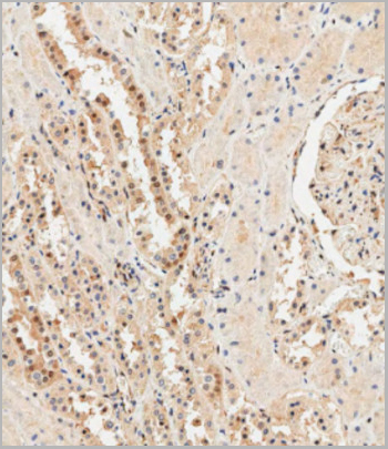
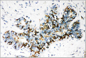
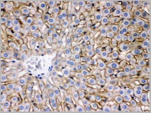
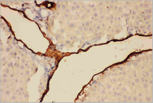
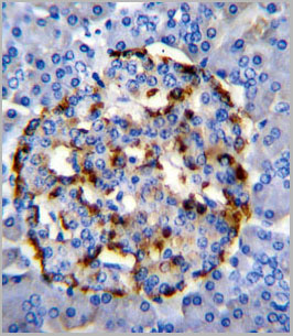
 IHC (Immunohistchemistry)
(Immunohistochemical analysis of paraffin-embedded Human breast carcinoma tissue using AAA28688 performed on the Leica® BOND RXm. Tissue was fixed with formaldehyde at room temperature, antigen retrieval was by heat mediation with a EDTA buffer (pH9. 0). Samples were incubated with primary antibody(1:500) for 1 hours at room temperature. A undiluted biotinylated CRF Anti-Polyvalent HRP Polymer antibody was used as the secondary antibody)
IHC (Immunohistchemistry)
(Immunohistochemical analysis of paraffin-embedded Human breast carcinoma tissue using AAA28688 performed on the Leica® BOND RXm. Tissue was fixed with formaldehyde at room temperature, antigen retrieval was by heat mediation with a EDTA buffer (pH9. 0). Samples were incubated with primary antibody(1:500) for 1 hours at room temperature. A undiluted biotinylated CRF Anti-Polyvalent HRP Polymer antibody was used as the secondary antibody)
Ubiquitin, Polyclonal Antibody (Cat# AAA28688)
Full Name
Ubiquitin Antibody (N-term)
Gene Names
UBC; HMG20
Reactivity
Human \Human, Mouse, Rat
Predicted Reactivity: Bovine, Drosophila, Monkey, Pig, Sheep, Chicken, Horse, Hamster, Xenopus, Rabbit
Predicted Reactivity: Bovine, Drosophila, Monkey, Pig, Sheep, Chicken, Horse, Hamster, Xenopus, Rabbit
Applications
Western Blot, Immunohistochemistry
Purity
This antibody is purified through a protein A column, followed by peptide affinity purification.
Pricing
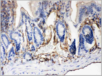
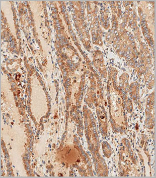
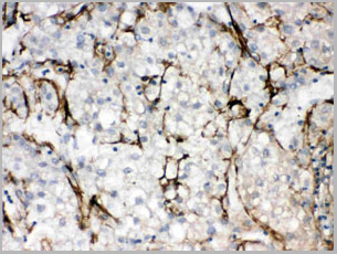
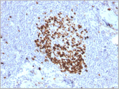
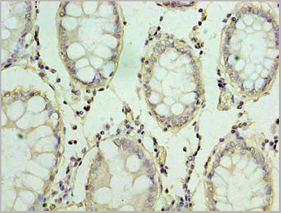
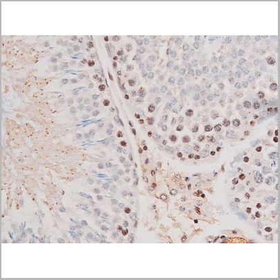
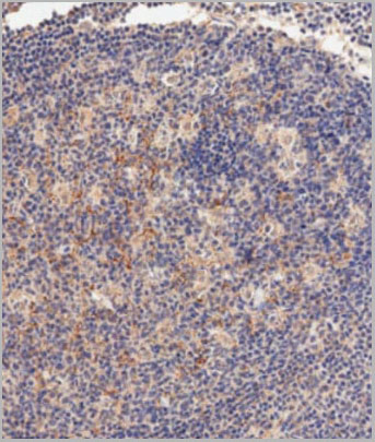
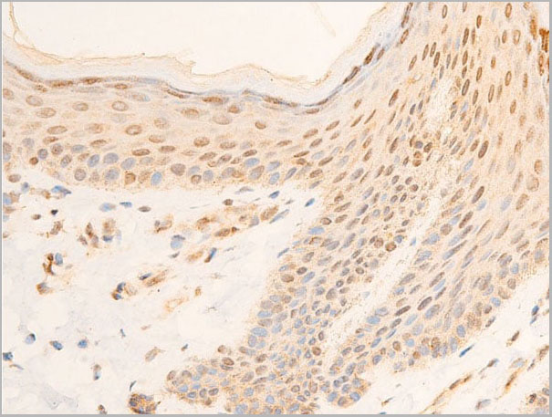
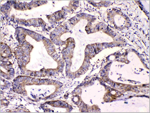
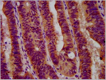 IHC (Immunohistochemistry)
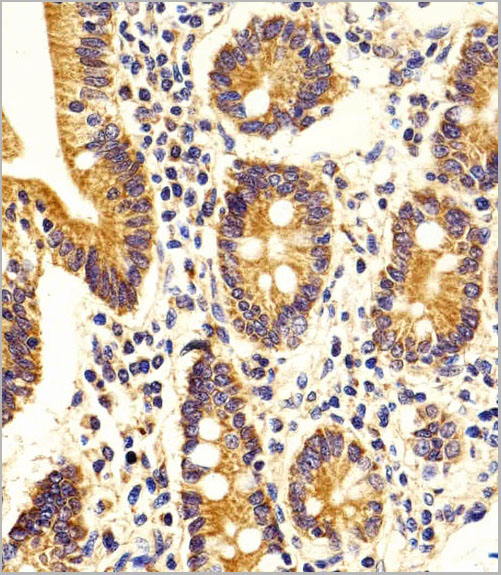
(IHC image of AAA27004 diluted at 1:600 and staining in paraffin-embedded human colon cancer performed on a Leica BondTM system. After dewaxing and hydration, antigen retrieval was mediated by high pressure in a citrate buffer (pH 6.0). Section was blocked with 10% normal goat serum 30min at RT. Then primary antibody (1% BSA) was incubated at 4 degree C overnight. The primary is detected by a biotinylated secondary antibody and visualized using an HRP conjugated SP system.)
IHC (Immunohistochemistry)
(IHC image of AAA27004 diluted at 1:600 and staining in paraffin-embedded human colon cancer performed on a Leica BondTM system. After dewaxing and hydration, antigen retrieval was mediated by high pressure in a citrate buffer (pH 6.0). Section was blocked with 10% normal goat serum 30min at RT. Then primary antibody (1% BSA) was incubated at 4 degree C overnight. The primary is detected by a biotinylated secondary antibody and visualized using an HRP conjugated SP system.)
GBA, Polyclonal Antibody (Cat# AAA27004)
Full Name
GBA Antibody
Gene Names
GBA; GCB; GBA1; GLUC
Reactivity
Human
Applications
Western Blot, Immunohistochemistry
Purity
>95%, Protein G purified
Pricing
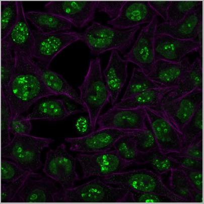
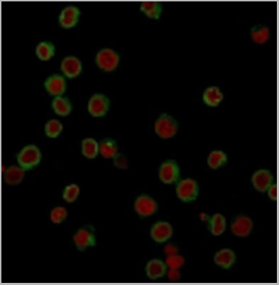
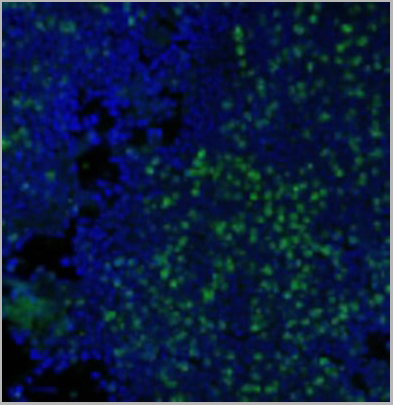
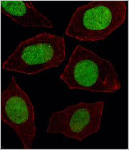
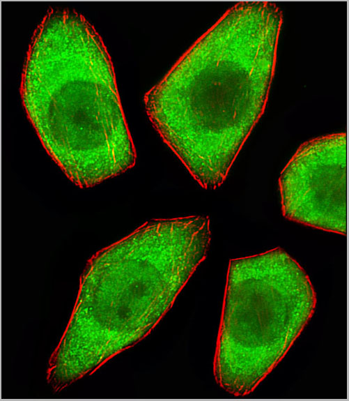
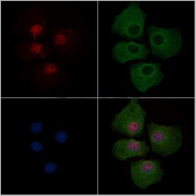
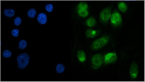
 IF (Immunofluorescence)
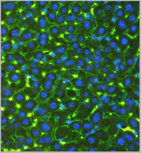
(Fluorescent confocal image of Hela cell stained with NRK Antibody (Center) (AAA28689). Hela cells were fixed with 4% PFA (20 min), permeabilized with Triton X-100 (0.1%, 10 min), then incubated with NRK primary antibody (1:25, 1 h at 37 degree). For secondary antibody, Alexa Fluor 488 conjugated donkey anti-rabbit antibody (green) was used (1:400, 50 min at 37 degree).Cytoplasmic actin was counterstained with Alexa Fluor 555 (red) conjugated Phalloidin (7units/ml, 1 h at 37 degree). Nuclei were counterstained with DAPI (blue) (10 ug/ml, 10 min). NRK immunoreactivity is localized to Cytoplasm and Nucleus significantly.)
IF (Immunofluorescence)
(Fluorescent confocal image of Hela cell stained with NRK Antibody (Center) (AAA28689). Hela cells were fixed with 4% PFA (20 min), permeabilized with Triton X-100 (0.1%, 10 min), then incubated with NRK primary antibody (1:25, 1 h at 37 degree). For secondary antibody, Alexa Fluor 488 conjugated donkey anti-rabbit antibody (green) was used (1:400, 50 min at 37 degree).Cytoplasmic actin was counterstained with Alexa Fluor 555 (red) conjugated Phalloidin (7units/ml, 1 h at 37 degree). Nuclei were counterstained with DAPI (blue) (10 ug/ml, 10 min). NRK immunoreactivity is localized to Cytoplasm and Nucleus significantly.)
NRK, Polyclonal Antibody (Cat# AAA28689)
Full Name
NRK Antibody (Center)
Gene Names
NRK; NESK
Reactivity
Human
Applications
Western Blot, Immunofluorescence
Purity
This antibody is purified through a protein A column, followed by peptide affinity purification.
Pricing
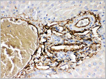
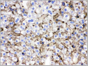
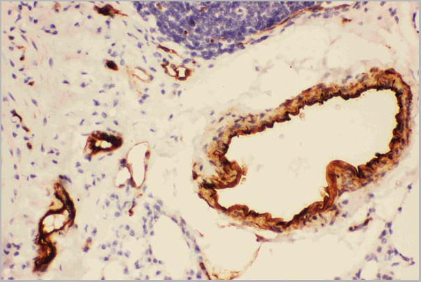
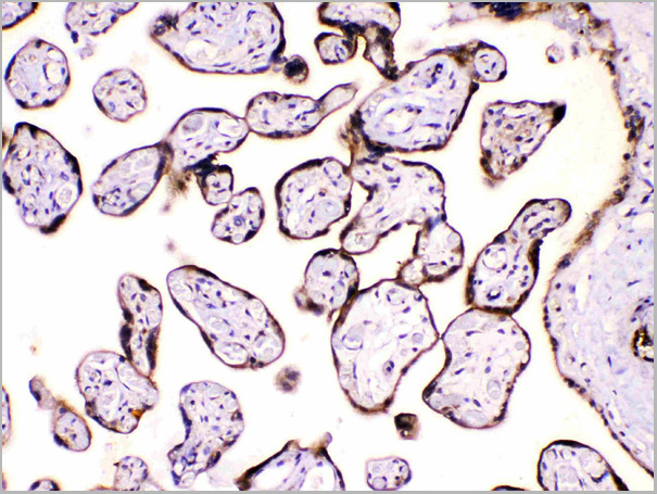
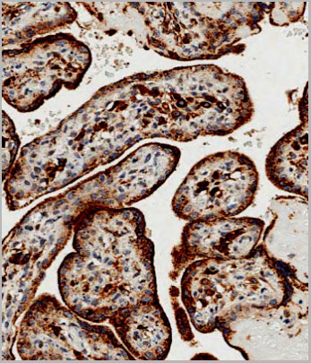 IHC (Immunohistchemistry)
(Immunohistochemical analysis of paraffin-embedded Human placenta tissue using AAA28695 performed on the Leica® BOND RXm. Tissue was fixed with formaldehyde at room temperature, antigen retrieval was by heat mediation with a EDTA buffer (pH9. 0). Samples were incubated with primary antibody(1:500) for 1 hours at room temperature. A undiluted biotinylated CRF Anti-Polyvalent HRP Polymer antibody was used as the secondary antibody.)
IHC (Immunohistchemistry)
(Immunohistochemical analysis of paraffin-embedded Human placenta tissue using AAA28695 performed on the Leica® BOND RXm. Tissue was fixed with formaldehyde at room temperature, antigen retrieval was by heat mediation with a EDTA buffer (pH9. 0). Samples were incubated with primary antibody(1:500) for 1 hours at room temperature. A undiluted biotinylated CRF Anti-Polyvalent HRP Polymer antibody was used as the secondary antibody.)
GAA, Polyclonal Antibody (Cat# AAA28695)
Full Name
GAA Antibody (N-term)
Gene Names
GAA; LYAG
Reactivity
Human
Applications
Western Blot, Immunohistochemistry
Purity
Peptide Affinity Purified Rabbit Polyclonal Antibody (Pab)
Pricing
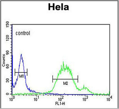
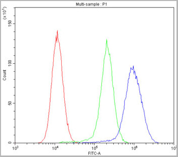
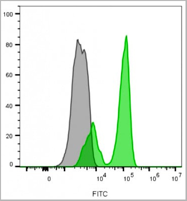
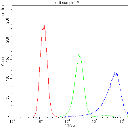
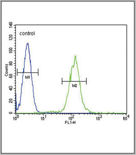
 FCM (Flow Cytometry)
(Overlay histogram showing Hela cells stained with AAA28750 (green line). The cells were fixed with 2% paraformaldehyde (10 min) and then permeabilized with 90% methanol for 10 min. The cells were then icubated in 2% bovine serum albumin to block non-specific protein-protein interactions followed by the antibody (AAA28750, 1:25 dilution) for 60 min at 37ºC. The secondary antibody used was Goat-Anti-Rabbit IgG, DyLight® 488 Conjugated Highly Cross-Adsorbed (1583138) at 1/200 dilution for 40 min at 37°C. Isotype control antibody (blue line) was rabbit IgG1 (1ug/1x106 cells) used under the same conditions. Acquisition of >10, 000 events wasperformed.)
FCM (Flow Cytometry)
(Overlay histogram showing Hela cells stained with AAA28750 (green line). The cells were fixed with 2% paraformaldehyde (10 min) and then permeabilized with 90% methanol for 10 min. The cells were then icubated in 2% bovine serum albumin to block non-specific protein-protein interactions followed by the antibody (AAA28750, 1:25 dilution) for 60 min at 37ºC. The secondary antibody used was Goat-Anti-Rabbit IgG, DyLight® 488 Conjugated Highly Cross-Adsorbed (1583138) at 1/200 dilution for 40 min at 37°C. Isotype control antibody (blue line) was rabbit IgG1 (1ug/1x106 cells) used under the same conditions. Acquisition of >10, 000 events wasperformed.)
WNT5A, Polyclonal Antibody (Cat# AAA28750)
Full Name
WNT5A Antibody (Center)
Gene Names
WNT5A; hWNT5A
Reactivity
Human, Mouse, Rat
Predicted: Rabbit
Predicted: Rabbit
Applications
Immunohistochemistry, Immunohistochemistry, Flow Cytometry, Western Blot
Purity
This antibody is purified through a protein A column, followed by peptide affinity purification.
Pricing
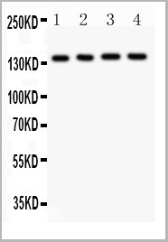
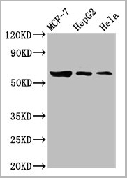
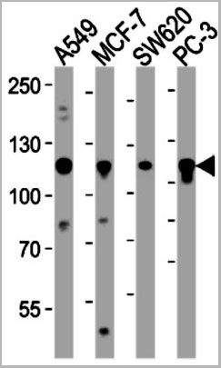
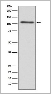
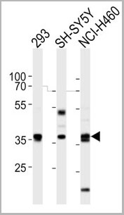
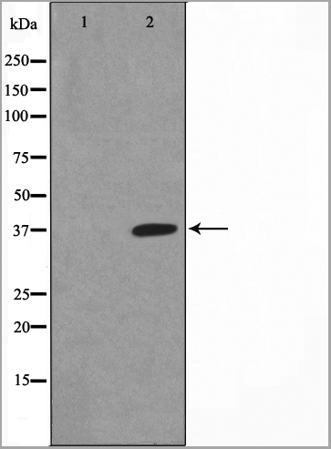
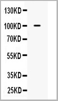
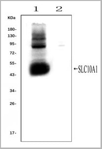 WB (Western Blot)
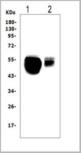
(Western blot analysis of SLC10A1 using anti- SLC10A1 antibody (AAA11653).Electrophoresis was performed on a 5-20% SDS-PAGE gel at 70V (Stacking gel) / 90V (Resolving gel) for 2-3 hours. The sample well of each lane was loaded with 50ug of sample under reducing conditions.Lane 1: rat liver tissue lysates (positive control), Lane 2: rat kidney tissue lysates, (negative control)After Electrophoresis, proteins were transferred to a Nitrocellulose membrane at 150mA for 50-90 minutes. Blocked the membrane with 5% Non-fat Milk/ TBS for 1.5 hour at RT. The membrane was incubated with rabbit anti-SLC10A1 antigen affinity purified polyclonal antibody (AAA11653) at 0.25ug/mL overnight at 4°C, then washed with TBS-0.1%Tween 3 times with 5 minutes each and probed with a goat anti-rabbit IgG-HRP secondary antibody at a dilution of 1:10000 for 1.5 hour at RT. The signal is developed using an Enhanced Chemiluminescent detection (ECL) kit with Tanon 5200 system. A specific band was detected for SLC10A1 at approximately 50KD. The expected band size for SLC10A1 is at 38KD.)
WB (Western Blot)
(Western blot analysis of SLC10A1 using anti- SLC10A1 antibody (AAA11653).Electrophoresis was performed on a 5-20% SDS-PAGE gel at 70V (Stacking gel) / 90V (Resolving gel) for 2-3 hours. The sample well of each lane was loaded with 50ug of sample under reducing conditions.Lane 1: rat liver tissue lysates (positive control), Lane 2: rat kidney tissue lysates, (negative control)After Electrophoresis, proteins were transferred to a Nitrocellulose membrane at 150mA for 50-90 minutes. Blocked the membrane with 5% Non-fat Milk/ TBS for 1.5 hour at RT. The membrane was incubated with rabbit anti-SLC10A1 antigen affinity purified polyclonal antibody (AAA11653) at 0.25ug/mL overnight at 4°C, then washed with TBS-0.1%Tween 3 times with 5 minutes each and probed with a goat anti-rabbit IgG-HRP secondary antibody at a dilution of 1:10000 for 1.5 hour at RT. The signal is developed using an Enhanced Chemiluminescent detection (ECL) kit with Tanon 5200 system. A specific band was detected for SLC10A1 at approximately 50KD. The expected band size for SLC10A1 is at 38KD.)
SLC10A1, Polyclonal Antibody (Cat# AAA11653)
Full Name
Anti-SLC10A1 Antibody
Gene Names
Slc10a1; Ntcp
Reactivity
Mouse, Rat
Applications
Flow Cytometry, Immunofluorescence, Immunohistochemistry, Immunohistochemistry, Immunocytochemistry, Western Blot
Purity
Immunogen affinity purified.
Pricing
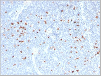
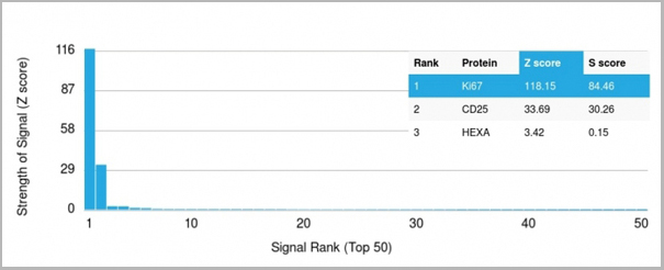 Application Data
(Analysis of Protein Array containing more than 19,000 full-length human proteins using Ki67 Mouse Monoclonal Antibody (MKI67/2466). Z- and S- Score: The Z-score represents the strength of a signal that a monoclonal antibody (MAb) (in combination with a fluorescently-tagged anti-IgG secondary antibody) produces when binding to a particular protein on the HuProtTM array. Z-scores are described in units of standard deviations (SD's) above the mean value of all signals generated on that array. If targets on HuProtTM are arranged in descending order of the Z-score, the S-score is the difference (also in units of SD's) between the Z-score. S-score therefore represents the relative target specificity of a MAb to its intended target. A MAb is considered to specific to its intended target, if the MAb has an S-score of at least 2.5. For example, if a MAb binds to protein X with a Z-score of 43 and to protein Y with a Z-score of 14, then the S-score for the binding of that MAb to protein X is equal to 29.)
Application Data
(Analysis of Protein Array containing more than 19,000 full-length human proteins using Ki67 Mouse Monoclonal Antibody (MKI67/2466). Z- and S- Score: The Z-score represents the strength of a signal that a monoclonal antibody (MAb) (in combination with a fluorescently-tagged anti-IgG secondary antibody) produces when binding to a particular protein on the HuProtTM array. Z-scores are described in units of standard deviations (SD's) above the mean value of all signals generated on that array. If targets on HuProtTM are arranged in descending order of the Z-score, the S-score is the difference (also in units of SD's) between the Z-score. S-score therefore represents the relative target specificity of a MAb to its intended target. A MAb is considered to specific to its intended target, if the MAb has an S-score of at least 2.5. For example, if a MAb binds to protein X with a Z-score of 43 and to protein Y with a Z-score of 14, then the S-score for the binding of that MAb to protein X is equal to 29.)
Ki-67, Monoclonal Antibody (Cat# AAA23909)
Full Name
Ki-67 (Proliferating Cell Marker)
Gene Names
MKI67; KIA; MIB-; MIB-1; PPP1R105
Reactivity
Human. Others not known.
Applications
Flow Cytometry, Immunofluorescence, Immunohistochemistry
Purity
Purified Ab with BSA and Azide at 200ug/ml OR Purified Ab WITHOUT BSA and Azide at 1.0mg/ml
Pricing


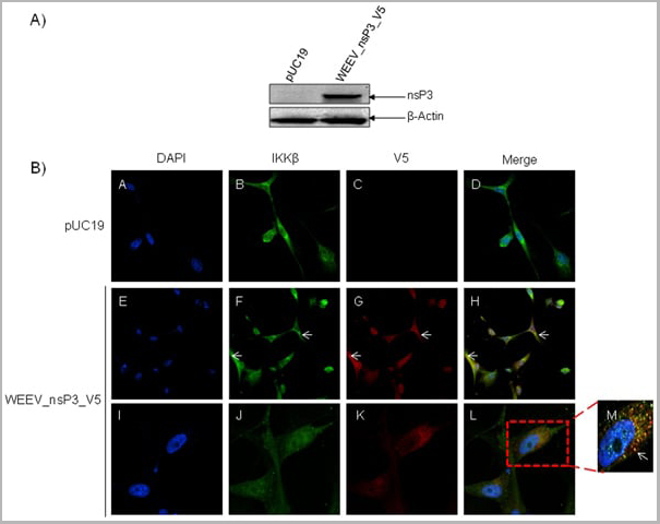
 Application Data
(Published customer image: Mouse anti V5 tag antibody, clone SV5-Pk1 used for the detection of V5 tagged WEEV_nsP3 protein by western blotting and immunofluorescenceImage caption: WEEV nsP3 interaction with host IKKbeta. A) U87MGs were transfected in a 6-well plate with 5 ug of pUC19 and WEEV_nsP3_HA for 24 hours. Cell lysates were resolved using SDS-PAGE and subsequently immunoblotted with V5 antibody and beta-actin served as a loading control. B) U87MGs were transfected with WEEV_nsP3_V5; cells were fixed after 24 hours and stained with antibodies against the endogenous IKKbeta and the V5 tag. Cells were incubated with appropriate secondary Alexa Fluor antibodies and the nuclei stained with DAPI. Co-localization of IKKbeta with WEEV_nsP3_V5 (yellow) was observed as shown by the arrows. B) Panels E -H serve as an example of transfected cells in a given field of view that show co-localization of IKKbeta and WEEV_nsP3_V5 24 hours post transfection. Panels I-L represent magnified images of other cells showing co-localization of IKKbeta and WEEV_nsP3_V5. Panel M is a magnified image of panel L. The co-localization was confirmed by Z-stack analysis. Co-localization was calculated to be approximately in 61% of cells (163 cells were counted of which 44% demonstrated expression of nsP3. Of those cells that expressed nsP3, 61% showed co-localization of both proteins). Images were taken using Nikon Eclipse TE2000-U at 60x magnification and are representative of 2 independent experiments.From: Amaya M, Voss K, Sampey G, Senina S, de la Fuente C, et al. (2014) The Role of IKKbeta in Venezuelan Equine Encephalitis Virus Infection. PLoS ONE 9(2): e86745.)
Application Data
(Published customer image: Mouse anti V5 tag antibody, clone SV5-Pk1 used for the detection of V5 tagged WEEV_nsP3 protein by western blotting and immunofluorescenceImage caption: WEEV nsP3 interaction with host IKKbeta. A) U87MGs were transfected in a 6-well plate with 5 ug of pUC19 and WEEV_nsP3_HA for 24 hours. Cell lysates were resolved using SDS-PAGE and subsequently immunoblotted with V5 antibody and beta-actin served as a loading control. B) U87MGs were transfected with WEEV_nsP3_V5; cells were fixed after 24 hours and stained with antibodies against the endogenous IKKbeta and the V5 tag. Cells were incubated with appropriate secondary Alexa Fluor antibodies and the nuclei stained with DAPI. Co-localization of IKKbeta with WEEV_nsP3_V5 (yellow) was observed as shown by the arrows. B) Panels E -H serve as an example of transfected cells in a given field of view that show co-localization of IKKbeta and WEEV_nsP3_V5 24 hours post transfection. Panels I-L represent magnified images of other cells showing co-localization of IKKbeta and WEEV_nsP3_V5. Panel M is a magnified image of panel L. The co-localization was confirmed by Z-stack analysis. Co-localization was calculated to be approximately in 61% of cells (163 cells were counted of which 44% demonstrated expression of nsP3. Of those cells that expressed nsP3, 61% showed co-localization of both proteins). Images were taken using Nikon Eclipse TE2000-U at 60x magnification and are representative of 2 independent experiments.From: Amaya M, Voss K, Sampey G, Senina S, de la Fuente C, et al. (2014) The Role of IKKbeta in Venezuelan Equine Encephalitis Virus Infection. PLoS ONE 9(2): e86745.)
V5-TAG, Monoclonal Antibody (Cat# AAA11864)
Full Name
MOUSE ANTI V5-TAG:FITC
Applications
Immunofluorescence
Pricing


 Application Data
(Published customer image: Mouse anti V5 tag antibody, clone SV5-Pk1 used for the detection of V5 tagged WEEV_nsP3 protein by western blotting and immunofluorescenceImage caption: WEEV nsP3 interaction with host IKKbeta. A) U87MGs were transfected in a 6-well plate with 5 ug of pUC19 and WEEV_nsP3_HA for 24 hours. Cell lysates were resolved using SDS-PAGE and subsequently immunoblotted with V5 antibody and beta-actin served as a loading control. B) U87MGs were transfected with WEEV_nsP3_V5; cells were fixed after 24 hours and stained with antibodies against the endogenous IKKbeta and the V5 tag. Cells were incubated with appropriate secondary Alexa Fluor antibodies and the nuclei stained with DAPI. Co-localization of IKKbeta with WEEV_nsP3_V5 (yellow) was observed as shown by the arrows. B) Panels E -H serve as an example of transfected cells in a given field of view that show co-localization of IKKbeta and WEEV_nsP3_V5 24 hours post transfection. Panels I-L represent magnified images of other cells showing co-localization of IKKbeta and WEEV_nsP3_V5. Panel M is a magnified image of panel L. The co-localization was confirmed by Z-stack analysis. Co-localization was calculated to be approximately in 61% of cells (163 cells were counted of which 44% demonstrated expression of nsP3. Of those cells that expressed nsP3, 61% showed co-localization of both proteins). Images were taken using Nikon Eclipse TE2000-U at 60x magnification and are representative of 2 independent experiments.From: Amaya M, Voss K, Sampey G, Senina S, de la Fuente C, et al. (2014) The Role of IKKbeta in Venezuelan Equine Encephalitis Virus Infection. PLoS ONE 9(2): e86745.)
Application Data
(Published customer image: Mouse anti V5 tag antibody, clone SV5-Pk1 used for the detection of V5 tagged WEEV_nsP3 protein by western blotting and immunofluorescenceImage caption: WEEV nsP3 interaction with host IKKbeta. A) U87MGs were transfected in a 6-well plate with 5 ug of pUC19 and WEEV_nsP3_HA for 24 hours. Cell lysates were resolved using SDS-PAGE and subsequently immunoblotted with V5 antibody and beta-actin served as a loading control. B) U87MGs were transfected with WEEV_nsP3_V5; cells were fixed after 24 hours and stained with antibodies against the endogenous IKKbeta and the V5 tag. Cells were incubated with appropriate secondary Alexa Fluor antibodies and the nuclei stained with DAPI. Co-localization of IKKbeta with WEEV_nsP3_V5 (yellow) was observed as shown by the arrows. B) Panels E -H serve as an example of transfected cells in a given field of view that show co-localization of IKKbeta and WEEV_nsP3_V5 24 hours post transfection. Panels I-L represent magnified images of other cells showing co-localization of IKKbeta and WEEV_nsP3_V5. Panel M is a magnified image of panel L. The co-localization was confirmed by Z-stack analysis. Co-localization was calculated to be approximately in 61% of cells (163 cells were counted of which 44% demonstrated expression of nsP3. Of those cells that expressed nsP3, 61% showed co-localization of both proteins). Images were taken using Nikon Eclipse TE2000-U at 60x magnification and are representative of 2 independent experiments.From: Amaya M, Voss K, Sampey G, Senina S, de la Fuente C, et al. (2014) The Role of IKKbeta in Venezuelan Equine Encephalitis Virus Infection. PLoS ONE 9(2): e86745.)
V5-TAG, Monoclonal Antibody (Cat# AAA11930)
Full Name
MOUSE ANTI V5-TAG
Applications
Immunohistochemistry, Flow Cytometry, Immunofluorescence, Immunoprecipitation, Western Blot, Radioimmunoassay
Pricing
TCR gamma delta, Monoclonal Antibody (Cat# AAA17922)
Full Name
TCR gamma delta antibody
Reactivity
Recognizes the gamma/delta T-cell receptor (TCR).
Applications
Flow Cytometry, Immunohistochemistry, Immunoprecipitation
Pricing
DNA (Double Stranded) (dsDNA), Antibody (Cat# AAA13410)
Full Name
Human anti dsDNA
Applications
Immunofluorescence
Purity
Delipidized and defibrinated, then the immunoglobulin fraction is precipitated and collected.
Pricing
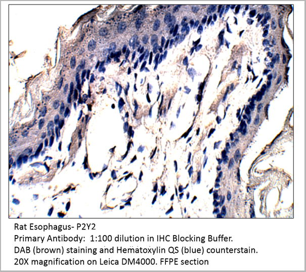 IHC (Immunohistochemistry)
P2Y2, Polyclonal Antibody (Cat# AAA14402)
Full Name
P2Y2 Antibody
Gene Names
P2RY2; P2U; HP2U; P2U1; P2UR; P2Y2; P2RU1; P2Y2R
Reactivity
Cat, Human, Mouse, Rat
Applications
Confocal Microscopy, Immunocytochemistry, Immunofluorescence, Immunohistochemistry, Immunoprecipitation, Western Blot
Pricing

 Assay Principle
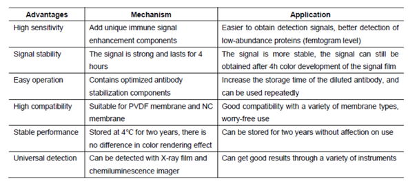
(After years of development on chemiluminescence technology, there have been a variety of luminescence systems, but the most commonly used one in laboratories is still a technology system based on Luminol or its derivatives (isoluminol, etc.). Western luminescent detection reagent ECL immunoblotting chemiluminescence solution is a non-radioactive (horseradish peroxidase) luminescence system designed to detect femtogram-level trace proteins immobilized on a solid membrane (such as NC, PVDF, etc.). It is an experimental auxiliary reagent for the photosensitive recording of its immunoblotting by X-ray film (radiograph).)
Assay Principle
(After years of development on chemiluminescence technology, there have been a variety of luminescence systems, but the most commonly used one in laboratories is still a technology system based on Luminol or its derivatives (isoluminol, etc.). Western luminescent detection reagent ECL immunoblotting chemiluminescence solution is a non-radioactive (horseradish peroxidase) luminescence system designed to detect femtogram-level trace proteins immobilized on a solid membrane (such as NC, PVDF, etc.). It is an experimental auxiliary reagent for the photosensitive recording of its immunoblotting by X-ray film (radiograph).)
West Femto Maximum Sensitivity Substrate, Reagent (Cat# AAA31545)
Full Name
West Femto Maximum Sensitivity Substrate
Applications
Western Blot, Immunoprecipitation
Pricing
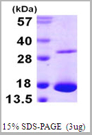
 SDS-PAGE
(3ug by SDS-PAGE under reducing condition and visualized by coomasie blue stain)
SDS-PAGE
(3ug by SDS-PAGE under reducing condition and visualized by coomasie blue stain)
PTP-1B (Protein Tyrosine Phosphatase1B), Active Protein (Cat# AAA11740)
Full Name
PTP-1B (Protein Tyrosine Phosphatase1B), 1-321aa, Human, E Coli (Bioactivity Validated)
Gene Names
PTPN1; PTP1B
Applications
Enzyme Activity, SDS-PAGE
Purity
> 95% by SDS-PAGE
Pricing
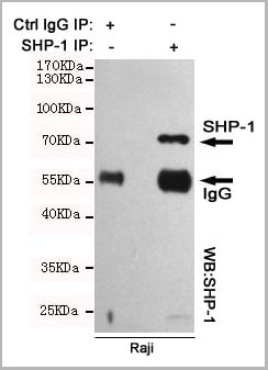 IP (Immunoprecipitation)
(Immunoprecipitation analysis of Raji cell lysates using SHP-1 mouse mAb.)
IP (Immunoprecipitation)
(Immunoprecipitation analysis of Raji cell lysates using SHP-1 mouse mAb.)
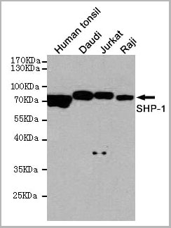
SHP-1, Monoclonal Antibody (Cat# AAA14102)
Full Name
Anti-SHP-1 Mouse mAb
Gene Names
PTPN6; HCP; HCPH; SHP1; SHP-1; HPTP1C; PTP-1C; SHP-1L; SH-PTP1
Reactivity
Human
Applications
Western Blot, Immunoprecipitation
Purity
Affinity purified
Pricing
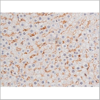
 WB (Western Blot)
(Western blot analysis of C3 expression in HepG2 cell lysate (AAA11693). Electrophoresis was performed on a 5-20% SDS-PAGE gel at 70V (stacking gel) / 90V (resolving gel) for 2-3 hours. The sample well of each lane was loaded with 50ug of sample under reducing conditions. After Electrophoresis, proteins were transferred to a Nitrocellulose membrane at 150mA for 50-90 minutes. Blocked the membrane with 5% Non-fat Milk/TBS for 1.5 hour at RT. The membrane was incubated with rabbit anti-C3 monoclonal antibody (AAA11693) overnight at 4°C, then washed with TBS-0.1%Tween 3 times with 5 minutes each and probed with goat anti-rabbit IgG-HRP secondary antibody at a dilution of 1:10000 for 1.5 hour at RT. The signal is developed using an Enhanced Chemiluminescent detection (ECL) kit (please inquire) with Tanon 5200 system. A specific band was detected for C3.)
WB (Western Blot)
(Western blot analysis of C3 expression in HepG2 cell lysate (AAA11693). Electrophoresis was performed on a 5-20% SDS-PAGE gel at 70V (stacking gel) / 90V (resolving gel) for 2-3 hours. The sample well of each lane was loaded with 50ug of sample under reducing conditions. After Electrophoresis, proteins were transferred to a Nitrocellulose membrane at 150mA for 50-90 minutes. Blocked the membrane with 5% Non-fat Milk/TBS for 1.5 hour at RT. The membrane was incubated with rabbit anti-C3 monoclonal antibody (AAA11693) overnight at 4°C, then washed with TBS-0.1%Tween 3 times with 5 minutes each and probed with goat anti-rabbit IgG-HRP secondary antibody at a dilution of 1:10000 for 1.5 hour at RT. The signal is developed using an Enhanced Chemiluminescent detection (ECL) kit (please inquire) with Tanon 5200 system. A specific band was detected for C3.)
C3/Complement C3, Monoclonal Antibody (Cat# AAA11693)
Full Name
Anti-C3/Complement C3 Rabbit Monoclonal Antibody
Gene Names
C3; ASP; C3a; C3b; AHUS5; ARMD9; CPAMD1; HEL-S-62p
Reactivity
Human
Applications
Flow Cytometry, Immunofluorescence, Immunocytochemistry, Western Blot
Purity
Affinity-chromatography
Pricing
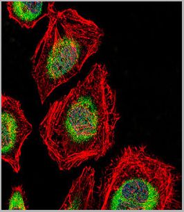
 IF (Immunofluorescence)
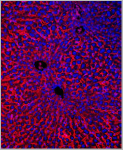
(Immunofluorescence Analysis of methanol-fixed human tonsil cryosection stained with CF488A CD3e clone RIV9 (green), mounted in EverBrite’ Mounting Medium with DAPI (nuclei, blue).)
IF (Immunofluorescence)
(Immunofluorescence Analysis of methanol-fixed human tonsil cryosection stained with CF488A CD3e clone RIV9 (green), mounted in EverBrite’ Mounting Medium with DAPI (nuclei, blue).)
CD3, Monoclonal Antibody (Cat# AAA13839)
Full Name
CD3 (T-Cell Marker) Mouse Monoclonal Antibody
Gene Names
CD3E; T3E; TCRE; IMD18
Reactivity
Human, Mouse, Rat
Applications
Functional Assay, Flow Cytometry, Immunofluorescence
Pricing
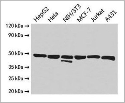
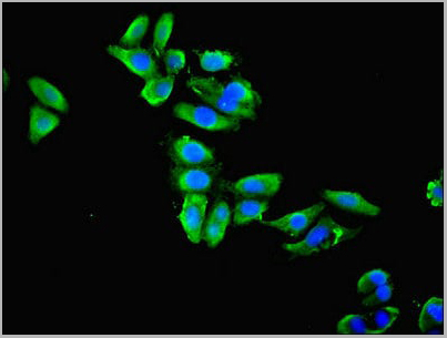 IF (Immunofluorescence)
(Immunofluorescent analysis of HepG2 cells cells using AAA15913 at a dilution of 1:100 and Alexa Fluor 488-congugated AffiniPure Goat Anti-Rabbit IgG(H+L))
IF (Immunofluorescence)
(Immunofluorescent analysis of HepG2 cells cells using AAA15913 at a dilution of 1:100 and Alexa Fluor 488-congugated AffiniPure Goat Anti-Rabbit IgG(H+L))
Alpha-enolase, Polyclonal Antibody (Cat# AAA15913)
Full Name
Rabbit anti- human Alpha-enolase polyclonal Antibody
Gene Names
ENO1; NNE; PPH; MPB1; ENO1L1; HEL-S-17
Reactivity
Human, Mouse
Applications
Western Blot, Immunohistochemistry, Immunofluorescence
Purity
>95%, Protein G purified
Pricing
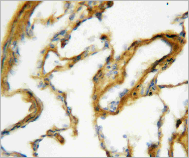
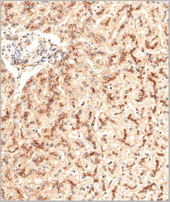
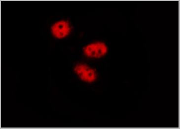
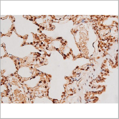
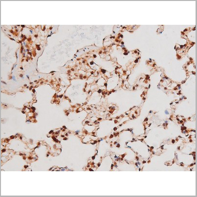
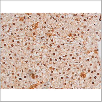
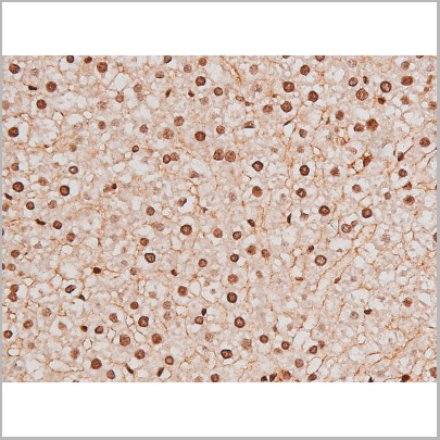
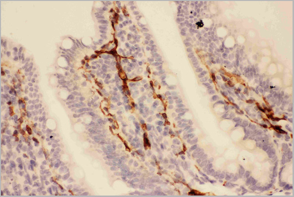
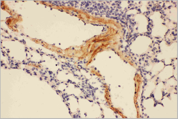
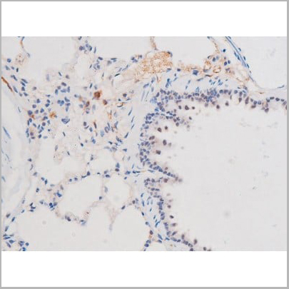
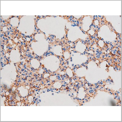
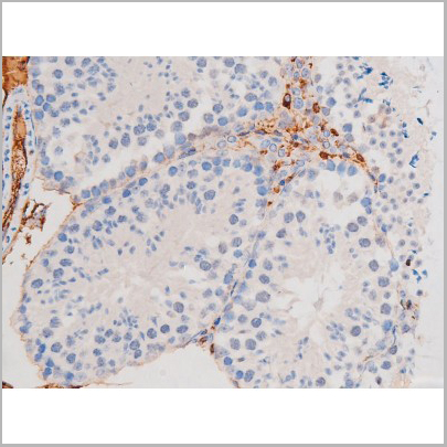
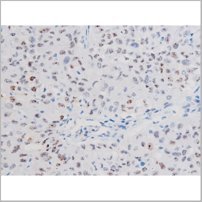
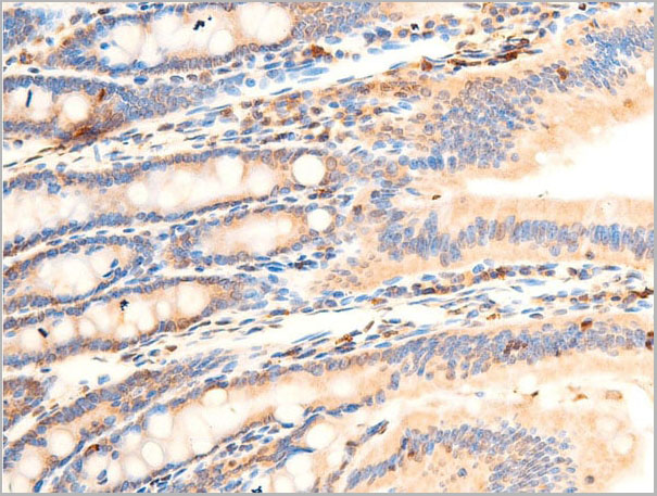
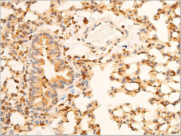
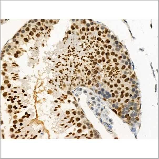
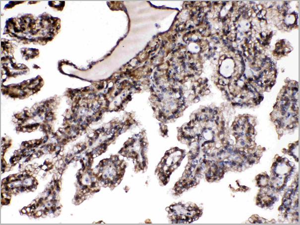
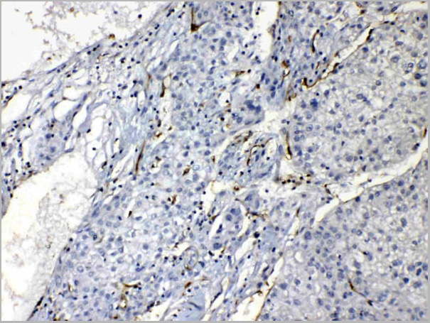
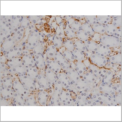
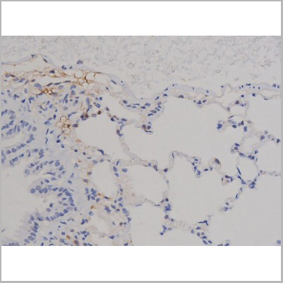
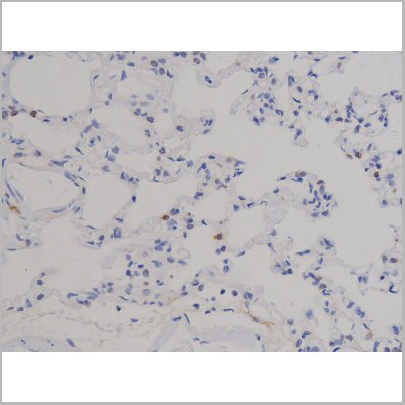
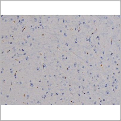
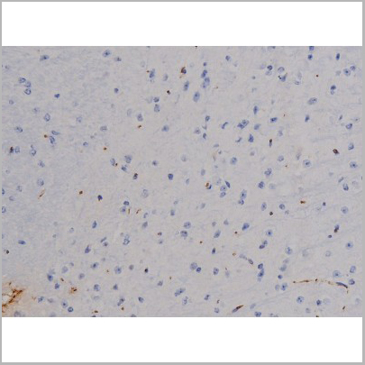
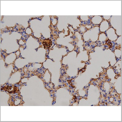
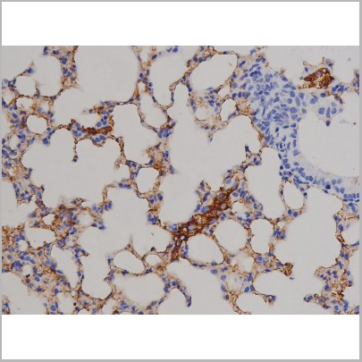
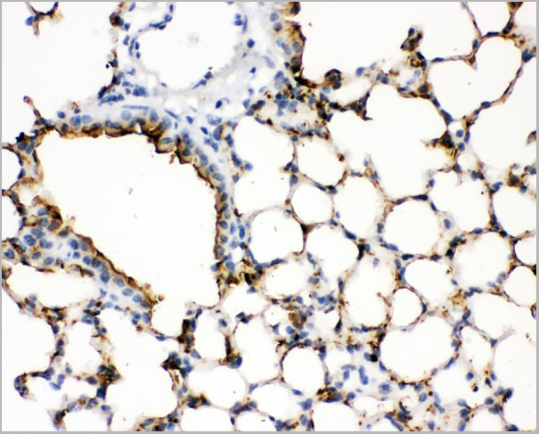 IHC (Immunohistochemistry)
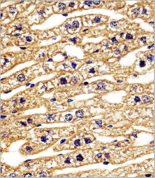
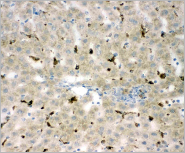
(Figure 3. IHC analysis of SFTPA1 using anti- SFTPA1 antibody (AAA11521).SFTPA1 was detected in paraffin-embedded section of mouse lung tissues. Heat mediated antigen retrieval was performed in citrate buffer (pH6, epitope retrieval solution) for 20 mins. The tissue section was blocked with 10% goat serum. The tissue section was then incubated with 1ug/ml rabbit anti- SFTPA1 Antibody (AAA11521) overnight at 4 degree C. Biotinylated goat anti-rabbit IgG was used as secondary antibody and incubated for 30 minutes at 37 degree C. The tissue section was developed using Strepavidin-Biotin-Complex (SABC) with DAB as the chromogen.)
IHC (Immunohistochemistry)
(Figure 3. IHC analysis of SFTPA1 using anti- SFTPA1 antibody (AAA11521).SFTPA1 was detected in paraffin-embedded section of mouse lung tissues. Heat mediated antigen retrieval was performed in citrate buffer (pH6, epitope retrieval solution) for 20 mins. The tissue section was blocked with 10% goat serum. The tissue section was then incubated with 1ug/ml rabbit anti- SFTPA1 Antibody (AAA11521) overnight at 4 degree C. Biotinylated goat anti-rabbit IgG was used as secondary antibody and incubated for 30 minutes at 37 degree C. The tissue section was developed using Strepavidin-Biotin-Complex (SABC) with DAB as the chromogen.)
Surfactant Protein A, Polyclonal Antibody (Cat# AAA11521)
Full Name
Polyclonal Anti-SFTPA1 Antibody
Gene Names
Sftpa1; SP-A; Sftp1; Sftp-1
Reactivity
Mouse, Rat
Applications
Western Blot, Immunohistochemistry
Purity
Immunogen affinity purified.
Pricing

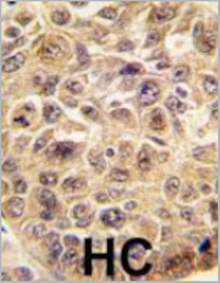
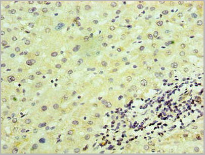
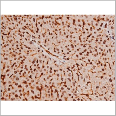
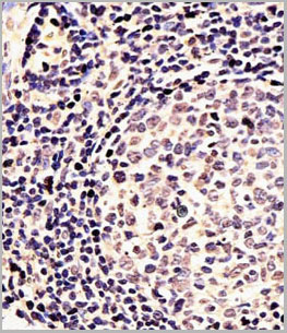
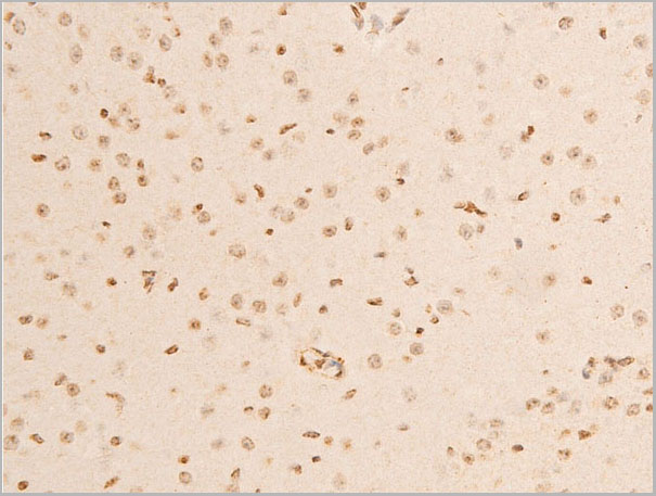
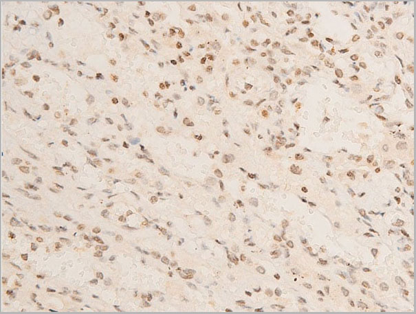
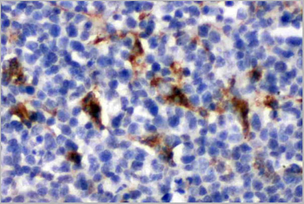
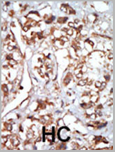 IHC (Immunohistochemistry)
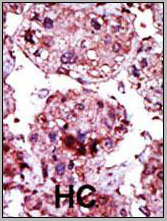
(Formalin-fixed and paraffin-embedded human cancer tissue reacted with the primary antibody, which was peroxidase-conjugated to the secondary antibody, followed by AEC staining. This data demonstrates the use of this antibody for immunohistochemistry; clinical relevance has not been evaluated. BC = breast carcinoma; HC = hepatocarcinoma.)
IHC (Immunohistochemistry)
(Formalin-fixed and paraffin-embedded human cancer tissue reacted with the primary antibody, which was peroxidase-conjugated to the secondary antibody, followed by AEC staining. This data demonstrates the use of this antibody for immunohistochemistry; clinical relevance has not been evaluated. BC = breast carcinoma; HC = hepatocarcinoma.)
HRI, Polyclonal Antibody (Cat# AAA28727)
Full Name
HRI Antibody (N-term)
Gene Names
EIF2AK1; HCR; HRI
Reactivity
Human
Applications
Western Blot, Immunohistochemistry
Purity
Purified Rabbit Polyclonal Antibody (Pab)
Pricing
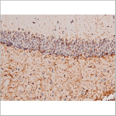
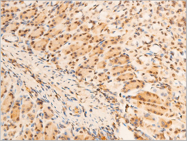
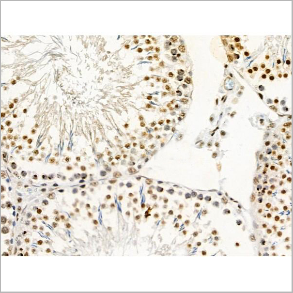
 IHC-P (Immunohistochemistry Paraffin)
(AAA31041 at 1/100 staining Mouse colon tissue by IHC-P. The sample was formaldehyde fixed and a heat mediated antigen retrieval step in citrate buffer was performed. The sample was then blocked and incubated with the primary antibody at 4°C overnight. An HRP conjugated anti-Rabbit antibody was use das the secondary antibody.)
IHC-P (Immunohistochemistry Paraffin)
(AAA31041 at 1/100 staining Mouse colon tissue by IHC-P. The sample was formaldehyde fixed and a heat mediated antigen retrieval step in citrate buffer was performed. The sample was then blocked and incubated with the primary antibody at 4°C overnight. An HRP conjugated anti-Rabbit antibody was use das the secondary antibody.)
STAT3, Polyclonal Antibody (Cat# AAA31041)
Full Name
Phospho-STAT3 (Tyr705) Antibody
Gene Names
STAT3; APRF; HIES; ADMIO; ADMIO1
Reactivity
Human, Mouse, Rat
Applications
Western Blot, Immunohistochemistry, Immunofluorescence, Immunoprecipitation, Peptide ELISA
Purity
Peptide affinity purification.
Pricing


 Application Data
(Published customer image: Mouse anti V5 tag antibody, clone SV5-Pk1 used for the detection of V5 tagged WEEV_nsP3 protein by western blotting and immunofluorescenceImage caption: WEEV nsP3 interaction with host IKKbeta. A) U87MGs were transfected in a 6-well plate with 5 ug of pUC19 and WEEV_nsP3_HA for 24 hours. Cell lysates were resolved using SDS-PAGE and subsequently immunoblotted with V5 antibody and beta-actin served as a loading control. B) U87MGs were transfected with WEEV_nsP3_V5; cells were fixed after 24 hours and stained with antibodies against the endogenous IKKbeta and the V5 tag. Cells were incubated with appropriate secondary Alexa Fluor antibodies and the nuclei stained with DAPI. Co-localization of IKKbeta with WEEV_nsP3_V5 (yellow) was observed as shown by the arrows. B) Panels E -H serve as an example of transfected cells in a given field of view that show co-localization of IKKbeta and WEEV_nsP3_V5 24 hours post transfection. Panels I-L represent magnified images of other cells showing co-localization of IKKbeta and WEEV_nsP3_V5. Panel M is a magnified image of panel L. The co-localization was confirmed by Z-stack analysis. Co-localization was calculated to be approximately in 61% of cells (163 cells were counted of which 44% demonstrated expression of nsP3. Of those cells that expressed nsP3, 61% showed co-localization of both proteins). Images were taken using Nikon Eclipse TE2000-U at 60x magnification and are representative of 2 independent experiments.From: Amaya M, Voss K, Sampey G, Senina S, de la Fuente C, et al. (2014) The Role of IKKbeta in Venezuelan Equine Encephalitis Virus Infection. PLoS ONE 9(2): e86745.)
Application Data
(Published customer image: Mouse anti V5 tag antibody, clone SV5-Pk1 used for the detection of V5 tagged WEEV_nsP3 protein by western blotting and immunofluorescenceImage caption: WEEV nsP3 interaction with host IKKbeta. A) U87MGs were transfected in a 6-well plate with 5 ug of pUC19 and WEEV_nsP3_HA for 24 hours. Cell lysates were resolved using SDS-PAGE and subsequently immunoblotted with V5 antibody and beta-actin served as a loading control. B) U87MGs were transfected with WEEV_nsP3_V5; cells were fixed after 24 hours and stained with antibodies against the endogenous IKKbeta and the V5 tag. Cells were incubated with appropriate secondary Alexa Fluor antibodies and the nuclei stained with DAPI. Co-localization of IKKbeta with WEEV_nsP3_V5 (yellow) was observed as shown by the arrows. B) Panels E -H serve as an example of transfected cells in a given field of view that show co-localization of IKKbeta and WEEV_nsP3_V5 24 hours post transfection. Panels I-L represent magnified images of other cells showing co-localization of IKKbeta and WEEV_nsP3_V5. Panel M is a magnified image of panel L. The co-localization was confirmed by Z-stack analysis. Co-localization was calculated to be approximately in 61% of cells (163 cells were counted of which 44% demonstrated expression of nsP3. Of those cells that expressed nsP3, 61% showed co-localization of both proteins). Images were taken using Nikon Eclipse TE2000-U at 60x magnification and are representative of 2 independent experiments.From: Amaya M, Voss K, Sampey G, Senina S, de la Fuente C, et al. (2014) The Role of IKKbeta in Venezuelan Equine Encephalitis Virus Infection. PLoS ONE 9(2): e86745.)
V5-TAG, Monoclonal Antibody (Cat# AAA12211)
Full Name
MOUSE ANTI V5-TAG
Applications
Immunohistochemistry, Flow Cytometry, Immunofluorescence, Immunoprecipitation, Western Blot, Radioimmunoassay
Pricing
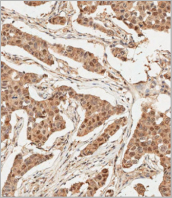
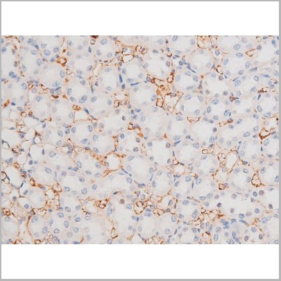
 IHC (Immunohistochemistry)
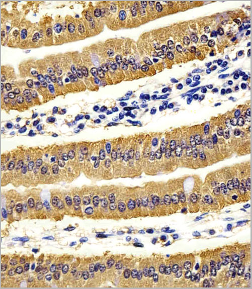
(Immunohistochemical analysis of paraffin-embedded human tonsil tissue using AAA28679 performed on the Leica® BOND RXm. Samples were incubated with primary antibody(1/500) for 1 hours at room temperature. A undiluted biotinylated CRF Anti-Polyvalent HRP Polymer antibody was used as the secondary antibody.)
IHC (Immunohistochemistry)
(Immunohistochemical analysis of paraffin-embedded human tonsil tissue using AAA28679 performed on the Leica® BOND RXm. Samples were incubated with primary antibody(1/500) for 1 hours at room temperature. A undiluted biotinylated CRF Anti-Polyvalent HRP Polymer antibody was used as the secondary antibody.)
HK1 (Hexokinase), Polyclonal Antibody (Cat# AAA28679)
Full Name
HK1 (Hexokinase) Antibody (N-term)
Gene Names
HK1; HKD; HKI; HXK1; HMSNR; HK1-ta; HK1-tb; HK1-tc
Reactivity
Human, Rat
Applications
Western Blot, Immunohistochemistry
Purity
Purified Rabbit Polyclonal Antibody (Pab)
Pricing
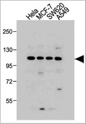
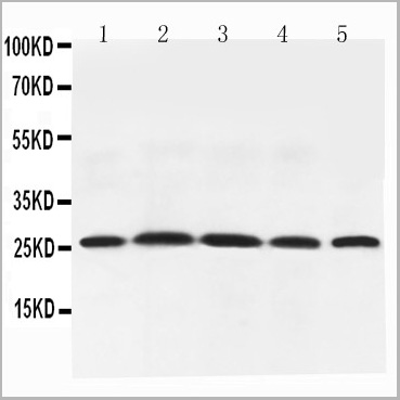 WB (Western Blot)
(Figure 6. Western blot analysis of Adiponectin using anti-Adiponectin antibody (AAA11602).Electrophoresis was performed on a 5-20% SDS-PAGE gel at 70V (Stacking gel) / 90V (Resolving gel) for 2-3 hours. The sample well of each lane was loaded with 50ug of sample under reducing conditions.Lane 1: Rat Testis Tissue Lysate,Lane 2: Rat Spleen Tissue Lysate,Lane 3: Rat Skeletal Muscle Tissue Lysate,Lane 4: Rat Cardiac Muscle Tissue Lysate,Lane 5: Rat Liver Tissue Lysate,After Electrophoresis, proteins were transferred to a Nitrocellulose membrane at 150mA for 50-90 minutes. Blocked the membrane with 5% Non-fat Milk/ TBS for 1.5 hour at RT. The membrane was incubated with rabbit anti-Adiponectin antigen affinity purified polyclonal antibody at 0.5ug/mL overnight at 4 degree C, then washed with TBS-0.1%Tween 3 times with 5 minutes each and probed with a goat anti-rabbit IgG-HRP secondary antibody at a dilution of 1:10000 for 1.5 hour at RT. The signal is developed using an Enhanced Chemiluminescent detection (ECL) kit with Tanon 5200 system. A specific band was detected for Adiponectin at approximately 26KD. The expected band size for Adiponectin is at 26KD.)
WB (Western Blot)
(Figure 6. Western blot analysis of Adiponectin using anti-Adiponectin antibody (AAA11602).Electrophoresis was performed on a 5-20% SDS-PAGE gel at 70V (Stacking gel) / 90V (Resolving gel) for 2-3 hours. The sample well of each lane was loaded with 50ug of sample under reducing conditions.Lane 1: Rat Testis Tissue Lysate,Lane 2: Rat Spleen Tissue Lysate,Lane 3: Rat Skeletal Muscle Tissue Lysate,Lane 4: Rat Cardiac Muscle Tissue Lysate,Lane 5: Rat Liver Tissue Lysate,After Electrophoresis, proteins were transferred to a Nitrocellulose membrane at 150mA for 50-90 minutes. Blocked the membrane with 5% Non-fat Milk/ TBS for 1.5 hour at RT. The membrane was incubated with rabbit anti-Adiponectin antigen affinity purified polyclonal antibody at 0.5ug/mL overnight at 4 degree C, then washed with TBS-0.1%Tween 3 times with 5 minutes each and probed with a goat anti-rabbit IgG-HRP secondary antibody at a dilution of 1:10000 for 1.5 hour at RT. The signal is developed using an Enhanced Chemiluminescent detection (ECL) kit with Tanon 5200 system. A specific band was detected for Adiponectin at approximately 26KD. The expected band size for Adiponectin is at 26KD.)
Adiponectin, Polyclonal Antibody (Cat# AAA11602)
Full Name
Anti-Adiponectin antibody
Gene Names
Adipoq; Acdc; Acrp30
Reactivity
Mouse, Rat
Applications
Western Blot, Immunohistochemistry
Purity
Immunogen affinity purified.
Pricing
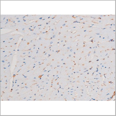

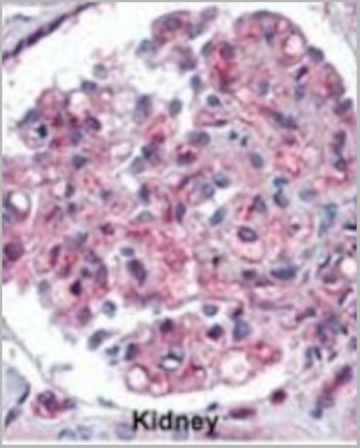
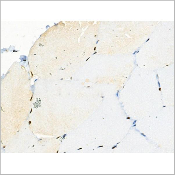
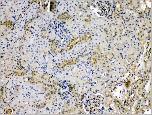
 IHC (Immunohistochemistry)
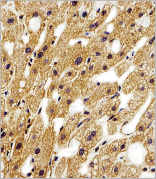
(AAA31043 at 1/200 staining Rat kidney tissue sections by IHC-P. The tissue was formaldehyde fixed and a heat mediated antigen retrieval step in citrate buffer was performed. The tissue was then blocked and incubated with the antibody for 1.5 hours at 22 degree C. An HRP conjugated goat anti-rabbit antibody was used as the secondary.)
IHC (Immunohistochemistry)
(AAA31043 at 1/200 staining Rat kidney tissue sections by IHC-P. The tissue was formaldehyde fixed and a heat mediated antigen retrieval step in citrate buffer was performed. The tissue was then blocked and incubated with the antibody for 1.5 hours at 22 degree C. An HRP conjugated goat anti-rabbit antibody was used as the secondary.)
STAT1, Polyclonal Antibody (Cat# AAA31043)
Full Name
Phospho-STAT1 (Ser727) Antibody
Gene Names
STAT1; CANDF7; IMD31A; IMD31B; IMD31C; ISGF-3; STAT91
Reactivity
Human, Mouse, Rat
Applications
Western Blot, Immunohistochemistry, Immunofluorescence, Immunocytochemistry
Purity
From purified rabbit serum by affinity purification via sequential chromatography on phospho-and non-phospho-peptide affinity columns.
Pricing
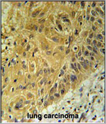
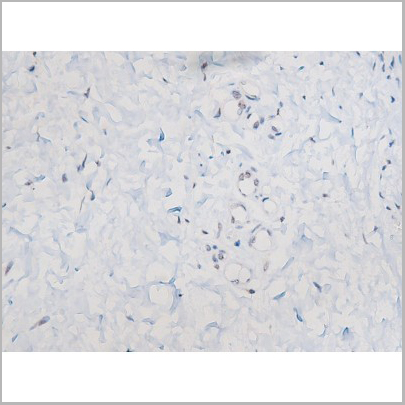
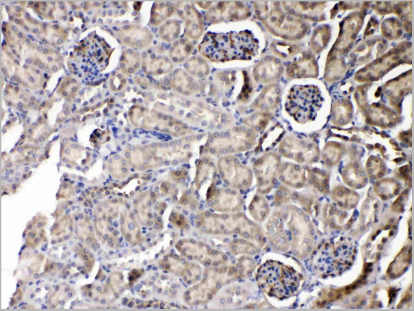
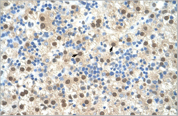
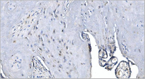
 IHC (Immunohistchemistry)
(Formalin-fixed and paraffin-embedded human lung carcinoma tissue reacted with EN1 antibody (N-term) (Cat.#AAA28756), which was peroxidase-conjugated to the secondary antibody, followed by DAB staining. This data demonstrates the use of this antibody for immunohistochemistry; clinical relevance has not been evaluated.)
IHC (Immunohistchemistry)
(Formalin-fixed and paraffin-embedded human lung carcinoma tissue reacted with EN1 antibody (N-term) (Cat.#AAA28756), which was peroxidase-conjugated to the secondary antibody, followed by DAB staining. This data demonstrates the use of this antibody for immunohistochemistry; clinical relevance has not been evaluated.)
EN1 (Engrailed 1), Polyclonal Antibody (Cat# AAA28756)
Full Name
EN1 (Engrailed 1) Antibody (N-term)
Reactivity
Human, Mouse
Applications
Western Blot, Immunohistochemistry, Immunofluorescence
Purity
This antibody is purified through a protein A column, followed by peptide affinity purification.
Pricing
CD20, Monoclonal Antibody (Cat# AAA14177)
Full Name
anti-CD20 monoclonal antibody
Gene Names
MS4A1; CD20
Reactivity
Human, mouse, rat
Applications
Flow Cytometry, Fluorescence Microscopy, Immunohistochemistry, Immunoprecipitation
Purity
Protein G purified.
Pricing
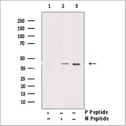
 DB (Dot Blot)
(Dot blot analysis of anti-AKT3-pS472 Phospho-specific Pab on nitrocellulose membrane. 50ng of Phospho-peptide or Non Phospho-peptide per dot were adsorbed. Antibody working concentrations are 0.5ug per ml.)
DB (Dot Blot)
(Dot blot analysis of anti-AKT3-pS472 Phospho-specific Pab on nitrocellulose membrane. 50ng of Phospho-peptide or Non Phospho-peptide per dot were adsorbed. Antibody working concentrations are 0.5ug per ml.)
Phospho-AKT3 (S472), Polyclonal Antibody (Cat# AAA28777)
Full Name
Phospho-AKT3 (S472) Antibody
Gene Names
AKT3; MPPH; PKBG; MPPH2; PRKBG; STK-2; PKB-GAMMA; RAC-gamma; RAC-PK-gamma
Reactivity
Human
Predicted: Mouse, Rat.
Predicted: Mouse, Rat.
Applications
Dot Blot
Purity
Peptide Affinity Purified Rabbit Polyclonal Antibody (Pab)
Pricing
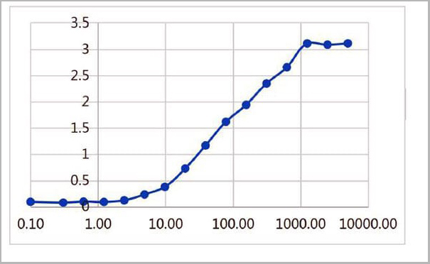 ELISA
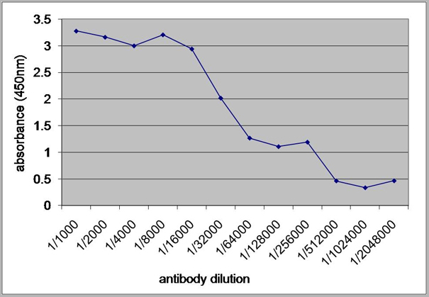
(ELISA plate was coated by the 2019-nCoV S1 protein, 100 uL/cell at 5ug/ml. The indirect ELISA analysis was performed by loading 100 uL per well of the anti-2019-nCoV S1 mAb (S1) at various concentrations. The plate was incubated for 1 hours at 37 degree C, then washed 5 times. An anti hFc HRP conjugated mAb at a concentration of 1:2000, 100uL/well was used as the secondary antibody. Again, the plate was incubated for 1 hours at 37 degree C, then washed 5 times. Detection was performed using TMB substrate for 10 minutes at room temperature in the dark. The plate was stopped with 2M sulfuric acid. Signal was read on a spectrophotometer at 450 nm.)
ELISA
(ELISA plate was coated by the 2019-nCoV S1 protein, 100 uL/cell at 5ug/ml. The indirect ELISA analysis was performed by loading 100 uL per well of the anti-2019-nCoV S1 mAb (S1) at various concentrations. The plate was incubated for 1 hours at 37 degree C, then washed 5 times. An anti hFc HRP conjugated mAb at a concentration of 1:2000, 100uL/well was used as the secondary antibody. Again, the plate was incubated for 1 hours at 37 degree C, then washed 5 times. Detection was performed using TMB substrate for 10 minutes at room temperature in the dark. The plate was stopped with 2M sulfuric acid. Signal was read on a spectrophotometer at 450 nm.)
COVID 19 Spike S1 Protein Coronavirus, Monoclonal Antibody (Cat# AAA27959)
Full Name
human anti-2019-nCoV Spike S1 mAb (S1)
Applications
ELISA
Purity
Purity: ≥ 95% as determined by SDS-PAGE
Purification: Protein A
Purification: Protein A
Pricing
ERO1L, Monoclonal Antibody (Cat# AAA24203)
Full Name
ERO1L (ERO1-like Protein alpha, ERO1-L, ERO1-L-alpha, Endoplasmic Oxidoreductin-1-like Protein, Oxidoreductin-1-L-alpha, UNQ434/PRO865) (AP)
Gene Names
ERO1A; ERO1L; ERO1-L; ERO1LA; Ero1alpha; ERO1-alpha; ERO1-L-alpha
Reactivity
Human, Rat
Applications
EIA, IHC, WB
Purity
Purified by Protein A Affinity Chromatography.
Pricing
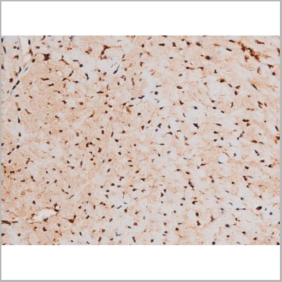
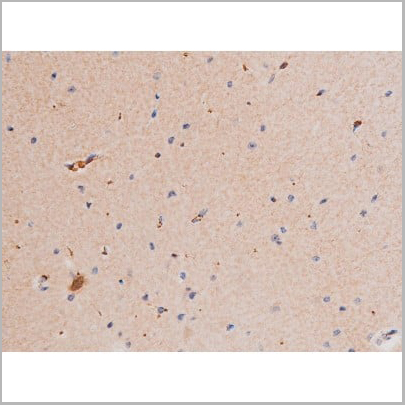
 IHC (Immunohistochemistry)
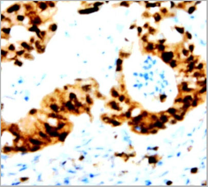
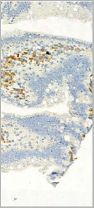
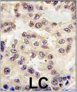
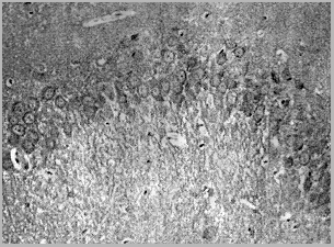
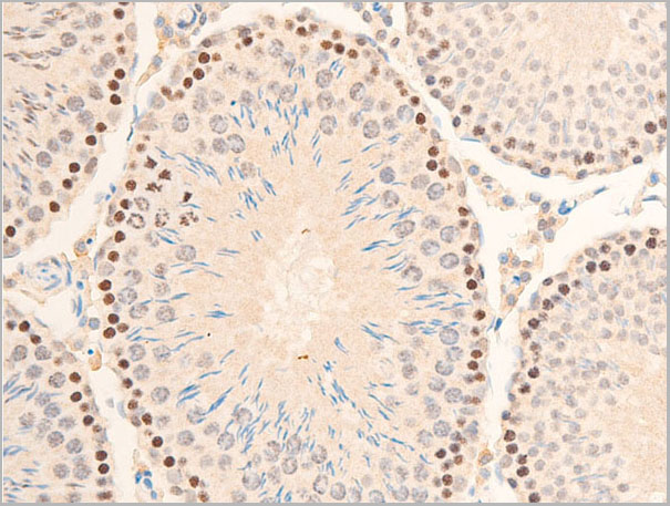
(Immunohistochemical analysis of paraffin-embedded rat hippocampal region tissue from a model with Alzheinmer's Disease useing Tau (phospho-Thr181) antibody)
IHC (Immunohistochemistry)
(Immunohistochemical analysis of paraffin-embedded rat hippocampal region tissue from a model with Alzheinmer's Disease useing Tau (phospho-Thr181) antibody)
Tau, Antibody (Cat# AAA17984)
Full Name
Tau (Phospho-Thr181) Antibody
Gene Names
MAPT; TAU; MSTD; PPND; DDPAC; MAPTL; MTBT1; MTBT2; FTDP-17; PPP1R103
Reactivity
Human, Mouse, Rat
Applications
Western Blot, Immunohistochemistry
Purity
Affinity-purified from rabbit antiserum by affinity-chromatography using epitope-specific phosphopeptide. The antibody against non-phosphopeptide was removed by chromatogramphy using non-phosphopeptide corresponding to the phosphorylation site.
Pricing
 IHC (Immunohistochemistry)
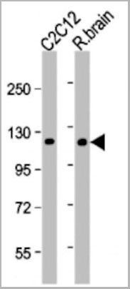
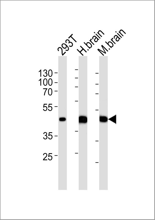
(Western blot analysis of lysates from 293T cell line human brain and mouse brain tissue lysate(from left to right), using STRADA Antibody (C-term). AAA28673 was diluted at 1:1000 at each lane. A goat anti-rabbit IgG H&L(HRP) at 1:5000 dilution was used as the secondary antibody. Lysates at 35ug per lane.)
IHC (Immunohistochemistry)
(Western blot analysis of lysates from 293T cell line human brain and mouse brain tissue lysate(from left to right), using STRADA Antibody (C-term). AAA28673 was diluted at 1:1000 at each lane. A goat anti-rabbit IgG H&L(HRP) at 1:5000 dilution was used as the secondary antibody. Lysates at 35ug per lane.)
STRADA, Polyclonal Antibody (Cat# AAA28673)
Full Name
STRADA Antibody (C-term)
Gene Names
STRADA; LYK5; PMSE; Stlk; STRAD; NY-BR-96
Reactivity
Human, mouse (Predicted Reactivity: Monkey)
Applications
Western Blot, Flow Cytometry, Immunofluorescence, Immunohistochemistry
Purity
Purified Rabbit Polyclonal Antibody (Pab)
Pricing
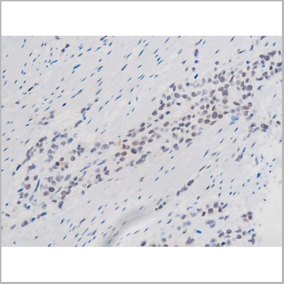
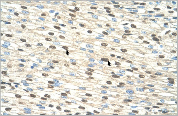
 IHC (Immunohistochemistry)
(AAA30934 at 1/100 staining human TB tissue sections by IHC-P. The tissue was formaldehyde fixed and a heat mediated antigen retrieval step in citrate buffer was performed. The tissue was then blocked and incubated with the antibody for 1.5 hours at 22 degree C. An HRP conjugated goat anti-rabbit antibody was used as the secondary.)
IHC (Immunohistochemistry)
(AAA30934 at 1/100 staining human TB tissue sections by IHC-P. The tissue was formaldehyde fixed and a heat mediated antigen retrieval step in citrate buffer was performed. The tissue was then blocked and incubated with the antibody for 1.5 hours at 22 degree C. An HRP conjugated goat anti-rabbit antibody was used as the secondary.)
I kappaB alpha, Polyclonal Antibody (Cat# AAA30934)
Full Name
Phospho-I kappaB alpha (Ser32/Ser36) Antibody
Gene Names
NFKBIA; IKBA; MAD-3; NFKBI
Reactivity
Human, Mouse, Rat
Applications
Western Blot, Immunohistochemistry, Immunofluorescence, Immunocytochemistry
Purity
From purified rabbit serum by affinity purification via sequential chromatography on phospho-and non-phospho-peptide affinity columns.
Pricing
 Application Data
(At 25 degree C. Samples were then incubated with primary Ab(At 37 degree C. An AlexaFluor594 conjugated goat anti-rabbit IgG(H+L) Ab(Red) and an AlexaFluor488 conjugated goat anti-mouse IgG(H+L) Ab(Green) were used as the secondary antibody.The nuclear counter stain is DAPI(blue).)
Application Data
(At 25 degree C. Samples were then incubated with primary Ab(At 37 degree C. An AlexaFluor594 conjugated goat anti-rabbit IgG(H+L) Ab(Red) and an AlexaFluor488 conjugated goat anti-mouse IgG(H+L) Ab(Green) were used as the secondary antibody.The nuclear counter stain is DAPI(blue).)
PAR4, Polyclonal Antibody (Cat# AAA31375)
Full Name
Phospho-PAR4 (Thr163) Antibody
Gene Names
PAWR; PAR4; Par-4
Reactivity
Human, Mouse, Rat
Predicted Reactivity: Pig (100%), Zebrafish (92%), Rabbit (100%), Chicken (100%), Xenopus (92%)
Predicted Reactivity: Pig (100%), Zebrafish (92%), Rabbit (100%), Chicken (100%), Xenopus (92%)
Applications
Western Blot, Immunohistochemistry, Immunofluorescence, Immunocytochemistry, Peptide ELISA
Purity
The antibody is from purified rabbit serum by affinity purification via sequential chromatography on phospho-peptide and non-phospho-peptide affinity columns.
Pricing
 FCM (Flow Cytometry)
(Figure 6. Flow Cytometry analysis of U20S cells using anti-Calpastatin antibody (AAA11679).Overlay histogram showing U20S cells stained with AAA11679 (Blue line).The cells were blocked with 10% normal goat serum. And then incubated with rabbit anti-Calpastatin Antibody (AAA11679,1ug/1x10^6 cells) for 30 min at 20 degree C. DyLight ®488 conjugated goat anti-rabbit IgG (5-10ug/1x10^6 cells) was used as secondary antibody for 30 minutes at 20 degree C. Isotype control antibody (Green line) was rabbit IgG (1ug/1x106) used under the same conditions. Unlabelled sample (Red line) was also used as a control.)
FCM (Flow Cytometry)
(Figure 6. Flow Cytometry analysis of U20S cells using anti-Calpastatin antibody (AAA11679).Overlay histogram showing U20S cells stained with AAA11679 (Blue line).The cells were blocked with 10% normal goat serum. And then incubated with rabbit anti-Calpastatin Antibody (AAA11679,1ug/1x10^6 cells) for 30 min at 20 degree C. DyLight ®488 conjugated goat anti-rabbit IgG (5-10ug/1x10^6 cells) was used as secondary antibody for 30 minutes at 20 degree C. Isotype control antibody (Green line) was rabbit IgG (1ug/1x106) used under the same conditions. Unlabelled sample (Red line) was also used as a control.)
Calpastatin, Polyclonal Antibody (Cat# AAA11679)
Full Name
Anti-Calpastatin Antibody
Gene Names
CAST; BS-17; PLACK
Reactivity
Human
Applications
Western Blot, Immunohistochemistry
Purity
Immunogen affinity purified.
Pricing

 FCM (Flow Cytometry)
(Figure 7. Flow Cytometry analysis of U251 cells using anti-PNP antibody (AAA11680).Overlay histogram showing U251 cells stained with AAA11680 (Blue line).The cells were blocked with 10% normal goat serum. And then incubated with rabbit anti-PNP Antibody (AAA11680,1ug/1x10^6 cells) for 30 min at 20 degree C. DyLight ®488 conjugated goat anti-rabbit IgG (5-10ug/1x10^6 cells) was used as secondary antibody for 30 minutes at 20 degree C. Isotype control antibody (Green line) was rabbit IgG (1ug/1x106) used under the same conditions. Unlabelled sample (Red line) was also used as a control.)
FCM (Flow Cytometry)
(Figure 7. Flow Cytometry analysis of U251 cells using anti-PNP antibody (AAA11680).Overlay histogram showing U251 cells stained with AAA11680 (Blue line).The cells were blocked with 10% normal goat serum. And then incubated with rabbit anti-PNP Antibody (AAA11680,1ug/1x10^6 cells) for 30 min at 20 degree C. DyLight ®488 conjugated goat anti-rabbit IgG (5-10ug/1x10^6 cells) was used as secondary antibody for 30 minutes at 20 degree C. Isotype control antibody (Green line) was rabbit IgG (1ug/1x106) used under the same conditions. Unlabelled sample (Red line) was also used as a control.)
PNP, Polyclonal Antibody (Cat# AAA11680)
Full Name
Anti-PNP Antibody
Gene Names
PNP; NP; PUNP; PRO1837
Reactivity
Human, Mouse, Rat
Applications
Western Blot, Immunohistochemistry
Purity
Immunogen affinity purified.
Pricing
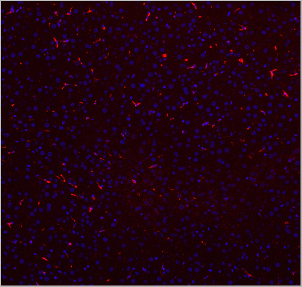
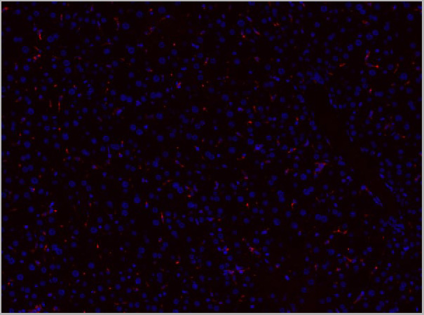

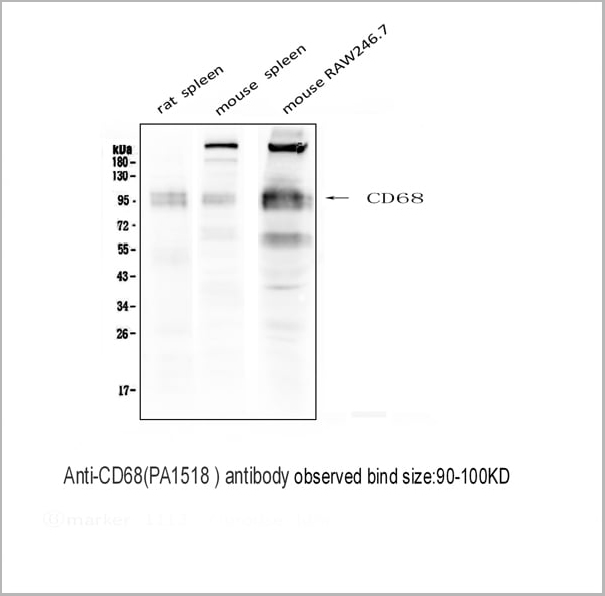
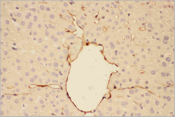
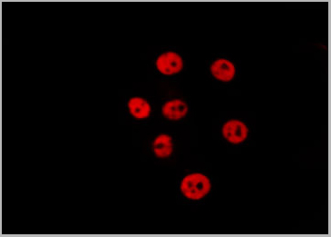
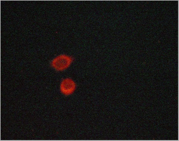
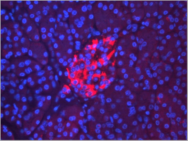
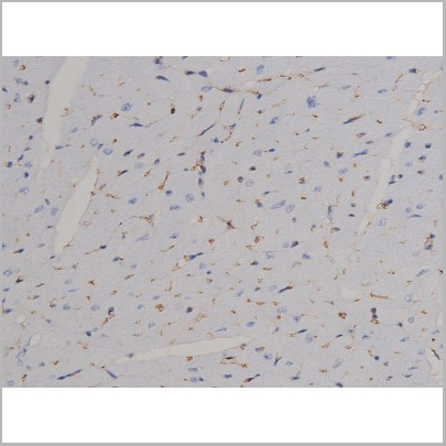
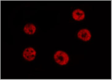
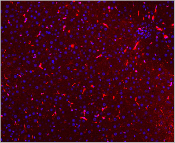 IF (Immunofluorescence)
(Figure 7. IF analysis of CD68 using anti- CD68 antibody CD68 was detected in paraffin-embedded section of mouse liver tissues. Heat mediated antigen retrieval was performed in citrate buffer (pH6, epitope retrieval solution) for 20 mins. The tissue section was blocked with 10% goat serum. The tissue section was then incubated with 1ug/mL rabbit anti- CD68 Antibody overnight at 4 degree C. Cy3 Conjugated Goat Anti-Rabbit IgG was used as secondary antibody at 1:100 dilution and incubated for 30 minutes at 37 degree C. The section was counterstained with DAPI. Visualize using a fluorescence microscope and filter sets appropriate for the label used.)
IF (Immunofluorescence)
(Figure 7. IF analysis of CD68 using anti- CD68 antibody CD68 was detected in paraffin-embedded section of mouse liver tissues. Heat mediated antigen retrieval was performed in citrate buffer (pH6, epitope retrieval solution) for 20 mins. The tissue section was blocked with 10% goat serum. The tissue section was then incubated with 1ug/mL rabbit anti- CD68 Antibody overnight at 4 degree C. Cy3 Conjugated Goat Anti-Rabbit IgG was used as secondary antibody at 1:100 dilution and incubated for 30 minutes at 37 degree C. The section was counterstained with DAPI. Visualize using a fluorescence microscope and filter sets appropriate for the label used.)
CD68, Polyclonal Antibody (Cat# AAA11512)
Full Name
Anti-CD68 antibody
Gene Names
Cd68; Lamp4; gp110; Scard1
Reactivity
Mouse, Rat
Applications
Western Blot, Immunohistochemistry, Immunohistochemistry
Purity
Immunogen affinity purified.
Pricing


 Application Data
(Published customer image: Mouse anti V5 tag antibody, clone SV5-Pk1 used for the detection of V5 tagged WEEV_nsP3 protein by western blotting and immunofluorescenceImage caption: WEEV nsP3 interaction with host IKKbeta. A) U87MGs were transfected in a 6-well plate with 5 ug of pUC19 and WEEV_nsP3_HA for 24 hours. Cell lysates were resolved using SDS-PAGE and subsequently immunoblotted with V5 antibody and beta-actin served as a loading control. B) U87MGs were transfected with WEEV_nsP3_V5; cells were fixed after 24 hours and stained with antibodies against the endogenous IKKbeta and the V5 tag. Cells were incubated with appropriate secondary Alexa Fluor antibodies and the nuclei stained with DAPI. Co-localization of IKKbeta with WEEV_nsP3_V5 (yellow) was observed as shown by the arrows. B) Panels E -H serve as an example of transfected cells in a given field of view that show co-localization of IKKbeta and WEEV_nsP3_V5 24 hours post transfection. Panels I-L represent magnified images of other cells showing co-localization of IKKbeta and WEEV_nsP3_V5. Panel M is a magnified image of panel L. The co-localization was confirmed by Z-stack analysis. Co-localization was calculated to be approximately in 61% of cells (163 cells were counted of which 44% demonstrated expression of nsP3. Of those cells that expressed nsP3, 61% showed co-localization of both proteins). Images were taken using Nikon Eclipse TE2000-U at 60x magnification and are representative of 2 independent experiments.From: Amaya M, Voss K, Sampey G, Senina S, de la Fuente C, et al. (2014) The Role of IKKbeta in Venezuelan Equine Encephalitis Virus Infection. PLoS ONE 9(2): e86745.)
Application Data
(Published customer image: Mouse anti V5 tag antibody, clone SV5-Pk1 used for the detection of V5 tagged WEEV_nsP3 protein by western blotting and immunofluorescenceImage caption: WEEV nsP3 interaction with host IKKbeta. A) U87MGs were transfected in a 6-well plate with 5 ug of pUC19 and WEEV_nsP3_HA for 24 hours. Cell lysates were resolved using SDS-PAGE and subsequently immunoblotted with V5 antibody and beta-actin served as a loading control. B) U87MGs were transfected with WEEV_nsP3_V5; cells were fixed after 24 hours and stained with antibodies against the endogenous IKKbeta and the V5 tag. Cells were incubated with appropriate secondary Alexa Fluor antibodies and the nuclei stained with DAPI. Co-localization of IKKbeta with WEEV_nsP3_V5 (yellow) was observed as shown by the arrows. B) Panels E -H serve as an example of transfected cells in a given field of view that show co-localization of IKKbeta and WEEV_nsP3_V5 24 hours post transfection. Panels I-L represent magnified images of other cells showing co-localization of IKKbeta and WEEV_nsP3_V5. Panel M is a magnified image of panel L. The co-localization was confirmed by Z-stack analysis. Co-localization was calculated to be approximately in 61% of cells (163 cells were counted of which 44% demonstrated expression of nsP3. Of those cells that expressed nsP3, 61% showed co-localization of both proteins). Images were taken using Nikon Eclipse TE2000-U at 60x magnification and are representative of 2 independent experiments.From: Amaya M, Voss K, Sampey G, Senina S, de la Fuente C, et al. (2014) The Role of IKKbeta in Venezuelan Equine Encephalitis Virus Infection. PLoS ONE 9(2): e86745.)
V5-TAG, Monoclonal Antibody (Cat# AAA11850)
Full Name
MOUSE ANTI V5-TAG:Biotin
Applications
Immunohistochemistry, Western Blot
Pricing
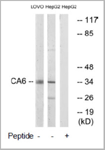 WB (Western Blot)
(Western blot analysis of extracts from LOVO cells and HepG2 cells, using CA6 antibody.)
WB (Western Blot)
(Western blot analysis of extracts from LOVO cells and HepG2 cells, using CA6 antibody.)
CA6, Antibody (Cat# AAA17978)
Full Name
CA6 Antibody
Gene Names
CA6; CA-VI; GUSTIN
Reactivity
Human (Identities = 100%, Positives = 100%)
Applications
Western Blot
Purity
Affinity-purified from rabbit antiserum by affinity-chromatography using epitope-specific immunogen.
Pricing
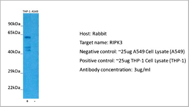
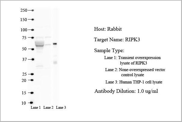
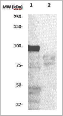
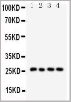
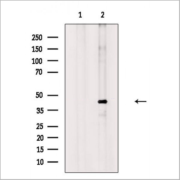
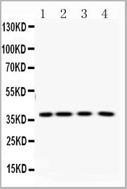
 WB (Western Blot)
(25 ug of the indicated Human whole cell extracts was loaded onto a 12% SDS-PAGE gel. 1 ug/mL of the antibody was used in this experiment.)
WB (Western Blot)
(25 ug of the indicated Human whole cell extracts was loaded onto a 12% SDS-PAGE gel. 1 ug/mL of the antibody was used in this experiment.)
RIPK3, Polyclonal Antibody (Cat# AAA23385)
Full Name
RIPK3 antibody - N-terminal region
Gene Names
RIPK3; RIP3
Reactivity
Tested Species Reactivty: Human
Predicted Species Reactivity: Cow, Dog, Horse, Human, Mouse, Pig, Rat
Predicted Species Reactivity: Cow, Dog, Horse, Human, Mouse, Pig, Rat
Applications
Immunohistochemistry, Western Blot
Purity
Protein A purified
Pricing
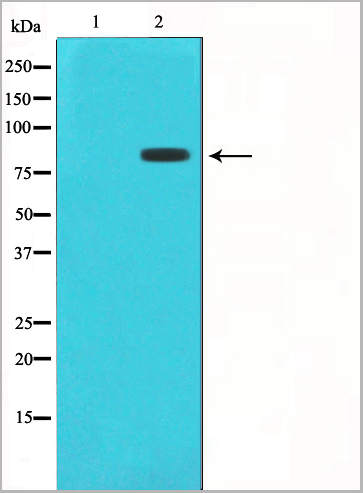
 WB (Western Blot)
(Figure. Western blot analysis of IFN gamma using anti- IFN gamma antibody (AAA11517).Electrophoresis was performed on a 5-20% SDS-PAGE gel at 70V (Stacking gel) / 90V (Resolving gel) for 2-3 hours. The sample well of each lane was loaded with 50ug of sample under reducing conditions.Lane: Recombinant Human IFN gamma Protein 0.5ngAfter Electrophoresis, proteins were transferred to a Nitrocellulose membrane at 150mA for 50-90 minutes. Blocked the membrane with 5% Non-fat Milk/ TBS for 1.5 hour at RT. The membrane was incubated with rabbit anti- IFN gamma antigen affinity purified polyclonal antibody at 0.5ug/mL overnight at 4 degree C, then washed with TBS-0.1%Tween 3 times with 5 minutes each and probed with a goat anti-rabbit IgG-HRP secondary antibody at a dilution of 1:10000 for 1.5 hour at RT. The signal is developed using an Enhanced Chemiluminescent detection (ECL) kit with Tanon 5200 system. A specific band was detected for IFN gamma at approximately 17KD. The expected band size for IFN gamma is at 17KD.)
WB (Western Blot)
(Figure. Western blot analysis of IFN gamma using anti- IFN gamma antibody (AAA11517).Electrophoresis was performed on a 5-20% SDS-PAGE gel at 70V (Stacking gel) / 90V (Resolving gel) for 2-3 hours. The sample well of each lane was loaded with 50ug of sample under reducing conditions.Lane: Recombinant Human IFN gamma Protein 0.5ngAfter Electrophoresis, proteins were transferred to a Nitrocellulose membrane at 150mA for 50-90 minutes. Blocked the membrane with 5% Non-fat Milk/ TBS for 1.5 hour at RT. The membrane was incubated with rabbit anti- IFN gamma antigen affinity purified polyclonal antibody at 0.5ug/mL overnight at 4 degree C, then washed with TBS-0.1%Tween 3 times with 5 minutes each and probed with a goat anti-rabbit IgG-HRP secondary antibody at a dilution of 1:10000 for 1.5 hour at RT. The signal is developed using an Enhanced Chemiluminescent detection (ECL) kit with Tanon 5200 system. A specific band was detected for IFN gamma at approximately 17KD. The expected band size for IFN gamma is at 17KD.)
Interferon gamma, Polyclonal Antibody (Cat# AAA11517)
Full Name
Anti-human Interferon gamma antibody
Gene Names
IFNG; IFG; IFI
Reactivity
Human
Applications
Western Blot, Immunohistochemistry, Neutralization Assay, Immunoprecipitation
Purity
Immunogen affinity purified.
Pricing
 IF (Immunofluorescence)
(AAA31046 staining HeLa by IF/ICC. The sample were fixed with PFA and permeabilized in 0.1% Triton X-100, then blocked in 10% serum for 45 minutes at 25 degree C. The primary antibody was diluted at 1/200 and incubated with the sample for 1 hour at 37 degree C. An Alexa Fluor 594 conjugated goat anti-rabbit IgG (H+L) Ab, diluted at 1/600, was used as the secondary antibody.)
IF (Immunofluorescence)
(AAA31046 staining HeLa by IF/ICC. The sample were fixed with PFA and permeabilized in 0.1% Triton X-100, then blocked in 10% serum for 45 minutes at 25 degree C. The primary antibody was diluted at 1/200 and incubated with the sample for 1 hour at 37 degree C. An Alexa Fluor 594 conjugated goat anti-rabbit IgG (H+L) Ab, diluted at 1/600, was used as the secondary antibody.)
STAT6, Polyclonal Antibody (Cat# AAA31046)
Full Name
Phospho-STAT6 (Thr645) Antibody
Gene Names
STAT6; STAT6B; STAT6C; D12S1644; IL-4-STAT
Reactivity
Human, Mouse, Rat
Applications
Western Blot, Immunohistochemistry, Immunofluorescence, Immunocytochemistry, Immunoprecipitation
Purity
From purified rabbit serum by affinity purification via sequential chromatography on phospho-and non-phospho-peptide affinity columns.
Pricing
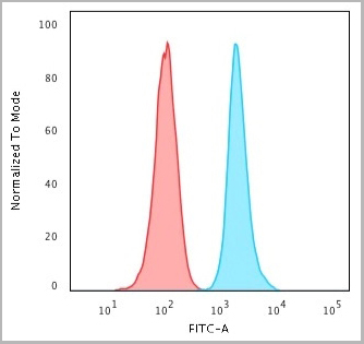
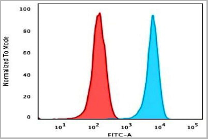
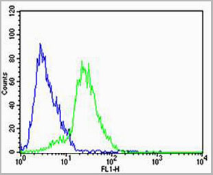
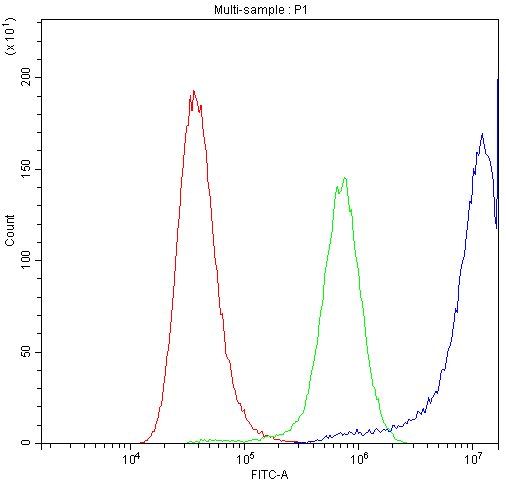
 FCM (Flow Cytometry)
(PLA2G2D Antibody (C-term) flow cytometric analysis of HL-60 cells (right histogram) compared to a negative control cell (left histogram).FITC-conjugated goat-anti-rabbit secondary antibodies were used for the analysis.)
FCM (Flow Cytometry)
(PLA2G2D Antibody (C-term) flow cytometric analysis of HL-60 cells (right histogram) compared to a negative control cell (left histogram).FITC-conjugated goat-anti-rabbit secondary antibodies were used for the analysis.)
PLA2G2D, Polyclonal Antibody (Cat# AAA28718)
Full Name
PLA2G2D Antibody (C-term)
Gene Names
PLA2G2D; SPLASH; sPLA2S; PLA2IID; sPLA2-IID
Reactivity
Human
Applications
Western Blot, Immunohistochemistry, Flow Cytometry
Purity
Peptide Affinity Purified Rabbit Polyclonal Antibody (Pab)
Pricing
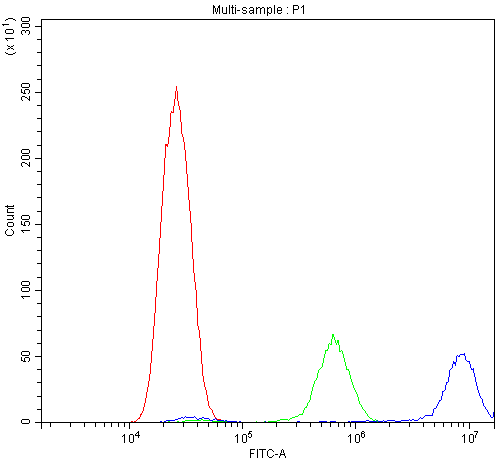
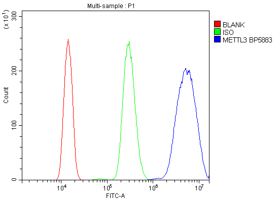 FCM (Flow Cytometry)
(Figure 6. Flow Cytometry analysis of HL-60 cells using anti-METTL3 antibody (AAA19247).Overlay histogram showing HL-60 cells stained with AAA19247 (Blue line). The cells were blocked with 10% normal goat serum. And then incubated with rabbit anti-METTL3 Antibody (AAA19247, 1μg/1x106 cells) for 30 min at 20 degree C. DyLight®488 conjugated goat anti-rabbit IgG (5-10μg/1x106 cells) was used as secondary antibody for 30 minutes at 20 degree C. Isotype control antibody (Green line) was rabbit IgG (1μg/1x106) used under the same conditions. Unlabelled sample (Red line) was also used as a control.)
FCM (Flow Cytometry)
(Figure 6. Flow Cytometry analysis of HL-60 cells using anti-METTL3 antibody (AAA19247).Overlay histogram showing HL-60 cells stained with AAA19247 (Blue line). The cells were blocked with 10% normal goat serum. And then incubated with rabbit anti-METTL3 Antibody (AAA19247, 1μg/1x106 cells) for 30 min at 20 degree C. DyLight®488 conjugated goat anti-rabbit IgG (5-10μg/1x106 cells) was used as secondary antibody for 30 minutes at 20 degree C. Isotype control antibody (Green line) was rabbit IgG (1μg/1x106) used under the same conditions. Unlabelled sample (Red line) was also used as a control.)
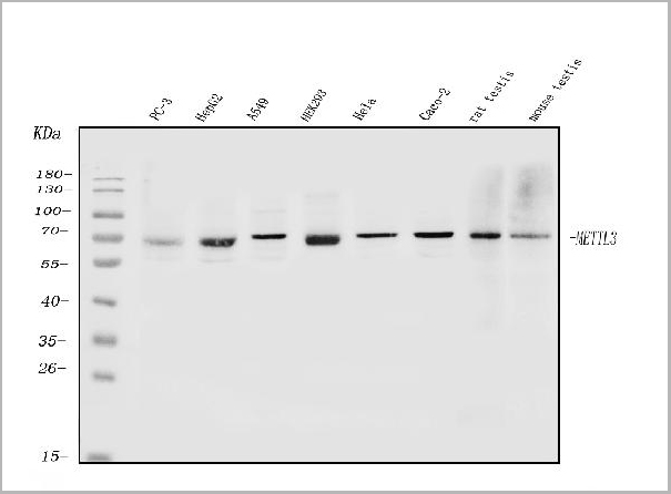
METTL3, Polyclonal Antibody (Cat# AAA19247)
Full Name
Anti-METTL3 Antibody
Gene Names
METTL3; M6A; IME4; Spo8; MT-A70
Reactivity
Human, Mouse, Rat
Applications
Western Blot, Immunohistochemistry, Immunocytochemistry, Immunofluorescence, Flow Cytometry, Direct ELISA
Purity
Immunogen affinity purified.
Pricing
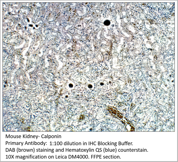
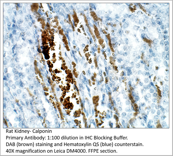
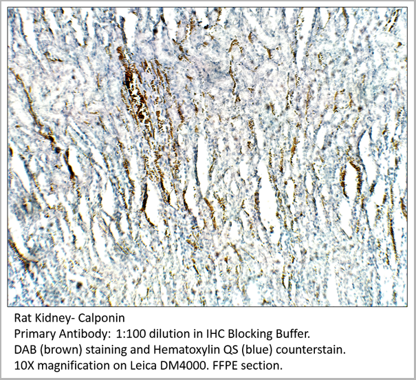
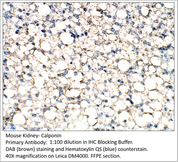 IHC (Immunohistochemistry)
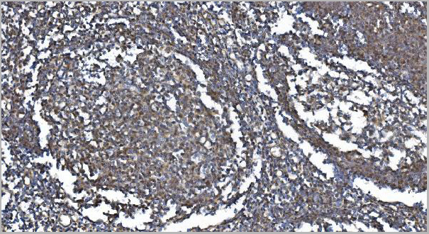
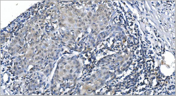
Calponin, Polyclonal Antibody (Cat# AAA14404)
Full Name
Calponin Antibody
Gene Names
CNN1; SMCC; Sm-Calp; HEL-S-14
Reactivity
Human, Mouse, Rat
Applications
Immunohistochemistry, Immunoprecipitation, Western Blot
Pricing