Filters
Clonality
Type
Reactivity
Gene Name
Isotype
Host
Application
Clone
2811 results for " C" - showing 950-1000
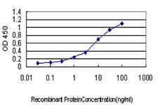
 Standard Curve (Sample)
Ubiquitin Carboxyl Terminal Hydrolase L1 (UCHL1), ELISA Kit (Cat# AAA23063)
Full Name
Multi-species Ubiquitin Carboxyl Terminal Hydrolase L1 (UCHL1) ELISA Kit
Gene Names
UCHL1; NDGOA; PARK5; PGP95; SPG79; PGP9.5; Uch-L1; HEL-117; PGP 9.5; HEL-S-53
Reactivity
Human, Mouse, Rat
Pricing


 Application Data
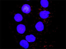
(Proximity Ligation Analysis (PLA) of protein-protein interactions between BAD and MAPK8 Mahlavu cells were stained with BAD rabbit purified polyclonal 1:1200 and anti-MAPK8 mouse monoclonal antibody 1:50. Signals were detected 30 Detection Kit 613 (red), and nuclei were counterstained with DAPI (blue). Each red dot represents the detection of protein-protein interaction complex.)
Application Data
(Proximity Ligation Analysis (PLA) of protein-protein interactions between BAD and MAPK8 Mahlavu cells were stained with BAD rabbit purified polyclonal 1:1200 and anti-MAPK8 mouse monoclonal antibody 1:50. Signals were detected 30 Detection Kit 613 (red), and nuclei were counterstained with DAPI (blue). Each red dot represents the detection of protein-protein interaction complex.)
MAPK8, Monoclonal Antibody (Cat# AAA25447)
Full Name
MAPK8 (JNK1, PRKM8, SAPK1, SAPK1C, Mitogen-activated Protein Kinase 8, MAP Kinase 8, MAPK 8, JNK-46, Stress-activated Protein Kinase 1c, Stress-activated Protein Kinase JNK1, c-Jun N-terminal Kinase 1) (HRP)
Gene Names
MAPK8; JNK; JNK1; PRKM8; SAPK1; JNK-46; JNK1A2; SAPK1c; JNK21B1/2
Reactivity
Human
Applications
Western Blot
Purity
Purified by Protein A Affinity Chromatography.
Pricing


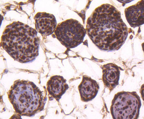
 IHC (Immunohistchemistry)
(Formalin-fixed and paraffin-embedded human testis tissue reacted with PARK2 (Parkin) antibody (C-term) , which was peroxidase-conjugated to the secondary antibody, followed by DAB staining. This data demonstrates the use of this antibody for immunohistochemistry; clinical relevance has not been evaluated.)
IHC (Immunohistchemistry)
(Formalin-fixed and paraffin-embedded human testis tissue reacted with PARK2 (Parkin) antibody (C-term) , which was peroxidase-conjugated to the secondary antibody, followed by DAB staining. This data demonstrates the use of this antibody for immunohistochemistry; clinical relevance has not been evaluated.)
Parkin, Polyclonal Antibody (Cat# AAA28668)
Full Name
Parkin Antibody (C-term)
Gene Names
PARK2; PDJ; PRKN; AR-JP; LPRS2
Reactivity
Human, mouse
Applications
WB, EIA, IF, FC/FACS, IHC
Purity
Purified Rabbit Polyclonal Antibody (Pab)
Pricing


 Application Data
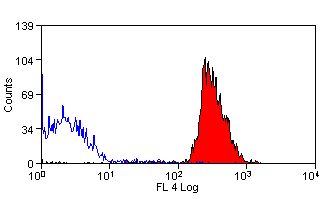
(Staining of human peripheral blood granulocytes with Mouse anti Human CD11b:Biotin)
Application Data
(Staining of human peripheral blood granulocytes with Mouse anti Human CD11b:Biotin)
CD11b, Monoclonal Antibody (Cat# AAA11992)
Full Name
MOUSE ANTI HUMAN CD11b
Gene Names
ITGAM; CR3A; MO1A; CD11B; MAC-1; MAC1A; SLEB6
Applications
Immunohistochemistry, Flow Cytometry, Immunoprecipitation
Pricing


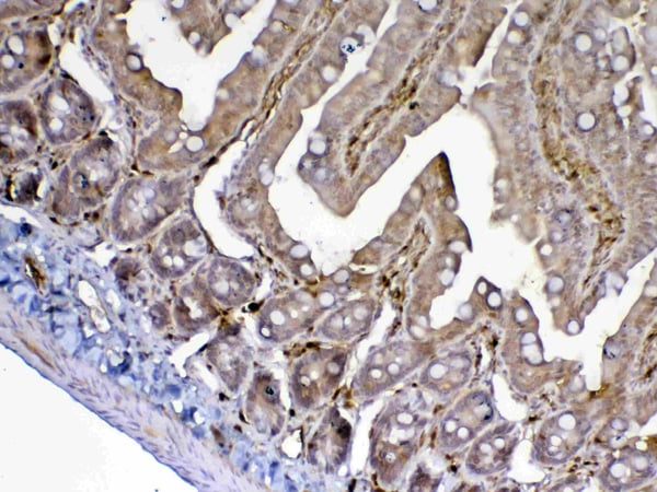
 IHC (Immunohistchemistry)
(At 1/100 staining Human gastric cancer by IHC-P. The sample was formaldehyde fixed and a heat mediated antigen retrieval step in citrate buffer was performed. The sample was then blocked and incubated with the primary antibody at 4 degree C overnight. An HRP conjugated anti-Rabbit antibody was used as the secondary antibody.)
IHC (Immunohistchemistry)
(At 1/100 staining Human gastric cancer by IHC-P. The sample was formaldehyde fixed and a heat mediated antigen retrieval step in citrate buffer was performed. The sample was then blocked and incubated with the primary antibody at 4 degree C overnight. An HRP conjugated anti-Rabbit antibody was used as the secondary antibody.)
SRPK2, Polyclonal Antibody (Cat# AAA31293)
Full Name
Phospho-SRPK2 (Ser494/Ser497) Antibody
Gene Names
SRPK2; SFRSK2
Reactivity
Human, Mouse, Rat
Applications
Immunohistochemistry, Peptide ELISA
Purity
The antibody is from purified rabbit serum by affinity purification via sequential chromatography on phospho-peptide and non-phospho-peptide affinity columns.
Pricing


 FCM (Flow Cytometry)
(Dual staining of pig peripheral blood lymphocytes with Mouse anti Pig CD335 detected with Goat anti Mouse IgG (H/L):FITC (STAR117F), and Mouse anti Pig wCD8a:RPE)
FCM (Flow Cytometry)
(Dual staining of pig peripheral blood lymphocytes with Mouse anti Pig CD335 detected with Goat anti Mouse IgG (H/L):FITC (STAR117F), and Mouse anti Pig wCD8a:RPE)
CD335, Monoclonal Antibody (Cat# AAA12253)
Full Name
MOUSE ANTI PIG CD335
Reactivity
Pig
Applications
Flow Cytometry, Immunofluorescence
Pricing
 Standard Curve (Sample)
Procollagen Type I C Terminal Propeptide, ELISA Kit (Cat# AAA16332)
Full Name
Human Procollagen Type I C Terminal Propeptide ELISA Kit
Reactivity
Human
Pricing
 Standard Curve (Sample)
Platelet Factor 4, PF-4, ELISA Kit (Cat# AAA11283)
Full Name
Human Platelet Factor 4, PF-4 ELISA Kit
Gene Names
PF4V1; PF4A; CXCL4L1; CXCL4V1; PF4-ALT; SCYB4V1
Reactivity
Human
Pricing
 Standard Curve (Sample)
Proteoglycan 4, ELISA Kit (Cat# AAA17068)
Full Name
Bovine Proteoglycan 4 ELISA Kit
Reactivity
Bovine
Pricing
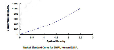 Standard Curve (Sample)
Bone Morphogenetic Protein 1 (BMP1), ELISA Kit (Cat# AAA20424)
Full Name
Human Bone Morphogenetic Protein 1 (BMP1) ELISA Kit
Gene Names
Bmp1; Pcp; Tld
Reactivity
Human
Pricing


 Application Data
(Detection limit for recombinant GST tagged SMN2 is ~0.3ng/ml as a capture antibody.)
Application Data
(Detection limit for recombinant GST tagged SMN2 is ~0.3ng/ml as a capture antibody.)
SMN2, Monoclonal Antibody (Cat# AAA25548)
Full Name
SMN2 (Survival Motor Neuron Protein, Component of Gems 1, SMN1, Gemin-1, SMN, SMNT, SMNC, FLJ76644, MGC20996, MGC5208) (HRP)
Gene Names
SMN2; SMNC; BCD541; GEMIN1; TDRD16B; C-BCD541
Reactivity
Human
Applications
EIA, IHC, WB
Purity
Purified by Protein A Affinity Chromatography.
Pricing


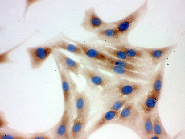
 IHC (Immunohistchemistry)
(Formalin-fixed and paraffin-embedded human cancer tissue reacted with the primary antibody, which was peroxidase-conjugated to the secondary antibody, followed by DAB staining. This data demonstrates the use of this antibody for immunohistochemistry; clinical relevance has not been evaluated. BC = breast carcinoma; HC = hepatocarcinoma.)
IHC (Immunohistchemistry)
(Formalin-fixed and paraffin-embedded human cancer tissue reacted with the primary antibody, which was peroxidase-conjugated to the secondary antibody, followed by DAB staining. This data demonstrates the use of this antibody for immunohistochemistry; clinical relevance has not been evaluated. BC = breast carcinoma; HC = hepatocarcinoma.)
PACSIN2, Polyclonal Antibody (Cat# AAA28758)
Full Name
PACSIN2 Antibody (C-term)
Gene Names
PACSIN2; SDPII
Reactivity
Human, mouse
Applications
Immunohistochemistry, Western Blot, Immunofluorescence
Purity
Purified Rabbit Polyclonal Antibody (Pab)
Pricing


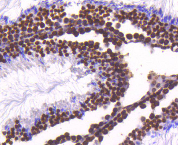
 IHC (Immunohistchemistry)
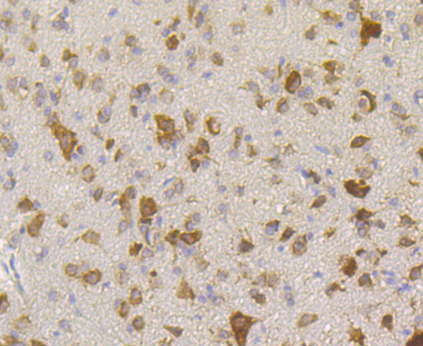
(Immunohistochemistry Analysis: Representative lot data. (Fig. 1 and 2) Paraffin-embedded mouse and human brain tissue was prepared using heat-induced epitope retrieval in citrate buffer, pH 6.0. Immunostaining was performed using a 1:100 dilution. Reactivity was detected using the IHC-Select Detection Kit. Staining pattern appears as cytoplasmic. (Fig. 3 and 4) Paraffin-embedded mouse and mouse olfactory lobe and cerebellum brain tissue was prepared using heat-induced epitope retrieval in citrate buffer, pH 6.0. Immunostaining was performed using a Chicken IgY Antibody 1:100 dilution of Cat. No. AB15894, anti-Tbr2. Reactivity was detected using the IHC-Select Detection Kit. Immunoreactivity seen here is mostly nuclear.)
IHC (Immunohistchemistry)
(Immunohistochemistry Analysis: Representative lot data. (Fig. 1 and 2) Paraffin-embedded mouse and human brain tissue was prepared using heat-induced epitope retrieval in citrate buffer, pH 6.0. Immunostaining was performed using a 1:100 dilution. Reactivity was detected using the IHC-Select Detection Kit. Staining pattern appears as cytoplasmic. (Fig. 3 and 4) Paraffin-embedded mouse and mouse olfactory lobe and cerebellum brain tissue was prepared using heat-induced epitope retrieval in citrate buffer, pH 6.0. Immunostaining was performed using a Chicken IgY Antibody 1:100 dilution of Cat. No. AB15894, anti-Tbr2. Reactivity was detected using the IHC-Select Detection Kit. Immunoreactivity seen here is mostly nuclear.)
EOMES, Polyclonal Antibody (Cat# AAA26886)
Full Name
EOMES (Eomesodermin Homolog, T-box Brain Protein 2, TBR2, T-brain-2, TBR-2)
Gene Names
Eomes; Tbr2; TBR-2; C77258
Reactivity
Mouse, Human, Rat
Applications
Immunohistochemistry, Western Blot
Purity
Purified by affinity chromatography.
Pricing


 DB (Dot Blot)
(Dot blot analysis using Rabbit Anti-Amyloid Fibrils (OC) Polyclonal Antibody (SPC-507). Tissue: Cell lysates. Species: Human. Primary Antibody: Rabbit Anti-Amyloid Fibrils (OC) Polyclonal Antibody (SPC-507) at 1:500, 1:5000. Beta Amyloid HEPES-NaCl aggregation, showing 1:500 (L) and 1:5000 (R) time lapse dot blot.)
DB (Dot Blot)
(Dot blot analysis using Rabbit Anti-Amyloid Fibrils (OC) Polyclonal Antibody (SPC-507). Tissue: Cell lysates. Species: Human. Primary Antibody: Rabbit Anti-Amyloid Fibrils (OC) Polyclonal Antibody (SPC-507) at 1:500, 1:5000. Beta Amyloid HEPES-NaCl aggregation, showing 1:500 (L) and 1:5000 (R) time lapse dot blot.)
Amyloid Fibrils (OC), Polyclonal Antibody (Cat# AAA17803)
Full Name
Amyloid Fibrils (OC) Antibody: ATTO 488
Reactivity
Human. Potentially mouse and rat based on species homology.
Applications
IP, ICC, IF, IHC, EIA, WB, DB
Purity
Protein A Purified
Pricing


 IHC (Immunohistchemistry)
(Figure 6. IHC analysis of ADA using anti-ADA antibody (AAA19140).ADA was detected in paraffin-embedded section of rat spleen tissue. Heat mediated antigen retrieval was performed in citrate buffer (pH6, epitope retrieval solution) for 20 mins. The tissue section was blocked with 10% goat serum. The tissue section was then incubated with 1ug/ml rabbit anti-ADA Antibody (AAA19140) overnight at 4 degree C. Biotinylated goat anti-rabbit IgG was used as secondary antibody and incubated for 30 minutes at 37 degree C. The tissue section was developed using Strepavidin-Biotin-Complex (SABC) with DAB as the chromogen.)
IHC (Immunohistchemistry)
(Figure 6. IHC analysis of ADA using anti-ADA antibody (AAA19140).ADA was detected in paraffin-embedded section of rat spleen tissue. Heat mediated antigen retrieval was performed in citrate buffer (pH6, epitope retrieval solution) for 20 mins. The tissue section was blocked with 10% goat serum. The tissue section was then incubated with 1ug/ml rabbit anti-ADA Antibody (AAA19140) overnight at 4 degree C. Biotinylated goat anti-rabbit IgG was used as secondary antibody and incubated for 30 minutes at 37 degree C. The tissue section was developed using Strepavidin-Biotin-Complex (SABC) with DAB as the chromogen.)
ADA/Adenosine Deaminase, Polyclonal Antibody (Cat# AAA19140)
Full Name
Anti-ADA/Adenosine Deaminase Picoband antibody
Reactivity
Mouse, Rat
No cross reactivity with other proteins.
No cross reactivity with other proteins.
Applications
EIA, IHC, WB
Pricing


 Application Data
(Analysis of Protein Array containing more than 19,000 full-length human proteins using HER-2 Mouse Monoclonal Antibody (ERBB2/3080). Z- and S- Score: The Z-score represents the strength of a signal that a monoclonal antibody (MAb) (in combination with a fluorescently-tagged anti-IgG secondary antibody) produces when binding to a particular protein on the HuProtTM array. Z-scores are described in units of standard deviations (SD's) above the mean value of all signals generated on that array. If targets on HuProtTM are arranged in descending order of the Z-score, the S-score is the difference (also in units of SD's) between the Z-score. S-score therefore represents the relative target specificity of a MAb to its intended target. A MAb is considered to specific to its intended target, if the MAb has an S-score of at least 2.5. For example, if a MAb binds to protein X with a Z-score of 43 and to protein Y with a Z-score of 14, then the S-score for the binding of that MAb to protein X is equal to 29.)
Application Data
(Analysis of Protein Array containing more than 19,000 full-length human proteins using HER-2 Mouse Monoclonal Antibody (ERBB2/3080). Z- and S- Score: The Z-score represents the strength of a signal that a monoclonal antibody (MAb) (in combination with a fluorescently-tagged anti-IgG secondary antibody) produces when binding to a particular protein on the HuProtTM array. Z-scores are described in units of standard deviations (SD's) above the mean value of all signals generated on that array. If targets on HuProtTM are arranged in descending order of the Z-score, the S-score is the difference (also in units of SD's) between the Z-score. S-score therefore represents the relative target specificity of a MAb to its intended target. A MAb is considered to specific to its intended target, if the MAb has an S-score of at least 2.5. For example, if a MAb binds to protein X with a Z-score of 43 and to protein Y with a Z-score of 14, then the S-score for the binding of that MAb to protein X is equal to 29.)
HER-2/c-erbB-2/neu/CD340, Monoclonal Antibody (Cat# AAA23912)
Full Name
HER-2/c-erbB-2/neu/CD340
Gene Names
ERBB2; NEU; NGL; HER2; TKR1; CD340; HER-2; MLN 19; HER-2/neu
Reactivity
Human
Applications
Immunohistochemistry
Purity
Purified Ab with BSA and Azide at 200ug/ml OR Purified Ab WITHOUT BSA and Azide at 1.0mg/ml
Pricing


 FCM (Flow Cytometry)
(Figure 8. Flow Cytometry analysis of U20S cells using anti-NFIA antibody (AAA11691).Overlay histogram showing U20S cells stained with AAA11691 (Blue line).The cells were blocked with 10% normal goat serum. And then incubated with rabbit anti-NFIA Antibody (AAA11691,1ug/1x10^6 cells) for 30 min at 20 degree C. DyLight®488 conjugated goat anti-rabbit IgG (5-10ug/1x10^6 cells) was used as secondary antibody for 30 minutes at 20 degree C. Isotype control antibody (Green line) was rabbit IgG (1ug/1x106) used under the same conditions. Unlabelled sample (Red line) was also used as a control.)
FCM (Flow Cytometry)
(Figure 8. Flow Cytometry analysis of U20S cells using anti-NFIA antibody (AAA11691).Overlay histogram showing U20S cells stained with AAA11691 (Blue line).The cells were blocked with 10% normal goat serum. And then incubated with rabbit anti-NFIA Antibody (AAA11691,1ug/1x10^6 cells) for 30 min at 20 degree C. DyLight®488 conjugated goat anti-rabbit IgG (5-10ug/1x10^6 cells) was used as secondary antibody for 30 minutes at 20 degree C. Isotype control antibody (Green line) was rabbit IgG (1ug/1x106) used under the same conditions. Unlabelled sample (Red line) was also used as a control.)
NFIA, Polyclonal Antibody (Cat# AAA11691)
Full Name
Anti-NFIA Antibody
Gene Names
NFIA; CTF; NF1-A; NFI-A; NFI-L; NF-I/A
Reactivity
Human, Mouse, Rat
Applications
Western Blot, Immunohistochemistry
Purity
Immunogen affinity purified.
Pricing


 Application Data
TCP1 delta, Polyclonal Antibody (Cat# AAA11672)
Full Name
Anti-TCP1 delta Antibody
Gene Names
CCT4; SRB; Cctd; CCT-DELTA
Reactivity
Human, Mouse, Rat
No cross reactivity with other proteins.
No cross reactivity with other proteins.
Applications
Western Blot, Immunohistochemistry
Purity
Immunogen affinity purified.
Pricing


 Application Data
(Published customer image Infiltration of GFP+ BM-cells in infarct and peri-infarct regions. (A-B) Dot plots of viable macrophages/granulocytes (CD11b+CD45high, top right quadrants) and microglia (CD11b+CD45dim, bottom right quadrants) in cortex from BM-chimeric unmanipulated mice and mice exposed to pMCAO. (C) Bar graph showing mean numbers of CD11b+CD45dim microglia and CD11b+CD45high macrophages/granulocytes in BM-chimeric mice 24 hours after pMCAO, subdivided based on expression of GFP (n = 5). Approximately 92% of of the CD45high population were GFP+. (D) Estimation and comparison of mean numbers of CD11b+CD45dim microglia in non-chimeric (n = 10) versus BM-chimeric mice (n = 5) 24 hours after of pMCAO shows significantly fewer CD11b+CD45dim microglial cells in irradiated mice. (E) Overview, showing distribution of infiltrating GFP+ BM-derived cells into infarct (IF) and peri-infarct (P-IF) regions 24 hours after pMCAO. (E-G) By 24 hours, GFP+ single cells (F) and vessel-associated aggregates of GFP+ cells (arrows in G) were observed in infarct and peri-infarct regions. Some of the vessel-associated cells were round, leukocyte-like cells (arrows) while others were elongated cells lining the vasculature (arrow heads in G and in insert). (H) Bar graph showing mean numbers of single GFP+ cells and vessel-associated aggregates of GFP+ cells in ipsi- and contralateral cortex 24 hours after surgery (n = 10). (I-P) Immunohistochemical staining of CD45.1 (I, K), CD45.2 (J, L), IgG2a (M, O) and CD45 (N, P) in ischemic tissue in BM-chimeric (I, J, M, N) and non-chimeric mice (K, L, O, P) 24 hours after pMCAO. N.D, none detected. Scale bars: 200 um (A), 10 um (B, C). 50 um (I-P) *P < 0.05, **P < 0.01, and ***P < 0.001.From: Clausen BH, Lambertsen KL, Babcock AA, Holm TH, Dagnaes-Hansen F, Finsen B. Interleukin-1beta and tumor necrosis factor-alpha are expressed by different subsets of microglia and macrophages after ischemic stroke in mice. J Neuroinflammation. 2008 Oct 23;5:46.)
Application Data
(Published customer image Infiltration of GFP+ BM-cells in infarct and peri-infarct regions. (A-B) Dot plots of viable macrophages/granulocytes (CD11b+CD45high, top right quadrants) and microglia (CD11b+CD45dim, bottom right quadrants) in cortex from BM-chimeric unmanipulated mice and mice exposed to pMCAO. (C) Bar graph showing mean numbers of CD11b+CD45dim microglia and CD11b+CD45high macrophages/granulocytes in BM-chimeric mice 24 hours after pMCAO, subdivided based on expression of GFP (n = 5). Approximately 92% of of the CD45high population were GFP+. (D) Estimation and comparison of mean numbers of CD11b+CD45dim microglia in non-chimeric (n = 10) versus BM-chimeric mice (n = 5) 24 hours after of pMCAO shows significantly fewer CD11b+CD45dim microglial cells in irradiated mice. (E) Overview, showing distribution of infiltrating GFP+ BM-derived cells into infarct (IF) and peri-infarct (P-IF) regions 24 hours after pMCAO. (E-G) By 24 hours, GFP+ single cells (F) and vessel-associated aggregates of GFP+ cells (arrows in G) were observed in infarct and peri-infarct regions. Some of the vessel-associated cells were round, leukocyte-like cells (arrows) while others were elongated cells lining the vasculature (arrow heads in G and in insert). (H) Bar graph showing mean numbers of single GFP+ cells and vessel-associated aggregates of GFP+ cells in ipsi- and contralateral cortex 24 hours after surgery (n = 10). (I-P) Immunohistochemical staining of CD45.1 (I, K), CD45.2 (J, L), IgG2a (M, O) and CD45 (N, P) in ischemic tissue in BM-chimeric (I, J, M, N) and non-chimeric mice (K, L, O, P) 24 hours after pMCAO. N.D, none detected. Scale bars: 200 um (A), 10 um (B, C). 50 um (I-P) *P < 0.05, **P < 0.01, and ***P < 0.001.From: Clausen BH, Lambertsen KL, Babcock AA, Holm TH, Dagnaes-Hansen F, Finsen B. Interleukin-1beta and tumor necrosis factor-alpha are expressed by different subsets of microglia and macrophages after ischemic stroke in mice. J Neuroinflammation. 2008 Oct 23;5:46.)
CD11b, Monoclonal Antibody (Cat# AAA12231)
Full Name
RAT ANTI MOUSE CD11b:Low Endotoxin
Gene Names
Itgam; CR3; CR3A; MAC1; Cd11b; Ly-40; Mac-1; Mac-1a; CD11b/CD18; F730045J24Rik
Applications
Immunohistochemistry, Flow Cytometry, Functional Assay, Immunofluorescence, Immunoprecipitation
Pricing
 Standard Curve (Sample)
Bone Morphogenetic Protein 1, ELISA Kit (Cat# AAA15985)
Full Name
Mouse Bone Morphogenetic Protein 1 ELISA Kit
Gene Names
BMP1; PCP; TLD; OI13; PCP2; PCOLC
Reactivity
Mouse
Pricing
 Standard Curve (Sample)
Mitochondrial Open Reading Frame Of The 12S rRNA-c (MOTS-c), ELISA Kit (Cat# AAA21096)
Full Name
Mouse Mitochondrial Open Reading Frame Of The 12S rRNA-c (MOTS-c) ELISA Kit
Reactivity
Mouse
Pricing


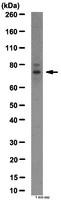
 WB (Western Blot)
(NME2 monoclonal antibody (M06), clone 1D3. Western Blot analysis of NME2 expression in NIH/3T3.)
WB (Western Blot)
(NME2 monoclonal antibody (M06), clone 1D3. Western Blot analysis of NME2 expression in NIH/3T3.)
NME2, Monoclonal Antibody (Cat# AAA25940)
Full Name
NME2 (Non-Metastatic Cells 2, Protein (NM23B) Expressed in, MGC111212, NDPK-B, NDPKB, NM23-H2, NM23B, puf) (AP)
Gene Names
NME2; PUF; NDKB; NDPKB; NM23B; NDPK-B; NM23-H2
Applications
Immunohistochemistry, Western Blot
Purity
Purified
Pricing
 Standard Curve (Sample)
myosin regulatory light chain interacting protein, ELISA Kit (Cat# AAA18064)
Full Name
Human E3 ubiquitin-protein ligase MYLIP, MYLIP ELISA Kit
Gene Names
MYLIP; MIR; IDOL
Reactivity
Human
Pricing
 Standard Curve (Sample)
Protein Disulfide-Isomerase A3 (PDIA3), ELISA Kit (Cat# AAA31491)
Full Name
Human Protein Disulfide-Isomerase A3 (PDIA3) ELISA Kit
Gene Names
PDIA3; P58; ER60; ERp57; ERp60; ERp61; GRP57; GRP58; PI-PLC; HsT17083; HEL-S-269; HEL-S-93n
Reactivity
Human
Pricing
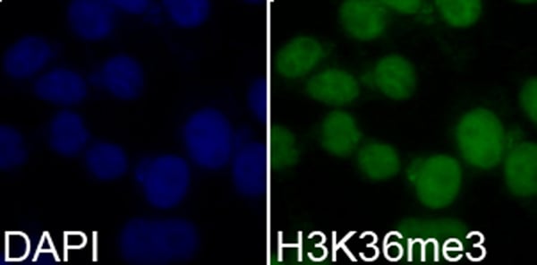


 ChIP (Chromatin Immunoprecipitation)
(Chromatin immunoprecipitation analysis extracts of 293T cells, using TriMethyl-Histone H3-K36 antibody and rabbit IgG. P1 and P2 were located on GAPDH gene. The amount of immunoprecipitated DNA was checked by quantitative PCR. Histogram was constructed by the ratios of the immunoprecipitated DNA to the input.)
ChIP (Chromatin Immunoprecipitation)
(Chromatin immunoprecipitation analysis extracts of 293T cells, using TriMethyl-Histone H3-K36 antibody and rabbit IgG. P1 and P2 were located on GAPDH gene. The amount of immunoprecipitated DNA was checked by quantitative PCR. Histogram was constructed by the ratios of the immunoprecipitated DNA to the input.)
H3K36me3, Antibody (Cat# AAA10657)
Full Name
Histone H3K36me3 Polyclonal Antibody
Gene Names
HIST3H3; H3t; H3.4; H3/g; H3FT
Reactivity
Human, Mouse, Rat, Other (Wide Range)
Applications
Western Blot, Immunohistochemistry, Immunofluorescence, Immunoprecipitation, Chromatin Immunoprecipitation, Chromatin Immunoprecipitation
Purity
Affinity Purification
Pricing


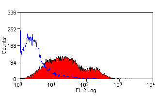
 FCM (Flow Cytometry)
(Flow cytometric analysis of PC-3M cells with BAP31 antibody at 1/100 dilution (purple) compared with an unlabelled control (cells without incubation with primary antibody; yellow). Alexa Fluor 488-conjugated goat anti-rabbit IgG was used as the secondary antibody.)
FCM (Flow Cytometry)
(Flow cytometric analysis of PC-3M cells with BAP31 antibody at 1/100 dilution (purple) compared with an unlabelled control (cells without incubation with primary antibody; yellow). Alexa Fluor 488-conjugated goat anti-rabbit IgG was used as the secondary antibody.)
BAP31, Monoclonal Antibody (Cat# AAA30501)
Full Name
BAP31 Antibody
Gene Names
BCAP31; CDM; BAP31; 6C6-AG; DXS1357E
Reactivity
Human, Mouse
Applications
Western Blot, Immunocytochemistry, Immunofluorescence, Immunohistochemistry, Flow Cytometry
Purity
ProA affinity purified
Pricing


 Application Data
(Staining of human peripheral blood granulocytes with Mouse anti Human CD45: Pacific Blue (AAA12014PB))
Application Data
(Staining of human peripheral blood granulocytes with Mouse anti Human CD45: Pacific Blue (AAA12014PB))
CD45, Monoclonal Antibody (Cat# AAA12014)
Full Name
MOUSE ANTI HUMAN CD45
Gene Names
PTPRC; LCA; LY5; B220; CD45; L-CA; T200; CD45R; GP180
Applications
Immunohistochemistry, Flow Cytometry, Immunoprecipitation, Immunohistochemistry
Pricing


 IHC (Immunohistochemistry)
(Figure 8. IHC analysis of RbAp48 using anti-RbAp48 antibody (AAA11671).RbAp48 was detected in frozen section of rat small intestine tissue. Heat mediated antigen retrieval was performed in citrate buffer (pH6, epitope retrieval solution) for 20 mins. The tissue section was blocked with 10% goat serum. The tissue section was then incubated with 1ug/ml rabbit anti-RbAp48 Antibody (AAA11671) overnight at 4 degree C. Biotinylated goat anti-rabbit IgG was used as secondary antibody and incubated for 30 minutes at 37 degree C. The tissue section was developed using Strepavidin-Biotin-Complex (SABC) with DAB as the chromogen.)
IHC (Immunohistochemistry)
(Figure 8. IHC analysis of RbAp48 using anti-RbAp48 antibody (AAA11671).RbAp48 was detected in frozen section of rat small intestine tissue. Heat mediated antigen retrieval was performed in citrate buffer (pH6, epitope retrieval solution) for 20 mins. The tissue section was blocked with 10% goat serum. The tissue section was then incubated with 1ug/ml rabbit anti-RbAp48 Antibody (AAA11671) overnight at 4 degree C. Biotinylated goat anti-rabbit IgG was used as secondary antibody and incubated for 30 minutes at 37 degree C. The tissue section was developed using Strepavidin-Biotin-Complex (SABC) with DAB as the chromogen.)
RbAp48, Polyclonal Antibody (Cat# AAA11671)
Full Name
Anti-RbAp48 Antibody
Gene Names
RBBP4; NURF55; RBAP48; lin-53
Reactivity
Human, Mouse, Rat
Applications
Western Blot, Immunohistochemistry
Purity
Immunogen Affinity Purified
Pricing


 FCM (Flow Cytometry)
(Figure 6. Flow Cytometry analysis of U20S cells using anti-PPID antibody (AAA19159).Overlay histogram showing U20S cells stained with AAA19159 (Blue line).The cells were blocked with 10% normal goat serum. And then incubated with rabbit anti-PPID Antibody (AAA19159,1ug/1x10^6 cells) for 30 min at 20 degree C. DyLight®488 conjugated goat anti-rabbit IgG (5-10ug/1x10^6 cells) was used as secondary antibody for 30 minutes at 20 degree C. Isotype control antibody (Green line) was rabbit IgG (1ug/1x106) used under the same conditions. Unlabelled sample (Red line) was also used as a control.)
FCM (Flow Cytometry)
(Figure 6. Flow Cytometry analysis of U20S cells using anti-PPID antibody (AAA19159).Overlay histogram showing U20S cells stained with AAA19159 (Blue line).The cells were blocked with 10% normal goat serum. And then incubated with rabbit anti-PPID Antibody (AAA19159,1ug/1x10^6 cells) for 30 min at 20 degree C. DyLight®488 conjugated goat anti-rabbit IgG (5-10ug/1x10^6 cells) was used as secondary antibody for 30 minutes at 20 degree C. Isotype control antibody (Green line) was rabbit IgG (1ug/1x106) used under the same conditions. Unlabelled sample (Red line) was also used as a control.)
PPID/Cyclophilin 40, Polyclonal Antibody (Cat# AAA19159)
Full Name
Anti-PPID/Cyclophilin 40 Picoband antibody
Gene Names
PPID; CYPD; CYP-40
Reactivity
Human, Mouse, Rat
No cross reactivity with other proteins.
No cross reactivity with other proteins.
Applications
EIA, FC/FACS, IHC, ICC, WB
Pricing


 FCM (Flow Cytometry)
(Figure 8. Flow Cytometry analysis of HepG2 cells using anti-MERTK antibody (AAA19219).Overlay histogram showing HepG2 cells stained with AAA19219 (Blue line). The cells were blocked with 10% normal goat serum. And then incubated with rabbit anti-MERTK Antibody (AAA19219, 1ug/1x106 cells) for 30 min at 20 degree C. DyLight488 conjugated goat anti-rabbit IgG (5-10ug/1x106 cells) was used as secondary antibody for 30 minutes at 20 degree C. Isotype control antibody (Green line) was rabbit IgG (1ug/1x106) used under the same conditions. Unlabelled sample (Red line) was also used as a control.)
FCM (Flow Cytometry)
(Figure 8. Flow Cytometry analysis of HepG2 cells using anti-MERTK antibody (AAA19219).Overlay histogram showing HepG2 cells stained with AAA19219 (Blue line). The cells were blocked with 10% normal goat serum. And then incubated with rabbit anti-MERTK Antibody (AAA19219, 1ug/1x106 cells) for 30 min at 20 degree C. DyLight488 conjugated goat anti-rabbit IgG (5-10ug/1x106 cells) was used as secondary antibody for 30 minutes at 20 degree C. Isotype control antibody (Green line) was rabbit IgG (1ug/1x106) used under the same conditions. Unlabelled sample (Red line) was also used as a control.)
MERTK, Polyclonal Antibody (Cat# AAA19219)
Full Name
Anti-MERTK Antibody
Gene Names
MERTK; MER; RP38; c-mer
Reactivity
Human, Mouse, Rat
Applications
Western Blot, Immunohistochemistry, Flow Cytometry, Direct ELISA
Purity
Immunogen affinity purified.
Pricing


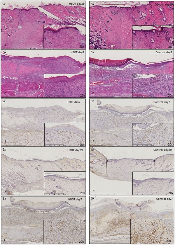 Application Data
(Published customer image: Histological staining of control and HBOT wounds at post-wounding days 7 and 29. A -D) H&E staining. E -H) CD34 immunohistochemistry. I+J) CD68 immunohistochemistry.From: uk B, Tong M, Fijneman EMG, van Neck JW (2014) Hyperbaric Oxygen Therapy to Treat Diabetes Impaired Wound Healing in Rats. PLoS ONE 9(10): e108533.)
Application Data
(Published customer image: Histological staining of control and HBOT wounds at post-wounding days 7 and 29. A -D) H&E staining. E -H) CD34 immunohistochemistry. I+J) CD68 immunohistochemistry.From: uk B, Tong M, Fijneman EMG, van Neck JW (2014) Hyperbaric Oxygen Therapy to Treat Diabetes Impaired Wound Healing in Rats. PLoS ONE 9(10): e108533.)
CD68, Monoclonal Antibody (Cat# AAA12148)
Full Name
MOUSE ANTI RAT CD68:FITC
Applications
Flow Cytometry
Pricing


 Application Data
(Published customer image: Mouse anti Human CD49d antibody, clone HP2/1 used for binding efficiency determinationImage caption:Binding efficiencies (BE) of different a4beta7 molecules composed of distinct a4 mutants to monoclonal antibodies against a4, beta7 or the a4beta7 heterodimer. Binding efficiency is determined by the ratio between the mean fluorescence of antibody binding to each a4 molecule and of the binding in a mock-transfected cell culture (see Materials and Methods for details). Dark gray bars represent binding to the human (wild type) a4 clone, whereas light gray bars are those of binding to the different a4 mutants (as shown in the x-axis). a4 mutants which included substitutions at codon 201 are boxed. A, binding of anti-a4 2b4 antibody. B, binding of anti-a4 HP2/1 antibody. C, BE of different anti-a4 and beta7 antibodies to the human a4 and the quintuple a4 mutant (5 aa mut). Bars represent the range of standard errors deduced from triplicate experiments. p-values of Student's t tests are shown above each comparison. NS, non-significant (> 0.05).From:Darc M, Hait SH, Soares EA, Cicala C, Seuanez HN, et al. (2011) Polymorphisms in the a4 Integrin of Neotropical Primates: Insights for Binding of Natural Ligands and HIV-1 gp120 to the Human a4beta7. PLoS ONE 6(9): e24461.)
Application Data
(Published customer image: Mouse anti Human CD49d antibody, clone HP2/1 used for binding efficiency determinationImage caption:Binding efficiencies (BE) of different a4beta7 molecules composed of distinct a4 mutants to monoclonal antibodies against a4, beta7 or the a4beta7 heterodimer. Binding efficiency is determined by the ratio between the mean fluorescence of antibody binding to each a4 molecule and of the binding in a mock-transfected cell culture (see Materials and Methods for details). Dark gray bars represent binding to the human (wild type) a4 clone, whereas light gray bars are those of binding to the different a4 mutants (as shown in the x-axis). a4 mutants which included substitutions at codon 201 are boxed. A, binding of anti-a4 2b4 antibody. B, binding of anti-a4 HP2/1 antibody. C, BE of different anti-a4 and beta7 antibodies to the human a4 and the quintuple a4 mutant (5 aa mut). Bars represent the range of standard errors deduced from triplicate experiments. p-values of Student's t tests are shown above each comparison. NS, non-significant (> 0.05).From:Darc M, Hait SH, Soares EA, Cicala C, Seuanez HN, et al. (2011) Polymorphisms in the a4 Integrin of Neotropical Primates: Insights for Binding of Natural Ligands and HIV-1 gp120 to the Human a4beta7. PLoS ONE 6(9): e24461.)
CD49d, Monoclonal Antibody (Cat# AAA12003)
Full Name
MOUSE ANTI HUMAN CD49d
Gene Names
ITGA4; IA4; CD49D
Applications
Immunohistochemistry, Flow Cytometry, Functional Assay, Immunoprecipitation
Pricing


 Application Data
(Published customer image Infiltration of GFP+ BM-cells in infarct and peri-infarct regions. (A-B) Dot plots of viable macrophages/granulocytes (CD11b+CD45high, top right quadrants) and microglia (CD11b+CD45dim, bottom right quadrants) in cortex from BM-chimeric unmanipulated mice and mice exposed to pMCAO. (C) Bar graph showing mean numbers of CD11b+CD45dim microglia and CD11b+CD45high macrophages/granulocytes in BM-chimeric mice 24 hours after pMCAO, subdivided based on expression of GFP (n = 5). Approximately 92% of of the CD45high population were GFP+. (D) Estimation and comparison of mean numbers of CD11b+CD45dim microglia in non-chimeric (n = 10) versus BM-chimeric mice (n = 5) 24 hours after of pMCAO shows significantly fewer CD11b+CD45dim microglial cells in irradiated mice. (E) Overview, showing distribution of infiltrating GFP+ BM-derived cells into infarct (IF) and peri-infarct (P-IF) regions 24 hours after pMCAO. (E-G) By 24 hours, GFP+ single cells (F) and vessel-associated aggregates of GFP+ cells (arrows in G) were observed in infarct and peri-infarct regions. Some of the vessel-associated cells were round, leukocyte-like cells (arrows) while others were elongated cells lining the vasculature (arrow heads in G and in insert). (H) Bar graph showing mean numbers of single GFP+ cells and vessel-associated aggregates of GFP+ cells in ipsi- and contralateral cortex 24 hours after surgery (n = 10). (I-P) Immunohistochemical staining of CD45.1 (I, K), CD45.2 (J, L), IgG2a (M, O) and CD45 (N, P) in ischemic tissue in BM-chimeric (I, J, M, N) and non-chimeric mice (K, L, O, P) 24 hours after pMCAO. N.D, none detected. Scale bars: 200 um (A), 10 um (B, C). 50 um (I-P) *P < 0.05, **P < 0.01, and ***P < 0.001.From: Clausen BH, Lambertsen KL, Babcock AA, Holm TH, Dagnaes-Hansen F, Finsen B. Interleukin-1beta and tumor necrosis factor-alpha are expressed by different subsets of microglia and macrophages after ischemic stroke in mice. J Neuroinflammation. 2008 Oct 23;5:46.)
Application Data
(Published customer image Infiltration of GFP+ BM-cells in infarct and peri-infarct regions. (A-B) Dot plots of viable macrophages/granulocytes (CD11b+CD45high, top right quadrants) and microglia (CD11b+CD45dim, bottom right quadrants) in cortex from BM-chimeric unmanipulated mice and mice exposed to pMCAO. (C) Bar graph showing mean numbers of CD11b+CD45dim microglia and CD11b+CD45high macrophages/granulocytes in BM-chimeric mice 24 hours after pMCAO, subdivided based on expression of GFP (n = 5). Approximately 92% of of the CD45high population were GFP+. (D) Estimation and comparison of mean numbers of CD11b+CD45dim microglia in non-chimeric (n = 10) versus BM-chimeric mice (n = 5) 24 hours after of pMCAO shows significantly fewer CD11b+CD45dim microglial cells in irradiated mice. (E) Overview, showing distribution of infiltrating GFP+ BM-derived cells into infarct (IF) and peri-infarct (P-IF) regions 24 hours after pMCAO. (E-G) By 24 hours, GFP+ single cells (F) and vessel-associated aggregates of GFP+ cells (arrows in G) were observed in infarct and peri-infarct regions. Some of the vessel-associated cells were round, leukocyte-like cells (arrows) while others were elongated cells lining the vasculature (arrow heads in G and in insert). (H) Bar graph showing mean numbers of single GFP+ cells and vessel-associated aggregates of GFP+ cells in ipsi- and contralateral cortex 24 hours after surgery (n = 10). (I-P) Immunohistochemical staining of CD45.1 (I, K), CD45.2 (J, L), IgG2a (M, O) and CD45 (N, P) in ischemic tissue in BM-chimeric (I, J, M, N) and non-chimeric mice (K, L, O, P) 24 hours after pMCAO. N.D, none detected. Scale bars: 200 um (A), 10 um (B, C). 50 um (I-P) *P < 0.05, **P < 0.01, and ***P < 0.001.From: Clausen BH, Lambertsen KL, Babcock AA, Holm TH, Dagnaes-Hansen F, Finsen B. Interleukin-1beta and tumor necrosis factor-alpha are expressed by different subsets of microglia and macrophages after ischemic stroke in mice. J Neuroinflammation. 2008 Oct 23;5:46.)
CD11b, Monoclonal Antibody (Cat# AAA12181)
Full Name
RAT ANTI MOUSE CD11b
Gene Names
Itgam; CR3; CR3A; MAC1; Cd11b; Ly-40; Mac-1; Mac-1a; CD11b/CD18; F730045J24Rik
Reactivity
Human
Applications
Immunohistochemistry, Flow Cytometry, Immunofluorescence, Immunoprecipitation
Pricing
 Standard Curve (Sample)
Cystic Fibrosis Transmembrane Conductance Regulator (CFTR), ELISA Kit (Cat# AAA20425)
Full Name
Human Cystic Fibrosis Transmembrane Conductance Regulator (CFTR) ELISA Kit
Gene Names
CFTR; CF; MRP7; ABC35; ABCC7; CFTR/MRP; TNR-CFTR; dJ760C5.1
Reactivity
Human
Pricing


 IF (Immunofluorescence)
(Immunofluorescence analysis of MCF7 cells using MYO1C antibody.)
IF (Immunofluorescence)
(Immunofluorescence analysis of MCF7 cells using MYO1C antibody.)
MYO1C, Polyclonal Antibody (Cat# AAA29832)
Full Name
MYO1C Polyclonal Antibody
Gene Names
MYO1C; NMI; MMIb; myr2; MMI-beta
Reactivity
Human, Mouse, Rat
Applications
Western Blot, Immunohistochemistry, Immunofluorescence
Purity
Affinity purification
Pricing
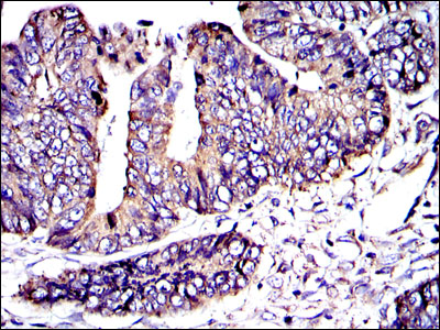


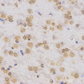
 IHC (Immunohistchemistry)
(Immunohistochemical analysis of paraffin-embedded bladder cancer tissues using CRK mouse mAb with DAB staining.)
IHC (Immunohistchemistry)
(Immunohistochemical analysis of paraffin-embedded bladder cancer tissues using CRK mouse mAb with DAB staining.)
CRK, Monoclonal Antibody (Cat# AAA14106)
Full Name
Anti-CRK Mouse mAb
Gene Names
CRK; p38; CRKII
Reactivity
Human
Applications
Western Blot, Immunohistochemistry, Immunocytochemistry, Flow Cytometry
Pricing


 Application Data
(Detection limit for recombinant GST tagged TFAP4 is approximately 0.1ng/ml as a capture antibody.)
Application Data
(Detection limit for recombinant GST tagged TFAP4 is approximately 0.1ng/ml as a capture antibody.)
TFAP4, Monoclonal Antibody (Cat# AAA26201)
Full Name
TFAP4 (Transcription Factor AP-4 (Activating Enhancer Binding Protein 4), AP-4, bHLHc41) (Biotin)
Gene Names
TFAP4; AP-4; bHLHc41
Applications
Immunofluorescence, Immunohistochemistry, Western Blot
Purity
Purified
Pricing


 Transfection Control
(Anti-XPR1 antibody immunocytochemistry (ICC) staining of untransfected HEK293 human embryonic kidney cells.)
Transfection Control
(Anti-XPR1 antibody immunocytochemistry (ICC) staining of untransfected HEK293 human embryonic kidney cells.)
XPR1, Polyclonal Antibody (Cat# AAA12332)
Full Name
Rabbit Polyclonal to Human XPR1
Gene Names
XPR1; X3; SYG1
Reactivity
Gibbon, Hamster, Human, Monkey, Mouse, Rabbit
Predicted Reactivity: Rat, Bovine, Dog, Horse, Pig (at least 90% immunogen sequence identity)
Predicted Reactivity: Rat, Bovine, Dog, Horse, Pig (at least 90% immunogen sequence identity)
Applications
Immunohistochemistry, Immunocytochemistry
Purity
Immunoaffinity Purified
Pricing


 Application Data
(Staining of human peripheral blood monocytes with Mouse anti Human CD33:RPE)
Application Data
(Staining of human peripheral blood monocytes with Mouse anti Human CD33:RPE)
CD33, Monoclonal Antibody (Cat# AAA12027)
Full Name
MOUSE ANTI HUMAN CD33:RPE
Gene Names
CD33; p67; SIGLEC3; SIGLEC-3
Applications
Flow Cytometry
Pricing


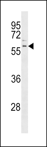
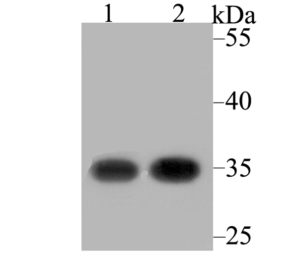
 WB (Western Blot)
(Western blot analysis of JNK1 on hybrid fish (crucian-carp) brain tissue lysate using anti-JNK1 antibody at 1/500 dilution.)
WB (Western Blot)
(Western blot analysis of JNK1 on hybrid fish (crucian-carp) brain tissue lysate using anti-JNK1 antibody at 1/500 dilution.)
JNK1, Polyclonal Antibody (Cat# AAA29866)
Full Name
JNK1 Antibody
Gene Names
MAPK8; JNK; JNK1; PRKM8; SAPK1; JNK-46; JNK1A2; SAPK1c; JNK21B1/2
Reactivity
Human, Mouse, Rat, Zebrafish
Applications
Western Blot, Immunocytochemistry, Immunohistochemistry, Flow Cytometry
Purity
Immunogen affinity purified
Pricing


 WB (Western Blot)
(Western blot analysis of CDC25C over-expressed 293 cell line, cotransfected with CDC25C Validated Chimera RNAi (Lane 2) or non-transfected control (Lane 1). Blot probed with CDC25C monoclonal antibody. GAPDH (36.1kD) used as specificity and loading control.)
WB (Western Blot)
(Western blot analysis of CDC25C over-expressed 293 cell line, cotransfected with CDC25C Validated Chimera RNAi (Lane 2) or non-transfected control (Lane 1). Blot probed with CDC25C monoclonal antibody. GAPDH (36.1kD) used as specificity and loading control.)
CDC25C, Monoclonal Antibody (Cat# AAA25051)
Full Name
CDC25C (Cell Division Cycle 25 Homolog C, Cdc 25C, CDC 25, Dual Specificity Phosphatase Cdc25C, M-phase Inducer Phosphatase 3, MPIP3, PPP1R60) (FITC)
Gene Names
CDC25C; CDC25; PPP1R60
Reactivity
Human
Applications
EIA, IHC, IP, WB
Purity
Purified by Protein A Affinity Chromatography.
Pricing


 ICC (Immunocytochemistry)
(ICC staining Histone H4 in PANC-1 cells (green). The nuclear counter stain is DAPI (blue). Cells were fixed in paraformaldehyde, permeabilised with 0.25% Triton X100/PBS.)
ICC (Immunocytochemistry)
(ICC staining Histone H4 in PANC-1 cells (green). The nuclear counter stain is DAPI (blue). Cells were fixed in paraformaldehyde, permeabilised with 0.25% Triton X100/PBS.)
Histone H4, Monoclonal Antibody (Cat# AAA30201)
Full Name
Histone H4 Antibody
Gene Names
HIST2H4B; H4/o
Reactivity
Human, Mouse, Rat
Applications
WB, ICC, IF, IHC
Purity
ProA affinity purified
Pricing


 IF (Immunofluorescence)
(Immunofluorescence analysis of U-2 OS cells using Phospho-Histone H3-S10/T11 Rabbit pAb at dilution of 100 (40x lens). Blue: DAPI for nuclear staining.)
IF (Immunofluorescence)
(Immunofluorescence analysis of U-2 OS cells using Phospho-Histone H3-S10/T11 Rabbit pAb at dilution of 100 (40x lens). Blue: DAPI for nuclear staining.)
Phospho-Histone H3-S10/T11, Polyclonal Antibody (Cat# AAA28391)
Full Name
Phospho-Histone H3-S10/T11 Rabbit pAb
Gene Names
HIST1H3A; H3/A; H3FA
Reactivity
Human, Mouse, Rat, Other (Wide Range)
Applications
WB, IHC, IF
Purity
Affinity purification
Pricing


 IHC (Immunohistochemistry)
(Figure 7. IHC analysis of GALNS using anti-GALNS antibody (AAA19164).GALNS was detected in paraffin-embedded section of rat small intestine tissue. Heat mediated antigen retrieval was performed in citrate buffer (pH6, epitope retrieval solution) for 20 mins. The tissue section was blocked with 10% goat serum. The tissue section was then incubated with 1ug/ml rabbit anti-GALNS Antibody (AAA19164) overnight at 4 degree C. Biotinylated goat anti-rabbit IgG was used as secondary antibody and incubated for 30 minutes at 37 degree C. The tissue section was developed using Strepavidin-Biotin-Complex (SABC) with DAB as the chromogen.)
IHC (Immunohistochemistry)
(Figure 7. IHC analysis of GALNS using anti-GALNS antibody (AAA19164).GALNS was detected in paraffin-embedded section of rat small intestine tissue. Heat mediated antigen retrieval was performed in citrate buffer (pH6, epitope retrieval solution) for 20 mins. The tissue section was blocked with 10% goat serum. The tissue section was then incubated with 1ug/ml rabbit anti-GALNS Antibody (AAA19164) overnight at 4 degree C. Biotinylated goat anti-rabbit IgG was used as secondary antibody and incubated for 30 minutes at 37 degree C. The tissue section was developed using Strepavidin-Biotin-Complex (SABC) with DAB as the chromogen.)
GALNS, Antibody (Cat# AAA19164)
Full Name
Anti-GALNS Picoband antibody
Gene Names
GALNS; GAS; MPS4A; GalN6S; GALNAC6S
Reactivity
Reacts with: Human, Mouse, Rat
Applications
WB, EIA
Purity
Immunogen affinity purified
Pricing


 Application Data
(Published clone specific image: Flow cytometric analysis of AM from NO2-exposed and control rats. Rats were exposed to NO2 for the indicated times and BAL cells were stained with antibodies to ED7, ED9, RM-4, and OX-6. To overcome autofluorescence signals, primary antibodies were detected using a biotin-PE/streptavidin-anti-streptavidin enhancing system and labeling of AM was analyzed by flow cytometry following gating by help of forward and sideward scatter properties. Shown are representative results of at least six animals per group.From: Garn H, Siese A, Stumpf S, Wensing A, Renz H, Gemsa D. Phenotypical and functional characterization of alveolar macrophage subpopulations in the lungs of NO2-exposed rats. Respir Res. 2006 Jan 6;7:4.)
Application Data
(Published clone specific image: Flow cytometric analysis of AM from NO2-exposed and control rats. Rats were exposed to NO2 for the indicated times and BAL cells were stained with antibodies to ED7, ED9, RM-4, and OX-6. To overcome autofluorescence signals, primary antibodies were detected using a biotin-PE/streptavidin-anti-streptavidin enhancing system and labeling of AM was analyzed by flow cytometry following gating by help of forward and sideward scatter properties. Shown are representative results of at least six animals per group.From: Garn H, Siese A, Stumpf S, Wensing A, Renz H, Gemsa D. Phenotypical and functional characterization of alveolar macrophage subpopulations in the lungs of NO2-exposed rats. Respir Res. 2006 Jan 6;7:4.)
CD172a, Monoclonal Antibody (Cat# AAA12001)
Full Name
MOUSE ANTI RAT CD172a
Gene Names
Sirpa; Bit; Ptpns1; SHPS-1
Applications
Immunohistochemistry, Flow Cytometry, Immunoprecipitation, Western Blot
Pricing


 FCM (Flow Cytometry)
(Figure 7. Flow Cytometry analysis of HEPA1-6 cells using anti-Rad9b antibody (AAA19340).Overlay histogram showing HEPA1-6 cells stained with AAA19340 (Blue line). The cells were blocked with 10% normal goat serum. And then incubated with rabbit anti-Rad9b Antibody (AAA19340, 1μg/1x106 cells) for 30 min at 20 degree C. DyLight®488 conjugated goat anti-rabbit IgG (5-10μg/1x106 cells) was used as secondary antibody for 30 minutes at 20 degree C. Isotype control antibody (Green line) was rabbit IgG (1μg/1x106) used under the same conditions. Unlabelled sample (Red line) was also used as a control.)
FCM (Flow Cytometry)
(Figure 7. Flow Cytometry analysis of HEPA1-6 cells using anti-Rad9b antibody (AAA19340).Overlay histogram showing HEPA1-6 cells stained with AAA19340 (Blue line). The cells were blocked with 10% normal goat serum. And then incubated with rabbit anti-Rad9b Antibody (AAA19340, 1μg/1x106 cells) for 30 min at 20 degree C. DyLight®488 conjugated goat anti-rabbit IgG (5-10μg/1x106 cells) was used as secondary antibody for 30 minutes at 20 degree C. Isotype control antibody (Green line) was rabbit IgG (1μg/1x106) used under the same conditions. Unlabelled sample (Red line) was also used as a control.)
Rad9b, Polyclonal Antibody (Cat# AAA19340)
Full Name
Anti-Rad9b Antibody
Gene Names
Rad9b; BC021784; A630082N15Rik
Reactivity
Mouse, Rat
Applications
WB, IHC-P, FC/FACS/FCM, EIA
Purity
Immunogen affinity purified.
Pricing
 Standard Curve (Sample)
Macrophage Inflammatory Protein-1alpha, MIP-1alpha, ELISA Kit (Cat# AAA19047)
Full Name
Monkey Macrophage Inflammatory Protein-1alpha, MIP-1alpha ELISA Kit
Reactivity
Monkey
Pricing


 Application Data
(Immunoperoxidase staining of rat lymph node cryosection with Mouse anti Rat CD86 antibody, clone 42F followed by horseradish peroxidase conjugated Goat anti Mouse IgG . Low power)
Application Data
(Immunoperoxidase staining of rat lymph node cryosection with Mouse anti Rat CD86 antibody, clone 42F followed by horseradish peroxidase conjugated Goat anti Mouse IgG . Low power)
CD86, Monoclonal Antibody (Cat# AAA12222)
Full Name
MOUSE ANTI RAT CD86
Gene Names
CD86; B70; B7-2; B7.2; LAB72; CD28LG2
Applications
Immunohistochemistry, Flow Cytometry, Immunoprecipitation
Pricing


 FCM (Flow Cytometry)
(Flow cytometric analysis of HepG2 cells with MTCO2 antibody at 1/50 dilution (red) compared with an unlabelled control (cells without incubation with primary antibody; black). Alexa Fluor 488-conjugated goat anti rabbit IgG was used as the secondary antibody)
FCM (Flow Cytometry)
(Flow cytometric analysis of HepG2 cells with MTCO2 antibody at 1/50 dilution (red) compared with an unlabelled control (cells without incubation with primary antibody; black). Alexa Fluor 488-conjugated goat anti rabbit IgG was used as the secondary antibody)
MTCO2, Monoclonal Antibody (Cat# AAA30139)
Full Name
MTCO2 Antibody
Gene Names
MT-CO2; COII; MTCO2; COX2
Reactivity
Human
Applications
Western Blot, Immunocytochemistry, Immunofluorescence, Immunohistochemistry, Immunoprecipitation, Flow Cytometry
Purity
ProA affinity purified
Pricing


 FCM (Flow Cytometry)
(Flow cytometric analysis of SH-SY-5Y cells with RACK1 antibody at 1/100 dilution (purple) compared with an unlabelled control (cells without incubation with primary antibody; yellow). Alexa Fluor 488-conjugated goat anti-rabbit IgG was used as the secondary antibody.)
FCM (Flow Cytometry)
(Flow cytometric analysis of SH-SY-5Y cells with RACK1 antibody at 1/100 dilution (purple) compared with an unlabelled control (cells without incubation with primary antibody; yellow). Alexa Fluor 488-conjugated goat anti-rabbit IgG was used as the secondary antibody.)
RACK1, Monoclonal Antibody (Cat# AAA30521)
Full Name
RACK1 Antibody
Gene Names
GNB2L1; H12.3; HLC-7; PIG21; RACK1; Gnb2-rs1
Reactivity
Human, Mouse, Rat
Applications
Western Blot, Immunohistochemistry, Flow Cytometry
Purity
ProA affinity purified
Pricing