Filters
Clonality
Type
Reactivity
Gene Name
Isotype
Host
Application
Clone
219 results for "Rat Anti Mouse IgG2a" - showing 50-100


 Application Data
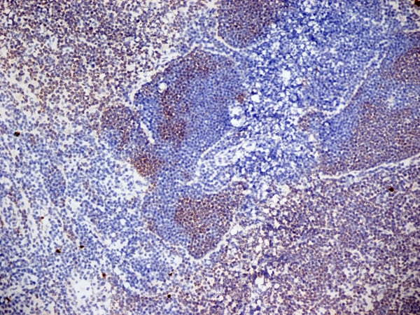
(Immunoperoxidase staining of human tonsil cryosection using Mouse anti Human CD3 antibody, clone UCHT1 followed by horseradish peroxidase Goat anti Mouse IgG2a antibody as a detection reagent. Medium power)
Application Data
(Immunoperoxidase staining of human tonsil cryosection using Mouse anti Human CD3 antibody, clone UCHT1 followed by horseradish peroxidase Goat anti Mouse IgG2a antibody as a detection reagent. Medium power)
CD3, Monoclonal Antibody (Cat# AAA11908)
Full Name
MOUSE ANTI HUMAN CD3:Low Endotoxin
Gene Names
CD3G; T3G; IMD17; CD3-GAMMA
Applications
Immunohistochemistry, Flow Cytometry
Pricing


 Application Data
(Published customer image: Leukocyte infiltration in COX-2-M/-M and COX-2+/+ mice. MPO enzymatic activity (panel A) was statistically similar in COX-2-M/-M and COX-2+/+ livers at 6 h and 24 h post-IRI. Ly-6G+ neutrophil (panel B) and granulocyte (panel C) infiltration were also comparable in COX-2-M/-M and COX-2+/+ livers after IRI. Mac-1+ (panel D) and CD68 (panel E) infiltrating macrophages were significantly reduced in COX-2-M/-M livers at 24 h post-reperfusion, but were statistically indistinguishable in COX-2-M/-M and COX-2+/+ livers at 6 h after IRI. No statistical differences in MMP-9 expression (panel F) could be demonstrated in livers of COX-2-M/-M and COX-2+/+ mice post-IRI. Representative immunostaining (panel G) of infiltrating Ly-6G+ (a,b,e,f) and Mac-1+ (c,d,g,h) leukocytes in livers of COX-2+/+ (a,c,e,g) and COX-2-M/-M (b,d,f,h) mice at 6 h (a to d) and 24 h (e to h) post IRI; (n = 5 -6/group; * indicates p)
Application Data
(Published customer image: Leukocyte infiltration in COX-2-M/-M and COX-2+/+ mice. MPO enzymatic activity (panel A) was statistically similar in COX-2-M/-M and COX-2+/+ livers at 6 h and 24 h post-IRI. Ly-6G+ neutrophil (panel B) and granulocyte (panel C) infiltration were also comparable in COX-2-M/-M and COX-2+/+ livers after IRI. Mac-1+ (panel D) and CD68 (panel E) infiltrating macrophages were significantly reduced in COX-2-M/-M livers at 24 h post-reperfusion, but were statistically indistinguishable in COX-2-M/-M and COX-2+/+ livers at 6 h after IRI. No statistical differences in MMP-9 expression (panel F) could be demonstrated in livers of COX-2-M/-M and COX-2+/+ mice post-IRI. Representative immunostaining (panel G) of infiltrating Ly-6G+ (a,b,e,f) and Mac-1+ (c,d,g,h) leukocytes in livers of COX-2+/+ (a,c,e,g) and COX-2-M/-M (b,d,f,h) mice at 6 h (a to d) and 24 h (e to h) post IRI; (n = 5 -6/group; * indicates p)
CD68, Monoclonal Antibody (Cat# AAA12105)
Full Name
RAT ANTI MOUSE CD68:FITC
Gene Names
Cd68; Lamp4; gp110; Scard1
Applications
Flow Cytometry
Pricing


 Application Data
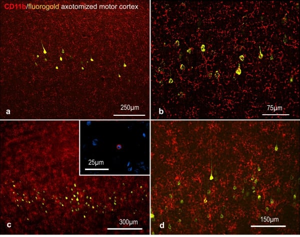
(Published customer image: Representative images of the inflammatory changes in the facial nucleus during axonal regeneration, one week following facial nerve transaction. a, b: CD11b immunoreactivity for microglia is increased in the axotomized facial nucleus, and microglia enwrap the facial motor neurons, e.g. at arrows. The regenerating neurons were retrogradely labelled with fluorogold. c, d: CD6- positive T-cells accumulated in the injured motor nucleus (arrows). They had little cytoplasm but dense nuclei (c) and were sometimes clustered around neurons retrogradely labelled with fluorogold (d). The scale bar in (a) also applies to (b) and that in (c) also applies to (d).From: Shokouhi et al. BMC Neuroscience 2010 11:13.)
Application Data
(Published customer image: Representative images of the inflammatory changes in the facial nucleus during axonal regeneration, one week following facial nerve transaction. a, b: CD11b immunoreactivity for microglia is increased in the axotomized facial nucleus, and microglia enwrap the facial motor neurons, e.g. at arrows. The regenerating neurons were retrogradely labelled with fluorogold. c, d: CD6- positive T-cells accumulated in the injured motor nucleus (arrows). They had little cytoplasm but dense nuclei (c) and were sometimes clustered around neurons retrogradely labelled with fluorogold (d). The scale bar in (a) also applies to (b) and that in (c) also applies to (d).From: Shokouhi et al. BMC Neuroscience 2010 11:13.)
CD11b, Monoclonal Antibody (Cat# AAA11971)
Full Name
MOUSE ANTI RAT CD11b
Gene Names
ITGAM; CD11B
Applications
Immunohistochemistry, Flow Cytometry, Immunofluorescence, Immunoprecipitation
Pricing


 Application Data
(Immunoperoxidase staining of human tonsil cryosection using Mouse anti Human CD3 antibody, clone UCHT1 followed by horseradish peroxidase Goat anti Mouse IgG2a antibody as a detection reagent. Medium power)
Application Data
(Immunoperoxidase staining of human tonsil cryosection using Mouse anti Human CD3 antibody, clone UCHT1 followed by horseradish peroxidase Goat anti Mouse IgG2a antibody as a detection reagent. Medium power)
CD3, Monoclonal Antibody (Cat# AAA12046)
Full Name
MOUSE ANTI HUMAN CD3:RPE
Gene Names
CD3G; T3G; IMD17; CD3-GAMMA
Applications
Flow Cytometry
Pricing


 Application Data
(Staining of Rat peripheral blood lymphocytes with Mouse anti Rat CD49d:RPE (MCA2872PE))
Application Data
(Staining of Rat peripheral blood lymphocytes with Mouse anti Rat CD49d:RPE (MCA2872PE))
CD49d, Monoclonal Antibody (Cat# AAA26748)
Full Name
CD49d (Antigen CD49d, CD49d Antigen, CDw49d, Alpha 4 Subunit of VLA-4 Receptor, Integrin alpha IV, Integrin alpha 4, IA4, ITGA4, LPAM23, MGC90518, Very Late Activation Protein 4 Receptor Alpha 4 Subunit, VLA4, VLA-4) (FITC)
Reactivity
Rat
Applications
Flow Cytometry, Immunoprecipitation
Purity
Purified by Protein G Affinity Chromatography
Pricing


 Application Data
(Immunoperoxidase staining of mouse lymph node cryosection stained with Rat antii Mouse CD8alpha antibody, clone KT15 followed by horseradish peroxidase conjugatedGoat anti Rat IgG s a detection reagent. High power)
Application Data
(Immunoperoxidase staining of mouse lymph node cryosection stained with Rat antii Mouse CD8alpha antibody, clone KT15 followed by horseradish peroxidase conjugatedGoat anti Rat IgG s a detection reagent. High power)
CD8 ALPHA, Monoclonal Antibody (Cat# AAA11999)
Full Name
RAT ANTI MOUSE CD8 ALPHA
Gene Names
Cd8a; Ly-2; Ly-B; Ly-35; Lyt-2; BB154331
Applications
Immunohistochemistry, Flow Cytometry, Immunofluorescence
Pricing


 FCM (Flow Cytometry)
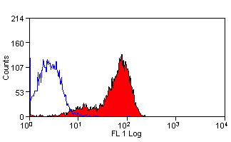
(Figure 12. Flow Cytometry analysis of A549 cells using anti-Transketolase/TKT antibody (AAA19367).Overlay histogram showing A549 cells stained with AAA19367 (Blue line). The cells were blocked with 10% normal goat serum. And then incubated with mouse anti- Transketolase/TKT Antibody (AAA19367, 1μg/1x106 cells) for 30 min at 20 degree C. DyLight®488 conjugated goat anti-mouse IgG (BA1126, 5-10μg/1x106 cells) was used as secondary antibody for 30 minutes at 20 degree C. Isotype control antibody (Green line) was mouse IgG (1μg/1x106) used under the same conditions. Unlabelled sample (Red line) was also used as a control.)
FCM (Flow Cytometry)
(Figure 12. Flow Cytometry analysis of A549 cells using anti-Transketolase/TKT antibody (AAA19367).Overlay histogram showing A549 cells stained with AAA19367 (Blue line). The cells were blocked with 10% normal goat serum. And then incubated with mouse anti- Transketolase/TKT Antibody (AAA19367, 1μg/1x106 cells) for 30 min at 20 degree C. DyLight®488 conjugated goat anti-mouse IgG (BA1126, 5-10μg/1x106 cells) was used as secondary antibody for 30 minutes at 20 degree C. Isotype control antibody (Green line) was mouse IgG (1μg/1x106) used under the same conditions. Unlabelled sample (Red line) was also used as a control.)
Transketolase/TKT, Monoclonal Antibody (Cat# AAA19367)
Full Name
Anti-Transketolase/TKT Antibody (monoclonal, 3E5)
Gene Names
TKT; TK; TKT1; HEL107
Reactivity
Human, Mouse, Rat
Applications
WB, IHC-P, ICC, IF, FC/FACS/FCM
Purity
Immunogen affinity purified.
Pricing
 Application Data
(Antibody Concentration Used: 1.0ug/ 10^6 cells Secondary Antibody Used: Goat anti-rat PE 1:500 dil.)
Application Data
(Antibody Concentration Used: 1.0ug/ 10^6 cells Secondary Antibody Used: Goat anti-rat PE 1:500 dil.)
CD200, Monoclonal Antibody (Cat# AAA14268)
Full Name
Anti-Mouse CD200, Purified (Clone OX90) (rat IgG2a)
Gene Names
CD200; MRC; MOX1; MOX2; OX-2
Reactivity
Mouse
Applications
Immunohistochemistry
Pricing


 Application Data
(Staining of mouse spleen cells with Rat anti Mouse CD3 Epsilon (T3): APC)
Application Data
(Staining of mouse spleen cells with Rat anti Mouse CD3 Epsilon (T3): APC)
CD3, Monoclonal Antibody (Cat# AAA12050)
Full Name
RAT ANTI MOUSE CD3:RPE
Gene Names
Cd3e; CD3; T3e; AI504783; CD3epsilon
Applications
Flow Cytometry
Pricing


 Application Data
(Staining of mouse spleen cells with Rat anti Mouse CD3 Epsilon (T3): APC)
Application Data
(Staining of mouse spleen cells with Rat anti Mouse CD3 Epsilon (T3): APC)
CD3, Monoclonal Antibody (Cat# AAA11840)
Full Name
RAT ANTI MOUSE CD3:APC
Gene Names
Cd3e; CD3; T3e; AI504783; CD3epsilon
Applications
Flow Cytometry
Pricing
 Application Data
CD62L, Monoclonal Antibody (Cat# AAA14858)
Full Name
anti-human CD62L-APC
Reactivity
Human, Horse, Mouse, Guinea Pig, Rat, Dog, Cow
Applications
Flow Cytometry
Pricing


 Application Data
(Published customer image: Leukocyte infiltration in COX-2-M/-M and COX-2+/+ mice. MPO enzymatic activity (panel A) was statistically similar in COX-2-M/-M and COX-2+/+ livers at 6 h and 24 h post-IRI. Ly-6G+ neutrophil (panel B) and granulocyte (panel C) infiltration were also comparable in COX-2-M/-M and COX-2+/+ livers after IRI. Mac-1+ (panel D) and CD68 (panel E) infiltrating macrophages were significantly reduced in COX-2-M/-M livers at 24 h post-reperfusion, but were statistically indistinguishable in COX-2-M/-M and COX-2+/+ livers at 6 h after IRI. No statistical differences in MMP-9 expression (panel F) could be demonstrated in livers of COX-2-M/-M and COX-2+/+ mice post-IRI. Representative immunostaining (panel G) of infiltrating Ly-6G+ (a,b,e,f) and Mac-1+ (c,d,g,h) leukocytes in livers of COX-2+/+ (a,c,e,g) and COX-2-M/-M (b,d,f,h) mice at 6 h (a to d) and 24 h (e to h) post IRI; (n = 5 -6/group; * indicates p)
Application Data
(Published customer image: Leukocyte infiltration in COX-2-M/-M and COX-2+/+ mice. MPO enzymatic activity (panel A) was statistically similar in COX-2-M/-M and COX-2+/+ livers at 6 h and 24 h post-IRI. Ly-6G+ neutrophil (panel B) and granulocyte (panel C) infiltration were also comparable in COX-2-M/-M and COX-2+/+ livers after IRI. Mac-1+ (panel D) and CD68 (panel E) infiltrating macrophages were significantly reduced in COX-2-M/-M livers at 24 h post-reperfusion, but were statistically indistinguishable in COX-2-M/-M and COX-2+/+ livers at 6 h after IRI. No statistical differences in MMP-9 expression (panel F) could be demonstrated in livers of COX-2-M/-M and COX-2+/+ mice post-IRI. Representative immunostaining (panel G) of infiltrating Ly-6G+ (a,b,e,f) and Mac-1+ (c,d,g,h) leukocytes in livers of COX-2+/+ (a,c,e,g) and COX-2-M/-M (b,d,f,h) mice at 6 h (a to d) and 24 h (e to h) post IRI; (n = 5 -6/group; * indicates p)
CD68, Monoclonal Antibody (Cat# AAA12103)
Full Name
RAT ANTI MOUSE CD68:Biotin
Gene Names
Cd68; Lamp4; gp110; Scard1
Applications
Flow Cytometry
Pricing


 Application Data
(Published customer image: Leukocyte infiltration in COX-2-M/-M and COX-2+/+ mice. MPO enzymatic activity (panel A) was statistically similar in COX-2-M/-M and COX-2+/+ livers at 6 h and 24 h post-IRI. Ly-6G+ neutrophil (panel B) and granulocyte (panel C) infiltration were also comparable in COX-2-M/-M and COX-2+/+ livers after IRI. Mac-1+ (panel D) and CD68 (panel E) infiltrating macrophages were significantly reduced in COX-2-M/-M livers at 24 h post-reperfusion, but were statistically indistinguishable in COX-2-M/-M and COX-2+/+ livers at 6 h after IRI. No statistical differences in MMP-9 expression (panel F) could be demonstrated in livers of COX-2-M/-M and COX-2+/+ mice post-IRI. Representative immunostaining (panel G) of infiltrating Ly-6G+ (a,b,e,f) and Mac-1+ (c,d,g,h) leukocytes in livers of COX-2+/+ (a,c,e,g) and COX-2-M/-M (b,d,f,h) mice at 6 h (a to d) and 24 h (e to h) post IRI; (n = 5 -6/group; * indicates p)
Application Data
(Published customer image: Leukocyte infiltration in COX-2-M/-M and COX-2+/+ mice. MPO enzymatic activity (panel A) was statistically similar in COX-2-M/-M and COX-2+/+ livers at 6 h and 24 h post-IRI. Ly-6G+ neutrophil (panel B) and granulocyte (panel C) infiltration were also comparable in COX-2-M/-M and COX-2+/+ livers after IRI. Mac-1+ (panel D) and CD68 (panel E) infiltrating macrophages were significantly reduced in COX-2-M/-M livers at 24 h post-reperfusion, but were statistically indistinguishable in COX-2-M/-M and COX-2+/+ livers at 6 h after IRI. No statistical differences in MMP-9 expression (panel F) could be demonstrated in livers of COX-2-M/-M and COX-2+/+ mice post-IRI. Representative immunostaining (panel G) of infiltrating Ly-6G+ (a,b,e,f) and Mac-1+ (c,d,g,h) leukocytes in livers of COX-2+/+ (a,c,e,g) and COX-2-M/-M (b,d,f,h) mice at 6 h (a to d) and 24 h (e to h) post IRI; (n = 5 -6/group; * indicates p)
CD68, Monoclonal Antibody (Cat# AAA12110)
Full Name
RAT ANTI MOUSE CD68
Gene Names
Cd68; Lamp4; gp110; Scard1
Applications
Immunohistochemistry, Flow Cytometry, Immunofluorescence, Immunoprecipitation, Immunohistochemistry, Western Blot
Pricing


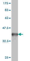
 WB (Western Blot)
(EXOC4 monoclonal antibody. Western Blot analysis of EXOC4 expression in Raw 264.7.)
WB (Western Blot)
(EXOC4 monoclonal antibody. Western Blot analysis of EXOC4 expression in Raw 264.7.)
EXOC4, Monoclonal Antibody (Cat# AAA24081)
Full Name
EXOC4 (Exocyst Complex Component 4, Exocyst Complex Component Sec8, KIAA1699, SEC8, SEC8L1, MGC27170)
Gene Names
EXOC4; SEC8; Sec8p; SEC8L1
Reactivity
Human, Mouse, Rat
Applications
Western Blot
Purity
Affinity Purified
Purified by Protein A affinity chromatography.
Purified by Protein A affinity chromatography.
Pricing


 Application Data
(Published customer image Infiltration of GFP+ BM-cells in infarct and peri-infarct regions. (A-B) Dot plots of viable macrophages/granulocytes (CD11b+CD45high, top right quadrants) and microglia (CD11b+CD45dim, bottom right quadrants) in cortex from BM-chimeric unmanipulated mice and mice exposed to pMCAO. (C) Bar graph showing mean numbers of CD11b+CD45dim microglia and CD11b+CD45high macrophages/granulocytes in BM-chimeric mice 24 hours after pMCAO, subdivided based on expression of GFP (n = 5). Approximately 92% of of the CD45high population were GFP+. (D) Estimation and comparison of mean numbers of CD11b+CD45dim microglia in non-chimeric (n = 10) versus BM-chimeric mice (n = 5) 24 hours after of pMCAO shows significantly fewer CD11b+CD45dim microglial cells in irradiated mice. (E) Overview, showing distribution of infiltrating GFP+ BM-derived cells into infarct (IF) and peri-infarct (P-IF) regions 24 hours after pMCAO. (E-G) By 24 hours, GFP+ single cells (F) and vessel-associated aggregates of GFP+ cells (arrows in G) were observed in infarct and peri-infarct regions. Some of the vessel-associated cells were round, leukocyte-like cells (arrows) while others were elongated cells lining the vasculature (arrow heads in G and in insert). (H) Bar graph showing mean numbers of single GFP+ cells and vessel-associated aggregates of GFP+ cells in ipsi- and contralateral cortex 24 hours after surgery (n = 10). (I-P) Immunohistochemical staining of CD45.1 (I, K), CD45.2 (J, L), IgG2a (M, O) and CD45 (N, P) in ischemic tissue in BM-chimeric (I, J, M, N) and non-chimeric mice (K, L, O, P) 24 hours after pMCAO. N.D, none detected. Scale bars: 200 um (A), 10 um (B, C). 50 um (I-P) *P < 0.05, **P < 0.01, and ***P < 0.001.From: Clausen BH, Lambertsen KL, Babcock AA, Holm TH, Dagnaes-Hansen F, Finsen B. Interleukin-1beta and tumor necrosis factor-alpha are expressed by different subsets of microglia and macrophages after ischemic stroke in mice. J Neuroinflammation. 2008 Oct 23;5:46.)
Application Data
(Published customer image Infiltration of GFP+ BM-cells in infarct and peri-infarct regions. (A-B) Dot plots of viable macrophages/granulocytes (CD11b+CD45high, top right quadrants) and microglia (CD11b+CD45dim, bottom right quadrants) in cortex from BM-chimeric unmanipulated mice and mice exposed to pMCAO. (C) Bar graph showing mean numbers of CD11b+CD45dim microglia and CD11b+CD45high macrophages/granulocytes in BM-chimeric mice 24 hours after pMCAO, subdivided based on expression of GFP (n = 5). Approximately 92% of of the CD45high population were GFP+. (D) Estimation and comparison of mean numbers of CD11b+CD45dim microglia in non-chimeric (n = 10) versus BM-chimeric mice (n = 5) 24 hours after of pMCAO shows significantly fewer CD11b+CD45dim microglial cells in irradiated mice. (E) Overview, showing distribution of infiltrating GFP+ BM-derived cells into infarct (IF) and peri-infarct (P-IF) regions 24 hours after pMCAO. (E-G) By 24 hours, GFP+ single cells (F) and vessel-associated aggregates of GFP+ cells (arrows in G) were observed in infarct and peri-infarct regions. Some of the vessel-associated cells were round, leukocyte-like cells (arrows) while others were elongated cells lining the vasculature (arrow heads in G and in insert). (H) Bar graph showing mean numbers of single GFP+ cells and vessel-associated aggregates of GFP+ cells in ipsi- and contralateral cortex 24 hours after surgery (n = 10). (I-P) Immunohistochemical staining of CD45.1 (I, K), CD45.2 (J, L), IgG2a (M, O) and CD45 (N, P) in ischemic tissue in BM-chimeric (I, J, M, N) and non-chimeric mice (K, L, O, P) 24 hours after pMCAO. N.D, none detected. Scale bars: 200 um (A), 10 um (B, C). 50 um (I-P) *P < 0.05, **P < 0.01, and ***P < 0.001.From: Clausen BH, Lambertsen KL, Babcock AA, Holm TH, Dagnaes-Hansen F, Finsen B. Interleukin-1beta and tumor necrosis factor-alpha are expressed by different subsets of microglia and macrophages after ischemic stroke in mice. J Neuroinflammation. 2008 Oct 23;5:46.)
CD11b, Monoclonal Antibody (Cat# AAA12183)
Full Name
RAT ANTI MOUSE CD11b:FITC
Gene Names
Itgam; CR3; CR3A; MAC1; Cd11b; Ly-40; Mac-1; Mac-1a; CD11b/CD18; F730045J24Rik
Applications
Flow Cytometry
Pricing


 Application Data
(Published customer image Infiltration of GFP+ BM-cells in infarct and peri-infarct regions. (A-B) Dot plots of viable macrophages/granulocytes (CD11b+CD45high, top right quadrants) and microglia (CD11b+CD45dim, bottom right quadrants) in cortex from BM-chimeric unmanipulated mice and mice exposed to pMCAO. (C) Bar graph showing mean numbers of CD11b+CD45dim microglia and CD11b+CD45high macrophages/granulocytes in BM-chimeric mice 24 hours after pMCAO, subdivided based on expression of GFP (n = 5). Approximately 92% of of the CD45high population were GFP+. (D) Estimation and comparison of mean numbers of CD11b+CD45dim microglia in non-chimeric (n = 10) versus BM-chimeric mice (n = 5) 24 hours after of pMCAO shows significantly fewer CD11b+CD45dim microglial cells in irradiated mice. (E) Overview, showing distribution of infiltrating GFP+ BM-derived cells into infarct (IF) and peri-infarct (P-IF) regions 24 hours after pMCAO. (E-G) By 24 hours, GFP+ single cells (F) and vessel-associated aggregates of GFP+ cells (arrows in G) were observed in infarct and peri-infarct regions. Some of the vessel-associated cells were round, leukocyte-like cells (arrows) while others were elongated cells lining the vasculature (arrow heads in G and in insert). (H) Bar graph showing mean numbers of single GFP+ cells and vessel-associated aggregates of GFP+ cells in ipsi- and contralateral cortex 24 hours after surgery (n = 10). (I-P) Immunohistochemical staining of CD45.1 (I, K), CD45.2 (J, L), IgG2a (M, O) and CD45 (N, P) in ischemic tissue in BM-chimeric (I, J, M, N) and non-chimeric mice (K, L, O, P) 24 hours after pMCAO. N.D, none detected. Scale bars: 200 um (A), 10 um (B, C). 50 um (I-P) *P < 0.05, **P < 0.01, and ***P < 0.001.From: Clausen BH, Lambertsen KL, Babcock AA, Holm TH, Dagnaes-Hansen F, Finsen B. Interleukin-1beta and tumor necrosis factor-alpha are expressed by different subsets of microglia and macrophages after ischemic stroke in mice. J Neuroinflammation. 2008 Oct 23;5:46.)
Application Data
(Published customer image Infiltration of GFP+ BM-cells in infarct and peri-infarct regions. (A-B) Dot plots of viable macrophages/granulocytes (CD11b+CD45high, top right quadrants) and microglia (CD11b+CD45dim, bottom right quadrants) in cortex from BM-chimeric unmanipulated mice and mice exposed to pMCAO. (C) Bar graph showing mean numbers of CD11b+CD45dim microglia and CD11b+CD45high macrophages/granulocytes in BM-chimeric mice 24 hours after pMCAO, subdivided based on expression of GFP (n = 5). Approximately 92% of of the CD45high population were GFP+. (D) Estimation and comparison of mean numbers of CD11b+CD45dim microglia in non-chimeric (n = 10) versus BM-chimeric mice (n = 5) 24 hours after of pMCAO shows significantly fewer CD11b+CD45dim microglial cells in irradiated mice. (E) Overview, showing distribution of infiltrating GFP+ BM-derived cells into infarct (IF) and peri-infarct (P-IF) regions 24 hours after pMCAO. (E-G) By 24 hours, GFP+ single cells (F) and vessel-associated aggregates of GFP+ cells (arrows in G) were observed in infarct and peri-infarct regions. Some of the vessel-associated cells were round, leukocyte-like cells (arrows) while others were elongated cells lining the vasculature (arrow heads in G and in insert). (H) Bar graph showing mean numbers of single GFP+ cells and vessel-associated aggregates of GFP+ cells in ipsi- and contralateral cortex 24 hours after surgery (n = 10). (I-P) Immunohistochemical staining of CD45.1 (I, K), CD45.2 (J, L), IgG2a (M, O) and CD45 (N, P) in ischemic tissue in BM-chimeric (I, J, M, N) and non-chimeric mice (K, L, O, P) 24 hours after pMCAO. N.D, none detected. Scale bars: 200 um (A), 10 um (B, C). 50 um (I-P) *P < 0.05, **P < 0.01, and ***P < 0.001.From: Clausen BH, Lambertsen KL, Babcock AA, Holm TH, Dagnaes-Hansen F, Finsen B. Interleukin-1beta and tumor necrosis factor-alpha are expressed by different subsets of microglia and macrophages after ischemic stroke in mice. J Neuroinflammation. 2008 Oct 23;5:46.)
CD11b, Monoclonal Antibody (Cat# AAA12185)
Full Name
RAT ANTI MOUSE CD11b
Gene Names
Itgam; CR3; CR3A; MAC1; Cd11b; Ly-40; Mac-1; Mac-1a; CD11b/CD18; F730045J24Rik
Applications
Immunohistochemistry, Flow Cytometry, Immunofluorescence, Immunoprecipitation
Pricing


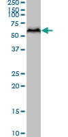
 WB (Western Blot)
(SF3B2 monoclonal antibody . Western Blot analysis of SF3B2 expression in PC-12.)
WB (Western Blot)
(SF3B2 monoclonal antibody . Western Blot analysis of SF3B2 expression in PC-12.)
SF3B2, Monoclonal Antibody (Cat# AAA24082)
Full Name
SF3B2 (Splicing Factor 3B Subunit 2, Pre-mRNA-splicing Factor SF3b 145 kDa Subunit, SF3b145, SF3b150, Spliceosome-associated Protein 145, SAP 145, SAP145)
Gene Names
SF3B2; Cus1; SF3b1; SAP145; SF3B145; SF3b150
Reactivity
Human, Mouse, Rat
Applications
Western Blot, Immunohistochemistry, Immunofluorescence
Purity
Affinity Purified
Purified by Protein A affinity chromatography.
Purified by Protein A affinity chromatography.
Pricing


 Application Data
(Published customer image: Representative images of the inflammatory changes in the facial nucleus during axonal regeneration, one week following facial nerve transaction. a, b: CD11b immunoreactivity for microglia is increased in the axotomized facial nucleus, and microglia enwrap the facial motor neurons, e.g. at arrows. The regenerating neurons were retrogradely labelled with fluorogold. c, d: CD6- positive T-cells accumulated in the injured motor nucleus (arrows). They had little cytoplasm but dense nuclei (c) and were sometimes clustered around neurons retrogradely labelled with fluorogold (d). The scale bar in (a) also applies to (b) and that in (c) also applies to (d).From: Shokouhi et al. BMC Neuroscience 2010 11:13.)
Application Data
(Published customer image: Representative images of the inflammatory changes in the facial nucleus during axonal regeneration, one week following facial nerve transaction. a, b: CD11b immunoreactivity for microglia is increased in the axotomized facial nucleus, and microglia enwrap the facial motor neurons, e.g. at arrows. The regenerating neurons were retrogradely labelled with fluorogold. c, d: CD6- positive T-cells accumulated in the injured motor nucleus (arrows). They had little cytoplasm but dense nuclei (c) and were sometimes clustered around neurons retrogradely labelled with fluorogold (d). The scale bar in (a) also applies to (b) and that in (c) also applies to (d).From: Shokouhi et al. BMC Neuroscience 2010 11:13.)
CD11b, Monoclonal Antibody (Cat# AAA11876)
Full Name
MOUSE ANTI RAT CD11b:FITC
Gene Names
ITGAM; CD11B
Applications
Flow Cytometry
Pricing


 Application Data
(Staining of Rat peripheral blood lymphocytes with Mouse anti Rat CD49d:RPE (MCA2872PE))
Application Data
(Staining of Rat peripheral blood lymphocytes with Mouse anti Rat CD49d:RPE (MCA2872PE))
CD49d, Monoclonal Antibody (Cat# AAA26760)
Full Name
CD49d (Antigen CD49d, CD49d Antigen, CDw49d, Alpha 4 Subunit of VLA-4 Receptor, Integrin alpha IV, Integrin alpha 4, IA4, ITGA4, LPAM23, MGC90518, Very Late Activation Protein 4 Receptor Alpha 4 Subunit, VLA4, VLA-4) (HRP)
Reactivity
Rat
Applications
Flow Cytometry, Immunoprecipitation
Purity
Purified by Protein G Affinity Chromatography
Pricing


 Application Data
(Figure A. FITC conjugated Rat anti Mouse CD19 . Figure B. FITC conjugated Rat anti Mouse CD19 and SBV440 conjugated Rat anti Mouse CD45R . All experiments performed on mouse splenocytes in the presence of 10% mouse seru)
Application Data
(Figure A. FITC conjugated Rat anti Mouse CD19 . Figure B. FITC conjugated Rat anti Mouse CD19 and SBV440 conjugated Rat anti Mouse CD45R . All experiments performed on mouse splenocytes in the presence of 10% mouse seru)
CD19, Monoclonal Antibody (Cat# AAA12286)
Full Name
Rat anti Mouse CD19:Amethyst Orange
Gene Names
Cd19; AW495831
Reactivity
Mouse
Applications
Flow Cytometry
Purity
Purified IgG prepared by affinity chromatography on Protein G from tissue culture supernatant
Pricing


 Application Data
(Immunoperoxidase staining of human tonsil cryosection using Mouse anti Human CD3 antibody, clone UCHT1 followed by horseradish peroxidase Goat anti Mouse IgG2a antibody as a detection reagent. Medium power)
Application Data
(Immunoperoxidase staining of human tonsil cryosection using Mouse anti Human CD3 antibody, clone UCHT1 followed by horseradish peroxidase Goat anti Mouse IgG2a antibody as a detection reagent. Medium power)
CD3, Monoclonal Antibody (Cat# AAA12156)
Full Name
MOUSE ANTI HUMAN CD3
Gene Names
CD3G; T3G; IMD17; CD3-GAMMA
Applications
Immunohistochemistry, Flow Cytometry
Pricing
 Application Data
(ATH mouse bone marrow stained with anti-CD44 (clone: KM81) (filled histogram) or Rat IgG2a isotype control (open histogram).N.B. Appropriate control samples should always be included in any labelling studies. * For optimal results in various applications, it is recommended that each investigator determine dilutions appropriate for individual use.)
Application Data
(ATH mouse bone marrow stained with anti-CD44 (clone: KM81) (filled histogram) or Rat IgG2a isotype control (open histogram).N.B. Appropriate control samples should always be included in any labelling studies. * For optimal results in various applications, it is recommended that each investigator determine dilutions appropriate for individual use.)
CD44, Monoclonal Antibody (Cat# AAA14269)
Full Name
Anti-Mouse CD44, Purified (Clone KM81) (rat IgG2a)
Gene Names
CD44; IN; LHR; MC56; MDU2; MDU3; MIC4; Pgp1; CDW44; CSPG8; HCELL; HUTCH-I; ECMR-III
Reactivity
Mouse
Applications
Immunohistochemistry, Flow Cytometry
Pricing


 IHC (Immunohistchemistry)
(Immunohistochemistry analysis using Mouse Anti-Hsp70 Monoclonal Antibody, Clone BB70 . Tissue: hepatocytes. Species: Rat. Fixation: Paraffin Embedded. Primary Antibody: Mouse Anti-Hsp70 Monoclonal Antibody at 1:200. Liver sections were paraffin embedded. First pictures in series show two hours after exposure to stress, the second shows the control. Courtesy of: G. Matic, University of Belgrade, Serbia.)
IHC (Immunohistchemistry)
(Immunohistochemistry analysis using Mouse Anti-Hsp70 Monoclonal Antibody, Clone BB70 . Tissue: hepatocytes. Species: Rat. Fixation: Paraffin Embedded. Primary Antibody: Mouse Anti-Hsp70 Monoclonal Antibody at 1:200. Liver sections were paraffin embedded. First pictures in series show two hours after exposure to stress, the second shows the control. Courtesy of: G. Matic, University of Belgrade, Serbia.)
HSP70/HSC70, Monoclonal Antibody (Cat# AAA27659)
Full Name
HSP70/HSC70 Antibody
Gene Names
HSPA2; HSP70
Reactivity
Human, Mouse, Rat, Bovine, Sheep, Dog, Beluga, Fish, Guinea Pig, Pig, Hamster, Rabbit, Chicken, African Clawed Frog, Fruit Fly, Yeast
Applications
Western Blot, Immunocytochemistry, Immunofluorescence, Immunohistochemistry, Immunoprecipitation
Purity
Protein G Purified
Pricing


 Application Data
(Staining of mouse spleen with Rat anti Mouse CD3 Epsilon (T3): RPE)
Application Data
(Staining of mouse spleen with Rat anti Mouse CD3 Epsilon (T3): RPE)
CD3, Monoclonal Antibody (Cat# AAA12243)
Full Name
RAT ANTI MOUSE CD3:Low Endotoxin
Gene Names
Cd3e; CD3; T3e; AI504783; CD3epsilon
Applications
Immunohistochemistry, Flow Cytometry
Pricing


 Application Data
(Published customer image: Increased accumulation of repair-associated macrophages surrounding collaterals in ischemic hind limbs is PAR2-dependent. (A) Stainings of CD206-positive macrophages (green) and SMA-positive vessels (red) in non-ischemic (control) and ischemic (ligated) hind limbs of WT, PAR1-/- and PAR2-/- mice are shown. Nuclei were visualized with DAPI (blue). Arrows indicate single macrophages in the non-ischemic adductor. Quantification of the average number of repair-associated macrophages per vessel is indicated on the right. (B) Correlation between the number of CD206-positive macrophages in the ischemic tissues and the expression of CD11b and (C) CD115 on monocytes. ** p)
Application Data
(Published customer image: Increased accumulation of repair-associated macrophages surrounding collaterals in ischemic hind limbs is PAR2-dependent. (A) Stainings of CD206-positive macrophages (green) and SMA-positive vessels (red) in non-ischemic (control) and ischemic (ligated) hind limbs of WT, PAR1-/- and PAR2-/- mice are shown. Nuclei were visualized with DAPI (blue). Arrows indicate single macrophages in the non-ischemic adductor. Quantification of the average number of repair-associated macrophages per vessel is indicated on the right. (B) Correlation between the number of CD206-positive macrophages in the ischemic tissues and the expression of CD11b and (C) CD115 on monocytes. ** p)
CD206, Monoclonal Antibody (Cat# AAA12118)
Full Name
RAT ANTI MOUSE CD206:Biotin
Gene Names
Mrc1; MR; CD206; AW259686
Applications
Flow Cytometry
Pricing


 Application Data
(Staining of LPS stimulated mouse spleen cells with Rat anti Mouse CD83:RPE)
Application Data
(Staining of LPS stimulated mouse spleen cells with Rat anti Mouse CD83:RPE)
CD83, Monoclonal Antibody (Cat# AAA12212)
Full Name
RAT ANTI MOUSE CD83
Applications
Flow Cytometry
Pricing


 Application Data
(Immunoperoxidase staining of mouse lymph node cryosection eith Rat anti Mouse antibody clone R3-63 followed by horseradish peroxidase Goat anti Rat IgG antibody . Medium power)
Application Data
(Immunoperoxidase staining of mouse lymph node cryosection eith Rat anti Mouse antibody clone R3-63 followed by horseradish peroxidase Goat anti Rat IgG antibody . Medium power)
CD13, Monoclonal Antibody (Cat# AAA12037)
Full Name
RAT ANTI MOUSE CD13:RPE
Gene Names
Anpep; Apn; AP-M; AP-N; Cd13; P150
Applications
Flow Cytometry
Pricing


 Application Data
(Published customer image: Leukocyte infiltration in COX-2-M/-M and COX-2+/+ mice. MPO enzymatic activity (panel A) was statistically similar in COX-2-M/-M and COX-2+/+ livers at 6 h and 24 h post-IRI. Ly-6G+ neutrophil (panel B) and granulocyte (panel C) infiltration were also comparable in COX-2-M/-M and COX-2+/+ livers after IRI. Mac-1+ (panel D) and CD68 (panel E) infiltrating macrophages were significantly reduced in COX-2-M/-M livers at 24 h post-reperfusion, but were statistically indistinguishable in COX-2-M/-M and COX-2+/+ livers at 6 h after IRI. No statistical differences in MMP-9 expression (panel F) could be demonstrated in livers of COX-2-M/-M and COX-2+/+ mice post-IRI. Representative immunostaining (panel G) of infiltrating Ly-6G+ (a,b,e,f) and Mac-1+ (c,d,g,h) leukocytes in livers of COX-2+/+ (a,c,e,g) and COX-2-M/-M (b,d,f,h) mice at 6 h (a to d) and 24 h (e to h) post IRI; (n = 5 -6/group; * indicates p)
Application Data
(Published customer image: Leukocyte infiltration in COX-2-M/-M and COX-2+/+ mice. MPO enzymatic activity (panel A) was statistically similar in COX-2-M/-M and COX-2+/+ livers at 6 h and 24 h post-IRI. Ly-6G+ neutrophil (panel B) and granulocyte (panel C) infiltration were also comparable in COX-2-M/-M and COX-2+/+ livers after IRI. Mac-1+ (panel D) and CD68 (panel E) infiltrating macrophages were significantly reduced in COX-2-M/-M livers at 24 h post-reperfusion, but were statistically indistinguishable in COX-2-M/-M and COX-2+/+ livers at 6 h after IRI. No statistical differences in MMP-9 expression (panel F) could be demonstrated in livers of COX-2-M/-M and COX-2+/+ mice post-IRI. Representative immunostaining (panel G) of infiltrating Ly-6G+ (a,b,e,f) and Mac-1+ (c,d,g,h) leukocytes in livers of COX-2+/+ (a,c,e,g) and COX-2-M/-M (b,d,f,h) mice at 6 h (a to d) and 24 h (e to h) post IRI; (n = 5 -6/group; * indicates p)
CD68, Monoclonal Antibody (Cat# AAA12104)
Full Name
RAT ANTI MOUSE CD68:Biotin
Gene Names
Cd68; Lamp4; gp110; Scard1
Applications
Flow Cytometry
Pricing


 Application Data
(Staining of Rat peripheral blood lymphocytes with Mouse anti Rat CD49d:RPE (MCA2872PE))
Application Data
(Staining of Rat peripheral blood lymphocytes with Mouse anti Rat CD49d:RPE (MCA2872PE))
CD49d, Monoclonal Antibody (Cat# AAA26735)
Full Name
CD49d (Antigen CD49d, CD49d Antigen, CDw49d, Alpha 4 Subunit of VLA-4 Receptor, Integrin alpha IV, Integrin alpha 4, IA4, ITGA4, LPAM23, MGC90518, Very Late Activation Protein 4 Receptor Alpha 4 Subunit, VLA4, VLA-4) (Biotin)
Reactivity
Rat
Applications
Flow Cytometry, Immunoprecipitation
Purity
Purified by Protein G Affinity Chromatography
Pricing


 WB (Western Blot)
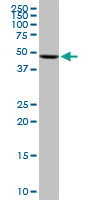
(Western blot analysis of UGP2 over-expressed 293 cell line, cotransfected with UGP2 Validated Chimera RNAi (Lane 2) or non-transfected control (Lane 1). Blot probed with UGP2 monoclonal antibody. GAPDH (36.1kD) used as specificity and loading control.)
WB (Western Blot)
(Western blot analysis of UGP2 over-expressed 293 cell line, cotransfected with UGP2 Validated Chimera RNAi (Lane 2) or non-transfected control (Lane 1). Blot probed with UGP2 monoclonal antibody. GAPDH (36.1kD) used as specificity and loading control.)
UGP2, Monoclonal Antibody (Cat# AAA24096)
Full Name
UGP2 (UTP--glucose-1-phosphate Uridylyltransferase, UDP-glucose Pyrophosphorylase, UDPGP, UGPase, UGP1)
Gene Names
UGP2; UDPG; UGP1; UDPGP; UGPP1; UGPP2; UDPGP2; pHC379
Reactivity
Human, Mouse, Rat
Applications
Western Blot
Purity
Affinity Purified
Purified by Protein A affinity chromatography.
Purified by Protein A affinity chromatography.
Pricing


 Application Data
(Staining of mouse spleen cells with Rat anti Mouse CD3 Epsilon (T3): APC)
Application Data
(Staining of mouse spleen cells with Rat anti Mouse CD3 Epsilon (T3): APC)
CD3, Monoclonal Antibody (Cat# AAA11985)
Full Name
RAT ANTI MOUSE CD3
Gene Names
Cd3e; CD3; T3e; AI504783; CD3epsilon
Applications
Immunohistochemistry, Flow Cytometry
Pricing


 Application Data
(Staining of mouse peripheral blood lymphocytes with Rat anti Mouse CD200R: Low Endotoxin)
Application Data
(Staining of mouse peripheral blood lymphocytes with Rat anti Mouse CD200R: Low Endotoxin)
CD200R, Monoclonal Antibody (Cat# AAA11957)
Full Name
RAT ANTI MOUSE CD200R
Gene Names
Cd200r1; OX2R; Mox2r; CD200R
Applications
Flow Cytometry
Pricing


 Application Data
(Proximity Ligation Analysis of protein-protein interactions between TP53 and PML HeLa cells were stained with anti-TP53 rabbit purified polyclonal 1:1200 and anti-PML mouse monoclonal antibody 1:50. Each red dot represents the detection of protein-protein interaction complex, and nuclei were counterstained with DAPI (blue).)
Application Data
(Proximity Ligation Analysis of protein-protein interactions between TP53 and PML HeLa cells were stained with anti-TP53 rabbit purified polyclonal 1:1200 and anti-PML mouse monoclonal antibody 1:50. Each red dot represents the detection of protein-protein interaction complex, and nuclei were counterstained with DAPI (blue).)
PML, Monoclonal Antibody (Cat# AAA24914)
Full Name
PML (Probable Transcription Factor PML, RING Finger Protein 71, Tripartite Motif-containing Protein 19, PML, MYL, RNF71, TRIM19) (Biotin)
Gene Names
PML; MYL; RNF71; PP8675; TRIM19
Reactivity
Human, Rat
Applications
Immunofluorescence, Western Blot
Purity
Purified by Protein A Affinity Chromatography.
Pricing


 Application Data
(Immunoperoxidase staining of mouse lymph node cryosection eith Rat anti Mouse antibody clone R3-63 followed by horseradish peroxidase Goat anti Rat IgG antibody . Medium power)
Application Data
(Immunoperoxidase staining of mouse lymph node cryosection eith Rat anti Mouse antibody clone R3-63 followed by horseradish peroxidase Goat anti Rat IgG antibody . Medium power)
CD13, Monoclonal Antibody (Cat# AAA12116)
Full Name
RAT ANTI MOUSE CD13
Gene Names
Anpep; Apn; AP-M; AP-N; Cd13; P150
Applications
Immunohistochemistry, Flow Cytometry, Immunohistochemistry
Pricing


 Application Data
(Published customer image: Representative images of the inflammatory changes in the facial nucleus during axonal regeneration, one week following facial nerve transaction. a, b: CD11b immunoreactivity for microglia is increased in the axotomized facial nucleus, and microglia enwrap the facial motor neurons, e.g. at arrows. The regenerating neurons were retrogradely labelled with fluorogold. c, d: CD6- positive T-cells accumulated in the injured motor nucleus (arrows). They had little cytoplasm but dense nuclei (c) and were sometimes clustered around neurons retrogradely labelled with fluorogold (d). The scale bar in (a) also applies to (b) and that in (c) also applies to (d).From: Shokouhi et al. BMC Neuroscience 2010 11:13.)
Application Data
(Published customer image: Representative images of the inflammatory changes in the facial nucleus during axonal regeneration, one week following facial nerve transaction. a, b: CD11b immunoreactivity for microglia is increased in the axotomized facial nucleus, and microglia enwrap the facial motor neurons, e.g. at arrows. The regenerating neurons were retrogradely labelled with fluorogold. c, d: CD6- positive T-cells accumulated in the injured motor nucleus (arrows). They had little cytoplasm but dense nuclei (c) and were sometimes clustered around neurons retrogradely labelled with fluorogold (d). The scale bar in (a) also applies to (b) and that in (c) also applies to (d).From: Shokouhi et al. BMC Neuroscience 2010 11:13.)
CD11b, Monoclonal Antibody (Cat# AAA11970)
Full Name
MOUSE ANTI RAT CD11b
Gene Names
ITGAM; CD11B
Applications
Immunohistochemistry, Flow Cytometry, Immunofluorescence, Immunoprecipitation
Pricing


 IHC (Immunohistochemistry)
(DAB staining on IHC-P; Samples:Human Cardiac Muscle Tissue;Primary Ab: 20ug/ml Mouse Anti-Human LOXL1 AntibodySecond Ab: 2?g/mL HRP-Linked Caprine Anti-Mouse IgG Polyclonal Antibody (Catalog:).)
IHC (Immunohistochemistry)
(DAB staining on IHC-P; Samples:Human Cardiac Muscle Tissue;Primary Ab: 20ug/ml Mouse Anti-Human LOXL1 AntibodySecond Ab: 2?g/mL HRP-Linked Caprine Anti-Mouse IgG Polyclonal Antibody (Catalog:).)
Lysyl Oxidase Like Protein 1 (LOXL1), Monoclonal Antibody (Cat# AAA20293)
Full Name
Monoclonal Antibody to Lysyl Oxidase Like Protein 1 (LOXL1)
Gene Names
LOXL1; LOL; LOXL
Reactivity
Human
Applications
Western Blot, Immunohistochemistry
Purity
Protein A + Protein G affinity chromatography
Pricing


 Application Data
(Figure A. Alexa Fluor 488 conjugated Rat anti Mouse CD4 . Figure B. Alexa Fluor 488 conjugated Rat anti Mouse CD4 and StarBright Violet 610 conjugated Rat anti Mouse CD3 . All experiments performed on red blood lys)
Application Data
(Figure A. Alexa Fluor 488 conjugated Rat anti Mouse CD4 . Figure B. Alexa Fluor 488 conjugated Rat anti Mouse CD4 and StarBright Violet 610 conjugated Rat anti Mouse CD3 . All experiments performed on red blood lys)
CD4, Monoclonal Antibody (Cat# AAA12287)
Full Name
Rat anti Mouse CD4:Amethyst Orange
Gene Names
Cd4; L3T4; Ly-4
Reactivity
Mouse
Applications
ELISA
Purity
Purified IgG prepared by affinity chromatography on Protein G from tissue culture supernatant
Pricing


 WB (Western Blot)
(Western blot analysis of CITED1 over-expressed 293 cell line, cotransfected with CITED1 Validated Chimera RNAi ((Lane 2) or non-transfected control (Lane 1). Blot probed with CITED1 monoclonal antibody GAPDH (36.1kD) used as specificity and loading control.)
WB (Western Blot)
(Western blot analysis of CITED1 over-expressed 293 cell line, cotransfected with CITED1 Validated Chimera RNAi ((Lane 2) or non-transfected control (Lane 1). Blot probed with CITED1 monoclonal antibody GAPDH (36.1kD) used as specificity and loading control.)
CITED1, Monoclonal Antibody (Cat# AAA24077)
Full Name
CITED1 (CBP/p300 Interacting Transactivator 1, Cbp/p300-interacting Transactivator with Glu/Asp-rich Carboxy-terminal Domain 1, Melanocyte-specific Protein1)
Gene Names
CITED1; MSG1
Reactivity
Human, Rat
Applications
Western Blot
Purity
Affinity Purified
Purified by Protein A affinity chromatography.
Purified by Protein A affinity chromatography.
Pricing


 Application Data
(Published customer image: Increased accumulation of repair-associated macrophages surrounding collaterals in ischemic hind limbs is PAR2-dependent. (A) Stainings of CD206-positive macrophages (green) and SMA-positive vessels (red) in non-ischemic (control) and ischemic (ligated) hind limbs of WT, PAR1-/- and PAR2-/- mice are shown. Nuclei were visualized with DAPI (blue). Arrows indicate single macrophages in the non-ischemic adductor. Quantification of the average number of repair-associated macrophages per vessel is indicated on the right. (B) Correlation between the number of CD206-positive macrophages in the ischemic tissues and the expression of CD11b and (C) CD115 on monocytes. ** p)
Application Data
(Published customer image: Increased accumulation of repair-associated macrophages surrounding collaterals in ischemic hind limbs is PAR2-dependent. (A) Stainings of CD206-positive macrophages (green) and SMA-positive vessels (red) in non-ischemic (control) and ischemic (ligated) hind limbs of WT, PAR1-/- and PAR2-/- mice are shown. Nuclei were visualized with DAPI (blue). Arrows indicate single macrophages in the non-ischemic adductor. Quantification of the average number of repair-associated macrophages per vessel is indicated on the right. (B) Correlation between the number of CD206-positive macrophages in the ischemic tissues and the expression of CD11b and (C) CD115 on monocytes. ** p)
CD206, Monoclonal Antibody (Cat# AAA12124)
Full Name
RAT ANTI MOUSE CD206:RPE
Gene Names
Mrc1; MR; CD206; AW259686
Applications
Flow Cytometry
Pricing


 Application Data
(Published customer image: Leukocyte infiltration in COX-2-M/-M and COX-2+/+ mice. MPO enzymatic activity (panel A) was statistically similar in COX-2-M/-M and COX-2+/+ livers at 6 h and 24 h post-IRI. Ly-6G+ neutrophil (panel B) and granulocyte (panel C) infiltration were also comparable in COX-2-M/-M and COX-2+/+ livers after IRI. Mac-1+ (panel D) and CD68 (panel E) infiltrating macrophages were significantly reduced in COX-2-M/-M livers at 24 h post-reperfusion, but were statistically indistinguishable in COX-2-M/-M and COX-2+/+ livers at 6 h after IRI. No statistical differences in MMP-9 expression (panel F) could be demonstrated in livers of COX-2-M/-M and COX-2+/+ mice post-IRI. Representative immunostaining (panel G) of infiltrating Ly-6G+ (a,b,e,f) and Mac-1+ (c,d,g,h) leukocytes in livers of COX-2+/+ (a,c,e,g) and COX-2-M/-M (b,d,f,h) mice at 6 h (a to d) and 24 h (e to h) post IRI; (n = 5 -6/group; * indicates p)
Application Data
(Published customer image: Leukocyte infiltration in COX-2-M/-M and COX-2+/+ mice. MPO enzymatic activity (panel A) was statistically similar in COX-2-M/-M and COX-2+/+ livers at 6 h and 24 h post-IRI. Ly-6G+ neutrophil (panel B) and granulocyte (panel C) infiltration were also comparable in COX-2-M/-M and COX-2+/+ livers after IRI. Mac-1+ (panel D) and CD68 (panel E) infiltrating macrophages were significantly reduced in COX-2-M/-M livers at 24 h post-reperfusion, but were statistically indistinguishable in COX-2-M/-M and COX-2+/+ livers at 6 h after IRI. No statistical differences in MMP-9 expression (panel F) could be demonstrated in livers of COX-2-M/-M and COX-2+/+ mice post-IRI. Representative immunostaining (panel G) of infiltrating Ly-6G+ (a,b,e,f) and Mac-1+ (c,d,g,h) leukocytes in livers of COX-2+/+ (a,c,e,g) and COX-2-M/-M (b,d,f,h) mice at 6 h (a to d) and 24 h (e to h) post IRI; (n = 5 -6/group; * indicates p)
CD68, Monoclonal Antibody (Cat# AAA12107)
Full Name
RAT ANTI MOUSE CD68
Gene Names
Cd68; Lamp4; gp110; Scard1
Applications
Immunohistochemistry, Flow Cytometry, Immunofluorescence, Immunoprecipitation, Immunohistochemistry, Western Blot
Purity
Purified
Purified IgG - liquid
Purified IgG - liquid
Pricing


 Application Data
(Staining of Rat peripheral blood lymphocytes with Mouse anti Rat CD49d:RPE (MCA2872PE))
Application Data
(Staining of Rat peripheral blood lymphocytes with Mouse anti Rat CD49d:RPE (MCA2872PE))
CD49d, Monoclonal Antibody (Cat# AAA26798)
Full Name
CD49d (Antigen CD49d, CD49d Antigen, CDw49d, Alpha 4 Subunit of VLA-4 Receptor, Integrin alpha IV, Integrin alpha 4, IA4, ITGA4, LPAM23, MGC90518, Very Late Activation Protein 4 Receptor Alpha 4 Subunit, VLA4, VLA-4) (MaxLight 490)
Reactivity
Rat
Applications
Flow Cytometry, Immunoprecipitation
Purity
Purified by Protein G Affinity Chromatography
Pricing


 Application Data
(Proximity Ligation Analysis of protein-protein interactions between PRKCZ and AKT3. HeLa cells were stained with anti-PRKCZ rabbit purified polyclonal 1:1200 and anti-AKT3 mouse monoclonal antibody 1:50. Each red dot represents the detection of protein-protein interaction complex, and nuclei were counterstained with DAPI (blue).)
Application Data
(Proximity Ligation Analysis of protein-protein interactions between PRKCZ and AKT3. HeLa cells were stained with anti-PRKCZ rabbit purified polyclonal 1:1200 and anti-AKT3 mouse monoclonal antibody 1:50. Each red dot represents the detection of protein-protein interaction complex, and nuclei were counterstained with DAPI (blue).)
AKT3, Monoclonal Antibody (Cat# AAA24135)
Full Name
AKT3 (RAC-gamma Serine/Threonine-protein Kinase, Protein Kinase Akt-3, Protein Kinase B gamma, PKB gamma, RAC-PK-gamma, STK-2, PKBG) (AP)
Gene Names
AKT3; MPPH; PKBG; MPPH2; PRKBG; STK-2; PKB-GAMMA; RAC-gamma; RAC-PK-gamma
Reactivity
Human, Mouse, Rat
Applications
Western Blot
Purity
Purified by Protein A Affinity Chromatography.
Pricing


 Application Data
(Published customer image: Leukocyte infiltration in COX-2-M/-M and COX-2+/+ mice. MPO enzymatic activity (panel A) was statistically similar in COX-2-M/-M and COX-2+/+ livers at 6 h and 24 h post-IRI. Ly-6G+ neutrophil (panel B) and granulocyte (panel C) infiltration were also comparable in COX-2-M/-M and COX-2+/+ livers after IRI. Mac-1+ (panel D) and CD68 (panel E) infiltrating macrophages were significantly reduced in COX-2-M/-M livers at 24 h post-reperfusion, but were statistically indistinguishable in COX-2-M/-M and COX-2+/+ livers at 6 h after IRI. No statistical differences in MMP-9 expression (panel F) could be demonstrated in livers of COX-2-M/-M and COX-2+/+ mice post-IRI. Representative immunostaining (panel G) of infiltrating Ly-6G+ (a,b,e,f) and Mac-1+ (c,d,g,h) leukocytes in livers of COX-2+/+ (a,c,e,g) and COX-2-M/-M (b,d,f,h) mice at 6 h (a to d) and 24 h (e to h) post IRI; (n = 5 -6/group; * indicates p)
Application Data
(Published customer image: Leukocyte infiltration in COX-2-M/-M and COX-2+/+ mice. MPO enzymatic activity (panel A) was statistically similar in COX-2-M/-M and COX-2+/+ livers at 6 h and 24 h post-IRI. Ly-6G+ neutrophil (panel B) and granulocyte (panel C) infiltration were also comparable in COX-2-M/-M and COX-2+/+ livers after IRI. Mac-1+ (panel D) and CD68 (panel E) infiltrating macrophages were significantly reduced in COX-2-M/-M livers at 24 h post-reperfusion, but were statistically indistinguishable in COX-2-M/-M and COX-2+/+ livers at 6 h after IRI. No statistical differences in MMP-9 expression (panel F) could be demonstrated in livers of COX-2-M/-M and COX-2+/+ mice post-IRI. Representative immunostaining (panel G) of infiltrating Ly-6G+ (a,b,e,f) and Mac-1+ (c,d,g,h) leukocytes in livers of COX-2+/+ (a,c,e,g) and COX-2-M/-M (b,d,f,h) mice at 6 h (a to d) and 24 h (e to h) post IRI; (n = 5 -6/group; * indicates p)
CD68, Monoclonal Antibody (Cat# AAA12108)
Full Name
RAT ANTI MOUSE CD68:RPE
Gene Names
Cd68; Lamp4; gp110; Scard1
Applications
Flow Cytometry
Pricing


 Application Data
(Detection limit for recombinant GST tagged PITPNA is 0.1ng/ml as a capture antibody.)
Application Data
(Detection limit for recombinant GST tagged PITPNA is 0.1ng/ml as a capture antibody.)
PITPNA, Monoclonal Antibody (Cat# AAA24090)
Full Name
PITPNA (Phosphatidylinositol Transfer Protein alpha Isoform, PI-TP-alpha, PtdIns Transfer Protein alpha, PtdInsTP alpha, PITPN, MGC99649)
Gene Names
PITPNA; PITPN; VIB1A; PI-TPalpha
Reactivity
Human, Mouse, Rat
Applications
Western Blot
Purity
Affinity Purified
Purified by Protein A affinity chromatography.
Purified by Protein A affinity chromatography.
Pricing


 IHC (Immunohistchemistry)
(Immunohistochemistry analysis using Mouse Anti-Hsp70 Monoclonal Antibody, Clone BB70. Tissue: hepatocytes. Species: Rat. Fixation: Paraffin Embedded. Primary Antibody: Mouse Anti-Hsp70 Monoclonal Antibody at 1:200. Liver sections were paraffin embedded. First pictures in series show two hours after exposure to stress, the second shows the control. Courtesy of: G. Matic, University of Belgrade, Serbia.)
IHC (Immunohistchemistry)
(Immunohistochemistry analysis using Mouse Anti-Hsp70 Monoclonal Antibody, Clone BB70. Tissue: hepatocytes. Species: Rat. Fixation: Paraffin Embedded. Primary Antibody: Mouse Anti-Hsp70 Monoclonal Antibody at 1:200. Liver sections were paraffin embedded. First pictures in series show two hours after exposure to stress, the second shows the control. Courtesy of: G. Matic, University of Belgrade, Serbia.)
Hsp70/Hsc70, Monoclonal Antibody (Cat# AAA17793)
Full Name
Hsp70/Hsc70 Antibody: HRP
Gene Names
HSPA2; HSP70
Reactivity
Human, Mouse, Rat, Sheep, Dog, Beluga, Bovine, Fish, Guinea pig, Scallop pig, Hamster, Rabbit, Chicken, Xenopus, Drosophila, Yeast
Applications
WB, IP, IHC
Pricing


 Application Data
(Staining of mouse peripheral blood platelets with Rat anti Mouse CD36:FITC)
Application Data
(Staining of mouse peripheral blood platelets with Rat anti Mouse CD36:FITC)
CD36, Monoclonal Antibody (Cat# AAA12234)
Full Name
RAT ANTI MOUSE CD36:RPE
Gene Names
Cd36; FAT; GPIV; Scarb3
Applications
Flow Cytometry
Pricing


 Application Data
(Published clone specific image Alloimmunity-associated cytotoxicity is mediated through the NKG2D receptor. (A) Liver expression of nkg2d on day ten after liver transplantation. (B) Representative NKG2D expression levels in blood NK cells (left) and monocytes (right) of allogeneic (black) and syngeneic (grey) recipients. Isotype was used as control (dashed lines). (C) Sorted blood NK cell cytotoxicity inhibition with anti-NKG2D antibody or with anti-NKp30 antibody. (D) Levels of NKG2D ligand (rae1l, rrlt and irp94) expression in the liver on day ten after transplantation. (E) Levels of NKG2D ligand (rae1l, rrlt and irp94) expression in rat HCC cell lines. (F) Representative level of recombinant NKG2D-Fc binding to rat HCC cells lines. *p)
Application Data
(Published clone specific image Alloimmunity-associated cytotoxicity is mediated through the NKG2D receptor. (A) Liver expression of nkg2d on day ten after liver transplantation. (B) Representative NKG2D expression levels in blood NK cells (left) and monocytes (right) of allogeneic (black) and syngeneic (grey) recipients. Isotype was used as control (dashed lines). (C) Sorted blood NK cell cytotoxicity inhibition with anti-NKG2D antibody or with anti-NKp30 antibody. (D) Levels of NKG2D ligand (rae1l, rrlt and irp94) expression in the liver on day ten after transplantation. (E) Levels of NKG2D ligand (rae1l, rrlt and irp94) expression in rat HCC cell lines. (F) Representative level of recombinant NKG2D-Fc binding to rat HCC cells lines. *p)
CD172a, Monoclonal Antibody (Cat# AAA11875)
Full Name
MOUSE ANTI RAT CD172a:FITC
Gene Names
Sirpa; Bit; Ptpns1; SHPS-1
Applications
Flow Cytometry
Pricing


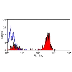
 FCM (Flow Cytometry)
(Figure 8. Flow Cytometry analysis of SiHa cells using anti- ASS1 antibody (AAA19369).Overlay histogram showing SiHa cells stained with AAA19369 (Blue line). The cells were blocked with 10% normal goat serum. And then incubated with mouse anti-ASS1 Antibody (AAA19369, 1μg/1x106 cells) for 30 min at 20 degree C. DyLight®488 conjugated goat anti-mouse IgG (BA1126, 5-10μg/1x106 cells) was used as secondary antibody for 30 minutes at 20 degree C. Isotype control antibody (Green line) was mouse IgG (1μg/1x106) used under the same conditions. Unlabelled sample (Red line) was also used as a control.)
FCM (Flow Cytometry)
(Figure 8. Flow Cytometry analysis of SiHa cells using anti- ASS1 antibody (AAA19369).Overlay histogram showing SiHa cells stained with AAA19369 (Blue line). The cells were blocked with 10% normal goat serum. And then incubated with mouse anti-ASS1 Antibody (AAA19369, 1μg/1x106 cells) for 30 min at 20 degree C. DyLight®488 conjugated goat anti-mouse IgG (BA1126, 5-10μg/1x106 cells) was used as secondary antibody for 30 minutes at 20 degree C. Isotype control antibody (Green line) was mouse IgG (1μg/1x106) used under the same conditions. Unlabelled sample (Red line) was also used as a control.)
ASS1, Monoclonal Antibody (Cat# AAA19369)
Full Name
Anti-ASS1 Antibody (monoclonal, 5I5)
Gene Names
ASS1; ASS; CTLN1
Reactivity
Human, Mouse, Rat, Monkey
Applications
WB, IHC-P, ICC, IF, FC/FACS/FCM
Purity
Immunogen affinity purified.
Pricing


 Application Data
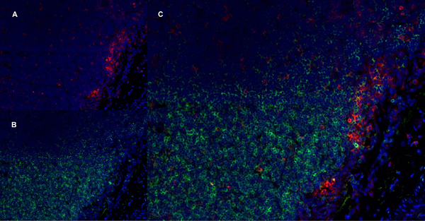
(Immunofluorescence staining of a mouse lymph node cryosection with Rat anti MouseCD19 antibody, clone 6D5 , green in A and Rat anti Mlouse CD8 antibody, clone YTS105.18 , red in B. Merged image in C with nuclei counterstained blue using DAPI. Low power)
Application Data
(Immunofluorescence staining of a mouse lymph node cryosection with Rat anti MouseCD19 antibody, clone 6D5 , green in A and Rat anti Mlouse CD8 antibody, clone YTS105.18 , red in B. Merged image in C with nuclei counterstained blue using DAPI. Low power)
CD19, Monoclonal Antibody (Cat# AAA11932)
Full Name
RAT ANTI MOUSE CD19
Gene Names
Cd19; AW495831
Applications
Immunohistochemistry, Flow Cytometry, Immunoprecipitation
Pricing


 Application Data
(Published customer image: Leukocyte infiltration in COX-2-M/-M and COX-2+/+ mice. MPO enzymatic activity (panel A) was statistically similar in COX-2-M/-M and COX-2+/+ livers at 6 h and 24 h post-IRI. Ly-6G+ neutrophil (panel B) and granulocyte (panel C) infiltration were also comparable in COX-2-M/-M and COX-2+/+ livers after IRI. Mac-1+ (panel D) and CD68 (panel E) infiltrating macrophages were significantly reduced in COX-2-M/-M livers at 24 h post-reperfusion, but were statistically indistinguishable in COX-2-M/-M and COX-2+/+ livers at 6 h after IRI. No statistical differences in MMP-9 expression (panel F) could be demonstrated in livers of COX-2-M/-M and COX-2+/+ mice post-IRI. Representative immunostaining (panel G) of infiltrating Ly-6G+ (a,b,e,f) and Mac-1+ (c,d,g,h) leukocytes in livers of COX-2+/+ (a,c,e,g) and COX-2-M/-M (b,d,f,h) mice at 6 h (a to d) and 24 h (e to h) post IRI; (n = 5 -6/group; * indicates p)
Application Data
(Published customer image: Leukocyte infiltration in COX-2-M/-M and COX-2+/+ mice. MPO enzymatic activity (panel A) was statistically similar in COX-2-M/-M and COX-2+/+ livers at 6 h and 24 h post-IRI. Ly-6G+ neutrophil (panel B) and granulocyte (panel C) infiltration were also comparable in COX-2-M/-M and COX-2+/+ livers after IRI. Mac-1+ (panel D) and CD68 (panel E) infiltrating macrophages were significantly reduced in COX-2-M/-M livers at 24 h post-reperfusion, but were statistically indistinguishable in COX-2-M/-M and COX-2+/+ livers at 6 h after IRI. No statistical differences in MMP-9 expression (panel F) could be demonstrated in livers of COX-2-M/-M and COX-2+/+ mice post-IRI. Representative immunostaining (panel G) of infiltrating Ly-6G+ (a,b,e,f) and Mac-1+ (c,d,g,h) leukocytes in livers of COX-2+/+ (a,c,e,g) and COX-2-M/-M (b,d,f,h) mice at 6 h (a to d) and 24 h (e to h) post IRI; (n = 5 -6/group; * indicates p)
CD68, Monoclonal Antibody (Cat# AAA12102)
Full Name
RAT ANTI MOUSE CD68
Gene Names
Cd68; Lamp4; gp110; Scard1
Applications
Immunohistochemistry, Flow Cytometry, Immunofluorescence, Immunoprecipitation, Immunohistochemistry, Western Blot
Pricing