Filters
Clonality
Type
Reactivity
Gene Name
Isotype
Host
Application
Clone
96 results for " neurons" - showing 50-96


 IF (Immunofluorescence)
(Immunofluorescence - anti-mScarlet Ab using hCEC cells transduced with mScarlet-Rab5a; cells were fixed with methanol and anti-mScarlet at 1/250;)
IF (Immunofluorescence)
(Immunofluorescence - anti-mScarlet Ab using hCEC cells transduced with mScarlet-Rab5a; cells were fixed with methanol and anti-mScarlet at 1/250;)
mScarlet, Polyclonal Antibody (Cat# AAA13885)
Full Name
anti-mScarlet
Reactivity
Reacts with Transfected cells proteins
Applications
Western Blot, Immunofluorescence, Immunohistochemistry, Immunohistochemistry, Immunoelectron Microscopy
Purity
This antibody is epitope-affinity purified from goat antiserum.
Pricing


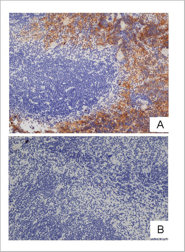
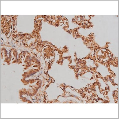
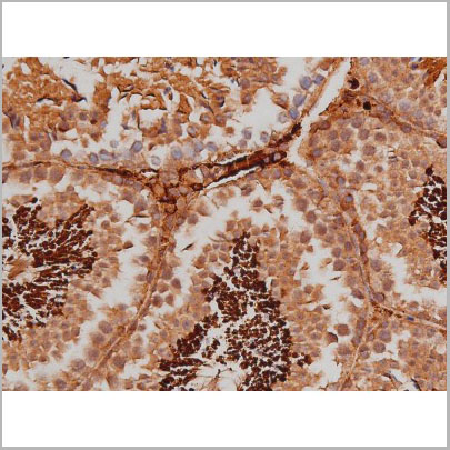
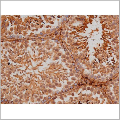
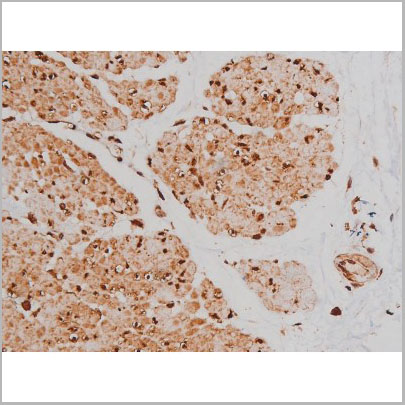
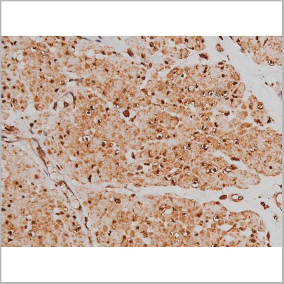
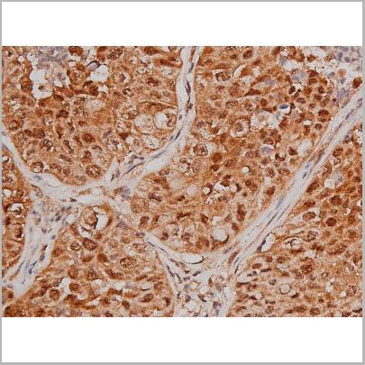
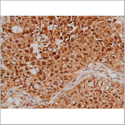
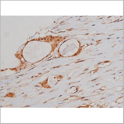
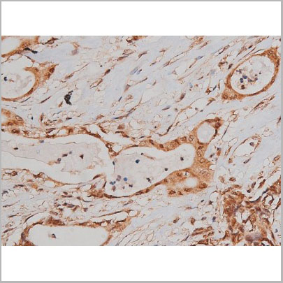
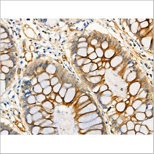
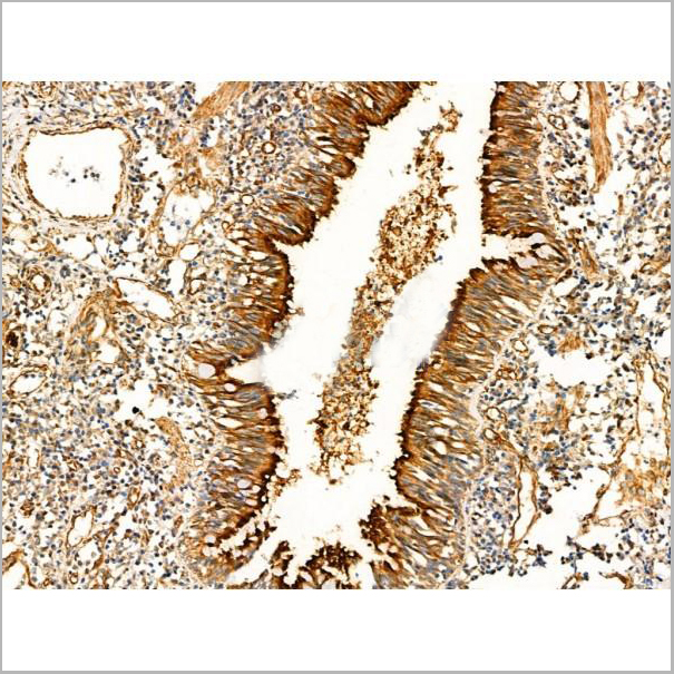
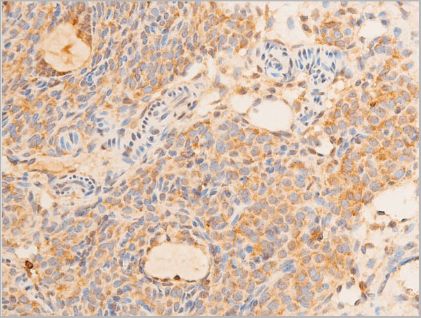
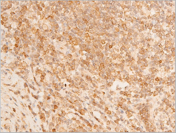
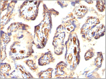
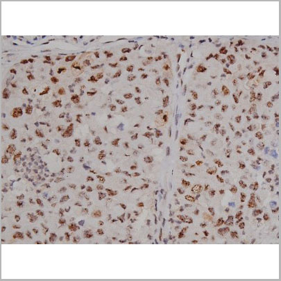
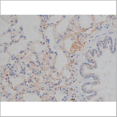
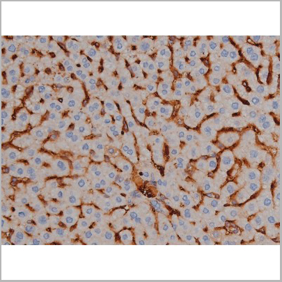
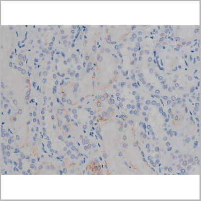
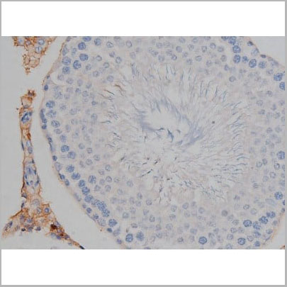
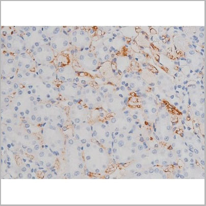
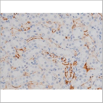
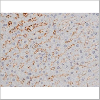
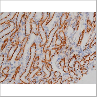
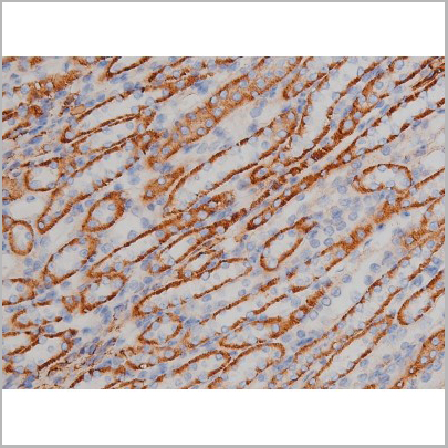
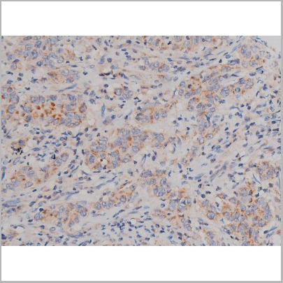
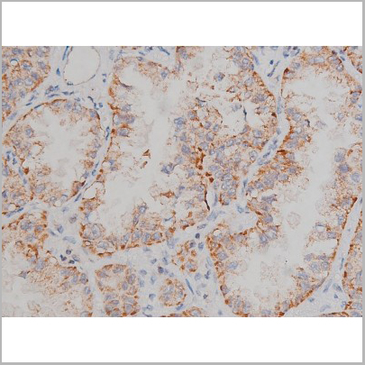
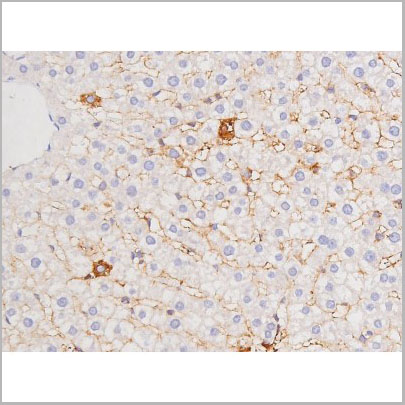
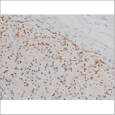
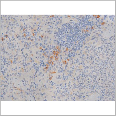
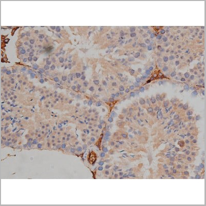
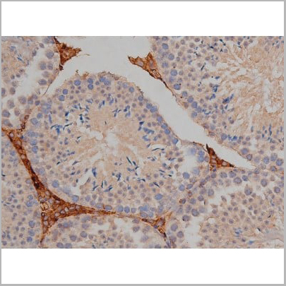
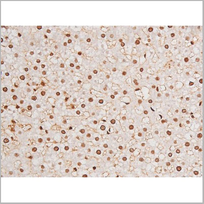
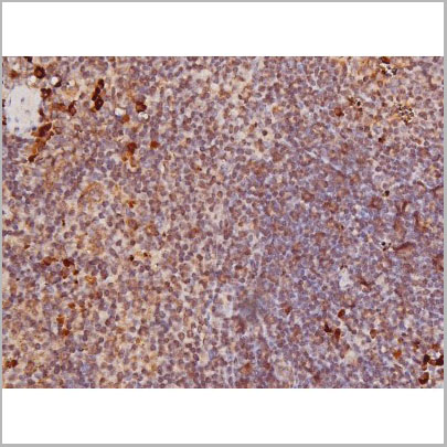
 IHC (Immunohistochemistry)
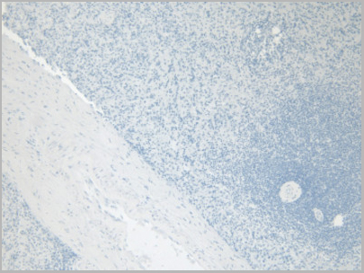
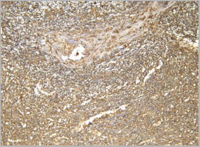
(Figure 8. IHC analysis of beta Catenin using anti-beta Catenin antibody (AAA19129).beta Catenin was detected in paraffin-embedded section of rat small intestine tissue. Heat mediated antigen retrieval was performed in citrate buffer (pH6, epitope retrieval solution) for 20 mins. The tissue section was blocked with 10% goat serum. The tissue section was then incubated with 1ug/ml rabbit anti-beta Catenin Antibody (AAA19129) overnight at 4 degree C. Biotinylated goat anti-rabbit IgG was used as secondary antibody and incubated for 30 minutes at 37 degree C. The tissue section was developed using Strepavidin-Biotin-Complex (SABC) with DAB as the chromogen.)
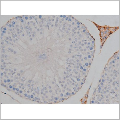
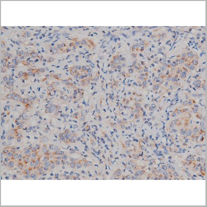
IHC (Immunohistochemistry)
(Figure 8. IHC analysis of beta Catenin using anti-beta Catenin antibody (AAA19129).beta Catenin was detected in paraffin-embedded section of rat small intestine tissue. Heat mediated antigen retrieval was performed in citrate buffer (pH6, epitope retrieval solution) for 20 mins. The tissue section was blocked with 10% goat serum. The tissue section was then incubated with 1ug/ml rabbit anti-beta Catenin Antibody (AAA19129) overnight at 4 degree C. Biotinylated goat anti-rabbit IgG was used as secondary antibody and incubated for 30 minutes at 37 degree C. The tissue section was developed using Strepavidin-Biotin-Complex (SABC) with DAB as the chromogen.)
beta Catenin, Polyclonal Antibody (Cat# AAA19129)
Full Name
Anti-beta Catenin Picoband Antibody
Gene Names
CTNNB1; EVR7; CTNNB; MRD19; armadillo
Reactivity
Human, Mouse, Rat
No cross reactivity with other proteins.
No cross reactivity with other proteins.
Applications
EIA, IHC, WB
Purity
Immunogen affinity purified
Pricing

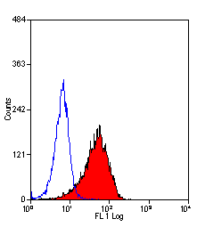
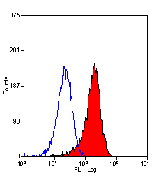
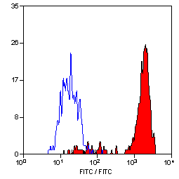
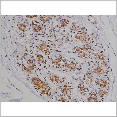
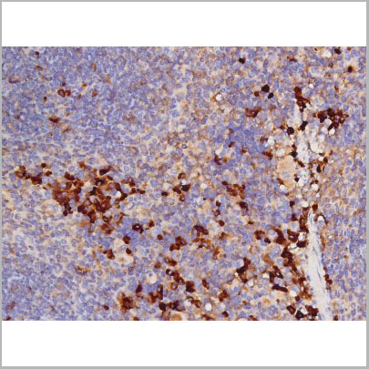
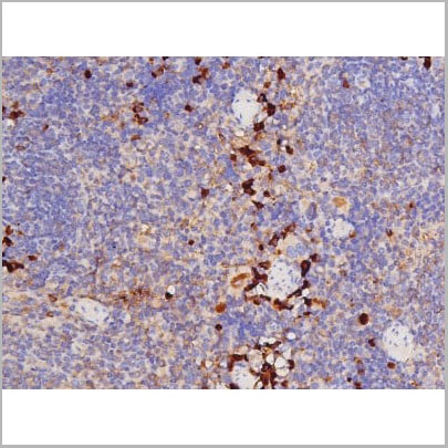
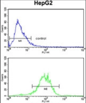
 FCM (Flow Cytometry)
(TMPRSS2 Antibody (Center)(AAA28646) flow cytometric analysis of HepG2 cells (right histogram) compared to a negative control cell (left histogram).FITC-conjugated goat-anti-rabbit secondary antibodies were used for the analysis.)
FCM (Flow Cytometry)
(TMPRSS2 Antibody (Center)(AAA28646) flow cytometric analysis of HepG2 cells (right histogram) compared to a negative control cell (left histogram).FITC-conjugated goat-anti-rabbit secondary antibodies were used for the analysis.)
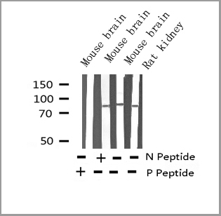
TMPRSS2, Polyclonal Antibody (Cat# AAA28646)
Full Name
TMPRSS2 Antibody (Center)
Gene Names
TMPRSS2; PP9284; PRSS10
Reactivity
Reactivity: Human, Mouse
Predicted: Mouse
Predicted: Mouse
Applications
WB, EIA, FC/FACS
Purity
Purified Rabbit Polyclonal Antibody (Pab)
Pricing


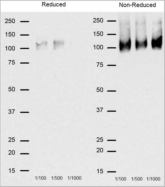
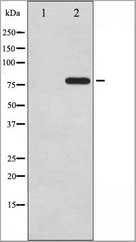
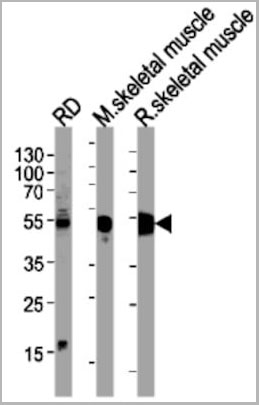
 WB (Western Blot)
(FG Pancreatic Carcinoma Cell Lines stably expressing vector along (FG-V) the b3 integrin subunit (FG-b3) or a b3 truncation mutant (FG-759x). Src Mab (AAA28639) was diluted 1:500 in 1% BSA/TBST and incubated Overnight at 4 degree C. After washing 3x 5 min. with TBST the blots were incubated with 1:5000 Goat anti-mouse or Goat anti-rabbit secondary antibody for 1 hr at Room temperature. The blots were again washed 3x 5 min. with TBST and developed using ECL reagent.Data and protocol kindly provided by Dr. Weis of Cheresh Lab, UCSD.)
WB (Western Blot)
(FG Pancreatic Carcinoma Cell Lines stably expressing vector along (FG-V) the b3 integrin subunit (FG-b3) or a b3 truncation mutant (FG-759x). Src Mab (AAA28639) was diluted 1:500 in 1% BSA/TBST and incubated Overnight at 4 degree C. After washing 3x 5 min. with TBST the blots were incubated with 1:5000 Goat anti-mouse or Goat anti-rabbit secondary antibody for 1 hr at Room temperature. The blots were again washed 3x 5 min. with TBST and developed using ECL reagent.Data and protocol kindly provided by Dr. Weis of Cheresh Lab, UCSD.)
SRC, Monoclonal Antibody (Cat# AAA28639)
Full Name
SRC Antibody
Gene Names
SRC; ASV; SRC1; c-SRC; p60-Src
Reactivity
Human, mouse
Applications
WB, EIA, IF
Purity
This antibody is purified through a protein G column, followed by dialysis against PBS.
Pricing


 Application Data
(Analysis of Protein Array containing more than 19,000 full-length human proteins using Calretinin Mouse Monoclonal Antibody (CALB2/2786). Z- and S- Score: The Z-score represents the strength of a signal that a monoclonal antibody (MAb) (in combination with a fluorescently-tagged anti-IgG secondary antibody) produces when binding to a particular protein on the HuProtTM array. Z-scores are described in units of standard deviations (SD's) above the mean value of all signals generated on that array. If targets on HuProtTM are arranged in descending order of the Z-score, the S-score is the difference (also in units of SD's) between the Z-score. S-score therefore represents the relative target specificity of a MAb to its intended target. A MAb is considered to specific to its intended target, if the MAb has an S-score of at least 2.5. For example, if a MAb binds to protein X with a Z-score of 43 and to protein Y with a Z-score of 14, then the S-score for the binding of that MAb to protein X is equal to 29.)
Application Data
(Analysis of Protein Array containing more than 19,000 full-length human proteins using Calretinin Mouse Monoclonal Antibody (CALB2/2786). Z- and S- Score: The Z-score represents the strength of a signal that a monoclonal antibody (MAb) (in combination with a fluorescently-tagged anti-IgG secondary antibody) produces when binding to a particular protein on the HuProtTM array. Z-scores are described in units of standard deviations (SD's) above the mean value of all signals generated on that array. If targets on HuProtTM are arranged in descending order of the Z-score, the S-score is the difference (also in units of SD's) between the Z-score. S-score therefore represents the relative target specificity of a MAb to its intended target. A MAb is considered to specific to its intended target, if the MAb has an S-score of at least 2.5. For example, if a MAb binds to protein X with a Z-score of 43 and to protein Y with a Z-score of 14, then the S-score for the binding of that MAb to protein X is equal to 29.)
Calretinin/Calbindin 2, Monoclonal Antibody (Cat# AAA23939)
Full Name
Calretinin/Calbindin 2 (Mesothelioma Marker)
Gene Names
CALB2; CR; CAL2; CAB29
Reactivity
Human
Applications
WB, IHC
Purity
Purified Ab with BSA and Azide at 200ug/ml OR Purified Ab WITHOUT BSA at 1.0mg/ml
Pricing


 Application Data
(Analysis of Protein Array containing more than 19,000 full-length human proteins using Calretinin Mouse Monoclonal Antibody (CALB2/2602). Z- and S- Score: The Z-score represents the strength of a signal that a monoclonal antibody (MAb) (in combination with a fluorescently-tagged anti-IgG secondary antibody) produces when binding to a particular protein on the HuProtTM array. Z-scores are described in units of standard deviations (SD's) above the mean value of all signals generated on that array. If targets on HuProtTM are arranged in descending order of the Z-score, the S-score is the difference (also in units of SD's) between the Z-score. S-score therefore represents the relative target specificity of a MAb to its intended target. A MAb is considered to specific to its intended target, if the MAb has an S-score of at least 2.5. For example, if a MAb binds to protein X with a Z-score of 43 and to protein Y with a Z-score of 14, then the S-score for the binding of that MAb to protein X is equal to 29.)
Application Data
(Analysis of Protein Array containing more than 19,000 full-length human proteins using Calretinin Mouse Monoclonal Antibody (CALB2/2602). Z- and S- Score: The Z-score represents the strength of a signal that a monoclonal antibody (MAb) (in combination with a fluorescently-tagged anti-IgG secondary antibody) produces when binding to a particular protein on the HuProtTM array. Z-scores are described in units of standard deviations (SD's) above the mean value of all signals generated on that array. If targets on HuProtTM are arranged in descending order of the Z-score, the S-score is the difference (also in units of SD's) between the Z-score. S-score therefore represents the relative target specificity of a MAb to its intended target. A MAb is considered to specific to its intended target, if the MAb has an S-score of at least 2.5. For example, if a MAb binds to protein X with a Z-score of 43 and to protein Y with a Z-score of 14, then the S-score for the binding of that MAb to protein X is equal to 29.)
Calretinin/Calbindin 2, Monoclonal Antibody (Cat# AAA23938)
Full Name
Calretinin/Calbindin 2 (Mesothelioma Marker)
Gene Names
CALB2; CR; CAL2; CAB29
Reactivity
Human
Applications
WB, IHC
Purity
Purified Ab with BSA and Azide at 200ug/ml OR Purified Ab WITHOUT BSA at 1.0mg/ml
Pricing
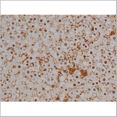
 Application Data
(Staining of J774 cells with Rat anti Mouse F4/80 antigen Biotin)
Application Data
(Staining of J774 cells with Rat anti Mouse F4/80 antigen Biotin)
F4/80, Monoclonal Antibody (Cat# AAA12225)
Full Name
RAT ANTI MOUSE F4/80
Gene Names
Emr1; Ly71; F4/80; Gpf480; TM7LN3; DD7A5-7; EGF-TM7
Applications
Flow Cytometry, Immunohistochemistry, Immunohistochemistry, Immunohistochemistry, Immunoprecipitation, Immunofluorescence, Radioimmunoassay, Fluorescence Microscopy
Pricing
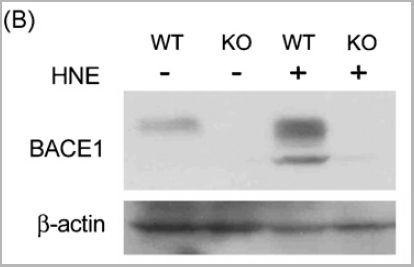
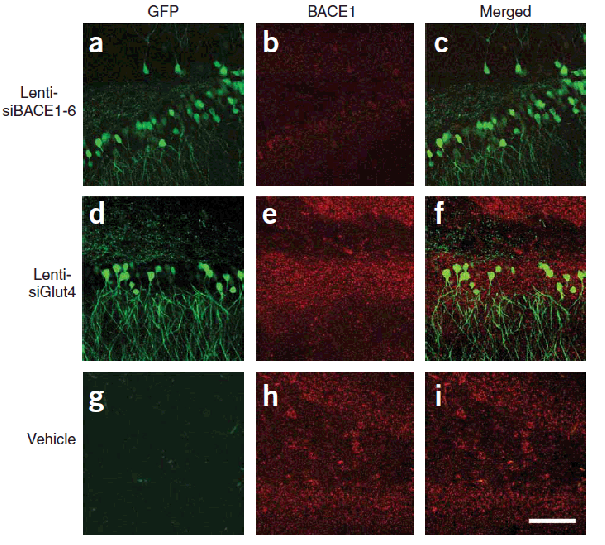
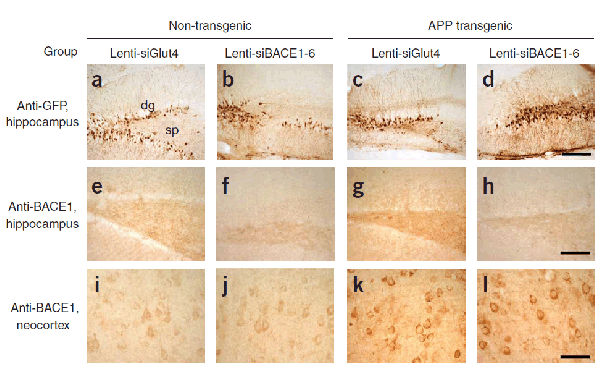

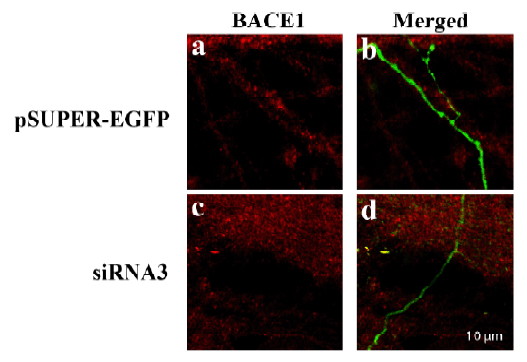 IF (Immunofluorescence)
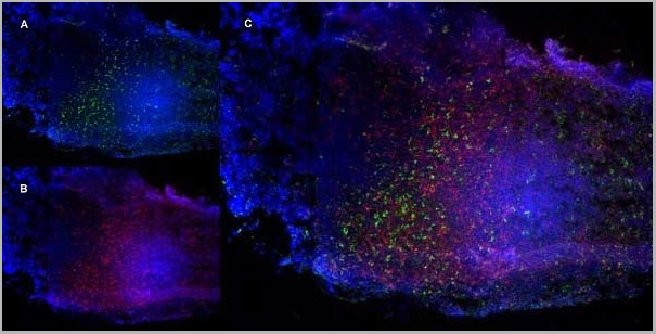
(Figure 10 KD Validation of BACE in DRG (Hyun, 2007)Decreased BACE1 expression in DRG following siRNA3 transfection. DRG neurons were transfected with 1 ug siRNA3 plasmid and incubated for 48 hours in 37°˚C. DRG neurons were stained for BACE1 us¬ing the Anti-BACE antibody (a,b) Neurons transfected with the control plas¬mid pSUPER-EGFP (green) did not display any changes in BACE1 expression (red). (c,d) DRG neurons transfected with siR¬NA3 displayed reduced BACE1 expression in the axon.)
IF (Immunofluorescence)
(Figure 10 KD Validation of BACE in DRG (Hyun, 2007)Decreased BACE1 expression in DRG following siRNA3 transfection. DRG neurons were transfected with 1 ug siRNA3 plasmid and incubated for 48 hours in 37°˚C. DRG neurons were stained for BACE1 us¬ing the Anti-BACE antibody (a,b) Neurons transfected with the control plas¬mid pSUPER-EGFP (green) did not display any changes in BACE1 expression (red). (c,d) DRG neurons transfected with siR¬NA3 displayed reduced BACE1 expression in the axon.)
BACE, Polyclonal Antibody (Cat# AAA10918)
Full Name
BACE Antibody
Gene Names
BACE1; ASP2; BACE; HSPC104
Reactivity
Human, Mouse
Applications
Immunocytochemistry, Immunofluorescence, Immunohistochemistry, Western Blot
Purity
BACE Antibody is affinity chromatography purified via peptide column.
Pricing

 IF (Immunofluorescence)
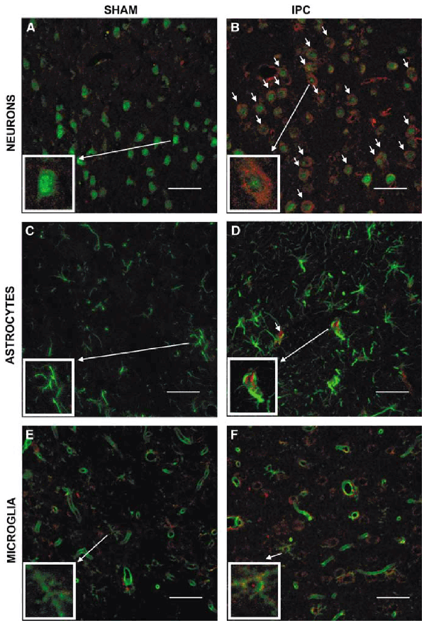
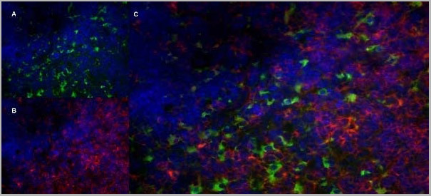
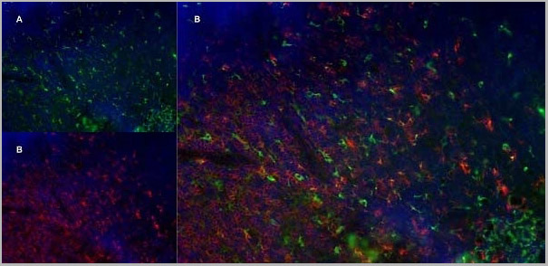
(Figure 10 Immunofluorescence Validation of TACE in Rat Brain (Pradillo et al, 2005)Cellular localization of TACE. Double immunofluorescence staining of brain sections from sham-operated (SHAM; A, C, E) and IPC-exposed animals (IPC; B, D, F) of TACE (red) and the cellular markers (green) NeuN (neurons; A, B), GFAP (astrocytes; C, D) and L. esculentum lectin (microglia and endothelium; E, F). White arrows indicate TACE-positive cells.)
IF (Immunofluorescence)
(Figure 10 Immunofluorescence Validation of TACE in Rat Brain (Pradillo et al, 2005)Cellular localization of TACE. Double immunofluorescence staining of brain sections from sham-operated (SHAM; A, C, E) and IPC-exposed animals (IPC; B, D, F) of TACE (red) and the cellular markers (green) NeuN (neurons; A, B), GFAP (astrocytes; C, D) and L. esculentum lectin (microglia and endothelium; E, F). White arrows indicate TACE-positive cells.)
TACE, Polyclonal Antibody (Cat# AAA10912)
Full Name
TACE Antibody
Gene Names
ADAM17; CSVP; TACE; NISBD; ADAM18; CD156B; NISBD1
Reactivity
Human, Mouse, Rat
Applications
Western Blot, Immunocytochemistry, Immunofluorescence, Flow Cytometry
Purity
TACE Antibody is affinity chromatography purified via peptide column.
Pricing
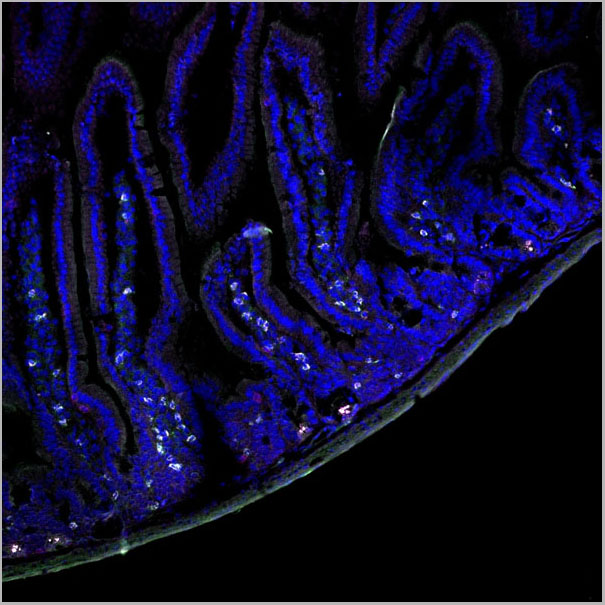
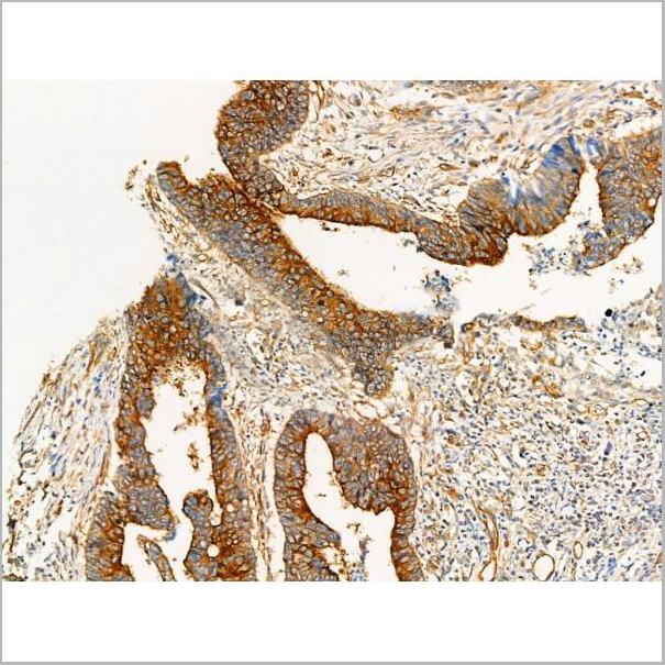
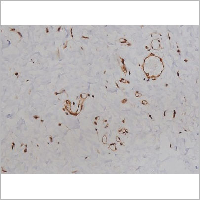
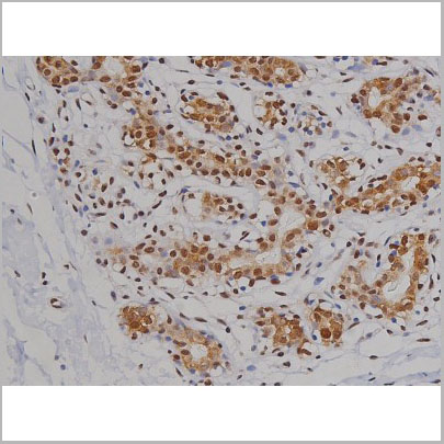
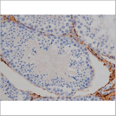
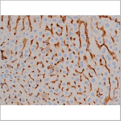
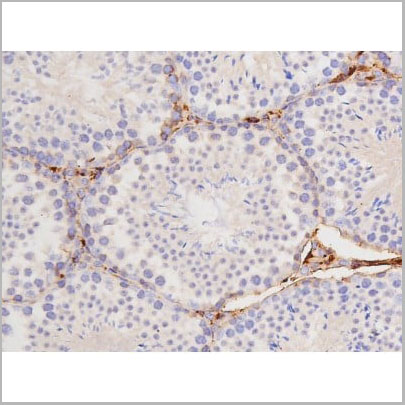
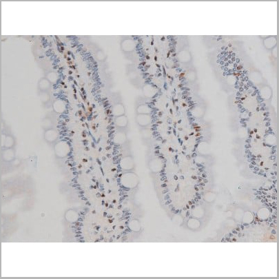
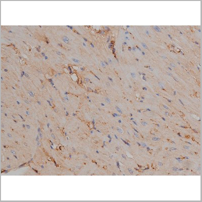
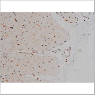
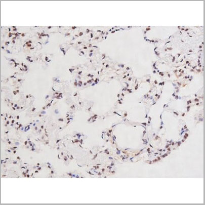
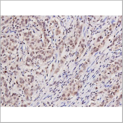
 IHC (Immunohistochemistry)
(AAA31005 at 1/200 staining Rat ganstric tissue sections by IHC-P. The tissue was formaldehyde fixed and a heat mediated antigen retrieval step in citrate buffer was performed. The tissue was then blocked and incubated with the antibody for 1.5 hours at 22 degree C. An HRP conjugated goat anti-rabbit antibody was used as the secondary.)
IHC (Immunohistochemistry)
(AAA31005 at 1/200 staining Rat ganstric tissue sections by IHC-P. The tissue was formaldehyde fixed and a heat mediated antigen retrieval step in citrate buffer was performed. The tissue was then blocked and incubated with the antibody for 1.5 hours at 22 degree C. An HRP conjugated goat anti-rabbit antibody was used as the secondary.)
Tau, Polyclonal Antibody (Cat# AAA31005)
Full Name
Phospho-Tau (Ser262) Antibody
Gene Names
MAPT; TAU; MSTD; PPND; DDPAC; MAPTL; MTBT1; MTBT2; FTDP-17; PPP1R103
Reactivity
Human, Mouse, Rat
Applications
Western Blot, Immunohistochemistry
Purity
From purified rabbit serum by affinity purification via sequential chromatography on phospho-and non-phospho-peptide affinity columns.
Pricing
 Application Data
(At 25 degree C. Samples were then incubated with primary Ab(At 37 degree C. An AlexaFluor594 conjugated goat anti-rabbit IgG(H+L) Ab(Red) and an AlexaFluor488 conjugated goat anti-mouse IgG(H+L) Ab(Green) were used as the secondary antibody.The nuclear counter stain is DAPI(blue).)
Application Data
(At 25 degree C. Samples were then incubated with primary Ab(At 37 degree C. An AlexaFluor594 conjugated goat anti-rabbit IgG(H+L) Ab(Red) and an AlexaFluor488 conjugated goat anti-mouse IgG(H+L) Ab(Green) were used as the secondary antibody.The nuclear counter stain is DAPI(blue).)
alpha Tubulin, Polyclonal Antibody (Cat# AAA31344)
Full Name
Acetyl-alpha Tubulin (Lys40) Antibody
Gene Names
TUBA1B; K-ALPHA-1
Reactivity
Human, Mouse, Rat
Applications
Western Blot, Immunohistochemistry, Immunofluorescence, Immunocytochemistry
Purity
The antiserum was purified by peptide affinity chromatography using SulfoLink Coupling Resin (Thermo Fisher Scientific).
Pricing
Tyrosine Hydroxylase, Antibody (Cat# AAA13664)
Full Name
Goat anti-Tyrosine Hydroxylase Antibody
Gene Names
TH; TYH; DYT14; DYT5b
Reactivity
Tested: Human; Expected from sequence similarity: Human, Rat, Dog
Applications
Peptide ELISA, Immunohistochemistry
Purity
Purified from goat serum by ammonium sulphate precipitation followed by antigen affinity chromatography using the immunizing peptide.
Pricing
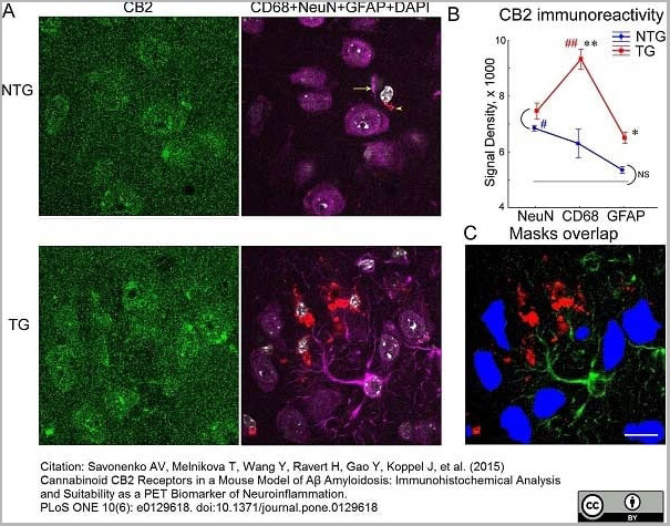 Application Data
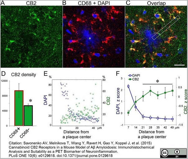
(Published customer image:Rat anti Mouse CD68 antibody, clone FA-11 used for the identification of microglia in mouse brain by immunofluorescence.Image caption:Comparison of CB2 immunoreactivity in neurons, activated microglia and astrocytes. ()
Application Data
(Published customer image:Rat anti Mouse CD68 antibody, clone FA-11 used for the identification of microglia in mouse brain by immunofluorescence.Image caption:Comparison of CB2 immunoreactivity in neurons, activated microglia and astrocytes. ()
CD68, Monoclonal Antibody (Cat# AAA12289)
Full Name
Rat anti Mouse CD68:Amethyst Orange
Gene Names
Cd68; Lamp4; gp110; Scard1
Reactivity
Mouse
Applications
Flow Cytometry
Purity
Purified IgG prepared by affinity chromatography on Protein G from tissue culture supernatant
Pricing
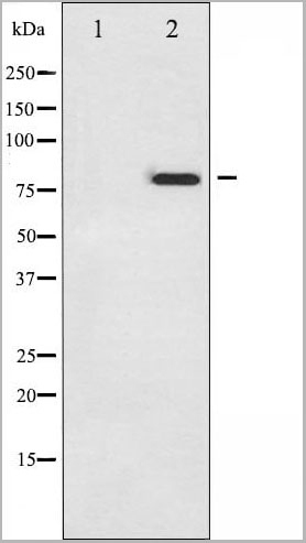
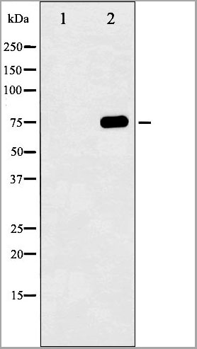
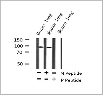 WB (Western Blot)
(Western blot analysis of Phospho-p73 (Tyr99) expression in Mouse lung lysate)
WB (Western Blot)
(Western blot analysis of Phospho-p73 (Tyr99) expression in Mouse lung lysate)
p73, Polyclonal Antibody (Cat# AAA30950)
Full Name
Phospho-p73 (Tyr99) Antibody
Gene Names
TP73; P73
Reactivity
Human, Mouse
Applications
Western Blot, Immunohistochemistry
Purity
From purified rabbit serum by affinity purification via sequential chromatography on phospho-and non-phospho-peptide affinity columns.
Pricing
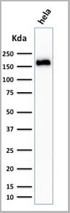
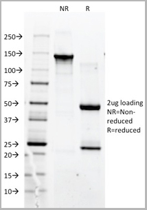
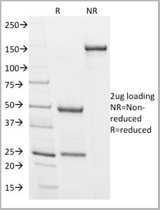 SDS-PAGE
(SDS-PAGE Analysis Purified Podocalyxin Mouse Monoclonal Antibody (3D3). Confirmation of Integrity and Purity of Antibody.)
SDS-PAGE
(SDS-PAGE Analysis Purified Podocalyxin Mouse Monoclonal Antibody (3D3). Confirmation of Integrity and Purity of Antibody.)
Podocalyxin (PODXL), Monoclonal Antibody (Cat# AAA13811)
Full Name
Podocalyxin (PODXL) (Hematopoietic Stem Cell Marker) Mouse Monoclonal Antibody
Gene Names
PODXL; PC; PCLP; Gp200; PCLP-1
Reactivity
Human, Rabbit, Rat
Applications
Flow Cytometry, Immunofluorescence, Immunoprecipitation, Western Blot, Immunohistochemistry
Pricing
 WB (Western Blot)
(Western blot analysis of Phospho-Tau (Ser396) expression in various lysates)
WB (Western Blot)
(Western blot analysis of Phospho-Tau (Ser396) expression in various lysates)
Tau, Polyclonal Antibody (Cat# AAA31003)
Full Name
Phospho-Tau (Ser396) Antibody
Gene Names
MAPT; TAU; MSTD; PPND; DDPAC; MAPTL; MTBT1; MTBT2; FTDP-17; PPP1R103
Reactivity
Human, Mouse, Rat
Applications
Western Blot, Immunohistochemistry
Purity
From purified rabbit serum by affinity purification via sequential chromatography on phospho-and non-phospho-peptide affinity columns.
Pricing
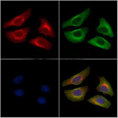
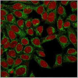
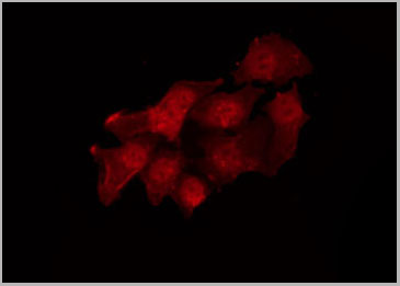
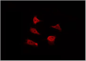
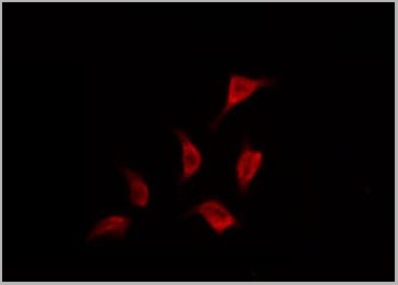 IF (Immunofluorescence)
(AAA30998 staining HepG2 by IF/ICC. The sample were fixed with PFA and permeabilized in 0.1% Triton X-100, then blocked in 10% serum for 45 minutes at 25 degree C. The primary antibody was diluted at 1/200 and incubated with the sample for 1 hour at 37 degree C. An Alexa Fluor 594 conjugated goat anti-rabbit IgG (H+L) Ab, diluted at 1/600, was used as the secondary antibody.)
IF (Immunofluorescence)
(AAA30998 staining HepG2 by IF/ICC. The sample were fixed with PFA and permeabilized in 0.1% Triton X-100, then blocked in 10% serum for 45 minutes at 25 degree C. The primary antibody was diluted at 1/200 and incubated with the sample for 1 hour at 37 degree C. An Alexa Fluor 594 conjugated goat anti-rabbit IgG (H+L) Ab, diluted at 1/600, was used as the secondary antibody.)
Tau, Polyclonal Antibody (Cat# AAA30998)
Full Name
Phospho-Tau (Ser356) Antibody
Gene Names
MAPT; TAU; MSTD; PPND; DDPAC; MAPTL; MTBT1; MTBT2; FTDP-17; PPP1R103
Reactivity
Human, Mouse, Rat
Applications
Western Blot, Immunohistochemistry, Immunofluorescence, Immunocytochemistry
Purity
From purified rabbit serum by affinity purification via sequential chromatography on phospho-and non-phospho-peptide affinity columns.
Pricing
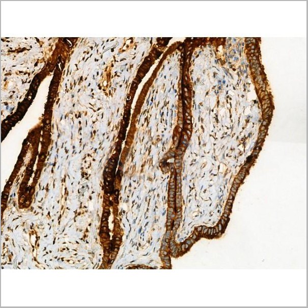
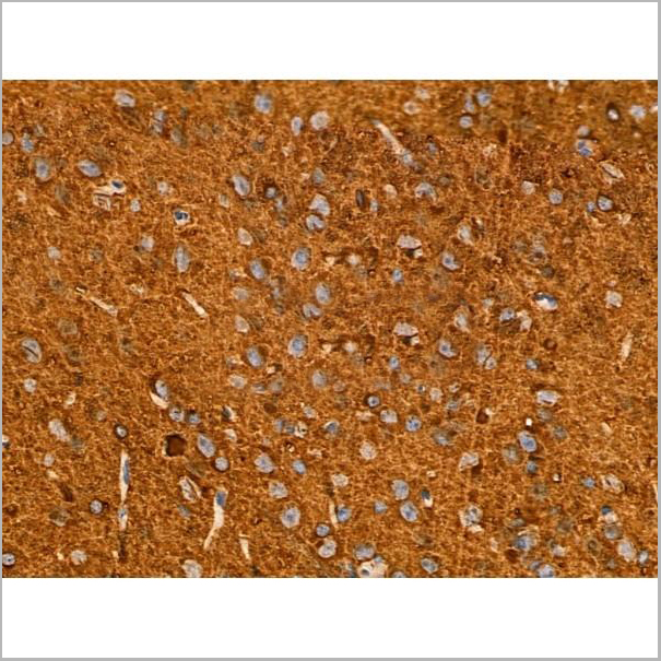
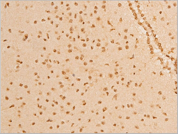
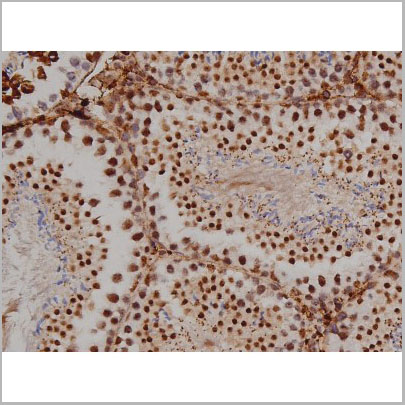
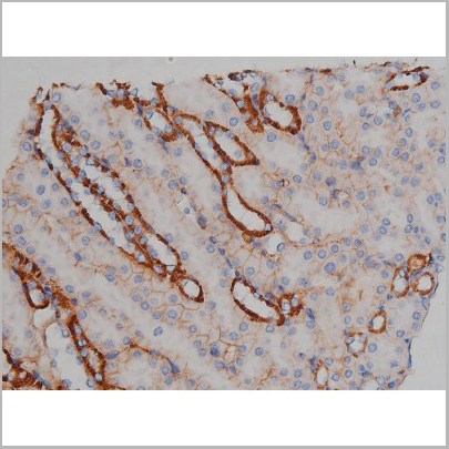
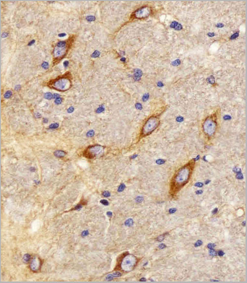
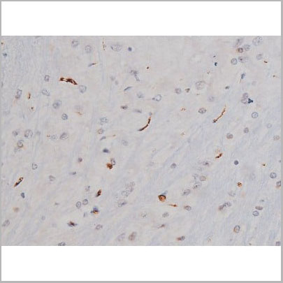
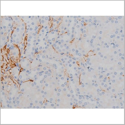
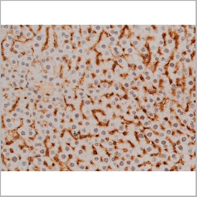
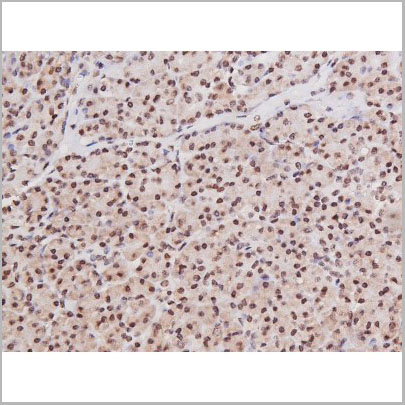
 IHC (Immunohistochemistry)
(IHC staining of human cerebellar neurons using AAA14700.)
IHC (Immunohistochemistry)
(IHC staining of human cerebellar neurons using AAA14700.)
SNAP 25, 26-27kD, Monoclonal Antibody (Cat# AAA14700)
Full Name
SNAP 25, 26-27kD (Synaptosomal-associated Protein)
Reactivity
Rat, Hamster Gerbil and Porcine
Applications
Western Blot, Immunohistochemistry
Purity
Purified by Protein A affinity chromatography from tissue culture supernatent
Pricing


 Application Data
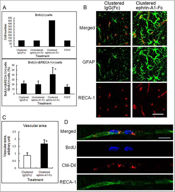
(Published customer image: Effect of clustered ephrin-A1-Fc on vascular formation in the rat striatum. Clustered ephrin-A1-Fc was injected into the lesioned side of the lateral ventricle in the unilaterally lesioned rats. Brains taken 6 weeks after injection were sectioned coronally and stained for GFAP (green) and RECA-1 (red) and with DAPI (nuclei; blue). The rectangular insets are shown in Fig. 8B. Scale bar: 100 um.From: Jing X, Miwa H, Sawada T, Nakanishi I, Kondo T, et al. (2012) Ephrin-A1-Mediated Dopaminergic Neurogenesis and Angiogenesis in a Rat Model of Parkinson's Disease. PLoS ONE 7(2): e32019.)
Application Data
(Published customer image: Effect of clustered ephrin-A1-Fc on vascular formation in the rat striatum. Clustered ephrin-A1-Fc was injected into the lesioned side of the lateral ventricle in the unilaterally lesioned rats. Brains taken 6 weeks after injection were sectioned coronally and stained for GFAP (green) and RECA-1 (red) and with DAPI (nuclei; blue). The rectangular insets are shown in Fig. 8B. Scale bar: 100 um.From: Jing X, Miwa H, Sawada T, Nakanishi I, Kondo T, et al. (2012) Ephrin-A1-Mediated Dopaminergic Neurogenesis and Angiogenesis in a Rat Model of Parkinson's Disease. PLoS ONE 7(2): e32019.)
RECA-1, Monoclonal Antibody (Cat# AAA12018)
Full Name
MOUSE ANTI RAT RECA-1
Applications
Immunohistochemistry, Immunofluorescence
Pricing


 Application Data
(Published customer image: Mouse anti V5 tag antibody, clone SV5-Pk1 used for the detection of V5 tagged WEEV_nsP3 protein by western blotting and immunofluorescenceImage caption: WEEV nsP3 interaction with host IKKbeta. A) U87MGs were transfected in a 6-well plate with 5 ug of pUC19 and WEEV_nsP3_HA for 24 hours. Cell lysates were resolved using SDS-PAGE and subsequently immunoblotted with V5 antibody and beta-actin served as a loading control. B) U87MGs were transfected with WEEV_nsP3_V5; cells were fixed after 24 hours and stained with antibodies against the endogenous IKKbeta and the V5 tag. Cells were incubated with appropriate secondary Alexa Fluor antibodies and the nuclei stained with DAPI. Co-localization of IKKbeta with WEEV_nsP3_V5 (yellow) was observed as shown by the arrows. B) Panels E -H serve as an example of transfected cells in a given field of view that show co-localization of IKKbeta and WEEV_nsP3_V5 24 hours post transfection. Panels I-L represent magnified images of other cells showing co-localization of IKKbeta and WEEV_nsP3_V5. Panel M is a magnified image of panel L. The co-localization was confirmed by Z-stack analysis. Co-localization was calculated to be approximately in 61% of cells (163 cells were counted of which 44% demonstrated expression of nsP3. Of those cells that expressed nsP3, 61% showed co-localization of both proteins). Images were taken using Nikon Eclipse TE2000-U at 60x magnification and are representative of 2 independent experiments.From: Amaya M, Voss K, Sampey G, Senina S, de la Fuente C, et al. (2014) The Role of IKKbeta in Venezuelan Equine Encephalitis Virus Infection. PLoS ONE 9(2): e86745.)
Application Data
(Published customer image: Mouse anti V5 tag antibody, clone SV5-Pk1 used for the detection of V5 tagged WEEV_nsP3 protein by western blotting and immunofluorescenceImage caption: WEEV nsP3 interaction with host IKKbeta. A) U87MGs were transfected in a 6-well plate with 5 ug of pUC19 and WEEV_nsP3_HA for 24 hours. Cell lysates were resolved using SDS-PAGE and subsequently immunoblotted with V5 antibody and beta-actin served as a loading control. B) U87MGs were transfected with WEEV_nsP3_V5; cells were fixed after 24 hours and stained with antibodies against the endogenous IKKbeta and the V5 tag. Cells were incubated with appropriate secondary Alexa Fluor antibodies and the nuclei stained with DAPI. Co-localization of IKKbeta with WEEV_nsP3_V5 (yellow) was observed as shown by the arrows. B) Panels E -H serve as an example of transfected cells in a given field of view that show co-localization of IKKbeta and WEEV_nsP3_V5 24 hours post transfection. Panels I-L represent magnified images of other cells showing co-localization of IKKbeta and WEEV_nsP3_V5. Panel M is a magnified image of panel L. The co-localization was confirmed by Z-stack analysis. Co-localization was calculated to be approximately in 61% of cells (163 cells were counted of which 44% demonstrated expression of nsP3. Of those cells that expressed nsP3, 61% showed co-localization of both proteins). Images were taken using Nikon Eclipse TE2000-U at 60x magnification and are representative of 2 independent experiments.From: Amaya M, Voss K, Sampey G, Senina S, de la Fuente C, et al. (2014) The Role of IKKbeta in Venezuelan Equine Encephalitis Virus Infection. PLoS ONE 9(2): e86745.)
V5-TAG, Monoclonal Antibody (Cat# AAA12081)
Full Name
MOUSE ANTI V5-TAG:HRP
Applications
Western Blot
Pricing


 Application Data
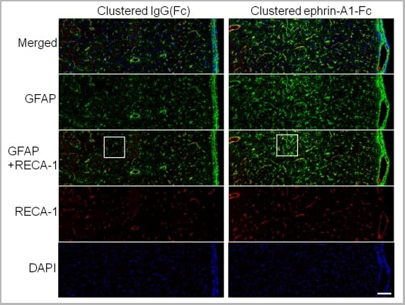
(Published customer image: Effect of clustered ephrin-A1-Fc on vascular formation in the rat striatum. Clustered ephrin-A1-Fc was injected into the lesioned side of the lateral ventricle in the unilaterally lesioned rats. Brains taken 6 weeks after injection were sectioned coronally and stained for GFAP (green) and RECA-1 (red) and with DAPI (nuclei; blue). The rectangular insets are shown in Fig. 8B. Scale bar: 100 um.From: Jing X, Miwa H, Sawada T, Nakanishi I, Kondo T, et al. (2012) Ephrin-A1-Mediated Dopaminergic Neurogenesis and Angiogenesis in a Rat Model of Parkinson's Disease. PLoS ONE 7(2): e32019.)
Application Data
(Published customer image: Effect of clustered ephrin-A1-Fc on vascular formation in the rat striatum. Clustered ephrin-A1-Fc was injected into the lesioned side of the lateral ventricle in the unilaterally lesioned rats. Brains taken 6 weeks after injection were sectioned coronally and stained for GFAP (green) and RECA-1 (red) and with DAPI (nuclei; blue). The rectangular insets are shown in Fig. 8B. Scale bar: 100 um.From: Jing X, Miwa H, Sawada T, Nakanishi I, Kondo T, et al. (2012) Ephrin-A1-Mediated Dopaminergic Neurogenesis and Angiogenesis in a Rat Model of Parkinson's Disease. PLoS ONE 7(2): e32019.)
RECA-1, Monoclonal Antibody (Cat# AAA12019)
Full Name
MOUSE ANTI RAT RECA-1
Applications
Immunohistochemistry, Immunofluorescence
Pricing
 Application Data
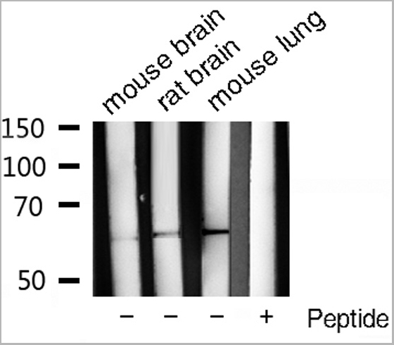
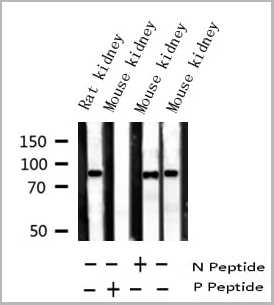
(AAA13666 (1.5ug/ml) as the reporter with EB002025 as the capture rabbit antibody (5ug/ml).)
Application Data
(AAA13666 (1.5ug/ml) as the reporter with EB002025 as the capture rabbit antibody (5ug/ml).)
FTL, Polyclonal Antibody (Cat# AAA13666)
Full Name
Goat anti-FTL Antibody
Gene Names
FTL; LFTD; NBIA3
Reactivity
Tested: Human, Mouse, Rat
Expected from sequence similarity: Human, Mouse, Rat, Dog, Cow
Expected from sequence similarity: Human, Mouse, Rat, Dog, Cow
Applications
Peptide ELISA, Western Blot, Immunohistochemistry
Purity
Purified from goat serum by ammonium sulphate precipitation followed by antigen affinity chromatography using the immunizing peptide.
Pricing

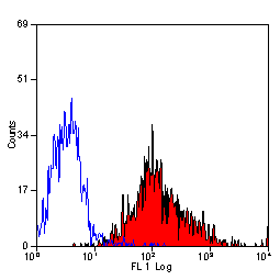
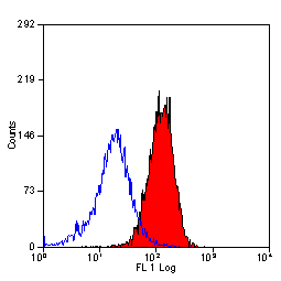
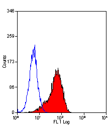
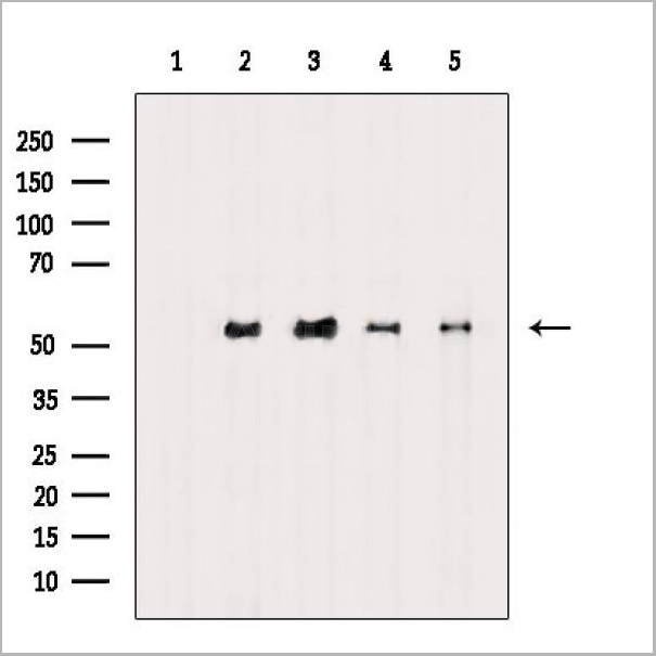
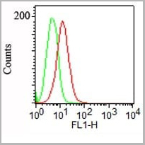
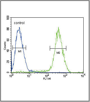 FCM (Flow Cytometry)
(CB2 Antibody (C-term) flow cytometric analysis of Jurkat cells (right histogram) compared to a negative control cell (left histogram).FITC-conjugated goat-anti-rabbit secondary antibodies were used for the analysis.)
FCM (Flow Cytometry)
(CB2 Antibody (C-term) flow cytometric analysis of Jurkat cells (right histogram) compared to a negative control cell (left histogram).FITC-conjugated goat-anti-rabbit secondary antibodies were used for the analysis.)
CB2, Polyclonal Antibody (Cat# AAA28672)
Full Name
CB2 Antibody (C-term)
Gene Names
CNR2; CB2; CX5; CB-2
Reactivity
Human
Applications
Immunohistochemistry, Flow Cytometry, Western Blot
Purity
This antibody is purified through a protein A column, followed by peptide affinity purification.
Pricing
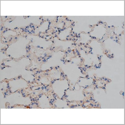

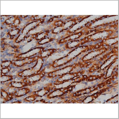
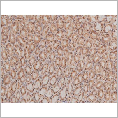
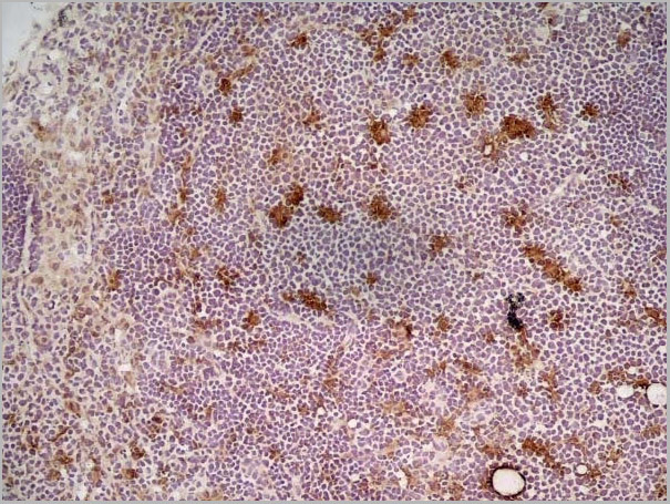
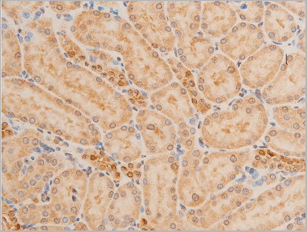
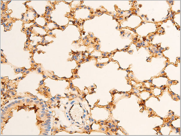
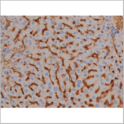
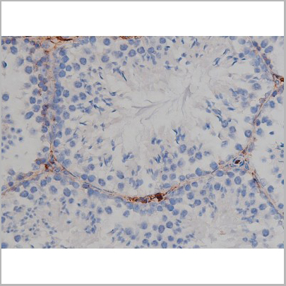
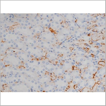
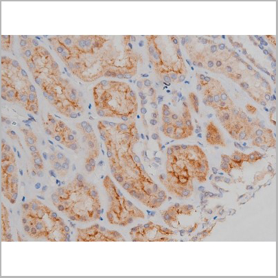
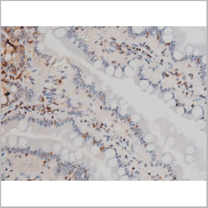
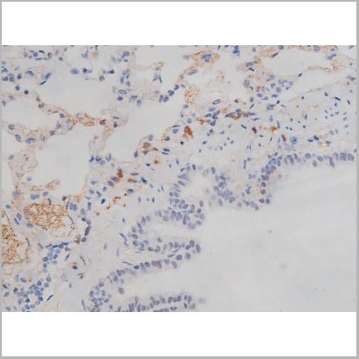
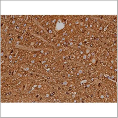
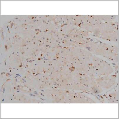
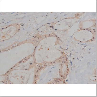
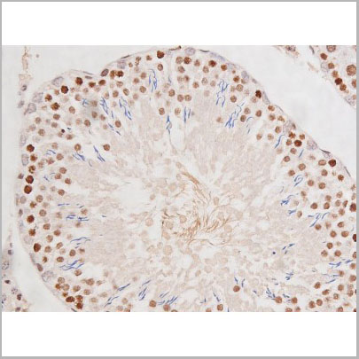
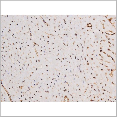
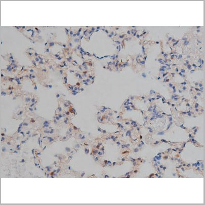 IHC (Immunohistochemistry)
(AAA31000 at 1/200 staining Rat lung tissue sections by IHC-P. The tissue was formaldehyde fixed and a heat mediated antigen retrieval step in citrate buffer was performed. The tissue was then blocked and incubated with the antibody for 1.5 hours at 22 degree C. An HRP conjugated goat anti-rabbit antibody was used as the secondary.)
IHC (Immunohistochemistry)
(AAA31000 at 1/200 staining Rat lung tissue sections by IHC-P. The tissue was formaldehyde fixed and a heat mediated antigen retrieval step in citrate buffer was performed. The tissue was then blocked and incubated with the antibody for 1.5 hours at 22 degree C. An HRP conjugated goat anti-rabbit antibody was used as the secondary.)
Tau, Polyclonal Antibody (Cat# AAA31000)
Full Name
Phospho-Tau (Ser422) Antibody
Gene Names
MAPT; TAU; MSTD; PPND; DDPAC; MAPTL; MTBT1; MTBT2; FTDP-17; PPP1R103
Reactivity
Human, Mouse, Rat
Applications
Western Blot, Immunohistochemistry
Purity
From purified rabbit serum by affinity purification via sequential chromatography on phospho-and non-phospho-peptide affinity columns.
Pricing
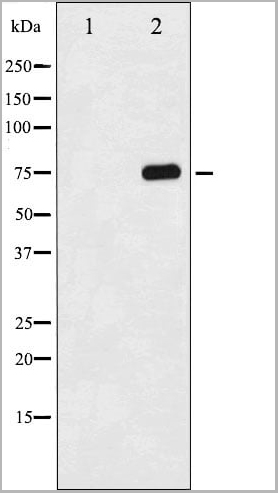
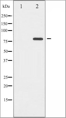
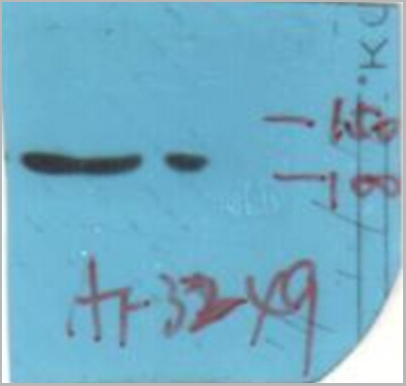
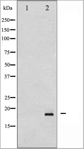
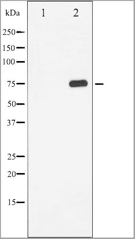
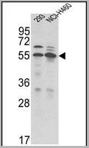
 WB (Western Blot)
(Western blot analysis of extracts of various celllines, using Phospho-Src (Tyr529) Antibody.)
WB (Western Blot)
(Western blot analysis of extracts of various celllines, using Phospho-Src (Tyr529) Antibody.)
Src, Polyclonal Antibody (Cat# AAA31009)
Full Name
Phospho-Src (Tyr530) Antibody
Gene Names
SRC; ASV; SRC1; THC6; c-SRC; p60-Src
Reactivity
Human, Mouse, Rat
Applications
Western Blot, Immunohistochemistry, Immunofluorescence, Immunocytochemistry
Purity
From purified rabbit serum by affinity purification via sequential chromatography on phospho-and non-phospho-peptide affinity columns.
Pricing


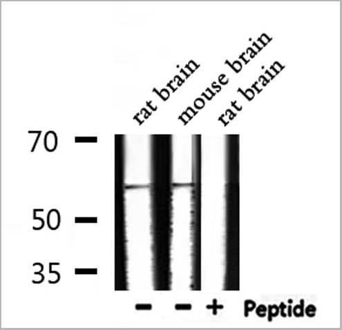
 WB (Western Blot)
(Western blot analysis of n-NOS phosphorylation expression in A549 whole cell lysates, The lane on the left is treated with the antigen-specific peptide.)
WB (Western Blot)
(Western blot analysis of n-NOS phosphorylation expression in A549 whole cell lysates, The lane on the left is treated with the antigen-specific peptide.)
n-NOS, Polyclonal Antibody (Cat# AAA31028)
Full Name
Phospho-n-NOS (Ser852) Antibody
Gene Names
NOS1; NOS; bNOS; nNOS; IHPS1; N-NOS; NC-NOS
Reactivity
Human, Mouse, Rat
Applications
Western Blot, Immunohistochemistry, Immunofluorescence, Immunocytochemistry
Purity
From purified rabbit serum by affinity purification via sequential chromatography on phospho-and non-phospho-peptide affinity columns.
Pricing
68 kDa Neurofilament, Native Protein (Cat# AAA14359)
Full Name
68 kDa Neurofilament Ag (Bovine)
Purity
> 95% by SDS Gelelectrophoresis analysis
Pricing
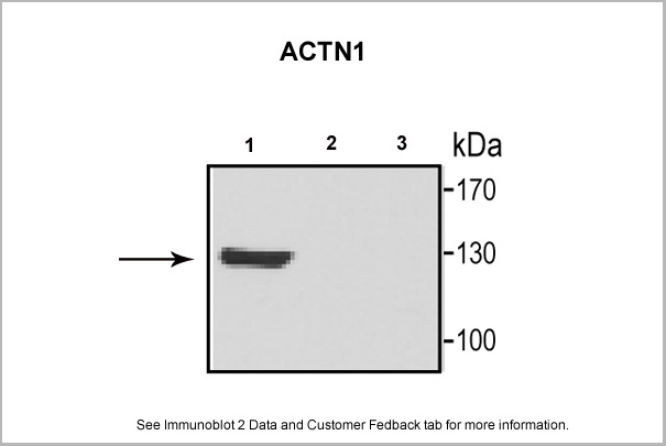
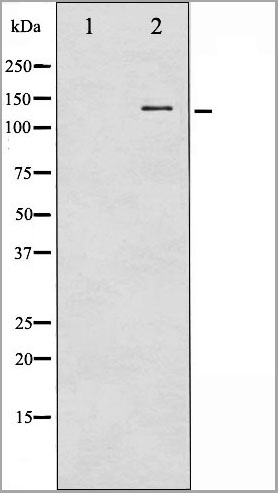
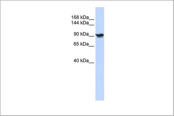 WB (Western Blot)
(WB Suggested Anti-ACTN1 Antibody Titration: 0.2-1 ug/mlELISA Titer: 1:312500Positive Control: 293T cell lysate)
WB (Western Blot)
(WB Suggested Anti-ACTN1 Antibody Titration: 0.2-1 ug/mlELISA Titer: 1:312500Positive Control: 293T cell lysate)
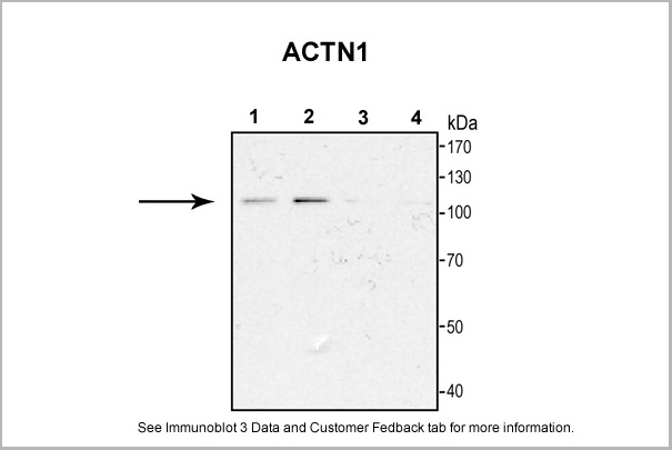
ACTN1, Polyclonal Antibody (Cat# AAA23579)
Full Name
ACTN1 antibody - N-terminal region
Gene Names
ACTN1; BDPLT15
Reactivity
Cow, Dog, Guinea Pig, Horse, Human, Mouse, Rabbit, Rat, Zebrafish
Applications
Immunohistochemistry, Western Blot
Purity
Affinity Purified
Pricing

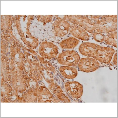
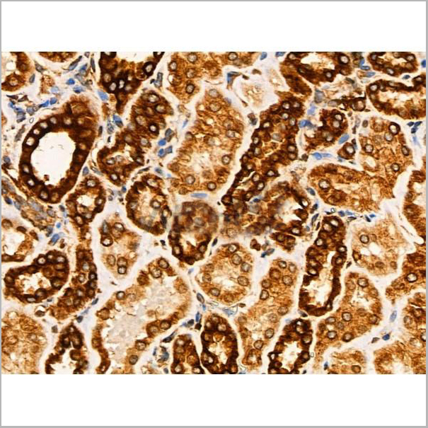
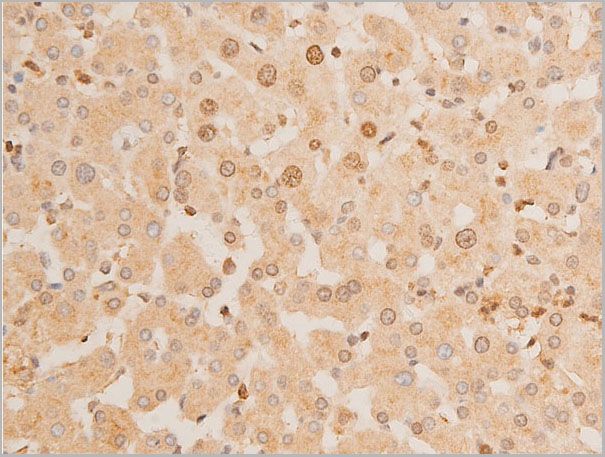
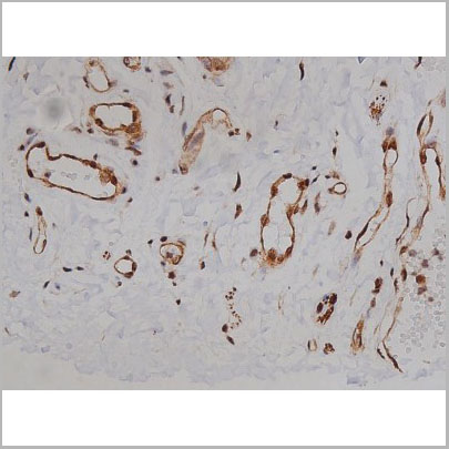
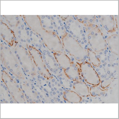
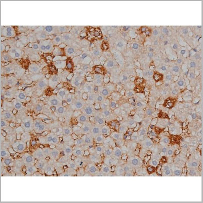
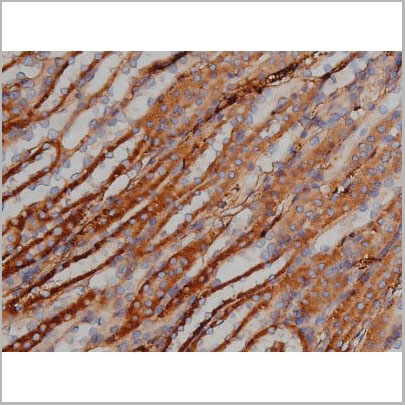
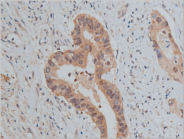
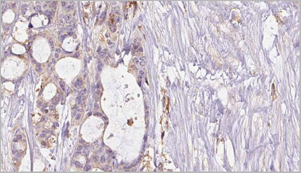
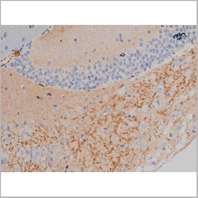
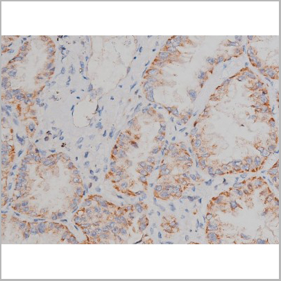
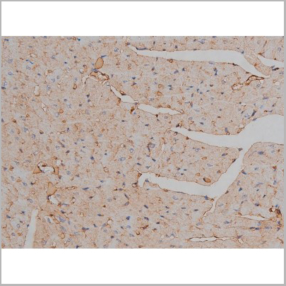
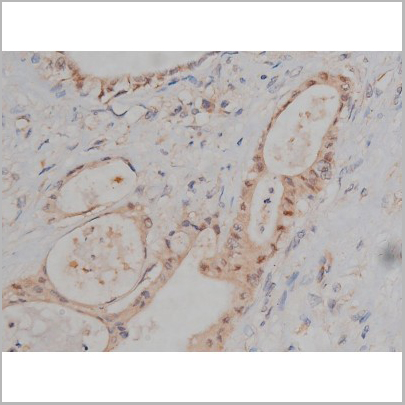
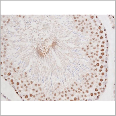
 IHC (Immunohistochemistry)
(AAA31004 at 1/200 staining Rat brain tissue sections by IHC-P. The tissue was formaldehyde fixed and a heat mediated antigen retrieval step in citrate buffer was performed. The tissue was then blocked and incubated with the antibody for 1.5 hours at 22 degree C. An HRP conjugated goat anti-rabbit antibody was used as the secondary.)
IHC (Immunohistochemistry)
(AAA31004 at 1/200 staining Rat brain tissue sections by IHC-P. The tissue was formaldehyde fixed and a heat mediated antigen retrieval step in citrate buffer was performed. The tissue was then blocked and incubated with the antibody for 1.5 hours at 22 degree C. An HRP conjugated goat anti-rabbit antibody was used as the secondary.)
Tau, Polyclonal Antibody (Cat# AAA31004)
Full Name
Phospho-Tau (Thr205) Antibody
Gene Names
MAPT; TAU; MSTD; PPND; DDPAC; MAPTL; MTBT1; MTBT2; FTDP-17; PPP1R103
Reactivity
Human, Mouse, Rat
Applications
Western Blot, Immunohistochemistry
Purity
From purified rabbit serum by affinity purification via sequential chromatography on phospho-and non-phospho-peptide affinity columns.
Pricing
 IS (Immunostaining)
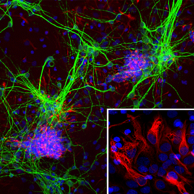
(Immunostaining of cultured E20 rat cortical neurons and glia stained with anti-nestin antibody (AAA14229, red, 1:500) and anti-MAP2 antibody (, green, 1:500). The blue is Hoechst staining for nuclear DNA. The nestin antibody labels developing astrocytes and neuronal stem cells in a clearly filamentous fashion, while the MAP2 antibody stains dendrites and perikarya of mature neurons.)
IS (Immunostaining)
(Immunostaining of cultured E20 rat cortical neurons and glia stained with anti-nestin antibody (AAA14229, red, 1:500) and anti-MAP2 antibody (, green, 1:500). The blue is Hoechst staining for nuclear DNA. The nestin antibody labels developing astrocytes and neuronal stem cells in a clearly filamentous fashion, while the MAP2 antibody stains dendrites and perikarya of mature neurons.)
Nestin, Monoclonal Antibody (Cat# AAA14229)
Full Name
Anti-Nestin
Gene Names
NES; Nbla00170
Applications
Western Blot, Immunofluorescence
Purity
Protein G purified culture supernatant
Pricing

 WB (Western Blot)
(Western blot analysis of Synuclein phosphorylation expression in Mouse brain tissue lysates, The lane on the left is treated with the antigen-specific peptide.)
WB (Western Blot)
(Western blot analysis of Synuclein phosphorylation expression in Mouse brain tissue lysates, The lane on the left is treated with the antigen-specific peptide.)
Synuclein, Polyclonal Antibody (Cat# AAA31040)
Full Name
Phospho-Synuclein (Ser129) Antibody
Gene Names
SNCA; PD1; NACP; PARK1; PARK4
Reactivity
Human, Mouse, Rat
Applications
Western Blot, Immunohistochemistry
Purity
From purified rabbit serum by affinity purification via sequential chromatography on phospho-and non-phospho-peptide affinity columns.
Pricing

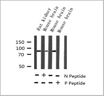
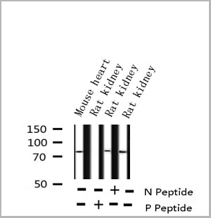
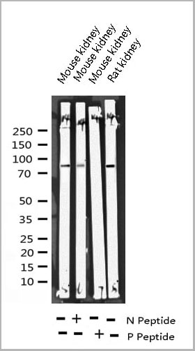
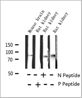
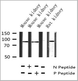
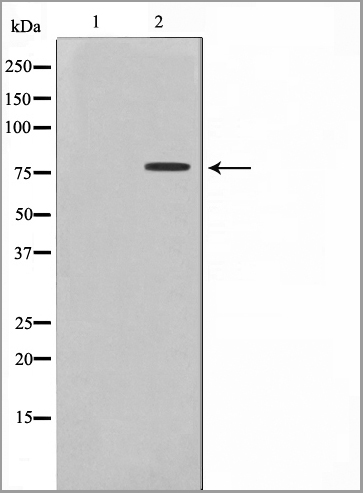
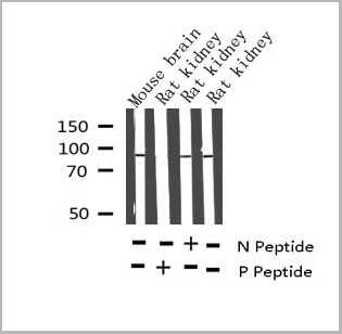 WB (Western Blot)
(Western blot analysis of Phospho-Tau (Thr212) Antibody expression in mouse brain and rat kidney tissues lysates.)
WB (Western Blot)
(Western blot analysis of Phospho-Tau (Thr212) Antibody expression in mouse brain and rat kidney tissues lysates.)
Tau, Polyclonal Antibody (Cat# AAA31001)
Full Name
Phospho-Tau (Thr212) Antibody
Gene Names
MAPT; TAU; MSTD; PPND; DDPAC; MAPTL; MTBT1; MTBT2; FTDP-17; PPP1R103
Reactivity
Human, Mouse, Rat
Applications
Western Blot, Immunohistochemistry
Purity
From purified rabbit serum by affinity purification via sequential chromatography on phospho-and non-phospho-peptide affinity columns.
Pricing


 Application Data
(Published customer image: Mouse anti V5 tag antibody, clone SV5-Pk1 used for the detection of V5 tagged WEEV_nsP3 protein by western blotting and immunofluorescenceImage caption: WEEV nsP3 interaction with host IKKbeta. A) U87MGs were transfected in a 6-well plate with 5 ug of pUC19 and WEEV_nsP3_HA for 24 hours. Cell lysates were resolved using SDS-PAGE and subsequently immunoblotted with V5 antibody and beta-actin served as a loading control. B) U87MGs were transfected with WEEV_nsP3_V5; cells were fixed after 24 hours and stained with antibodies against the endogenous IKKbeta and the V5 tag. Cells were incubated with appropriate secondary Alexa Fluor antibodies and the nuclei stained with DAPI. Co-localization of IKKbeta with WEEV_nsP3_V5 (yellow) was observed as shown by the arrows. B) Panels E -H serve as an example of transfected cells in a given field of view that show co-localization of IKKbeta and WEEV_nsP3_V5 24 hours post transfection. Panels I-L represent magnified images of other cells showing co-localization of IKKbeta and WEEV_nsP3_V5. Panel M is a magnified image of panel L. The co-localization was confirmed by Z-stack analysis. Co-localization was calculated to be approximately in 61% of cells (163 cells were counted of which 44% demonstrated expression of nsP3. Of those cells that expressed nsP3, 61% showed co-localization of both proteins). Images were taken using Nikon Eclipse TE2000-U at 60x magnification and are representative of 2 independent experiments.From: Amaya M, Voss K, Sampey G, Senina S, de la Fuente C, et al. (2014) The Role of IKKbeta in Venezuelan Equine Encephalitis Virus Infection. PLoS ONE 9(2): e86745.)
Application Data
(Published customer image: Mouse anti V5 tag antibody, clone SV5-Pk1 used for the detection of V5 tagged WEEV_nsP3 protein by western blotting and immunofluorescenceImage caption: WEEV nsP3 interaction with host IKKbeta. A) U87MGs were transfected in a 6-well plate with 5 ug of pUC19 and WEEV_nsP3_HA for 24 hours. Cell lysates were resolved using SDS-PAGE and subsequently immunoblotted with V5 antibody and beta-actin served as a loading control. B) U87MGs were transfected with WEEV_nsP3_V5; cells were fixed after 24 hours and stained with antibodies against the endogenous IKKbeta and the V5 tag. Cells were incubated with appropriate secondary Alexa Fluor antibodies and the nuclei stained with DAPI. Co-localization of IKKbeta with WEEV_nsP3_V5 (yellow) was observed as shown by the arrows. B) Panels E -H serve as an example of transfected cells in a given field of view that show co-localization of IKKbeta and WEEV_nsP3_V5 24 hours post transfection. Panels I-L represent magnified images of other cells showing co-localization of IKKbeta and WEEV_nsP3_V5. Panel M is a magnified image of panel L. The co-localization was confirmed by Z-stack analysis. Co-localization was calculated to be approximately in 61% of cells (163 cells were counted of which 44% demonstrated expression of nsP3. Of those cells that expressed nsP3, 61% showed co-localization of both proteins). Images were taken using Nikon Eclipse TE2000-U at 60x magnification and are representative of 2 independent experiments.From: Amaya M, Voss K, Sampey G, Senina S, de la Fuente C, et al. (2014) The Role of IKKbeta in Venezuelan Equine Encephalitis Virus Infection. PLoS ONE 9(2): e86745.)
V5-TAG, Monoclonal Antibody (Cat# AAA11864)
Full Name
MOUSE ANTI V5-TAG:FITC
Applications
Immunofluorescence
Pricing


 Application Data
(Published customer image: Mouse anti V5 tag antibody, clone SV5-Pk1 used for the detection of V5 tagged WEEV_nsP3 protein by western blotting and immunofluorescenceImage caption: WEEV nsP3 interaction with host IKKbeta. A) U87MGs were transfected in a 6-well plate with 5 ug of pUC19 and WEEV_nsP3_HA for 24 hours. Cell lysates were resolved using SDS-PAGE and subsequently immunoblotted with V5 antibody and beta-actin served as a loading control. B) U87MGs were transfected with WEEV_nsP3_V5; cells were fixed after 24 hours and stained with antibodies against the endogenous IKKbeta and the V5 tag. Cells were incubated with appropriate secondary Alexa Fluor antibodies and the nuclei stained with DAPI. Co-localization of IKKbeta with WEEV_nsP3_V5 (yellow) was observed as shown by the arrows. B) Panels E -H serve as an example of transfected cells in a given field of view that show co-localization of IKKbeta and WEEV_nsP3_V5 24 hours post transfection. Panels I-L represent magnified images of other cells showing co-localization of IKKbeta and WEEV_nsP3_V5. Panel M is a magnified image of panel L. The co-localization was confirmed by Z-stack analysis. Co-localization was calculated to be approximately in 61% of cells (163 cells were counted of which 44% demonstrated expression of nsP3. Of those cells that expressed nsP3, 61% showed co-localization of both proteins). Images were taken using Nikon Eclipse TE2000-U at 60x magnification and are representative of 2 independent experiments.From: Amaya M, Voss K, Sampey G, Senina S, de la Fuente C, et al. (2014) The Role of IKKbeta in Venezuelan Equine Encephalitis Virus Infection. PLoS ONE 9(2): e86745.)
Application Data
(Published customer image: Mouse anti V5 tag antibody, clone SV5-Pk1 used for the detection of V5 tagged WEEV_nsP3 protein by western blotting and immunofluorescenceImage caption: WEEV nsP3 interaction with host IKKbeta. A) U87MGs were transfected in a 6-well plate with 5 ug of pUC19 and WEEV_nsP3_HA for 24 hours. Cell lysates were resolved using SDS-PAGE and subsequently immunoblotted with V5 antibody and beta-actin served as a loading control. B) U87MGs were transfected with WEEV_nsP3_V5; cells were fixed after 24 hours and stained with antibodies against the endogenous IKKbeta and the V5 tag. Cells were incubated with appropriate secondary Alexa Fluor antibodies and the nuclei stained with DAPI. Co-localization of IKKbeta with WEEV_nsP3_V5 (yellow) was observed as shown by the arrows. B) Panels E -H serve as an example of transfected cells in a given field of view that show co-localization of IKKbeta and WEEV_nsP3_V5 24 hours post transfection. Panels I-L represent magnified images of other cells showing co-localization of IKKbeta and WEEV_nsP3_V5. Panel M is a magnified image of panel L. The co-localization was confirmed by Z-stack analysis. Co-localization was calculated to be approximately in 61% of cells (163 cells were counted of which 44% demonstrated expression of nsP3. Of those cells that expressed nsP3, 61% showed co-localization of both proteins). Images were taken using Nikon Eclipse TE2000-U at 60x magnification and are representative of 2 independent experiments.From: Amaya M, Voss K, Sampey G, Senina S, de la Fuente C, et al. (2014) The Role of IKKbeta in Venezuelan Equine Encephalitis Virus Infection. PLoS ONE 9(2): e86745.)
V5-TAG, Monoclonal Antibody (Cat# AAA11930)
Full Name
MOUSE ANTI V5-TAG
Applications
Immunohistochemistry, Flow Cytometry, Immunofluorescence, Immunoprecipitation, Western Blot, Radioimmunoassay
Pricing


 Application Data
(Published customer image: Mouse anti V5 tag antibody, clone SV5-Pk1 used for the detection of V5 tagged WEEV_nsP3 protein by western blotting and immunofluorescenceImage caption: WEEV nsP3 interaction with host IKKbeta. A) U87MGs were transfected in a 6-well plate with 5 ug of pUC19 and WEEV_nsP3_HA for 24 hours. Cell lysates were resolved using SDS-PAGE and subsequently immunoblotted with V5 antibody and beta-actin served as a loading control. B) U87MGs were transfected with WEEV_nsP3_V5; cells were fixed after 24 hours and stained with antibodies against the endogenous IKKbeta and the V5 tag. Cells were incubated with appropriate secondary Alexa Fluor antibodies and the nuclei stained with DAPI. Co-localization of IKKbeta with WEEV_nsP3_V5 (yellow) was observed as shown by the arrows. B) Panels E -H serve as an example of transfected cells in a given field of view that show co-localization of IKKbeta and WEEV_nsP3_V5 24 hours post transfection. Panels I-L represent magnified images of other cells showing co-localization of IKKbeta and WEEV_nsP3_V5. Panel M is a magnified image of panel L. The co-localization was confirmed by Z-stack analysis. Co-localization was calculated to be approximately in 61% of cells (163 cells were counted of which 44% demonstrated expression of nsP3. Of those cells that expressed nsP3, 61% showed co-localization of both proteins). Images were taken using Nikon Eclipse TE2000-U at 60x magnification and are representative of 2 independent experiments.From: Amaya M, Voss K, Sampey G, Senina S, de la Fuente C, et al. (2014) The Role of IKKbeta in Venezuelan Equine Encephalitis Virus Infection. PLoS ONE 9(2): e86745.)
Application Data
(Published customer image: Mouse anti V5 tag antibody, clone SV5-Pk1 used for the detection of V5 tagged WEEV_nsP3 protein by western blotting and immunofluorescenceImage caption: WEEV nsP3 interaction with host IKKbeta. A) U87MGs were transfected in a 6-well plate with 5 ug of pUC19 and WEEV_nsP3_HA for 24 hours. Cell lysates were resolved using SDS-PAGE and subsequently immunoblotted with V5 antibody and beta-actin served as a loading control. B) U87MGs were transfected with WEEV_nsP3_V5; cells were fixed after 24 hours and stained with antibodies against the endogenous IKKbeta and the V5 tag. Cells were incubated with appropriate secondary Alexa Fluor antibodies and the nuclei stained with DAPI. Co-localization of IKKbeta with WEEV_nsP3_V5 (yellow) was observed as shown by the arrows. B) Panels E -H serve as an example of transfected cells in a given field of view that show co-localization of IKKbeta and WEEV_nsP3_V5 24 hours post transfection. Panels I-L represent magnified images of other cells showing co-localization of IKKbeta and WEEV_nsP3_V5. Panel M is a magnified image of panel L. The co-localization was confirmed by Z-stack analysis. Co-localization was calculated to be approximately in 61% of cells (163 cells were counted of which 44% demonstrated expression of nsP3. Of those cells that expressed nsP3, 61% showed co-localization of both proteins). Images were taken using Nikon Eclipse TE2000-U at 60x magnification and are representative of 2 independent experiments.From: Amaya M, Voss K, Sampey G, Senina S, de la Fuente C, et al. (2014) The Role of IKKbeta in Venezuelan Equine Encephalitis Virus Infection. PLoS ONE 9(2): e86745.)
V5-TAG, Monoclonal Antibody (Cat# AAA12211)
Full Name
MOUSE ANTI V5-TAG
Applications
Immunohistochemistry, Flow Cytometry, Immunofluorescence, Immunoprecipitation, Western Blot, Radioimmunoassay
Pricing
 WB (Western Blot)
(Western blot analysis of Ezrin phosphorylation expression in Na3VO4 treated K562 whole cell lysates, The lane on the left is treated with the antigen-specific peptide.)
WB (Western Blot)
(Western blot analysis of Ezrin phosphorylation expression in Na3VO4 treated K562 whole cell lysates, The lane on the left is treated with the antigen-specific peptide.)
Ezrin, Polyclonal Antibody (Cat# AAA31010)
Full Name
Phospho-Ezrin (Tyr478) Antibody
Gene Names
EZR; CVL; CVIL; VIL2; HEL-S-105
Reactivity
Human, Mouse, Rat
Applications
Western Blot, Immunohistochemistry, Immunofluorescence, Immunocytochemistry
Purity
From purified rabbit serum by affinity purification via sequential chromatography on phospho-and non-phospho-peptide affinity columns.
Pricing

 WB (Western Blot)
(Western blot analysis of Phospho-Tau (Ser214) expression in various lysates)
WB (Western Blot)
(Western blot analysis of Phospho-Tau (Ser214) expression in various lysates)
Tau, Polyclonal Antibody (Cat# AAA30996)
Full Name
Phospho-Tau (Ser214) Antibody
Gene Names
MAPT; TAU; MSTD; PPND; DDPAC; MAPTL; MTBT1; MTBT2; FTDP-17; PPP1R103
Reactivity
Human, Mouse, Rat
Applications
Western Blot, Immunohistochemistry
Purity
From purified rabbit serum by affinity purification via sequential chromatography on phospho-and non-phospho-peptide affinity columns.
Pricing
Tau, Polyclonal Antibody (Cat# AAA31002)
Full Name
Phospho-Tau (Thr231) Antibody
Gene Names
MAPT; TAU; MSTD; PPND; DDPAC; MAPTL; MTBT1; MTBT2; FTDP-17; PPP1R103
Reactivity
Human, Mouse, Rat
Applications
Western Blot
Purity
The Ab is from purified rabbit serum by affinity purification via sequential chromatography on phospho-peptide and non-phospho- peptide affinity columns.
Pricing


 Application Data
(Published customer image: Mouse anti V5 tag antibody, clone SV5-Pk1 used for the detection of V5 tagged WEEV_nsP3 protein by western blotting and immunofluorescenceImage caption: WEEV nsP3 interaction with host IKKbeta. A) U87MGs were transfected in a 6-well plate with 5 ug of pUC19 and WEEV_nsP3_HA for 24 hours. Cell lysates were resolved using SDS-PAGE and subsequently immunoblotted with V5 antibody and beta-actin served as a loading control. B) U87MGs were transfected with WEEV_nsP3_V5; cells were fixed after 24 hours and stained with antibodies against the endogenous IKKbeta and the V5 tag. Cells were incubated with appropriate secondary Alexa Fluor antibodies and the nuclei stained with DAPI. Co-localization of IKKbeta with WEEV_nsP3_V5 (yellow) was observed as shown by the arrows. B) Panels E -H serve as an example of transfected cells in a given field of view that show co-localization of IKKbeta and WEEV_nsP3_V5 24 hours post transfection. Panels I-L represent magnified images of other cells showing co-localization of IKKbeta and WEEV_nsP3_V5. Panel M is a magnified image of panel L. The co-localization was confirmed by Z-stack analysis. Co-localization was calculated to be approximately in 61% of cells (163 cells were counted of which 44% demonstrated expression of nsP3. Of those cells that expressed nsP3, 61% showed co-localization of both proteins). Images were taken using Nikon Eclipse TE2000-U at 60x magnification and are representative of 2 independent experiments.From: Amaya M, Voss K, Sampey G, Senina S, de la Fuente C, et al. (2014) The Role of IKKbeta in Venezuelan Equine Encephalitis Virus Infection. PLoS ONE 9(2): e86745.)
Application Data
(Published customer image: Mouse anti V5 tag antibody, clone SV5-Pk1 used for the detection of V5 tagged WEEV_nsP3 protein by western blotting and immunofluorescenceImage caption: WEEV nsP3 interaction with host IKKbeta. A) U87MGs were transfected in a 6-well plate with 5 ug of pUC19 and WEEV_nsP3_HA for 24 hours. Cell lysates were resolved using SDS-PAGE and subsequently immunoblotted with V5 antibody and beta-actin served as a loading control. B) U87MGs were transfected with WEEV_nsP3_V5; cells were fixed after 24 hours and stained with antibodies against the endogenous IKKbeta and the V5 tag. Cells were incubated with appropriate secondary Alexa Fluor antibodies and the nuclei stained with DAPI. Co-localization of IKKbeta with WEEV_nsP3_V5 (yellow) was observed as shown by the arrows. B) Panels E -H serve as an example of transfected cells in a given field of view that show co-localization of IKKbeta and WEEV_nsP3_V5 24 hours post transfection. Panels I-L represent magnified images of other cells showing co-localization of IKKbeta and WEEV_nsP3_V5. Panel M is a magnified image of panel L. The co-localization was confirmed by Z-stack analysis. Co-localization was calculated to be approximately in 61% of cells (163 cells were counted of which 44% demonstrated expression of nsP3. Of those cells that expressed nsP3, 61% showed co-localization of both proteins). Images were taken using Nikon Eclipse TE2000-U at 60x magnification and are representative of 2 independent experiments.From: Amaya M, Voss K, Sampey G, Senina S, de la Fuente C, et al. (2014) The Role of IKKbeta in Venezuelan Equine Encephalitis Virus Infection. PLoS ONE 9(2): e86745.)
V5-TAG, Monoclonal Antibody (Cat# AAA11850)
Full Name
MOUSE ANTI V5-TAG:Biotin
Applications
Immunohistochemistry, Western Blot
Pricing
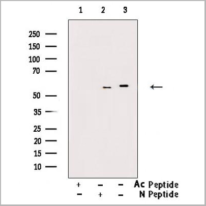
 WB (Western Blot)
(Western blot analysis of Phospho-Tau (Ser404) expression in various lysates)
WB (Western Blot)
(Western blot analysis of Phospho-Tau (Ser404) expression in various lysates)
Tau, Polyclonal Antibody (Cat# AAA30999)
Full Name
Phospho-Tau (Ser404) Antibody
Gene Names
MAPT; TAU; MSTD; PPND; DDPAC; MAPTL; MTBT1; MTBT2; FTDP-17; PPP1R103
Reactivity
Human, Mouse, Rat
Applications
Western Blot, Immunohistochemistry
Purity
From purified rabbit serum by affinity purification via sequential chromatography on phospho-and non-phospho-peptide affinity columns.
Pricing

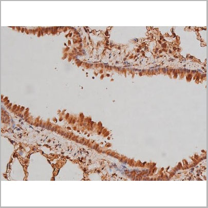
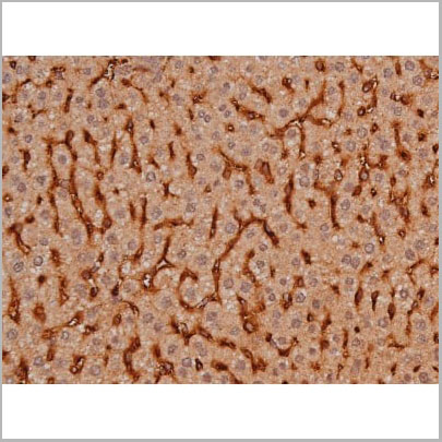
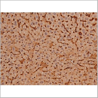
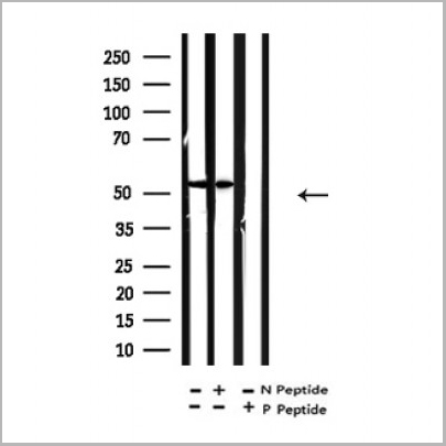
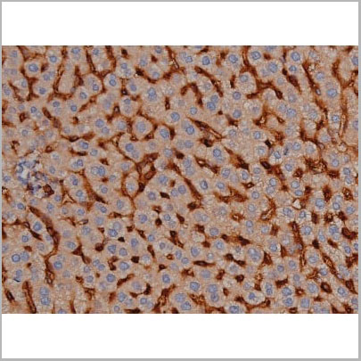
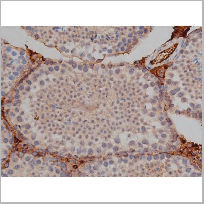
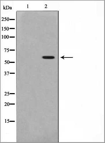
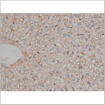
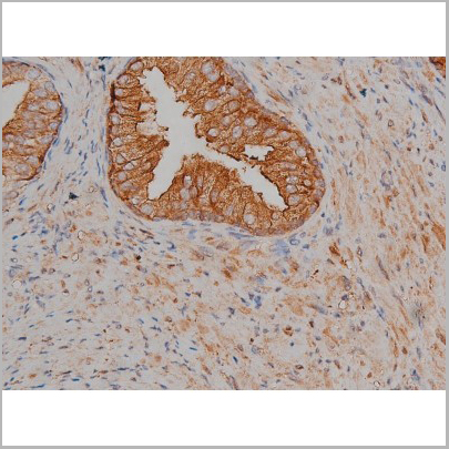
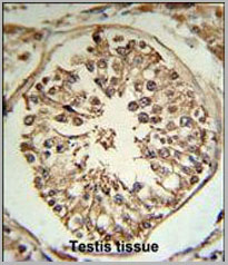 IHC (Immunohistochemistry)
(TMPRSS2 Antibody (N-term) (RB18784) IHC analysis in formalin fixed and paraffin embedded human testis tissue followed by peroxidase conjugation of the secondary antibody and DAB staining. This data demonstrates the use of the TMPRSS2 Antibody (N-term) for immunohistochemistry. Clinical relevance has not been evaluated.)
IHC (Immunohistochemistry)
(TMPRSS2 Antibody (N-term) (RB18784) IHC analysis in formalin fixed and paraffin embedded human testis tissue followed by peroxidase conjugation of the secondary antibody and DAB staining. This data demonstrates the use of the TMPRSS2 Antibody (N-term) for immunohistochemistry. Clinical relevance has not been evaluated.)
TMPRSS2, Polyclonal Antibody (Cat# AAA28791)
Full Name
TMPRSS2 Antibody (N-term)
Gene Names
TMPRSS2; PP9284; PRSS10
Reactivity
Human
Applications
Western Blot, Immunohistochemistry, Flow Cytometry
Purity
Purified Rabbit Polyclonal Antibody (Pab)
Pricing
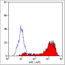
 FCM (Flow Cytometry)
(ENOB Antibody (Center) flow cytometry analysis of HepG2 cells (bottom histogram) compared to a negative control cell (top histogram). FITC-conjugated goat-anti-rabbit secondary antibodies were used for the analysis.)
FCM (Flow Cytometry)
(ENOB Antibody (Center) flow cytometry analysis of HepG2 cells (bottom histogram) compared to a negative control cell (top histogram). FITC-conjugated goat-anti-rabbit secondary antibodies were used for the analysis.)
ENOB, Polyclonal Antibody (Cat# AAA28761)
Full Name
ENOB Antibody (Center)
Gene Names
ENO3; MSE; GSD13
Reactivity
Human, Mouse, Rat (Predicted Reactivity: Bovine, Pig)
Applications
Western Blot, Immunohistochemistry, Flow Cytometry
Pricing
 WB (Western Blot)
(Western blot analysis of Phospho-Tau (Ser235) expression in various lysates)
WB (Western Blot)
(Western blot analysis of Phospho-Tau (Ser235) expression in various lysates)
Tau, Polyclonal Antibody (Cat# AAA30997)
Full Name
Phospho-Tau (Ser235) Antibody
Gene Names
MAPT; TAU; MSTD; PPND; DDPAC; MAPTL; MTBT1; MTBT2; FTDP-17; PPP1R103
Reactivity
Human, Mouse, Rat
Applications
Western Blot
Purity
From purified rabbit serum by affinity purification via sequential chromatography on phospho-and non-phospho-peptide affinity columns.
Pricing
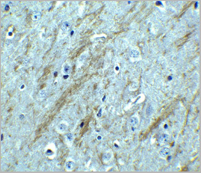
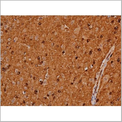
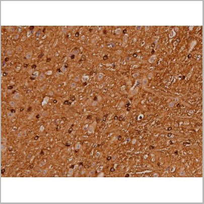
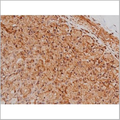
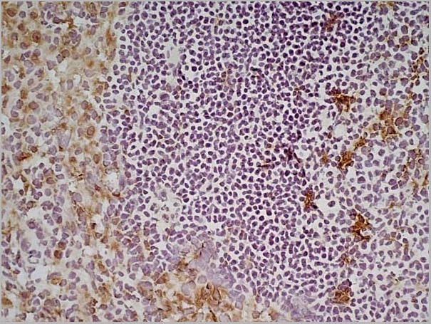
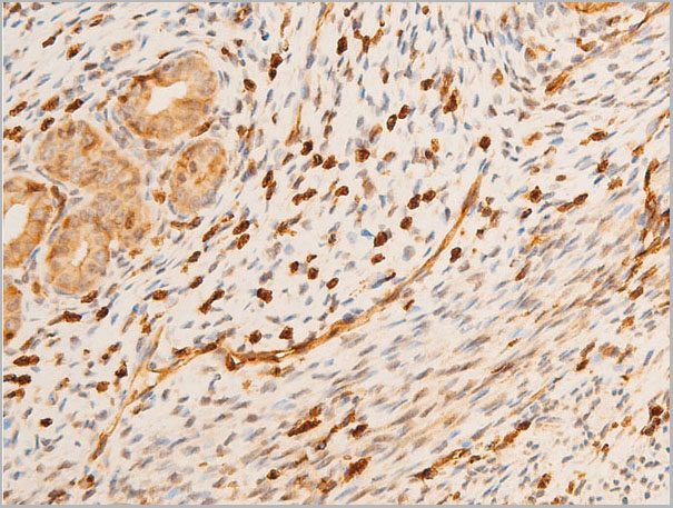
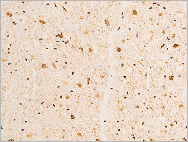
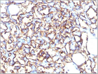
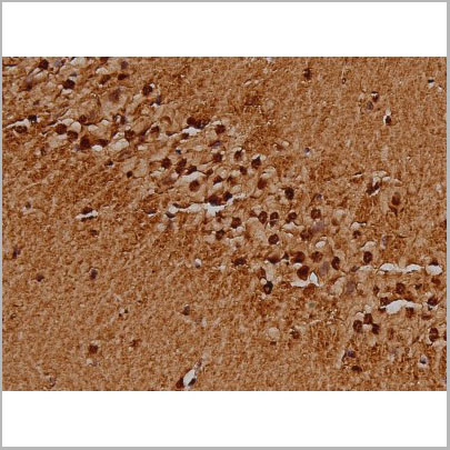
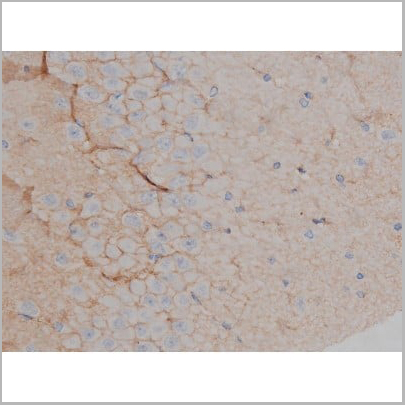
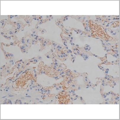
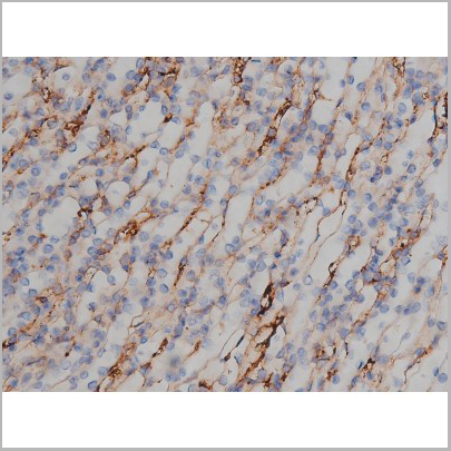
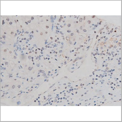
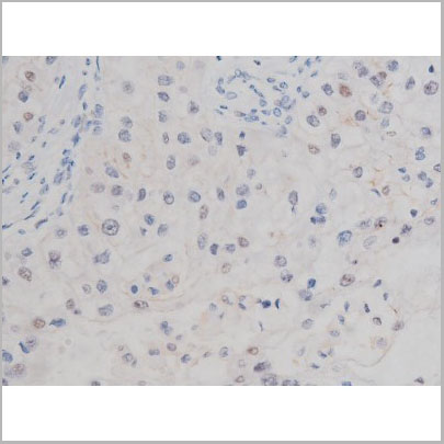
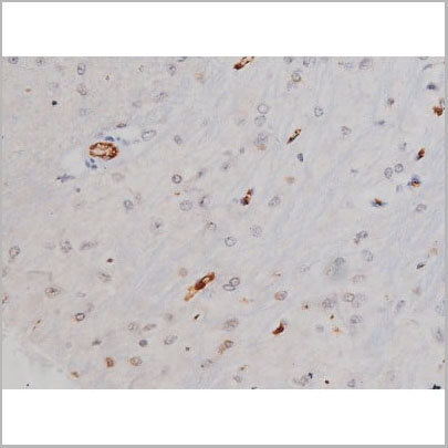
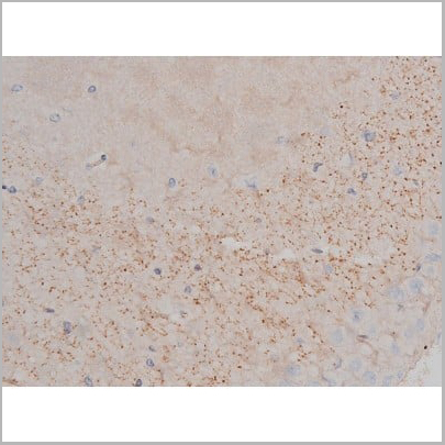
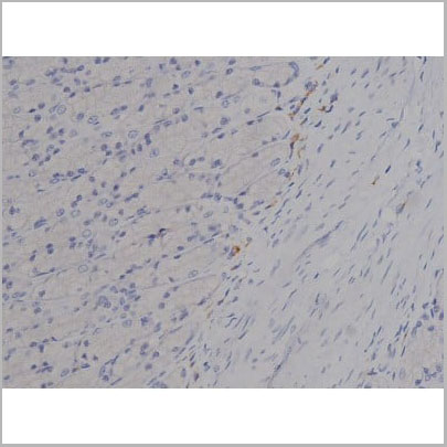
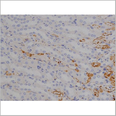
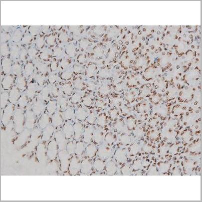
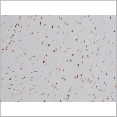
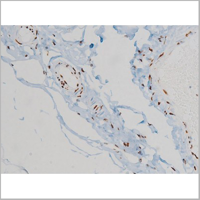
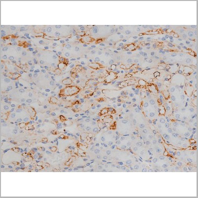
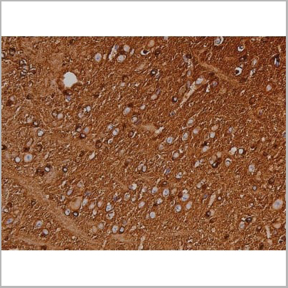
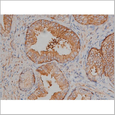
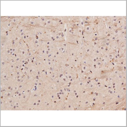
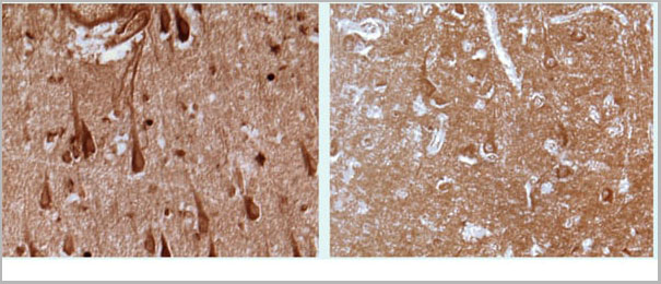 IHC (Immunohistochemistry)
(Immunohistochemistry analysis of isoprostane in brain of Alzheimer’s disease patient (left). Neurons in the hippocampus stained intensely with AAA14721. Results for age-matched control are shown on the right image.)
IHC (Immunohistochemistry)
(Immunohistochemistry analysis of isoprostane in brain of Alzheimer’s disease patient (left). Neurons in the hippocampus stained intensely with AAA14721. Results for age-matched control are shown on the right image.)
Isoprostane, Polyclonal Antibody (Cat# AAA14721)
Full Name
Isoprostane (8-iso-PGF2a)
Applications
Western Blot, Immunohistochemistry
Purity
Purified by ammonium sulfate precipitation.
Pricing
CD90, Monoclonal Antibody (Cat# AAA20024)
Full Name
Anti-Hu CD90 FITC
Gene Names
THY1; CD90; CDw90
Reactivity
Pig, Horse, Human, Non-human primates
Applications
Flow Cytometry
Purity
The purified antibody is conjugated with fluorescein isothiocyanate (FITC) under optimum conditions. The conjugate is purified by size-exclusion chromatography.
Pricing
 SDS-PAGE
(Analysis Purified CD90 Mouse Monoclonal antibody. Confirmation of Purity and Integrity of Antibody.)
SDS-PAGE
(Analysis Purified CD90 Mouse Monoclonal antibody. Confirmation of Purity and Integrity of Antibody.)
Cd90, Monoclonal Antibody (Cat# AAA13815)
Full Name
Cd90
Gene Names
THY1; CD90
Reactivity
Human, Monkey. Others not known.
Applications
Flow Cytometry, Immunofluorescence, Functional Assay
Pricing