Filters
Clonality
Type
Reactivity
Gene Name
Isotype
Host
Application
Clone
226 results for " adhesion" - showing 50-100


 WB (Western Blot)
(Western blot analysis of PAK1 expression in Etoposide treated 293 whole cell lysates, The lane on the left is treated with the antigen-specific peptide.)
WB (Western Blot)
(Western blot analysis of PAK1 expression in Etoposide treated 293 whole cell lysates, The lane on the left is treated with the antigen-specific peptide.)
PAK1, Polyclonal Antibody (Cat# AAA31096)
Full Name
PAK1 Antibody
Gene Names
PAK1; PAKalpha
Reactivity
Human, Mouse, Rat
Applications
Western Blot, Immunohistochemistry, Immunofluorescence, Immunocytochemistry
Purity
The antiserum was purified by peptide affinity chromatography using SulfoLink Coupling Resin.
Pricing


 Application Data
(Staining of human peripheral blood granulocytes with Mouse anti Human CD11b:Biotin)
Application Data
(Staining of human peripheral blood granulocytes with Mouse anti Human CD11b:Biotin)
CD11b, Monoclonal Antibody (Cat# AAA12053)
Full Name
MOUSE ANTI HUMAN CD11b:RPE
Gene Names
ITGAM; CR3A; MO1A; CD11B; MAC-1; MAC1A; SLEB6
Applications
Flow Cytometry
Pricing


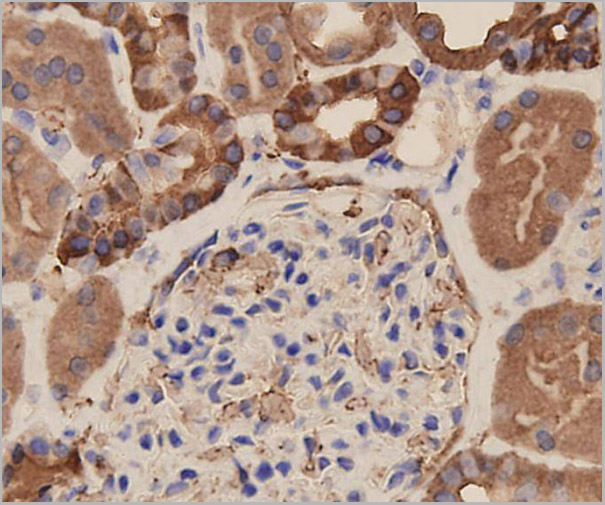
 IHC (Immunohistchemistry)
(At 1/100 staining Human ovarian cancer and adjacent normal tissues by IHC-P. The sample was formaldehyde fixed and a heat mediated antigen retrieval step in citrate buffer was performed. The sample was then blocked and incubated with the primary antibody at 4 degree C overnight. An HRP conjugated anti-Rabbit antibody was used as the secondary antibody.)
IHC (Immunohistchemistry)
(At 1/100 staining Human ovarian cancer and adjacent normal tissues by IHC-P. The sample was formaldehyde fixed and a heat mediated antigen retrieval step in citrate buffer was performed. The sample was then blocked and incubated with the primary antibody at 4 degree C overnight. An HRP conjugated anti-Rabbit antibody was used as the secondary antibody.)
alpha 1 Catenin, Polyclonal Antibody (Cat# AAA31319)
Full Name
Phospho-alpha 1 Catenin (Ser655/Thr658) Antibody
Gene Names
CTNNA1; CAP102
Reactivity
Human, Mouse, Rat
Applications
Western Blot, Immunohistochemistry, Peptide ELISA
Purity
The antibody is from purified rabbit serum by affinity purification via sequential chromatography on phospho-peptide and non-phospho-peptide affinity columns.
Pricing

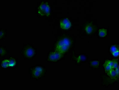
 IF (Immunofluorescence)
(Immunofluorescence analysis of HeLa cell using SPAM1 antibody. Blue: DAPI for nuclear staining.)
IF (Immunofluorescence)
(Immunofluorescence analysis of HeLa cell using SPAM1 antibody. Blue: DAPI for nuclear staining.)
SPAM1, Polyclonal Antibody (Cat# AAA29645)
Full Name
SPAM1 Antibody
Gene Names
SPAM1; HYA1; PH20; HYAL1; HYAL3; HYAL5; PH-20; SPAG15; HEL-S-96n
Reactivity
Human, Mouse, Rat
Applications
Western Blot, Immunohistochemistry, Immunofluorescence
Purity
Antibodies were purified by affinity purification using immunogen.
Pricing


 FCM (Flow Cytometry)
(Flow cytometric analysis of LOVO cells with Nectin 2 antibody at 1/100 dilution (purple) compared with an unlabelled control (cells without incubation with primary antibody; yellow). Alexa Fluor 488-conjugated goat anti-rabbit IgG was used as the secondary antibody.)
FCM (Flow Cytometry)
(Flow cytometric analysis of LOVO cells with Nectin 2 antibody at 1/100 dilution (purple) compared with an unlabelled control (cells without incubation with primary antibody; yellow). Alexa Fluor 488-conjugated goat anti-rabbit IgG was used as the secondary antibody.)
Nectin 2, Monoclonal Antibody (Cat# AAA30516)
Full Name
Nectin 2 Antibody
Gene Names
NECTIN2; HVEB; PRR2; CD112; PVRL2; PVRR2
Reactivity
Human, Mouse, Rat
Applications
Western Blot, Immunocytochemistry, Immunofluorescence, Immunohistochemistry, Flow Cytometry
Purity
ProA affinity purified
Pricing


 IHC (Immunohistochemistry)
(At 1/100 staining Human prostate cancer and adjacent normal tissues by IHC-P. The sample was formaldehyde fixed and a heat mediated antigen retrieval step in citrate buffer was performed. The sample was then blocked and incubated with the primary antibody at 4 degree C overnight. An HRP conjugated anti-Rabbit antibody was used as the secondary antibody.)
IHC (Immunohistochemistry)
(At 1/100 staining Human prostate cancer and adjacent normal tissues by IHC-P. The sample was formaldehyde fixed and a heat mediated antigen retrieval step in citrate buffer was performed. The sample was then blocked and incubated with the primary antibody at 4 degree C overnight. An HRP conjugated anti-Rabbit antibody was used as the secondary antibody.)
S100A9, Polyclonal Antibody (Cat# AAA31466)
Full Name
Phospho-S100A9 (Thr113) Antibody
Reactivity
Human
Applications
Western Blot, Immunohistochemistry, Peptide ELISA
Purity
The antibody is from purified rabbit serum by affinity purification via sequential chromatography on phospho-peptide and non-phospho-peptide affinity columns.
Pricing


 ICC (Immunocytochemistry)
(ICC staining ICAM1 in HUVEC cells (green). The nuclear counter stain is DAPI (blue). Cells were fixed in paraformaldehyde, permeabilised with 0.25% Triton X100/PBS.)
ICC (Immunocytochemistry)
(ICC staining ICAM1 in HUVEC cells (green). The nuclear counter stain is DAPI (blue). Cells were fixed in paraformaldehyde, permeabilised with 0.25% Triton X100/PBS.)
ICAM1, Monoclonal Antibody (Cat# AAA30099)
Full Name
ICAM1 Antibody
Gene Names
ICAM1; BB2; CD54; P3.58
Reactivity
Human
Applications
Western Blot, Immunocytochemistry, Immunofluorescence, Immunohistochemistry
Purity
ProA affinity purified
Pricing


 Application Data
Ep-CAM /CD326, Monoclonal Antibody (Cat# AAA13831)
Full Name
Ep-CAM /CD326 (Epithelial Marker) Mouse Monoclonal Antibody
Gene Names
EPCAM; ESA; KSA; M4S1; MK-1; DIAR5; EGP-2; EGP40; KS1/4; MIC18; TROP1; EGP314; HNPCC8; TACSTD1
Reactivity
Human, Mouse, Rat
Applications
Flow Cytometry, Immunofluorescence, Western Blot, Immunohistochemistry
Pricing


 Application Data
(Anti-ICAM1 Picoband antibody, AAA11608-6.jpgAll lanes: Anti-ICAM1(AAA11608) at 0.5ug/mlLane 1: Rat Thymus Tissue Lysate at 40ugLane 2: Rat Spleen Tissue Lysate at 40ugLane 3: PC12 Whole Cell Lysate at 40ugLane 4: NRK Whole Cell Lysate at 40ugLane 5: RH35 Whole Cell Lysate at 40ugPredicted bind size: 58KDObserved bind size: 90KD)
Application Data
(Anti-ICAM1 Picoband antibody, AAA11608-6.jpgAll lanes: Anti-ICAM1(AAA11608) at 0.5ug/mlLane 1: Rat Thymus Tissue Lysate at 40ugLane 2: Rat Spleen Tissue Lysate at 40ugLane 3: PC12 Whole Cell Lysate at 40ugLane 4: NRK Whole Cell Lysate at 40ugLane 5: RH35 Whole Cell Lysate at 40ugPredicted bind size: 58KDObserved bind size: 90KD)
ICAM1, Polyclonal Antibody (Cat# AAA11608)
Full Name
Anti-ICAM1 antibody
Gene Names
Icam1; CD54; Ly-47; Icam-1; MALA-2
Reactivity
Mouse, Rat
Applications
Western Blot, Immunohistochemistry
Purity
Immunogen affinity purified.
Pricing


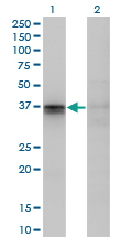

 WB (Western Blot)
(F11R monoclonal antibody, Western Blot analysis of F11R expression in HepG2.)
WB (Western Blot)
(F11R monoclonal antibody, Western Blot analysis of F11R expression in HepG2.)
Junctional Adhesion Molecule 1, Monoclonal Antibody (Cat# AAA24841)
Full Name
Junctional Adhesion Molecule 1 (JAM-1, CD321, F11 Receptor, F11R, Junctional Adhesion Molecule A, JAMA, JCAM, KAT, Platelet Adhesion Molecule 1, PAM1, Platelet F11 Receptor, UNQ264/PRO301) (Biotin)
Gene Names
F11R; JAM; KAT; JAM1; JAMA; JCAM; CD321; PAM-1
Reactivity
Human
Applications
Immunohistochemistry, Western Blot
Purity
Purified by Protein A Affinity Chromatography.
Pricing


 FCM (Flow Cytometry)
(Flow cytometric analysis of SH-SY-5Y cells with NCAM antibody at 1/50 dilution (red) compared with an unlabelled control (cells without incubation with primary antibody; black). Alexa Fluor 488-conjugated goat anti rabbit IgG was used as the secondary antibody.)
FCM (Flow Cytometry)
(Flow cytometric analysis of SH-SY-5Y cells with NCAM antibody at 1/50 dilution (red) compared with an unlabelled control (cells without incubation with primary antibody; black). Alexa Fluor 488-conjugated goat anti rabbit IgG was used as the secondary antibody.)
NCAM, Monoclonal Antibody (Cat# AAA30279)
Full Name
NCAM Antibody
Gene Names
NCAM1; CD56; NCAM; MSK39
Reactivity
Human, Mouse, Zebrafish
Applications
Western Blot, Immunocytochemistry, Immunofluorescence, Immunohistochemistry, Immunoprecipitation, Flow Cytometry
Purity
ProA affinity purified
Pricing


 FCM (Flow Cytometry)
(Overlay histogram showing Hela cells stained with AAA27026 (red line) at 1:200. The cells were incubated in 1x PBS /10% normal goat serum to block non-specific protein-protein interactions followed by primary antibody for 1 h at 4 degree C. The secondary antibody used was FITC goat anti-mouse IgG(H+L) at 1/200 dilution for 1 h at 4 degree C. Isotype control antibody (green line) was used under the same conditions. Acquisition of >10,000 events was performed.)
FCM (Flow Cytometry)
(Overlay histogram showing Hela cells stained with AAA27026 (red line) at 1:200. The cells were incubated in 1x PBS /10% normal goat serum to block non-specific protein-protein interactions followed by primary antibody for 1 h at 4 degree C. The secondary antibody used was FITC goat anti-mouse IgG(H+L) at 1/200 dilution for 1 h at 4 degree C. Isotype control antibody (green line) was used under the same conditions. Acquisition of >10,000 events was performed.)
CD44, Monoclonal Antibody (Cat# AAA27026)
Full Name
CD44 Monoclonal Antibody
Gene Names
CD44; IN; LHR; MC56; MDU2; MDU3; MIC4; Pgp1; CDW44; CSPG8; HCELL; HUTCH-I; ECMR-III
Reactivity
Human
Applications
Western Blot, Immunohistochemistry, Immunofluorescence, Flow Cytometry
Purity
>95%, Protein G Purified
Pricing


 IHC (Immunohistchemistry)
(Immunohistochemistry of paraffin-embedded human stomach using FAK antibody at dilution of 1:100 .)
IHC (Immunohistchemistry)
(Immunohistochemistry of paraffin-embedded human stomach using FAK antibody at dilution of 1:100 .)
FAK, Polyclonal Antibody (Cat# AAA30546)
Full Name
FAK Polyclonal Antibody
Gene Names
PTK2; FAK; FADK; FAK1; FRNK; PPP1R71; p125FAK; pp125FAK
Reactivity
Human, Mouse, Rat
Applications
Western Blot, Immunohistochemistry, Immunofluorescence
Purity
Affinity purification
Pricing


 FCM (Flow Cytometry)
(Flow cytometric analysis of Hela cells with CD44 antibody at 1/50 dilution (red) compared with an unlabelled control (cells without incubation with primary antibody; black). Alexa Fluor 488-conjugated goat anti rabbit IgG was used as the secondary antibody.)
FCM (Flow Cytometry)
(Flow cytometric analysis of Hela cells with CD44 antibody at 1/50 dilution (red) compared with an unlabelled control (cells without incubation with primary antibody; black). Alexa Fluor 488-conjugated goat anti rabbit IgG was used as the secondary antibody.)
CD44, Monoclonal Antibody (Cat# AAA30109)
Full Name
CD44 Antibody
Gene Names
CD44; IN; LHR; MC56; MDU2; MDU3; MIC4; Pgp1; CDW44; CSPG8; HCELL; HUTCH-I; ECMR-III
Reactivity
Human
Applications
Western Blot, Immunohistochemistry, Flow Cytometry
Purity
ProA affinity purified
Pricing


 Application Data
(Published customer image Infiltration of GFP+ BM-cells in infarct and peri-infarct regions. (A-B) Dot plots of viable macrophages/granulocytes (CD11b+CD45high, top right quadrants) and microglia (CD11b+CD45dim, bottom right quadrants) in cortex from BM-chimeric unmanipulated mice and mice exposed to pMCAO. (C) Bar graph showing mean numbers of CD11b+CD45dim microglia and CD11b+CD45high macrophages/granulocytes in BM-chimeric mice 24 hours after pMCAO, subdivided based on expression of GFP (n = 5). Approximately 92% of of the CD45high population were GFP+. (D) Estimation and comparison of mean numbers of CD11b+CD45dim microglia in non-chimeric (n = 10) versus BM-chimeric mice (n = 5) 24 hours after of pMCAO shows significantly fewer CD11b+CD45dim microglial cells in irradiated mice. (E) Overview, showing distribution of infiltrating GFP+ BM-derived cells into infarct (IF) and peri-infarct (P-IF) regions 24 hours after pMCAO. (E-G) By 24 hours, GFP+ single cells (F) and vessel-associated aggregates of GFP+ cells (arrows in G) were observed in infarct and peri-infarct regions. Some of the vessel-associated cells were round, leukocyte-like cells (arrows) while others were elongated cells lining the vasculature (arrow heads in G and in insert). (H) Bar graph showing mean numbers of single GFP+ cells and vessel-associated aggregates of GFP+ cells in ipsi- and contralateral cortex 24 hours after surgery (n = 10). (I-P) Immunohistochemical staining of CD45.1 (I, K), CD45.2 (J, L), IgG2a (M, O) and CD45 (N, P) in ischemic tissue in BM-chimeric (I, J, M, N) and non-chimeric mice (K, L, O, P) 24 hours after pMCAO. N.D, none detected. Scale bars: 200 um (A), 10 um (B, C). 50 um (I-P) *P < 0.05, **P < 0.01, and ***P < 0.001.From: Clausen BH, Lambertsen KL, Babcock AA, Holm TH, Dagnaes-Hansen F, Finsen B. Interleukin-1beta and tumor necrosis factor-alpha are expressed by different subsets of microglia and macrophages after ischemic stroke in mice. J Neuroinflammation. 2008 Oct 23;5:46.)
Application Data
(Published customer image Infiltration of GFP+ BM-cells in infarct and peri-infarct regions. (A-B) Dot plots of viable macrophages/granulocytes (CD11b+CD45high, top right quadrants) and microglia (CD11b+CD45dim, bottom right quadrants) in cortex from BM-chimeric unmanipulated mice and mice exposed to pMCAO. (C) Bar graph showing mean numbers of CD11b+CD45dim microglia and CD11b+CD45high macrophages/granulocytes in BM-chimeric mice 24 hours after pMCAO, subdivided based on expression of GFP (n = 5). Approximately 92% of of the CD45high population were GFP+. (D) Estimation and comparison of mean numbers of CD11b+CD45dim microglia in non-chimeric (n = 10) versus BM-chimeric mice (n = 5) 24 hours after of pMCAO shows significantly fewer CD11b+CD45dim microglial cells in irradiated mice. (E) Overview, showing distribution of infiltrating GFP+ BM-derived cells into infarct (IF) and peri-infarct (P-IF) regions 24 hours after pMCAO. (E-G) By 24 hours, GFP+ single cells (F) and vessel-associated aggregates of GFP+ cells (arrows in G) were observed in infarct and peri-infarct regions. Some of the vessel-associated cells were round, leukocyte-like cells (arrows) while others were elongated cells lining the vasculature (arrow heads in G and in insert). (H) Bar graph showing mean numbers of single GFP+ cells and vessel-associated aggregates of GFP+ cells in ipsi- and contralateral cortex 24 hours after surgery (n = 10). (I-P) Immunohistochemical staining of CD45.1 (I, K), CD45.2 (J, L), IgG2a (M, O) and CD45 (N, P) in ischemic tissue in BM-chimeric (I, J, M, N) and non-chimeric mice (K, L, O, P) 24 hours after pMCAO. N.D, none detected. Scale bars: 200 um (A), 10 um (B, C). 50 um (I-P) *P < 0.05, **P < 0.01, and ***P < 0.001.From: Clausen BH, Lambertsen KL, Babcock AA, Holm TH, Dagnaes-Hansen F, Finsen B. Interleukin-1beta and tumor necrosis factor-alpha are expressed by different subsets of microglia and macrophages after ischemic stroke in mice. J Neuroinflammation. 2008 Oct 23;5:46.)
CD11b, Monoclonal Antibody (Cat# AAA12183)
Full Name
RAT ANTI MOUSE CD11b:FITC
Gene Names
Itgam; CR3; CR3A; MAC1; Cd11b; Ly-40; Mac-1; Mac-1a; CD11b/CD18; F730045J24Rik
Applications
Flow Cytometry
Pricing


 Application Data
(Published customer image Infiltration of GFP+ BM-cells in infarct and peri-infarct regions. (A-B) Dot plots of viable macrophages/granulocytes (CD11b+CD45high, top right quadrants) and microglia (CD11b+CD45dim, bottom right quadrants) in cortex from BM-chimeric unmanipulated mice and mice exposed to pMCAO. (C) Bar graph showing mean numbers of CD11b+CD45dim microglia and CD11b+CD45high macrophages/granulocytes in BM-chimeric mice 24 hours after pMCAO, subdivided based on expression of GFP (n = 5). Approximately 92% of of the CD45high population were GFP+. (D) Estimation and comparison of mean numbers of CD11b+CD45dim microglia in non-chimeric (n = 10) versus BM-chimeric mice (n = 5) 24 hours after of pMCAO shows significantly fewer CD11b+CD45dim microglial cells in irradiated mice. (E) Overview, showing distribution of infiltrating GFP+ BM-derived cells into infarct (IF) and peri-infarct (P-IF) regions 24 hours after pMCAO. (E-G) By 24 hours, GFP+ single cells (F) and vessel-associated aggregates of GFP+ cells (arrows in G) were observed in infarct and peri-infarct regions. Some of the vessel-associated cells were round, leukocyte-like cells (arrows) while others were elongated cells lining the vasculature (arrow heads in G and in insert). (H) Bar graph showing mean numbers of single GFP+ cells and vessel-associated aggregates of GFP+ cells in ipsi- and contralateral cortex 24 hours after surgery (n = 10). (I-P) Immunohistochemical staining of CD45.1 (I, K), CD45.2 (J, L), IgG2a (M, O) and CD45 (N, P) in ischemic tissue in BM-chimeric (I, J, M, N) and non-chimeric mice (K, L, O, P) 24 hours after pMCAO. N.D, none detected. Scale bars: 200 um (A), 10 um (B, C). 50 um (I-P) *P < 0.05, **P < 0.01, and ***P < 0.001.From: Clausen BH, Lambertsen KL, Babcock AA, Holm TH, Dagnaes-Hansen F, Finsen B. Interleukin-1beta and tumor necrosis factor-alpha are expressed by different subsets of microglia and macrophages after ischemic stroke in mice. J Neuroinflammation. 2008 Oct 23;5:46.)
Application Data
(Published customer image Infiltration of GFP+ BM-cells in infarct and peri-infarct regions. (A-B) Dot plots of viable macrophages/granulocytes (CD11b+CD45high, top right quadrants) and microglia (CD11b+CD45dim, bottom right quadrants) in cortex from BM-chimeric unmanipulated mice and mice exposed to pMCAO. (C) Bar graph showing mean numbers of CD11b+CD45dim microglia and CD11b+CD45high macrophages/granulocytes in BM-chimeric mice 24 hours after pMCAO, subdivided based on expression of GFP (n = 5). Approximately 92% of of the CD45high population were GFP+. (D) Estimation and comparison of mean numbers of CD11b+CD45dim microglia in non-chimeric (n = 10) versus BM-chimeric mice (n = 5) 24 hours after of pMCAO shows significantly fewer CD11b+CD45dim microglial cells in irradiated mice. (E) Overview, showing distribution of infiltrating GFP+ BM-derived cells into infarct (IF) and peri-infarct (P-IF) regions 24 hours after pMCAO. (E-G) By 24 hours, GFP+ single cells (F) and vessel-associated aggregates of GFP+ cells (arrows in G) were observed in infarct and peri-infarct regions. Some of the vessel-associated cells were round, leukocyte-like cells (arrows) while others were elongated cells lining the vasculature (arrow heads in G and in insert). (H) Bar graph showing mean numbers of single GFP+ cells and vessel-associated aggregates of GFP+ cells in ipsi- and contralateral cortex 24 hours after surgery (n = 10). (I-P) Immunohistochemical staining of CD45.1 (I, K), CD45.2 (J, L), IgG2a (M, O) and CD45 (N, P) in ischemic tissue in BM-chimeric (I, J, M, N) and non-chimeric mice (K, L, O, P) 24 hours after pMCAO. N.D, none detected. Scale bars: 200 um (A), 10 um (B, C). 50 um (I-P) *P < 0.05, **P < 0.01, and ***P < 0.001.From: Clausen BH, Lambertsen KL, Babcock AA, Holm TH, Dagnaes-Hansen F, Finsen B. Interleukin-1beta and tumor necrosis factor-alpha are expressed by different subsets of microglia and macrophages after ischemic stroke in mice. J Neuroinflammation. 2008 Oct 23;5:46.)
CD11b, Monoclonal Antibody (Cat# AAA12185)
Full Name
RAT ANTI MOUSE CD11b
Gene Names
Itgam; CR3; CR3A; MAC1; Cd11b; Ly-40; Mac-1; Mac-1a; CD11b/CD18; F730045J24Rik
Applications
Immunohistochemistry, Flow Cytometry, Immunofluorescence, Immunoprecipitation
Pricing
 Standard Curve (Sample)
Intercellular Adhesion Molecule 1, sICAM-1, ELISA Kit (Cat# AAA11091)
Full Name
Rat Soluble Intercellular Adhesion Molecule 1, sICAM-1 ELISA Kit
Gene Names
ICAM1; BB2; CD54; P3.58
Reactivity
Rat
Pricing


 FCM (Flow Cytometry)
(Flow cytometric analysis of Hela cells with N Cadherin antibody at 1/50 dilution (red) compared with an unlabelled control (cells without incubation with primary antibody; black). Alexa Fluor 488-conjugated goat anti rabbit IgG was used as the secondary antibody.)
FCM (Flow Cytometry)
(Flow cytometric analysis of Hela cells with N Cadherin antibody at 1/50 dilution (red) compared with an unlabelled control (cells without incubation with primary antibody; black). Alexa Fluor 488-conjugated goat anti rabbit IgG was used as the secondary antibody.)
N Cadherin, Monoclonal Antibody (Cat# AAA30054)
Full Name
N Cadherin Antibody
Gene Names
CDH2; CDHN; NCAD; CD325; CDw325
Reactivity
Human, Mouse, Rat
Applications
Western Blot, Immunocytochemistry, Immunofluorescence, Immunohistochemistry, Flow Cytometry
Purity
ProA affinity purified
Pricing
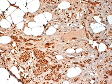
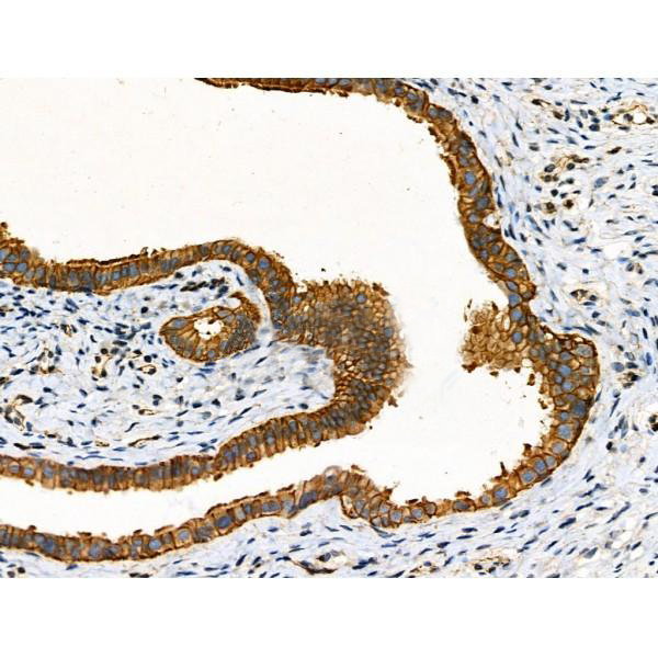
 IHC (Immunohistochemistry)
(Immunochemical staining of human CEACAM5 in human rectal cancer with mouse monoclonal antibody (1:60, formalin-fixed paraffin embedded sections). Positive staining was localized to intestinal gland.)
IHC (Immunohistochemistry)
(Immunochemical staining of human CEACAM5 in human rectal cancer with mouse monoclonal antibody (1:60, formalin-fixed paraffin embedded sections). Positive staining was localized to intestinal gland.)
CEACAM5, Monoclonal Antibody (Cat# AAA27742)
Full Name
Anti-CEACAM5 Antibody, Mouse Monoclonal
Gene Names
CEACAM5; CEA; CD66e
Reactivity
Human
Applications
Immunohistochemistry
Purity
Protein A
Pricing
 Standard Curve (Sample)
insulin-like growth factor binding protein 7, ELISA Kit (Cat# AAA15846)
Full Name
Human Insulin-like growth factor-binding protein 7, IGFBP7/MAC25/PSF ELISA Kit
Gene Names
IGFBP7; AGM; PSF; TAF; FSTL2; IBP-7; MAC25; IGFBP-7; RAMSVPS; IGFBP-7v; IGFBPRP1
Reactivity
Human
Pricing


 Application Data
(Staining of mouse peritoneal macrophages with Rat anti Mouse CD11b)
Application Data
(Staining of mouse peritoneal macrophages with Rat anti Mouse CD11b)
CD11b, Monoclonal Antibody (Cat# AAA12187)
Full Name
RAT ANTI MOUSE CD11b:Biotin
Gene Names
Itgam; CR3; CR3A; MAC1; Cd11b; Ly-40; Mac-1; Mac-1a; CD11b/CD18; F730045J24Rik
Applications
Flow Cytometry
Pricing
 WB (Western Blot)
(WB Suggested Anti-Tmem164 AntibodyTitration: 1.0 ug/mlPositive Control: Mouse Kidney)
WB (Western Blot)
(WB Suggested Anti-Tmem164 AntibodyTitration: 1.0 ug/mlPositive Control: Mouse Kidney)
Tmem164, Polyclonal Antibody (Cat# AAA23591)
Full Name
Tmem164 antibody - N-terminal region
Gene Names
Tmem164; AI316850; AI852450; AW547186; F730011B02
Reactivity
Tested Species Reactivity: Mouse
Predicted Species Reactivity: Human, Mouse, Rat, Cow, Dog, Guinea Pig, Horse, Rabbit
Predicted Species Reactivity: Human, Mouse, Rat, Cow, Dog, Guinea Pig, Horse, Rabbit
Applications
Western Blot
Purity
Affinity Purified
Pricing
 Application Data
(ATH mouse bone marrow stained with anti-CD44 (clone: KM81) (filled histogram) or Rat IgG2a isotype control (open histogram).N.B. Appropriate control samples should always be included in any labelling studies. * For optimal results in various applications, it is recommended that each investigator determine dilutions appropriate for individual use.)
Application Data
(ATH mouse bone marrow stained with anti-CD44 (clone: KM81) (filled histogram) or Rat IgG2a isotype control (open histogram).N.B. Appropriate control samples should always be included in any labelling studies. * For optimal results in various applications, it is recommended that each investigator determine dilutions appropriate for individual use.)
CD44, Monoclonal Antibody (Cat# AAA14269)
Full Name
Anti-Mouse CD44, Purified (Clone KM81) (rat IgG2a)
Gene Names
CD44; IN; LHR; MC56; MDU2; MDU3; MIC4; Pgp1; CDW44; CSPG8; HCELL; HUTCH-I; ECMR-III
Reactivity
Mouse
Applications
Immunohistochemistry, Flow Cytometry
Pricing


 Application Data
(Staining of CD209 transfected K562 cells with Mouse anti Human CD209: Biotin)
Application Data
(Staining of CD209 transfected K562 cells with Mouse anti Human CD209: Biotin)
CD209, Monoclonal Antibody (Cat# AAA11906)
Full Name
MOUSE ANTI HUMAN CD209:Low Endotoxin
Gene Names
CD209; CDSIGN; CLEC4L; DC-SIGN; DC-SIGN1
Applications
Flow Cytometry, Functional Assay, Immunofluorescence
Pricing
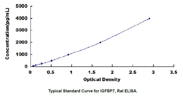 Standard Curve (Sample)
Insulin Like Growth Factor Binding Protein 7 (IGFBP7), ELISA Kit (Cat# AAA23011)
Full Name
Rat Insulin Like Growth Factor Binding Protein 7 (IGFBP7) ELISA Kit
Gene Names
IGFBP7; AGM; PSF; TAF; FSTL2; IBP-7; MAC25; IGFBP-7; RAMSVPS; IGFBP-7v; IGFBPRP1
Reactivity
Rat
Pricing
 Standard Curve (Sample)
platelet/endothelial cell adhesion molecule, ELISA Kit (Cat# AAA15520)
Full Name
Mouse soluble platelet endothelial cell adhesion molecule 1, sPECAM-1 ELISA Kit
Gene Names
Pecam1; Cd31; Pecam; C85791; PECAM-1
Reactivity
Mouse
Pricing


 WB (Western Blot)
(WB Suggested Anti-ADRM1 antibodyTitration: 1 ug/mlPositive Control: HeLa cells and cytosol from male mouse liver)
WB (Western Blot)
(WB Suggested Anti-ADRM1 antibodyTitration: 1 ug/mlPositive Control: HeLa cells and cytosol from male mouse liver)
ADRM1, Polyclonal Antibody (Cat# AAA23588)
Full Name
ADRM1 antibody - C-terminal region
Gene Names
ADRM1; ARM1; ARM-1; GP110
Reactivity
Guinea Pig, Human, Mouse, Rat
Applications
Western Blot
Purity
Affinity Purified
Pricing


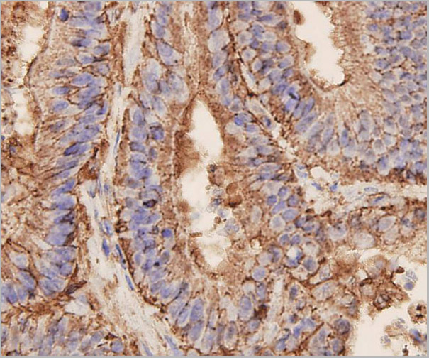
 IHC (Immunohistochemistry)
(At 1/100 staining Human colorectal cancer by IHC-P. The sample was formaldehyde fixed and a heat mediated antigen retrieval step in citrate buffer was performed. The sample was then blocked and incubated with the primary antibody at 4 degree C overnight. An HRP conjugated anti-Rabbit antibody was used as the secondary antibody.)
IHC (Immunohistochemistry)
(At 1/100 staining Human colorectal cancer by IHC-P. The sample was formaldehyde fixed and a heat mediated antigen retrieval step in citrate buffer was performed. The sample was then blocked and incubated with the primary antibody at 4 degree C overnight. An HRP conjugated anti-Rabbit antibody was used as the secondary antibody.)
CD227/MUC1, Polyclonal Antibody (Cat# AAA31426)
Full Name
Phospho-CD227/MUC1 (Ser1227) Antibody
Gene Names
MUC1; EMA; PEM; PUM; KL-6; MAM6; PEMT; CD227; H23AG; MUC-1; CA 15-3; MUC-1/X; MUC1/ZD; MUC-1/SEC
Reactivity
Human, Mouse, Rat
Applications
Western Blot, Immunohistochemistry, Peptide ELISA
Purity
The antibody is from purified rabbit serum by affinity purification via sequential chromatography on phospho-peptide and non-phospho-peptide affinity columns.
Pricing


 Application Data
(Formalin fixed, paraffin embedded human breast cancer biopsy stained with Mouse anti Human CD44 antibody followed by HRP-polymer detection and DAB substrate development (high power) following antigen retrieval using citrate buffer at pH6.2)
Application Data
(Formalin fixed, paraffin embedded human breast cancer biopsy stained with Mouse anti Human CD44 antibody followed by HRP-polymer detection and DAB substrate development (high power) following antigen retrieval using citrate buffer at pH6.2)
CD44, Monoclonal Antibody (Cat# AAA12208)
Full Name
MOUSE ANTI HUMAN CD44
Gene Names
CD44; IN; LHR; MC56; MDU2; MDU3; MIC4; Pgp1; CDW44; CSPG8; HCELL; HUTCH-I; ECMR-III
Applications
Immunohistochemistry, Flow Cytometry, Immunofluorescence, Immunohistochemistry
Pricing
 Standard Curve (Sample)
Fc Fragment Of IgG Low Affinity IIIb Receptor (FcgR3B), ELISA Kit (Cat# AAA20704)
Full Name
Human Fc Fragment Of IgG Low Affinity IIIb Receptor (FcgR3B) ELISA Kit
Gene Names
FCGR3B; CD16; FCG3; CD16b; FCGR3; FCR-10; FCRIII; FCRIIIb
Reactivity
Human
Pricing


 FCM (Flow Cytometry)
(Flow cytometric analysis of Jurkat cells with CD31 antibody at 1/100 dilution (blue) compared with an unlabelled control (cells without incubation with primary antibody; red). Goat anti rabbit IgG (FITC) was used as the secondary antibody.)
FCM (Flow Cytometry)
(Flow cytometric analysis of Jurkat cells with CD31 antibody at 1/100 dilution (blue) compared with an unlabelled control (cells without incubation with primary antibody; red). Goat anti rabbit IgG (FITC) was used as the secondary antibody.)
CD31, Polyclonal Antibody (Cat# AAA29956)
Full Name
CD31 Antibody
Gene Names
PECAM1; CD31; PECA1; GPIIA'; PECAM-1; endoCAM; CD31/EndoCAM
Reactivity
Human, Mouse
Applications
Western Blot, Immunocytochemistry, Immunohistochemistry, Flow Cytometry
Purity
Peptide affinity purified
Pricing


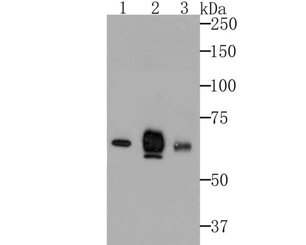
 WB (Western Blot)
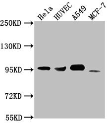
(Western Blot analysis using CD44 antibodyWestern Blot showing using CD44 antibody used against Hela (1) and HUVE-12 (2) cell lysate.)
WB (Western Blot)
(Western Blot analysis using CD44 antibodyWestern Blot showing using CD44 antibody used against Hela (1) and HUVE-12 (2) cell lysate.)
CD44, Monoclonal Antibody (Cat# AAA14314)
Full Name
CD44 antibody
Gene Names
CD44; IN; LHR; MC56; MDU2; MDU3; MIC4; Pgp1; CDW44; CSPG8; HCELL; HUTCH-I; ECMR-III
Applications
Flow Cytometry, Immunofluorescence, Immunohistochemistry, Western Blot
Pricing
 Standard Curve (Sample)
vascular cell adhesion protein 1 (VCAM-1/CD106), ELISA Kit (Cat# AAA15402)
Full Name
Rat vascular cell adhesion protein 1, VCAM-1/CD106 ELISA Kit
Gene Names
Vcam1; VCAM1B
Reactivity
Rat
Pricing


 FCM (Flow Cytometry)
(Flow cytometric analysis of Hela cells with FAK antibody at 1/50 dilution (blue) compared with an unlabelled control (cells without incubation with primary antibody; red). Alexa Fluor 488-conjugated goat anti rabbit IgG was used as the secondary antibody)
FCM (Flow Cytometry)
(Flow cytometric analysis of Hela cells with FAK antibody at 1/50 dilution (blue) compared with an unlabelled control (cells without incubation with primary antibody; red). Alexa Fluor 488-conjugated goat anti rabbit IgG was used as the secondary antibody)
FAK, Monoclonal Antibody (Cat# AAA29990)
Full Name
FAK Antibody
Gene Names
PTK2; FAK; FADK; FAK1; FRNK; PPP1R71; p125FAK; pp125FAK
Reactivity
Human, Mouse, Rat
Applications
Western Blot, Immunocytochemistry, Immunofluorescence, Immunohistochemistry, Immunoprecipitation, Flow Cytometry
Purity
ProA affinity purified
Pricing


 Application Data
(Formalin fixed, paraffin embedded human breast cancer biopsy stained with Mouse anti Human CD44 antibody followed by HRP-polymer detection and DAB substrate development (high power) following antigen retrieval using citrate buffer at pH6.2)
Application Data
(Formalin fixed, paraffin embedded human breast cancer biopsy stained with Mouse anti Human CD44 antibody followed by HRP-polymer detection and DAB substrate development (high power) following antigen retrieval using citrate buffer at pH6.2)
CD44, Monoclonal Antibody (Cat# AAA11893)
Full Name
MOUSE ANTI HUMAN CD44:FITC
Gene Names
CD44; IN; LHR; MC56; MDU2; MDU3; MIC4; Pgp1; CDW44; CSPG8; HCELL; HUTCH-I; ECMR-III
Applications
Flow Cytometry, Immunohistochemistry, Immunofluorescence
Purity
Purified
Pricing


 ICC (Immunocytochemistry)
(ICC staining EpCAM in SW1990 cells (green). The nuclear counter stain is DAPI (blue). Cells were fixed in paraformaldehyde, permeabilised with 0.25% Triton X100/PBS.)
ICC (Immunocytochemistry)
(ICC staining EpCAM in SW1990 cells (green). The nuclear counter stain is DAPI (blue). Cells were fixed in paraformaldehyde, permeabilised with 0.25% Triton X100/PBS.)
EP-CAM, Monoclonal Antibody (Cat# AAA29891)
Full Name
EP-CAM Antibody
Gene Names
EPCAM; ESA; KSA; M4S1; MK-1; DIAR5; EGP-2; EGP40; KS1/4; MIC18; TROP1; EGP314; HNPCC8; TACSTD1
Reactivity
Human, Mouse
Applications
Western Blot, Immunocytochemistry, Immunohistochemistry, Flow Cytometry
Purity
ProL affinity purified
Pricing


 FCM (Flow Cytometry)
(Flow cytometric analysis of K562 cells with CD11a antibody at 1/50 dilution (red) compared with an unlabelled control (cells without incubation with primary antibody; black). Alexa Fluor 488-conjugated goat anti rabbit IgG was used as the secondary antibody.)
FCM (Flow Cytometry)
(Flow cytometric analysis of K562 cells with CD11a antibody at 1/50 dilution (red) compared with an unlabelled control (cells without incubation with primary antibody; black). Alexa Fluor 488-conjugated goat anti rabbit IgG was used as the secondary antibody.)
CD11a, Monoclonal Antibody (Cat# AAA30112)
Full Name
CD11a Antibody
Gene Names
ITGAL; CD11A; LFA-1; LFA1A
Reactivity
Human
Applications
Western Blot, Immunocytochemistry, Immunofluorescence, Immunohistochemistry, Immunoprecipitation, Flow Cytometry
Purity
ProA affinity purified
Pricing


 Application Data
(Formalin fixed, paraffin embedded human breast cancer biopsy stained with Mouse anti Human CD44 antibody followed by HRP-polymer detection and DAB substrate development (high power) following antigen retrieval using citrate buffer at pH6.2)
Application Data
(Formalin fixed, paraffin embedded human breast cancer biopsy stained with Mouse anti Human CD44 antibody followed by HRP-polymer detection and DAB substrate development (high power) following antigen retrieval using citrate buffer at pH6.2)
CD44, Monoclonal Antibody (Cat# AAA12016)
Full Name
MOUSE ANTI HUMAN CD44
Gene Names
CD44; IN; LHR; MC56; MDU2; MDU3; MIC4; Pgp1; CDW44; CSPG8; HCELL; HUTCH-I; ECMR-III
Reactivity
Cynomolgus monkey
Applications
Immunohistochemistry, Flow Cytometry, Immunofluorescence, Immunohistochemistry
Pricing


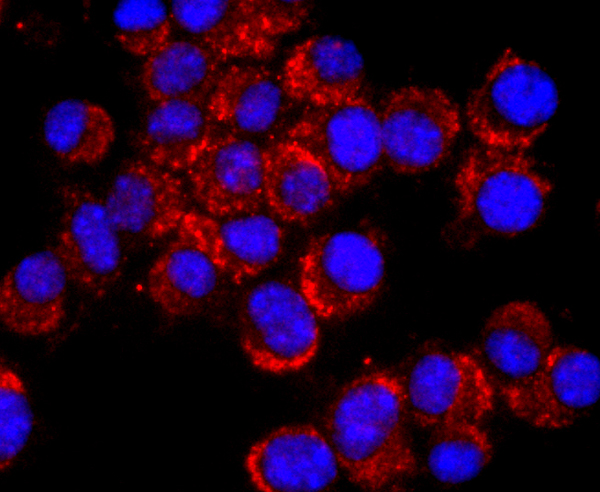
 IF (Immunofluorescence)
(Immunofluorescence analysis of Raw264.7 cells using CD44 antibody at dilution of 1:100. Blue: DAPI for nuclear staining.)
IF (Immunofluorescence)
(Immunofluorescence analysis of Raw264.7 cells using CD44 antibody at dilution of 1:100. Blue: DAPI for nuclear staining.)
CD44, Polyclonal Antibody (Cat# AAA28336)
Full Name
CD44 Rabbit pAb
Gene Names
CD44; IN; LHR; MC56; MDU2; MDU3; MIC4; Pgp1; CDW44; CSPG8; HCELL; HUTCH-I; ECMR-III
Reactivity
Human, Mouse, Rat
Applications
Western Blot, Immunohistochemistry, Immunofluorescence
Purity
Affinity purification
Pricing


 Application Data
(Staining of CD209 transfected K562 cells with Mouse anti Human CD209: Biotin)
Application Data
(Staining of CD209 transfected K562 cells with Mouse anti Human CD209: Biotin)
CD209, Monoclonal Antibody (Cat# AAA12139)
Full Name
MOUSE ANTI HUMAN CD209
Gene Names
CD209; CDSIGN; CLEC4L; DC-SIGN; DC-SIGN1
Applications
Flow Cytometry, Immunofluorescence
Pricing


 IF (Immunofluorescence)
(Confocal Immunofluorescent analysis of SK-OV-3 cells using AF488-labeled Isotype Control Monoclonal Antibody (IgG1) (Green).DAPI was used to stain the cell nuclei (blue). (Negative Control))
IF (Immunofluorescence)
(Confocal Immunofluorescent analysis of SK-OV-3 cells using AF488-labeled Isotype Control Monoclonal Antibody (IgG1) (Green).DAPI was used to stain the cell nuclei (blue). (Negative Control))
Ep-CAM /CD326, Monoclonal Antibody (Cat# AAA13838)
Full Name
Ep-CAM /CD326 (Epithelial Marker) Mouse Monoclonal Antibody
Gene Names
EPCAM; ESA; KSA; M4S1; MK-1; DIAR5; EGP-2; EGP40; KS1/4; MIC18; TROP1; EGP314; HNPCC8; TACSTD1
Reactivity
Human.
Does not react with Mouse and Rat
Does not react with Mouse and Rat
Applications
Flow Cytometry, Immunofluorescence, Western Blot, Immunohistochemistry
Pricing


 ICC (Immunocytochemistry)
(Figure 6. IHC analysis of LYRIC using anti-LYRIC antibody (AAA11650).LYRIC was detected in immunocytochemical section of SMMC-7721 cell. Heat mediated antigen retrieval was performed in citrate buffer (pH6, epitope retrieval solution) for 20 mins. The tissue section was blocked with 10% goat serum. The tissue section was then incubated with 1ug/ml rabbit anti-LYRIC Antibody (AAA11650) overnight at 4 degree C. Biotinylated goat anti-rabbit IgG was used as secondary antibody and incubated for 30 minutes at 37 degree C. The tissue section was developed using Strepavidin-Biotin-Complex (SABC) with DAB as the chromogen.)
ICC (Immunocytochemistry)
(Figure 6. IHC analysis of LYRIC using anti-LYRIC antibody (AAA11650).LYRIC was detected in immunocytochemical section of SMMC-7721 cell. Heat mediated antigen retrieval was performed in citrate buffer (pH6, epitope retrieval solution) for 20 mins. The tissue section was blocked with 10% goat serum. The tissue section was then incubated with 1ug/ml rabbit anti-LYRIC Antibody (AAA11650) overnight at 4 degree C. Biotinylated goat anti-rabbit IgG was used as secondary antibody and incubated for 30 minutes at 37 degree C. The tissue section was developed using Strepavidin-Biotin-Complex (SABC) with DAB as the chromogen.)
LYRIC, Polyclonal Antibody (Cat# AAA11650)
Full Name
Anti-LYRIC Antibody
Gene Names
MTDH; 3D3; AEG1; AEG-1; LYRIC; LYRIC/3D3
Reactivity
Human, Mouse, Rat
Applications
Western Blot, Immunohistochemistry
Purity
Immunogen Affinity Purified
Pricing


 Application Data
(Staining of mouse spleen cells with HAMSTER ANTI MOUSE CD29: Alexa Fluor® 647)
Application Data
(Staining of mouse spleen cells with HAMSTER ANTI MOUSE CD29: Alexa Fluor® 647)
Integrin, beta1, Monoclonal Antibody (Cat# AAA26815)
Full Name
Integrin, beta1 (CD29, Very Late Antigen, VLAb1, Platelet gplla, ITGB1, Fibrinogen Receptor beta Subunit, FNRB, Integrin VLA4 Subunit beta, MDF2, MSK12, Very Late Activation Protein beta Polypeptide, VLAB, VLAbeta) (MaxLight 550)
Reactivity
Mouse, Rat
Applications
FC/FACS, IP
Purity
Purified by Protein G Affinity Chromatography
Pricing


 Application Data
(Staining of mouse spleen cells with HAMSTER ANTI MOUSE CD29: Alexa Fluor® 647)
Application Data
(Staining of mouse spleen cells with HAMSTER ANTI MOUSE CD29: Alexa Fluor® 647)
Integrin, beta1, Monoclonal Antibody (Cat# AAA26829)
Full Name
Integrin, beta1 (CD29, Very Late Antigen, VLAb1, Platelet gplla, ITGB1, Fibrinogen Receptor beta Subunit, FNRB, Integrin VLA4 Subunit beta, MDF2, MSK12, Very Late Activation Protein beta Polypeptide, VLAB, VLAbeta) (MaxLight 650)
Reactivity
Mouse, Rat
Applications
FC/FACS, IP
Purity
Purified by Protein G Affinity Chromatography
Pricing
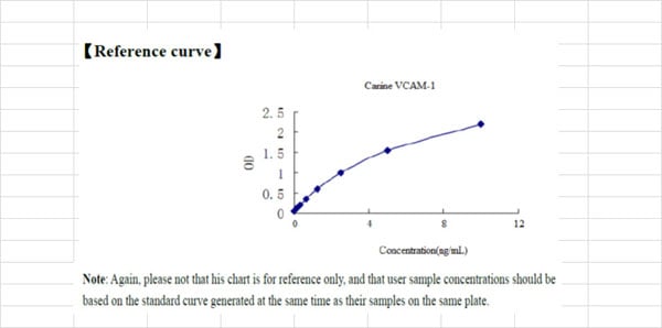 Standard Curve (Sample)
Vascular Cell Adhesion Molecule 1, ELISA Kit (Cat# AAA22583)
Full Name
Canine Vascular Cell Adhesion Molecule 1 (VCAM-1) ELISA Kit
Gene Names
VCAM1; CD106; INCAM-100
Reactivity
Canine
Pricing
 SDS-PAGE
Vinculin, Recombinant Protein (Cat# AAA18464)
Full Name
Recombinant Human Vinculin
Gene Names
VCL; MV; MVCL; CMD1W; CMH15; HEL114
Purity
Greater than 90% as determined by SDS-PAGE.
Pricing


 WB (Western Blot)
(F11R monoclonal antibody, Western Blot analysis of F11R expression in HepG2.)
WB (Western Blot)
(F11R monoclonal antibody, Western Blot analysis of F11R expression in HepG2.)
Junctional Adhesion Molecule 1, Monoclonal Antibody (Cat# AAA24250)
Full Name
Junctional Adhesion Molecule 1 (JAM-1, CD321, F11 Receptor, F11R, Junctional Adhesion Molecule A, JAMA, JCAM, KAT, Platelet Adhesion Molecule 1, PAM1, Platelet F11 Receptor, UNQ264/PRO301) (AP)
Gene Names
F11R; JAM; KAT; JAM1; JAMA; JCAM; CD321; PAM-1
Reactivity
Human
Applications
EIA, IHC, WB
Purity
Purified by Protein A Affinity Chromatography.
Pricing


 Application Data
(Staining of mouse spleen cells with HAMSTER ANTI MOUSE CD29: Alexa Fluor® 647)
Application Data
(Staining of mouse spleen cells with HAMSTER ANTI MOUSE CD29: Alexa Fluor® 647)
Integrin, beta1, Monoclonal Antibody (Cat# AAA14704)
Full Name
Integrin, beta1 (CD29, Very Late Antigen, VLAb1, Platelet gplla, ITGB1, Fibrinogen Receptor beta Subunit, FNRB, Integrin VLA4 Subunit beta, MDF2, MSK12, Very Late Activation Protein beta Polypeptide, VLAB, VLAbeta)
Gene Names
ITGB1; CD29; FNRB; MDF2; VLAB; GPIIA; MSK12; VLA-BETA
Reactivity
Mouse, Rat
Applications
Immunoprecipitation, Flow Cytometry
Purity
Affinity Purified
Purified by Protein G affinity chromatography.
Purified by Protein G affinity chromatography.
Pricing


 Application Data
(Published customer image Infiltration of GFP+ BM-cells in infarct and peri-infarct regions. (A-B) Dot plots of viable macrophages/granulocytes (CD11b+CD45high, top right quadrants) and microglia (CD11b+CD45dim, bottom right quadrants) in cortex from BM-chimeric unmanipulated mice and mice exposed to pMCAO. (C) Bar graph showing mean numbers of CD11b+CD45dim microglia and CD11b+CD45high macrophages/granulocytes in BM-chimeric mice 24 hours after pMCAO, subdivided based on expression of GFP (n = 5). Approximately 92% of of the CD45high population were GFP+. (D) Estimation and comparison of mean numbers of CD11b+CD45dim microglia in non-chimeric (n = 10) versus BM-chimeric mice (n = 5) 24 hours after of pMCAO shows significantly fewer CD11b+CD45dim microglial cells in irradiated mice. (E) Overview, showing distribution of infiltrating GFP+ BM-derived cells into infarct (IF) and peri-infarct (P-IF) regions 24 hours after pMCAO. (E-G) By 24 hours, GFP+ single cells (F) and vessel-associated aggregates of GFP+ cells (arrows in G) were observed in infarct and peri-infarct regions. Some of the vessel-associated cells were round, leukocyte-like cells (arrows) while others were elongated cells lining the vasculature (arrow heads in G and in insert). (H) Bar graph showing mean numbers of single GFP+ cells and vessel-associated aggregates of GFP+ cells in ipsi- and contralateral cortex 24 hours after surgery (n = 10). (I-P) Immunohistochemical staining of CD45.1 (I, K), CD45.2 (J, L), IgG2a (M, O) and CD45 (N, P) in ischemic tissue in BM-chimeric (I, J, M, N) and non-chimeric mice (K, L, O, P) 24 hours after pMCAO. N.D, none detected. Scale bars: 200 um (A), 10 um (B, C). 50 um (I-P) *P < 0.05, **P < 0.01, and ***P < 0.001.From: Clausen BH, Lambertsen KL, Babcock AA, Holm TH, Dagnaes-Hansen F, Finsen B. Interleukin-1beta and tumor necrosis factor-alpha are expressed by different subsets of microglia and macrophages after ischemic stroke in mice. J Neuroinflammation. 2008 Oct 23;5:46.)
Application Data
(Published customer image Infiltration of GFP+ BM-cells in infarct and peri-infarct regions. (A-B) Dot plots of viable macrophages/granulocytes (CD11b+CD45high, top right quadrants) and microglia (CD11b+CD45dim, bottom right quadrants) in cortex from BM-chimeric unmanipulated mice and mice exposed to pMCAO. (C) Bar graph showing mean numbers of CD11b+CD45dim microglia and CD11b+CD45high macrophages/granulocytes in BM-chimeric mice 24 hours after pMCAO, subdivided based on expression of GFP (n = 5). Approximately 92% of of the CD45high population were GFP+. (D) Estimation and comparison of mean numbers of CD11b+CD45dim microglia in non-chimeric (n = 10) versus BM-chimeric mice (n = 5) 24 hours after of pMCAO shows significantly fewer CD11b+CD45dim microglial cells in irradiated mice. (E) Overview, showing distribution of infiltrating GFP+ BM-derived cells into infarct (IF) and peri-infarct (P-IF) regions 24 hours after pMCAO. (E-G) By 24 hours, GFP+ single cells (F) and vessel-associated aggregates of GFP+ cells (arrows in G) were observed in infarct and peri-infarct regions. Some of the vessel-associated cells were round, leukocyte-like cells (arrows) while others were elongated cells lining the vasculature (arrow heads in G and in insert). (H) Bar graph showing mean numbers of single GFP+ cells and vessel-associated aggregates of GFP+ cells in ipsi- and contralateral cortex 24 hours after surgery (n = 10). (I-P) Immunohistochemical staining of CD45.1 (I, K), CD45.2 (J, L), IgG2a (M, O) and CD45 (N, P) in ischemic tissue in BM-chimeric (I, J, M, N) and non-chimeric mice (K, L, O, P) 24 hours after pMCAO. N.D, none detected. Scale bars: 200 um (A), 10 um (B, C). 50 um (I-P) *P < 0.05, **P < 0.01, and ***P < 0.001.From: Clausen BH, Lambertsen KL, Babcock AA, Holm TH, Dagnaes-Hansen F, Finsen B. Interleukin-1beta and tumor necrosis factor-alpha are expressed by different subsets of microglia and macrophages after ischemic stroke in mice. J Neuroinflammation. 2008 Oct 23;5:46.)
CD11b, Monoclonal Antibody (Cat# AAA12186)
Full Name
RAT ANTI MOUSE CD11b:RPE
Gene Names
Itgam; CR3; CR3A; MAC1; Cd11b; Ly-40; Mac-1; Mac-1a; CD11b/CD18; F730045J24Rik
Applications
Flow Cytometry
Pricing


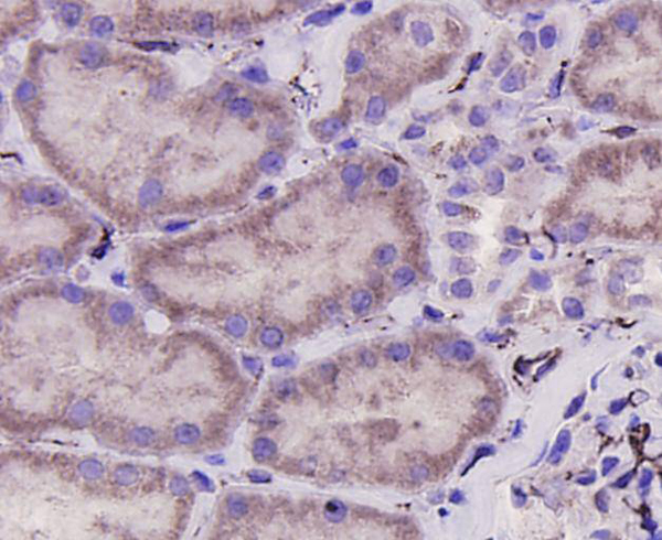
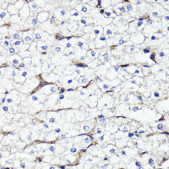
 IHC (Immunohistochemistry)
(At 1/100 staining Human gastric cancer by IHC-P. The sample was formaldehyde fixed and a heat mediated antigen retrieval step in citrate buffer was performed. The sample was then blocked and incubated with the primary antibody at 4 degree C overnight. An HRP conjugated anti-Rabbit antibody was used as the secondary antibody.)
IHC (Immunohistochemistry)
(At 1/100 staining Human gastric cancer by IHC-P. The sample was formaldehyde fixed and a heat mediated antigen retrieval step in citrate buffer was performed. The sample was then blocked and incubated with the primary antibody at 4 degree C overnight. An HRP conjugated anti-Rabbit antibody was used as the secondary antibody.)
alpha 1 Catenin, Polyclonal Antibody (Cat# AAA31379)
Full Name
alpha 1 Catenin Antibody
Gene Names
CTNNA1; CAP102
Reactivity
Human, Mouse, Rat
Applications
Western Blot, Immunohistochemistry, Peptide ELISA
Purity
The antiserum was purified by peptide affinity chromatography using SulfoLink Coupling Resin
Pricing