


 FCM (Flow Cytometry)
(Flow cytometric analysis of Hela cells with N Cadherin antibody at 1/50 dilution (red) compared with an unlabelled control (cells without incubation with primary antibody; black). Alexa Fluor 488-conjugated goat anti rabbit IgG was used as the secondary antibody.)
FCM (Flow Cytometry)
(Flow cytometric analysis of Hela cells with N Cadherin antibody at 1/50 dilution (red) compared with an unlabelled control (cells without incubation with primary antibody; black). Alexa Fluor 488-conjugated goat anti rabbit IgG was used as the secondary antibody.)
Cadherin-2 Preproprotein (NCAD) Antibody – Rabbit Monoclonal
N Cadherin Antibody
Product Overview
IHC: 1:50-1:200
ICC: 1:50-1:200
FC/FACS: 1:50-1:100
FCM (Flow Cytometry)
(Flow cytometric analysis of Hela cells with N Cadherin antibody at 1/50 dilution (red) compared with an unlabelled control (cells without incubation with primary antibody; black). Alexa Fluor 488-conjugated goat anti rabbit IgG was used as the secondary antibody.)
FCM (Flow Cytometry)
(Flow cytometric analysis of Hela cells with N Cadherin antibody at 1/50 dilution (red) compared with an unlabelled control (cells without incubation with primary antibody; black). Alexa Fluor 488-conjugated goat anti rabbit IgG was used as the secondary antibody.)
ICC (Immunocytochemistry)
(ICC staining N Cadherin in RH-35 cells (green). The nuclear counter stain is DAPI (blue). Cells were fixed in paraformaldehyde, permeabilised with 0.25% Triton X100/PBS.)
ICC (Immunocytochemistry)
(ICC staining N Cadherin in RH-35 cells (green). The nuclear counter stain is DAPI (blue). Cells were fixed in paraformaldehyde, permeabilised with 0.25% Triton X100/PBS.)
ICC (Immunocytochemistry)
(ICC staining N Cadherin in NCCIT cells (green). The nuclear counter stain is DAPI (blue). Cells were fixed in paraformaldehyde, permeabilised with 0.25% Triton X100/PBS.)
ICC (Immunocytochemistry)
(ICC staining N Cadherin in NCCIT cells (green). The nuclear counter stain is DAPI (blue). Cells were fixed in paraformaldehyde, permeabilised with 0.25% Triton X100/PBS.)
ICC (Immunocytochemistry)
(ICC staining N Cadherin in HepG2 cells (green). The nuclear counter stain is DAPI (blue). Cells were fixed in paraformaldehyde, permeabilised with 0.25% Triton X100/PBS.)
ICC (Immunocytochemistry)
(ICC staining N Cadherin in HepG2 cells (green). The nuclear counter stain is DAPI (blue). Cells were fixed in paraformaldehyde, permeabilised with 0.25% Triton X100/PBS.)
ICC (Immunocytochemistry)
(ICC staining N Cadherin in F9 cells (green). The nuclear counter stain is DAPI (blue). Cells were fixed in paraformaldehyde, permeabilised with 0.25% Triton X100/PBS.)
ICC (Immunocytochemistry)
(ICC staining N Cadherin in F9 cells (green). The nuclear counter stain is DAPI (blue). Cells were fixed in paraformaldehyde, permeabilised with 0.25% Triton X100/PBS.)
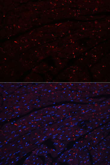
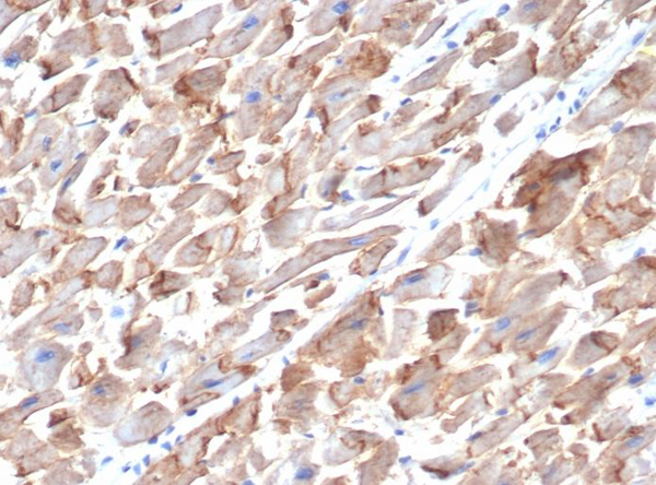
IHC (Immunohistochemistry)
(Immunohistochemical analysis of paraffin-embedded mouse heart tissue using anti-N Cadherin antibody. Counter stained with hematoxylin.)
IHC (Immunohistochemistry)
(Immunohistochemical analysis of paraffin-embedded mouse heart tissue using anti-N Cadherin antibody. Counter stained with hematoxylin.)
IHC (Immunohistochemistry)
(Immunohistochemical analysis of paraffin-embedded mouse embryo tissue using anti-N Cadherin antibody. Counter stained with hematoxylin.)
IHC (Immunohistochemistry)
(Immunohistochemical analysis of paraffin-embedded mouse embryo tissue using anti-N Cadherin antibody. Counter stained with hematoxylin.)
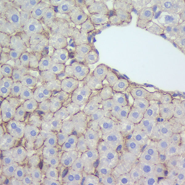
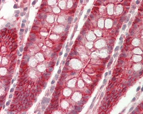
IHC (Immunohistochemistry)
(Immunohistochemical analysis of paraffin-embedded human liver cancer tissue using anti-N Cadherin antibody. Counter stained with hematoxylin.)
IHC (Immunohistochemistry)
(Immunohistochemical analysis of paraffin-embedded human liver cancer tissue using anti-N Cadherin antibody. Counter stained with hematoxylin.)
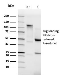
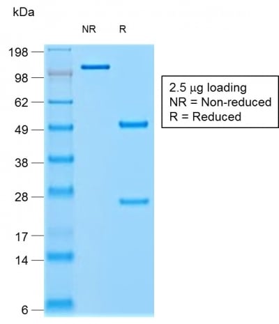
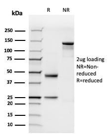
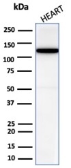
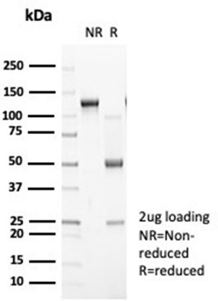
WB (Western Blot)
(Western blot analysis of N Cadherin on different lysates using anti-N Cadherin antibody at 1/1, 000 dilution. Positive control: Lane 1: Hela Lane 2: NIH/3T3 Lane 3: Mouse brain)
WB (Western Blot)
(Western blot analysis of N Cadherin on different lysates using anti-N Cadherin antibody at 1/1, 000 dilution. Positive control: Lane 1: Hela Lane 2: NIH/3T3 Lane 3: Mouse brain)
NCBI and Uniprot Product Information
Customer Reviews
Loading reviews...
Share Your Experience
Similar Products


Additional Details
Product Notes
The NCAD cdh2 (Catalog #AAA30054) is an Antibody produced from Rabbit and is intended for research purposes only. The product is available for immediate purchase. The N Cadherin Antibody reacts with Human, Mouse, Rat and may cross-react with other species as described in the data sheet. AAA Biotech's N Cadherin can be used in a range of immunoassay formats including, but not limited to, WB (Western Blot), ICC (Immunocytochemistry), IF (Immunofluorescence), IHC (Immunohistochemistry), FCM/FACS (Flow Cytometry). WB: 1:1000-5000 IHC: 1:50-1:200 ICC: 1:50-1:200 FC/FACS: 1:50-1:100. Researchers should empirically determine the suitability of the NCAD cdh2 for an application not listed in the data sheet. Researchers commonly develop new applications and it is an integral, important part of the investigative research process. It is sometimes possible for the material contained within the vial of "N Cadherin, Monoclonal Antibody" to become dispersed throughout the inside of the vial, particularly around the seal of said vial, during shipment and storage. We always suggest centrifuging these vials to consolidate all of the liquid away from the lid and to the bottom of the vial prior to opening. Please be advised that certain products may require dry ice for shipping and that, if this is the case, an additional dry ice fee may also be required.Precautions
All products in the AAA Biotech catalog are strictly for research-use only, and are absolutely not suitable for use in any sort of medical, therapeutic, prophylactic, in-vivo, or diagnostic capacity. By purchasing a product from AAA Biotech, you are explicitly certifying that said products will be properly tested and used in line with industry standard. AAA Biotech and its authorized distribution partners reserve the right to refuse to fulfill any order if we have any indication that a purchaser may be intending to use a product outside of our accepted criteria.Disclaimer
Though we do strive to guarantee the information represented in this datasheet, AAA Biotech cannot be held responsible for any oversights or imprecisions. AAA Biotech reserves the right to adjust any aspect of this datasheet at any time and without notice. It is the responsibility of the customer to inform AAA Biotech of any product performance issues observed or experienced within 30 days of receipt of said product. To see additional details on this or any of our other policies, please see our Terms & Conditions page.Frequently Asked Questions
What is N-cadherin used as a marker for in cell adhesion and EMT studies?
N-cadherin is a transmembrane protein primarily used as a marker for mesenchymal cells and neural tissues. In the context of the Epithelial-Mesenchymal Transition (EMT), it serves as a key indicator of cellular transformation. Using the AAA Biotech N-cadherin antibody helps researchers identify cells that have lost epithelial characteristics and gained the migratory properties associated with mesenchymal phenotypes.
Can a rabbit N-cadherin antibody be used for immunofluorescence or Western blot?
Yes, the rabbit N-cadherin antibody is suitable for both immunofluorescence and Western blotting. In IF, it reveals the localization of N-cadherin at cell-cell junctions or within the cytoplasm. In Western blot, it is used to quantify the total expression level of the protein in tissue or cell lysates, providing a reliable measurement for AAA Biotech customers studying cell adhesion.
Is N-cadherin expression associated with epithelial–mesenchymal transition (EMT) in cancer?
Yes, N-cadherin is a hallmark of EMT. During cancer progression, cells often undergo a "cadherin switch," where E-cadherin expression decreases, and N-cadherin expression increases. This switch, which can be monitored using AAA Biotech antibodies, is associated with increased tumor invasiveness, motility, and a higher potential for metastasis in various types of carcinomas.
What’s the difference between N-cadherin and E-cadherin detection?
The main difference lies in their tissue distribution and biological roles. E-cadherin is primarily found in epithelial cells and maintains tight cell-cell junctions. N-cadherin is typically found in neural, skeletal, and mesenchymal cells. Detecting both using AAA Biotech reagents allows researchers to determine the differentiation state of a cell and whether it is undergoing transition between epithelial and mesenchymal states.
Can this antibody detect N-cadherin in tissue sections by IHC?
Yes, this antibody is validated for immunohistochemistry (IHC) on tissue sections. It allows for the spatial visualization of N-cadherin in various tissues, such as the brain, heart, or cancerous tumors. Using AAA Biotech N-cadherin antibodies in IHC helps researchers map the distribution of mesenchymal cells within the complex architecture of a tissue sample.
How does fixation method affect N-cadherin staining quality?
The fixation method is critical for maintaining the integrity of cell junctions where N-cadherin is localized. While PFA fixation is common for IHC and IF, some epitopes may require specific antigen retrieval steps to be accessible. AAA Biotech recommends following optimized fixation protocols to ensure sharp, clear staining of N-cadherin at the plasma membrane and to avoid background noise.
Item has been added to Shopping Cart
If you are ready to order, navigate to Shopping Cart and get ready to checkout.