Filters
Clonality
Type
Reactivity
Gene Name
Isotype
Host
Application
Clone
7408 results for " Serum" - showing 6950-7000
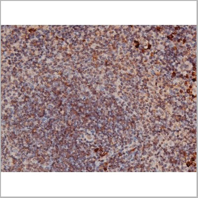
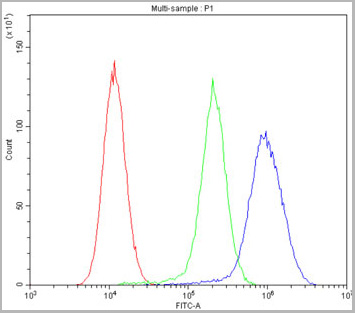
 Application Data
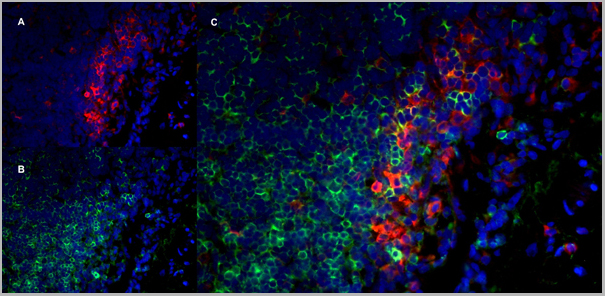
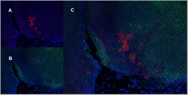
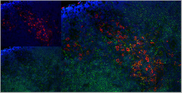
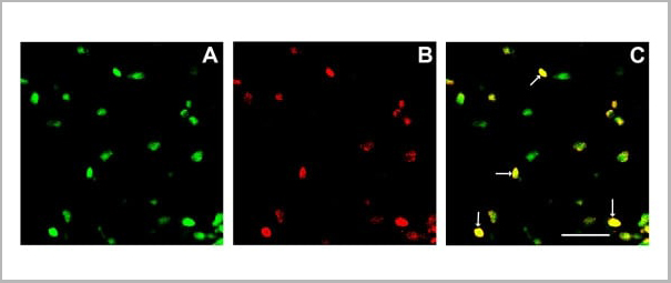
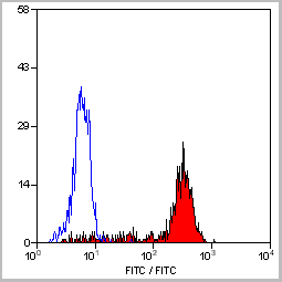
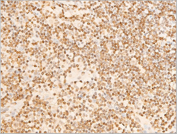
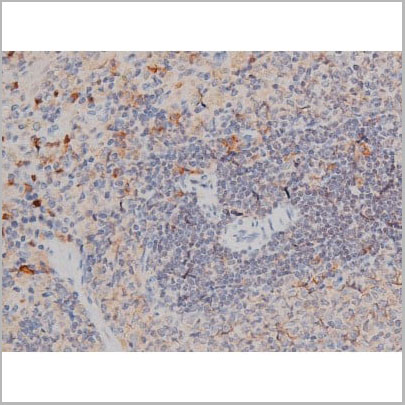
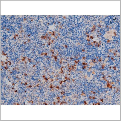
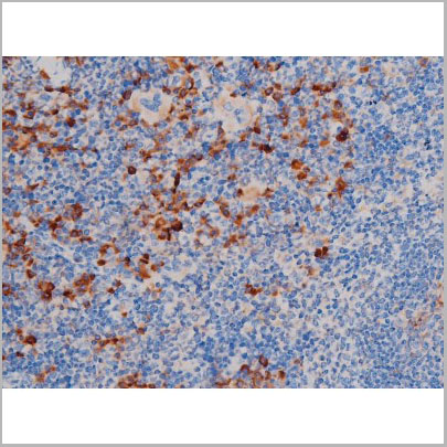
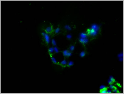
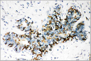
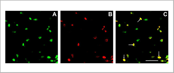
(Immunofluorescence staining of rat lymph node cryosection with Mouse anti Rat CD163 antibody , red an A and Mouse anti Rat CD8 MCA48), green in B. C is the merged image with nuclei counter-stained blue using DAPI. High power)
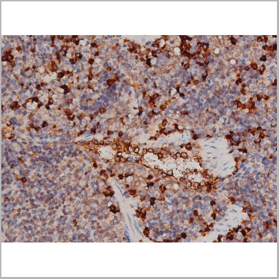
Application Data
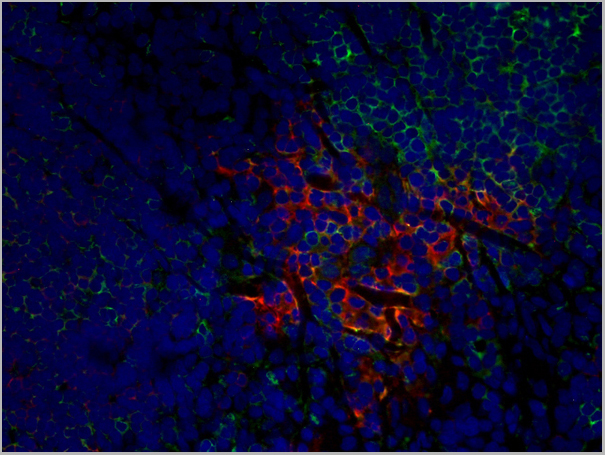
(Immunofluorescence staining of rat lymph node cryosection with Mouse anti Rat CD163 antibody , red an A and Mouse anti Rat CD8 MCA48), green in B. C is the merged image with nuclei counter-stained blue using DAPI. High power)
CD8 ALPHA, Monoclonal Antibody (Cat# AAA11981)
Full Name
MOUSE ANTI RAT CD8 ALPHA
Applications
Immunohistochemistry, Flow Cytometry, Immunofluorescence, Immunoprecipitation, Immunohistochemistry, Western Blot
Pricing
 Application Data
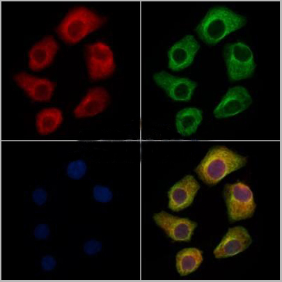
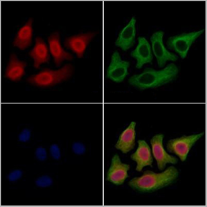
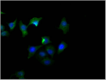
(AAA31434 staining Hela cells(4h of LPS treatment) by IF/ICC. The samples were fixed with PFA and permeabilized in 0.1% Triton X-100,then blocked in 10% serum for 45 minutes at 25°C. Samples were then incubated with primary Ab(AAA31434 1:200) and mouse anti-beta tubulin Ab( 1:200) for 1 hour at 37°C. An AlexaFluor594 conjugated goat anti-rabbit IgG(H+L) Ab(Red) and an AlexaFluor488 conjugated goat anti-mouse IgG(H+L) Ab(Green) were used as the secondary antibody. The nuclear counter stain is DAPI(blue).)
Application Data
(AAA31434 staining Hela cells(4h of LPS treatment) by IF/ICC. The samples were fixed with PFA and permeabilized in 0.1% Triton X-100,then blocked in 10% serum for 45 minutes at 25°C. Samples were then incubated with primary Ab(AAA31434 1:200) and mouse anti-beta tubulin Ab( 1:200) for 1 hour at 37°C. An AlexaFluor594 conjugated goat anti-rabbit IgG(H+L) Ab(Red) and an AlexaFluor488 conjugated goat anti-mouse IgG(H+L) Ab(Green) were used as the secondary antibody. The nuclear counter stain is DAPI(blue).)
Smad3, Polyclonal Antibody (Cat# AAA31434)
Full Name
Phospho-Smad3 (Ser423+Ser425) Antibody
Gene Names
SMAD3; LDS3; LDS1C; MADH3; JV15-2; HSPC193; HsT17436
Reactivity
Human, Mouse, Rat
Applications
Western Blot, Immunohistochemistry, Immunofluorescence, Immunocytochemistry
Purity
The antibody is from purified rabbit serum by affinity purification via sequential chromatography on phospho-peptide and non-phospho-peptide affinity columns.
Pricing

 Application Data
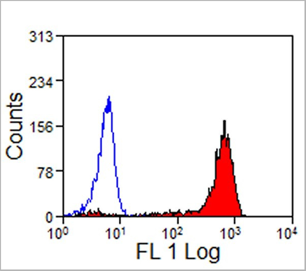
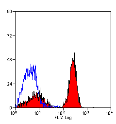
(Staining of New Zealand Black mouse peripheral blood granulocytes with Rat anti Mouse Ly-6B.2 conjugated to FITC Data)
Application Data
(Staining of New Zealand Black mouse peripheral blood granulocytes with Rat anti Mouse Ly-6B.2 conjugated to FITC Data)
Ly-6B.2 ALLOANTIGEN, Monoclonal Antibody (Cat# AAA12188)
Full Name
RAT ANTI MOUSE Ly-6B.2 ALLOANTIGEN:Biotin
Applications
Flow Cytometry
Pricing
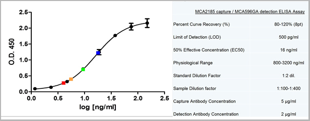
Corynebacterium diphtheriae toxin IgG, ELISA Kit (Cat# AAA14224)
Full Name
Corynebacterium diphtheriae toxin IgG ELISA Kit
Pricing
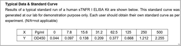 Standard Curve (Sample)
sTNFR1, ELISA Kit (Cat# AAA13496)
Full Name
sTNFR1 Human ELISA Kit
Reactivity
Human
Applications
ELISA
Pricing
Thrombocyte, Polyclonal Antibody (Cat# AAA14283)
Full Name
Anti-Mouse Thrombocyte, Antiserum, (Polyclonal) (rabbit serum)
Reactivity
Mouse
Applications
Agglutination Assay, Invivo Depletion
Pricing
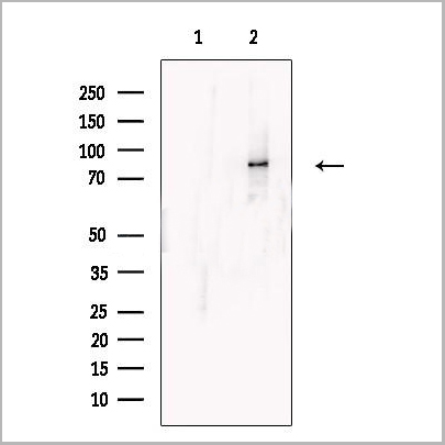
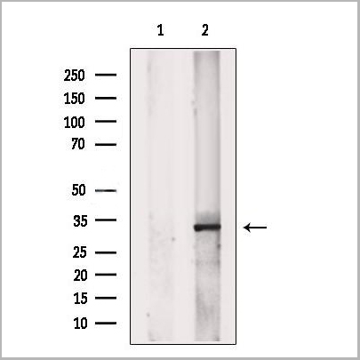
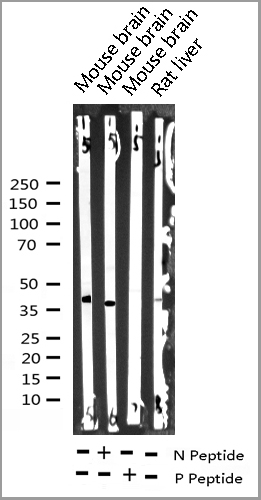
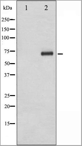
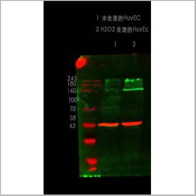
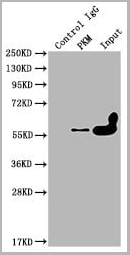
 WB (Western Blot)
(Western blot analysis on human serum using anti- Complement C3 mouse monoclonal antibody.)
WB (Western Blot)
(Western blot analysis on human serum using anti- Complement C3 mouse monoclonal antibody.)
Complement C3, Antibody (Cat# AAA17956)
Full Name
Complement C3 (betachain) Antibody
Gene Names
C3; ASP; C3a; C3b; AHUS5; ARMD9; CPAMD1; HEL-S-62p
Reactivity
Human
Applications
Western Blot
Pricing
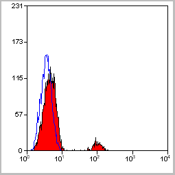
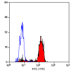
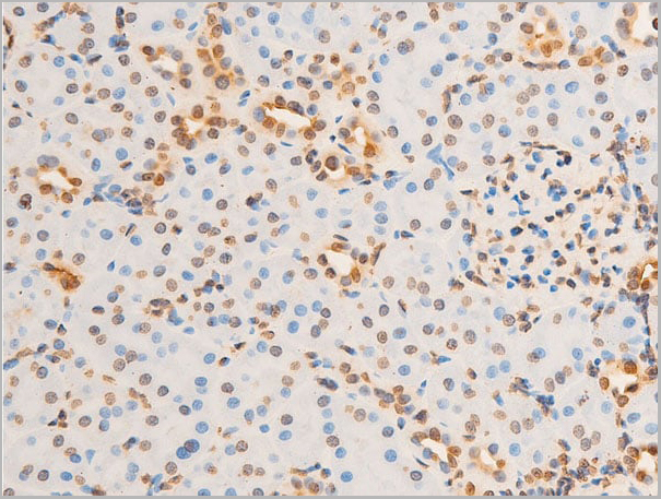
 Application Data
(Staining of human peripheral blood lymphocytes with Mouse anti Human CD19: RPE- Alexa Fluor 647)
Application Data
(Staining of human peripheral blood lymphocytes with Mouse anti Human CD19: RPE- Alexa Fluor 647)
CD19, Monoclonal Antibody (Cat# AAA11870)
Full Name
MOUSE ANTI HUMAN CD19:FITC
Gene Names
CD19; B4; CVID3
Applications
Flow Cytometry
Pricing
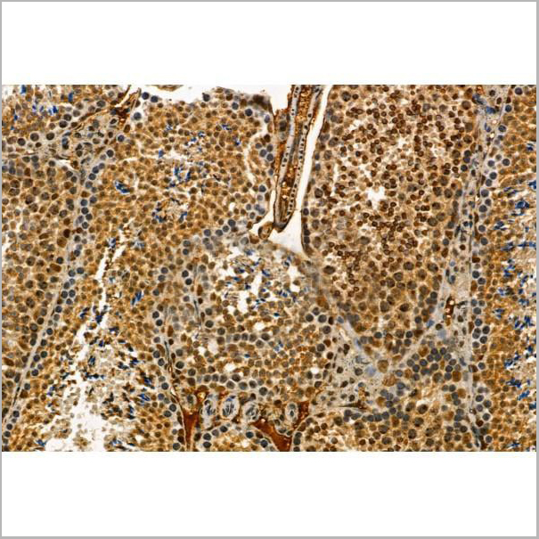
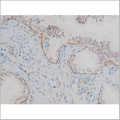
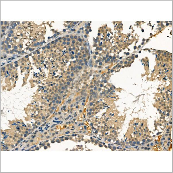
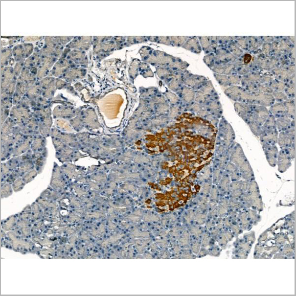
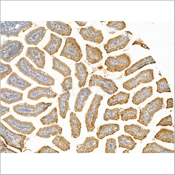
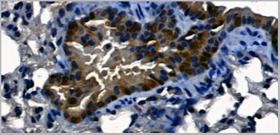
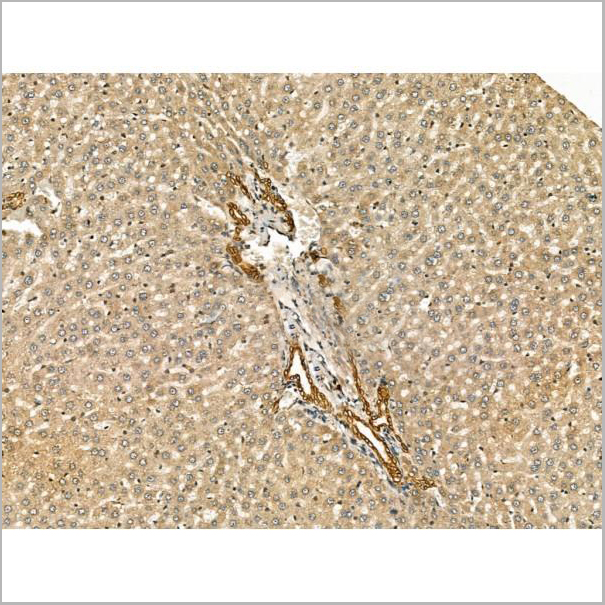
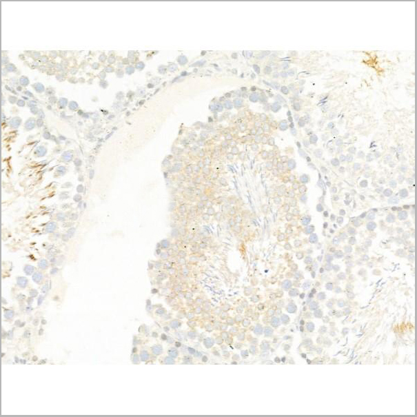
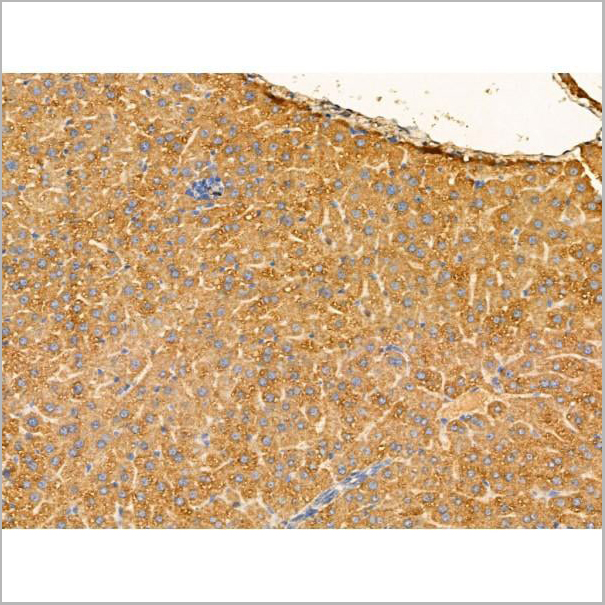
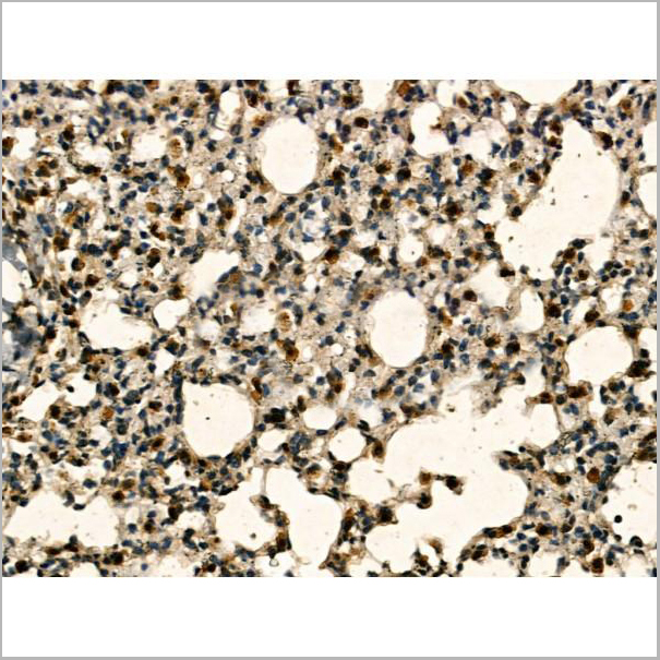
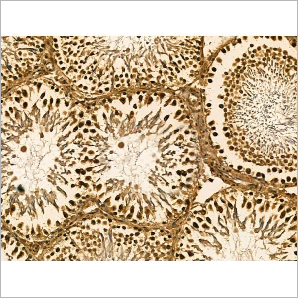
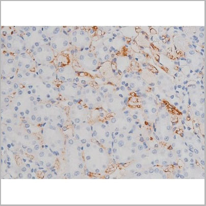
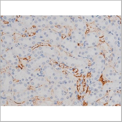
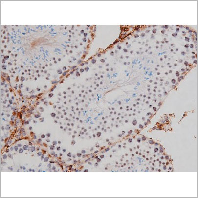
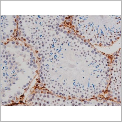
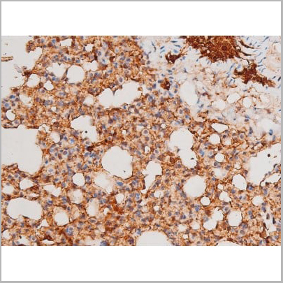
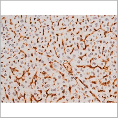
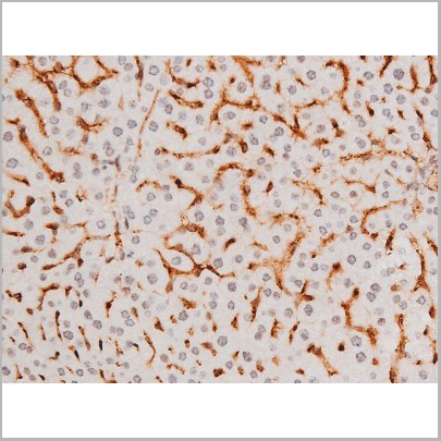
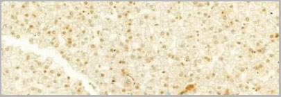
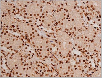
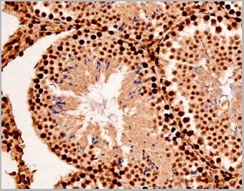
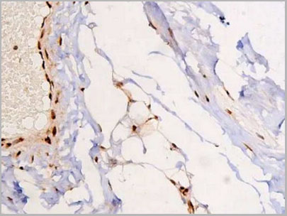
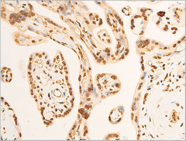
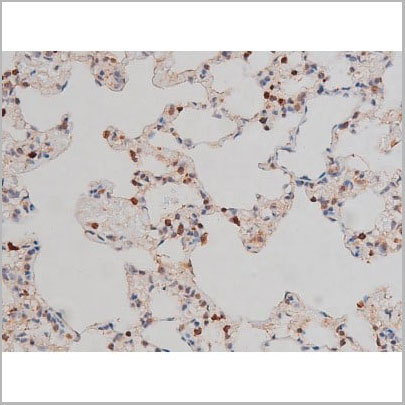
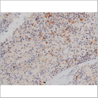
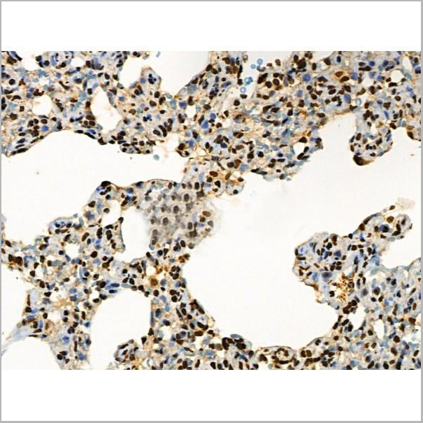
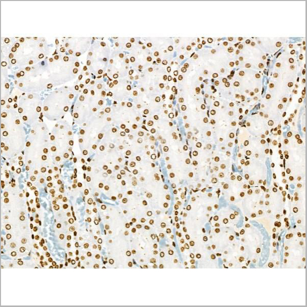
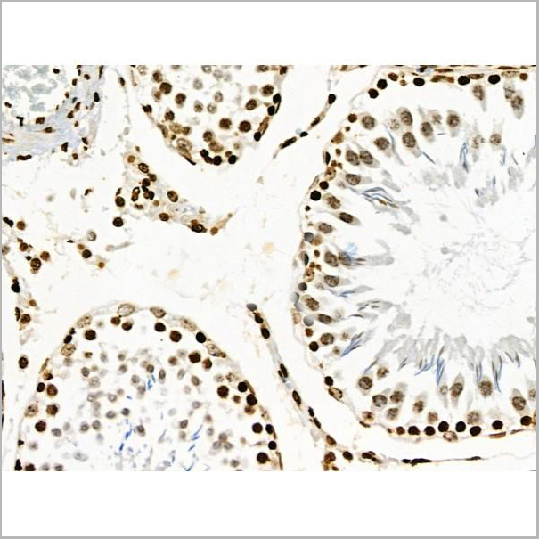
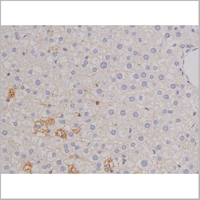
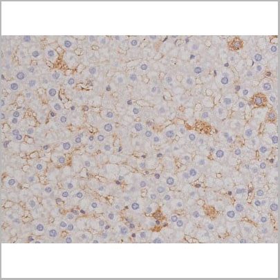
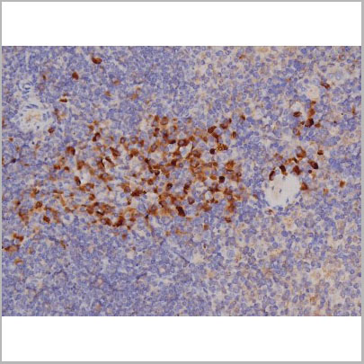
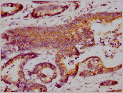
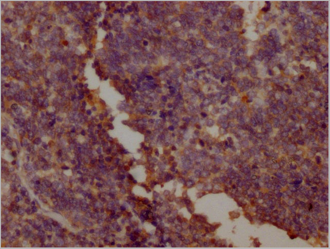
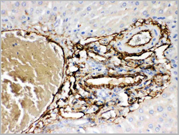
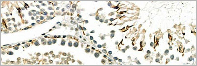 IHC (Immunohistochemistry)
(AAA31112 at 1/100 staining Rat testis tissue by IHC-P. The sample was formaldehyde fixed and a heat mediated antigen retrieval step in citrate buffer was performed. The sample was then blocked and incubated with the primary Ab at 4°C overnight. An HRP conjugated anti-Rabbit Ab was used as the secondary Ab.)
IHC (Immunohistochemistry)
(AAA31112 at 1/100 staining Rat testis tissue by IHC-P. The sample was formaldehyde fixed and a heat mediated antigen retrieval step in citrate buffer was performed. The sample was then blocked and incubated with the primary Ab at 4°C overnight. An HRP conjugated anti-Rabbit Ab was used as the secondary Ab.)
OSR2, Polyclonal Antibody (Cat# AAA31112)
Full Name
OSR2 Antibody
Reactivity
Human, Mouse, Rat
Applications
Western Blot, Immunofluorescence, Immunocytochemistry
Purity
The antiserum was purified by peptide affinity chromatography using SulfoLink Coupling Resin.
Pricing
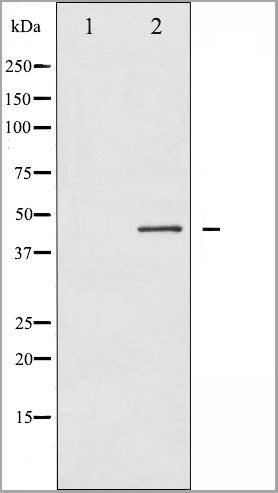
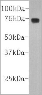
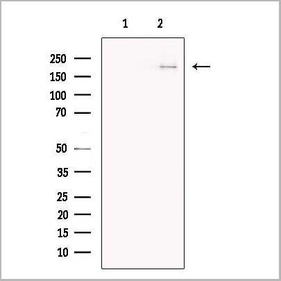
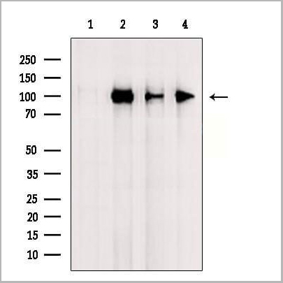
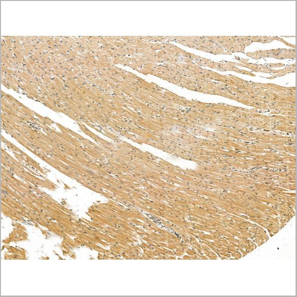
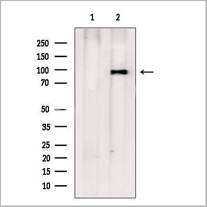
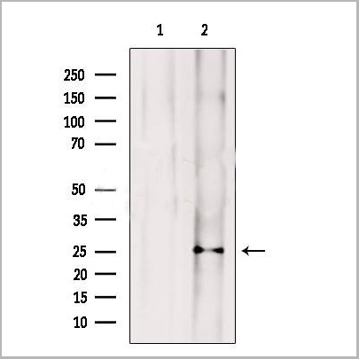
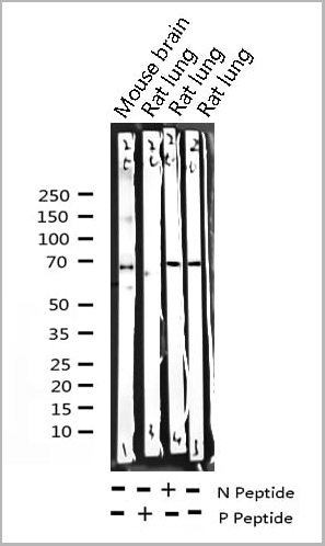
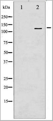
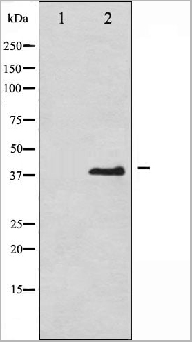
 WB (Western Blot)
(Western blot analysis of Phospho-Connexin 43 (Ser367) expression in various lysates)
WB (Western Blot)
(Western blot analysis of Phospho-Connexin 43 (Ser367) expression in various lysates)
Connexin 43, Polyclonal Antibody (Cat# AAA31018)
Full Name
Phospho-Connexin 43 (Ser367) Antibody
Gene Names
GJA1; HSS; CMDR; CX43; EKVP; GJAL; ODDD; AVSD3; EKVP3; HLHS1; PPKCA
Reactivity
Human, Mouse, Rat
Applications
Western Blot, Immunohistochemistry
Purity
From purified rabbit serum by affinity purification via sequential chromatography on phospho-and non-phospho-peptide affinity columns.
Pricing
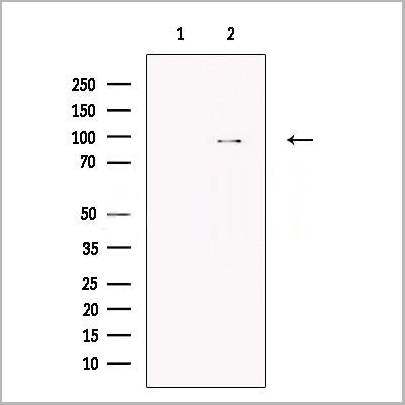
 WB (Western Blot)
(Western blot analysis of extracts from Hela, using Phospho-TLR4(Ser800) Antibody.)
WB (Western Blot)
(Western blot analysis of extracts from Hela, using Phospho-TLR4(Ser800) Antibody.)
TLR4, Polyclonal Antibody (Cat# AAA31162)
Full Name
Phospho-TLR4 (Ser800) Antibody
Gene Names
TLR4; TOLL; CD284; TLR-4; ARMD10
Reactivity
Human, Mouse, Rat
Applications
Western Blot, Immunohistochemistry, Immunofluorescence, Immunocytochemistry
Purity
Purified rabbit serum by affinity purification via sequential chromatography on phospho- and non-phospho-peptide affinity columns.
Pricing
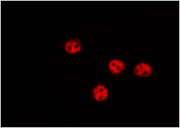
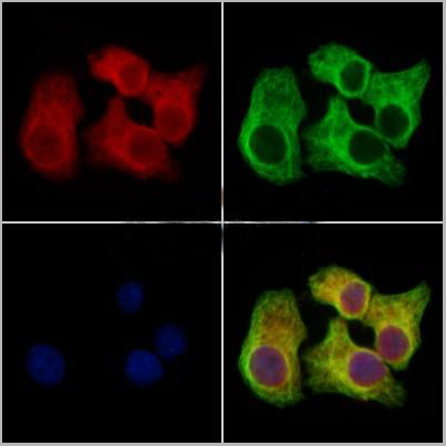 IF (Immunofluorescence)
(AAA31170 staining Hela cells by IF/ICC. The samples were fixed with PFA and permeabilized in 0.1% Triton X-100,then blocked in 10% serum for 45 minutes at 25°C. Samples were then incubated with primary Ab(AAA31170 1:200) and mouse anti-beta tubulin Ab(T0023 1:200) for 1 hour at 37°C. An AlexaFluor594 conjugated goat anti-rabbit IgG(H+L) Ab(Red) and an AlexaFluor488 conjugated goat anti-mouse IgG(H+L) Ab(Green) were used as the secondary antibody.The nuclear counter stain is DAPI(blue).)
IF (Immunofluorescence)
(AAA31170 staining Hela cells by IF/ICC. The samples were fixed with PFA and permeabilized in 0.1% Triton X-100,then blocked in 10% serum for 45 minutes at 25°C. Samples were then incubated with primary Ab(AAA31170 1:200) and mouse anti-beta tubulin Ab(T0023 1:200) for 1 hour at 37°C. An AlexaFluor594 conjugated goat anti-rabbit IgG(H+L) Ab(Red) and an AlexaFluor488 conjugated goat anti-mouse IgG(H+L) Ab(Green) were used as the secondary antibody.The nuclear counter stain is DAPI(blue).)
Clathrin heavy chain, Polyclonal Antibody (Cat# AAA31170)
Full Name
Clathrin heavy chain Antibody
Gene Names
CLTCL1; CLTD; CHC22; CLH22; CLTCL
Reactivity
Human, Mouse, Rat
Predicted Reactivity: Zebrafish(88%), Rabbit(88%), Dog(100%), Chicken(88%)
Predicted Reactivity: Zebrafish(88%), Rabbit(88%), Dog(100%), Chicken(88%)
Applications
Immunofluorescence, Immunocytochemistry
Purity
The antiserum was purified by peptide affinity chromatography using SulfoLink Coupling Resin (Thermo Fisher Scientific).
Pricing
 IF (Immunofluorescence)
(AAA31241 staining Hela cells by IF/ICC. The samples were fixed with PFA and permeabilized in 0.1% Triton X-100,then blocked in 10% serum for 45 minutes at 25°C. Samples were then incubated with primary Ab(AAA31241 1:200) and mouse anti-beta tubulin Ab(T0023 1:200) for 1 hour at 37°C. An AlexaFluor594 conjugated goat anti-rabbit IgG(H+L) Ab(Red) and an AlexaFluor488 conjugated goat anti-mouse IgG(H+L) Ab(Green) were used as the secondary antibody.The nuclear counter stain is DAPI(blue).)
IF (Immunofluorescence)
(AAA31241 staining Hela cells by IF/ICC. The samples were fixed with PFA and permeabilized in 0.1% Triton X-100,then blocked in 10% serum for 45 minutes at 25°C. Samples were then incubated with primary Ab(AAA31241 1:200) and mouse anti-beta tubulin Ab(T0023 1:200) for 1 hour at 37°C. An AlexaFluor594 conjugated goat anti-rabbit IgG(H+L) Ab(Red) and an AlexaFluor488 conjugated goat anti-mouse IgG(H+L) Ab(Green) were used as the secondary antibody.The nuclear counter stain is DAPI(blue).)
Sulfatase 1, Polyclonal Antibody (Cat# AAA31241)
Full Name
Sulfatase 1 Antibody
Gene Names
SULF1; SULF-1
Reactivity
Human, Mouse, Rat
Predicted Reactivity: Pig(100%), Bovine(100%), Horse(100%), Sheep(100%), Rabbit(100%), Dog(100%), Chicken(88%), Xenopus(100%)
Predicted Reactivity: Pig(100%), Bovine(100%), Horse(100%), Sheep(100%), Rabbit(100%), Dog(100%), Chicken(88%), Xenopus(100%)
Applications
ELISA
Purity
The antiserum was purified by peptide affinity chromatography using SulfoLink Coupling Resin (Thermo Fisher Scientific).
Pricing
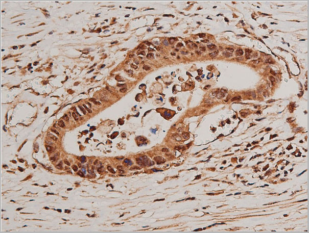
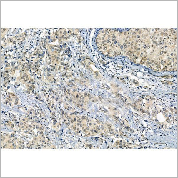
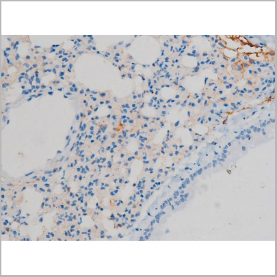
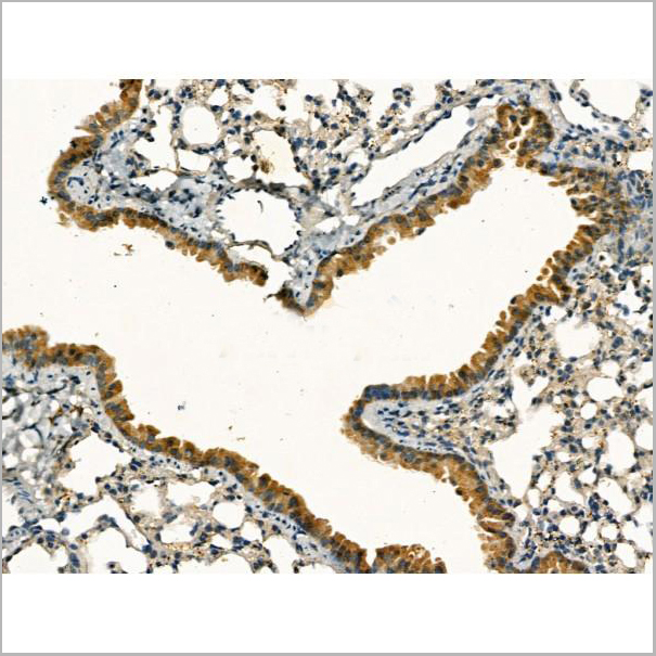
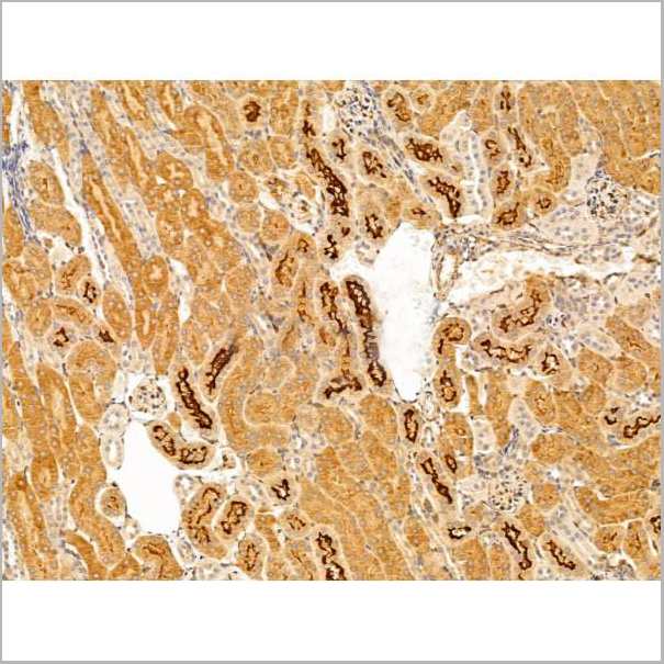
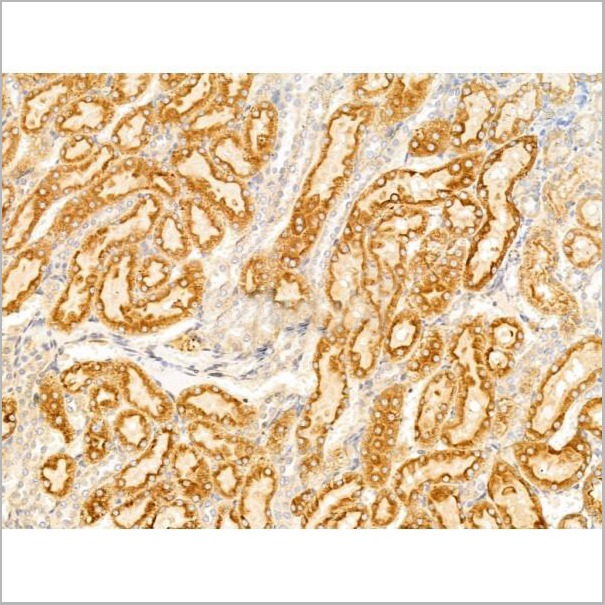
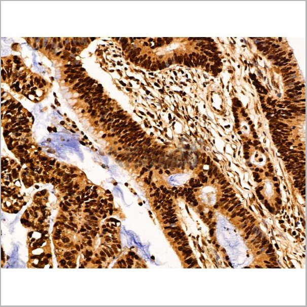
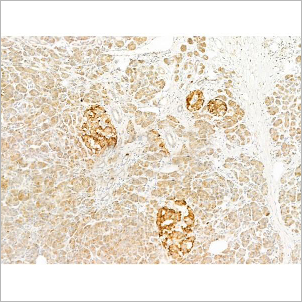
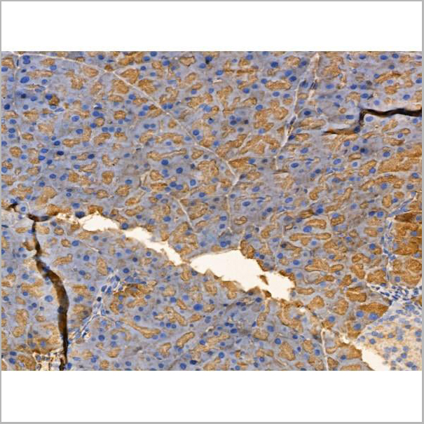
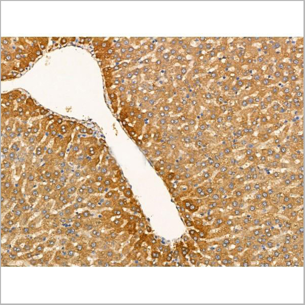
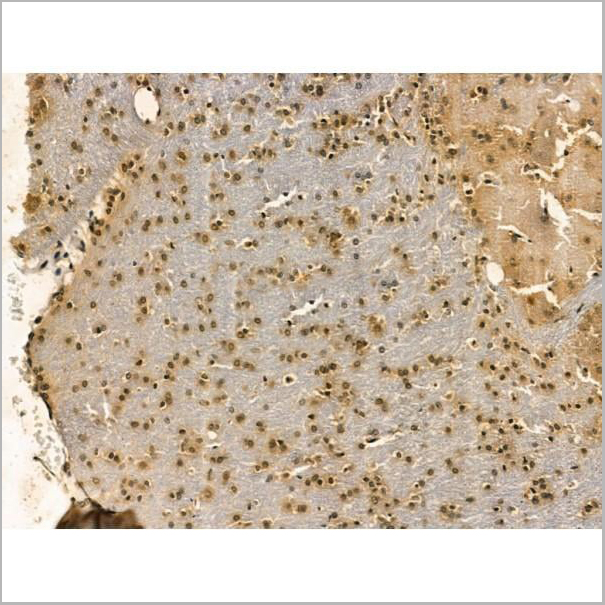
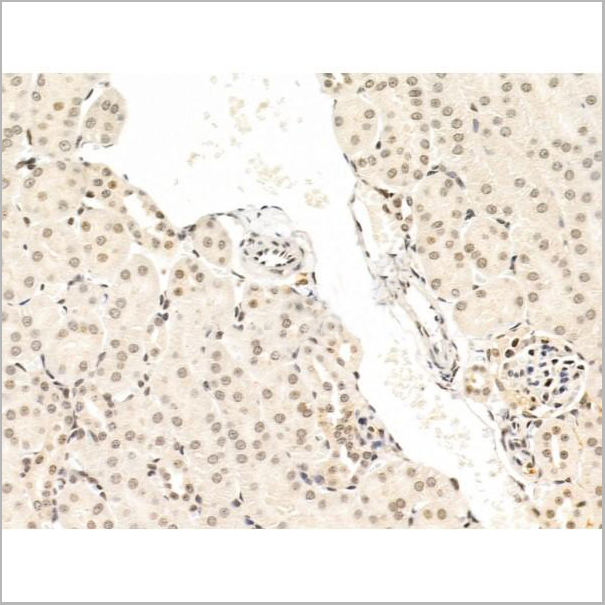
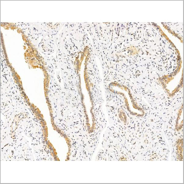
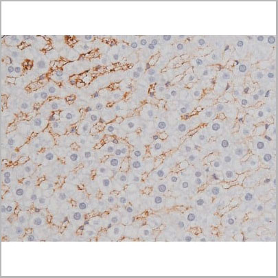
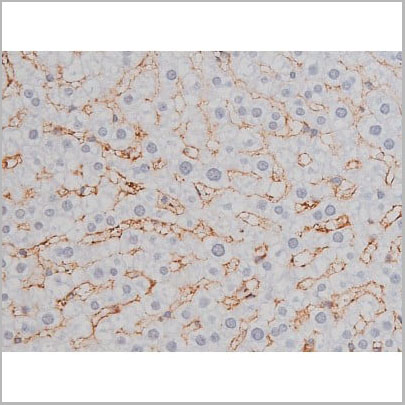
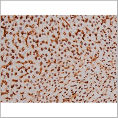
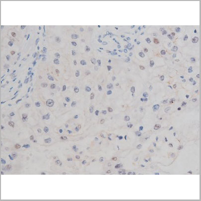
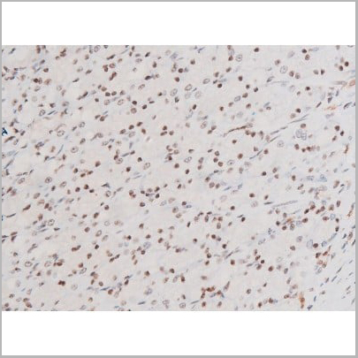
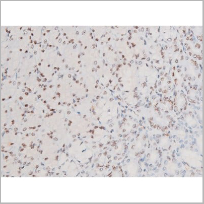
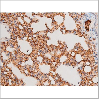
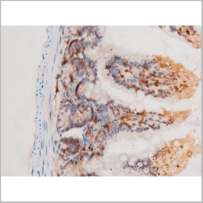
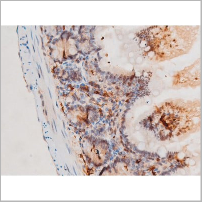
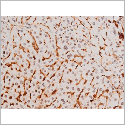
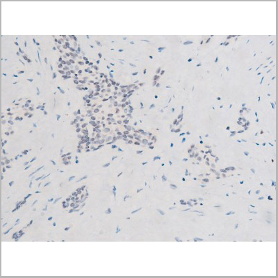
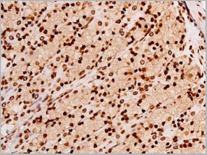
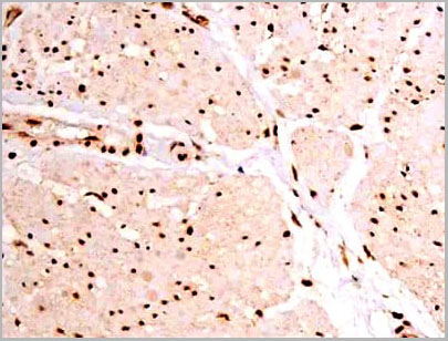
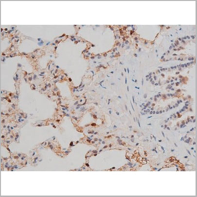
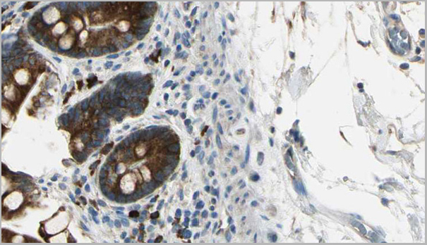
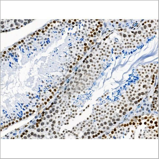
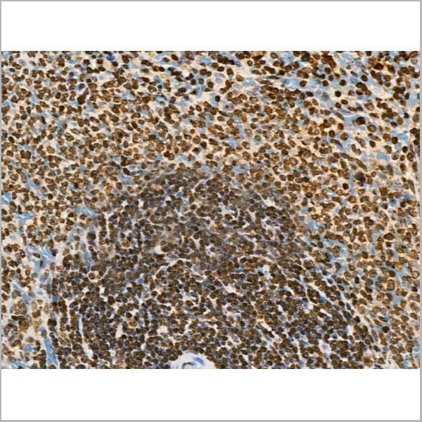
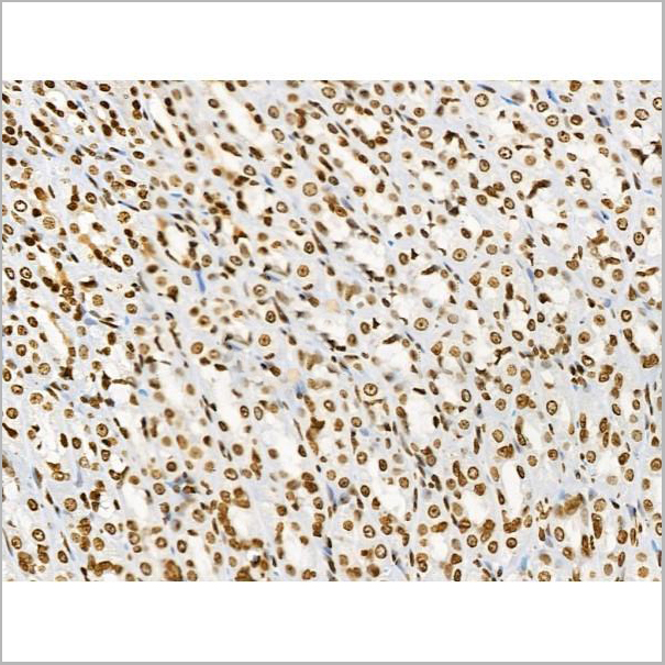
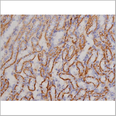
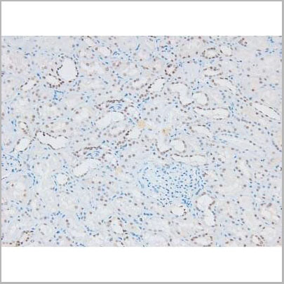
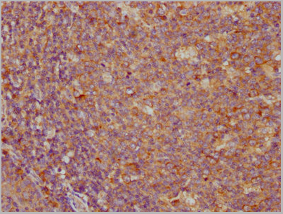
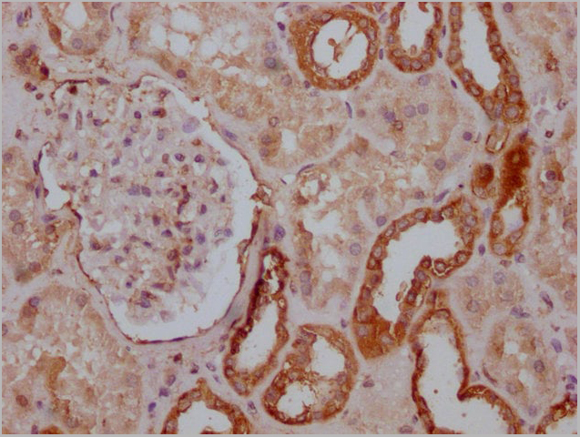
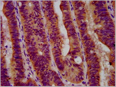
 IHC (Immunohistochemistry)
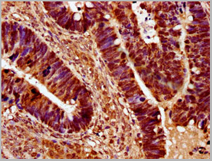
(AAA31069 at 1/50 staining human colon cancer tissue sections by IHC-P. The tissue was formaldehyde fixed and a heat mediated antigen retrieval step in citrate buffer was performed. The tissue was then blocked and incubated with the antibody for 1.5 hours at 22 degree C. An HRP conjugated goat anti-rabbit antibody was used as the secondary.)
IHC (Immunohistochemistry)
(AAA31069 at 1/50 staining human colon cancer tissue sections by IHC-P. The tissue was formaldehyde fixed and a heat mediated antigen retrieval step in citrate buffer was performed. The tissue was then blocked and incubated with the antibody for 1.5 hours at 22 degree C. An HRP conjugated goat anti-rabbit antibody was used as the secondary.)
FKHR, Polyclonal Antibody (Cat# AAA31069)
Full Name
Phospho-FKHR (Ser319) Antibody
Gene Names
FOXO1; FKH1; FKHR; FOXO1A
Reactivity
Human, Mouse, Rat
Applications
Western Blot, Immunohistochemistry, Immunofluorescence, Immunocytochemistry
Purity
From purified rabbit serum by affinity purification via sequential chromatography on phospho-and non-phospho-peptide affinity columns.
Pricing
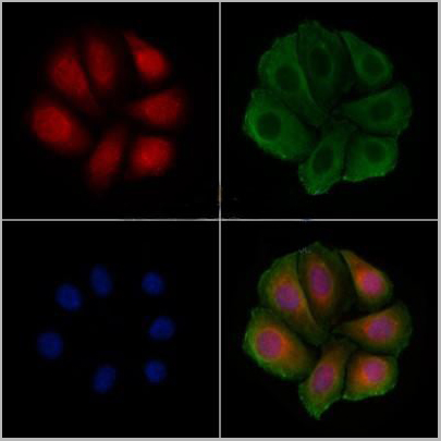
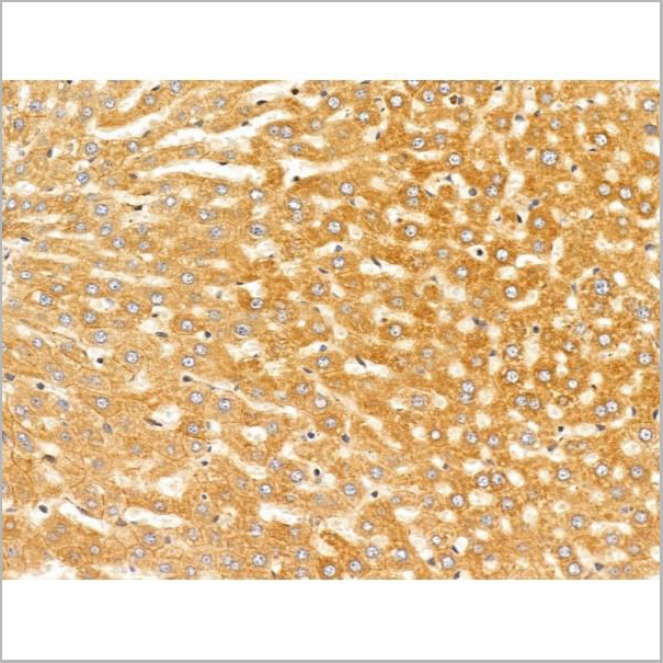
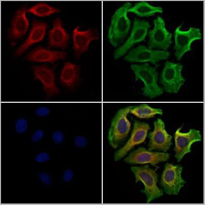
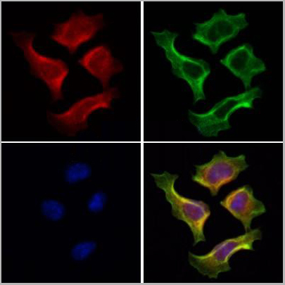
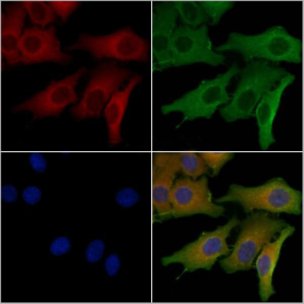
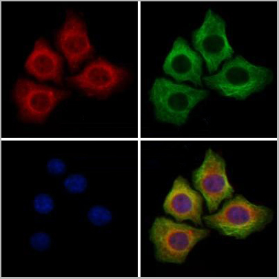
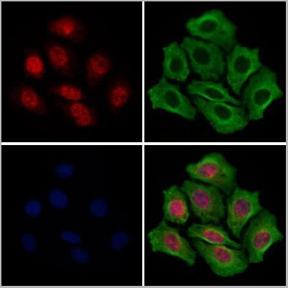
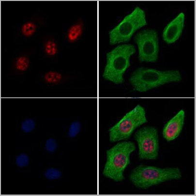
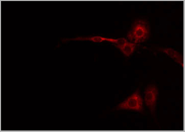
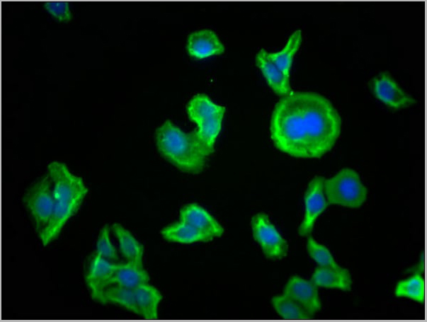
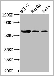
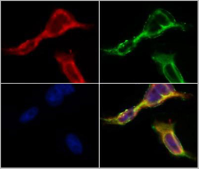 IF (Immunofluorescence)
(AAA31077 staining HepG2 by IF/ICC. The samples were fixed with PFA and permeabilized in 0.1% Triton X-100,then blocked in 10% serum for 45 minutes at 25°C. Samples were then incubated with primary Ab(AAA31077 1:200) and mouse anti-beta tubulin Ab for 1 hour at 37°C. An AlexaFluor594 conjugated goat anti-rabbit IgG(H+L) Ab(Red) and an AlexaFluor488 conjugated goat anti-mouse IgG(H+L) Ab(Green) were used as the secondary antibody.The nuclear counter stain is DAPI(blue).)
IF (Immunofluorescence)
(AAA31077 staining HepG2 by IF/ICC. The samples were fixed with PFA and permeabilized in 0.1% Triton X-100,then blocked in 10% serum for 45 minutes at 25°C. Samples were then incubated with primary Ab(AAA31077 1:200) and mouse anti-beta tubulin Ab for 1 hour at 37°C. An AlexaFluor594 conjugated goat anti-rabbit IgG(H+L) Ab(Red) and an AlexaFluor488 conjugated goat anti-mouse IgG(H+L) Ab(Green) were used as the secondary antibody.The nuclear counter stain is DAPI(blue).)
ACE2, Polyclonal Antibody (Cat# AAA31077)
Full Name
ACE2 Antibody
Gene Names
ACE2; ACEH
Reactivity
Human, Mouse, Rat
Applications
Western Blot
Purity
Peptide affinity purification
Pricing
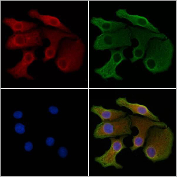
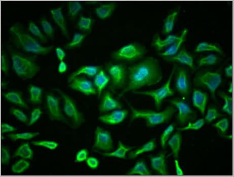
 IF (Immunofluorescence)
(AAA31168 staining Hela cells by IF/ICC. The samples were fixed with PFA and permeabilized in 0.1% Triton X-100,then blocked in 10% serum for 45 minutes at 25°C. Samples were then incubated with primary Ab(AAA31168 1:200) and mouse anti-beta tubulin Ab(T0023 1:200) for 1 hour at 37°C. An AlexaFluor594 conjugated goat anti-rabbit IgG(H+L) Ab(Red) and an AlexaFluor488 conjugated goat anti-mouse IgG(H+L) Ab(Green) were used as the secondary antibody.The nuclear counter stain is DAPI(blue).)
IF (Immunofluorescence)
(AAA31168 staining Hela cells by IF/ICC. The samples were fixed with PFA and permeabilized in 0.1% Triton X-100,then blocked in 10% serum for 45 minutes at 25°C. Samples were then incubated with primary Ab(AAA31168 1:200) and mouse anti-beta tubulin Ab(T0023 1:200) for 1 hour at 37°C. An AlexaFluor594 conjugated goat anti-rabbit IgG(H+L) Ab(Red) and an AlexaFluor488 conjugated goat anti-mouse IgG(H+L) Ab(Green) were used as the secondary antibody.The nuclear counter stain is DAPI(blue).)
DLL4, Polyclonal Antibody (Cat# AAA31168)
Full Name
DLL4 Antibody
Gene Names
DLL4; AOS6; delta4; hdelta2
Reactivity
Human, Mouse, Rat
Predicted Reactivity: Pig(100%), Bovine(100%), Horse(100%), Rabbit(100%), Dog(100%), Chicken(88%)
Predicted Reactivity: Pig(100%), Bovine(100%), Horse(100%), Rabbit(100%), Dog(100%), Chicken(88%)
Applications
Immunofluorescence, Immunocytochemistry
Purity
The antiserum was purified by peptide affinity chromatography using SulfoLink Coupling Resin (Thermo Fisher Scientific).
Pricing
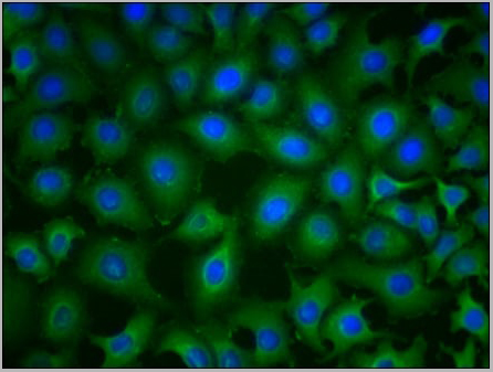
 IF (Immunofluorescence)
(AAA31227 staining Hela cells by IF/ICC. The samples were fixed with PFA and permeabilized in 0.1% Triton X-100,then blocked in 10% serum for 45 minutes at 25°C. Samples were then incubated with primary Ab(AAA31227 1:200) and mouse anti-beta tubulin Ab(T0023 1:200) for 1 hour at 37°C. An AlexaFluor594 conjugated goat anti-rabbit IgG(H+L) Ab(Red) and an AlexaFluor488 conjugated goat anti-mouse IgG(H+L) Ab(Green) were used as the secondary antibody.The nuclear counter stain is DAPI(blue).)
IF (Immunofluorescence)
(AAA31227 staining Hela cells by IF/ICC. The samples were fixed with PFA and permeabilized in 0.1% Triton X-100,then blocked in 10% serum for 45 minutes at 25°C. Samples were then incubated with primary Ab(AAA31227 1:200) and mouse anti-beta tubulin Ab(T0023 1:200) for 1 hour at 37°C. An AlexaFluor594 conjugated goat anti-rabbit IgG(H+L) Ab(Red) and an AlexaFluor488 conjugated goat anti-mouse IgG(H+L) Ab(Green) were used as the secondary antibody.The nuclear counter stain is DAPI(blue).)
SET, Polyclonal Antibody (Cat# AAA31227)
Full Name
SET Antibody
Gene Names
SET; 2PP2A; IGAAD; MRD58; TAF-I; I2PP2A; IPP2A2; PHAPII; TAF-IBETA
Reactivity
Human, Rat
Applications
ELISA
Purity
The antiserum was purified by peptide affinity chromatography using SulfoLink Coupling Resin (Thermo Fisher Scientific).
Pricing
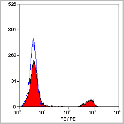
 Application Data
(Staining of human peripheral blood monocytes with Mouse anti Human CD14:RPE)
Application Data
(Staining of human peripheral blood monocytes with Mouse anti Human CD14:RPE)
CD14, Monoclonal Antibody (Cat# AAA12179)
Full Name
MOUSE ANTI HUMAN CD14
Applications
Immunohistochemistry, Flow Cytometry, Immunoprecipitation
Pricing
 IF (Immunofluorescence)
(AAA31274 staining Hela cells by IF/ICC. The samples were fixed with PFA and permeabilized in 0.1% Triton X-100,then blocked in 10% serum for 45 minutes at 25°C. Samples were then incubated with primary Ab(AAA31274 1:200) and mouse anti-beta tubulin Ab(T0023 1:200) for 1 hour at 37°C. An AlexaFluor594 conjugated goat anti-rabbit IgG(H+L) Ab(Red) and an AlexaFluor488 conjugated goat anti-mouse IgG(H+L) Ab(Green) were used as the secondary antibody.The nuclear counter stain is DAPI(blue).)
IF (Immunofluorescence)
(AAA31274 staining Hela cells by IF/ICC. The samples were fixed with PFA and permeabilized in 0.1% Triton X-100,then blocked in 10% serum for 45 minutes at 25°C. Samples were then incubated with primary Ab(AAA31274 1:200) and mouse anti-beta tubulin Ab(T0023 1:200) for 1 hour at 37°C. An AlexaFluor594 conjugated goat anti-rabbit IgG(H+L) Ab(Red) and an AlexaFluor488 conjugated goat anti-mouse IgG(H+L) Ab(Green) were used as the secondary antibody.The nuclear counter stain is DAPI(blue).)
TTC7A, Polyclonal Antibody (Cat# AAA31274)
Full Name
TTC7A Antibody
Gene Names
TTC7A; TTC7; GIDID; MINAT
Reactivity
Human, Mouse, Rat
Predicted Reactivity: Pig(88%), Bovine(88%), Horse(88%), Sheep(88%), Rabbit(100%), Dog(86%)
Predicted Reactivity: Pig(88%), Bovine(88%), Horse(88%), Sheep(88%), Rabbit(100%), Dog(86%)
Applications
ELISA
Purity
The antiserum was purified by peptide affinity chromatography using SulfoLink Coupling Resin (Thermo Fisher Scientific).
Pricing
 IF (Immunofluorescence)
(AAA31210 staining Hela cells by IF/ICC. The samples were fixed with PFA and permeabilized in 0.1% Triton X-100,then blocked in 10% serum for 45 minutes at 25°C. Samples were then incubated with primary Ab(AAA31210) and mouse anti-beta tubulin Ab(T0023) for 1 hour at 37°C. An AlexaFluor594 conjugated goat anti-rabbit IgG(H+L) Ab(Red) and an AlexaFluor488 conjugated goat anti-mouse IgG(H+L) Ab(Green) were used as the secondary antibody.The nuclear counter stain is DAPI(blue).)
IF (Immunofluorescence)
(AAA31210 staining Hela cells by IF/ICC. The samples were fixed with PFA and permeabilized in 0.1% Triton X-100,then blocked in 10% serum for 45 minutes at 25°C. Samples were then incubated with primary Ab(AAA31210) and mouse anti-beta tubulin Ab(T0023) for 1 hour at 37°C. An AlexaFluor594 conjugated goat anti-rabbit IgG(H+L) Ab(Red) and an AlexaFluor488 conjugated goat anti-mouse IgG(H+L) Ab(Green) were used as the secondary antibody.The nuclear counter stain is DAPI(blue).)
GLS2, Polyclonal Antibody (Cat# AAA31210)
Full Name
GLS2 Antibody
Gene Names
GLS2; GA; GLS; LGA; hLGA
Reactivity
Human, Mouse, Rat
Predicted Reactivity: Pig(86%), Horse(100%), Sheep(100%), Dog(88%)
Predicted Reactivity: Pig(86%), Horse(100%), Sheep(100%), Dog(88%)
Applications
ELISA
Purity
The antiserum was purified by peptide affinity chromatography using SulfoLink Coupling Resin (Thermo Fisher Scientific).
Pricing
 IF (Immunofluorescence)
(AAA31166 staining Hela cells by IF/ICC. The samples were fixed with PFA and permeabilized in 0.1% Triton X-100,then blocked in 10% serum for 45 minutes at 25°C. Samples were then incubated with primary Ab(AAA31166 1:200) and mouse anti-beta tubulin Ab(T0023 1:200) for 1 hour at 37°C. An AlexaFluor594 conjugated goat anti-rabbit IgG(H+L) Ab(Red) and an AlexaFluor488 conjugated goat anti-mouse IgG(H+L) Ab(Green) were used as the secondary antibody.The nuclear counter stain is DAPI(blue).)
IF (Immunofluorescence)
(AAA31166 staining Hela cells by IF/ICC. The samples were fixed with PFA and permeabilized in 0.1% Triton X-100,then blocked in 10% serum for 45 minutes at 25°C. Samples were then incubated with primary Ab(AAA31166 1:200) and mouse anti-beta tubulin Ab(T0023 1:200) for 1 hour at 37°C. An AlexaFluor594 conjugated goat anti-rabbit IgG(H+L) Ab(Red) and an AlexaFluor488 conjugated goat anti-mouse IgG(H+L) Ab(Green) were used as the secondary antibody.The nuclear counter stain is DAPI(blue).)
SC35, Polyclonal Antibody (Cat# AAA31166)
Full Name
SC35 Antibody
Gene Names
SRSF2; SC35; PR264; SC-35; SFRS2; SFRS2A; SRp30b
Reactivity
Human, Mouse, Rat
Predicted Reactivity: Pig(100%), Bovine(100%), Dog(100%), Chicken(100%), Xenopus(86%)
Predicted Reactivity: Pig(100%), Bovine(100%), Dog(100%), Chicken(100%), Xenopus(86%)
Applications
Immunofluorescence, Immunocytochemistry
Purity
The antiserum was purified by peptide affinity chromatography using SulfoLink Coupling Resin (Thermo Fisher Scientific).
Pricing

 IF (Immunofluorescence)
(AAA31271 staining Hela cells by IF/ICC. The samples were fixed with PFA and permeabilized in 0.1% Triton X-100,then blocked in 10% serum for 45 minutes at 25°C. Samples were then incubated with primary Ab(AAA31271 1:200) and mouse anti-beta tubulin Ab(T0023 1:200) for 1 hour at 37°C. An AlexaFluor594 conjugated goat anti-rabbit IgG(H+L) Ab(Red) and an AlexaFluor488 conjugated goat anti-mouse IgG(H+L) Ab(Green) were used as the secondary antibody.The nuclear counter stain is DAPI(blue).)
IF (Immunofluorescence)
(AAA31271 staining Hela cells by IF/ICC. The samples were fixed with PFA and permeabilized in 0.1% Triton X-100,then blocked in 10% serum for 45 minutes at 25°C. Samples were then incubated with primary Ab(AAA31271 1:200) and mouse anti-beta tubulin Ab(T0023 1:200) for 1 hour at 37°C. An AlexaFluor594 conjugated goat anti-rabbit IgG(H+L) Ab(Red) and an AlexaFluor488 conjugated goat anti-mouse IgG(H+L) Ab(Green) were used as the secondary antibody.The nuclear counter stain is DAPI(blue).)
Sts1, Polyclonal Antibody (Cat# AAA31271)
Full Name
Sts1 Antibody
Gene Names
UBASH3B; p70; STS1; STS-1; TULA2; TULA-2
Reactivity
Human, Mouse, Rat
Predicted Reactivity: Pig(100%), Bovine(100%), Horse(100%), Sheep(100%), Rabbit(100%), Dog(100%), Chicken(100%), Xenopus(100%)
Predicted Reactivity: Pig(100%), Bovine(100%), Horse(100%), Sheep(100%), Rabbit(100%), Dog(100%), Chicken(100%), Xenopus(100%)
Applications
ELISA
Purity
The antiserum was purified by peptide affinity chromatography using SulfoLink Coupling Resin (Thermo Fisher Scientific).
Pricing
 IF (Immunofluorescence)
(AAA31232 staining Hela cells by IF/ICC. The samples were fixed with PFA and permeabilized in 0.1% Triton X-100,then blocked in 10% serum for 45 minutes at 25°C. Samples were then incubated with primary Ab(AAA31232 1:200) and mouse anti-beta tubulin Ab(T0023 1:200) for 1 hour at 37°C. An AlexaFluor594 conjugated goat anti-rabbit IgG(H+L) Ab(Red) and an AlexaFluor488 conjugated goat anti-mouse IgG(H+L) Ab(Green) were used as the secondary antibody.The nuclear counter stain is DAPI(blue).)
IF (Immunofluorescence)
(AAA31232 staining Hela cells by IF/ICC. The samples were fixed with PFA and permeabilized in 0.1% Triton X-100,then blocked in 10% serum for 45 minutes at 25°C. Samples were then incubated with primary Ab(AAA31232 1:200) and mouse anti-beta tubulin Ab(T0023 1:200) for 1 hour at 37°C. An AlexaFluor594 conjugated goat anti-rabbit IgG(H+L) Ab(Red) and an AlexaFluor488 conjugated goat anti-mouse IgG(H+L) Ab(Green) were used as the secondary antibody.The nuclear counter stain is DAPI(blue).)
SMARCA1, Polyclonal Antibody (Cat# AAA31232)
Full Name
SMARCA1 Antibody
Gene Names
SMARCA1; SWI; ISWI; SWI2; SNF2L; SNF2L1; SNF2LB; SNF2LT; hSNF2L; NURF140
Reactivity
Human, Mouse, Rat
Predicted Reactivity: Pig(100%), Zebrafish(100%), Bovine(100%), Horse(100%), Sheep(100%), Rabbit(100%), Dog(100%), Chicken(100%)
Predicted Reactivity: Pig(100%), Zebrafish(100%), Bovine(100%), Horse(100%), Sheep(100%), Rabbit(100%), Dog(100%), Chicken(100%)
Applications
ELISA
Purity
The antiserum was purified by peptide affinity chromatography using SulfoLink Coupling Resin (Thermo Fisher Scientific).
Pricing

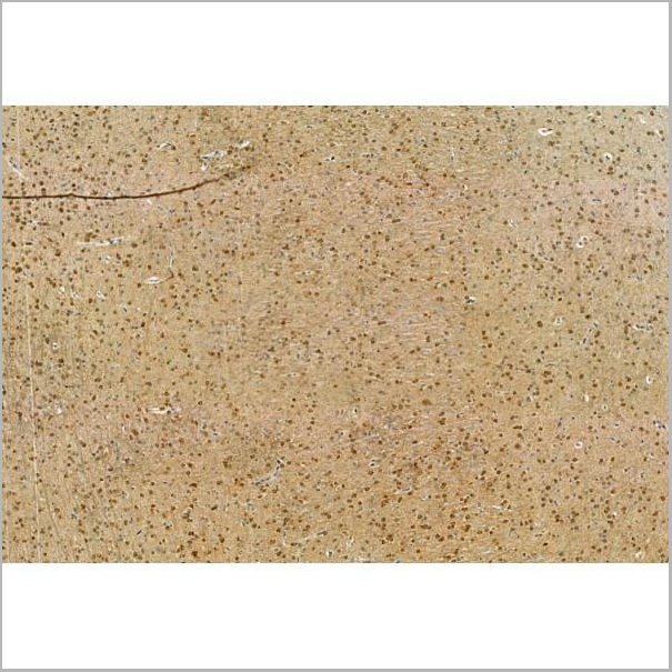
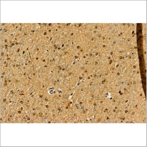
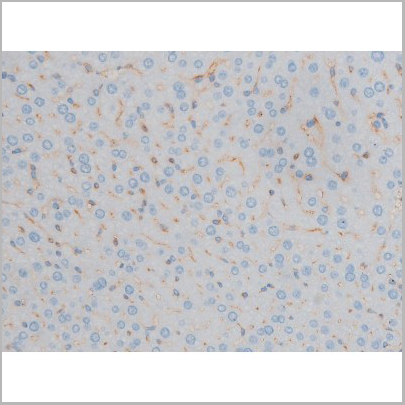
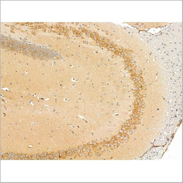
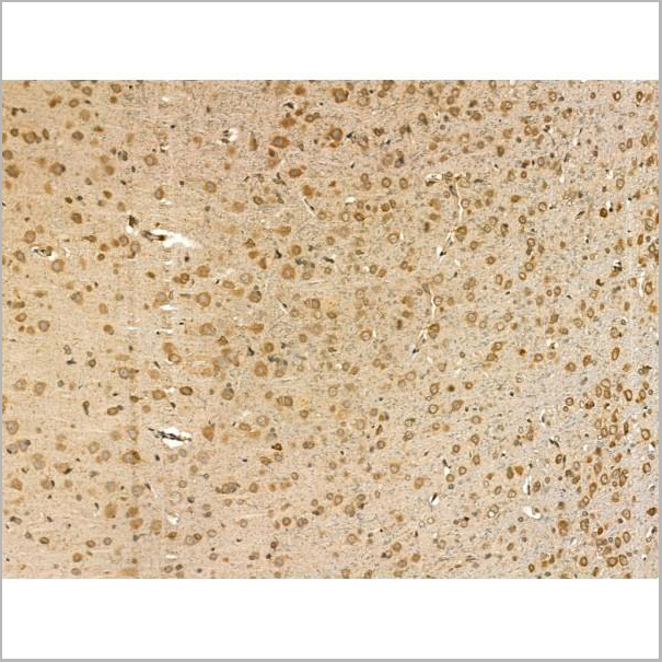
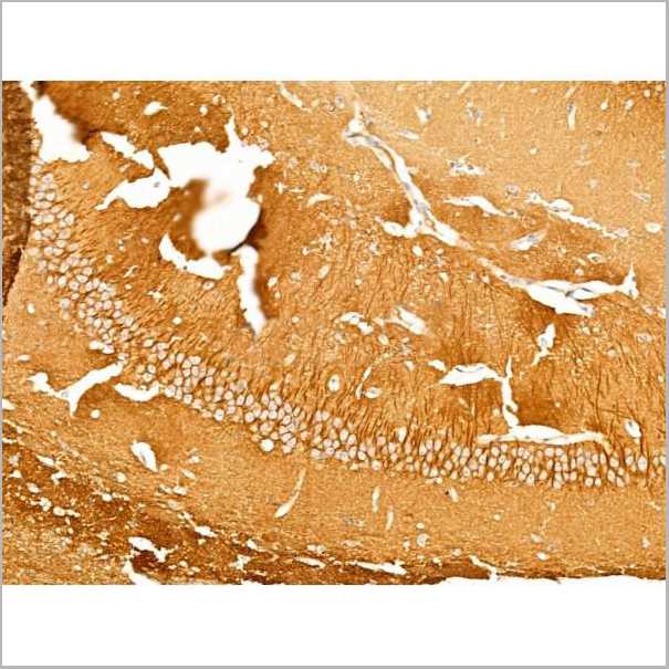
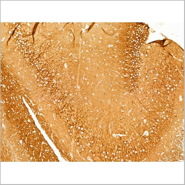
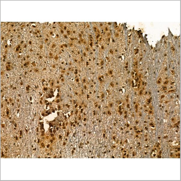
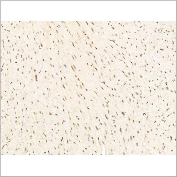
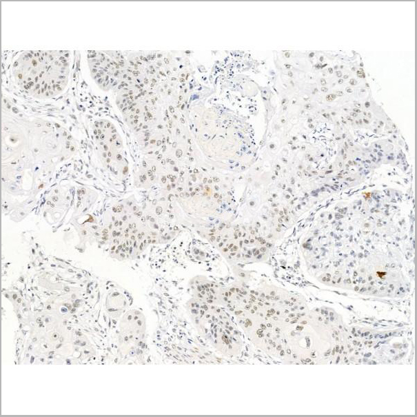
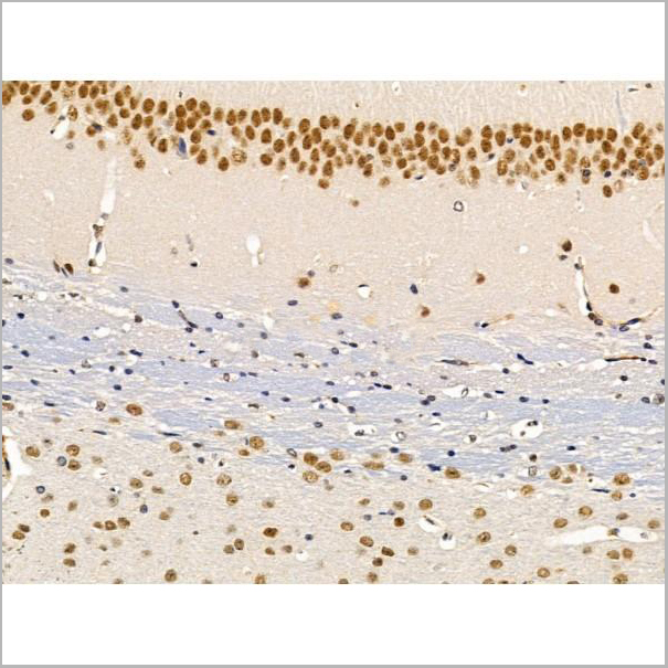
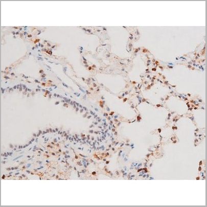
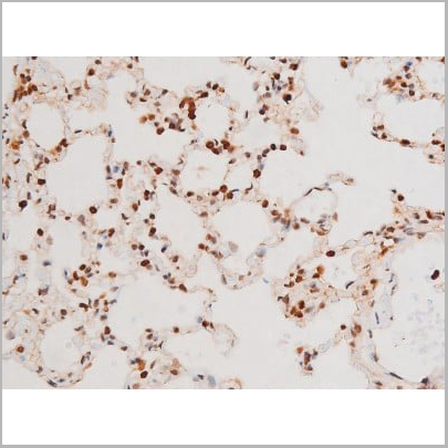
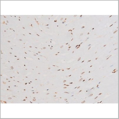
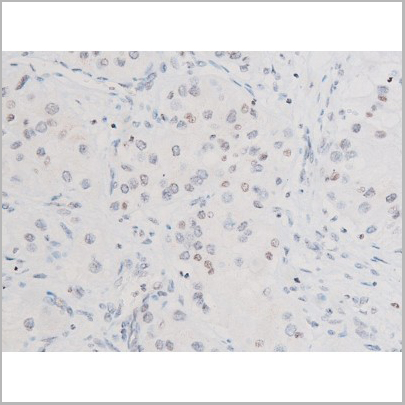
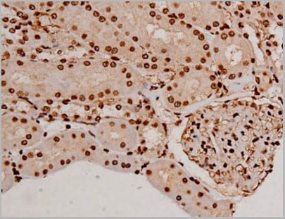
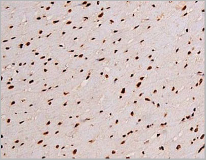
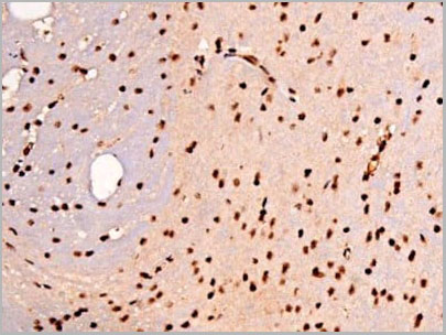
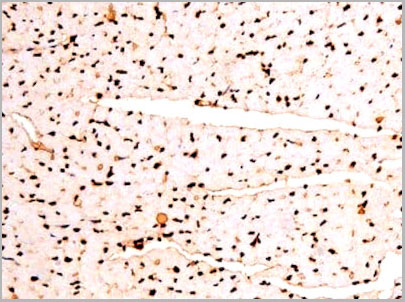
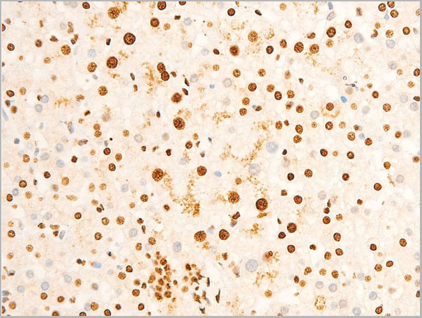
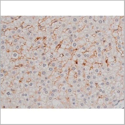
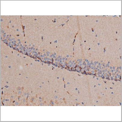
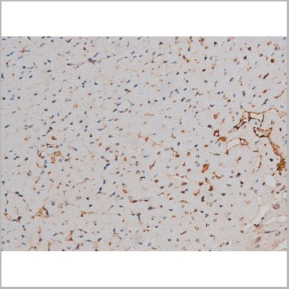
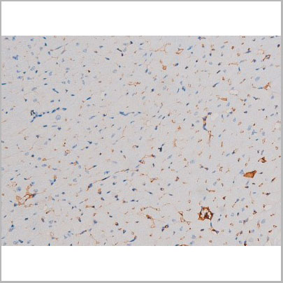
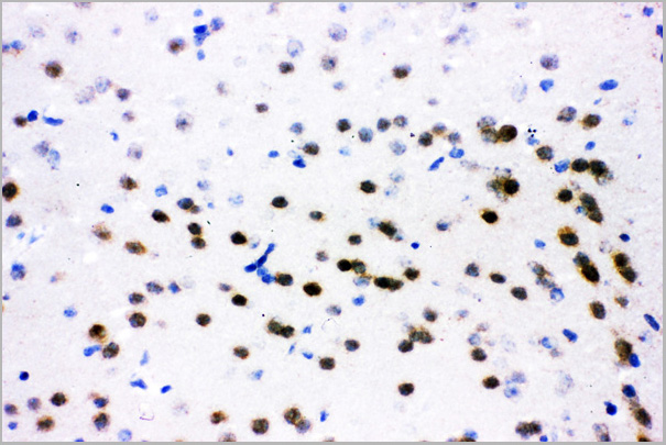
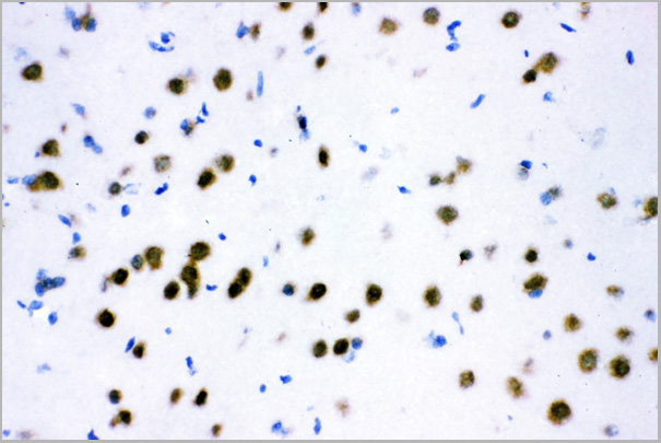
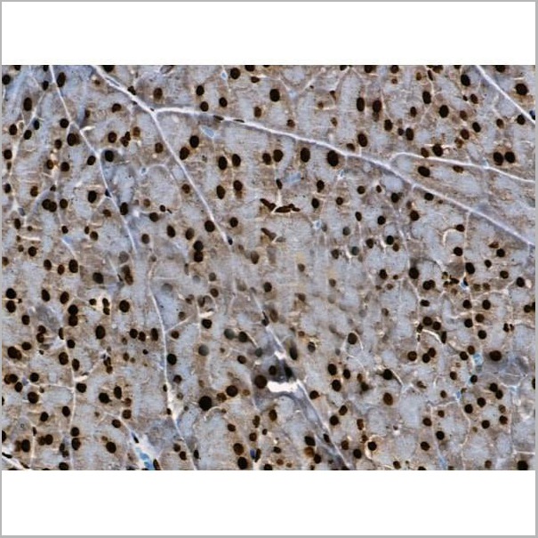
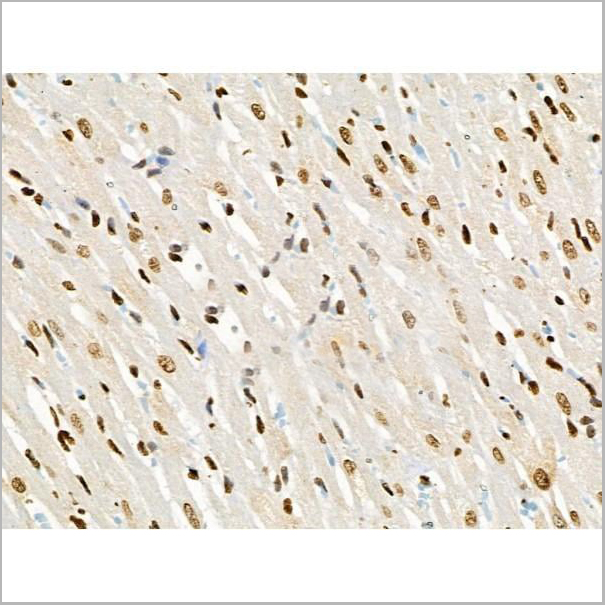
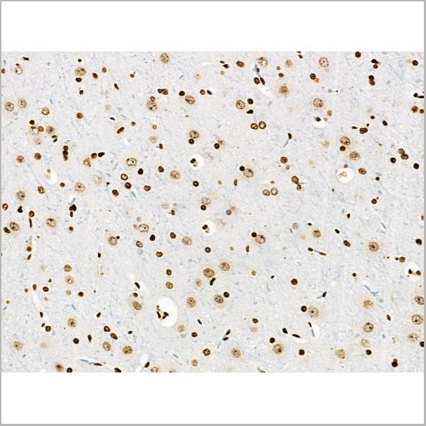
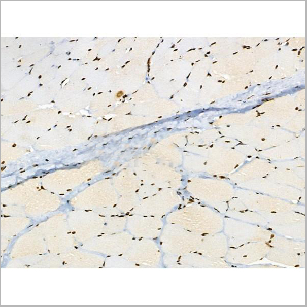
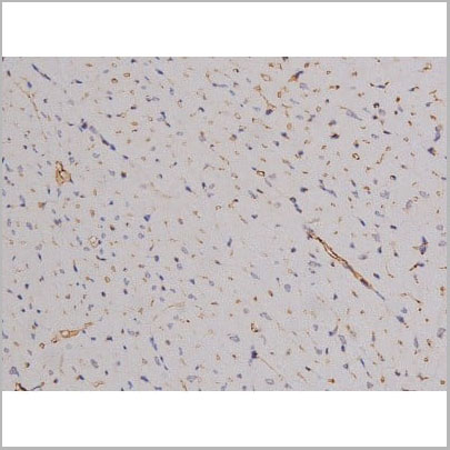
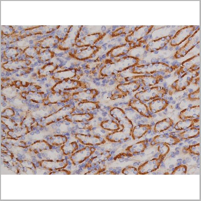
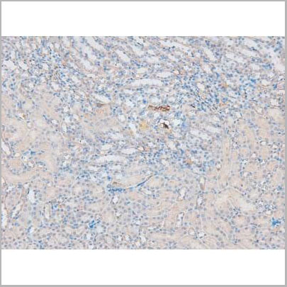
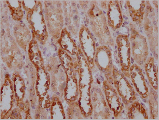
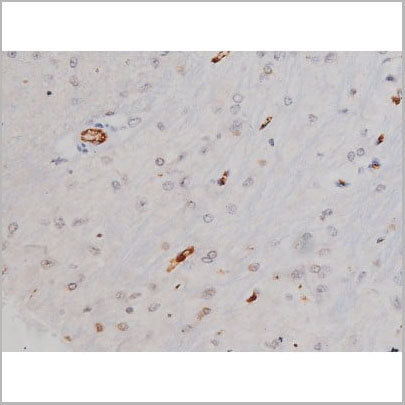 IHC (Immunohistochemistry)
(AAA31004 at 1/200 staining Rat brain tissue sections by IHC-P. The tissue was formaldehyde fixed and a heat mediated antigen retrieval step in citrate buffer was performed. The tissue was then blocked and incubated with the antibody for 1.5 hours at 22 degree C. An HRP conjugated goat anti-rabbit antibody was used as the secondary.)
IHC (Immunohistochemistry)
(AAA31004 at 1/200 staining Rat brain tissue sections by IHC-P. The tissue was formaldehyde fixed and a heat mediated antigen retrieval step in citrate buffer was performed. The tissue was then blocked and incubated with the antibody for 1.5 hours at 22 degree C. An HRP conjugated goat anti-rabbit antibody was used as the secondary.)
Tau, Polyclonal Antibody (Cat# AAA31004)
Full Name
Phospho-Tau (Thr205) Antibody
Gene Names
MAPT; TAU; MSTD; PPND; DDPAC; MAPTL; MTBT1; MTBT2; FTDP-17; PPP1R103
Reactivity
Human, Mouse, Rat
Applications
Western Blot, Immunohistochemistry
Purity
From purified rabbit serum by affinity purification via sequential chromatography on phospho-and non-phospho-peptide affinity columns.
Pricing
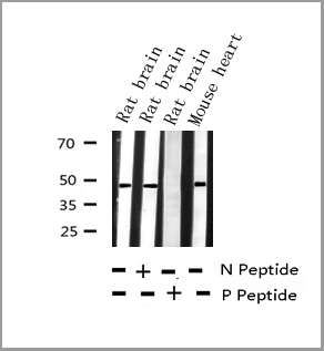
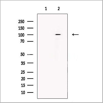
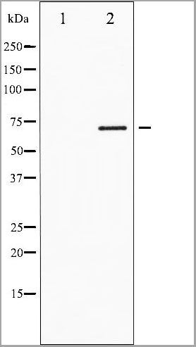
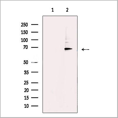
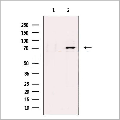
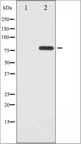
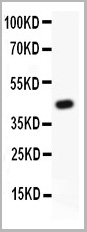
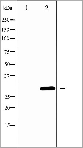
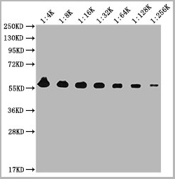
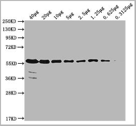
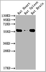
 WB (Western Blot)
(Western blot analysis of Phospho-SYK (Tyr525) expression in various lysates)
WB (Western Blot)
(Western blot analysis of Phospho-SYK (Tyr525) expression in various lysates)
SYK, Polyclonal Antibody (Cat# AAA31052)
Full Name
Phospho-SYK (Tyr525) Antibody
Gene Names
SYK; p72-Syk
Reactivity
Human, Mouse, Rat, Monkey
Applications
Western Blot, Immunohistochemistry, Immunofluorescence, Immunocytochemistry
Purity
From purified rabbit serum by affinity purification via sequential chromatography on phospho-and non-phospho-peptide affinity columns.
Pricing
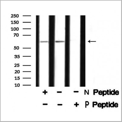
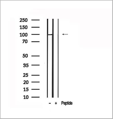
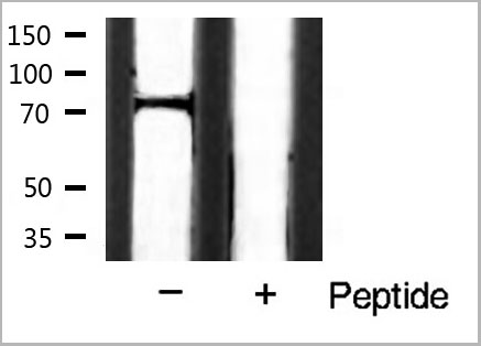
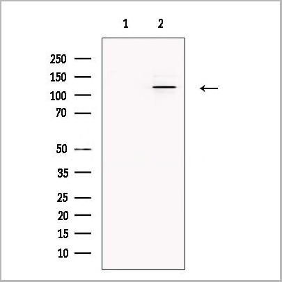
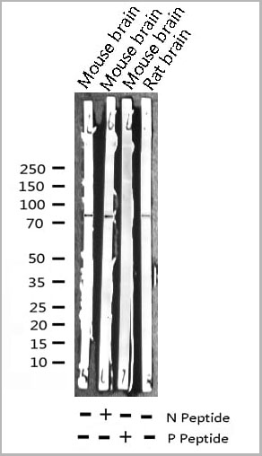
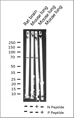
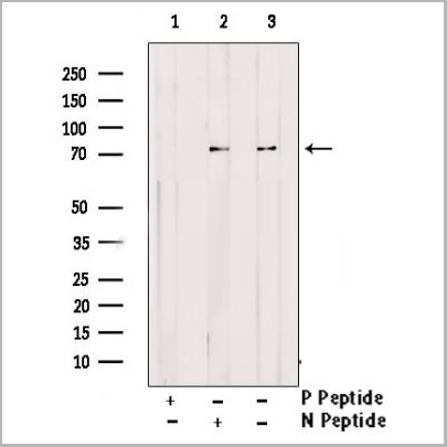
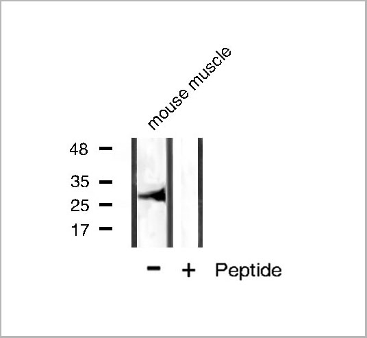
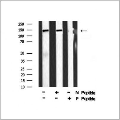
 WB (Western Blot)
(Western blot analysis of FAK phosphorylation expression in EGF treated 293 whole cell lysates, The lane on the left is treated with the antigen-specific peptide.)
WB (Western Blot)
(Western blot analysis of FAK phosphorylation expression in EGF treated 293 whole cell lysates, The lane on the left is treated with the antigen-specific peptide.)
FAK, Polyclonal Antibody (Cat# AAA31067)
Full Name
Phospho-FAK (Tyr397) Antibody
Gene Names
PTK2; FAK; FADK; FAK1; FRNK; PPP1R71; p125FAK; pp125FAK
Reactivity
Human, Mouse, Rat
Applications
Western Blot, Immunohistochemistry, Immunofluorescence, Immunocytochemistry
Purity
From purified rabbit serum by affinity purification via sequential chromatography on phospho-and non-phospho-peptide affinity columns.
Pricing
 WB (Western Blot)
(Western blot analysis of eIF2 alpha phosphorylation expression in IFN-alpha treated K562 whole cell lysates, The lane on the left is treated with the antigen-specific peptide.)
WB (Western Blot)
(Western blot analysis of eIF2 alpha phosphorylation expression in IFN-alpha treated K562 whole cell lysates, The lane on the left is treated with the antigen-specific peptide.)
eIF2 alpha, Polyclonal Antibody (Cat# AAA30978)
Full Name
Phospho-eIF2 alpha (Ser51) Antibody
Gene Names
EIF2S1; EIF2; EIF-2; EIF2A; EIF-2A; EIF-2alpha
Reactivity
Human, Mouse, Rat
Applications
Western Blot, Immunohistochemistry, Immunofluorescence, Immunocytochemistry
Purity
From purified rabbit serum by affinity purification via sequential chromatography on phospho-and non-phospho-peptide affinity columns.
Pricing
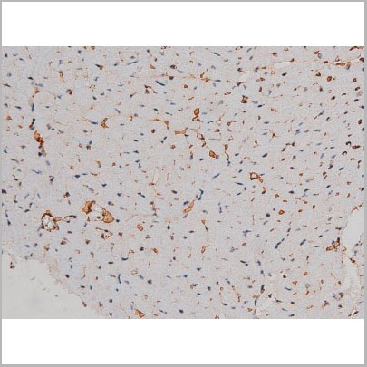
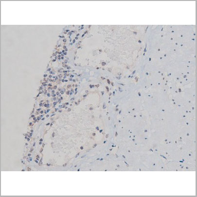
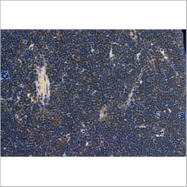
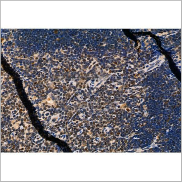
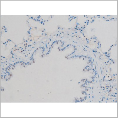
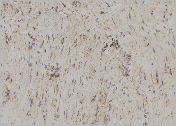
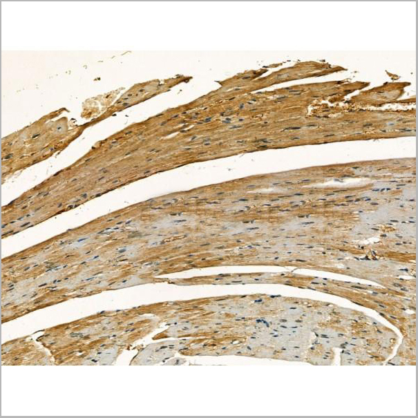
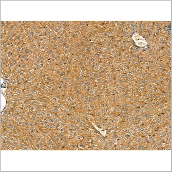
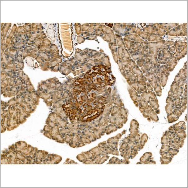
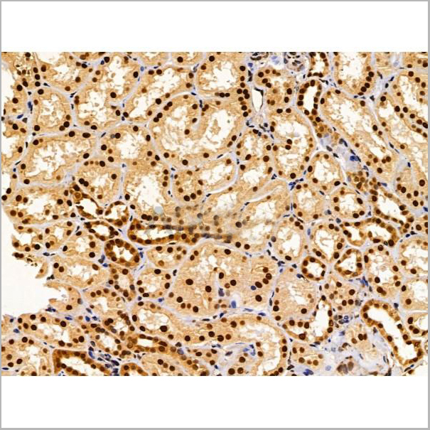
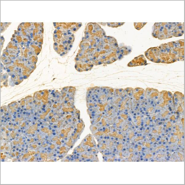
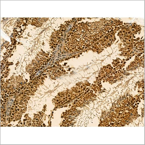
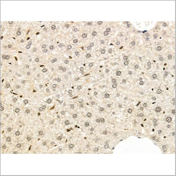
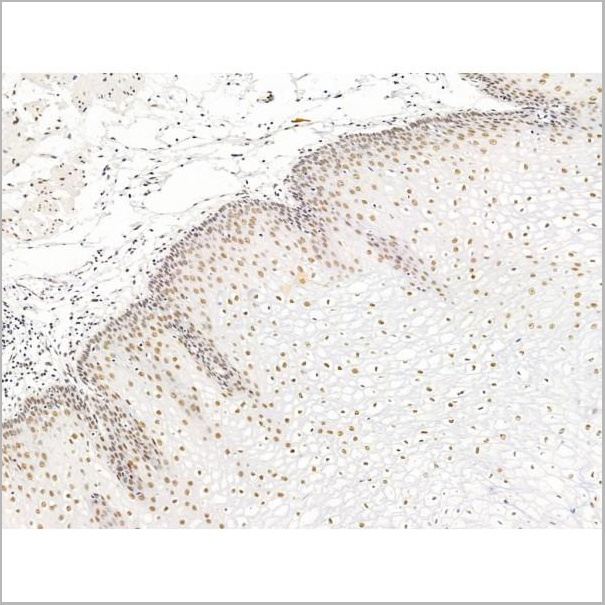
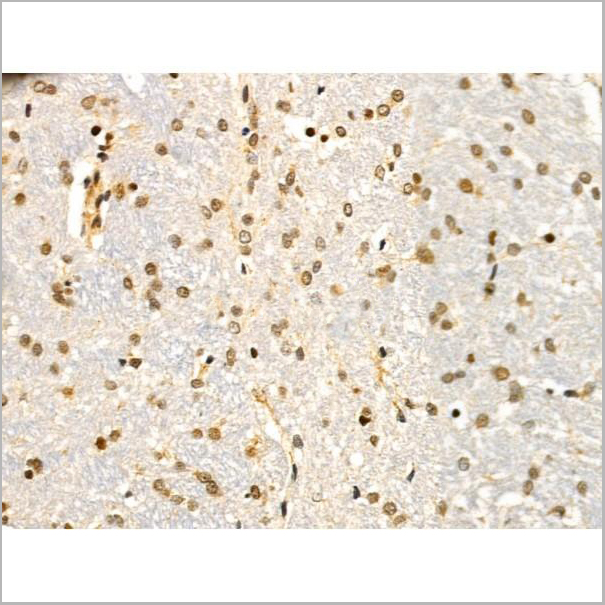
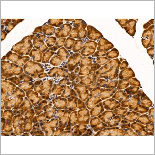
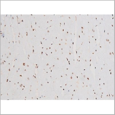
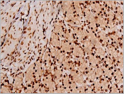
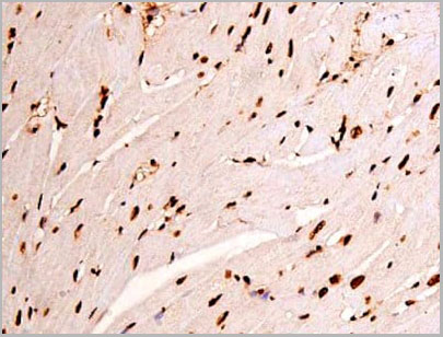
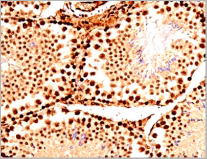
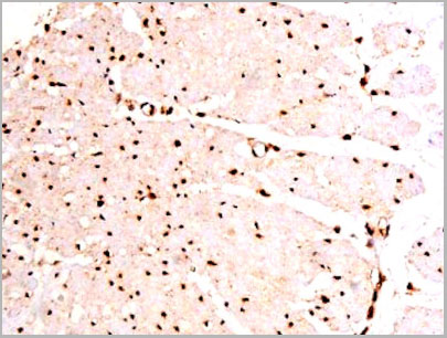
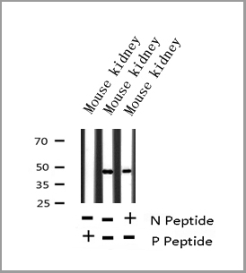
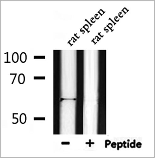
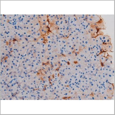
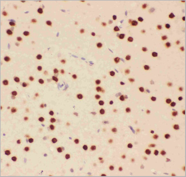
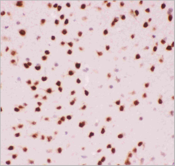
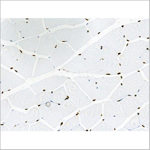
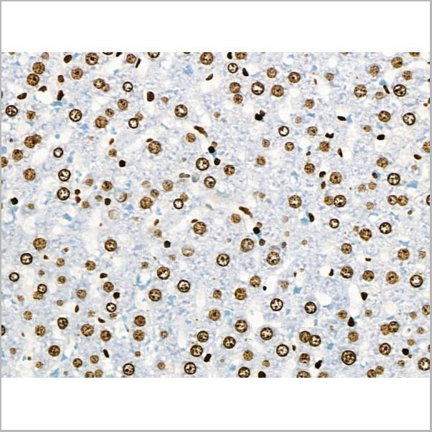
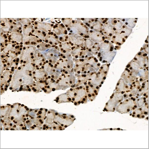
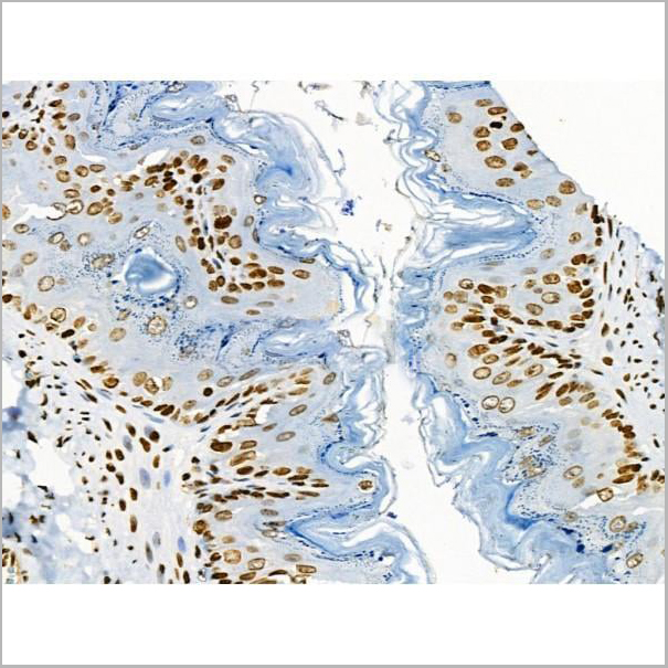
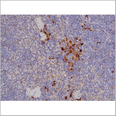
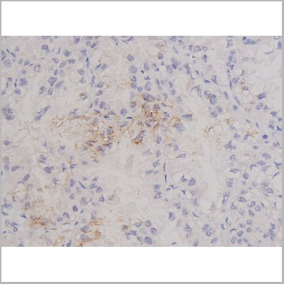
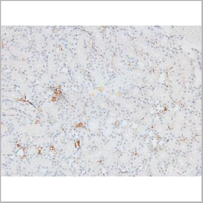
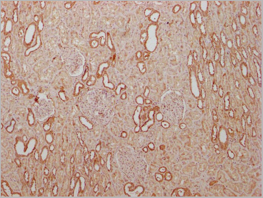
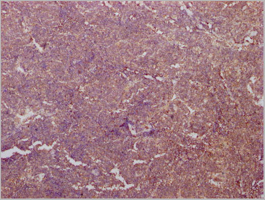
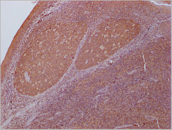
 IHC (Immunohistochemistry)
(AAA31023 at 1/200 staining Rat spleen tissue sections by IHC-P. The tissue was formaldehyde fixed and a heat mediated antigen retrieval step in citrate buffer was performed. The tissue was then blocked and incubated with the antibody for 1.5 hours at 22 degree C. An HRP conjugated goat anti-rabbit antibody was used as the secondary.)
IHC (Immunohistochemistry)
(AAA31023 at 1/200 staining Rat spleen tissue sections by IHC-P. The tissue was formaldehyde fixed and a heat mediated antigen retrieval step in citrate buffer was performed. The tissue was then blocked and incubated with the antibody for 1.5 hours at 22 degree C. An HRP conjugated goat anti-rabbit antibody was used as the secondary.)
p70 S6 Kinase, Polyclonal Antibody (Cat# AAA31023)
Full Name
Phospho-p70 S6 Kinase (Thr229) Antibody
Gene Names
RPS6KB1; S6K; PS6K; S6K1; STK14A; p70-S6K; p70 S6KA; p70-alpha; S6K-beta-1; p70(S6K)-alpha
Reactivity
Human, Mouse, Rat
Applications
Western Blot, Immunohistochemistry, Immunofluorescence, Immunocytochemistry
Purity
From purified rabbit serum by affinity purification via sequential chromatography on phospho-and non-phospho-peptide affinity columns.
Pricing

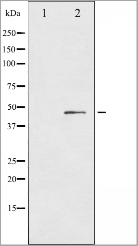
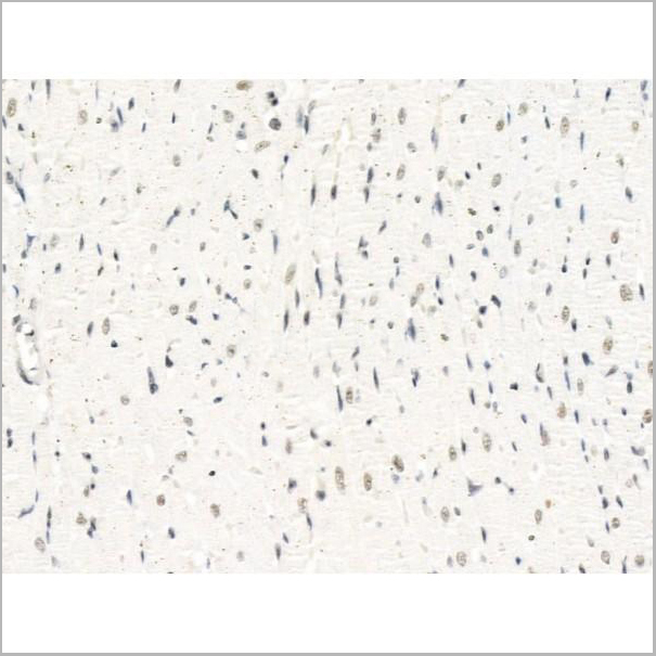
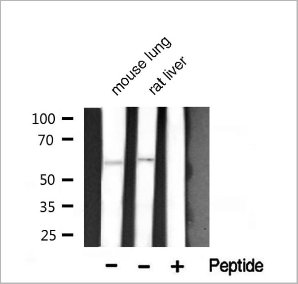
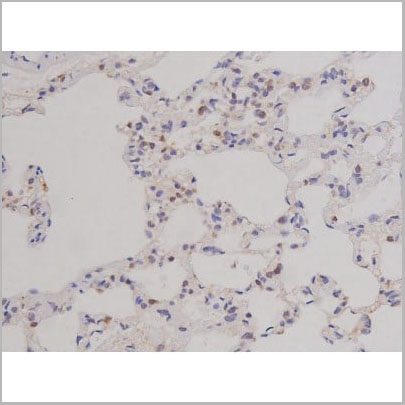
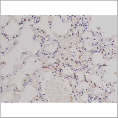
 WB (Western Blot)
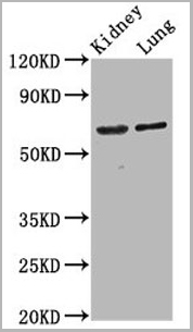
(Western blot analysis of Phospho-IL-8R beta/CDw128 beta (Ser347) expression in Mouse kidney lysate)
WB (Western Blot)
(Western blot analysis of Phospho-IL-8R beta/CDw128 beta (Ser347) expression in Mouse kidney lysate)
IL-8R beta/CDw128 beta, Polyclonal Antibody (Cat# AAA31026)
Full Name
Phospho-IL-8R beta/CDw128 beta (Ser347) Antibody
Gene Names
CXCR2; CD182; IL8R2; IL8RA; IL8RB; CMKAR2; CDw128b
Reactivity
Human, Mouse, Monkey
Applications
Western Blot, Immunohistochemistry, Immunofluorescence, Immunocytochemistry
Purity
From purified rabbit serum by affinity purification via sequential chromatography on phospho-and non-phospho-peptide affinity columns.
Pricing
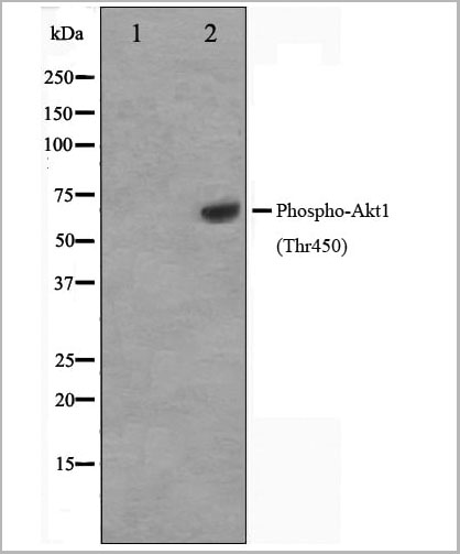
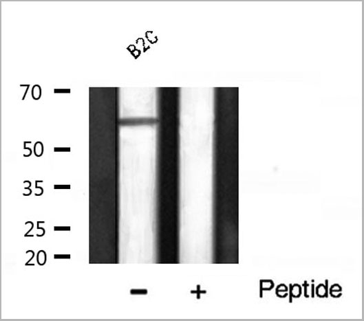 WB (Western Blot)
(Western blot analysis of Akt1 phosphorylation expression in B2C whole cell lysates, The lane on the right is treated with the antigen-specific peptide.)
WB (Western Blot)
(Western blot analysis of Akt1 phosphorylation expression in B2C whole cell lysates, The lane on the right is treated with the antigen-specific peptide.)
Akt1, Polyclonal Antibody (Cat# AAA31034)
Full Name
Phospho-Akt1 (Thr450) Antibody
Gene Names
AKT1; AKT; PKB; RAC; CWS6; PRKBA; PKB-ALPHA; RAC-ALPHA
Reactivity
Human, Mouse, Rat
Applications
Western Blot, Immunohistochemistry, Immunofluorescence, Immunocytochemistry
Purity
From purified rabbit serum by affinity purification via sequential chromatography on phospho-and non-phospho-peptide affinity columns.
Pricing
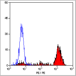
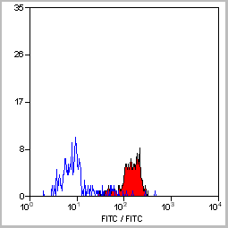
 FCM (Flow Cytometry)
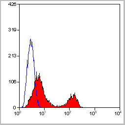
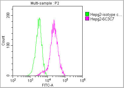
(Figure 8. Flow Cytometry analysis of HepG2 cells using anti-ATF2 antibody (AAA11634).Overlay histogram showing HepG2 cells stained with AAA11634 (Blue line).The cells were blocked with 10% normal goat serum. And then incubated with rabbit anti-ATF2 Antibody (AAA11634,1ug/1x10^6 cells) for 30 min at 20 degree C. DyLight ®488 conjugated goat anti-rabbit IgG (5-10ug/1x10^6 cells) was used as secondary antibody for 30 minutes at 20 degree C. Isotype control antibody (Green line) was rabbit IgG (1ug/1x106) used under the same conditions. Unlabelled sample (Red line) was also used as a control.)
FCM (Flow Cytometry)
(Figure 8. Flow Cytometry analysis of HepG2 cells using anti-ATF2 antibody (AAA11634).Overlay histogram showing HepG2 cells stained with AAA11634 (Blue line).The cells were blocked with 10% normal goat serum. And then incubated with rabbit anti-ATF2 Antibody (AAA11634,1ug/1x10^6 cells) for 30 min at 20 degree C. DyLight ®488 conjugated goat anti-rabbit IgG (5-10ug/1x10^6 cells) was used as secondary antibody for 30 minutes at 20 degree C. Isotype control antibody (Green line) was rabbit IgG (1ug/1x106) used under the same conditions. Unlabelled sample (Red line) was also used as a control.)
Cyclic AMP-dependent transcription factor ATF-2, Polyclonal Antibody (Cat# AAA11634)
Full Name
Anti-ATF2 Antibody
Gene Names
ATF2; HB16; CREB2; TREB7; CREB-2; CRE-BP1
Reactivity
Human, Mouse, Rat. No cross reactivity with other proteins.
Applications
Western Blot, Immunohistochemistry, Immunohistochemistry
Purity
Immunogen affinity purified.
Pricing

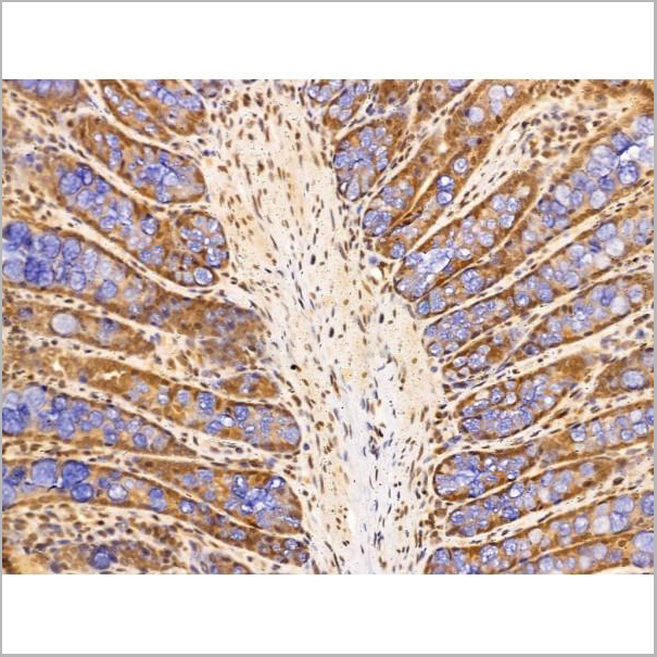
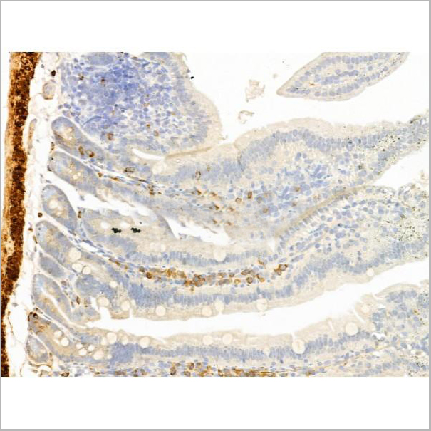
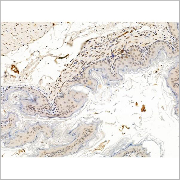
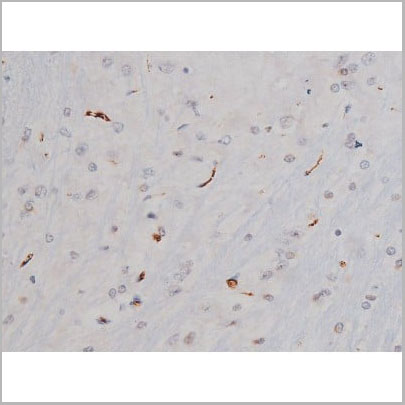
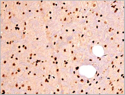
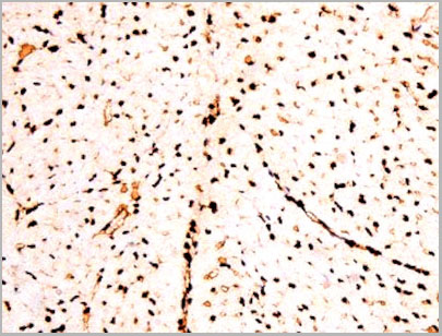
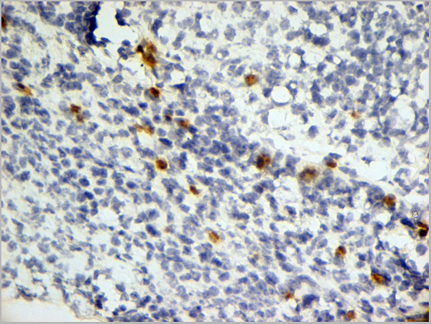
 IHC (Immunohistochemistry)
(At 1/100 staining Mouse stomach tissue by IHC-P. The sample was formaldehyde fixed and a heat mediated antigen retrieval step in citrate buffer was performed. The sample was then blocked and incubated with the primary antibody at 4 degree C overnight. An HRP conjugated anti-Rabbit antibody was used as the secondary antibody.)
IHC (Immunohistochemistry)
(At 1/100 staining Mouse stomach tissue by IHC-P. The sample was formaldehyde fixed and a heat mediated antigen retrieval step in citrate buffer was performed. The sample was then blocked and incubated with the primary antibody at 4 degree C overnight. An HRP conjugated anti-Rabbit antibody was used as the secondary antibody.)
BRK, Polyclonal Antibody (Cat# AAA31371)
Full Name
Phospho-BRK (Tyr447) Antibody
Gene Names
PTK6; BRK
Reactivity
Human, Mouse, Rat
Predicted Reactivity: Horse (89%)
Predicted Reactivity: Horse (89%)
Applications
Western Blot, Immunohistochemistry, Peptide ELISA
Purity
The antibody is from purified rabbit serum by affinity purification via sequential chromatography on phospho-peptide and non-phospho-peptide affinity columns.
Pricing
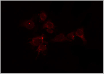
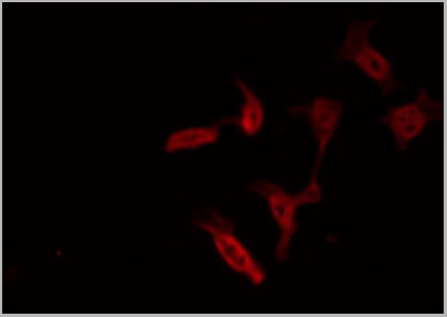
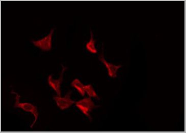
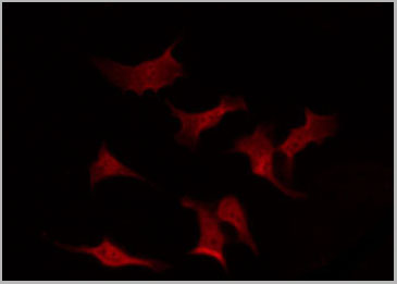
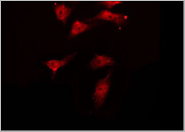
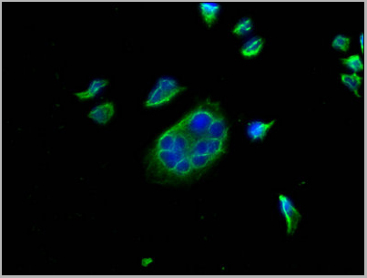
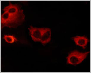 IF (Immunofluorescence)
(AAA31068 staining NIH/3T3 cells by ICC/IF. Cells were fixed with PFA and permeabilized in 0.1% saponin prior to blocking in 10% serum for 45 minutes at 37 degree C. The primary antibody was diluted 1/400 and incubated with the sample for 1 hour at 37 degree C. A Alexa Fluor 594 conjugated goat polyclonal to rabbit IgG (H+L), diluted 1/600 was used as secondary antibody.)
IF (Immunofluorescence)
(AAA31068 staining NIH/3T3 cells by ICC/IF. Cells were fixed with PFA and permeabilized in 0.1% saponin prior to blocking in 10% serum for 45 minutes at 37 degree C. The primary antibody was diluted 1/400 and incubated with the sample for 1 hour at 37 degree C. A Alexa Fluor 594 conjugated goat polyclonal to rabbit IgG (H+L), diluted 1/600 was used as secondary antibody.)
BCL-XL, Polyclonal Antibody (Cat# AAA31068)
Full Name
Phospho-BCL-XL (Thr47) Antibody
Gene Names
BCL2L1; BCLX; BCL2L; Bcl-X; PPP1R52; BCL-XL/S
Reactivity
Human, Mouse, Rat
Applications
Western Blot, Immunohistochemistry, Immunofluorescence, Immunocytochemistry
Purity
From purified rabbit serum by affinity purification via sequential chromatography on phospho-and non-phospho-peptide affinity columns.
Pricing
 Application Data
(At 25 degree C. The primary antibody was diluted at 1/200 and incubated with the sample for 1 hour at 37 degree C. An Alexa Fluor 594 conjugated goat anti-rabbit IgG (H+L) Ab, diluted at 1/600, was used as the secondary antibody.)
Application Data
(At 25 degree C. The primary antibody was diluted at 1/200 and incubated with the sample for 1 hour at 37 degree C. An Alexa Fluor 594 conjugated goat anti-rabbit IgG (H+L) Ab, diluted at 1/600, was used as the secondary antibody.)
eNOS, Polyclonal Antibody (Cat# AAA31394)
Full Name
Phospho-eNOS (Ser1179) Antibody
Gene Names
NOS3; eNOS; ECNOS
Reactivity
Human, Mouse, Rat
Predicted Reactivity: Pig (100%), Bovine (100%), Rabbit (100%), Dog (100%)
Predicted Reactivity: Pig (100%), Bovine (100%), Rabbit (100%), Dog (100%)
Applications
Western Blot, Immunohistochemistry, Immunofluorescence, Immunocytochemistry, Peptide ELISA
Purity
The antibody is from purified rabbit serum by affinity purification via sequential chromatography on phospho-peptide and non-phospho-peptide affinity columns.
Pricing
RAD50, Polyclonal Antibody (Cat# AAA31432)
Full Name
Phospho-RAD50 (Ser635) Antibody
Gene Names
RAD50; NBSLD; RAD502; hRad50
Reactivity
Human, Mouse, Rat
Applications
Western Blot, Immunohistochemistry
Purity
The antibody is from purified rabbit serum by affinity purification via sequential chromatography on phospho-peptide and non-phospho-peptide affinity columns.
Pricing
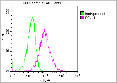
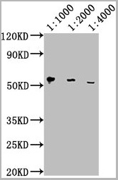
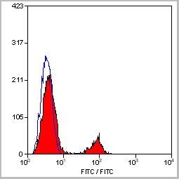
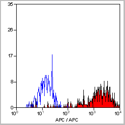
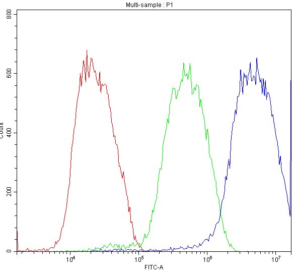
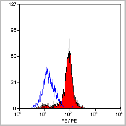
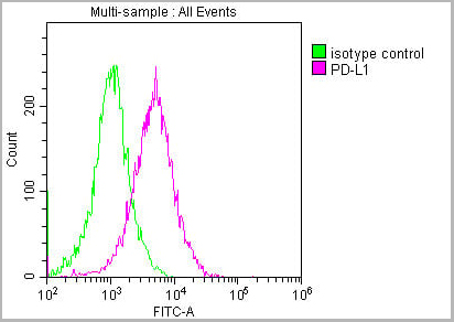 FCM (Flow Cytometry)
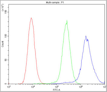
(Overlay histogram showing Hela cells stained with AAA27016 (red line) at 1:150. The cells were incubated in 1x PBS /10% normal goat serum to block non-specific protein-protein interactions followed by primary antibody for 1 h at 4 degree C. The secondary antibody used was FITC goat anti-mouse IgG(H+L) at 1/200 dilution for 1 h at 4 degree C. Isotype control antibody (green line) was used under the same conditions. Acquisition of >10,000 events was performed.)
FCM (Flow Cytometry)
(Overlay histogram showing Hela cells stained with AAA27016 (red line) at 1:150. The cells were incubated in 1x PBS /10% normal goat serum to block non-specific protein-protein interactions followed by primary antibody for 1 h at 4 degree C. The secondary antibody used was FITC goat anti-mouse IgG(H+L) at 1/200 dilution for 1 h at 4 degree C. Isotype control antibody (green line) was used under the same conditions. Acquisition of >10,000 events was performed.)
PD-L1, Monoclonal Antibody (Cat# AAA27016)
Full Name
PD-L1 Monoclonal Antibody
Gene Names
CD274; B7-H; B7H1; PDL1; PD-L1; PDCD1L1; PDCD1LG1
Reactivity
Human
Applications
Western Blot, Immunohistochemistry, Immunofluorescence, Flow Cytometry
Purity
>95%, Protein G purified
Pricing
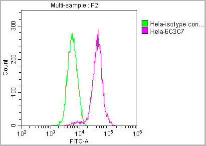
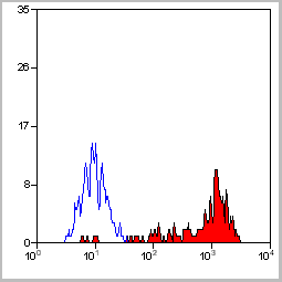
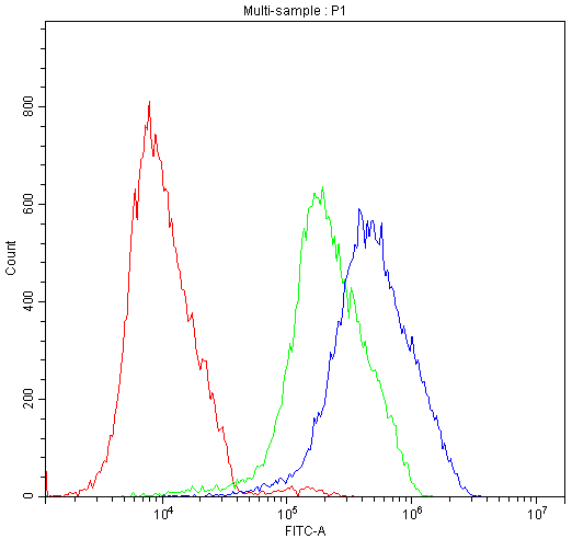
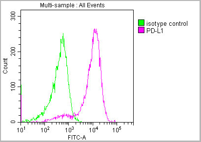
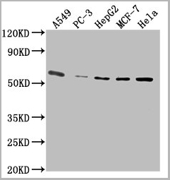
 FCM (Flow Cytometry)
(Overlay histogram showing HepG2 cells stained with AAA27041 (red line) at 1:100. The cells were incubated in 1x PBS /10% normal goat serum to block non-specific protein-protein interactions followed by primary antibody for 1 h at 4 degree C. The secondary antibody used was FITC goat anti-mouse IgG(H+L) at 1/200 dilution for 1 h at 4 degree C. Isotype control antibody (green line) was used under the same conditions. Acquisition of >10,000 events was performed.)
FCM (Flow Cytometry)
(Overlay histogram showing HepG2 cells stained with AAA27041 (red line) at 1:100. The cells were incubated in 1x PBS /10% normal goat serum to block non-specific protein-protein interactions followed by primary antibody for 1 h at 4 degree C. The secondary antibody used was FITC goat anti-mouse IgG(H+L) at 1/200 dilution for 1 h at 4 degree C. Isotype control antibody (green line) was used under the same conditions. Acquisition of >10,000 events was performed.)
PKM, Monoclonal Antibody (Cat# AAA27041)
Full Name
PKM Monoclonal Antibody
Reactivity
Human, Rat, Mouse, Rabbit
Applications
Western Blot, Immunohistochemistry, Immunofluorescence, Flow Cytometry, Immunoprecipitation
Purity
>95%, Protein G purified
Pricing
 FCM (Flow Cytometry)
(Figure 6. Flow Cytometry analysis of A549 cells using anti-MDR3 antibody (AAA11652).Overlay histogram showing A549 cells stained with AAA11652 (Blue line).The cells were blocked with 10% normal goat serum. And then incubated with rabbit anti-MDR3 Antibody (AAA11652,1ug/1x106 cells) for 30 min at 20°C. DyLight®488 conjugated goat anti-rabbit IgG (5-10ug/1x106 cells) was used as secondary antibody for 30 minutes at 20°C. Isotype control antibody (Green line) was rabbit IgG (1ug/1x106) used under the same conditions. Unlabelled sample (Red line) was also used as a control.)
FCM (Flow Cytometry)
(Figure 6. Flow Cytometry analysis of A549 cells using anti-MDR3 antibody (AAA11652).Overlay histogram showing A549 cells stained with AAA11652 (Blue line).The cells were blocked with 10% normal goat serum. And then incubated with rabbit anti-MDR3 Antibody (AAA11652,1ug/1x106 cells) for 30 min at 20°C. DyLight®488 conjugated goat anti-rabbit IgG (5-10ug/1x106 cells) was used as secondary antibody for 30 minutes at 20°C. Isotype control antibody (Green line) was rabbit IgG (1ug/1x106) used under the same conditions. Unlabelled sample (Red line) was also used as a control.)
ABCB4, Polyclonal Antibody (Cat# AAA11652)
Full Name
Anti-ABCB4 Antibody
Gene Names
ABCB4; GBD1; ICP3; MDR2; MDR3; PGY3; ABC21; MDR2/3; PFIC-3
Reactivity
Human, Mouse, Rat
Applications
Western Blot, Immunohistochemistry, Immunofluorescence, Flow Cytometry
Purity
Immunogen Affinity Purified
Pricing
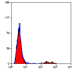
 Application Data
(Staining of mouse peritoneal macrophages with Rat anti Mouse Beta-glucan Receptor: FITC)
Application Data
(Staining of mouse peritoneal macrophages with Rat anti Mouse Beta-glucan Receptor: FITC)
DECTIN-1, Monoclonal Antibody (Cat# AAA12127)
Full Name
RAT ANTI MOUSE DECTIN-1:Biotin
Gene Names
Clec7a; BGR; beta-GR; Clecsf12
Applications
Flow Cytometry
Pricing
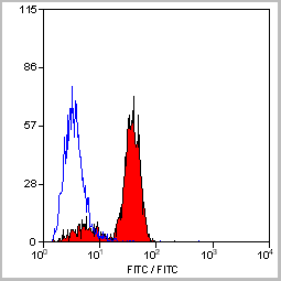
 FCM (Flow Cytometry)
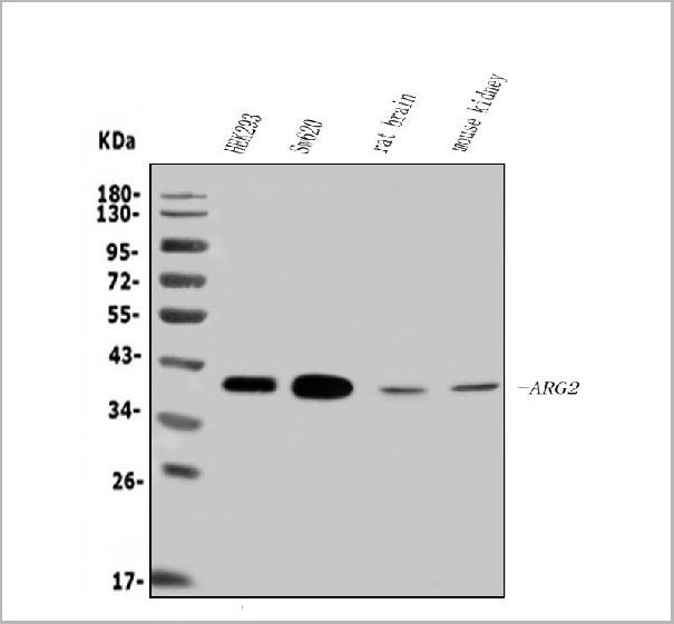
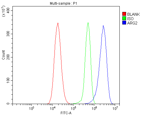
(Figure 7. Flow Cytometry analysis of 293T cells using anti-ARG2 antibody (AAA19256).Overlay histogram showing 293T cells stained with AAA19256 (Blue line). The cells were blocked with 10% normal goat serum. And then incubated with rabbit anti-ARG2 Antibody (AAA19256, 1μg/1x106 cells) for 30 min at 20 degree C. DyLight®488 conjugated goat anti-rabbit IgG (5-10μg/1x106 cells) was used as secondary antibody for 30 minutes at 20 degree C. Isotype control antibody (Green line) was rabbit IgG (1μg/1x106) used under the same conditions. Unlabelled sample (Red line) was also used as a control.)
FCM (Flow Cytometry)
(Figure 7. Flow Cytometry analysis of 293T cells using anti-ARG2 antibody (AAA19256).Overlay histogram showing 293T cells stained with AAA19256 (Blue line). The cells were blocked with 10% normal goat serum. And then incubated with rabbit anti-ARG2 Antibody (AAA19256, 1μg/1x106 cells) for 30 min at 20 degree C. DyLight®488 conjugated goat anti-rabbit IgG (5-10μg/1x106 cells) was used as secondary antibody for 30 minutes at 20 degree C. Isotype control antibody (Green line) was rabbit IgG (1μg/1x106) used under the same conditions. Unlabelled sample (Red line) was also used as a control.)
ARG2, Polyclonal Antibody (Cat# AAA19256)
Full Name
Anti-ARG2 Antibody
Reactivity
Human, Mouse, Rat
Applications
Western Blot, Immunohistochemistry, Immunocytochemistry, Immunofluorescence, Flow Cytometry, Direct ELISA
Purity
Immunogen affinity purified.
Pricing
Double-stranded RNA (dsRNA), Monoclonal ELISA Kit (Cat# AAA14576)
Full Name
Double-stranded RNA (dsRNA) ELISA kit (J2 based)
Reactivity
General
Applications
ELISA
Pricing


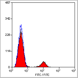
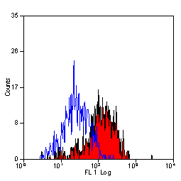
 Application Data
(Staining of New Zealand Black mouse peripheral blood granulocytes with Rat anti Mouse Ly-6B.2 conjugated to FITC Data)
Application Data
(Staining of New Zealand Black mouse peripheral blood granulocytes with Rat anti Mouse Ly-6B.2 conjugated to FITC Data)
Ly-6B.2 ALLOANTIGEN, Monoclonal Antibody (Cat# AAA12199)
Full Name
RAT ANTI MOUSE Ly-6B.2 ALLOANTIGEN:RPE
Applications
Flow Cytometry
Pricing
Reovirus, Polyclonal Antibody (Cat# AAA14712)
Full Name
Reovirus
Applications
ELISA
Purity
Sterile filtered serum
Pricing
Malondialdehyde (MDA), Assay Kit (Cat# AAA22069)
Full Name
Malondialdehyde (MDA) assay kit (TBA method)
Pricing
HIV-1 tat, Recombinant Protein (Cat# AAA14180)
Full Name
HIV-1 TAT antigen (recombinant) full length 14 kDa
Purity
> 95% pure
Purity of proteins is evaluated by SDS-PAGE by measuring optimal density at 280 nm.
Purity of proteins is evaluated by SDS-PAGE by measuring optimal density at 280 nm.
Pricing
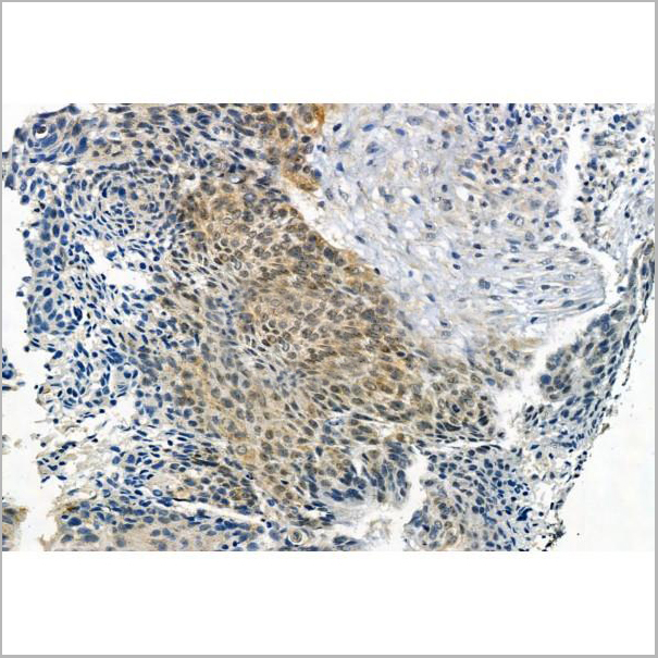
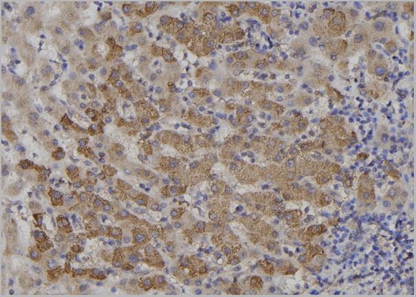
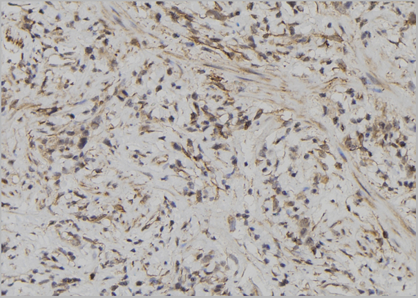
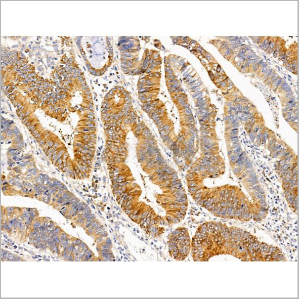
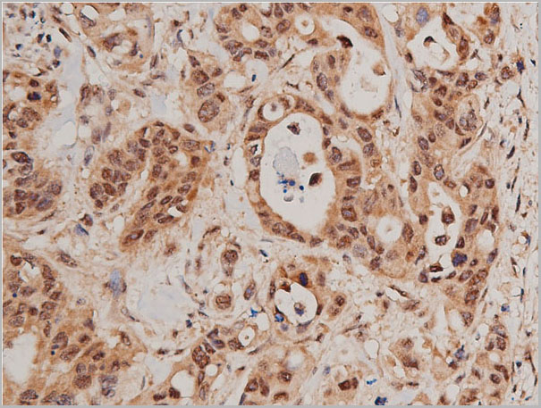
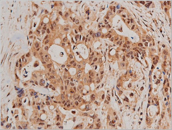
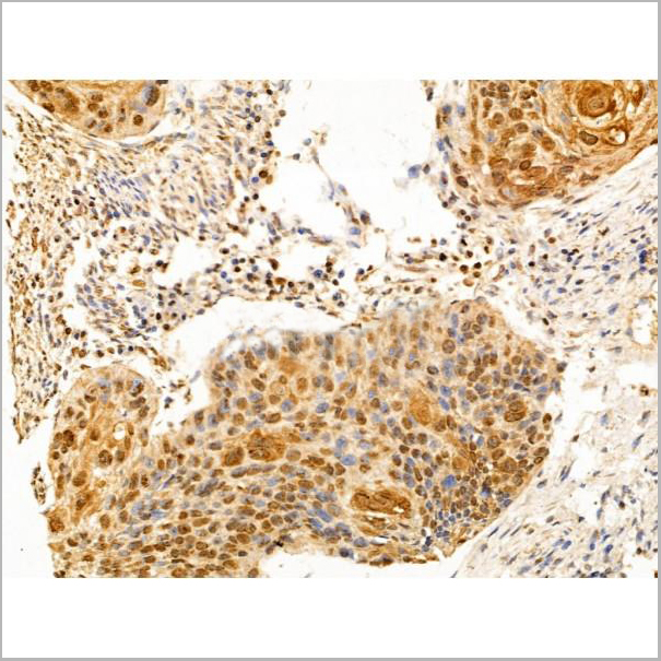
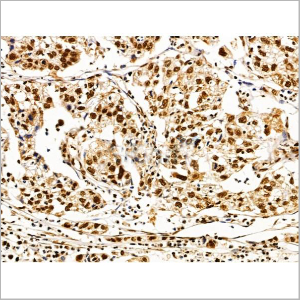
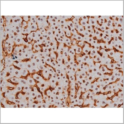
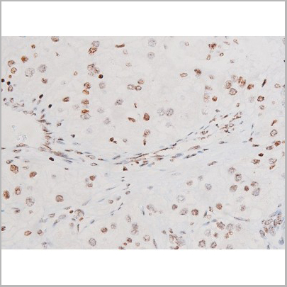
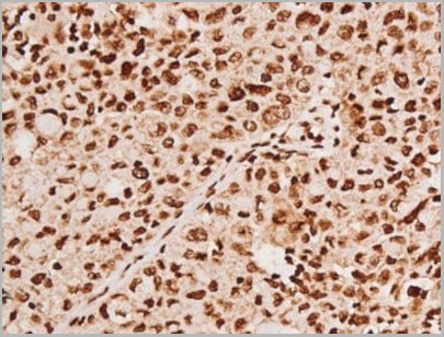
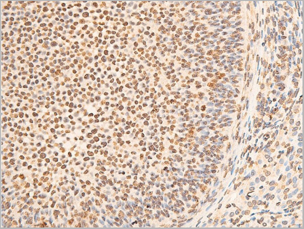
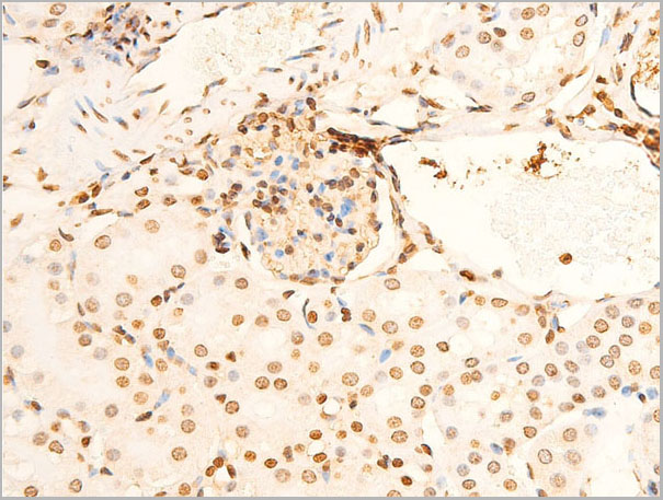
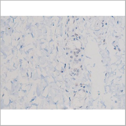
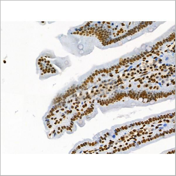
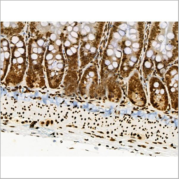
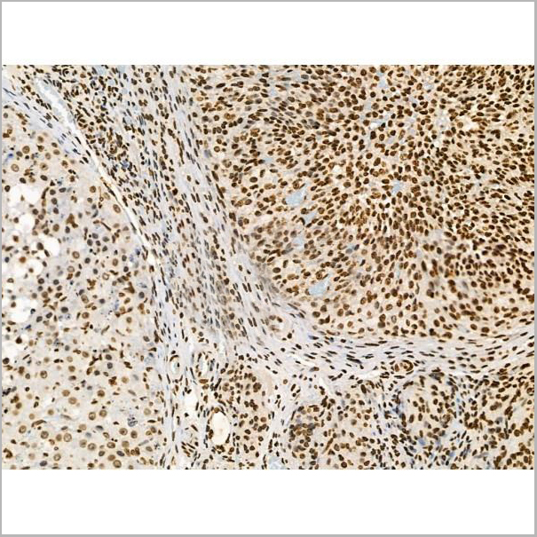
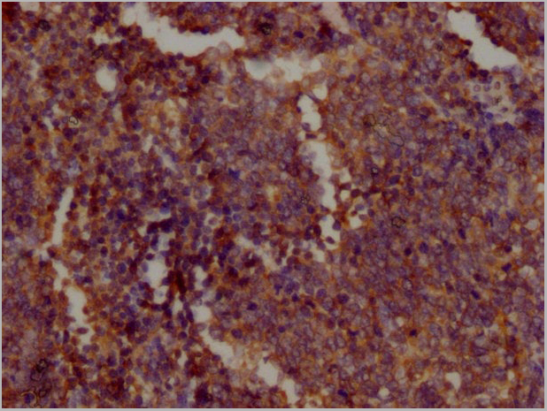
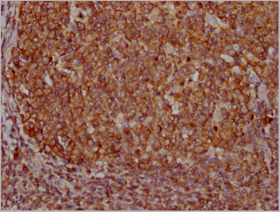
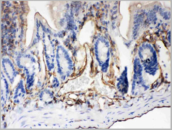
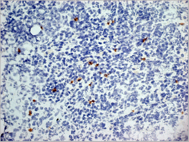
 IHC (Immunohistochemistry)
(IHC image of AAA27005 diluted at 1:500 and staining in paraffin-embedded human ovarian cancer performed on a Leica Bond™ system. After dewaxing and hydration, antigen retrieval was mediated by high pressure in a citrate buffer (pH 6.0). Section was blocked with 10% normal goat serum 30min at RT. Then primary antibody (1% BSA) was incubated at 4°C overnight. The primary is detected by a biotinylated secondary antibody and visualized using an HRP conjugated SP system.)
IHC (Immunohistochemistry)
(IHC image of AAA27005 diluted at 1:500 and staining in paraffin-embedded human ovarian cancer performed on a Leica Bond™ system. After dewaxing and hydration, antigen retrieval was mediated by high pressure in a citrate buffer (pH 6.0). Section was blocked with 10% normal goat serum 30min at RT. Then primary antibody (1% BSA) was incubated at 4°C overnight. The primary is detected by a biotinylated secondary antibody and visualized using an HRP conjugated SP system.)
SRRM4, Polyclonal Antibody (Cat# AAA27005)
Full Name
SRRM4 Antibody
Gene Names
SRRM4; nSR100; KIAA1853; MU-MB-2.76
Reactivity
Human, Mouse, Rat
Applications
Western Blot, Immunohistochemistry
Purity
>95%, Protein G purified
Pricing
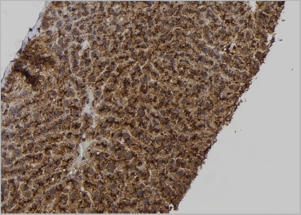
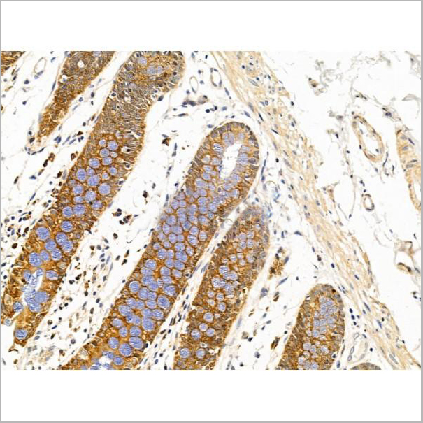
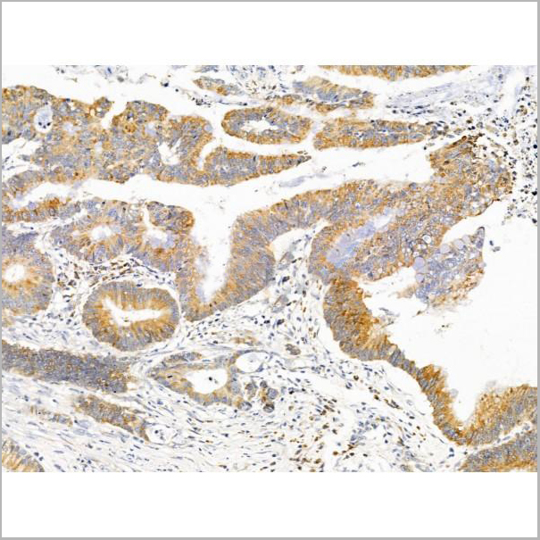
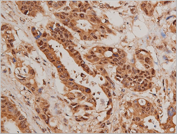
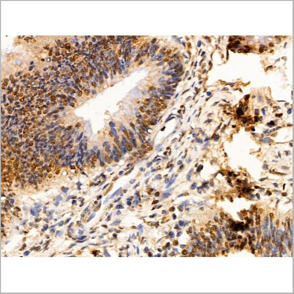
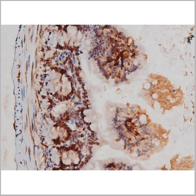
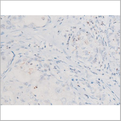
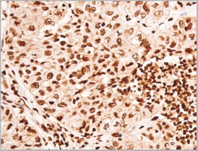
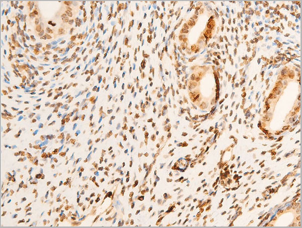
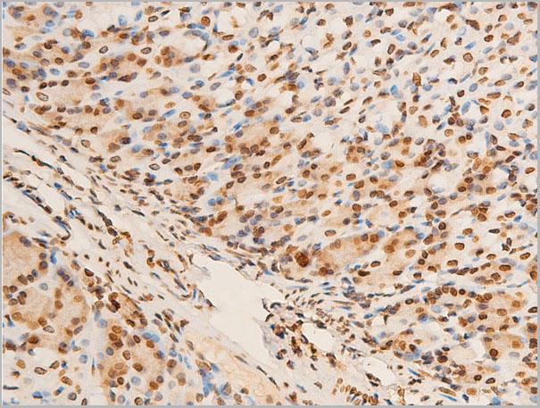
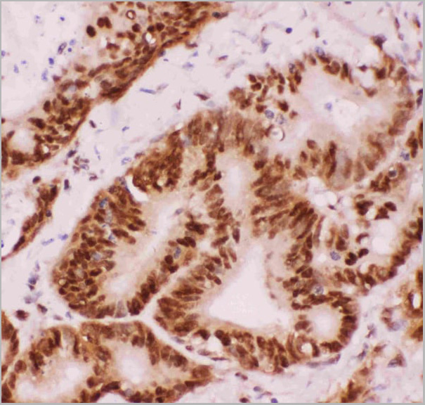
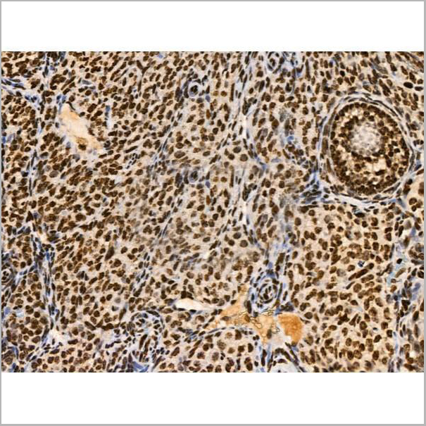
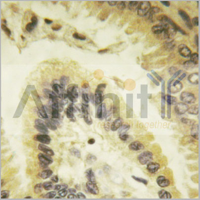
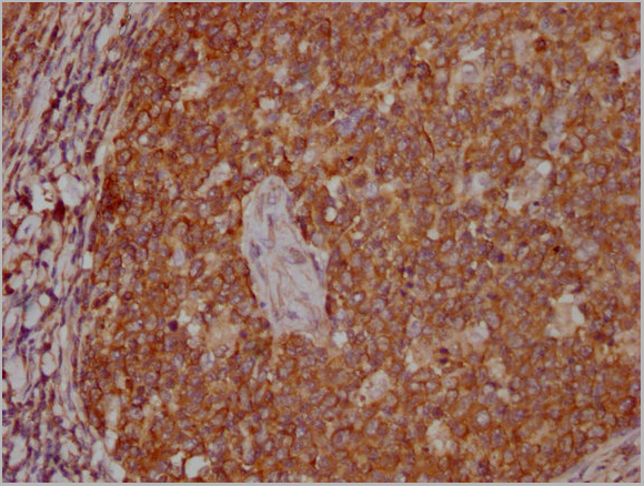
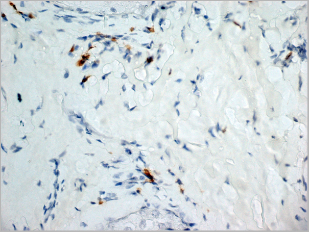
 IHC (Immunohistochemistry)
(IHC image of AAA27004 diluted at 1:600 and staining in paraffin-embedded human colon cancer performed on a Leica BondTM system. After dewaxing and hydration, antigen retrieval was mediated by high pressure in a citrate buffer (pH 6.0). Section was blocked with 10% normal goat serum 30min at RT. Then primary antibody (1% BSA) was incubated at 4 degree C overnight. The primary is detected by a biotinylated secondary antibody and visualized using an HRP conjugated SP system.)
IHC (Immunohistochemistry)
(IHC image of AAA27004 diluted at 1:600 and staining in paraffin-embedded human colon cancer performed on a Leica BondTM system. After dewaxing and hydration, antigen retrieval was mediated by high pressure in a citrate buffer (pH 6.0). Section was blocked with 10% normal goat serum 30min at RT. Then primary antibody (1% BSA) was incubated at 4 degree C overnight. The primary is detected by a biotinylated secondary antibody and visualized using an HRP conjugated SP system.)
GBA, Polyclonal Antibody (Cat# AAA27004)
Full Name
GBA Antibody
Gene Names
GBA; GCB; GBA1; GLUC
Reactivity
Human
Applications
Western Blot, Immunohistochemistry
Purity
>95%, Protein G purified
Pricing