Filters
Clonality
Type
Reactivity
Gene Name
Isotype
Host
Application
Clone
7408 results for " Serum" - showing 6750-6800
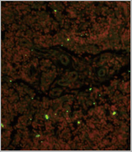
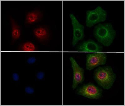
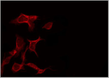
 IF (Immunofluorescence)
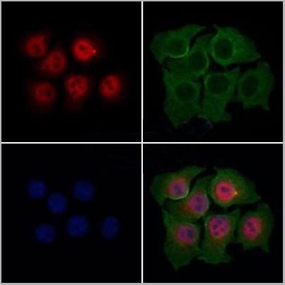
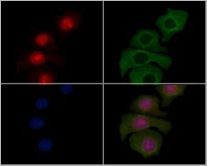
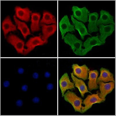
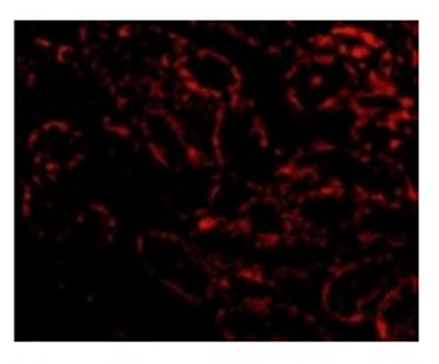
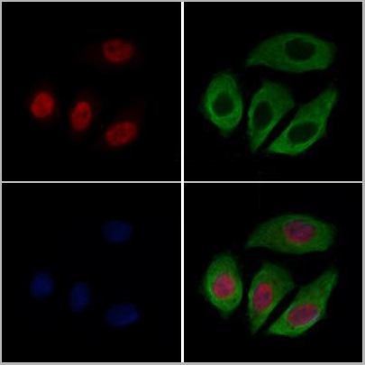
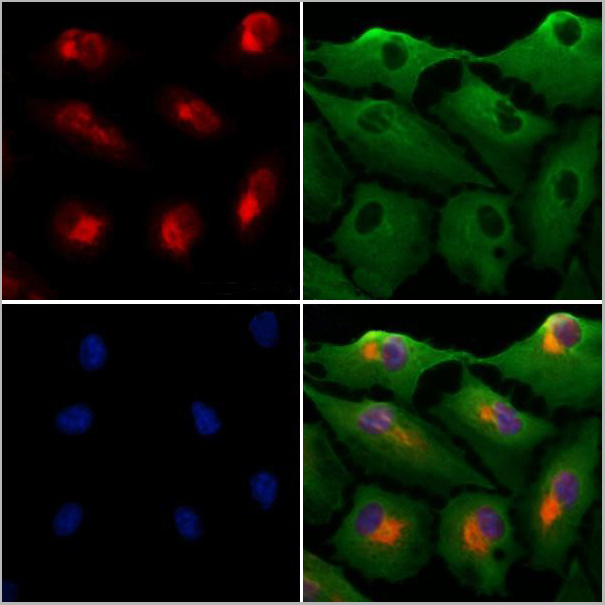
(AAA31242 staining Hela cells by IF/ICC. The samples were fixed with PFA and permeabilized in 0.1% Triton X-100,then blocked in 10% serum for 45 minutes at 25°C. Samples were then incubated with primary Ab(AAA31242 1:200) and mouse anti-beta tubulin Ab(T0023 1:200) for 1 hour at 37°C. An AlexaFluor594 conjugated goat anti-rabbit IgG(H+L) Ab(Red) and an AlexaFluor488 conjugated goat anti-mouse IgG(H+L) Ab(Green) were used as the secondary antibody.The nuclear counter stain is DAPI(blue).)
IF (Immunofluorescence)
(AAA31242 staining Hela cells by IF/ICC. The samples were fixed with PFA and permeabilized in 0.1% Triton X-100,then blocked in 10% serum for 45 minutes at 25°C. Samples were then incubated with primary Ab(AAA31242 1:200) and mouse anti-beta tubulin Ab(T0023 1:200) for 1 hour at 37°C. An AlexaFluor594 conjugated goat anti-rabbit IgG(H+L) Ab(Red) and an AlexaFluor488 conjugated goat anti-mouse IgG(H+L) Ab(Green) were used as the secondary antibody.The nuclear counter stain is DAPI(blue).)
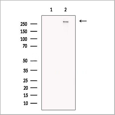
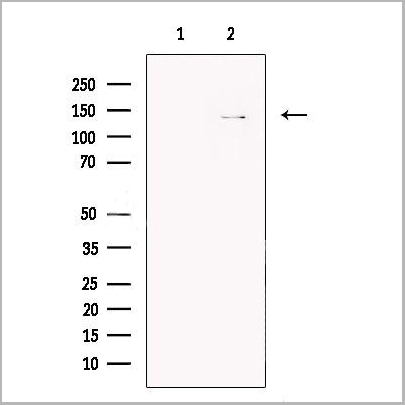
Arc, Polyclonal Antibody (Cat# AAA31242)
Full Name
Arc Antibody
Gene Names
ARC; hArc; Arg3.1
Reactivity
Human, Mouse, Rat
Predicted Reactivity: Pig(100%), Bovine(100%), Chicken(88%), Xenopus(83%)
Predicted Reactivity: Pig(100%), Bovine(100%), Chicken(88%), Xenopus(83%)
Applications
ELISA
Purity
The antiserum was purified by peptide affinity chromatography using SulfoLink Coupling Resin (Thermo Fisher Scientific).
Pricing
 IF (Immunofluorescence)
(AAA31033 staining NIH-3T3 by IF/ICC. The sample were fixed with PFA and permeabilized in 0.1% Triton X-100, then blocked in 10% serum for 45 minutes at 25 degree C. The primary antibody was diluted at 1/200 and incubated with the sample for 1 hour at 37 degree C. An Alexa Fluor 594 conjugated goat anti-rabbit IgG (H+L) Ab, diluted at 1/600, was used as the secondary antibody.)
IF (Immunofluorescence)
(AAA31033 staining NIH-3T3 by IF/ICC. The sample were fixed with PFA and permeabilized in 0.1% Triton X-100, then blocked in 10% serum for 45 minutes at 25 degree C. The primary antibody was diluted at 1/200 and incubated with the sample for 1 hour at 37 degree C. An Alexa Fluor 594 conjugated goat anti-rabbit IgG (H+L) Ab, diluted at 1/600, was used as the secondary antibody.)
CDC25B, Polyclonal Antibody (Cat# AAA31033)
Full Name
Phospho-CDC25B (Ser323) Antibody
Reactivity
Human, Mouse, Rat
Applications
Western Blot, Immunohistochemistry, Immunofluorescence, Immunocytochemistry
Purity
From purified rabbit serum by affinity purification via sequential chromatography on phospho-and non-phospho-peptide affinity columns.
Pricing

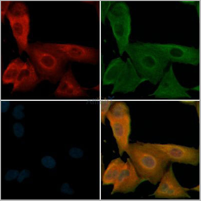
 IF (Immunofluorescence)
(AAA31249 staining HepG2 cells by IF/ICC. The samples were fixed with PFA and permeabilized in 0.1% Triton X-100,then blocked in 10% serum for 45 minutes at 25°C. Samples were then incubated with primary Ab(AAA31249 1:200) and mouse anti-beta tubulin Ab(T0023 1:200) for 1 hour at 37°C. An AlexaFluor594 conjugated goat anti-rabbit IgG(H+L) Ab(Red) and an AlexaFluor488 conjugated goat anti-mouse IgG(H+L) Ab(Green) were used as the secondary antibody.The nuclear counter stain is DAPI(blue).)
IF (Immunofluorescence)
(AAA31249 staining HepG2 cells by IF/ICC. The samples were fixed with PFA and permeabilized in 0.1% Triton X-100,then blocked in 10% serum for 45 minutes at 25°C. Samples were then incubated with primary Ab(AAA31249 1:200) and mouse anti-beta tubulin Ab(T0023 1:200) for 1 hour at 37°C. An AlexaFluor594 conjugated goat anti-rabbit IgG(H+L) Ab(Red) and an AlexaFluor488 conjugated goat anti-mouse IgG(H+L) Ab(Green) were used as the secondary antibody.The nuclear counter stain is DAPI(blue).)
Amino-terminal enhancer of split, Polyclonal Antibody (Cat# AAA31249)
Full Name
Amino-terminal enhancer of split Antibody
Gene Names
TLE5; AES; GRG; ESP1; GRG5; AES-1; AES-2; Grg-5
Reactivity
Human, Mouse, Rat
Predicted Reactivity: Pig(100%), Bovine(100%), Sheep(100%), Xenopus(86%)
Predicted Reactivity: Pig(100%), Bovine(100%), Sheep(100%), Xenopus(86%)
Applications
ELISA
Purity
The antiserum was purified by peptide affinity chromatography using SulfoLink Coupling Resin (Thermo Fisher Scientific).
Pricing

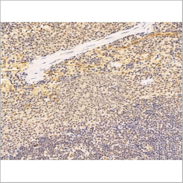
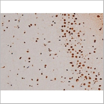
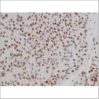
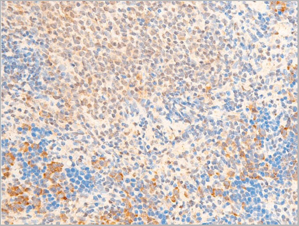
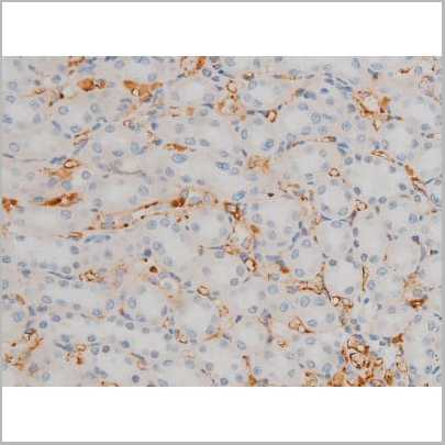
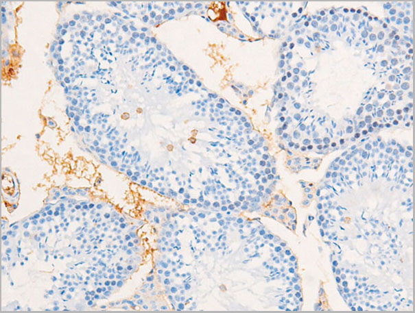
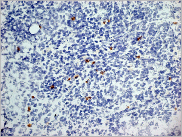
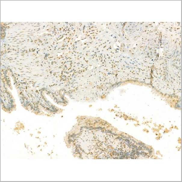
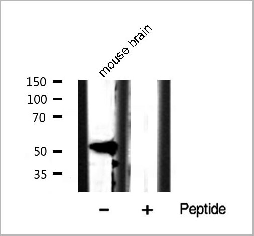
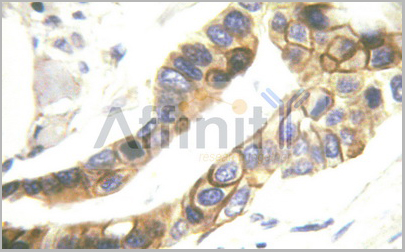
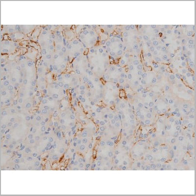
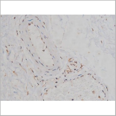
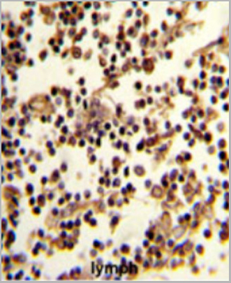 IHC (Immunohistchemistry)
(Formalin-fixed and paraffin-embedded human lymph reacted with CD19 Antibody (N-term), which was peroxidase-conjugated to the secondary antibody, followed by DAB staining. This data demonstrates the use of this antibody for immunohistochemistry; clinical relevance has not been evaluated.)
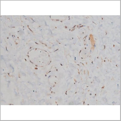
IHC (Immunohistchemistry)
(Formalin-fixed and paraffin-embedded human lymph reacted with CD19 Antibody (N-term), which was peroxidase-conjugated to the secondary antibody, followed by DAB staining. This data demonstrates the use of this antibody for immunohistochemistry; clinical relevance has not been evaluated.)
CD19, Polyclonal Antibody (Cat# AAA28685)
Full Name
CD19 Antibody (N-term)
Gene Names
CD19; B4; CVID3
Reactivity
Human
Applications
Western Blot, Immunofluorescence, Flow Cytometry, Immunohistochemistry
Purity
This antibody is purified through a protein A column, followed by peptide affinity purification.
Pricing

 IF (Immunoflouorescence)
(AAA31011 staining Hela by IF/ICC. The sample were fixed with PFA and permeabilized in 0.1% Triton X-100, then blocked in 10% serum for 45 minutes at 25 degree C. The primary antibody was diluted at 1/200 and incubated with the sample for 1 hour at 37 degree C. An Alexa Fluor 594 conjugated goat anti-rabbit IgG (H+L) Ab, diluted at 1/600, was used as the secondary antibody.)
IF (Immunoflouorescence)
(AAA31011 staining Hela by IF/ICC. The sample were fixed with PFA and permeabilized in 0.1% Triton X-100, then blocked in 10% serum for 45 minutes at 25 degree C. The primary antibody was diluted at 1/200 and incubated with the sample for 1 hour at 37 degree C. An Alexa Fluor 594 conjugated goat anti-rabbit IgG (H+L) Ab, diluted at 1/600, was used as the secondary antibody.)
ATF2, Polyclonal Antibody (Cat# AAA31011)
Full Name
Phospho-ATF2 (Thr69/51) Antibody
Gene Names
ATF2; HB16; CREB2; TREB7; CREB-2; CRE-BP1
Reactivity
Human, Mouse, Rat
Applications
Western Blot, Immunohistochemistry, Immunofluorescence, Immunocytochemistry, Immunoprecipitation
Purity
From purified rabbit serum by affinity purification via sequential chromatography on phospho-and non-phospho-peptide affinity columns.
Pricing


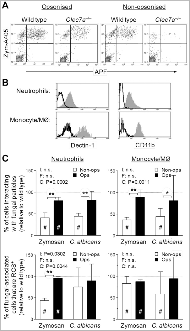
 Application Data
(Staining of mouse peritoneal macrophages with Rat anti Mouse Beta-glucan Receptor: FITC)
Application Data
(Staining of mouse peritoneal macrophages with Rat anti Mouse Beta-glucan Receptor: FITC)
DECTIN-1, Monoclonal Antibody (Cat# AAA12129)
Full Name
RAT ANTI MOUSE DECTIN-1:Low Endotoxin
Gene Names
Clec7a; BGR; beta-GR; Clecsf12
Applications
Immunohistochemistry, Flow Cytometry, Functional Assay, Immunoprecipitation
Pricing
Polyadenylate-binding protein 2, ELISA Kit (Cat# AAA13205)
Full Name
Human Polyadenylate-binding protein 2 (PABPN1) ELISA Kit
Gene Names
PABPN1; OPMD; PAB2; PABII; PABP2; PABP-2
Reactivity
Human
Pricing
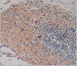

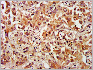
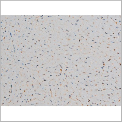
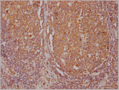
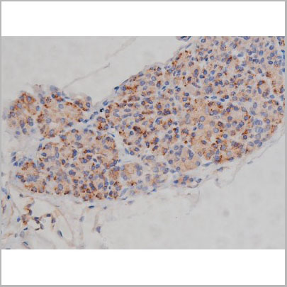
 IHC (Immunohistochemistry)
(DAB staining on IHC-P;Samples: Human Skin cancer Tissue;Primary Ab: 10ug/ml Rabbit Anti-Human F12 AntibodySecond Ab: 2ug/mL HRP-Linked Caprine Anti-Rabbit IgG Polyclonal Antibody (Immunohistochemistry))
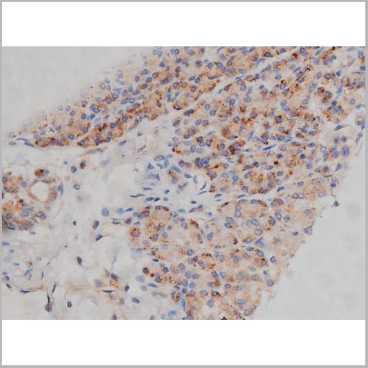
IHC (Immunohistochemistry)
(DAB staining on IHC-P;Samples: Human Skin cancer Tissue;Primary Ab: 10ug/ml Rabbit Anti-Human F12 AntibodySecond Ab: 2ug/mL HRP-Linked Caprine Anti-Rabbit IgG Polyclonal Antibody (Immunohistochemistry))
Prothrombin Fragment 1+2 (F1+2), Polyclonal Antibody (Cat# AAA20072)
Full Name
Polyclonal Antibody to Prothrombin Fragment 1+2 (F1+2)
Gene Names
F2; PT; THPH1; RPRGL2
Reactivity
Human, Mouse, Rat, Pig
Applications
WB, IHC, ICC, IP
Purity
Antigen-specific affinity chromatography followed by Protein A affinity chromatography
Pricing
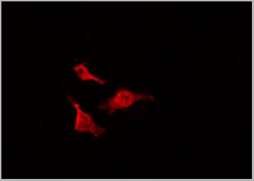
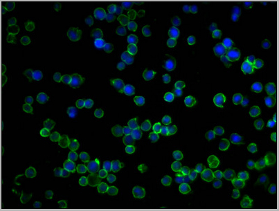
 IF (Immunofluorescence)
(AAA31156 staining HepG2 cells(4h of LPS treatment) by IF/ICC. The samples were fixed with PFA and permeabilized in 0.1% Triton X-100,then blocked in 10% serum for 45 minutes at 25°C. Samples were then incubated with primary Ab(AAA31156 1:200) and mouse anti-beta tubulin Ab( 1:200) for 1 hour at 37°C. An AlexaFluor594 conjugated goat anti-rabbit IgG(H+L) Ab(Red) and an AlexaFluor488 conjugated goat anti-mouse IgG(H+L) Ab(Green) were used as the secondary Ab. The nuclear counter stain is DAPI(blue).)
IF (Immunofluorescence)
(AAA31156 staining HepG2 cells(4h of LPS treatment) by IF/ICC. The samples were fixed with PFA and permeabilized in 0.1% Triton X-100,then blocked in 10% serum for 45 minutes at 25°C. Samples were then incubated with primary Ab(AAA31156 1:200) and mouse anti-beta tubulin Ab( 1:200) for 1 hour at 37°C. An AlexaFluor594 conjugated goat anti-rabbit IgG(H+L) Ab(Red) and an AlexaFluor488 conjugated goat anti-mouse IgG(H+L) Ab(Green) were used as the secondary Ab. The nuclear counter stain is DAPI(blue).)
Beclin-1, Polyclonal Antibody (Cat# AAA31156)
Full Name
Phospho-Beclin-1 (Ser90/93/96) Antibody
Gene Names
BECN1; ATG6; VPS30; beclin1
Reactivity
Human, Mouse, Rat
Predicted: Pig, Bovine, Horse, Sheep, Rabbit, Dog, Chicken, Xenopus
Predicted: Pig, Bovine, Horse, Sheep, Rabbit, Dog, Chicken, Xenopus
Applications
Western Blot, Immunohistochemistry, Peptide ELISA
Purity
Purified rabbit serum by affinity purification via sequential chromatography on phospho- and non-phospho-peptide affinity columns.
Pricing
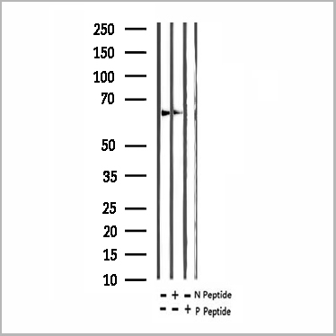
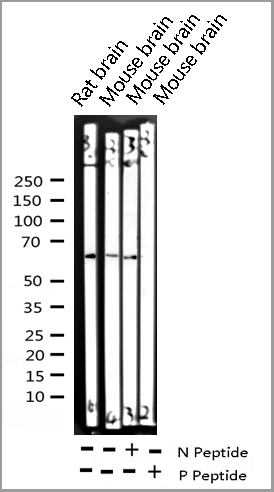
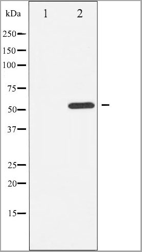
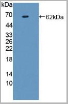
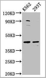
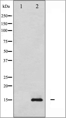
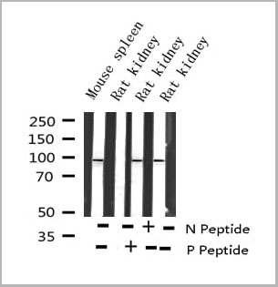
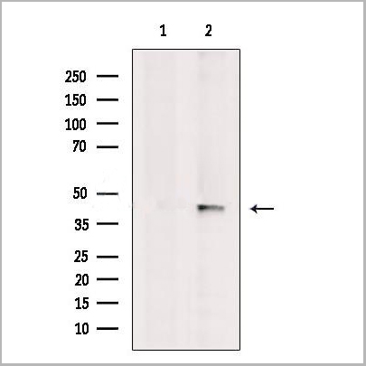
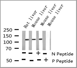
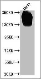
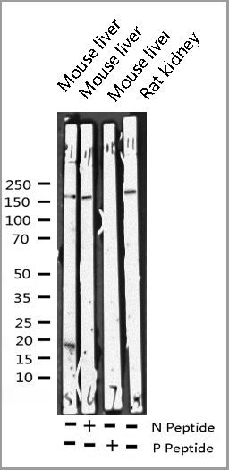
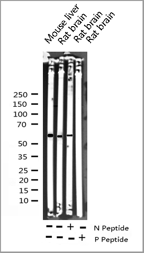
 WB (Western Blot)
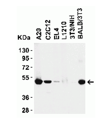
(Western blot analysis of extracts from A431, using Phospho-SHP-2 (Tyr62) Antibody. Lane1 was treated with phospho-blocking peptide, Lane2 was treated with non-phospho-blocking peptide.)
WB (Western Blot)
(Western blot analysis of extracts from A431, using Phospho-SHP-2 (Tyr62) Antibody. Lane1 was treated with phospho-blocking peptide, Lane2 was treated with non-phospho-blocking peptide.)
SHP-2, Polyclonal Antibody (Cat# AAA31155)
Full Name
Phospho-SHP-2 (Tyr62) Antibody
Gene Names
PTPN11; CFC; NS1; JMML; SHP2; BPTP3; PTP2C; METCDS; PTP-1D; SH-PTP2; SH-PTP3
Reactivity
Human, Mouse, Rat
Predicted: Bovine, Horse, Sheep, Rabbit, Dog, Chicken, Xenopus
Predicted: Bovine, Horse, Sheep, Rabbit, Dog, Chicken, Xenopus
Applications
Western Blot, Immunohistochemistry, Peptide ELISA
Purity
Purified rabbit serum by affinity purification via sequential chromatography on phospho- and non-phospho-peptide affinity columns.
Pricing

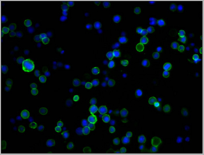
 IF (Immunofluorescence)
(AAA30956 staining HepG2 cells(4h of LPS treatment) by IF/ICC. The samples were fixed with PFA and permeabilized in 0.1% Triton X-100,then blocked in 10% serum for 45 minutes at 25°C. Samples were then incubated with primary Ab(AAA30956 1:200) and mouse anti-beta tubulin Ab( 1:200) for 1 hour at 37°C. An AlexaFluor594 conjugated goat anti-rabbit IgG(H+L) Ab(Red) and an AlexaFluor488 conjugated goat anti-mouse IgG(H+L) Ab(Green) were used as the secondary Ab. The nuclear counter stain is DAPI(blue).)
IF (Immunofluorescence)
(AAA30956 staining HepG2 cells(4h of LPS treatment) by IF/ICC. The samples were fixed with PFA and permeabilized in 0.1% Triton X-100,then blocked in 10% serum for 45 minutes at 25°C. Samples were then incubated with primary Ab(AAA30956 1:200) and mouse anti-beta tubulin Ab( 1:200) for 1 hour at 37°C. An AlexaFluor594 conjugated goat anti-rabbit IgG(H+L) Ab(Red) and an AlexaFluor488 conjugated goat anti-mouse IgG(H+L) Ab(Green) were used as the secondary Ab. The nuclear counter stain is DAPI(blue).)
MITF, Polyclonal Antibody (Cat# AAA30956)
Full Name
Phospho-MITF (Ser180/73) Antibody
Gene Names
MITF; MI; WS2; CMM8; WS2A; COMMAD; bHLHe32
Reactivity
Human, Mouse, Rat, Monkey
Applications
Western Blot, Immunohistochemistry, Immunofluorescence, Immunocytochemistry
Purity
Peptide affinity puriification
Pricing

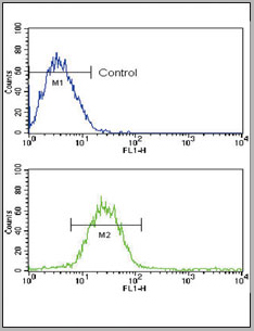
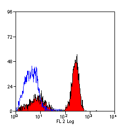
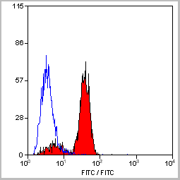
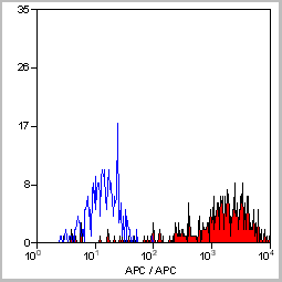
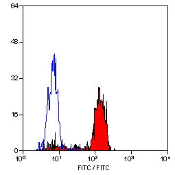
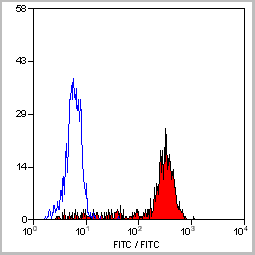
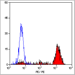
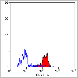
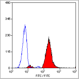
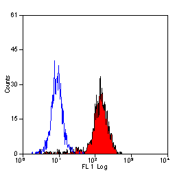
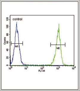 FCM (Flow Cytometry)
(LTF Antibody flow cytometric analysis of MDA-MB231 cells (right histogram) compared to a negative control cell (lefthistogram).FITC-conjugated goat-anti-rabbit secondary antibodies were used for theanalysis.)
FCM (Flow Cytometry)
(LTF Antibody flow cytometric analysis of MDA-MB231 cells (right histogram) compared to a negative control cell (lefthistogram).FITC-conjugated goat-anti-rabbit secondary antibodies were used for theanalysis.)
LTF, Polyclonal Antibody (Cat# AAA28680)
Full Name
LTF Antibody
Gene Names
LTF; LF; HLF2; GIG12; HEL110
Reactivity
Human, mouse
Applications
Western Blot, Flow Cytometry
Purity
Peptide Affinity Purified Rabbit Polyclonal Antibody (Pab)
Pricing
b2-Microglobulin, Monoclonal Antibody (Cat# AAA14654)
Full Name
b2-Microglobulin (B2M)
Gene Names
HLA-G; MHC-G
Applications
Immunohistochemistry, Flow Cytometry, Immunofluorescence
Purity
Purified
Pricing
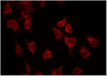
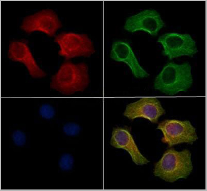
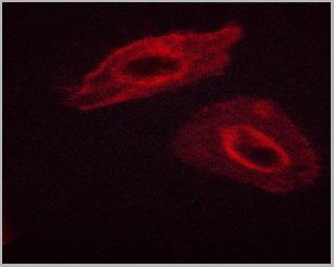
 IF (Immunofluorescence)
(Immunofluorescence staining of HepG2 cells with AAA26952 at 1:333, counter- stained with DAPI. The cells were fixed in 4% formaldehyde, permeabilized using 0.2% Triton X-100 and blocked in 10% normal Goat Serum. The cells were then incubated with the antibody overnight at 4°C. The secondary antibody was Alexa Fluor 488-congugated AffiniPure Goat Anti-Rabbit IgG(H+L)
IF (Immunofluorescence)
(Immunofluorescence staining of HepG2 cells with AAA26952 at 1:333, counter- stained with DAPI. The cells were fixed in 4% formaldehyde, permeabilized using 0.2% Triton X-100 and blocked in 10% normal Goat Serum. The cells were then incubated with the antibody overnight at 4°C. The secondary antibody was Alexa Fluor 488-congugated AffiniPure Goat Anti-Rabbit IgG(H+L)
XKR8, Polyclonal Antibody (Cat# AAA26952)
Full Name
XKR8 Antibody
Gene Names
XKR8; XRG8; hXkr8
Reactivity
Human
Applications
Western Blot, Immunohistochemistry, Immunofluorescence
Purity
>95%, Protein G purified
Pricing
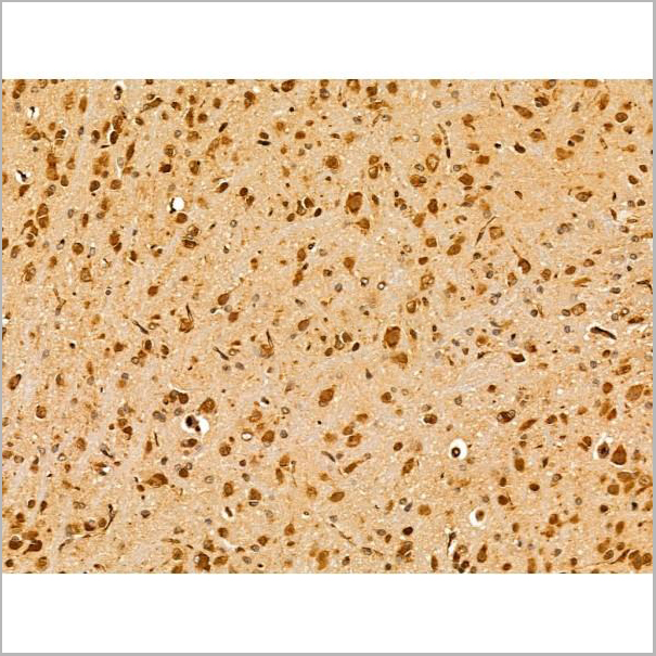
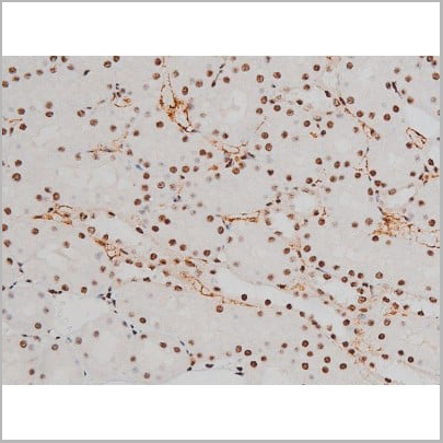
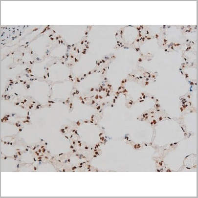
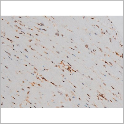
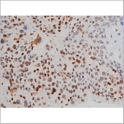
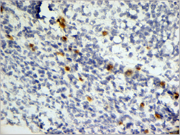
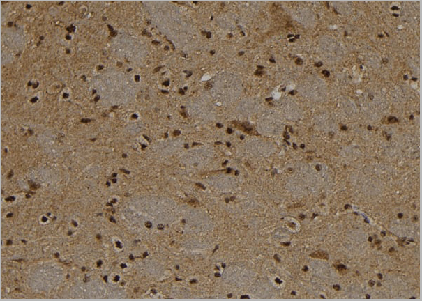
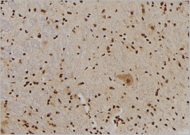
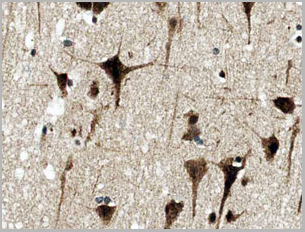
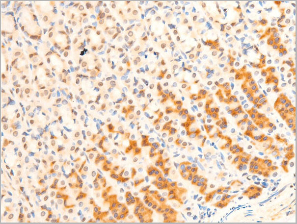
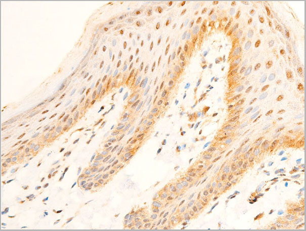
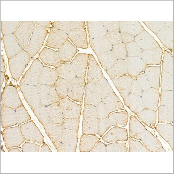
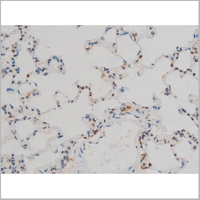
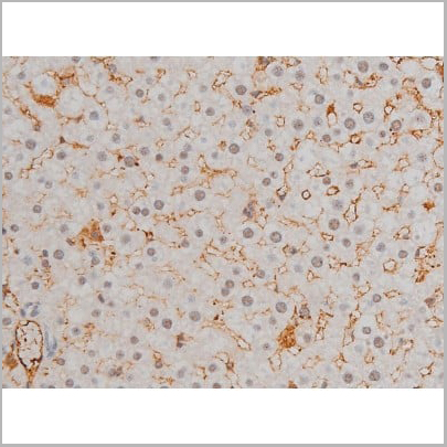
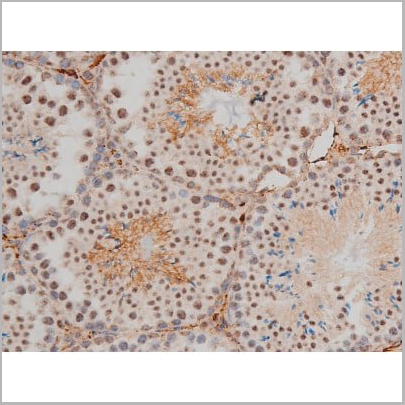
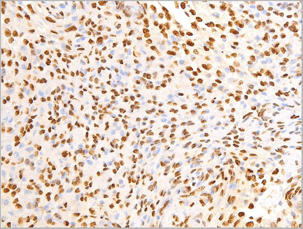
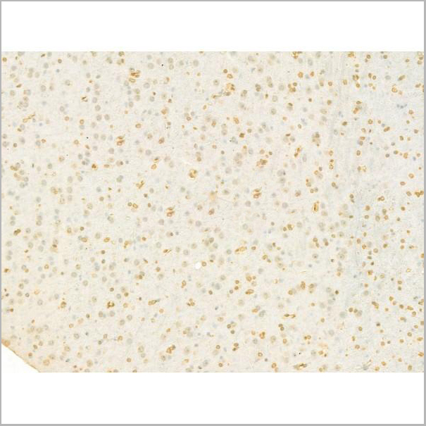
 IHC (Immunohistochemistry)
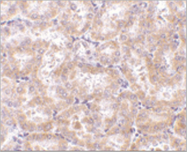
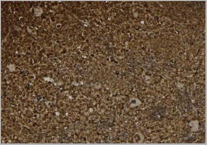
(AAA31014 at 1/200 staining Rat ganstric tissue sections by IHC-P. The tissue was formaldehyde fixed and a heat mediated antigen retrieval step in citrate buffer was performed. The tissue was then blocked and incubated with the antibody for 1.5 hours at 22 degree C. An HRP conjugated goat anti-rabbit antibody was used as the secondary.)
IHC (Immunohistochemistry)
(AAA31014 at 1/200 staining Rat ganstric tissue sections by IHC-P. The tissue was formaldehyde fixed and a heat mediated antigen retrieval step in citrate buffer was performed. The tissue was then blocked and incubated with the antibody for 1.5 hours at 22 degree C. An HRP conjugated goat anti-rabbit antibody was used as the secondary.)
Histone H2A.X, Polyclonal Antibody (Cat# AAA31014)
Full Name
Phospho-Histone H2A.X (Ser139) Antibody
Gene Names
H2AFX; H2AX; H2A.X; H2A/X
Reactivity
Human, Mouse, Rat
Applications
Western Blot, Immunohistochemistry
Purity
From purified rabbit serum by affinity purification via sequential chromatography on phospho-and non-phospho-peptide affinity columns.
Pricing


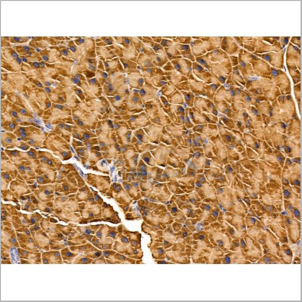
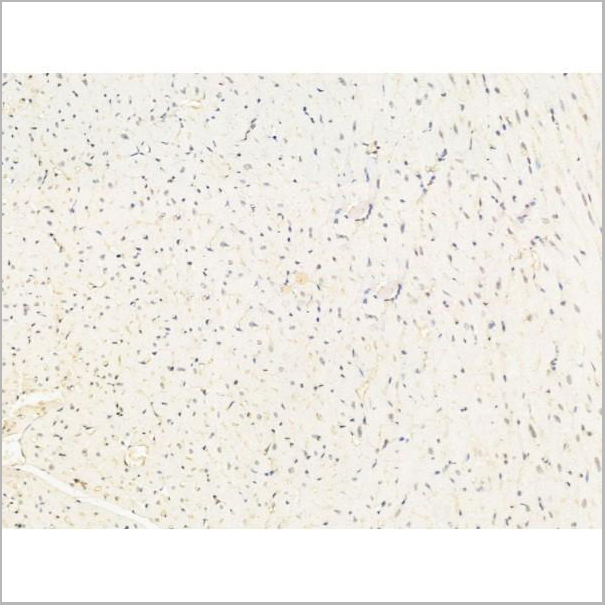
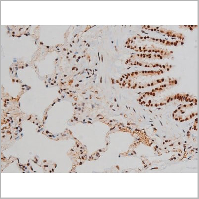
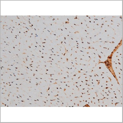
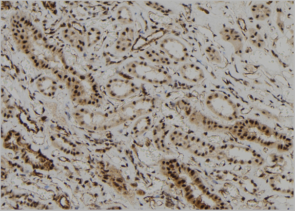
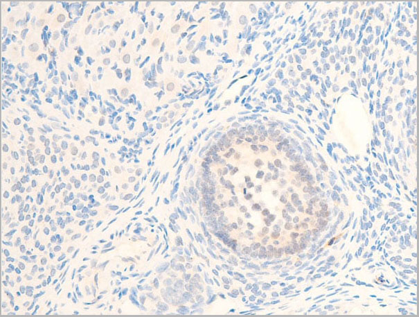
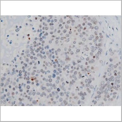
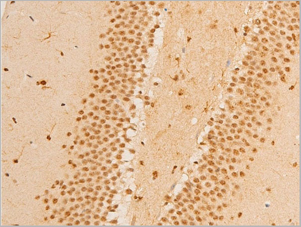
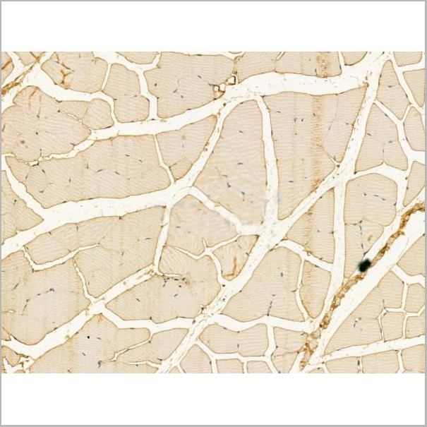
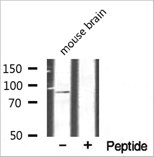
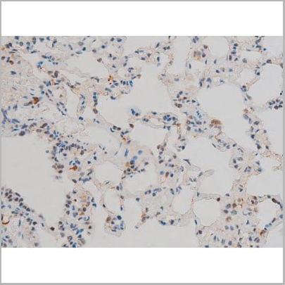
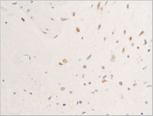
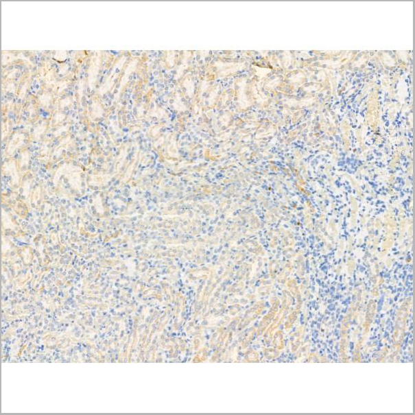
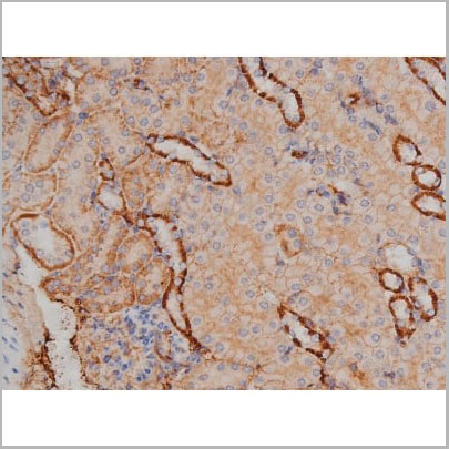
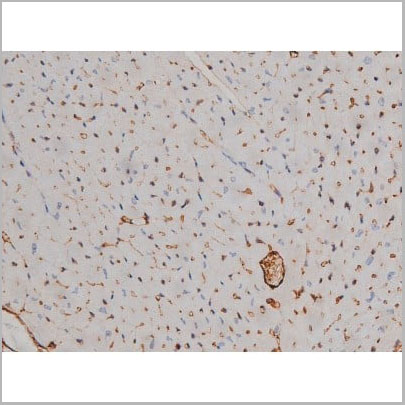
 IHC (Immunohistchemistry)
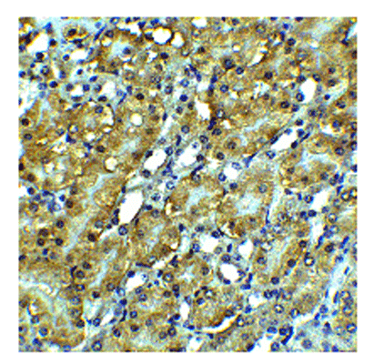
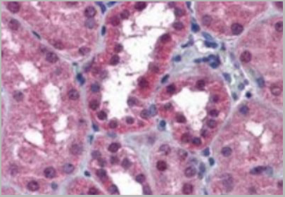
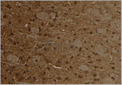
(AAA30928 at 1/100 staining Rat kidney tissue by IHC-P. The sample was formaldehyde fixed and a heat mediated antigen retrieval step in citrate buffer was performed. The sample was then blocked and incubated with the primary antibody at 4°C overnight. An HRP conjugated anti-Rabbit antibody was used as the secondary antibody.)
IHC (Immunohistchemistry)
(AAA30928 at 1/100 staining Rat kidney tissue by IHC-P. The sample was formaldehyde fixed and a heat mediated antigen retrieval step in citrate buffer was performed. The sample was then blocked and incubated with the primary antibody at 4°C overnight. An HRP conjugated anti-Rabbit antibody was used as the secondary antibody.)
Nrf2, Polyclonal Antibody (Cat# AAA30928)
Full Name
Nrf2 Antibody
Gene Names
NFE2L2; NRF2; HEBP1; IMDDHH
Reactivity
Human, Mouse, Rat
Applications
Western Blot, Immunohistochemistry, Immunofluorescence, Immunocytochemistry
Purity
Peptide affinity purification
Pricing
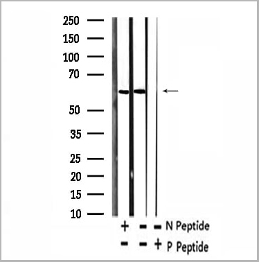
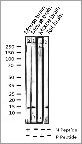
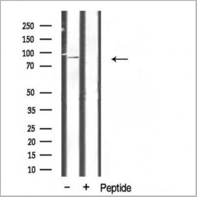
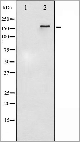
 WB (Western Blot)
(Western blot analysis of FKHRL1 phosphorylation expression in serum treated NIH-3T3 whole cell lysates, The lane on the left is treated with the antigen-specific peptide.)
WB (Western Blot)
(Western blot analysis of FKHRL1 phosphorylation expression in serum treated NIH-3T3 whole cell lysates, The lane on the left is treated with the antigen-specific peptide.)
FKHRL1, Polyclonal Antibody (Cat# AAA30954)
Full Name
Phospho-FKHRL1 (Ser253) Antibody
Gene Names
FOXO3; FOXO2; AF6q21; FKHRL1; FOXO3A; FKHRL1P2
Reactivity
Human, Mouse, Rat
Applications
Western Blot, Immunohistochemistry
Purity
From purified rabbit serum by affinity purification via sequential chromatography on phospho-and non-phospho-peptide affinity columns.
Pricing
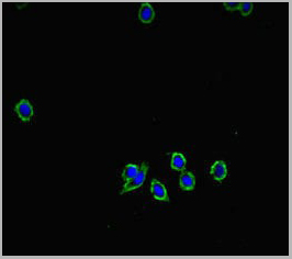
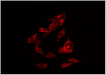
 IF (Immunofluorescence)
(AAA31197 staining Hela cells by IF/ICC. The samples were fixed with PFA and permeabilized in 0.1% Triton X-100,then blocked in 10% serum for 45 minutes at 25°C. Samples were then incubated with primary Ab(AAA31197 1:200) and mouse anti-beta tubulin Ab(T0023 1:200) for 1 hour at 37°C. An AlexaFluor594 conjugated goat anti-rabbit IgG(H+L) Ab(Red) and an AlexaFluor488 conjugated goat anti-mouse IgG(H+L) Ab(Green) were used as the secondary antibody.The nuclear counter stain is DAPI(blue).)
IF (Immunofluorescence)
(AAA31197 staining Hela cells by IF/ICC. The samples were fixed with PFA and permeabilized in 0.1% Triton X-100,then blocked in 10% serum for 45 minutes at 25°C. Samples were then incubated with primary Ab(AAA31197 1:200) and mouse anti-beta tubulin Ab(T0023 1:200) for 1 hour at 37°C. An AlexaFluor594 conjugated goat anti-rabbit IgG(H+L) Ab(Red) and an AlexaFluor488 conjugated goat anti-mouse IgG(H+L) Ab(Green) were used as the secondary antibody.The nuclear counter stain is DAPI(blue).)
CXCL16, Polyclonal Antibody (Cat# AAA31197)
Full Name
CXCL16 Antibody
Gene Names
CXCL16; SRPSOX; CXCLG16; SR-PSOX
Reactivity
Human, Mouse, Rat
Applications
Western Blot, Immunofluorescence, Immunocytochemistry
Purity
The antiserum was purified by peptide affinity chromatography using SulfoLink Coupling Resin (Thermo Fisher Scientific).
Pricing
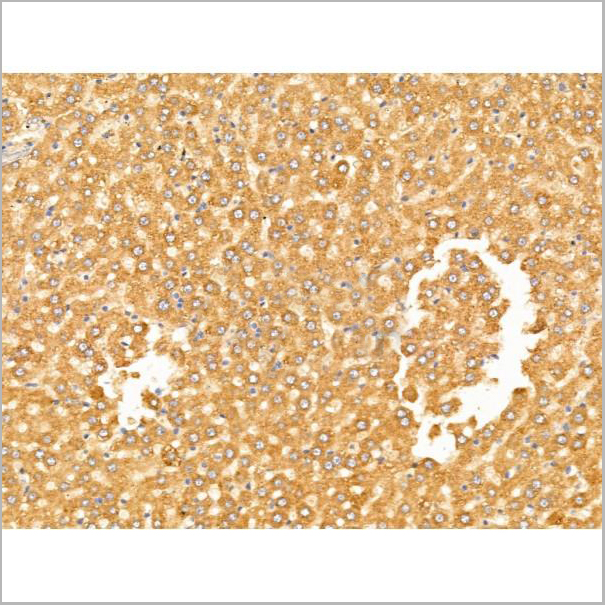
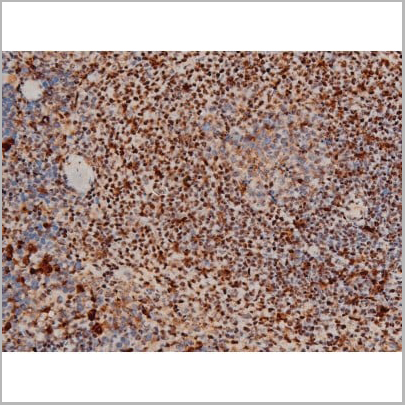
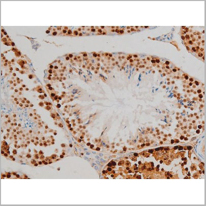
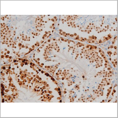
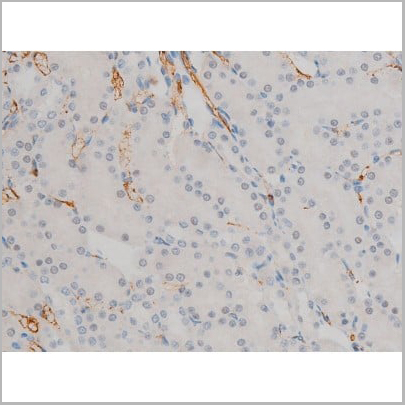
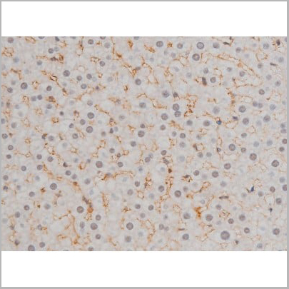
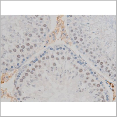
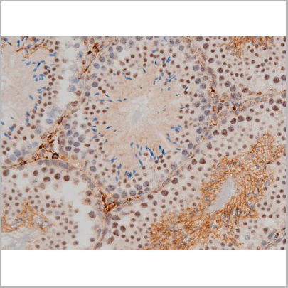
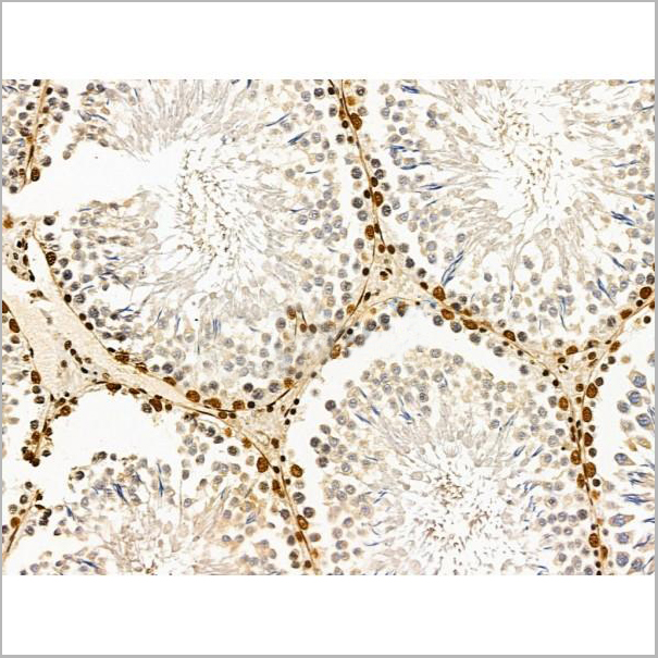
 IHC (Immunohistchemistry)
(AAA31110 at 1/100 staining Rat testis tissue by IHC-P. The sample was formaldehyde fixed and a heat mediated antigen retrieval step in citrate buffer was performed. The sample was then blocked and incubated with the primary antibody at 4°C overnight. An HRP conjugated anti-Rabbit antibody was used as the secondary antibody.)
IHC (Immunohistchemistry)
(AAA31110 at 1/100 staining Rat testis tissue by IHC-P. The sample was formaldehyde fixed and a heat mediated antigen retrieval step in citrate buffer was performed. The sample was then blocked and incubated with the primary antibody at 4°C overnight. An HRP conjugated anti-Rabbit antibody was used as the secondary antibody.)
CHSY1, Polyclonal Antibody (Cat# AAA31110)
Full Name
CHSY1 Antibody
Gene Names
CHSY1; CHSY; CSS1; TPBS; ChSy-1
Reactivity
Human, Mouse, Rat
Applications
Western Blot, Immunohistochemistry, Immunofluorescence, Immunocytochemistry
Purity
The antiserum was purified by peptide affinity chromatography using SulfoLink™ Coupling Resin (Thermo Fisher Scientific).
Pricing
 Application Data
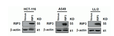
(The cancer cell lines were stably expressing DYKDDDDK-tagged RIP3 and RIP expression was detected by anti-RIP3 antibodies (AAA10922) in RIP3-overexpressed cells.)
Application Data
(The cancer cell lines were stably expressing DYKDDDDK-tagged RIP3 and RIP expression was detected by anti-RIP3 antibodies (AAA10922) in RIP3-overexpressed cells.)
RIP3, Polyclonal Antibody (Cat# AAA10922)
Full Name
RIP3 Antibody
Gene Names
Ripk3; Rip3; AW107945; 2610528K09Rik
Reactivity
Human, Mouse, Rat
Applications
Immunofluorescence, Immunohistochemistry, Immunoprecipitation, Western Blot
Purity
RIP3 Antibody is affinity chromatography purified via peptide column.
Pricing


 Application Data
(Staining of mouse peritoneal macrophages with Rat anti Mouse Beta-glucan Receptor: FITC)
Application Data
(Staining of mouse peritoneal macrophages with Rat anti Mouse Beta-glucan Receptor: FITC)
DECTIN-1, Monoclonal Antibody (Cat# AAA12130)
Full Name
RAT ANTI MOUSE DECTIN-1:FITC
Gene Names
Clec7a; BGR; beta-GR; Clecsf12
Applications
Flow Cytometry
Pricing
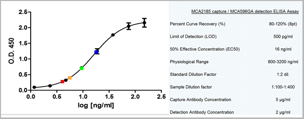
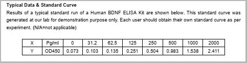 Standard Curve (Sample)
BDNF, ELISA Kit (Cat# AAA13497)
Full Name
BDNF Human ELISA Kit
Reactivity
Human
Applications
ELISA
Pricing
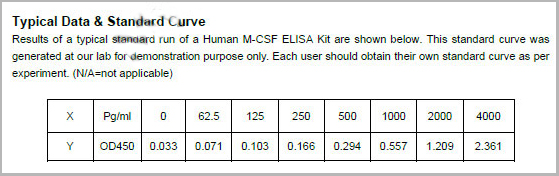 Standard Curve (Sample)
M-CSF, ELISA Kit (Cat# AAA13520)
Full Name
M-CSF Human ELISA Kit
Reactivity
Human
Applications
ELISA
Pricing
interleukin 10 receptor, alpha, ELISA Kit (Cat# AAA18041)
Full Name
Mouse Interleukin-10 receptor subunit alpha, IL10RA ELISA Kit
Gene Names
Il10ra; Il10r; CDw210; CDw210a; mIL-10R; AW553859
Reactivity
Mouse
Pricing
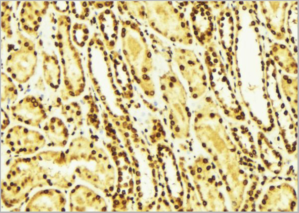
 IHC (Immunohistochemistry)
(AAA13663 (3.75µg/ml) staining of paraffin embedded Human Kidney. Steamed antigen retrieval with citrate buffer pH 6, AP-staining.)
IHC (Immunohistochemistry)
(AAA13663 (3.75µg/ml) staining of paraffin embedded Human Kidney. Steamed antigen retrieval with citrate buffer pH 6, AP-staining.)
P2RX7/P2X7 receptor, Polyclonal Antibody (Cat# AAA13663)
Full Name
Goat anti-P2RX7/P2X7 receptor Antibody
Gene Names
P2RX7; P2X7
Reactivity
Tested: Human; Expected from sequence similarity: Human, Mouse, Rat, Dog
Applications
Peptide ELISA, Western Blot, Immunohistochemistry
Purity
Purified from goat serum by ammonium sulphate precipitation followed by antigen affinity chromatography using the immunizing peptide.
Pricing
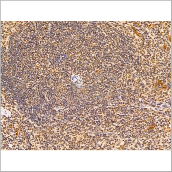
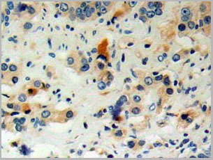
 IHC (Immunohistchemistry)
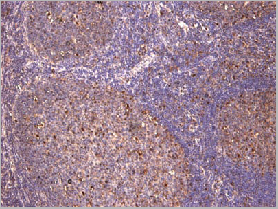
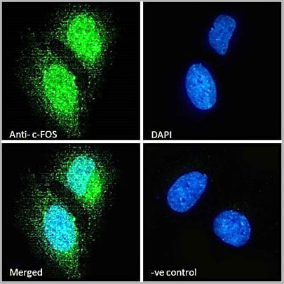
(Negative Control showing staining of paraffin embedded Human Tonsil, with no primary antibody.)
IHC (Immunohistchemistry)
(Negative Control showing staining of paraffin embedded Human Tonsil, with no primary antibody.)
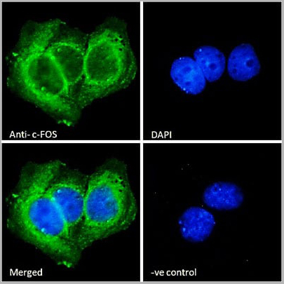
c-FOS, Antibody (Cat# AAA13673)
Full Name
Goat anti-c-FOS (aa283-295) Antibody
Gene Names
FOS; p55; AP-1; C-FOS
Reactivity
Tested: Human; Expected from sequence similarity: Human, Dog, Pig, Cow
Applications
Peptide ELISA, Western Blot, Immunofluorescence, Immunohistochemistry
Purity
Purified from goat serum by ammonium sulphate precipitation followed by antigen affinity chromatography using the immunizing peptide.
Pricing
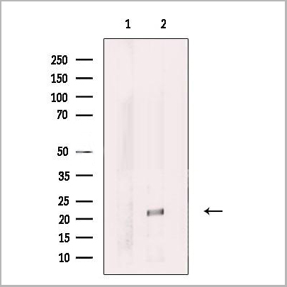
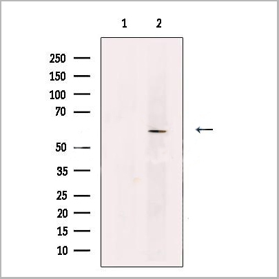
 WB (Western Blot)
(Western blot analysis of JunD phosphorylation expression in 293 whole cell lysates, The lane on the left is treated with the antigen-specific peptide.)
WB (Western Blot)
(Western blot analysis of JunD phosphorylation expression in 293 whole cell lysates, The lane on the left is treated with the antigen-specific peptide.)
JunD, Polyclonal Antibody (Cat# AAA31019)
Full Name
Phospho-JunD (Ser255) Antibody
Gene Names
JUND; AP-1
Reactivity
Human, Mouse, Rat
Applications
Western Blot, Immunohistochemistry, Immunofluorescence, Immunocytochemistry, Immunoprecipitation
Purity
From purified rabbit serum by affinity purification via sequential chromatography on phospho-and non-phospho-peptide affinity columns.
Pricing
 Application Data
(Staining of human peripheral blood monocytes with Mouse anti Human CD14:RPE)
Application Data
(Staining of human peripheral blood monocytes with Mouse anti Human CD14:RPE)
CD14, Monoclonal Antibody (Cat# AAA11841)
Full Name
MOUSE ANTI HUMAN CD14:APC
Applications
Flow Cytometry
Pricing
 IF (Immunofluorescence)
(AAA31224 staining Hela cells by IF/ICC. The samples were fixed with PFA and permeabilized in 0.1% Triton X-100,then blocked in 10% serum for 45 minutes at 25°C. Samples were then incubated with primary Ab(AAA31224 1:200) and mouse anti-beta tubulin Ab(T0023 1:200) for 1 hour at 37°C. An AlexaFluor594 conjugated goat anti-rabbit IgG(H+L) Ab(Red) and an AlexaFluor488 conjugated goat anti-mouse IgG(H+L) Ab(Green) were used as the secondary antibody.The nuclear counter stain is DAPI(blue).)
IF (Immunofluorescence)
(AAA31224 staining Hela cells by IF/ICC. The samples were fixed with PFA and permeabilized in 0.1% Triton X-100,then blocked in 10% serum for 45 minutes at 25°C. Samples were then incubated with primary Ab(AAA31224 1:200) and mouse anti-beta tubulin Ab(T0023 1:200) for 1 hour at 37°C. An AlexaFluor594 conjugated goat anti-rabbit IgG(H+L) Ab(Red) and an AlexaFluor488 conjugated goat anti-mouse IgG(H+L) Ab(Green) were used as the secondary antibody.The nuclear counter stain is DAPI(blue).)
CASC5, Polyclonal Antibody (Cat# AAA31224)
Full Name
CASC5 Antibody
Gene Names
KNL1; D40; CT29; Spc7; CASC5; MCPH4; hKNL-1; AF15Q14; PPP1R55; hSpc105
Reactivity
Human, Mouse, Rat
Predicted Reactivity: Pig(88%), Horse(88%), Rabbit(86%)
Predicted Reactivity: Pig(88%), Horse(88%), Rabbit(86%)
Applications
ELISA
Purity
The antiserum was purified by peptide affinity chromatography using SulfoLink Coupling Resin (Thermo Fisher Scientific).
Pricing
 IF (Immunofluorescence)
(AAA29758 staining HepG2 cells(4h of LPS treatment) by IF/ICC. The samples were fixed with PFA and permeabilized in 0.1% Triton X-100,then blocked in 10% serum for 45 minutes at 25°C. Samples were then incubated with primary Ab(1:200) and mouse anti-beta tubulin Ab(1:200) for 1 hour at 37°C. An AlexaFluor594 conjugated goat anti-rabbit IgG(H+L) Ab(Red) and an AlexaFluor488 conjugated goat anti-mouse IgG(H+L) Ab(Green) were used as the secondary antibody. The nuclear counter stain is DAPI(blue).)
IF (Immunofluorescence)
(AAA29758 staining HepG2 cells(4h of LPS treatment) by IF/ICC. The samples were fixed with PFA and permeabilized in 0.1% Triton X-100,then blocked in 10% serum for 45 minutes at 25°C. Samples were then incubated with primary Ab(1:200) and mouse anti-beta tubulin Ab(1:200) for 1 hour at 37°C. An AlexaFluor594 conjugated goat anti-rabbit IgG(H+L) Ab(Red) and an AlexaFluor488 conjugated goat anti-mouse IgG(H+L) Ab(Green) were used as the secondary antibody. The nuclear counter stain is DAPI(blue).)
STING, Polyclonal Antibody (Cat# AAA29758)
Full Name
STING (Phospho-Ser366) Antibody
Gene Names
TMEM173; ERIS; MITA; MPYS; SAVI; NET23; STING; hMITA; hSTING
Reactivity
Human, Mouse
Applications
Westen Blot, Immunohistochemistry, Immunofluorescence
Purity
The antibody is from purified rabbit serum by affinity purification via sequential chromatography on phospho- and non-phospho-peptide affinity columns.
Pricing
 WB (Western Blot)
(Western blot analysis of EGFR phosphorylation expression in 293 whole cell lysates, The lane on the left is treated with the antigen-specific peptide.)
WB (Western Blot)
(Western blot analysis of EGFR phosphorylation expression in 293 whole cell lysates, The lane on the left is treated with the antigen-specific peptide.)
EGFR, Polyclonal Antibody (Cat# AAA30962)
Full Name
Phospho-EGFR (Ser695) Antibody
Gene Names
EGFR; ERBB; HER1; mENA; ERBB1; PIG61; NISBD2
Reactivity
Human, Mouse, Rat
Applications
Western Blot, Immunohistochemistry, Immunofluorescence, Immunocytochemistry
Purity
From purified rabbit serum by affinity purification via sequential chromatography on phospho-and non-phospho-peptide affinity columns.
Pricing


 Application Data
(Staining of mouse peritoneal macrophages with Rat anti Mouse Beta-glucan Receptor: FITC)
Application Data
(Staining of mouse peritoneal macrophages with Rat anti Mouse Beta-glucan Receptor: FITC)
DECTIN-1, Monoclonal Antibody (Cat# AAA12136)
Full Name
RAT ANTI MOUSE DECTIN-1:RPE
Gene Names
Clec7a; BGR; beta-GR; Clecsf12
Applications
Flow Cytometry
Pricing
Stromal cell Derived factor 1Alpha, ELISA Kit (Cat# AAA16170)
Full Name
Human Stromal cell Derived factor 1Alpha ELISA Kit
Gene Names
CXCL12; IRH; PBSF; SDF1; TLSF; TPAR1; SCYB12
Reactivity
Human
Pricing
Albumin Serum, Glycated, Monoclonal Antibody (Cat# AAA14687)
Full Name
Albumin, Human Serum, Glycated (HSA)
Gene Names
ALB; PRO0883; PRO0903; PRO1341; DKFZp779N1935
Reactivity
Human
Applications
Western Blot
Purity
Purified by Protein G affinity chromatography.
Pricing
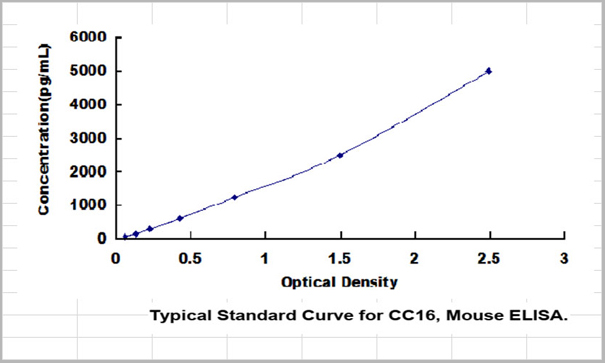 Standard Curve (Sample)
Clara Cell Protein 16 (CC16), ELISA Kit (Cat# AAA23117)
Full Name
Mouse Clara Cell Protein 16 (CC16) Mini Samples ELISA Kit
Gene Names
Scgb1a1; UG; UGB; Utg; CC10; CC16; CCSP; PCB-BP
Reactivity
Mouse
Applications
ELISA
Pricing
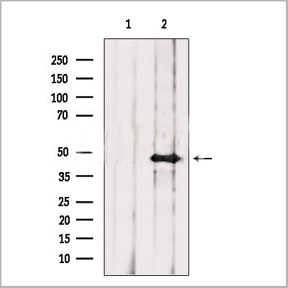
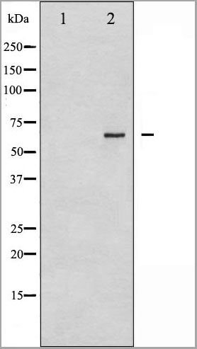
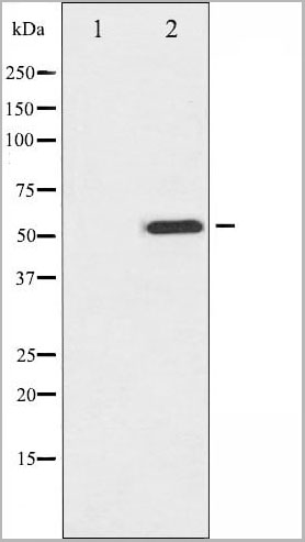
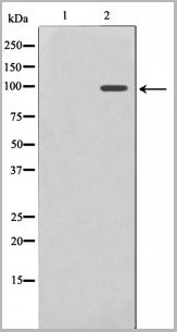
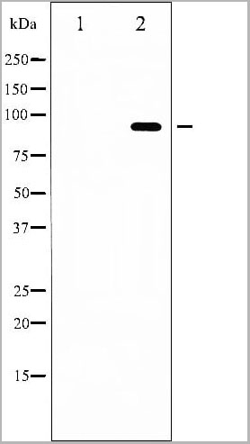
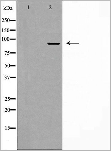
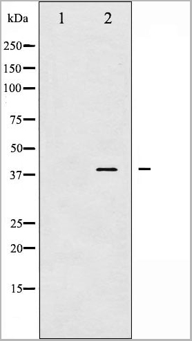
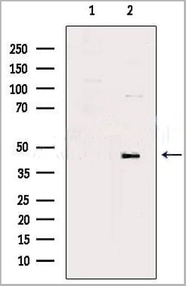
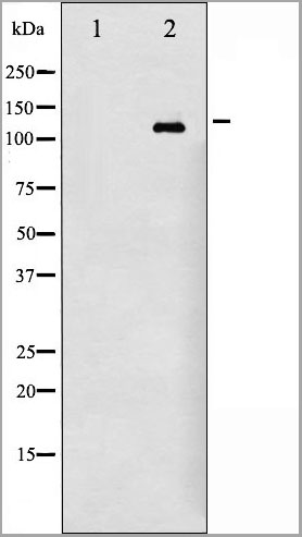
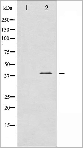
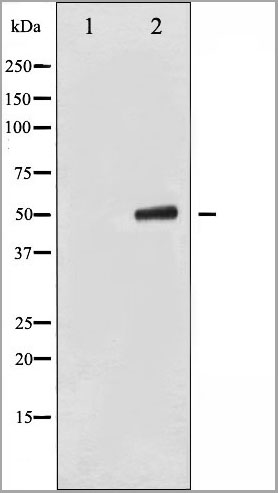
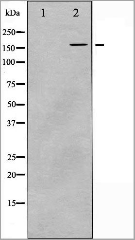
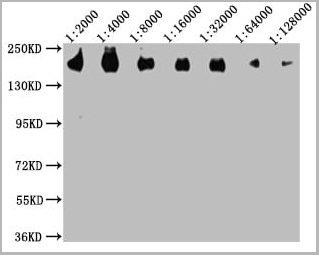
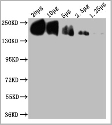
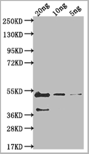 WB (Western Blot)
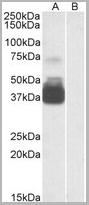
(Positive WB detected in Recombinant proteinAll lanes: gB antibody at 1:2000SecondaryGoat polyclonal to rabbit IgG at 1/50000 dilutionPredicted band size: 43.3 kDaObserved band size: 46 kDa)
WB (Western Blot)
(Positive WB detected in Recombinant proteinAll lanes: gB antibody at 1:2000SecondaryGoat polyclonal to rabbit IgG at 1/50000 dilutionPredicted band size: 43.3 kDaObserved band size: 46 kDa)
gB, Polyclonal Antibody (Cat# AAA27058)
Full Name
Rabbit anti-Epstein-Barr virus (strain B95-8)(HHV-4)(Human herpesvirus 4) gB Polyclonal Antibody
Reactivity
Epstein-Barr virus
Applications
Western Blot
Purity
Protein G
Pricing
 IF (Immunofluorescence)
(AAA31206 staining HepG2 cells by IF/ICC. The samples were fixed with PFA and permeabilized in 0.1% Triton X-100,then blocked in 10% serum for 45 minutes at 25°C. Samples were then incubated with primary Ab(AAA31206) and mouse anti-beta tubulin Ab(T0023) for 1 hour at 37°C. An AlexaFluor594 conjugated goat anti-rabbit IgG(H+L) Ab(Red) and an AlexaFluor488 conjugated goat anti-mouse IgG(H+L) Ab(Green) were used as the secondary antibody.The nuclear counter stain is DAPI(blue).)
IF (Immunofluorescence)
(AAA31206 staining HepG2 cells by IF/ICC. The samples were fixed with PFA and permeabilized in 0.1% Triton X-100,then blocked in 10% serum for 45 minutes at 25°C. Samples were then incubated with primary Ab(AAA31206) and mouse anti-beta tubulin Ab(T0023) for 1 hour at 37°C. An AlexaFluor594 conjugated goat anti-rabbit IgG(H+L) Ab(Red) and an AlexaFluor488 conjugated goat anti-mouse IgG(H+L) Ab(Green) were used as the secondary antibody.The nuclear counter stain is DAPI(blue).)
Activin Receptor Type IA, Polyclonal Antibody (Cat# AAA31206)
Full Name
Activin Receptor Type IA Antibody
Gene Names
ACVR1; FOP; ALK2; SKR1; TSRI; ACTRI; ACVR1A; ACVRLK2
Reactivity
Human, Mouse, Rat
Applications
Western Blot, Immunohistochemistry, Immunofluorescence, Immunocytochemistry
Purity
The antiserum was purified by peptide affinity chromatography using SulfoLink Coupling Resin (Thermo Fisher Scientific).
Pricing
 IF (Immunofluorescence)
(AAA31196 staining A549 cells by IF/ICC. The samples were fixed with PFA and permeabilized in 0.1% Triton X-100,then blocked in 10% serum for 45 minutes at 25°C. Samples were then incubated with primary Ab(AAA31196) and mouse anti-beta tubulin Ab(T0023) for 1 hour at 37°C. An AlexaFluor594 conjugated goat anti-rabbit IgG(H+L) Ab(Red) and an AlexaFluor488 conjugated goat anti-mouse IgG(H+L) Ab(Green) were used as the secondary antibody.The nuclear counter stain is DAPI (blue).)
IF (Immunofluorescence)
(AAA31196 staining A549 cells by IF/ICC. The samples were fixed with PFA and permeabilized in 0.1% Triton X-100,then blocked in 10% serum for 45 minutes at 25°C. Samples were then incubated with primary Ab(AAA31196) and mouse anti-beta tubulin Ab(T0023) for 1 hour at 37°C. An AlexaFluor594 conjugated goat anti-rabbit IgG(H+L) Ab(Red) and an AlexaFluor488 conjugated goat anti-mouse IgG(H+L) Ab(Green) were used as the secondary antibody.The nuclear counter stain is DAPI (blue).)
PRDM16, Polyclonal Antibody (Cat# AAA31196)
Full Name
PRDM16 Antibody
Gene Names
PRDM16; MEL1; KMT8F; LVNC8; PFM13; CMD1LL
Reactivity
Human, Mouse, Rat
Predicted Reactivity: Pig(100%), Horse(100%), Sheep(100%), Rabbit(100%), Dog(100%), Chicken(100%)
Predicted Reactivity: Pig(100%), Horse(100%), Sheep(100%), Rabbit(100%), Dog(100%), Chicken(100%)
Applications
ELISA
Purity
The antiserum was purified by peptide affinity chromatography using SulfoLink Coupling Resin (Thermo Fisher Scientific).
Pricing
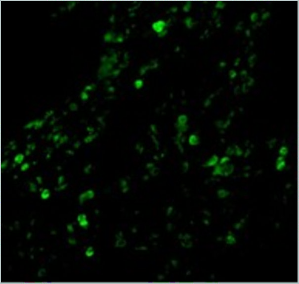
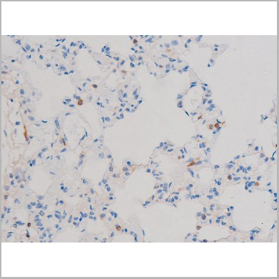
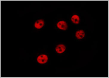
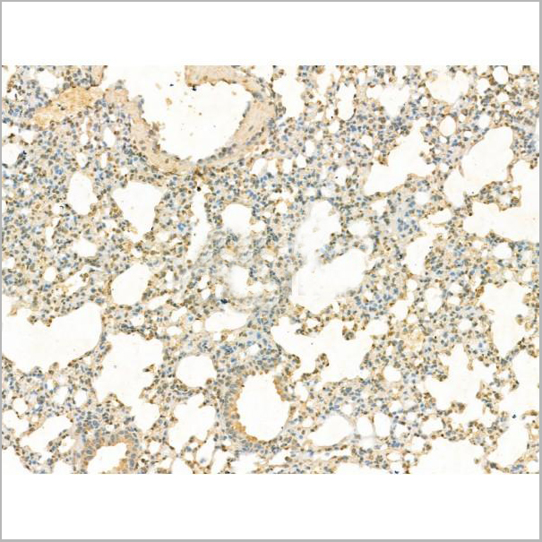
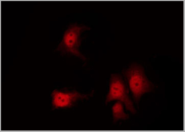
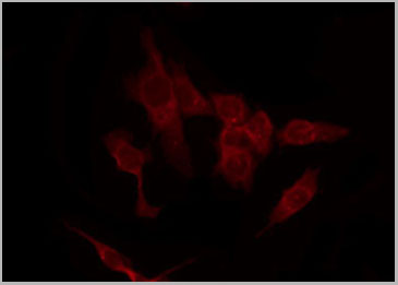
 IF (Immunofluorescence)
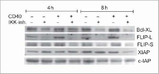
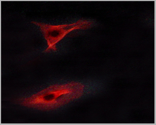
(Immunofluorescence Validation of cIAP in Human Lung Tissue Immunofluorescent analysis of 4% paraformaldehydefixed human lung tissue abeling cIAP with 3325 at 20 ug/mL, followed by goat anti-rabbit IgG secondary antibody at 1/500 dilution (green).)
IF (Immunofluorescence)
(Immunofluorescence Validation of cIAP in Human Lung Tissue Immunofluorescent analysis of 4% paraformaldehydefixed human lung tissue abeling cIAP with 3325 at 20 ug/mL, followed by goat anti-rabbit IgG secondary antibody at 1/500 dilution (green).)
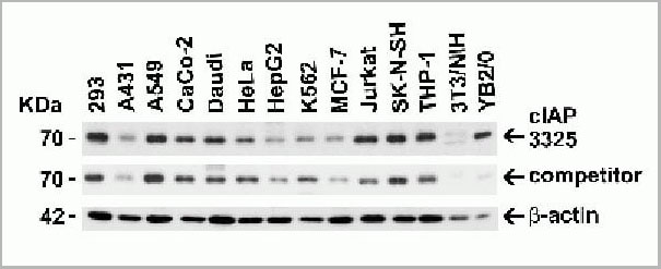
cIAP, Polyclonal Antibody (Cat# AAA10954)
Full Name
cIAP Antibody
Gene Names
BIRC2; API1; MIHB; HIAP2; RNF48; cIAP1; Hiap-2; c-IAP1
Reactivity
Human, Mouse
Applications
Western Blot, Immunohistochemistry, Immunofluorescence
Purity
cIAP Antibody is affinity chromatography purified via peptide column.
Pricing
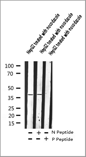
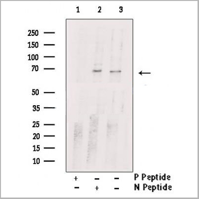
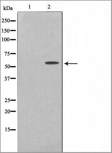
 WB (Western Blot)
(Western blot analysis of Adrenergic Receptor beta2 phosphorylation expression in nocodazole treated HepG2 whole cell lysates, The lane on the left is treated with the antigen-specific peptide.)
WB (Western Blot)
(Western blot analysis of Adrenergic Receptor beta2 phosphorylation expression in nocodazole treated HepG2 whole cell lysates, The lane on the left is treated with the antigen-specific peptide.)
Adrenergic Receptor beta2, Polyclonal Antibody (Cat# AAA30989)
Full Name
Phospho-Adrenergic Receptor beta2 (Ser346) Antibody
Gene Names
ADRB2; BAR; B2AR; ADRBR; ADRB2R; BETA2AR
Reactivity
Human, Mouse, Rat
Applications
Western Blot, Immunohistochemistry, Immunofluorescence, Immunocytochemistry
Purity
Peptide affinity purification
Pricing
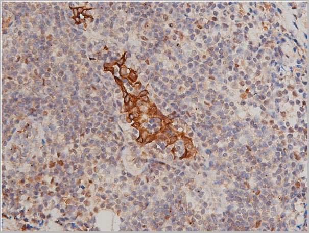
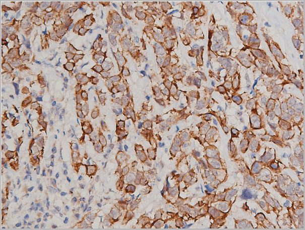
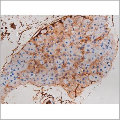
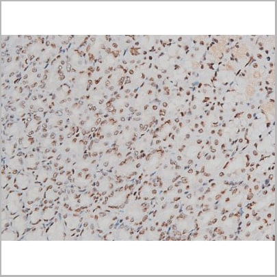
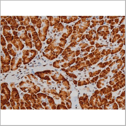
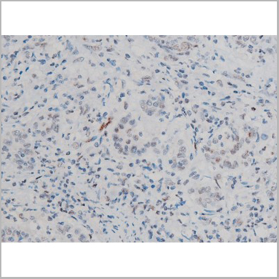
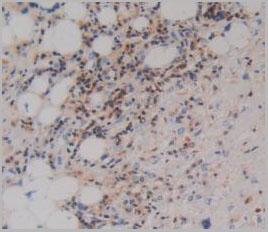
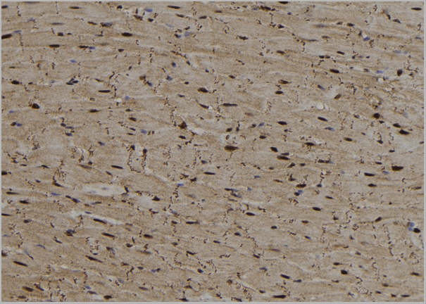
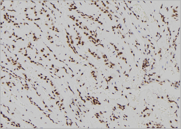
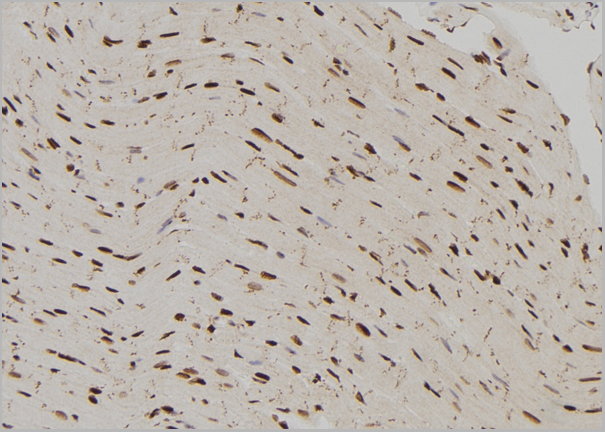
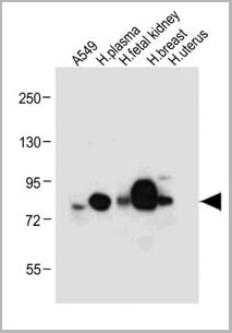
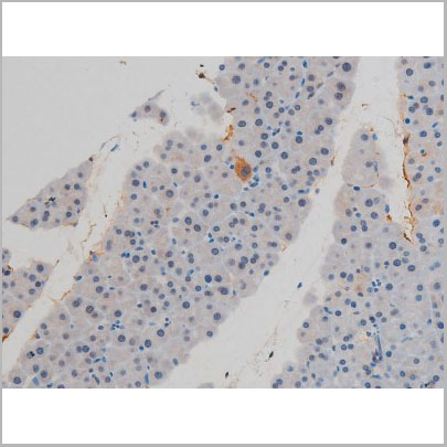
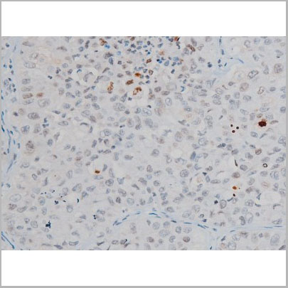
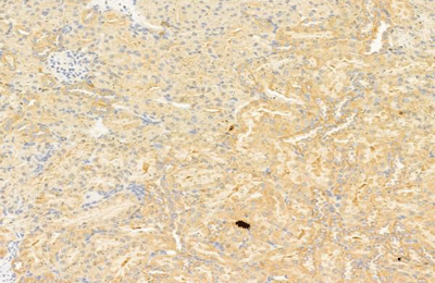
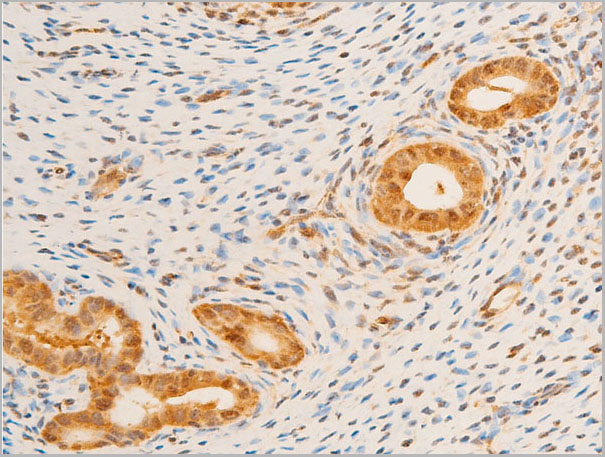
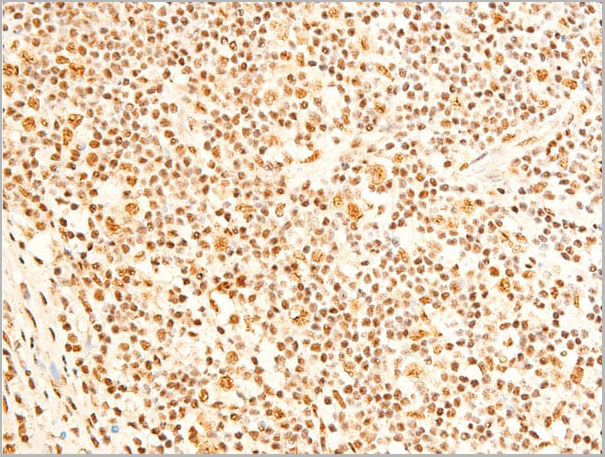
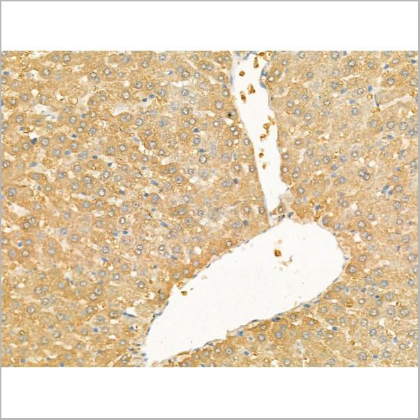
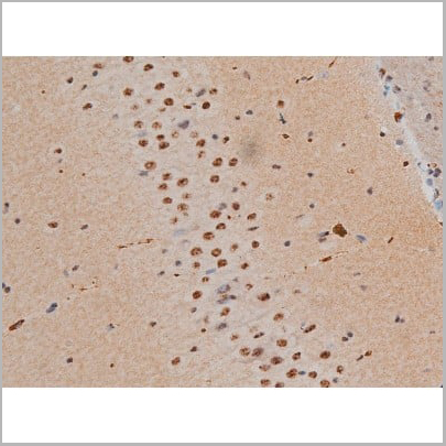
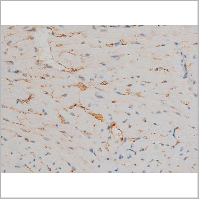
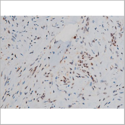
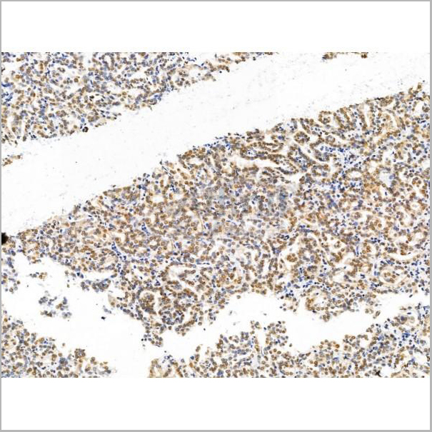
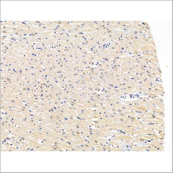
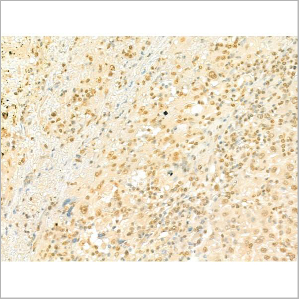
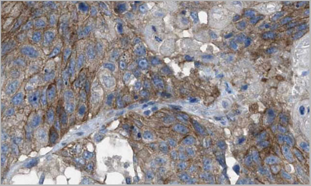
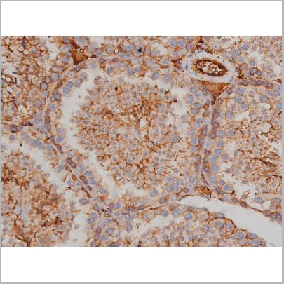
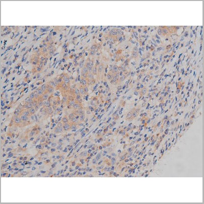
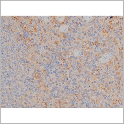
 IHC (Immunohistochemistry)
(AAA30985 at 1/50 staining human breast cancer tissue sections by IHC-P. The tissue was formaldehyde fixed and a heat mediated antigen retrieval step in citrate buffer was performed. The tissue was then blocked and incubated with the antibody for 1.5 hours at 22 degree C. An HRP conjugated goat anti-rabbit antibody was used as the secondary.)
IHC (Immunohistochemistry)
(AAA30985 at 1/50 staining human breast cancer tissue sections by IHC-P. The tissue was formaldehyde fixed and a heat mediated antigen retrieval step in citrate buffer was performed. The tissue was then blocked and incubated with the antibody for 1.5 hours at 22 degree C. An HRP conjugated goat anti-rabbit antibody was used as the secondary.)
Keratin 8, Polyclonal Antibody (Cat# AAA30985)
Full Name
Phospho-Keratin 8 (Ser432) Antibody
Gene Names
KRT8; K8; KO; CK8; CK-8; CYK8; K2C8; CARD2
Reactivity
Human, Mouse, Rat
Applications
Western Blot, Immunohistochemistry, Immunofluorescence, Immunocytochemistry
Purity
From purified rabbit serum by affinity purification via sequential chromatography on phospho-and non-phospho-peptide affinity columns.
Pricing


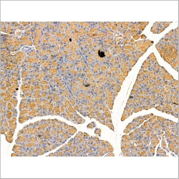
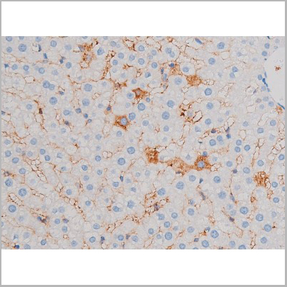
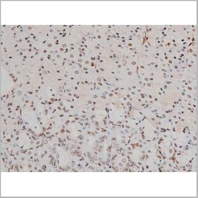
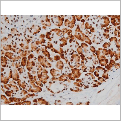
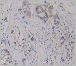
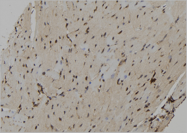
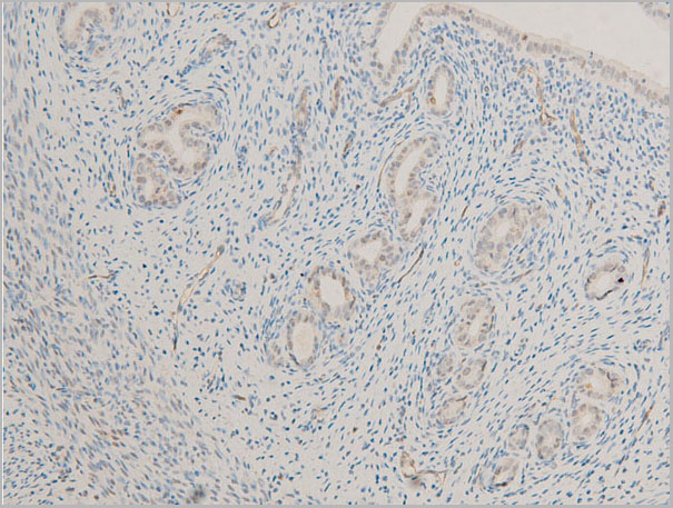
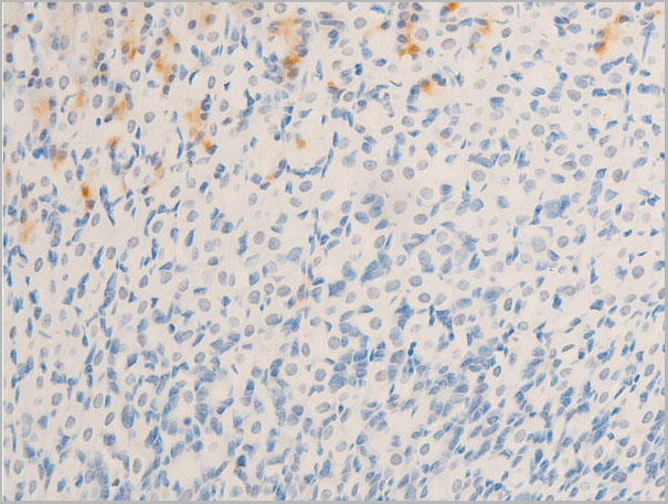
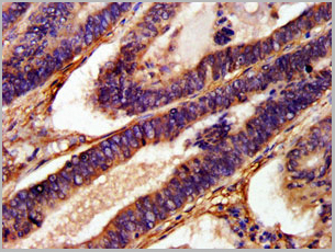
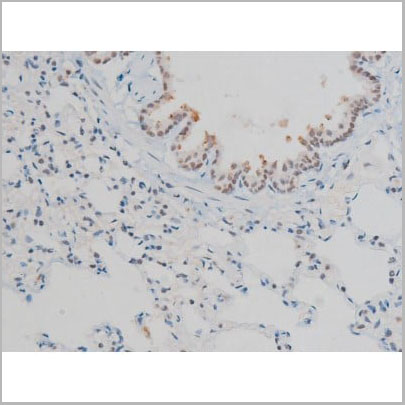
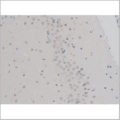
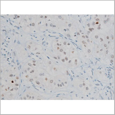
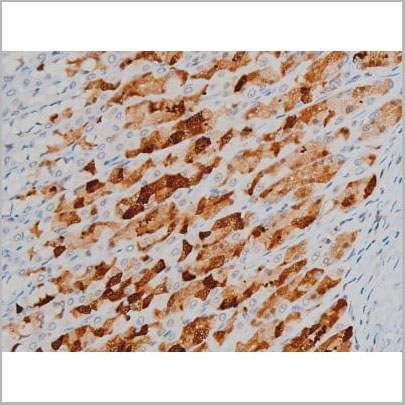
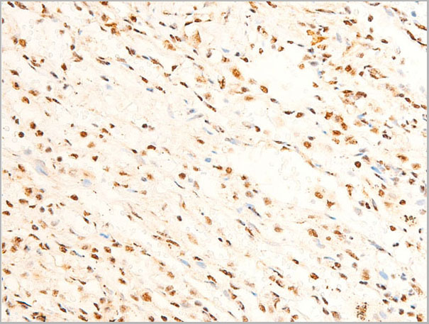
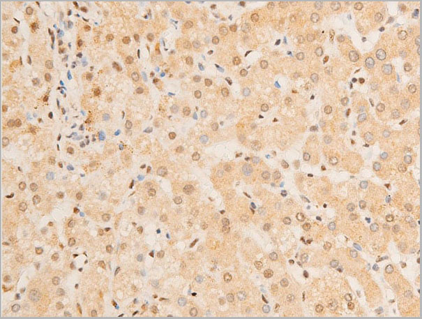
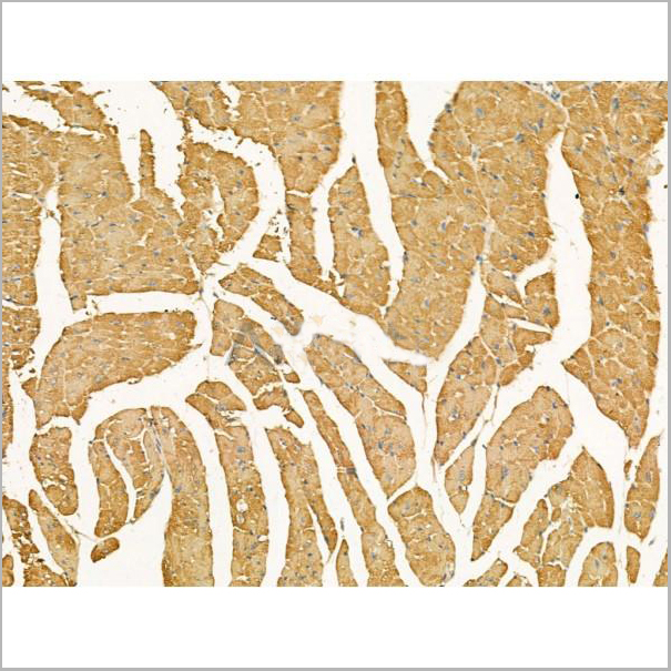
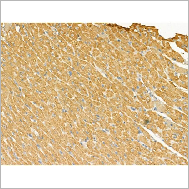
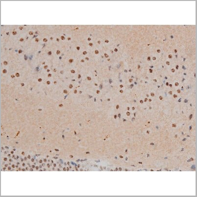
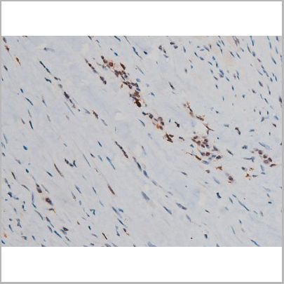
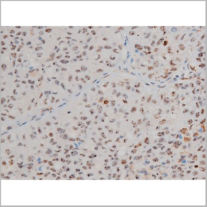
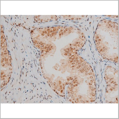
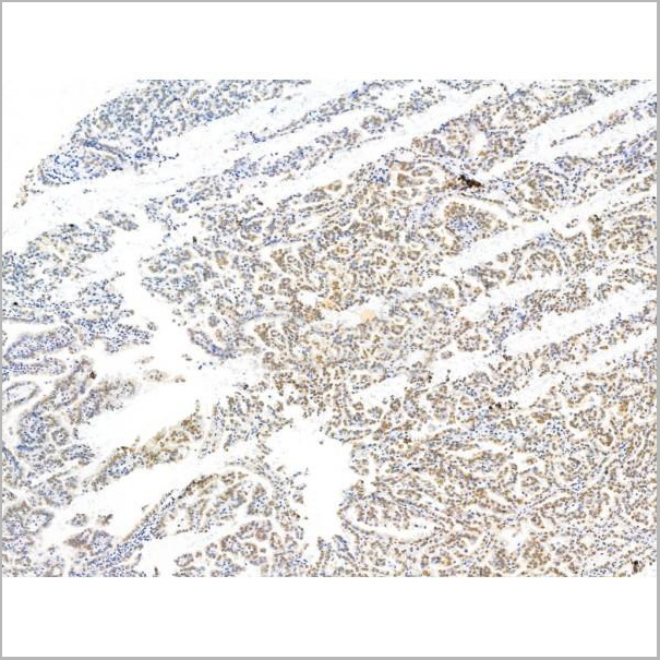
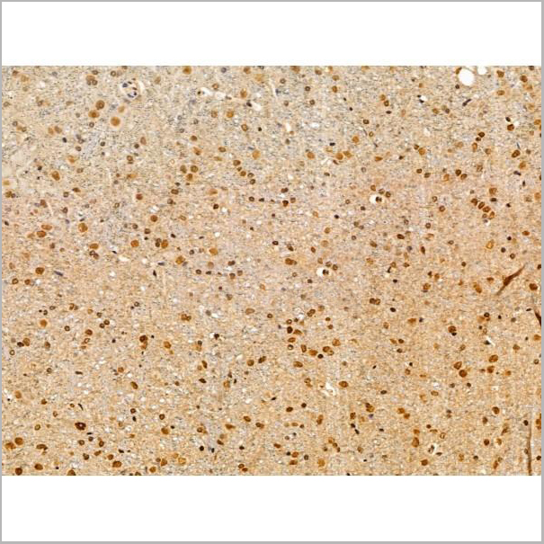
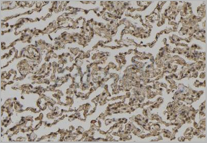
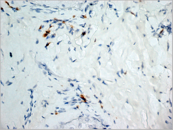
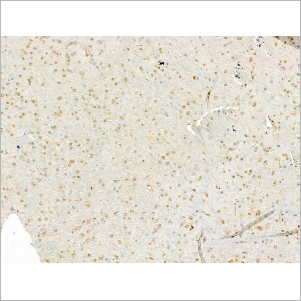
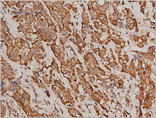
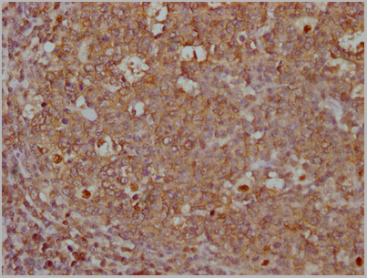
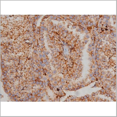
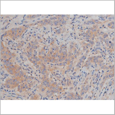
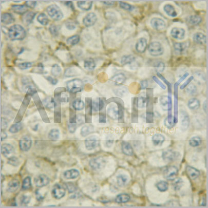
 IHC (Immunohistochemistry)
(AAA31083 at 1/100 staining human breast tissues sections by IHC-P. The tissue was formaldehyde fixed and a heat mediated antigen retrieval step in citrate buffer was performed. The tissue was then blocked and incubated with the antibody for 1.5 hours at 92)
IHC (Immunohistochemistry)
(AAA31083 at 1/100 staining human breast tissues sections by IHC-P. The tissue was formaldehyde fixed and a heat mediated antigen retrieval step in citrate buffer was performed. The tissue was then blocked and incubated with the antibody for 1.5 hours at 92)
EGFR, Polyclonal Antibody (Cat# AAA31083)
Full Name
EGFR Antibody
Gene Names
EGFR; ERBB; HER1; mENA; ERBB1; PIG61; NISBD2
Reactivity
Human, Mouse, Rat
Applications
Western Blot, Immunohistochemistry, Immunofluorescence, Immunocytochemistry
Purity
The antiserum was purified by peptide affinity chromatography using SulfoLink Coupling Resin.
Pricing

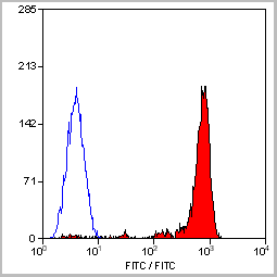
 Application Data
(Staining of human peripheral blood granulocytes with CD18: Alexa Fluor 647)
Application Data
(Staining of human peripheral blood granulocytes with CD18: Alexa Fluor 647)
CD18, Monoclonal Antibody (Cat# AAA11881)
Full Name
RAT ANTI HUMAN CD18:FITC
Gene Names
ITGB2; LAD; CD18; MF17; MFI7; LCAMB; LFA-1; MAC-1
Applications
Flow Cytometry
Pricing


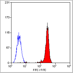
 Application Data
(Staining of human peripheral blood granulocytes with CD18: Alexa Fluor 647)
Application Data
(Staining of human peripheral blood granulocytes with CD18: Alexa Fluor 647)
CD18, Monoclonal Antibody (Cat# AAA11987)
Full Name
RAT ANTI HUMAN CD18
Gene Names
ITGB2; LAD; CD18; MF17; MFI7; LCAMB; LFA-1; MAC-1
Applications
Immunohistochemistry, Flow Cytometry, Immunoprecipitation
Pricing
MERS-COV Spike S1 Antibody IgG (MCSS1-IgG), ELISA Kit (Cat# AAA31554)
Full Name
Human MERS-COV Spike S1 Antibody IgG (MCSS1-IgG) ELISA Kit
Reactivity
Human
Pricing
Cardiolipin, Polyclonal Antibody (Cat# AAA14711)
Full Name
Cardiolipin
Reactivity
Human
Applications
ELISA
Purity
Purified by delipidation, defibrination and ammonium sulfate precipitation.
Pricing
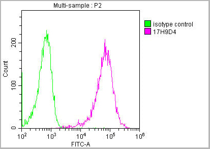
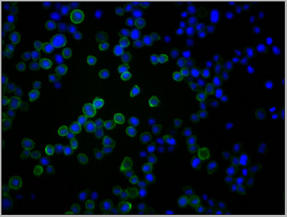
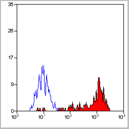
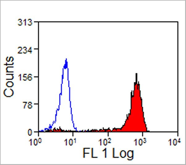
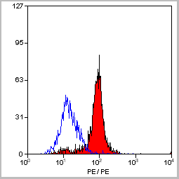
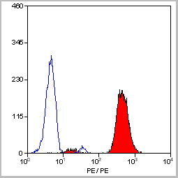
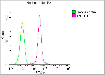 FCM (Flow Cytometry)
(Overlay histogram showing Raji cells stained with (red line) at 1:500. The cells were incubated in 10% normal goat serum to block non-specific protein-protein interactions followed by the antibody (1ug/1*106cells) for 1 h at 4 degree C. The secondary antibody used was FITC-conjugated Goat Anti-Mouse IgG(H+L) at 1/100 dilution for 30min at 4 degree C. Isotype control antibody (green line) was mouse IgG2b (1ug/1*106cells) used under the same conditions. Acquisition of >10,000 events was performed.)
FCM (Flow Cytometry)
(Overlay histogram showing Raji cells stained with (red line) at 1:500. The cells were incubated in 10% normal goat serum to block non-specific protein-protein interactions followed by the antibody (1ug/1*106cells) for 1 h at 4 degree C. The secondary antibody used was FITC-conjugated Goat Anti-Mouse IgG(H+L) at 1/100 dilution for 30min at 4 degree C. Isotype control antibody (green line) was mouse IgG2b (1ug/1*106cells) used under the same conditions. Acquisition of >10,000 events was performed.)
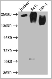
CD45, Monoclonal Antibody (Cat# AAA27049)
Full Name
CD45 Monoclonal Antibody
Gene Names
PTPRC; LCA; LY5; B220; CD45; L-CA; T200; CD45R; GP180
Reactivity
Human
Applications
Western Blot, Immunohistochemistry, Immunofluorescence, Flow Cytometry
Purity
>95%, Protein A purified
Pricing


 IF (Immunofluorescence)
(AAA31216 staining A549 cells by IF/ICC. The samples were fixed with PFA and permeabilized in 0.1% Triton X-100,then blocked in 10% serum for 45 minutes at 25°C. Samples were then incubated with primary Ab(AAA31216) and mouse anti-beta tubulin Ab(T0023) for 1 hour at 37°C. An AlexaFluor594 conjugated goat anti-rabbit IgG(H+L) Ab(Red) and an AlexaFluor488 conjugated goat anti-mouse IgG(H+L) Ab(Green) were used as the secondary antibody.The nuclear counter stain is DAPI (blue).)
IF (Immunofluorescence)
(AAA31216 staining A549 cells by IF/ICC. The samples were fixed with PFA and permeabilized in 0.1% Triton X-100,then blocked in 10% serum for 45 minutes at 25°C. Samples were then incubated with primary Ab(AAA31216) and mouse anti-beta tubulin Ab(T0023) for 1 hour at 37°C. An AlexaFluor594 conjugated goat anti-rabbit IgG(H+L) Ab(Red) and an AlexaFluor488 conjugated goat anti-mouse IgG(H+L) Ab(Green) were used as the secondary antibody.The nuclear counter stain is DAPI (blue).)
FKBP25, Polyclonal Antibody (Cat# AAA31216)
Full Name
FKBP25 Antibody
Gene Names
FKBP3; FKBP-3; FKBP25; PPIase; FKBP-25
Reactivity
Human, Mouse, Rat
Predicted Reactivity: Pig(100%), Bovine(100%), Horse(100%), Sheep(100%), Rabbit(100%), Dog(100%), Xenopus(100%)
Predicted Reactivity: Pig(100%), Bovine(100%), Horse(100%), Sheep(100%), Rabbit(100%), Dog(100%), Xenopus(100%)
Applications
ELISA
Purity
The antiserum was purified by peptide affinity chromatography using SulfoLink Coupling Resin (Thermo Fisher Scientific).
Pricing
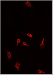
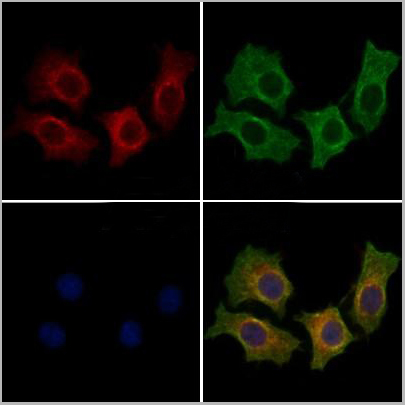
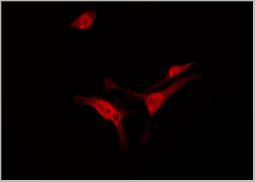
 IF (Immunofluorescence)
(AAA31071 staining HuvEc by IF/ICC. The sample were fixed with PFA and permeabilized in 0.1% Triton X-100, then blocked in 10% serum for 45 minutes at 25 degree C. The primary antibody was diluted at 1/200 and incubated with the sample for 1 hour at 37 degree C. An Alexa Fluor 594 conjugated goat anti-rabbit IgG (H+L) Ab, diluted at 1/600, was used as the secondary antibody.)
IF (Immunofluorescence)
(AAA31071 staining HuvEc by IF/ICC. The sample were fixed with PFA and permeabilized in 0.1% Triton X-100, then blocked in 10% serum for 45 minutes at 25 degree C. The primary antibody was diluted at 1/200 and incubated with the sample for 1 hour at 37 degree C. An Alexa Fluor 594 conjugated goat anti-rabbit IgG (H+L) Ab, diluted at 1/600, was used as the secondary antibody.)
HER4, Polyclonal Antibody (Cat# AAA31071)
Full Name
Phospho-HER4 (Tyr1284) Antibody
Gene Names
ERBB4; HER4; ALS19; p180erbB4
Reactivity
Human, Mouse, Rat
Applications
Western Blot, Immunohistochemistry, Immunofluorescence, Immunocytochemistry
Purity
From purified rabbit serum by affinity purification via sequential chromatography on phospho-and non-phospho-peptide affinity columns.
Pricing
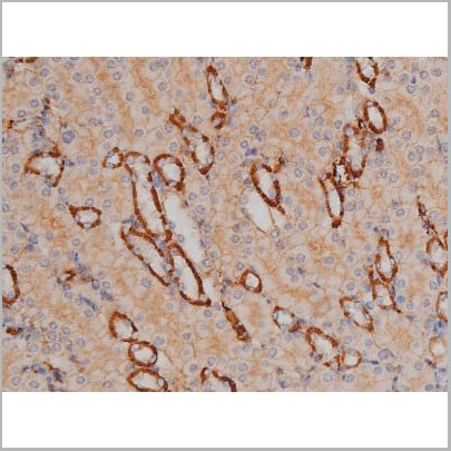
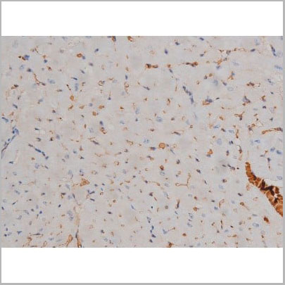
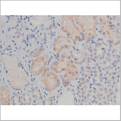
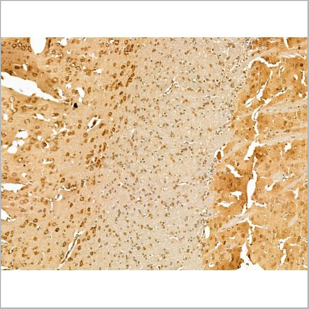
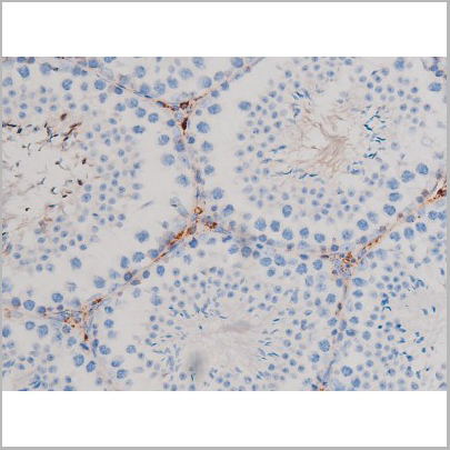
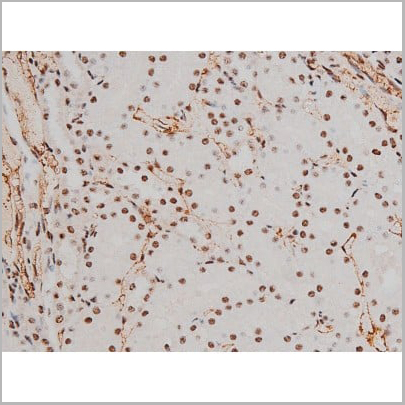
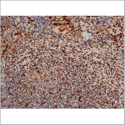
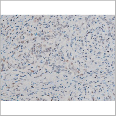
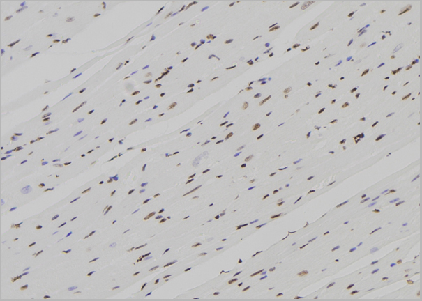
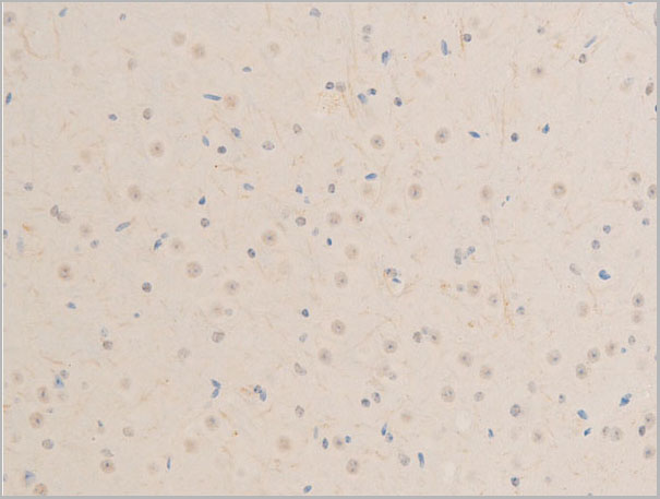
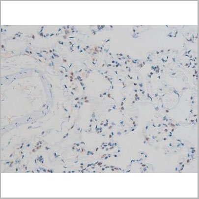
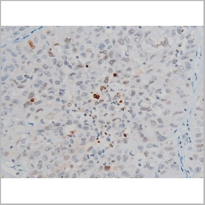
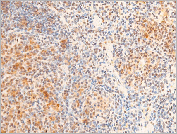
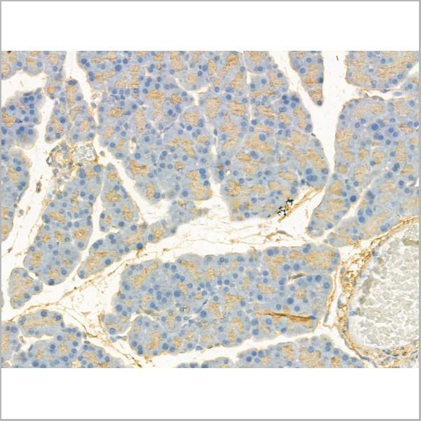
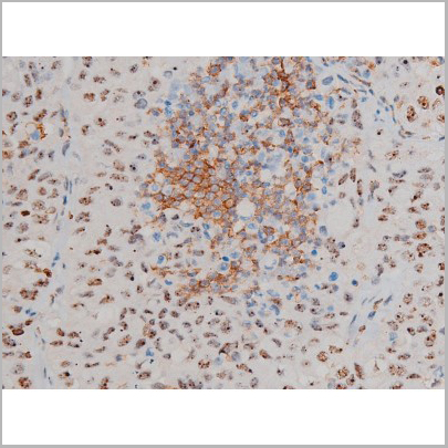
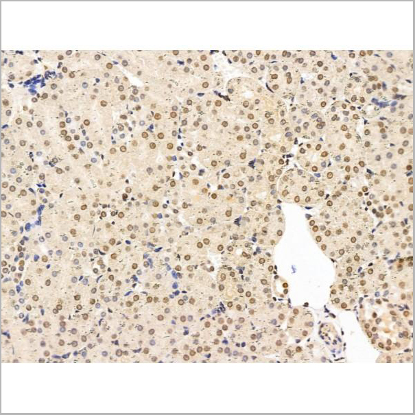
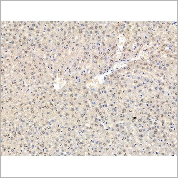
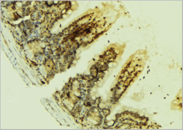
 IHC (Immunohistochemistry)
(AAA30986 at 1/200 staining Rat spleen tissue sections by IHC-P. The tissue was formaldehyde fixed and a heat mediated antigen retrieval step in citrate buffer was performed. The tissue was then blocked and incubated with the antibody for 1.5 hours at 22 degree C. An HRP conjugated goat anti-rabbit antibody was used as the secondary.)
IHC (Immunohistochemistry)
(AAA30986 at 1/200 staining Rat spleen tissue sections by IHC-P. The tissue was formaldehyde fixed and a heat mediated antigen retrieval step in citrate buffer was performed. The tissue was then blocked and incubated with the antibody for 1.5 hours at 22 degree C. An HRP conjugated goat anti-rabbit antibody was used as the secondary.)
Fyn, Polyclonal Antibody (Cat# AAA30986)
Full Name
Phospho-Fyn (Tyr530) Antibody
Gene Names
FYN; SLK; SYN; p59-FYN
Reactivity
Human, Mouse, Rat
Applications
Western Blot, Immunohistochemistry, Immunofluorescence, Immunocytochemistry
Purity
From purified rabbit serum by affinity purification via sequential chromatography on phospho-and non-phospho-peptide affinity columns.
Pricing