Filters
Clonality
Type
Reactivity
Gene Name
Isotype
Host
Application
Clone
628 results for "Loading control Antibodies" - showing 600-628
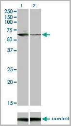
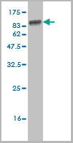
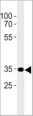
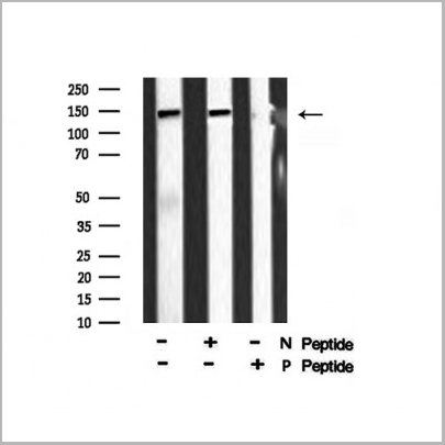
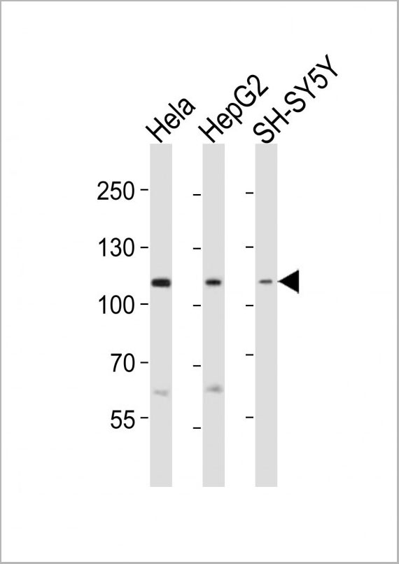
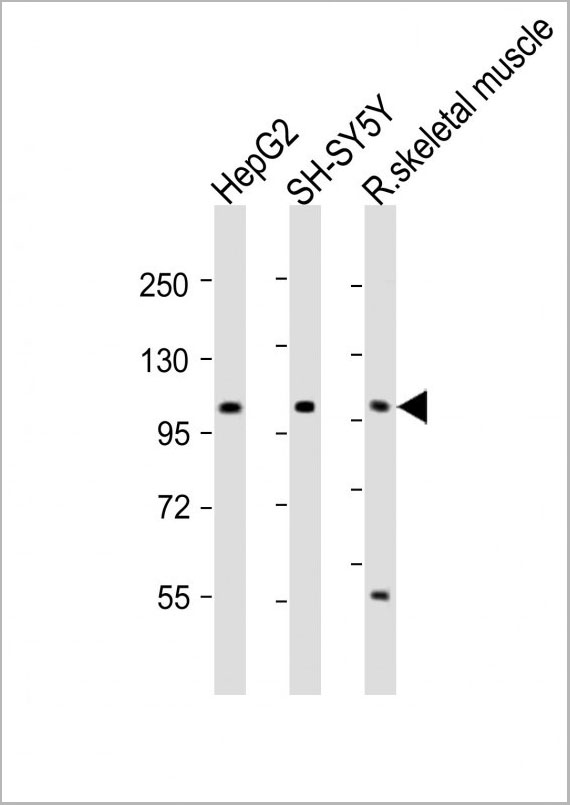
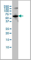
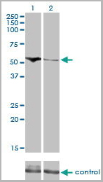
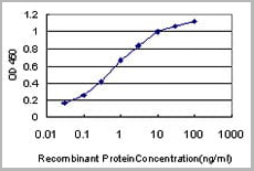
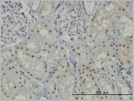
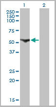
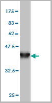
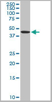
ENO3, Monoclonal Antibody (Cat# AAA24202)
Full Name
ENO3 (Enolase 3, 2-phospho-D-glycerate Hydro-lyase, beta-Enolase, Muscle-specific Enolase, MSE, Skeletal Muscle Enolase) (AP)
Gene Names
ENO3; MSE; GSD13
Reactivity
Human
Applications
EIA, IHC, WB
Purity
Purified by Protein A Affinity Chromatography.
Pricing
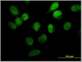
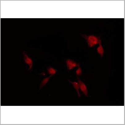
 IF (Immunofluorescence)
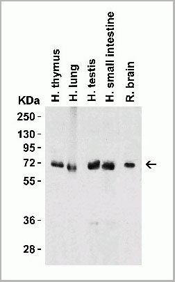
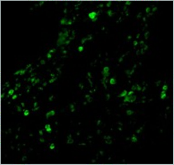
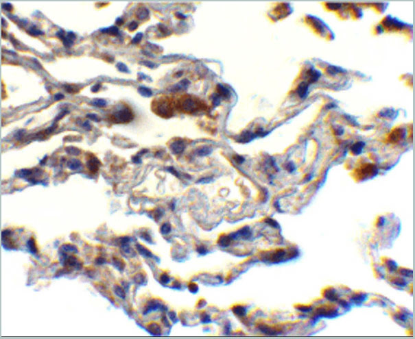
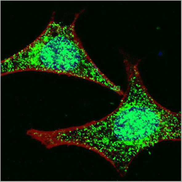
(Immunofluorescence Validation of cIAP in Human Lung Tissue Immunofluorescent analysis of 4% paraformaldehydefixed human lung tissue abeling cIAP with 3325 at 20 ug/mL, followed by goat anti-rabbit IgG secondary antibody at 1/500 dilution (green).)
IF (Immunofluorescence)
(Immunofluorescence Validation of cIAP in Human Lung Tissue Immunofluorescent analysis of 4% paraformaldehydefixed human lung tissue abeling cIAP with 3325 at 20 ug/mL, followed by goat anti-rabbit IgG secondary antibody at 1/500 dilution (green).)
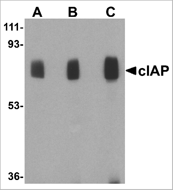
cIAP, Polyclonal Antibody (Cat# AAA10954)
Full Name
cIAP Antibody
Gene Names
BIRC2; API1; MIHB; HIAP2; RNF48; cIAP1; Hiap-2; c-IAP1
Reactivity
Human, Mouse
Applications
Western Blot, Immunohistochemistry, Immunofluorescence
Purity
cIAP Antibody is affinity chromatography purified via peptide column.
Pricing
 WB (Western Blot)
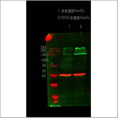
(Western blot analysis of ZO-1 using various lysatesLanes 1 - 2: Merged signal (red and green). Green - AAA31075 observed at 195 kDa. Red - loading control, , observed at 55 kDa. Blots were developed with Goat Anti- Rabbit IgG(H+L) FITC–conjugated and Goat Anti-Mouse IgG(H+L) Alexa Fluor 594–conjugated secondary antibodies)
WB (Western Blot)
(Western blot analysis of ZO-1 using various lysatesLanes 1 - 2: Merged signal (red and green). Green - AAA31075 observed at 195 kDa. Red - loading control, , observed at 55 kDa. Blots were developed with Goat Anti- Rabbit IgG(H+L) FITC–conjugated and Goat Anti-Mouse IgG(H+L) Alexa Fluor 594–conjugated secondary antibodies)
ZO 1, Polyclonal Antibody (Cat# AAA31075)
Full Name
ZO 1 Antibody
Gene Names
TJP1; ZO-1
Reactivity
Human, Mouse, Rat, Pig, Monkey
Applications
Western Blot, Immunohistochemistry, Immunofluorescence, Immunocytochemistry
Purity
The antiserum was purified by peptide affinity chromatography using SulfoLink Coupling Resin
Pricing


 Application Data
(Published customer image: Mouse anti V5 tag antibody, clone SV5-Pk1 used for the detection of V5 tagged WEEV_nsP3 protein by western blotting and immunofluorescenceImage caption: WEEV nsP3 interaction with host IKKbeta. A) U87MGs were transfected in a 6-well plate with 5 ug of pUC19 and WEEV_nsP3_HA for 24 hours. Cell lysates were resolved using SDS-PAGE and subsequently immunoblotted with V5 antibody and beta-actin served as a loading control. B) U87MGs were transfected with WEEV_nsP3_V5; cells were fixed after 24 hours and stained with antibodies against the endogenous IKKbeta and the V5 tag. Cells were incubated with appropriate secondary Alexa Fluor antibodies and the nuclei stained with DAPI. Co-localization of IKKbeta with WEEV_nsP3_V5 (yellow) was observed as shown by the arrows. B) Panels E -H serve as an example of transfected cells in a given field of view that show co-localization of IKKbeta and WEEV_nsP3_V5 24 hours post transfection. Panels I-L represent magnified images of other cells showing co-localization of IKKbeta and WEEV_nsP3_V5. Panel M is a magnified image of panel L. The co-localization was confirmed by Z-stack analysis. Co-localization was calculated to be approximately in 61% of cells (163 cells were counted of which 44% demonstrated expression of nsP3. Of those cells that expressed nsP3, 61% showed co-localization of both proteins). Images were taken using Nikon Eclipse TE2000-U at 60x magnification and are representative of 2 independent experiments.From: Amaya M, Voss K, Sampey G, Senina S, de la Fuente C, et al. (2014) The Role of IKKbeta in Venezuelan Equine Encephalitis Virus Infection. PLoS ONE 9(2): e86745.)
Application Data
(Published customer image: Mouse anti V5 tag antibody, clone SV5-Pk1 used for the detection of V5 tagged WEEV_nsP3 protein by western blotting and immunofluorescenceImage caption: WEEV nsP3 interaction with host IKKbeta. A) U87MGs were transfected in a 6-well plate with 5 ug of pUC19 and WEEV_nsP3_HA for 24 hours. Cell lysates were resolved using SDS-PAGE and subsequently immunoblotted with V5 antibody and beta-actin served as a loading control. B) U87MGs were transfected with WEEV_nsP3_V5; cells were fixed after 24 hours and stained with antibodies against the endogenous IKKbeta and the V5 tag. Cells were incubated with appropriate secondary Alexa Fluor antibodies and the nuclei stained with DAPI. Co-localization of IKKbeta with WEEV_nsP3_V5 (yellow) was observed as shown by the arrows. B) Panels E -H serve as an example of transfected cells in a given field of view that show co-localization of IKKbeta and WEEV_nsP3_V5 24 hours post transfection. Panels I-L represent magnified images of other cells showing co-localization of IKKbeta and WEEV_nsP3_V5. Panel M is a magnified image of panel L. The co-localization was confirmed by Z-stack analysis. Co-localization was calculated to be approximately in 61% of cells (163 cells were counted of which 44% demonstrated expression of nsP3. Of those cells that expressed nsP3, 61% showed co-localization of both proteins). Images were taken using Nikon Eclipse TE2000-U at 60x magnification and are representative of 2 independent experiments.From: Amaya M, Voss K, Sampey G, Senina S, de la Fuente C, et al. (2014) The Role of IKKbeta in Venezuelan Equine Encephalitis Virus Infection. PLoS ONE 9(2): e86745.)
V5-TAG, Monoclonal Antibody (Cat# AAA12081)
Full Name
MOUSE ANTI V5-TAG:HRP
Applications
Western Blot
Pricing
 WB (Western Blot)
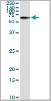
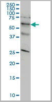
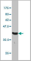
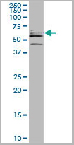
(STK33 monoclonal antibody, Western Blot analysis of STK33 expression in HeLa.)
WB (Western Blot)
(STK33 monoclonal antibody, Western Blot analysis of STK33 expression in HeLa.)
STK33, Monoclonal Antibody (Cat# AAA25856)
Full Name
STK33 (Serine/Threonine Kinase 33) (PE)
Reactivity
Human
Applications
Immunofluorescence, Immunoprecipitation, Western Blot
Purity
Purified by Protein A Affinity Chromatography.
Pricing


 Application Data
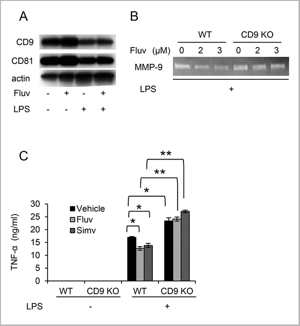
(Staining of mouse spleen with Hamster anti Mouse CD81: Alexa Fluor 488)
Application Data
(Staining of mouse spleen with Hamster anti Mouse CD81: Alexa Fluor 488)
CD81, Monoclonal Antibody (Cat# AAA11869)
Full Name
HAMSTER ANTI MOUSE CD81:FITC
Gene Names
Cd81; Tapa1; Tapa-1; Tspan28
Applications
Flow Cytometry
Pricing


 Application Data
(Staining of mouse spleen with Hamster anti Mouse CD81: Alexa Fluor 488)
Application Data
(Staining of mouse spleen with Hamster anti Mouse CD81: Alexa Fluor 488)
CD81, Monoclonal Antibody (Cat# AAA12033)
Full Name
HAMSTER ANTI MOUSE CD81:RPE
Gene Names
Cd81; Tapa1; Tapa-1; Tspan28
Applications
Flow Cytometry
Pricing
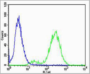
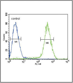 FCM (Flow Cytometry)
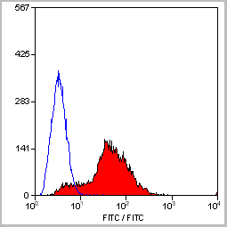
(CB2 Antibody (C-term) flow cytometric analysis of Jurkat cells (right histogram) compared to a negative control cell (left histogram).FITC-conjugated goat-anti-rabbit secondary antibodies were used for the analysis.)
FCM (Flow Cytometry)
(CB2 Antibody (C-term) flow cytometric analysis of Jurkat cells (right histogram) compared to a negative control cell (left histogram).FITC-conjugated goat-anti-rabbit secondary antibodies were used for the analysis.)
CB2, Polyclonal Antibody (Cat# AAA28672)
Full Name
CB2 Antibody (C-term)
Gene Names
CNR2; CB2; CX5; CB-2
Reactivity
Human
Applications
Immunohistochemistry, Flow Cytometry, Western Blot
Purity
This antibody is purified through a protein A column, followed by peptide affinity purification.
Pricing
 Application Data
(At 25 degree C. The primary antibody was diluted at 1/200 and incubated with the sample for 1 hour at 37 degree C. An Alexa Fluor 594 conjugated goat anti-rabbit IgG (H+L) Ab, diluted at 1/600, was used as the secondary antibody.)
Application Data
(At 25 degree C. The primary antibody was diluted at 1/200 and incubated with the sample for 1 hour at 37 degree C. An Alexa Fluor 594 conjugated goat anti-rabbit IgG (H+L) Ab, diluted at 1/600, was used as the secondary antibody.)
eNOS, Polyclonal Antibody (Cat# AAA31394)
Full Name
Phospho-eNOS (Ser1179) Antibody
Gene Names
NOS3; eNOS; ECNOS
Reactivity
Human, Mouse, Rat
Predicted Reactivity: Pig (100%), Bovine (100%), Rabbit (100%), Dog (100%)
Predicted Reactivity: Pig (100%), Bovine (100%), Rabbit (100%), Dog (100%)
Applications
Western Blot, Immunohistochemistry, Immunofluorescence, Immunocytochemistry, Peptide ELISA
Purity
The antibody is from purified rabbit serum by affinity purification via sequential chromatography on phospho-peptide and non-phospho-peptide affinity columns.
Pricing


 Application Data
(Published customer image: Mouse anti V5 tag antibody, clone SV5-Pk1 used for the detection of V5 tagged WEEV_nsP3 protein by western blotting and immunofluorescenceImage caption: WEEV nsP3 interaction with host IKKbeta. A) U87MGs were transfected in a 6-well plate with 5 ug of pUC19 and WEEV_nsP3_HA for 24 hours. Cell lysates were resolved using SDS-PAGE and subsequently immunoblotted with V5 antibody and beta-actin served as a loading control. B) U87MGs were transfected with WEEV_nsP3_V5; cells were fixed after 24 hours and stained with antibodies against the endogenous IKKbeta and the V5 tag. Cells were incubated with appropriate secondary Alexa Fluor antibodies and the nuclei stained with DAPI. Co-localization of IKKbeta with WEEV_nsP3_V5 (yellow) was observed as shown by the arrows. B) Panels E -H serve as an example of transfected cells in a given field of view that show co-localization of IKKbeta and WEEV_nsP3_V5 24 hours post transfection. Panels I-L represent magnified images of other cells showing co-localization of IKKbeta and WEEV_nsP3_V5. Panel M is a magnified image of panel L. The co-localization was confirmed by Z-stack analysis. Co-localization was calculated to be approximately in 61% of cells (163 cells were counted of which 44% demonstrated expression of nsP3. Of those cells that expressed nsP3, 61% showed co-localization of both proteins). Images were taken using Nikon Eclipse TE2000-U at 60x magnification and are representative of 2 independent experiments.From: Amaya M, Voss K, Sampey G, Senina S, de la Fuente C, et al. (2014) The Role of IKKbeta in Venezuelan Equine Encephalitis Virus Infection. PLoS ONE 9(2): e86745.)
Application Data
(Published customer image: Mouse anti V5 tag antibody, clone SV5-Pk1 used for the detection of V5 tagged WEEV_nsP3 protein by western blotting and immunofluorescenceImage caption: WEEV nsP3 interaction with host IKKbeta. A) U87MGs were transfected in a 6-well plate with 5 ug of pUC19 and WEEV_nsP3_HA for 24 hours. Cell lysates were resolved using SDS-PAGE and subsequently immunoblotted with V5 antibody and beta-actin served as a loading control. B) U87MGs were transfected with WEEV_nsP3_V5; cells were fixed after 24 hours and stained with antibodies against the endogenous IKKbeta and the V5 tag. Cells were incubated with appropriate secondary Alexa Fluor antibodies and the nuclei stained with DAPI. Co-localization of IKKbeta with WEEV_nsP3_V5 (yellow) was observed as shown by the arrows. B) Panels E -H serve as an example of transfected cells in a given field of view that show co-localization of IKKbeta and WEEV_nsP3_V5 24 hours post transfection. Panels I-L represent magnified images of other cells showing co-localization of IKKbeta and WEEV_nsP3_V5. Panel M is a magnified image of panel L. The co-localization was confirmed by Z-stack analysis. Co-localization was calculated to be approximately in 61% of cells (163 cells were counted of which 44% demonstrated expression of nsP3. Of those cells that expressed nsP3, 61% showed co-localization of both proteins). Images were taken using Nikon Eclipse TE2000-U at 60x magnification and are representative of 2 independent experiments.From: Amaya M, Voss K, Sampey G, Senina S, de la Fuente C, et al. (2014) The Role of IKKbeta in Venezuelan Equine Encephalitis Virus Infection. PLoS ONE 9(2): e86745.)
V5-TAG, Monoclonal Antibody (Cat# AAA11864)
Full Name
MOUSE ANTI V5-TAG:FITC
Applications
Immunofluorescence
Pricing


 Application Data
(Published customer image: Mouse anti V5 tag antibody, clone SV5-Pk1 used for the detection of V5 tagged WEEV_nsP3 protein by western blotting and immunofluorescenceImage caption: WEEV nsP3 interaction with host IKKbeta. A) U87MGs were transfected in a 6-well plate with 5 ug of pUC19 and WEEV_nsP3_HA for 24 hours. Cell lysates were resolved using SDS-PAGE and subsequently immunoblotted with V5 antibody and beta-actin served as a loading control. B) U87MGs were transfected with WEEV_nsP3_V5; cells were fixed after 24 hours and stained with antibodies against the endogenous IKKbeta and the V5 tag. Cells were incubated with appropriate secondary Alexa Fluor antibodies and the nuclei stained with DAPI. Co-localization of IKKbeta with WEEV_nsP3_V5 (yellow) was observed as shown by the arrows. B) Panels E -H serve as an example of transfected cells in a given field of view that show co-localization of IKKbeta and WEEV_nsP3_V5 24 hours post transfection. Panels I-L represent magnified images of other cells showing co-localization of IKKbeta and WEEV_nsP3_V5. Panel M is a magnified image of panel L. The co-localization was confirmed by Z-stack analysis. Co-localization was calculated to be approximately in 61% of cells (163 cells were counted of which 44% demonstrated expression of nsP3. Of those cells that expressed nsP3, 61% showed co-localization of both proteins). Images were taken using Nikon Eclipse TE2000-U at 60x magnification and are representative of 2 independent experiments.From: Amaya M, Voss K, Sampey G, Senina S, de la Fuente C, et al. (2014) The Role of IKKbeta in Venezuelan Equine Encephalitis Virus Infection. PLoS ONE 9(2): e86745.)
Application Data
(Published customer image: Mouse anti V5 tag antibody, clone SV5-Pk1 used for the detection of V5 tagged WEEV_nsP3 protein by western blotting and immunofluorescenceImage caption: WEEV nsP3 interaction with host IKKbeta. A) U87MGs were transfected in a 6-well plate with 5 ug of pUC19 and WEEV_nsP3_HA for 24 hours. Cell lysates were resolved using SDS-PAGE and subsequently immunoblotted with V5 antibody and beta-actin served as a loading control. B) U87MGs were transfected with WEEV_nsP3_V5; cells were fixed after 24 hours and stained with antibodies against the endogenous IKKbeta and the V5 tag. Cells were incubated with appropriate secondary Alexa Fluor antibodies and the nuclei stained with DAPI. Co-localization of IKKbeta with WEEV_nsP3_V5 (yellow) was observed as shown by the arrows. B) Panels E -H serve as an example of transfected cells in a given field of view that show co-localization of IKKbeta and WEEV_nsP3_V5 24 hours post transfection. Panels I-L represent magnified images of other cells showing co-localization of IKKbeta and WEEV_nsP3_V5. Panel M is a magnified image of panel L. The co-localization was confirmed by Z-stack analysis. Co-localization was calculated to be approximately in 61% of cells (163 cells were counted of which 44% demonstrated expression of nsP3. Of those cells that expressed nsP3, 61% showed co-localization of both proteins). Images were taken using Nikon Eclipse TE2000-U at 60x magnification and are representative of 2 independent experiments.From: Amaya M, Voss K, Sampey G, Senina S, de la Fuente C, et al. (2014) The Role of IKKbeta in Venezuelan Equine Encephalitis Virus Infection. PLoS ONE 9(2): e86745.)
V5-TAG, Monoclonal Antibody (Cat# AAA11930)
Full Name
MOUSE ANTI V5-TAG
Applications
Immunohistochemistry, Flow Cytometry, Immunofluorescence, Immunoprecipitation, Western Blot, Radioimmunoassay
Pricing


 Application Data
(Published customer image: Mouse anti V5 tag antibody, clone SV5-Pk1 used for the detection of V5 tagged WEEV_nsP3 protein by western blotting and immunofluorescenceImage caption: WEEV nsP3 interaction with host IKKbeta. A) U87MGs were transfected in a 6-well plate with 5 ug of pUC19 and WEEV_nsP3_HA for 24 hours. Cell lysates were resolved using SDS-PAGE and subsequently immunoblotted with V5 antibody and beta-actin served as a loading control. B) U87MGs were transfected with WEEV_nsP3_V5; cells were fixed after 24 hours and stained with antibodies against the endogenous IKKbeta and the V5 tag. Cells were incubated with appropriate secondary Alexa Fluor antibodies and the nuclei stained with DAPI. Co-localization of IKKbeta with WEEV_nsP3_V5 (yellow) was observed as shown by the arrows. B) Panels E -H serve as an example of transfected cells in a given field of view that show co-localization of IKKbeta and WEEV_nsP3_V5 24 hours post transfection. Panels I-L represent magnified images of other cells showing co-localization of IKKbeta and WEEV_nsP3_V5. Panel M is a magnified image of panel L. The co-localization was confirmed by Z-stack analysis. Co-localization was calculated to be approximately in 61% of cells (163 cells were counted of which 44% demonstrated expression of nsP3. Of those cells that expressed nsP3, 61% showed co-localization of both proteins). Images were taken using Nikon Eclipse TE2000-U at 60x magnification and are representative of 2 independent experiments.From: Amaya M, Voss K, Sampey G, Senina S, de la Fuente C, et al. (2014) The Role of IKKbeta in Venezuelan Equine Encephalitis Virus Infection. PLoS ONE 9(2): e86745.)
Application Data
(Published customer image: Mouse anti V5 tag antibody, clone SV5-Pk1 used for the detection of V5 tagged WEEV_nsP3 protein by western blotting and immunofluorescenceImage caption: WEEV nsP3 interaction with host IKKbeta. A) U87MGs were transfected in a 6-well plate with 5 ug of pUC19 and WEEV_nsP3_HA for 24 hours. Cell lysates were resolved using SDS-PAGE and subsequently immunoblotted with V5 antibody and beta-actin served as a loading control. B) U87MGs were transfected with WEEV_nsP3_V5; cells were fixed after 24 hours and stained with antibodies against the endogenous IKKbeta and the V5 tag. Cells were incubated with appropriate secondary Alexa Fluor antibodies and the nuclei stained with DAPI. Co-localization of IKKbeta with WEEV_nsP3_V5 (yellow) was observed as shown by the arrows. B) Panels E -H serve as an example of transfected cells in a given field of view that show co-localization of IKKbeta and WEEV_nsP3_V5 24 hours post transfection. Panels I-L represent magnified images of other cells showing co-localization of IKKbeta and WEEV_nsP3_V5. Panel M is a magnified image of panel L. The co-localization was confirmed by Z-stack analysis. Co-localization was calculated to be approximately in 61% of cells (163 cells were counted of which 44% demonstrated expression of nsP3. Of those cells that expressed nsP3, 61% showed co-localization of both proteins). Images were taken using Nikon Eclipse TE2000-U at 60x magnification and are representative of 2 independent experiments.From: Amaya M, Voss K, Sampey G, Senina S, de la Fuente C, et al. (2014) The Role of IKKbeta in Venezuelan Equine Encephalitis Virus Infection. PLoS ONE 9(2): e86745.)
V5-TAG, Monoclonal Antibody (Cat# AAA12211)
Full Name
MOUSE ANTI V5-TAG
Applications
Immunohistochemistry, Flow Cytometry, Immunofluorescence, Immunoprecipitation, Western Blot, Radioimmunoassay
Pricing


 Application Data
(Published customer image: Mouse anti V5 tag antibody, clone SV5-Pk1 used for the detection of V5 tagged WEEV_nsP3 protein by western blotting and immunofluorescenceImage caption: WEEV nsP3 interaction with host IKKbeta. A) U87MGs were transfected in a 6-well plate with 5 ug of pUC19 and WEEV_nsP3_HA for 24 hours. Cell lysates were resolved using SDS-PAGE and subsequently immunoblotted with V5 antibody and beta-actin served as a loading control. B) U87MGs were transfected with WEEV_nsP3_V5; cells were fixed after 24 hours and stained with antibodies against the endogenous IKKbeta and the V5 tag. Cells were incubated with appropriate secondary Alexa Fluor antibodies and the nuclei stained with DAPI. Co-localization of IKKbeta with WEEV_nsP3_V5 (yellow) was observed as shown by the arrows. B) Panels E -H serve as an example of transfected cells in a given field of view that show co-localization of IKKbeta and WEEV_nsP3_V5 24 hours post transfection. Panels I-L represent magnified images of other cells showing co-localization of IKKbeta and WEEV_nsP3_V5. Panel M is a magnified image of panel L. The co-localization was confirmed by Z-stack analysis. Co-localization was calculated to be approximately in 61% of cells (163 cells were counted of which 44% demonstrated expression of nsP3. Of those cells that expressed nsP3, 61% showed co-localization of both proteins). Images were taken using Nikon Eclipse TE2000-U at 60x magnification and are representative of 2 independent experiments.From: Amaya M, Voss K, Sampey G, Senina S, de la Fuente C, et al. (2014) The Role of IKKbeta in Venezuelan Equine Encephalitis Virus Infection. PLoS ONE 9(2): e86745.)
Application Data
(Published customer image: Mouse anti V5 tag antibody, clone SV5-Pk1 used for the detection of V5 tagged WEEV_nsP3 protein by western blotting and immunofluorescenceImage caption: WEEV nsP3 interaction with host IKKbeta. A) U87MGs were transfected in a 6-well plate with 5 ug of pUC19 and WEEV_nsP3_HA for 24 hours. Cell lysates were resolved using SDS-PAGE and subsequently immunoblotted with V5 antibody and beta-actin served as a loading control. B) U87MGs were transfected with WEEV_nsP3_V5; cells were fixed after 24 hours and stained with antibodies against the endogenous IKKbeta and the V5 tag. Cells were incubated with appropriate secondary Alexa Fluor antibodies and the nuclei stained with DAPI. Co-localization of IKKbeta with WEEV_nsP3_V5 (yellow) was observed as shown by the arrows. B) Panels E -H serve as an example of transfected cells in a given field of view that show co-localization of IKKbeta and WEEV_nsP3_V5 24 hours post transfection. Panels I-L represent magnified images of other cells showing co-localization of IKKbeta and WEEV_nsP3_V5. Panel M is a magnified image of panel L. The co-localization was confirmed by Z-stack analysis. Co-localization was calculated to be approximately in 61% of cells (163 cells were counted of which 44% demonstrated expression of nsP3. Of those cells that expressed nsP3, 61% showed co-localization of both proteins). Images were taken using Nikon Eclipse TE2000-U at 60x magnification and are representative of 2 independent experiments.From: Amaya M, Voss K, Sampey G, Senina S, de la Fuente C, et al. (2014) The Role of IKKbeta in Venezuelan Equine Encephalitis Virus Infection. PLoS ONE 9(2): e86745.)
V5-TAG, Monoclonal Antibody (Cat# AAA11850)
Full Name
MOUSE ANTI V5-TAG:Biotin
Applications
Immunohistochemistry, Western Blot
Pricing

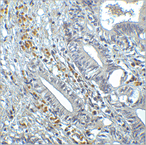
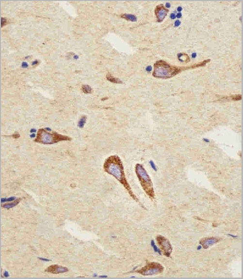
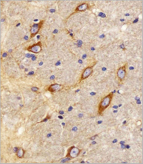
 IHC (Immunohistchemistry)
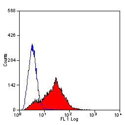
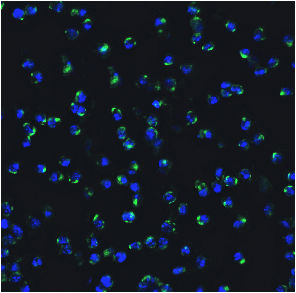
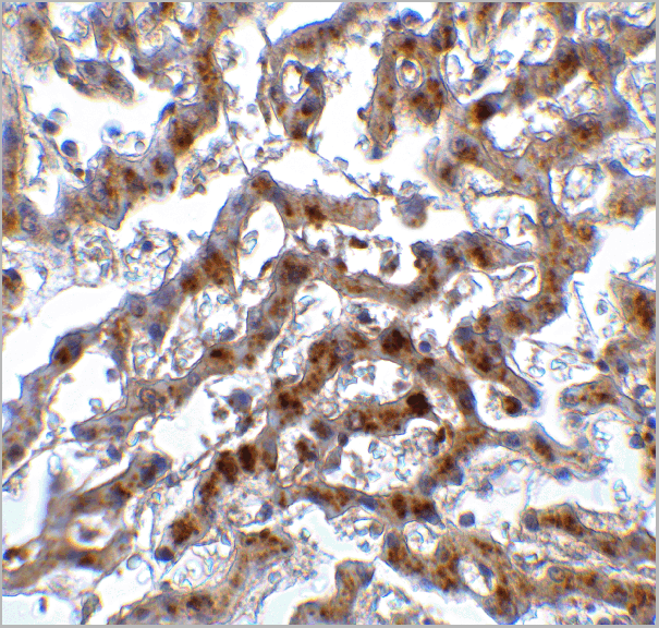
(Figure 9 Immunohistochemistry Validation of IRF7 in Human Liver Tissue Immunohistochemical analysis of paraffin-embedded human liver tissue using anti-IRF7 antibody (8991) at 2 μg/ml. Tissue was fixed with formaldehyde and blocked with 10% serum for 1 h at RT; antigen retrieval was by heat mediation with a citrate buffer (pH6). Samples were incubated with primary antibody overnight at 4˚C. A goat anti-rabbit IgG H&L (HRP) at 1/250 was used as secondary. Counter stained with Hematoxylin.)
IHC (Immunohistchemistry)
(Figure 9 Immunohistochemistry Validation of IRF7 in Human Liver Tissue Immunohistochemical analysis of paraffin-embedded human liver tissue using anti-IRF7 antibody (8991) at 2 μg/ml. Tissue was fixed with formaldehyde and blocked with 10% serum for 1 h at RT; antigen retrieval was by heat mediation with a citrate buffer (pH6). Samples were incubated with primary antibody overnight at 4˚C. A goat anti-rabbit IgG H&L (HRP) at 1/250 was used as secondary. Counter stained with Hematoxylin.)
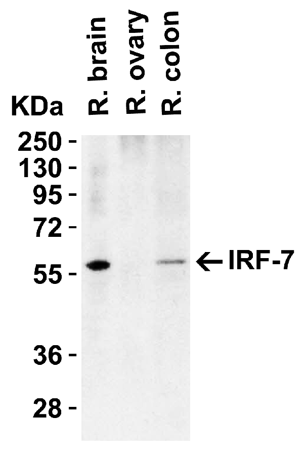
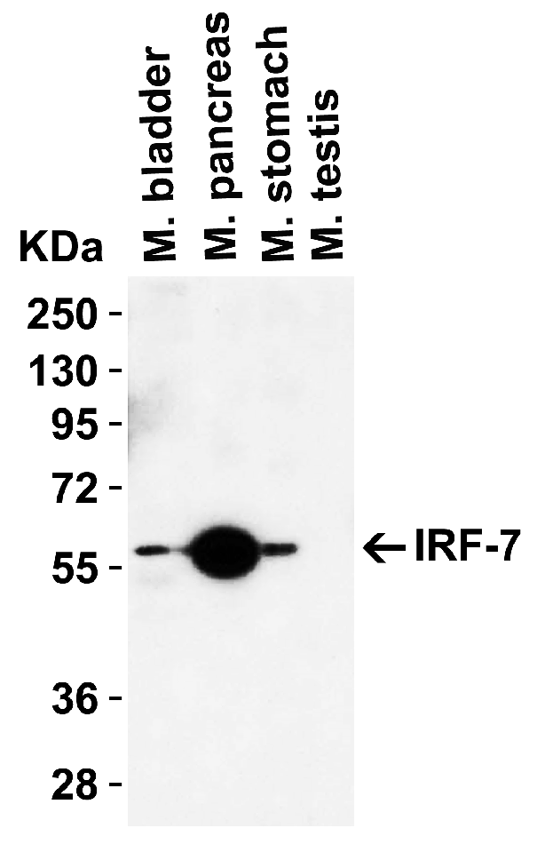
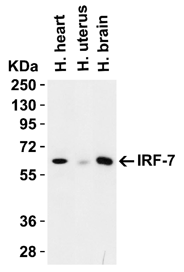
IRF7, Polyclonal Antibody (Cat# AAA11032)
Full Name
IRF7 Antibody
Gene Names
IRF7; IRF7A; IRF7B; IRF7C; IRF7H; IRF-7H
Reactivity
Human, Mouse, Rat
Applications
Western Blot, Immunohistochemistry
Purity
IRF7 Antibody is Protein A purified.
Pricing
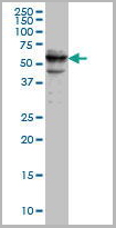
DUSP6, Monoclonal Antibody (Cat# AAA24194)
Full Name
DUSP6 (Dual Specificity Protein Phosphatase 6, Dual Specificity Protein Phosphatase PYST1, Mitogen-activated Protein Kinase Phosphatase 3, MAP Kinase Phosphatase 3, MKP3, MKP-3, PYST1) (AP)
Gene Names
DUSP6; HH19; MKP3; PYST1
Reactivity
Human
Applications
EIA, IHC, WB
Purity
Purified by Protein A Affinity Chromatography.
Pricing
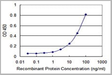
 IF (Immunofluorescence)
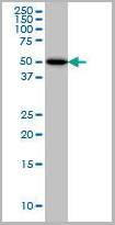
(LGR5/GPR49 Antibody (loop2) western blot analysis in HepG2 cell line lysates (35ug/lane).This demonstrates the LGR5/GPR49 antibody detected the LGR5/GPR49 protein (arrow).)
IF (Immunofluorescence)
(LGR5/GPR49 Antibody (loop2) western blot analysis in HepG2 cell line lysates (35ug/lane).This demonstrates the LGR5/GPR49 antibody detected the LGR5/GPR49 protein (arrow).)
LGR5/GPR49, Polyclonal Antibody (Cat# AAA28712)
Full Name
LGR5/GPR49 Antibody (loop2)
Gene Names
LGR5; FEX; HG38; GPR49; GPR67; GRP49
Reactivity
Human, mouse, rat
Applications
Western Blot, Immunofluorescence, Flow Cytometry
Purity
Purified Rabbit Polyclonal Antibody (Pab)
Pricing
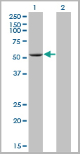
 WB (Western Blot)
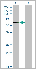
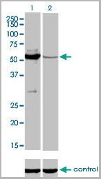
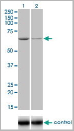
(Western blot analysis of MUTYH over-expressed 293 cell line, cotransfected with MUTYH Validated Chimera RNAi (Lane 2) or non-transfected control (Lane 1). Blot probed with MUTYH monoclonal antibody GAPDH (36.1kD) used as specificity and loading control.)
WB (Western Blot)
(Western blot analysis of MUTYH over-expressed 293 cell line, cotransfected with MUTYH Validated Chimera RNAi (Lane 2) or non-transfected control (Lane 1). Blot probed with MUTYH monoclonal antibody GAPDH (36.1kD) used as specificity and loading control.)
MUTYH, Monoclonal Antibody (Cat# AAA24577)
Full Name
MUTYH (A/G-specific Adenine DNA Glycosylase, MutY Homolog, hMYH, MYH) APC
Gene Names
MUTYH; MYH
Reactivity
Human
Applications
Immunoprecipitation, Western Blot
Purity
Purified by Protein A Affinity Chromatography.
Pricing
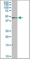
 WB (Western Blot)
(CDADC1 monoclonal antibody. Western Blot analysis of CDADC1 expression in NIH/3T3.)
WB (Western Blot)
(CDADC1 monoclonal antibody. Western Blot analysis of CDADC1 expression in NIH/3T3.)
CDADC1, Monoclonal Antibody (Cat# AAA25345)
Full Name
CDADC1 (Cytidine and dCMP Deaminase Domain-containing Protein 1, Testis Development Protein NYD-SP15, MGC150615, MGC41774, MGC57136, NYD-SP15, BA103J18.1) (HRP)
Gene Names
CDADC1; NYD-SP15; bA103J18.1
Reactivity
Human, Mouse
Applications
Immunohistochemistry, Western Blot
Purity
Purified by Protein A Affinity Chromatography.
Pricing
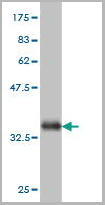
 WB (Western Blot)
(ADPGK monoclonal antibody. Western Blot analysis of ADPGK expression in Raw 264.7.)
WB (Western Blot)
(ADPGK monoclonal antibody. Western Blot analysis of ADPGK expression in Raw 264.7.)
ADPGK, Monoclonal Antibody (Cat# AAA25600)
Full Name
ADPGK (ADP-dependent Glucokinase, ADP-GK, RbBP-35, PSEC0260, 2610017G09Rik, DKFZp434B195) (PE)
Gene Names
ADPGK; ADP-GK; 2610017G09Rik
Reactivity
Human, Mouse, Rat
Applications
Western Blot
Purity
Purified by Protein A Affinity Chromatography.
Pricing
PKNOX2, Monoclonal Antibody (Cat# AAA24320)
Full Name
PKNOX2 (PREP2, Homeobox Protein PKNOX2, Homeobox Protein PREP-2, PBX/Knotted Homeobox 2, FLJ13074) (AP)
Gene Names
PKNOX2; PREP2
Reactivity
Human, Mouse
Applications
Western Blot
Purity
Purified by Protein A Affinity Chromatography.
Pricing
PITX1, Monoclonal Antibody (Cat# AAA24319)
Full Name
PITX1 (Pituitary Homeobox 1, Paired-like Homeodomain Transcription Factor 1, Homeobox Protein PITX1, Hindlimb-expressed Homeobox Protein Backfoot, PTX1, BFT) (AP)
Gene Names
PITX1; BFT; CCF; POTX; PTX1; LBNBG
Reactivity
Human
Applications
EIA, IP, WB
Purity
Purified by Protein A Affinity Chromatography.
Pricing
DUSP6, Monoclonal Antibody (Cat# AAA24490)
Full Name
DUSP6 (Dual Specificity Protein Phosphatase 6, Dual Specificity Protein Phosphatase PYST1, Mitogen-activated Protein Kinase Phosphatase 3, MAP Kinase Phosphatase 3, MKP3, MKP-3, PYST1) APC
Gene Names
DUSP6; HH19; MKP3; PYST1
Reactivity
Human
Applications
EIA, IF, IHC, WB
Purity
Purified by Protein A Affinity Chromatography.
Pricing
CDK2, Monoclonal Antibody (Cat# AAA24460)
Full Name
CDK2 (Cell Division Protein Kinase 2, p33 Protein Kinase) APC
Gene Names
CDK2; CDKN2; p33(CDK2)
Reactivity
Human
Applications
EIA, WB
Purity
Purified by Protein A Affinity Chromatography.
Pricing
MRPL12, Monoclonal Antibody (Cat# AAA24279)
Full Name
MRPL12 (Mitochondrial Ribosomal Protein L12, 39S Ribosomal Protein L12, Mitochondrial, 5c5-2, FLJ60124, L12mt, MGC8610, MRP-L12, MRP-L31/34, MRPL7, MRPL7/L12, RPML12) (AP)
Gene Names
MRPL12; 5c5-2; L12mt; MRPL7; RPML12; MRPL7/L12; MRP-L31/34
Reactivity
Human
Applications
EIA, IHC, WB
Purity
Purified by Protein A Affinity Chromatography.
Pricing
IRAK1, Monoclonal Antibody (Cat# AAA24248)
Full Name
IRAK1 (Interleukin-1 Receptor-associated Kinase 1, IRAK-1, IRAK) (AP)
Gene Names
IRAK1; IRAK; pelle
Reactivity
Human
Applications
EIA, IHC, IP, WB
Purity
Purified by Protein A Affinity Chromatography.
Pricing
CRKL, Monoclonal Antibody (Cat# AAA24472)
Full Name
CRKL (Crk-like Protein) APC
Reactivity
Human
Applications
EIA, IF, WB
Purity
Purified by Protein A Affinity Chromatography.
Pricing
CDC25C, Monoclonal Antibody (Cat# AAA24459)
Full Name
CDC25C (Cell Division Cycle 25 Homolog C, Cdc 25C, CDC 25, Dual Specificity Phosphatase Cdc25C, M-phase Inducer Phosphatase 3, MPIP3, PPP1R60) APC
Gene Names
CDC25C; CDC25; PPP1R60
Reactivity
Human
Applications
EIA, IHC, IP, WB
Purity
Purified by Protein A Affinity Chromatography.
Pricing
LCK, Monoclonal Antibody (Cat# AAA24254)
Full Name
LCK (Tyrosine-protein Kinase Lck, Leukocyte C-terminal Src Kinase, LSK, Lymphocyte Cell-specific Protein-tyrosine Kinase, Protein YT16, Proto-oncogene Lck, T Cell-specific Protein-tyrosine Kinase, p56-LCK) (AP)
Gene Names
LCK; LSK; YT16; IMD22; p56lck; pp58lck
Reactivity
Human
Applications
EIA, IP, WB
Purity
Purified by Protein A Affinity Chromatography.
Pricing