Filters
Clonality
Type
Reactivity
Gene Name
Isotype
Host
Application
Clone
1529 results for " Green" - showing 550-600


 Application Data
(Published customer image Infiltration of GFP+ BM-cells in infarct and peri-infarct regions. (A-B) Dot plots of viable macrophages/granulocytes (CD11b+CD45high, top right quadrants) and microglia (CD11b+CD45dim, bottom right quadrants) in cortex from BM-chimeric unmanipulated mice and mice exposed to pMCAO. (C) Bar graph showing mean numbers of CD11b+CD45dim microglia and CD11b+CD45high macrophages/granulocytes in BM-chimeric mice 24 hours after pMCAO, subdivided based on expression of GFP (n = 5). Approximately 92% of of the CD45high population were GFP+. (D) Estimation and comparison of mean numbers of CD11b+CD45dim microglia in non-chimeric (n = 10) versus BM-chimeric mice (n = 5) 24 hours after of pMCAO shows significantly fewer CD11b+CD45dim microglial cells in irradiated mice. (E) Overview, showing distribution of infiltrating GFP+ BM-derived cells into infarct (IF) and peri-infarct (P-IF) regions 24 hours after pMCAO. (E-G) By 24 hours, GFP+ single cells (F) and vessel-associated aggregates of GFP+ cells (arrows in G) were observed in infarct and peri-infarct regions. Some of the vessel-associated cells were round, leukocyte-like cells (arrows) while others were elongated cells lining the vasculature (arrow heads in G and in insert). (H) Bar graph showing mean numbers of single GFP+ cells and vessel-associated aggregates of GFP+ cells in ipsi- and contralateral cortex 24 hours after surgery (n = 10). (I-P) Immunohistochemical staining of CD45.1 (I, K), CD45.2 (J, L), IgG2a (M, O) and CD45 (N, P) in ischemic tissue in BM-chimeric (I, J, M, N) and non-chimeric mice (K, L, O, P) 24 hours after pMCAO. N.D, none detected. Scale bars: 200 um (A), 10 um (B, C). 50 um (I-P) *P < 0.05, **P < 0.01, and ***P < 0.001.From: Clausen BH, Lambertsen KL, Babcock AA, Holm TH, Dagnaes-Hansen F, Finsen B. Interleukin-1beta and tumor necrosis factor-alpha are expressed by different subsets of microglia and macrophages after ischemic stroke in mice. J Neuroinflammation. 2008 Oct 23;5:46.)
Application Data
(Published customer image Infiltration of GFP+ BM-cells in infarct and peri-infarct regions. (A-B) Dot plots of viable macrophages/granulocytes (CD11b+CD45high, top right quadrants) and microglia (CD11b+CD45dim, bottom right quadrants) in cortex from BM-chimeric unmanipulated mice and mice exposed to pMCAO. (C) Bar graph showing mean numbers of CD11b+CD45dim microglia and CD11b+CD45high macrophages/granulocytes in BM-chimeric mice 24 hours after pMCAO, subdivided based on expression of GFP (n = 5). Approximately 92% of of the CD45high population were GFP+. (D) Estimation and comparison of mean numbers of CD11b+CD45dim microglia in non-chimeric (n = 10) versus BM-chimeric mice (n = 5) 24 hours after of pMCAO shows significantly fewer CD11b+CD45dim microglial cells in irradiated mice. (E) Overview, showing distribution of infiltrating GFP+ BM-derived cells into infarct (IF) and peri-infarct (P-IF) regions 24 hours after pMCAO. (E-G) By 24 hours, GFP+ single cells (F) and vessel-associated aggregates of GFP+ cells (arrows in G) were observed in infarct and peri-infarct regions. Some of the vessel-associated cells were round, leukocyte-like cells (arrows) while others were elongated cells lining the vasculature (arrow heads in G and in insert). (H) Bar graph showing mean numbers of single GFP+ cells and vessel-associated aggregates of GFP+ cells in ipsi- and contralateral cortex 24 hours after surgery (n = 10). (I-P) Immunohistochemical staining of CD45.1 (I, K), CD45.2 (J, L), IgG2a (M, O) and CD45 (N, P) in ischemic tissue in BM-chimeric (I, J, M, N) and non-chimeric mice (K, L, O, P) 24 hours after pMCAO. N.D, none detected. Scale bars: 200 um (A), 10 um (B, C). 50 um (I-P) *P < 0.05, **P < 0.01, and ***P < 0.001.From: Clausen BH, Lambertsen KL, Babcock AA, Holm TH, Dagnaes-Hansen F, Finsen B. Interleukin-1beta and tumor necrosis factor-alpha are expressed by different subsets of microglia and macrophages after ischemic stroke in mice. J Neuroinflammation. 2008 Oct 23;5:46.)
CD11b, Monoclonal Antibody (Cat# AAA12183)
Full Name
RAT ANTI MOUSE CD11b:FITC
Gene Names
Itgam; CR3; CR3A; MAC1; Cd11b; Ly-40; Mac-1; Mac-1a; CD11b/CD18; F730045J24Rik
Applications
Flow Cytometry
Pricing


 Application Data
(Published customer image Infiltration of GFP+ BM-cells in infarct and peri-infarct regions. (A-B) Dot plots of viable macrophages/granulocytes (CD11b+CD45high, top right quadrants) and microglia (CD11b+CD45dim, bottom right quadrants) in cortex from BM-chimeric unmanipulated mice and mice exposed to pMCAO. (C) Bar graph showing mean numbers of CD11b+CD45dim microglia and CD11b+CD45high macrophages/granulocytes in BM-chimeric mice 24 hours after pMCAO, subdivided based on expression of GFP (n = 5). Approximately 92% of of the CD45high population were GFP+. (D) Estimation and comparison of mean numbers of CD11b+CD45dim microglia in non-chimeric (n = 10) versus BM-chimeric mice (n = 5) 24 hours after of pMCAO shows significantly fewer CD11b+CD45dim microglial cells in irradiated mice. (E) Overview, showing distribution of infiltrating GFP+ BM-derived cells into infarct (IF) and peri-infarct (P-IF) regions 24 hours after pMCAO. (E-G) By 24 hours, GFP+ single cells (F) and vessel-associated aggregates of GFP+ cells (arrows in G) were observed in infarct and peri-infarct regions. Some of the vessel-associated cells were round, leukocyte-like cells (arrows) while others were elongated cells lining the vasculature (arrow heads in G and in insert). (H) Bar graph showing mean numbers of single GFP+ cells and vessel-associated aggregates of GFP+ cells in ipsi- and contralateral cortex 24 hours after surgery (n = 10). (I-P) Immunohistochemical staining of CD45.1 (I, K), CD45.2 (J, L), IgG2a (M, O) and CD45 (N, P) in ischemic tissue in BM-chimeric (I, J, M, N) and non-chimeric mice (K, L, O, P) 24 hours after pMCAO. N.D, none detected. Scale bars: 200 um (A), 10 um (B, C). 50 um (I-P) *P < 0.05, **P < 0.01, and ***P < 0.001.From: Clausen BH, Lambertsen KL, Babcock AA, Holm TH, Dagnaes-Hansen F, Finsen B. Interleukin-1beta and tumor necrosis factor-alpha are expressed by different subsets of microglia and macrophages after ischemic stroke in mice. J Neuroinflammation. 2008 Oct 23;5:46.)
Application Data
(Published customer image Infiltration of GFP+ BM-cells in infarct and peri-infarct regions. (A-B) Dot plots of viable macrophages/granulocytes (CD11b+CD45high, top right quadrants) and microglia (CD11b+CD45dim, bottom right quadrants) in cortex from BM-chimeric unmanipulated mice and mice exposed to pMCAO. (C) Bar graph showing mean numbers of CD11b+CD45dim microglia and CD11b+CD45high macrophages/granulocytes in BM-chimeric mice 24 hours after pMCAO, subdivided based on expression of GFP (n = 5). Approximately 92% of of the CD45high population were GFP+. (D) Estimation and comparison of mean numbers of CD11b+CD45dim microglia in non-chimeric (n = 10) versus BM-chimeric mice (n = 5) 24 hours after of pMCAO shows significantly fewer CD11b+CD45dim microglial cells in irradiated mice. (E) Overview, showing distribution of infiltrating GFP+ BM-derived cells into infarct (IF) and peri-infarct (P-IF) regions 24 hours after pMCAO. (E-G) By 24 hours, GFP+ single cells (F) and vessel-associated aggregates of GFP+ cells (arrows in G) were observed in infarct and peri-infarct regions. Some of the vessel-associated cells were round, leukocyte-like cells (arrows) while others were elongated cells lining the vasculature (arrow heads in G and in insert). (H) Bar graph showing mean numbers of single GFP+ cells and vessel-associated aggregates of GFP+ cells in ipsi- and contralateral cortex 24 hours after surgery (n = 10). (I-P) Immunohistochemical staining of CD45.1 (I, K), CD45.2 (J, L), IgG2a (M, O) and CD45 (N, P) in ischemic tissue in BM-chimeric (I, J, M, N) and non-chimeric mice (K, L, O, P) 24 hours after pMCAO. N.D, none detected. Scale bars: 200 um (A), 10 um (B, C). 50 um (I-P) *P < 0.05, **P < 0.01, and ***P < 0.001.From: Clausen BH, Lambertsen KL, Babcock AA, Holm TH, Dagnaes-Hansen F, Finsen B. Interleukin-1beta and tumor necrosis factor-alpha are expressed by different subsets of microglia and macrophages after ischemic stroke in mice. J Neuroinflammation. 2008 Oct 23;5:46.)
CD11b, Monoclonal Antibody (Cat# AAA12185)
Full Name
RAT ANTI MOUSE CD11b
Gene Names
Itgam; CR3; CR3A; MAC1; Cd11b; Ly-40; Mac-1; Mac-1a; CD11b/CD18; F730045J24Rik
Applications
Immunohistochemistry, Flow Cytometry, Immunofluorescence, Immunoprecipitation
Pricing


 Application Data
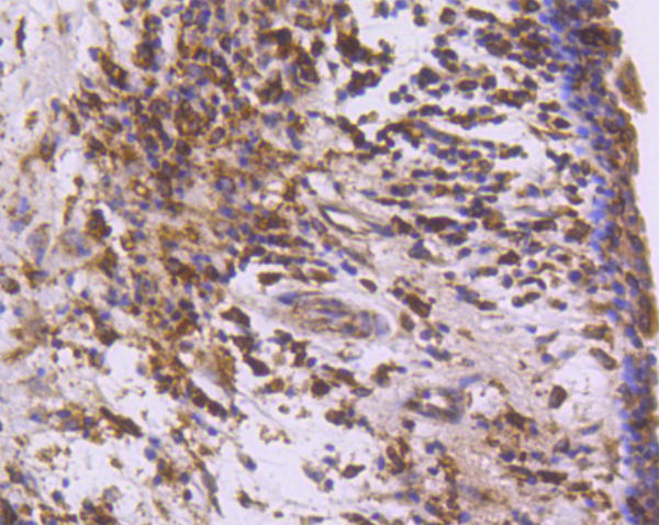
(Immunoperoxidase staining of a human tonsil cryosection with Mouse anti Human CD163 antibody, clone EDHu-1 followed by the Histar detection system . Low power)
Application Data
(Immunoperoxidase staining of a human tonsil cryosection with Mouse anti Human CD163 antibody, clone EDHu-1 followed by the Histar detection system . Low power)
CD163, Monoclonal Antibody (Cat# AAA12100)
Full Name
MOUSE ANTI HUMAN CD163
Gene Names
CD163; M130; MM130
Applications
Immunohistochemistry, Flow Cytometry, Immunofluorescence, Immunoassay, Immunohistochemistry, Western Blot
Pricing


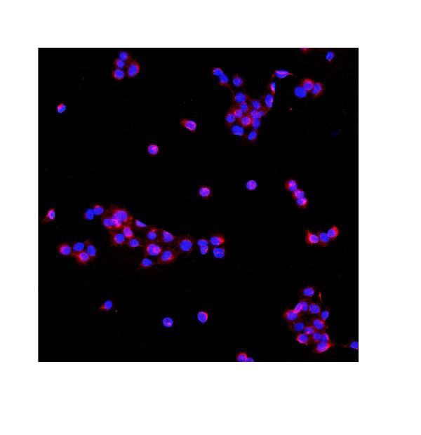
 ICC (Immunocytochemistry)
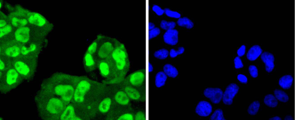
(ICC staining catalase in HepG2 cells (green). Cells were fixed in paraformaldehyde, permeabilised with 0.25% Triton X100/PBS.)
ICC (Immunocytochemistry)
(ICC staining catalase in HepG2 cells (green). Cells were fixed in paraformaldehyde, permeabilised with 0.25% Triton X100/PBS.)
Catalase, Polyclonal Antibody (Cat# AAA29962)
Full Name
Catalase Antibody
Reactivity
Human, Mouse, Rat
Applications
Western Blot, Immunocytochemistry, Immunohistochemistry
Purity
Peptide affinity purified
Pricing


 FCM (Flow Cytometry)
(Flow cytometric analysis of Hela cells with gamma Synuclein antibody at 1/100 dilution (red) compared with an unlabelled control (cells without incubation with primary antibody; black).)
FCM (Flow Cytometry)
(Flow cytometric analysis of Hela cells with gamma Synuclein antibody at 1/100 dilution (red) compared with an unlabelled control (cells without incubation with primary antibody; black).)
gamma Synuclein, Monoclonal Antibody (Cat# AAA30372)
Full Name
gamma Synuclein Antibody
Gene Names
SNCG; SR; BCSG1
Reactivity
Human, Rat
Applications
Western Blot, Immunocytochemistry, Immunohistochemistry, Immunoprecipitation, Flow Cytometry
Purity
ProA affinity purified
Pricing


 FCM (Flow Cytometry)
(Flow cytometric analysis of Hela cells with beta Arrestin 1 antibody at 1/50 dilution (blue) compared with an unlabelled control (cells without incubation with primary antibody; red). Alexa Fluor 488-conjugated goat anti rabbit IgG was used as the secondary antibody.)
FCM (Flow Cytometry)
(Flow cytometric analysis of Hela cells with beta Arrestin 1 antibody at 1/50 dilution (blue) compared with an unlabelled control (cells without incubation with primary antibody; red). Alexa Fluor 488-conjugated goat anti rabbit IgG was used as the secondary antibody.)
beta Arrestin 1, Monoclonal Antibody (Cat# AAA30095)
Full Name
beta Arrestin 1 Antibody
Gene Names
ARRB1; ARB1; ARR1
Reactivity
Human, Mouse, Rat
Applications
Western Blot, Immunocytochemistry, Immunofluorescence, Immunohistochemistry, Immunoprecipitation, Flow Cytometry
Purity
ProA affinity purified
Pricing


 FCM (Flow Cytometry)
(Flow cytometric analysis of Hela cells with 53BP1 antibody at 1/50 dilution (red) compared with an unlabelled control (cells without incubation with primary antibody; black). Alexa Fluor 488-conjugated goat anti rabbit IgG was used as the secondary antibody.)
FCM (Flow Cytometry)
(Flow cytometric analysis of Hela cells with 53BP1 antibody at 1/50 dilution (red) compared with an unlabelled control (cells without incubation with primary antibody; black). Alexa Fluor 488-conjugated goat anti rabbit IgG was used as the secondary antibody.)
53BP1, Monoclonal Antibody (Cat# AAA30337)
Full Name
53BP1 Antibody
Gene Names
TP53BP1; p202; 53BP1
Reactivity
Human, Mouse, Rat
Applications
Western Blot, Immunocytochemistry, Immunofluorescence, Immunohistochemistry, Flow Cytometry
Purity
ProA affinity purified
Pricing


 IF (Immunofluorescence)
(Immunofluorescence of Clusterin in mouse brain tissue with Clusterin Antibody at 20 μg/mL.Green: Clusterin Antibody (3856)Red: Phylloidin stainingBlue: DAPI staining)
IF (Immunofluorescence)
(Immunofluorescence of Clusterin in mouse brain tissue with Clusterin Antibody at 20 μg/mL.Green: Clusterin Antibody (3856)Red: Phylloidin stainingBlue: DAPI staining)
Clusterin, Polyclonal Antibody (Cat# AAA10914)
Full Name
Clusterin Antibody
Gene Names
CLU; CLI; AAG4; APOJ; CLU1; CLU2; KUB1; SGP2; APO-J; SGP-2; SP-40; TRPM2; TRPM-2; NA1/NA2
Reactivity
Human
Applications
Western Blot, Immunohistochemistry
Purity
Clusterin Antibody is affinity chromatography purified via peptide column.
Pricing


 FCM (Flow Cytometry)
(Flow cytometric analysis of SH-SY-5Y cells with EB3 antibody at 1/100 dilution (red) compared with an unlabelled control (cells without incubation with primary antibody; black). Alexa Fluor 488-conjugated goat anti-rabbit IgG was used as the secondary antibody.)
FCM (Flow Cytometry)
(Flow cytometric analysis of SH-SY-5Y cells with EB3 antibody at 1/100 dilution (red) compared with an unlabelled control (cells without incubation with primary antibody; black). Alexa Fluor 488-conjugated goat anti-rabbit IgG was used as the secondary antibody.)
EB3, Monoclonal Antibody (Cat# AAA30433)
Full Name
EB3 Antibody
Gene Names
MAPRE3; EB3; RP3; EBF3; EBF3-S
Reactivity
Human, Mouse, Rat
Applications
Western Blot, Immunocytochemistry, Immunofluorescence, Immunohistochemistry, Flow Cytometry
Purity
ProA affinity purified
Pricing


 ICC (Immunocytochemistry)
(ICC staining Cyclin D1 in N2A cells (green). The nuclear counter stain is DAPI (blue). Cells were fixed in paraformaldehyde, permeabilised with 0.25% Triton X100/PBS.)
ICC (Immunocytochemistry)
(ICC staining Cyclin D1 in N2A cells (green). The nuclear counter stain is DAPI (blue). Cells were fixed in paraformaldehyde, permeabilised with 0.25% Triton X100/PBS.)
Cyclin D1, Monoclonal Antibody (Cat# AAA29979)
Full Name
Cyclin D1 Antibody
Gene Names
CCND1; BCL1; PRAD1; U21B31; D11S287E
Reactivity
Human, Mouse, Rat
Applications
Western Blot, Immunocytochemistry, Immunofluorescence, Immunohistochemistry, Immunoprecipitation
Purity
ProA affinity purified
Pricing


 FCM (Flow Cytometry)
(Flow cytometric analysis of Hela cells with N Cadherin antibody at 1/50 dilution (red) compared with an unlabelled control (cells without incubation with primary antibody; black). Alexa Fluor 488-conjugated goat anti rabbit IgG was used as the secondary antibody.)
FCM (Flow Cytometry)
(Flow cytometric analysis of Hela cells with N Cadherin antibody at 1/50 dilution (red) compared with an unlabelled control (cells without incubation with primary antibody; black). Alexa Fluor 488-conjugated goat anti rabbit IgG was used as the secondary antibody.)
N Cadherin, Monoclonal Antibody (Cat# AAA30054)
Full Name
N Cadherin Antibody
Gene Names
CDH2; CDHN; NCAD; CD325; CDw325
Reactivity
Human, Mouse, Rat
Applications
Western Blot, Immunocytochemistry, Immunofluorescence, Immunohistochemistry, Flow Cytometry
Purity
ProA affinity purified
Pricing


 WB (Western Blot)
(Western Blot Analysis: Representative lot data. Lysate from HEK-293 cells was resolved by electrophoresis, transferred to PVDF and probed with anti-PI3 Kinase, p110beta (0.05 ug/mL). Proteins were visualized using donkey anti-rabbit secondary antibody conjugated to HRP and chemiluminescence detection. Arrow indicates PI3 Kinase, p110beta (~110kD).)
WB (Western Blot)
(Western Blot Analysis: Representative lot data. Lysate from HEK-293 cells was resolved by electrophoresis, transferred to PVDF and probed with anti-PI3 Kinase, p110beta (0.05 ug/mL). Proteins were visualized using donkey anti-rabbit secondary antibody conjugated to HRP and chemiluminescence detection. Arrow indicates PI3 Kinase, p110beta (~110kD).)
Phosphoinositide 3 Kinase, p110 beta, Polyclonal Antibody (Cat# AAA26898)
Full Name
Phosphoinositide 3 Kinase, p110 beta (DKFZp779K1237, MGC133043, p110beta, Phosphatidylinositol 3 Kinase Catalytic beta Polypeptide, Phosphatidylinositol-4,5-bisphosphate 3-kinase Catalytic Subunit beta Isoform, Phosphoinositide 3 Kinase Catalytic beta Pol
Gene Names
PIK3CA; MCM; CWS5; MCAP; PI3K; CLOVE; MCMTC; p110-alpha
Reactivity
Human
Applications
Immunocytochemistry, Western Blot, Immunoprecipitation, Immunohistochemistry
Purity
Purified by Immunoaffinity chromatography.
Pricing


 Application Data
(At 25 degree C. Samples were then incubated with primary Ab(At 37 degree C. An AlexaFluor594 conjugated goat anti-rabbit IgG(H+L) Ab(Red) and an AlexaFluor488 conjugated goat anti-mouse IgG(H+L) Ab(Green) were used as the secondary antibody.The nuclear counter stain is DAPI (blue).)
Application Data
(At 25 degree C. Samples were then incubated with primary Ab(At 37 degree C. An AlexaFluor594 conjugated goat anti-rabbit IgG(H+L) Ab(Red) and an AlexaFluor488 conjugated goat anti-mouse IgG(H+L) Ab(Green) were used as the secondary antibody.The nuclear counter stain is DAPI (blue).)
TOPBP1, Polyclonal Antibody (Cat# AAA31285)
Full Name
Phospho-TOPBP1 (Ser1138) Antibody
Gene Names
TOPBP1; TOP2BP1
Reactivity
Human, Mouse, Rat
Applications
Immunohistochemistry, Immunofluorescence, Immunocytochemistry, Peptide ELISA
Purity
The antibody is from purified rabbit serum by affinity purification via sequential chromatography on phospho-peptide and non-phospho-peptide affinity columns.
Pricing


 FCM (Flow Cytometry)
(Figure 7. Flow Cytometry analysis of U251 cells using anti-Serum Response Factor/SRF antibody (AAA19221).Overlay histogram showing U251 cells stained with AAA19221 (Blue line). The cells were blocked with 10% normal goat serum. And then incubated with rabbit anti-Serum Response Factor/SRF Antibody (AAA19221, 1μg/1x106 cells) for 30 min at 20 degree C. DyLight®488 conjugated goat anti-rabbit IgG (5-10μg/1x106 cells) was used as secondary antibody for 30 minutes at 20 degree C. Isotype control antibody (Green line) was rabbit IgG (1μg/1x106) used under the same conditions. Unlabelled sample (Red line) was also used as a control.)
FCM (Flow Cytometry)
(Figure 7. Flow Cytometry analysis of U251 cells using anti-Serum Response Factor/SRF antibody (AAA19221).Overlay histogram showing U251 cells stained with AAA19221 (Blue line). The cells were blocked with 10% normal goat serum. And then incubated with rabbit anti-Serum Response Factor/SRF Antibody (AAA19221, 1μg/1x106 cells) for 30 min at 20 degree C. DyLight®488 conjugated goat anti-rabbit IgG (5-10μg/1x106 cells) was used as secondary antibody for 30 minutes at 20 degree C. Isotype control antibody (Green line) was rabbit IgG (1μg/1x106) used under the same conditions. Unlabelled sample (Red line) was also used as a control.)
Serum Response Factor/SRF, Polyclonal Antibody (Cat# AAA19221)
Full Name
Anti-Serum Response Factor/SRF Antibody
Gene Names
SRF; MCM1
Reactivity
Human, Mouse, Rat
Applications
Western Blot, Immunohistochemistry, Immunocytochemistry, Immunofluorescence, Flow Cytometry, Direct ELISA
Purity
Immunogen affinity purified.
Pricing


 Application Data
(At 25 degree C. Samples were then incubated with primary Ab(At 37 degree C. An AlexaFluor594 conjugated goat anti-rabbit IgG(H+L) Ab(Red) and an AlexaFluor488 conjugated goat anti-mouse IgG(H+L) Ab(Green) were used as the secondary antibody.The nuclear counter stain is DAPI(blue).)
Application Data
(At 25 degree C. Samples were then incubated with primary Ab(At 37 degree C. An AlexaFluor594 conjugated goat anti-rabbit IgG(H+L) Ab(Red) and an AlexaFluor488 conjugated goat anti-mouse IgG(H+L) Ab(Green) were used as the secondary antibody.The nuclear counter stain is DAPI(blue).)
MARK2, Polyclonal Antibody (Cat# AAA31392)
Full Name
Phospho-MARK2 (Thr596) Antibody
Gene Names
MARK2; EMK1; EMK-1; PAR-1; Par1b; Par-1b
Reactivity
Human, Mouse, Rat
Predicted Reactivity: Pig (100%), Bovine (100%), Horse (100%), Sheep (100%), Rabbit (100%), Dog (100%), Xenopus (100%)
Predicted Reactivity: Pig (100%), Bovine (100%), Horse (100%), Sheep (100%), Rabbit (100%), Dog (100%), Xenopus (100%)
Applications
Western Blot, Immunohistochemistry, Immunofluorescence, Immunocytochemistry, Peptide ELISA
Purity
The antibody is from purified rabbit serum by affinity purification via sequential chromatography on phospho-peptide and non-phospho-peptide affinity columns.
Pricing


 IF (Immunofluorescence)
(Immunofluorescence of monoclonal antibody to POU5F1 on HeLa cell. [antibody concentration 10 ug/ml])
IF (Immunofluorescence)
(Immunofluorescence of monoclonal antibody to POU5F1 on HeLa cell. [antibody concentration 10 ug/ml])
POU5F1, Monoclonal Antibody (Cat# AAA26229)
Full Name
POU5F1 (POU Class 5 Homeobox 1, MGC22487, OCT3, OCT4, OTF3, OTF4) (Biotin)
Applications
Immunofluorescence, Western Blot
Purity
Purified
Pricing


 FCM (Flow Cytometry)
(Flow cytometric analysis of Jurkat cells with CaMKII gamma antibody at 1/100 dilution (green) compared with an unlabelled control (cells without incubation with primary antibody; red).)
FCM (Flow Cytometry)
(Flow cytometric analysis of Jurkat cells with CaMKII gamma antibody at 1/100 dilution (green) compared with an unlabelled control (cells without incubation with primary antibody; red).)
CAMK2G, Monoclonal Antibody (Cat# AAA29896)
Full Name
CAMK2G Antibody
Gene Names
CAMK2G; CAMK; CAMKG; CAMK-II
Reactivity
Human, Rat
Applications
Western Blot, Immunohistochemistry, Flow Cytometry
Purity
ProA affinity purified
Pricing


 Application Data
(Published customer image: Representative images of the inflammatory changes in the facial nucleus during axonal regeneration, one week following facial nerve transaction. a, b: CD11b immunoreactivity for microglia is increased in the axotomized facial nucleus, and microglia enwrap the facial motor neurons, e.g. at arrows. The regenerating neurons were retrogradely labelled with fluorogold. c, d: CD6- positive T-cells accumulated in the injured motor nucleus (arrows). They had little cytoplasm but dense nuclei (c) and were sometimes clustered around neurons retrogradely labelled with fluorogold (d). The scale bar in (a) also applies to (b) and that in (c) also applies to (d).From: Shokouhi et al. BMC Neuroscience 2010 11:13.)
Application Data
(Published customer image: Representative images of the inflammatory changes in the facial nucleus during axonal regeneration, one week following facial nerve transaction. a, b: CD11b immunoreactivity for microglia is increased in the axotomized facial nucleus, and microglia enwrap the facial motor neurons, e.g. at arrows. The regenerating neurons were retrogradely labelled with fluorogold. c, d: CD6- positive T-cells accumulated in the injured motor nucleus (arrows). They had little cytoplasm but dense nuclei (c) and were sometimes clustered around neurons retrogradely labelled with fluorogold (d). The scale bar in (a) also applies to (b) and that in (c) also applies to (d).From: Shokouhi et al. BMC Neuroscience 2010 11:13.)
CD11b, Monoclonal Antibody (Cat# AAA11876)
Full Name
MOUSE ANTI RAT CD11b:FITC
Gene Names
ITGAM; CD11B
Applications
Flow Cytometry
Pricing


 Application Data
(Staining of J774 cells with Rat anti Mouse F4/80 antigen Biotin)
Application Data
(Staining of J774 cells with Rat anti Mouse F4/80 antigen Biotin)
F4/80, Monoclonal Antibody (Cat# AAA12169)
Full Name
RAT ANTI MOUSE F4/80
Gene Names
Emr1; Ly71; F4/80; Gpf480; TM7LN3; DD7A5-7; EGF-TM7
Applications
Immunohistochemistry, Fluorescence Microscopy, Flow Cytometry, Immunofluorescence, Immunoprecipitation, Immunohistochemistry, Radioimmunoassay, Immunohistochemistry, Western Blot
Pricing


 Application Data
(At 25 degree C. Samples were then incubated with primary Ab(At 37 degree C. An AlexaFluor594 conjugated goat anti-rabbit IgG(H+L) Ab(Red) and an AlexaFluor488 conjugated goat anti-mouse IgG(H+L) Ab(Green) were used as the secondary antibody.The nuclear counter stain is DAPI (blue).)
Application Data
(At 25 degree C. Samples were then incubated with primary Ab(At 37 degree C. An AlexaFluor594 conjugated goat anti-rabbit IgG(H+L) Ab(Red) and an AlexaFluor488 conjugated goat anti-mouse IgG(H+L) Ab(Green) were used as the secondary antibody.The nuclear counter stain is DAPI (blue).)
Histone H3, Polyclonal Antibody (Cat# AAA31322)
Full Name
Phospho-Histone H3 (Thr3) Antibody
Gene Names
HIST1H3A; H3/A; H3FA
Reactivity
Human, Mouse, Rat
Applications
Immunohistochemistry, Immunofluorescence, Immunocytochemistry, Peptide ELISA
Purity
The antibody is from purified rabbit serum by affinity purification via sequential chromatography on phospho-peptide and non-phospho-peptide affinity columns.
Pricing


 FCM (Flow Cytometry)
(FACS analysis of ES-2 cells stained with STIP1 monoclonal antibody clone 2E1 (Green) and non-stained ES-2 cells (Black) as negative control.)
FCM (Flow Cytometry)
(FACS analysis of ES-2 cells stained with STIP1 monoclonal antibody clone 2E1 (Green) and non-stained ES-2 cells (Black) as negative control.)
STIP1, Monoclonal Antibody (Cat# AAA26407)
Full Name
STIP1 (Stress-Induced-Phosphoprotein 1, HOP, IEF-SSP-3521, P60, STI1, STI1L) (FITC)
Gene Names
STIP1; HOP; P60; STI1; STI1L; HEL-S-94n; IEF-SSP-3521
Applications
Flow Cytometry, Immunofluorescence, Immunohistochemistry, Western Blot
Purity
Purified
Pricing


 ELISA
(Titration curve analysis of PD-1 mAbs to detect recombinant PD-1 in ELISA with abs at decreasing concentrations.)
ELISA
(Titration curve analysis of PD-1 mAbs to detect recombinant PD-1 in ELISA with abs at decreasing concentrations.)
PD-1, Monoclonal Antibody (Cat# AAA10981)
Full Name
PD-1 Antibody [10B3]
Gene Names
PDCD1; PD1; PD-1; CD279; SLEB2; hPD-1; hPD-l; hSLE1
Reactivity
Human
Applications
Western Blot, Immunohistochemistry, Immunocytochemistry, Immunofluorescence, Flow Cytometry
Purity
Protein A purified IgG1.
Pricing


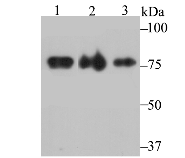
 WB (Western Blot)
(Lanes:Lane1: 400ug rat hippocampalLane2: 300ug rat hippocampalLane3: 200ug rat hippocampalLane4: 100ug rat hippocampalPrimary Antibody Dilution:1:1000Secondary Antibody:Anti-rabbit HRPSecondary Antibody Dilution:1:8000Gene Name:GPM6ASubmitted by:Anonymous)
WB (Western Blot)
(Lanes:Lane1: 400ug rat hippocampalLane2: 300ug rat hippocampalLane3: 200ug rat hippocampalLane4: 100ug rat hippocampalPrimary Antibody Dilution:1:1000Secondary Antibody:Anti-rabbit HRPSecondary Antibody Dilution:1:8000Gene Name:GPM6ASubmitted by:Anonymous)
GPM6A, Polyclonal Antibody (Cat# AAA23595)
Full Name
GPM6A antibody - C-terminal region
Gene Names
GPM6A; M6A; GPM6
Reactivity
Cow, Dog, Guinea Pig, Horse, Human, Mouse, Rabbit, Rat, Zebrafish
Applications
Immunohistochemistry, Western Blot
Purity
Affinity Purified
Pricing


 FCM (Flow Cytometry)
(Flow cytometric analysis of Hela cells with Ring1 antibody at 1/100 dilution (green) compared with an unlabelled control (cells without incubation with primary antibody; red).)
FCM (Flow Cytometry)
(Flow cytometric analysis of Hela cells with Ring1 antibody at 1/100 dilution (green) compared with an unlabelled control (cells without incubation with primary antibody; red).)
RING1, Monoclonal Antibody (Cat# AAA29906)
Full Name
RING1 Antibody
Gene Names
RING1; RNF1; RING1A
Reactivity
Human
Applications
Western Blot, Immunohistochemistry, Flow Cytometry
Purity
ProA affinity purified
Pricing


 FCM (Flow Cytometry)
(Flow cytometric analysis of Hela cells with Villin1 antibody at 1/100 dilution (red) compared with an unlabelled control (cells without incubation with primary antibody; black). Alexa Fluor 488-conjugated goat anti rabbit IgG was used as the secondary antibody.)
FCM (Flow Cytometry)
(Flow cytometric analysis of Hela cells with Villin1 antibody at 1/100 dilution (red) compared with an unlabelled control (cells without incubation with primary antibody; black). Alexa Fluor 488-conjugated goat anti rabbit IgG was used as the secondary antibody.)
Villin1, Monoclonal Antibody (Cat# AAA30420)
Full Name
Villin1 Antibody
Gene Names
VIL1; VIL; D2S1471
Reactivity
Human, Mouse, Rat
Applications
Western Blot, Immunocytochemistry, Immunohistochemistry, Flow Cytometry
Purity
ProA affinity purified
Pricing


 FCM (Flow Cytometry)
(Flow cytometric analysis of MCF-7 cells with Androgen receptor antibody at 1/50 dilution (red) compared with an unlabelled control (cells without incubation with primary antibody; black). Alexa Fluor 488-conjugated goat anti rabbit IgG was used as the secondary antibody)
FCM (Flow Cytometry)
(Flow cytometric analysis of MCF-7 cells with Androgen receptor antibody at 1/50 dilution (red) compared with an unlabelled control (cells without incubation with primary antibody; black). Alexa Fluor 488-conjugated goat anti rabbit IgG was used as the secondary antibody)
Androgen, Monoclonal Antibody (Cat# AAA30084)
Full Name
Androgen Receptor Antibody
Gene Names
AR; KD; AIS; TFM; DHTR; SBMA; HYSP1; NR3C4; SMAX1; HUMARA
Reactivity
Human
Applications
Western Blot, Immunocytochemistry, Immunofluorescence, Immunohistochemistry, Flow Cytometry
Purity
ProA affinity purified
Pricing


 ICC (Immunocytochemistry)
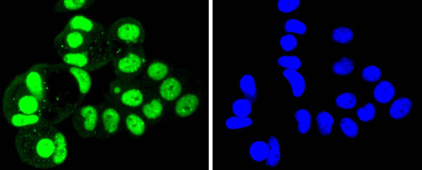
(ICC staining Aldolase in SK-Br-3 cells (green). The nuclear counter stain is DAPI (blue). Cells were fixed in paraformaldehyde, permeabilised with 0.25% Triton X100/PBS.)
ICC (Immunocytochemistry)
(ICC staining Aldolase in SK-Br-3 cells (green). The nuclear counter stain is DAPI (blue). Cells were fixed in paraformaldehyde, permeabilised with 0.25% Triton X100/PBS.)
Aldolase, Monoclonal Antibody (Cat# AAA30386)
Full Name
Aldolase Antibody
Gene Names
ALDOA; ALDA; GSD12
Reactivity
Human, Mouse, Rat, Zebrafish
Applications
Western Blot, Immunocytochemistry, Immunofluorescence, Immunohistochemistry
Purity
ProA affinity purified
Pricing


 ICC (Immunocytochemistry)
(ICC staining Phospho-Smad5 (S463/465) in HepG2 cells (green). The nuclear counter stain is DAPI (blue). Cells were fixed in paraformaldehyde, permeabilised with 0.25% Triton X100/PBS.)
ICC (Immunocytochemistry)
(ICC staining Phospho-Smad5 (S463/465) in HepG2 cells (green). The nuclear counter stain is DAPI (blue). Cells were fixed in paraformaldehyde, permeabilised with 0.25% Triton X100/PBS.)
SMAD5, Monoclonal Antibody (Cat# AAA29770)
Full Name
SMAD5 (Phospho-S463/465) Antibody
Gene Names
SMAD5; DWFC; JV5-1; MADH5
Reactivity
Human, Mouse, Rat, Zebrafish
Applications
Western Blot, Immunocytochemistry, Immunofluorescence, Immunohistochemistry, Immunoprecipitation, Flow Cytometry
Purity
ProA affinity purified
Pricing


 ICC (Immunocytochemistry)
(ICC staining IRF7 in HepG2 cells (green). The nuclear counter stain is DAPI (blue). Cells were fixed in paraformaldehyde, permeabilised with 0.25% Triton X100/PBS.)
ICC (Immunocytochemistry)
(ICC staining IRF7 in HepG2 cells (green). The nuclear counter stain is DAPI (blue). Cells were fixed in paraformaldehyde, permeabilised with 0.25% Triton X100/PBS.)
IRF7, Monoclonal Antibody (Cat# AAA30150)
Full Name
IRF7 Antibody
Gene Names
IRF7; IRF7A; IRF7B; IRF7C; IRF7H; IRF-7H
Reactivity
Human, Mouse, Rat, Zebrafish
Applications
Western Blot, Immunocytochemistry, Immunofluorescence, Immunohistochemistry, Immunoprecipitation, Flow Cytometry
Purity
ProA affinity purified
Pricing


 Application Data
(Figure A. FITC conjugated Rat anti Mouse CD19 . Figure B. FITC conjugated Rat anti Mouse CD19 and SBV440 conjugated Rat anti Mouse CD45R . All experiments performed on mouse splenocytes in the presence of 10% mouse seru)
Application Data
(Figure A. FITC conjugated Rat anti Mouse CD19 . Figure B. FITC conjugated Rat anti Mouse CD19 and SBV440 conjugated Rat anti Mouse CD45R . All experiments performed on mouse splenocytes in the presence of 10% mouse seru)
CD19, Monoclonal Antibody (Cat# AAA12286)
Full Name
Rat anti Mouse CD19:Amethyst Orange
Gene Names
Cd19; AW495831
Reactivity
Mouse
Applications
Flow Cytometry
Purity
Purified IgG prepared by affinity chromatography on Protein G from tissue culture supernatant
Pricing


 FCM (Flow Cytometry)
(Flow cytometric analysis of MCF-7 cells with Cytokeratin 18 antibody at 1/50 dilution (red) compared with an unlabelled control (cells without incubation with primary antibody; black). Alexa Fluor 488-conjugated goat anti rabbit IgG was used as the secondary antibody)
FCM (Flow Cytometry)
(Flow cytometric analysis of MCF-7 cells with Cytokeratin 18 antibody at 1/50 dilution (red) compared with an unlabelled control (cells without incubation with primary antibody; black). Alexa Fluor 488-conjugated goat anti rabbit IgG was used as the secondary antibody)
Cytokeratin 18, Monoclonal Antibody (Cat# AAA30013)
Full Name
Cytokeratin 18 Antibody
Gene Names
KRT18; K18; CYK18
Reactivity
Human, Mouse, Rat
Applications
Western Blot, Immunocytochemistry, Immunofluorescence, Immunohistochemistry, Flow Cytometry
Purity
ProA affinity purified
Pricing


 Application Data
(Immunoperoxidase staining of human tonsil cryosection using Mouse anti Human CD3 antibody, clone UCHT1 followed by horseradish peroxidase Goat anti Mouse IgG2a antibody as a detection reagent. Medium power)
Application Data
(Immunoperoxidase staining of human tonsil cryosection using Mouse anti Human CD3 antibody, clone UCHT1 followed by horseradish peroxidase Goat anti Mouse IgG2a antibody as a detection reagent. Medium power)
CD3, Monoclonal Antibody (Cat# AAA12156)
Full Name
MOUSE ANTI HUMAN CD3
Gene Names
CD3G; T3G; IMD17; CD3-GAMMA
Applications
Immunohistochemistry, Flow Cytometry
Pricing


 Application Data
(At 25 degree C. Samples were then incubated with primary Ab(At 37 degree C. An AlexaFluor594 conjugated goat anti-rabbit IgG(H+L) Ab(Red) and an AlexaFluor488 conjugated goat anti-mouse IgG(H+L) Ab(Green) were used as the secondary antibody.The nuclear counter stain is DAPI (blue).)
Application Data
(At 25 degree C. Samples were then incubated with primary Ab(At 37 degree C. An AlexaFluor594 conjugated goat anti-rabbit IgG(H+L) Ab(Red) and an AlexaFluor488 conjugated goat anti-mouse IgG(H+L) Ab(Green) were used as the secondary antibody.The nuclear counter stain is DAPI (blue).)
PFKFB3, Polyclonal Antibody (Cat# AAA31288)
Full Name
Phospho-PFKFB3 (Ser461) Antibody
Gene Names
PFKFB3; PFK2; IPFK2
Reactivity
Human, Rat
Applications
Western Blot, Immunohistochemistry, Immunofluorescence, Immunocytochemistry, Peptide ELISA
Purity
The antibody is from purified rabbit serum by affinity purification via sequential chromatography on phospho-peptide and non-phospho-peptide affinity columns.
Pricing


 IP (Immunoprecipitation)
(Immunoprecipitating HSPA8 in Hela whole cell lysate Lane 1: Mouse control IgG2b instead dilution for 30min at 4 degree C. Isotype control antibody (green line) was mouse IgG2b used under the same conditions. Acquisition of >10,000 events was performed.)
IP (Immunoprecipitation)
(Immunoprecipitating HSPA8 in Hela whole cell lysate Lane 1: Mouse control IgG2b instead dilution for 30min at 4 degree C. Isotype control antibody (green line) was mouse IgG2b used under the same conditions. Acquisition of >10,000 events was performed.)
HSPA8, Monoclonal Antibody (Cat# AAA27045)
Full Name
HSPA8 Monoclonal Antibody
Gene Names
HSPA8; LAP1; HSC54; HSC70; HSC71; HSP71; HSP73; NIP71; HSPA10
Reactivity
Human, Rat, Mouse
Applications
Western Blot, Immunohistochemistry, Flow Cytometry, Immunoprecipitation
Purity
>95%, Protein A purified
Pricing


 Application Data
(Staining of J774 cells with Rat anti Mouse F4/80 antigen Biotin)
Application Data
(Staining of J774 cells with Rat anti Mouse F4/80 antigen Biotin)
F4/80, Monoclonal Antibody (Cat# AAA12163)
Full Name
RAT ANTI MOUSE F4/80:Biotin
Gene Names
Emr1; Ly71; F4/80; Gpf480; TM7LN3; DD7A5-7; EGF-TM7
Applications
Flow Cytometry
Pricing


 FCM (Flow Cytometry)
(Figure 7. Flow Cytometry analysis of U87 cells using anti-PDE6 beta/PDE6B antibody (AAA19261).Overlay histogram showing U87 cells stained with AAA19261 (Blue line). The cells were blocked with 10% normal goat serum. And then incubated with rabbit anti-PDE6 beta/PDE6B Antibody (AAA19261,1μg/1x106 cells) for 30 min at 20 degree C. DyLight®488 conjugated goat anti-rabbit IgG (5-10μg/1x106 cells) was used as secondary antibody for 30 minutes at 20 degree C. Isotype control antibody (Green line) was rabbit IgG (1μg/1x106) used under the same conditions. Unlabelled sample (Red line) was also used as a control.)
FCM (Flow Cytometry)
(Figure 7. Flow Cytometry analysis of U87 cells using anti-PDE6 beta/PDE6B antibody (AAA19261).Overlay histogram showing U87 cells stained with AAA19261 (Blue line). The cells were blocked with 10% normal goat serum. And then incubated with rabbit anti-PDE6 beta/PDE6B Antibody (AAA19261,1μg/1x106 cells) for 30 min at 20 degree C. DyLight®488 conjugated goat anti-rabbit IgG (5-10μg/1x106 cells) was used as secondary antibody for 30 minutes at 20 degree C. Isotype control antibody (Green line) was rabbit IgG (1μg/1x106) used under the same conditions. Unlabelled sample (Red line) was also used as a control.)
PDE6 beta/PDE6B, Polyclonal Antibody (Cat# AAA19261)
Full Name
Anti-PDE6 beta/PDE6B Antibody
Reactivity
Human, Mouse, Rat
Applications
Western Blot, Immunohistochemistry, Immunocytochemistry, Immunofluorescence, Flow Cytometry, Direct ELISA
Purity
Immunogen affinity purified.
Pricing


 FCM (Flow Cytometry)
(Flow cytometric analysis of Hela cells with ASK1 antibody at 1/50 dilution (red) compared with an unlabelled control (cells without incubation with primary antibody; black). Alexa Fluor 488-conjugated goat anti rabbit IgG was used as the secondary antibody.)
FCM (Flow Cytometry)
(Flow cytometric analysis of Hela cells with ASK1 antibody at 1/50 dilution (red) compared with an unlabelled control (cells without incubation with primary antibody; black). Alexa Fluor 488-conjugated goat anti rabbit IgG was used as the secondary antibody.)
ASK1, Monoclonal Antibody (Cat# AAA30075)
Full Name
ASK1 Antibody
Gene Names
MAP3K5; ASK1; MEKK5; MAPKKK5
Reactivity
Human, Mouse
Applications
Western Blot, Immunocytochemistry, Immunofluorescence, Immunohistochemistry, Flow Cytometry
Purity
ProA affinity purified
Pricing


 ICC (Immunocytochemistry)
(ICC staining Phospho-GSK3 (alpha+beta) (Y216+Y279) in MCF-7 cells (green). The nuclear counter stain is DAPI (blue). Cells were fixed in paraformaldehyde, permeabilised with 0.25% Triton X100/PBS.)
ICC (Immunocytochemistry)
(ICC staining Phospho-GSK3 (alpha+beta) (Y216+Y279) in MCF-7 cells (green). The nuclear counter stain is DAPI (blue). Cells were fixed in paraformaldehyde, permeabilised with 0.25% Triton X100/PBS.)
GSK3 (alpha+beta), Monoclonal Antibody (Cat# AAA29772)
Full Name
GSK3 (alpha+beta) (Phospho-Y216+Y279) Antibody
Reactivity
Human, Mouse, Rat
Applications
Western Blot, Immunocytochemistry, Immunohistochemistry, Immunoprecipitation
Purity
ProA affinity purified
Pricing


 FCM (Flow Cytometry)
(Flow cytometric analysis of Hela cells with PBK antibody at 1/100 dilution (green) compared with an unlabelled control (cells without incubation with primary antibody; red).)
FCM (Flow Cytometry)
(Flow cytometric analysis of Hela cells with PBK antibody at 1/100 dilution (green) compared with an unlabelled control (cells without incubation with primary antibody; red).)
PBK, Monoclonal Antibody (Cat# AAA29912)
Full Name
PBK Antibody
Gene Names
PBK; SPK; CT84; TOPK; Nori-3
Reactivity
Human
Applications
Western Blot, Immunocytochemistry, Immunohistochemistry, Flow Cytometry
Purity
ProA affinity purified
Pricing


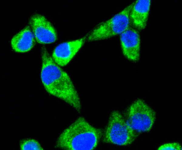
 ICC (Immunocytochemistry)
(ICC staining KHSRP in SH-SY-5Y cells (green). The nuclear counter stain is DAPI (blue). Cells were fixed in paraformaldehyde, permeabilised with 0.25% Triton X100/PBS.)
ICC (Immunocytochemistry)
(ICC staining KHSRP in SH-SY-5Y cells (green). The nuclear counter stain is DAPI (blue). Cells were fixed in paraformaldehyde, permeabilised with 0.25% Triton X100/PBS.)
KHSRP, Monoclonal Antibody (Cat# AAA30534)
Full Name
KHSRP Antibody
Gene Names
KHSRP; FBP2; KSRP; FUBP2
Reactivity
Human, Mouse, Rat
Applications
Western Blot, Immunocytochemistry, Immunofluorescence, Immunohistochemistry, Immunoprecipitation
Purity
ProA affinity purified
Pricing


 FCM (Flow Cytometry)
(Flow cytometric analysis of SH-SY5Y cells with PKC epsilon antibody at 1/100 dilution (red) compared with an unlabelled control (cells without incubation with primary antibody; black).)
FCM (Flow Cytometry)
(Flow cytometric analysis of SH-SY5Y cells with PKC epsilon antibody at 1/100 dilution (red) compared with an unlabelled control (cells without incubation with primary antibody; black).)
PKC epsilon, Monoclonal Antibody (Cat# AAA30361)
Full Name
PKC epsilon Antibody
Gene Names
PRKCE; PKCE; nPKC-epsilon
Reactivity
Human, Mouse, Rat
Applications
Western Blot, Immunocytochemistry, Immunohistochemistry, Flow Cytometry
Purity
ProA affinity purified
Pricing


 FCM (Flow Cytometry)
(Flow cytometric analysis of HepG2 cells with KDEL antibody at 1/100 dilution (red) compared with an unlabelled control (cells without incubation with primary antibody; black). Alexa Fluor 488-conjugated goat anti rabbit IgG was used as the secondary antibody.)
FCM (Flow Cytometry)
(Flow cytometric analysis of HepG2 cells with KDEL antibody at 1/100 dilution (red) compared with an unlabelled control (cells without incubation with primary antibody; black). Alexa Fluor 488-conjugated goat anti rabbit IgG was used as the secondary antibody.)
KDEL, Monoclonal Antibody (Cat# AAA30471)
Full Name
KDEL Antibody
Gene Names
KDELR1; ERD2; HDEL; PM23; ERD2.1
Reactivity
Human, Mouse, Rat
Applications
Western Blot, Immunocytochemistry, Immunofluorescence, Immunohistochemistry, Flow Cytometry
Purity
ProA affinity purified
Pricing


 FCM (Flow Cytometry)
(Flow cytometric analysis of Hela cells with Sonic Hedgehog Protein antibody at 1/50 dilution (red) compared with an unlabelled control (cells without incubation with primary antibody; black). Alexa Fluor 488-conjugated goat anti rabbit IgG was used as the secondary antibody.)
FCM (Flow Cytometry)
(Flow cytometric analysis of Hela cells with Sonic Hedgehog Protein antibody at 1/50 dilution (red) compared with an unlabelled control (cells without incubation with primary antibody; black). Alexa Fluor 488-conjugated goat anti rabbit IgG was used as the secondary antibody.)
Sonic Hedgehog Protein/SHH, Monoclonal Antibody (Cat# AAA30131)
Full Name
Sonic Hedgehog Protein/SHH (C-Product) Antibody
Gene Names
SHH; TPT; HHG1; HLP3; HPE3; SMMCI; TPTPS; MCOPCB5
Reactivity
Human
Applications
Western Blot, Immunocytochemistry, Immunofluorescence, Immunohistochemistry, Flow Cytometry
Purity
ProA affinity purified
Pricing


 WB (Western Blot)
(Western blot analysis of COX1 on different lysates using anti-COX1 antibody at 1/1, 000 dilution. Positive control: Lane 1: C2C12 Lane 2: A431)
WB (Western Blot)
(Western blot analysis of COX1 on different lysates using anti-COX1 antibody at 1/1, 000 dilution. Positive control: Lane 1: C2C12 Lane 2: A431)
COX1/Cyclooxygenase 1, Monoclonal Antibody (Cat# AAA30153)
Full Name
COX1/Cyclooxygenase 1 Antibody
Gene Names
PTGS1; COX1; COX3; PHS1; PCOX1; PES-1; PGHS1; PTGHS; PGG/HS; PGHS-1
Reactivity
Human, Mouse, Rat
Applications
Western Blot, Immunocytochemistry, Immunohistochemistry, Flow Cytometry, Immunoprecipitation
Purity
ProA affinity purified
Pricing


 SDS-PAGE
(SDS-PAGE Analysis Purified HSP60 Mouse Monoclonal Antibody (CPTC-HSPD1-1). Confirmation of Purity and Integrity of Antibody.)
SDS-PAGE
(SDS-PAGE Analysis Purified HSP60 Mouse Monoclonal Antibody (CPTC-HSPD1-1). Confirmation of Purity and Integrity of Antibody.)
HSP60 (Heat Shock Protein 60), Monoclonal Antibody (Cat# AAA23919)
Full Name
HSP60 (Heat Shock Protein 60) (Mitochondrial Marker)
Gene Names
HSPD1; HLD4; CPN60; GROEL; HSP60; HSP65; SPG13; HSP-60; HuCHA60
Reactivity
Human
Applications
Flow Cytometry, Immunofluorescence, Western Blot, Immunohistochemistry
Purity
Purified Ab with BSA and Azide at 200ug/ml OR Purified Ab WITHOUT BSA and Azide at 1.0mg/ml
Pricing


 FCM (Flow Cytometry)
(Figure 6. Flow Cytometry analysis of PC-3 cells using anti-ADK antibody (AAA11690).Overlay histogram showing PC-3 cells stained with AAA11690 (Blue line).The cells were blocked with 10% normal goat serum. And then incubated with rabbit anti-ADK Antibody (AAA11690,1ug/1x10^6 cells) for 30 min at 20 degree C. DyLight®488 conjugated goat anti-rabbit IgG (5-10ug/1x10^6 cells) was used as secondary antibody for 30 minutes at 20 degree C. Isotype control antibody (Green line) was rabbit IgG (1ug/1x106) used under the same conditions. Unlabelled sample (Red line) was also used as a control.)
FCM (Flow Cytometry)
(Figure 6. Flow Cytometry analysis of PC-3 cells using anti-ADK antibody (AAA11690).Overlay histogram showing PC-3 cells stained with AAA11690 (Blue line).The cells were blocked with 10% normal goat serum. And then incubated with rabbit anti-ADK Antibody (AAA11690,1ug/1x10^6 cells) for 30 min at 20 degree C. DyLight®488 conjugated goat anti-rabbit IgG (5-10ug/1x10^6 cells) was used as secondary antibody for 30 minutes at 20 degree C. Isotype control antibody (Green line) was rabbit IgG (1ug/1x106) used under the same conditions. Unlabelled sample (Red line) was also used as a control.)
ADK, Polyclonal Antibody (Cat# AAA11690)
Full Name
Anti-ADK Antibody
Gene Names
ADK; AK
Reactivity
Human, Mouse, Rat
Applications
Western Blot, Immunohistochemistry
Purity
Immunogen affinity purified.
Pricing


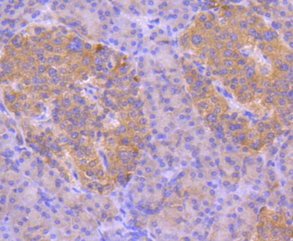
 IHC (Immunohistchemistry)
(Immunohistochemical analysis of paraffin-embedded rectum cancer tissues using ABCG5 mouse mAb with DAB staining.)
IHC (Immunohistchemistry)
(Immunohistochemical analysis of paraffin-embedded rectum cancer tissues using ABCG5 mouse mAb with DAB staining.)
ABCG5, Monoclonal Antibody (Cat# AAA14137)
Full Name
Anti-ABCG5 Mouse mAb
Gene Names
ABCG5; STSL
Reactivity
Human
Applications
Western Blot, Immunohistochemistry, Flow Cytometry
Pricing


 FCM (Flow Cytometry)
(Flow cytometric analysis of 293 cells with SUMO4 antibody at 1/50 dilution (red) compared with an unlabelled control (cells without incubation with primary antibody; black). Alexa Fluor 488-conjugated goat anti rabbit IgG was used as the secondary antibody)
FCM (Flow Cytometry)
(Flow cytometric analysis of 293 cells with SUMO4 antibody at 1/50 dilution (red) compared with an unlabelled control (cells without incubation with primary antibody; black). Alexa Fluor 488-conjugated goat anti rabbit IgG was used as the secondary antibody)
SUMO4, Monoclonal Antibody (Cat# AAA30248)
Full Name
SUMO4 Antibody
Gene Names
SUMO4; IDDM5; SMT3H4; SUMO-4; dJ281H8.4
Reactivity
Human, Mouse, Rat
Applications
Western Blot, Immunocytochemistry, Immunofluorescence, Immunoprecipitation, Flow Cytometry
Purity
ProA affinity purified
Pricing


 ELISA
(Titration curve analysis of VISTA antibody to detect recombinant VISTA in ELISA at decreasing concentrations.)
ELISA
(Titration curve analysis of VISTA antibody to detect recombinant VISTA in ELISA at decreasing concentrations.)
VISTA, Monoclonal Antibody (Cat# AAA11014)
Full Name
VISTA Antibody [8E11]
Gene Names
VSIR; B7H5; GI24; B7-H5; PD-1H; SISP1; VISTA; PP2135; C10orf54; DD1alpha
Reactivity
Human
Applications
Immunohistochemistry, Immunocytochemistry, Immunofluorescence, Flow Cytometry
Purity
Protein A purified
Pricing


 FCM (Flow Cytometry)
(Flow cytometric analysis of Hela cells with Phospho-eIF-2a (S51) antibody at 1/50 dilution (blue) compared with an unlabelled control (cells without incubation with primary antibody; red). Alexa Fluor 488-conjugated goat anti rabbit IgG was used as the secondary antibody.)
FCM (Flow Cytometry)
(Flow cytometric analysis of Hela cells with Phospho-eIF-2a (S51) antibody at 1/50 dilution (blue) compared with an unlabelled control (cells without incubation with primary antibody; red). Alexa Fluor 488-conjugated goat anti rabbit IgG was used as the secondary antibody.)
eIF-2a, Monoclonal Antibody (Cat# AAA29766)
Full Name
eIF-2a (Phospho-S51) Antibody
Gene Names
EIF2A; CDA02; EIF-2A; MST089; MSTP004; MSTP089
Reactivity
Human, Mouse, Rat
Applications
Western Blot, Immunohistochemistry, Flow Cytometry, Immunocytochemistry, Immunofluorescence, Immunohistochemistry, Immunoprecipitation, Flow Cytometry
Purity
ProA affinity purified
Pricing