Filters
Clonality
Type
Reactivity
Gene Name
Isotype
Host
Application
Clone
592 results for " DNA" - showing 500-550
IL-11 receptor alpha, Active Protein (Cat# AAA14431)
Full Name
Mouse IL-11 receptor alpha Recombinant
Purity
>85%, as determined by SDS-PAGE and HPLC
Pricing
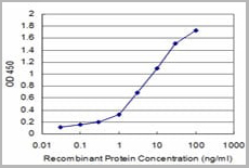
 Application Data
(Z- and S- Score: The Z-score represents the strength of a signal that a monoclonal antibody (MAb) (in combination with a fluorescently-tagged anti-IgG secondary antibody) produces when binding to a particular protein on the HuProtTM array. Z-scores are described in units of standard deviations (SD's) above the mean value of all signals generated on that array. If targets on HuProtTM are arranged in descending order of the Z-score, the S-score is the difference (also in units of SD's) between the Z-score. S-score therefore represents the relative target specificity of a MAb to its intended target. A MAb is considered to specific to its intended target, if the MAb has an S-score of at least 2.5. For example, if a MAb binds to protein X with a Z-score of 43 and to protein Y with a Z-score of 14, then the S-score for the binding of that MAb to protein X is equal to 29.)
Application Data
(Z- and S- Score: The Z-score represents the strength of a signal that a monoclonal antibody (MAb) (in combination with a fluorescently-tagged anti-IgG secondary antibody) produces when binding to a particular protein on the HuProtTM array. Z-scores are described in units of standard deviations (SD's) above the mean value of all signals generated on that array. If targets on HuProtTM are arranged in descending order of the Z-score, the S-score is the difference (also in units of SD's) between the Z-score. S-score therefore represents the relative target specificity of a MAb to its intended target. A MAb is considered to specific to its intended target, if the MAb has an S-score of at least 2.5. For example, if a MAb binds to protein X with a Z-score of 43 and to protein Y with a Z-score of 14, then the S-score for the binding of that MAb to protein X is equal to 29.)
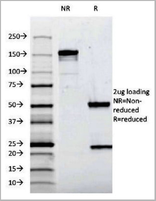
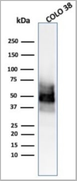
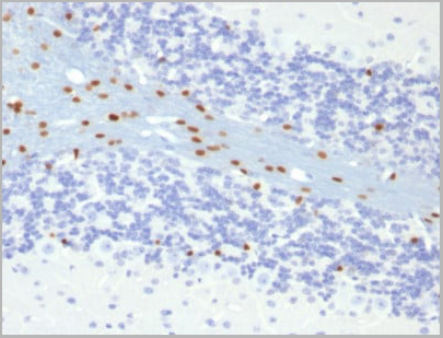
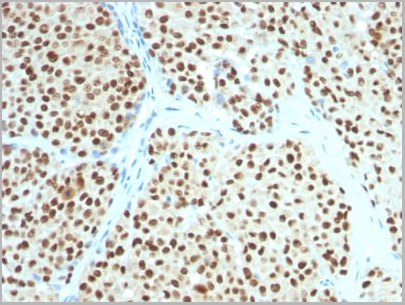
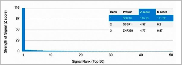
SOX10, Monoclonal Antibody (Cat# AAA13845)
Full Name
SOX10 (Melanoma Marker) Mouse Monoclonal Antibody
Gene Names
SOX10; DOM; WS4; PCWH; WS2E; WS4C
Reactivity
Human, Mouse
Applications
Flow Cytometry, Immunofluorescence, Western Blot, Immunohistochemistry
Pricing
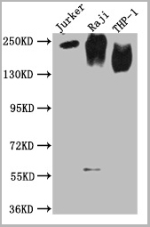
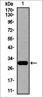
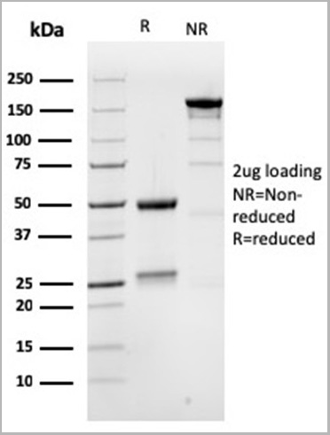
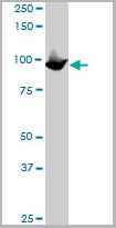
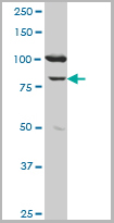
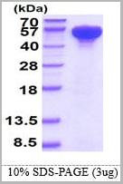 SDS-PAGE
HSP70, Recombinant Protein (Cat# AAA11739)
Full Name
HSP70, 1-641aa, Human, His tag, E Coli
Gene Names
HSPA1A; HSP72; HSPA1; HSP70I; HSP70-1; HSP70.1; HSP70-1A; HEL-S-103
Applications
SDS-PAGE
Purity
> 95% by SDS-PAGE
Pricing
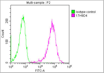
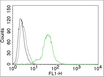
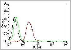
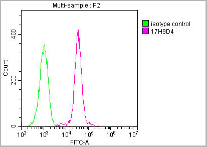 FCM (Flow Cytometry)
(Overlay histogram showing Raji cells stained with (red line) at 1:500. The cells were incubated in 10% normal goat serum to block non-specific protein-protein interactions followed by the antibody (1ug/1*106cells) for 1 h at 4 degree C. The secondary antibody used was FITC-conjugated Goat Anti-Mouse IgG(H+L) at 1/100 dilution for 30min at 4 degree C. Isotype control antibody (green line) was mouse IgG2b (1ug/1*106cells) used under the same conditions. Acquisition of >10,000 events was performed.)
FCM (Flow Cytometry)
(Overlay histogram showing Raji cells stained with (red line) at 1:500. The cells were incubated in 10% normal goat serum to block non-specific protein-protein interactions followed by the antibody (1ug/1*106cells) for 1 h at 4 degree C. The secondary antibody used was FITC-conjugated Goat Anti-Mouse IgG(H+L) at 1/100 dilution for 30min at 4 degree C. Isotype control antibody (green line) was mouse IgG2b (1ug/1*106cells) used under the same conditions. Acquisition of >10,000 events was performed.)
CD45, Monoclonal Antibody (Cat# AAA27049)
Full Name
CD45 Monoclonal Antibody
Gene Names
PTPRC; LCA; LY5; B220; CD45; L-CA; T200; CD45R; GP180
Reactivity
Human
Applications
Western Blot, Immunohistochemistry, Immunofluorescence, Flow Cytometry
Purity
>95%, Protein A purified
Pricing
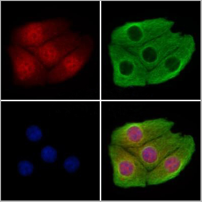
 Application Data
(AAA31369 staining Hela cells(4h of LPS treatment) by IF/ICC. The samples were fixed with PFA and permeabilized in 0.1% Triton X-100,then blocked in 10% serum for 45 minutes at 25°C. Samples were then incubated with primary Ab(AAA31369 1:200) and mouse anti-beta tubulin Ab(T0023 1:200) for 1 hour at 37°C. An AlexaFluor594 conjugated goat anti-rabbit IgG(H+L) Ab(Red) and an AlexaFluor488 conjugated goat anti-mouse IgG(H+L) Ab(Green) were used as the secondary Ab. The nuclear counter stain is DAPI(blue).)
Application Data
(AAA31369 staining Hela cells(4h of LPS treatment) by IF/ICC. The samples were fixed with PFA and permeabilized in 0.1% Triton X-100,then blocked in 10% serum for 45 minutes at 25°C. Samples were then incubated with primary Ab(AAA31369 1:200) and mouse anti-beta tubulin Ab(T0023 1:200) for 1 hour at 37°C. An AlexaFluor594 conjugated goat anti-rabbit IgG(H+L) Ab(Red) and an AlexaFluor488 conjugated goat anti-mouse IgG(H+L) Ab(Green) were used as the secondary Ab. The nuclear counter stain is DAPI(blue).)
FOXM1, Polyclonal Antibody (Cat# AAA31369)
Full Name
Phospho-FOXM1 (Ser35) Antibody
Gene Names
FOXM1; MPP2; TGT3; HFH11; HNF-3; INS-1; MPP-2; PIG29; FKHL16; FOXM1B; HFH-11; TRIDENT; MPHOSPH2
Reactivity
Human, Mouse, Rat
Applications
Western Blot, Immunohistochemistry, Immunofluorescence, Immunocytochemistry
Purity
The antibody is from purified rabbit serum by affinity purification via sequential chromatography on phospho-peptide and non-phospho-peptide affinity columns.
Pricing
Neuregulin-4, Active Protein (Cat# AAA14428)
Full Name
Human Neuregulin-4 Recombinant
Purity
>96% as determined by SDS-PAGE and HPLC
Pricing
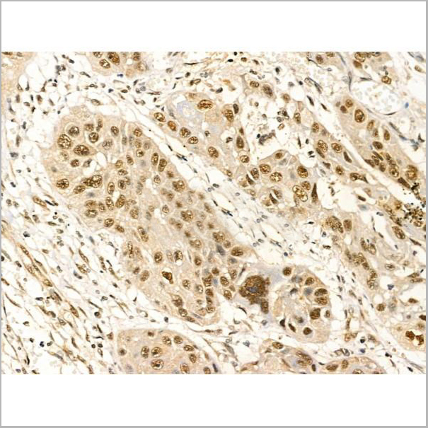
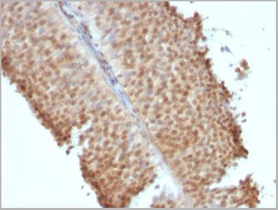
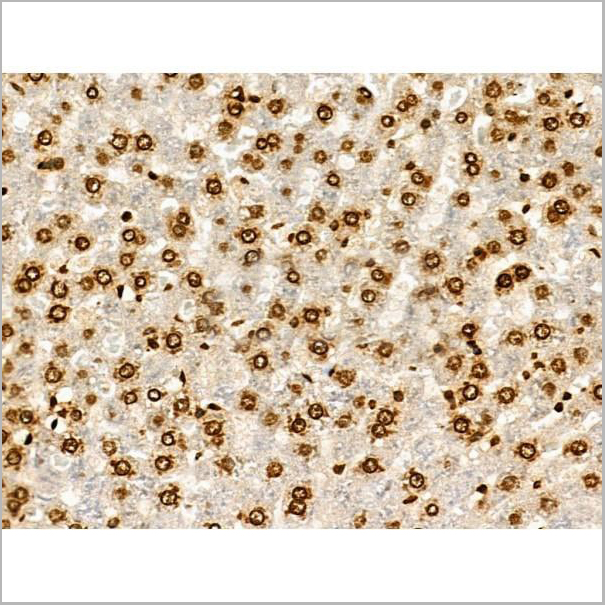
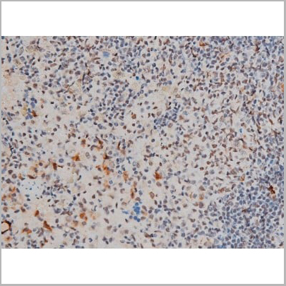
 IHC (Immunohistochemistry)
(ICH of Macrophage HAM-56 on an FFPE Liver Tissue)
IHC (Immunohistochemistry)
(ICH of Macrophage HAM-56 on an FFPE Liver Tissue)
Macrophage HAM-56, Monoclonal Antibody (Cat# AAA13562)
Full Name
Macrophage HAM-56
Reactivity
Human, Monkey
Applications
Immunohistochemistry
Pricing
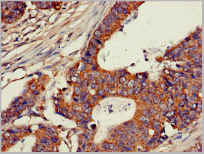
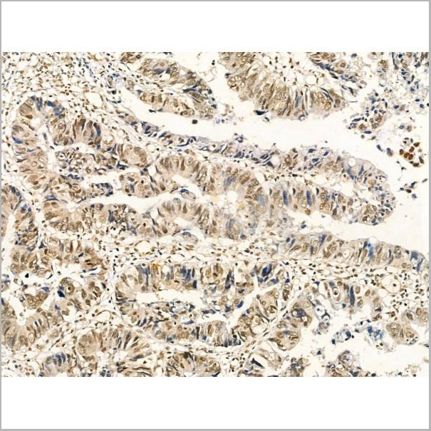
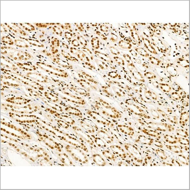
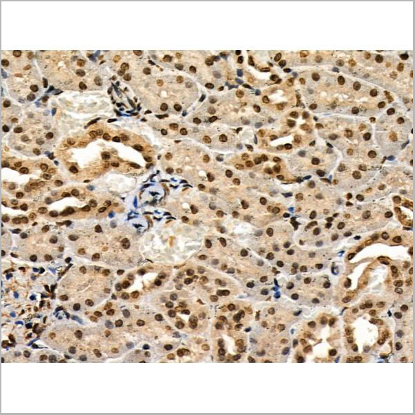
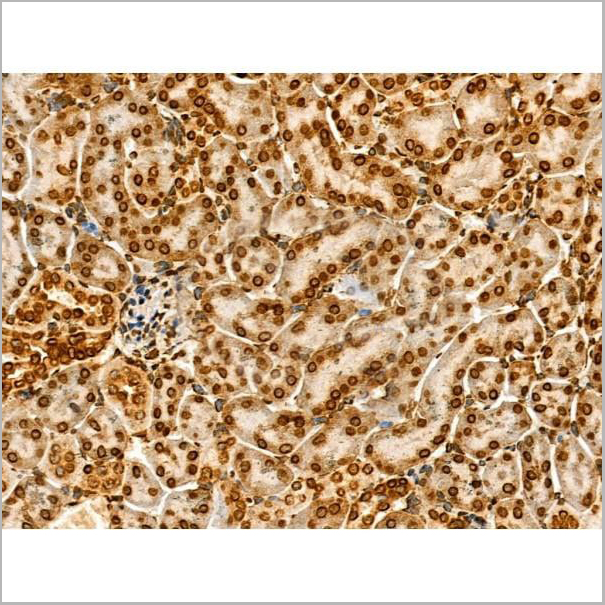
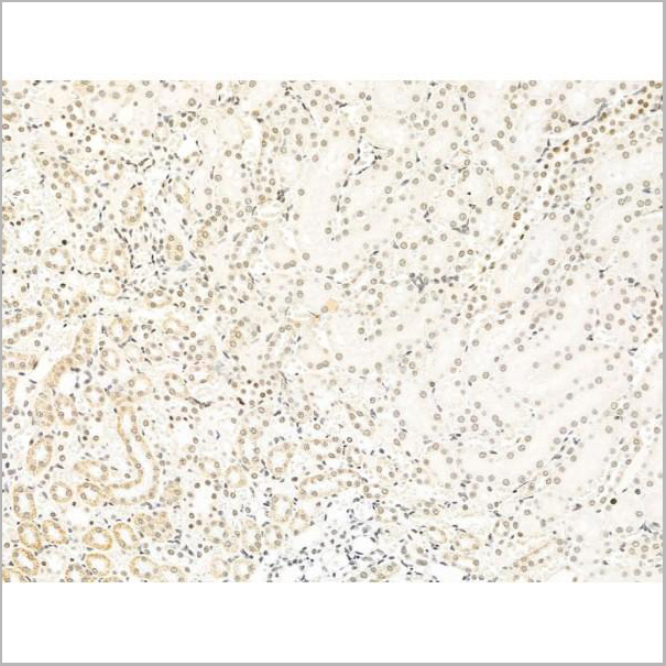
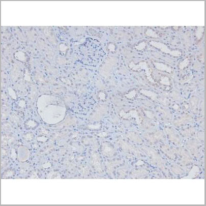
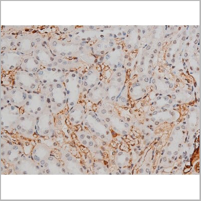
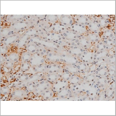
 IHC (Immunohistochemistry)
(Immunohistochemistry of paraffin-embedded human kidney tissue using AAA26983 at dilution of 1:100)
IHC (Immunohistochemistry)
(Immunohistochemistry of paraffin-embedded human kidney tissue using AAA26983 at dilution of 1:100)
CH25H, Polyclonal Antibody (Cat# AAA26983)
Full Name
CH25H Antibody
Gene Names
CH25H; C25H
Reactivity
Human
Applications
Immunohistochemistry
Purity
>95%, Protein G purified
Pricing
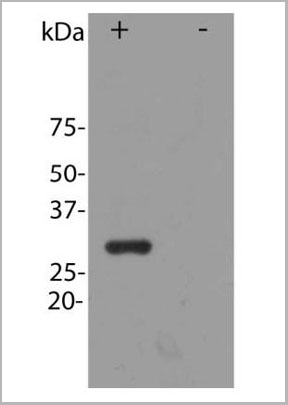 WB (Western Blot)
(Image: Blot of HEK293 cells transfected with pFin-EF1-mCherry vector, in the lane marked "+". HEK293 cells which were not transfected with this vector show no protein band in lane marked)
WB (Western Blot)
(Image: Blot of HEK293 cells transfected with pFin-EF1-mCherry vector, in the lane marked "+". HEK293 cells which were not transfected with this vector show no protein band in lane marked)
mCherry, Antibody (Cat# AAA14440)
Full Name
mCherry, Affinity Purified Antibody, Rabbit
Reactivity
All mammalian
Applications
Immunofluorescence, Western Blot
Pricing
 Biological Activity
(Human TNF-alpha induces cell cytotoxicity in the L-929 mouse fibroblast cells in the presence of the metabolic inhibitor actinomycin D.)
Biological Activity
(Human TNF-alpha induces cell cytotoxicity in the L-929 mouse fibroblast cells in the presence of the metabolic inhibitor actinomycin D.)
Tumor Necrosis Factor-alpha, Active Protein (Cat# AAA11742)
Full Name
Tumor Necrosis Factor-alpha, 77-233 aa, Human, E Coli (Bioactivity Validated)
Gene Names
TNF; DIF; TNFA; TNFSF2; TNLG1F; TNF-alpha
Applications
SDS-PAGE, Bioactivity
Purity
> 95% by SDS-PAGE
Pricing

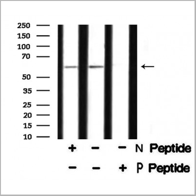
 Application Data
(At 25 degree C. Samples were then incubated with primary Ab(At 37 degree C. An AlexaFluor594 conjugated goat anti-rabbit IgG(H+L) Ab(Red) and an AlexaFluor488 conjugated goat anti-mouse IgG(H+L) Ab(Green) were used as the secondary antibody.The nuclear counter stain is DAPI (blue).)
Application Data
(At 25 degree C. Samples were then incubated with primary Ab(At 37 degree C. An AlexaFluor594 conjugated goat anti-rabbit IgG(H+L) Ab(Red) and an AlexaFluor488 conjugated goat anti-mouse IgG(H+L) Ab(Green) were used as the secondary antibody.The nuclear counter stain is DAPI (blue).)
IRF7, Polyclonal Antibody (Cat# AAA31330)
Full Name
Phospho-IRF7 (Ser471/Ser472) Antibody
Gene Names
IRF7; IRF7A; IRF7B; IRF7C; IRF7H; IRF-7H
Reactivity
Human, Mouse, Rat
Applications
Western Blot, Immunohistochemistry, Immunofluorescence, Immunocytochemistry, Peptide ELISA
Purity
The antibody is from purified rabbit serum by affinity purification via sequential chromatography on phospho-peptide and non-phospho-peptide affinity columns.
Pricing
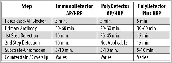
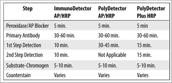
 Staining Procedure
(1. Cut and mount 3-5 micron formalin-fixed paraffin-embedded tissues on positive charged slides such as Hydrophilic Plus Slides.2. Air dry for 2 hours at 58° C.3. Deparaffinize, dehydrate and rehydrate tissues.4. Subject tissues to heat epitope retrieval using a suitable retrieval solution such as ImmunoDNA Retriever with Citrate or EDTA.5. Any of three heating methods may be used:a. TintoRetriever Pressure Cooker or EquivalentPlace tissues/slides in a staining dish or coplin jar containing the ImmunoDNA Retriever with Citrate or EDTA, and place in the pressure cooker. Add 1-2 inches of distilled water to the pressure cooker and turn heat to high. Incubate for 15 minutes. Open and immediately transfer slides to room temperature. b. TintoRetriever PT Module or Water Bath Method Place tissues/slides in a pre-warmed staining dish or coplin jar containing the ImmunoDNA Retriever with Citrate or EDTA at 95°-99° C. Incubate for 30-60 minutes.c. Conventional Steamer MethodPlace tissues/slides in a pre-warmed staining dish or coplin jar containing the ImmunoDNA Retriever with Citrate or EDTA in a Steamer, cover and steam for 30-60 minutes.6. After heat treatment, transfer slides in ImmunoDNA Retriever with Citrate or EDTA to room temperature and let stand for 15-20 minutes.7. For manual staining, perform antibody incubation at ambient temperature. For automated staining methods, perform antibody incubation according to instrument manufacturer’s instructions. 8. Wash slides with ImmunoDNA washer or DI water. 9. Continue IHC staining protocol.)
Staining Procedure
(1. Cut and mount 3-5 micron formalin-fixed paraffin-embedded tissues on positive charged slides such as Hydrophilic Plus Slides.2. Air dry for 2 hours at 58° C.3. Deparaffinize, dehydrate and rehydrate tissues.4. Subject tissues to heat epitope retrieval using a suitable retrieval solution such as ImmunoDNA Retriever with Citrate or EDTA.5. Any of three heating methods may be used:a. TintoRetriever Pressure Cooker or EquivalentPlace tissues/slides in a staining dish or coplin jar containing the ImmunoDNA Retriever with Citrate or EDTA, and place in the pressure cooker. Add 1-2 inches of distilled water to the pressure cooker and turn heat to high. Incubate for 15 minutes. Open and immediately transfer slides to room temperature. b. TintoRetriever PT Module or Water Bath Method Place tissues/slides in a pre-warmed staining dish or coplin jar containing the ImmunoDNA Retriever with Citrate or EDTA at 95°-99° C. Incubate for 30-60 minutes.c. Conventional Steamer MethodPlace tissues/slides in a pre-warmed staining dish or coplin jar containing the ImmunoDNA Retriever with Citrate or EDTA in a Steamer, cover and steam for 30-60 minutes.6. After heat treatment, transfer slides in ImmunoDNA Retriever with Citrate or EDTA to room temperature and let stand for 15-20 minutes.7. For manual staining, perform antibody incubation at ambient temperature. For automated staining methods, perform antibody incubation according to instrument manufacturer’s instructions. 8. Wash slides with ImmunoDNA washer or DI water. 9. Continue IHC staining protocol.)
Podoplanin/D2-40, Monoclonal Antibody (Cat# AAA13558)
Full Name
Podoplanin/D2-40
Reactivity
Human, Rat, Mouse
Applications
Immunohistochemistry
Pricing
Ribonuclease H1, Recombinant Protein (Cat# AAA10894)
Full Name
Recombinant E Coli Ribonuclease H1
Gene Names
rnhA; cer; dasF; ECK0214; herA; JW0204; rnh; sdrA; sin
Purity
Greater than 95% as determined by SDS-PAGE.
Pricing
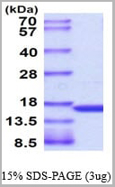
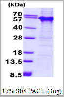 SDS-PAGE
(3ug by SDS-PAGE under reducing condition and visualized by coomassie blue stain.)
SDS-PAGE
(3ug by SDS-PAGE under reducing condition and visualized by coomassie blue stain.)
TDG, Recombinant Protein (Cat# AAA11810)
Full Name
TDG, 1-410aa, Human, His tag, E Coli (Denatured)
Gene Names
TDG; hTDG
Applications
SDS-PAGE
Purity
> 85% by SDS-PAGE
Pricing
 IF (Immunofluorescence)
(AAA31193 staining Hela cells by IF/ICC. The samples were fixed with PFA and permeabilized in 0.1% Triton X-100,then blocked in 10% serum for 45 minutes at 25°C. Samples were then incubated with primary Ab(AAA31193 1:200) and mouse anti-beta tubulin Ab(T0023 1:200) for 1 hour at 37°C. An AlexaFluor594 conjugated goat anti-rabbit IgG(H+L) Ab(Red) and an AlexaFluor488 conjugated goat anti-mouse IgG(H+L) Ab(Green) were used as the secondary antibody.The nuclear counter stain is DAPI(blue).)
IF (Immunofluorescence)
(AAA31193 staining Hela cells by IF/ICC. The samples were fixed with PFA and permeabilized in 0.1% Triton X-100,then blocked in 10% serum for 45 minutes at 25°C. Samples were then incubated with primary Ab(AAA31193 1:200) and mouse anti-beta tubulin Ab(T0023 1:200) for 1 hour at 37°C. An AlexaFluor594 conjugated goat anti-rabbit IgG(H+L) Ab(Red) and an AlexaFluor488 conjugated goat anti-mouse IgG(H+L) Ab(Green) were used as the secondary antibody.The nuclear counter stain is DAPI(blue).)
CHD4, Polyclonal Antibody (Cat# AAA31193)
Full Name
CHD4 Antibody
Gene Names
CHD4; CHD-4; Mi-2b; SIHIWES; Mi2-BETA
Reactivity
Human, Mouse, Rat
Predicted Reactivity: Pig(100%), Horse(100%), Rabbit(100%), Dog(100%), Chicken(100%), Xenopus(100%)
Predicted Reactivity: Pig(100%), Horse(100%), Rabbit(100%), Dog(100%), Chicken(100%), Xenopus(100%)
Applications
Immunofluorescence, Immunocytochemistry
Purity
The antiserum was purified by peptide affinity chromatography using SulfoLink Coupling Resin (Thermo Fisher Scientific).
Pricing
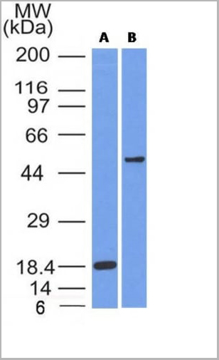
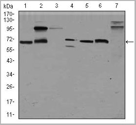
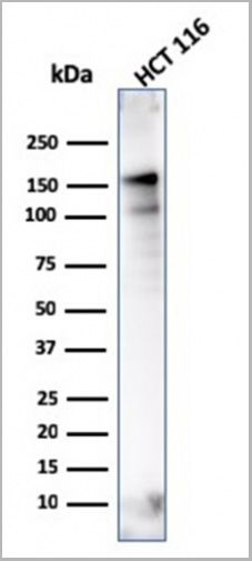
 WB (Western Blot)
(Western Blot Analysis of human MCF-7 cell lysate using ER-beta1 Mouse Monoclonal Antibody (ERb455).)
WB (Western Blot)
(Western Blot Analysis of human MCF-7 cell lysate using ER-beta1 Mouse Monoclonal Antibody (ERb455).)
ER-beta1 (Estrogen Receptor beta-1), Monoclonal Antibody (Cat# AAA13814)
Full Name
ER-beta1 (Estrogen Receptor beta-1) Mouse Monoclonal Antibody
Gene Names
ESR2; Erb; ESRB; ESTRB; NR3A2; ER-BETA; ESR-BETA
Reactivity
Human
Applications
Flow Cytometry, Immunofluorescence, Western Blot, Immunohistochemistry
Pricing


 Application Data
(Published customer image: Spindle abnormalities in embryos derived from imp-a2D14/imp-betaKetRE34 and imp-a2D14/imp-betac02473; NLSB-/+ females. (A -D) Wild-type and mutant embryos stained for a-tubulin (green) and DNA (blue). (A) Mitotic spindles in wild-type embryos at metaphase and anaphase. (B, C) Categories of spindle abnormalities found in embryos derived from (B) imp-a2D14/imp-betaKetRE34 and (C) imp-a2D14/imp-betac02743; NLSB-/+ females. (D) Formation of aster networks found in both genotypes. Scale bar: 10 um. (E) Frequency of spindle defects in embryos from both types of mutant females. Female genotypes are displayed at the upper right corner. At least 200 spindles were scored for both genotypes.From: Specific Cooperation Between Imp-a2 and Imp-beta/Ketel in Spindle Assembly During Drosophila Early Nuclear Divisions Erika Vir¡gh, M¡ty¡s Gorj¡n¡cz, Istv¡n T¶r¶k, Tolga Eichhorn, Sowjanya Kallakuri, Tam¡s Szlanka, Istv¡n Kiss, and Bernard M. Mechler G3 January 2012 2:1-14.)
Application Data
(Published customer image: Spindle abnormalities in embryos derived from imp-a2D14/imp-betaKetRE34 and imp-a2D14/imp-betac02473; NLSB-/+ females. (A -D) Wild-type and mutant embryos stained for a-tubulin (green) and DNA (blue). (A) Mitotic spindles in wild-type embryos at metaphase and anaphase. (B, C) Categories of spindle abnormalities found in embryos derived from (B) imp-a2D14/imp-betaKetRE34 and (C) imp-a2D14/imp-betac02743; NLSB-/+ females. (D) Formation of aster networks found in both genotypes. Scale bar: 10 um. (E) Frequency of spindle defects in embryos from both types of mutant females. Female genotypes are displayed at the upper right corner. At least 200 spindles were scored for both genotypes.From: Specific Cooperation Between Imp-a2 and Imp-beta/Ketel in Spindle Assembly During Drosophila Early Nuclear Divisions Erika Vir¡gh, M¡ty¡s Gorj¡n¡cz, Istv¡n T¶r¶k, Tolga Eichhorn, Sowjanya Kallakuri, Tam¡s Szlanka, Istv¡n Kiss, and Bernard M. Mechler G3 January 2012 2:1-14.)
TUBULIN ALPHA, Monoclonal Antibody (Cat# AAA12232)
Full Name
RAT ANTI TUBULIN ALPHA:HRP
Applications
Immunohistochemistry, Western Blot
Pricing

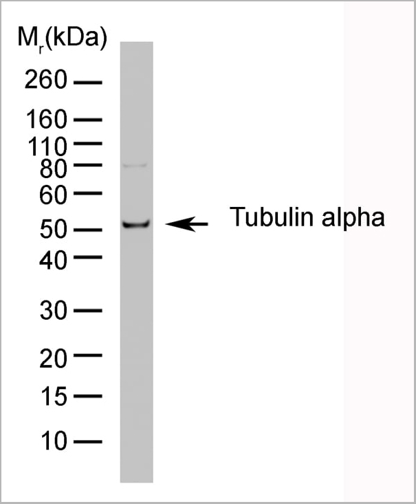

 Application Data
(Published customer image: Spindle abnormalities in embryos derived from imp-a2D14/imp-betaKetRE34 and imp-a2D14/imp-betac02473; NLSB-/+ females. (A -D) Wild-type and mutant embryos stained for a-tubulin (green) and DNA (blue). (A) Mitotic spindles in wild-type embryos at metaphase and anaphase. (B, C) Categories of spindle abnormalities found in embryos derived from (B) imp-a2D14/imp-betaKetRE34 and (C) imp-a2D14/imp-betac02743; NLSB-/+ females. (D) Formation of aster networks found in both genotypes. Scale bar: 10 um. (E) Frequency of spindle defects in embryos from both types of mutant females. Female genotypes are displayed at the upper right corner. At least 200 spindles were scored for both genotypes.From: Specific Cooperation Between Imp-a2 and Imp-beta/Ketel in Spindle Assembly During Drosophila Early Nuclear Divisions Erika Vir¡gh, M¡ty¡s Gorj¡n¡cz, Istv¡n T¶r¶k, Tolga Eichhorn, Sowjanya Kallakuri, Tam¡s Szlanka, Istv¡n Kiss, and Bernard M. Mechler G3 January 2012 2:1-14.)
Application Data
(Published customer image: Spindle abnormalities in embryos derived from imp-a2D14/imp-betaKetRE34 and imp-a2D14/imp-betac02473; NLSB-/+ females. (A -D) Wild-type and mutant embryos stained for a-tubulin (green) and DNA (blue). (A) Mitotic spindles in wild-type embryos at metaphase and anaphase. (B, C) Categories of spindle abnormalities found in embryos derived from (B) imp-a2D14/imp-betaKetRE34 and (C) imp-a2D14/imp-betac02743; NLSB-/+ females. (D) Formation of aster networks found in both genotypes. Scale bar: 10 um. (E) Frequency of spindle defects in embryos from both types of mutant females. Female genotypes are displayed at the upper right corner. At least 200 spindles were scored for both genotypes.From: Specific Cooperation Between Imp-a2 and Imp-beta/Ketel in Spindle Assembly During Drosophila Early Nuclear Divisions Erika Vir¡gh, M¡ty¡s Gorj¡n¡cz, Istv¡n T¶r¶k, Tolga Eichhorn, Sowjanya Kallakuri, Tam¡s Szlanka, Istv¡n Kiss, and Bernard M. Mechler G3 January 2012 2:1-14.)
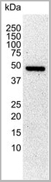
TUBULIN ALPHA, Monoclonal Antibody (Cat# AAA12009)
Full Name
RAT ANTI TUBULIN ALPHA
Applications
Immunohistochemistry, Immunofluorescence, Immunoprecipitation, Radioimmunoassay, Western Blot
Pricing


 Application Data
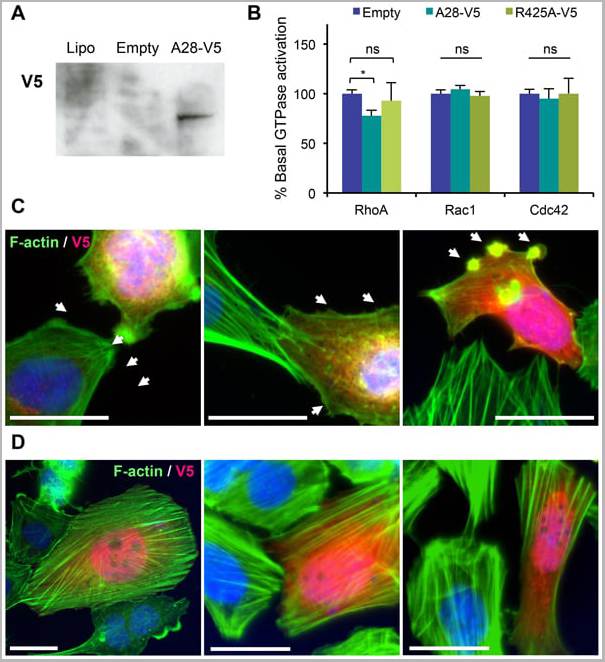
(Published customer image: Mouse anti V5 tag antibody, clone SV5-Pk1 used for the detection of V5 tagged WEEV_nsP3 protein by western blotting and immunofluorescenceImage caption: WEEV nsP3 interaction with host IKKbeta. A) U87MGs were transfected in a 6-well plate with 5 ug of pUC19 and WEEV_nsP3_HA for 24 hours. Cell lysates were resolved using SDS-PAGE and subsequently immunoblotted with V5 antibody and beta-actin served as a loading control. B) U87MGs were transfected with WEEV_nsP3_V5; cells were fixed after 24 hours and stained with antibodies against the endogenous IKKbeta and the V5 tag. Cells were incubated with appropriate secondary Alexa Fluor antibodies and the nuclei stained with DAPI. Co-localization of IKKbeta with WEEV_nsP3_V5 (yellow) was observed as shown by the arrows. B) Panels E -H serve as an example of transfected cells in a given field of view that show co-localization of IKKbeta and WEEV_nsP3_V5 24 hours post transfection. Panels I-L represent magnified images of other cells showing co-localization of IKKbeta and WEEV_nsP3_V5. Panel M is a magnified image of panel L. The co-localization was confirmed by Z-stack analysis. Co-localization was calculated to be approximately in 61% of cells (163 cells were counted of which 44% demonstrated expression of nsP3. Of those cells that expressed nsP3, 61% showed co-localization of both proteins). Images were taken using Nikon Eclipse TE2000-U at 60x magnification and are representative of 2 independent experiments.From: Amaya M, Voss K, Sampey G, Senina S, de la Fuente C, et al. (2014) The Role of IKKbeta in Venezuelan Equine Encephalitis Virus Infection. PLoS ONE 9(2): e86745.)
Application Data
(Published customer image: Mouse anti V5 tag antibody, clone SV5-Pk1 used for the detection of V5 tagged WEEV_nsP3 protein by western blotting and immunofluorescenceImage caption: WEEV nsP3 interaction with host IKKbeta. A) U87MGs were transfected in a 6-well plate with 5 ug of pUC19 and WEEV_nsP3_HA for 24 hours. Cell lysates were resolved using SDS-PAGE and subsequently immunoblotted with V5 antibody and beta-actin served as a loading control. B) U87MGs were transfected with WEEV_nsP3_V5; cells were fixed after 24 hours and stained with antibodies against the endogenous IKKbeta and the V5 tag. Cells were incubated with appropriate secondary Alexa Fluor antibodies and the nuclei stained with DAPI. Co-localization of IKKbeta with WEEV_nsP3_V5 (yellow) was observed as shown by the arrows. B) Panels E -H serve as an example of transfected cells in a given field of view that show co-localization of IKKbeta and WEEV_nsP3_V5 24 hours post transfection. Panels I-L represent magnified images of other cells showing co-localization of IKKbeta and WEEV_nsP3_V5. Panel M is a magnified image of panel L. The co-localization was confirmed by Z-stack analysis. Co-localization was calculated to be approximately in 61% of cells (163 cells were counted of which 44% demonstrated expression of nsP3. Of those cells that expressed nsP3, 61% showed co-localization of both proteins). Images were taken using Nikon Eclipse TE2000-U at 60x magnification and are representative of 2 independent experiments.From: Amaya M, Voss K, Sampey G, Senina S, de la Fuente C, et al. (2014) The Role of IKKbeta in Venezuelan Equine Encephalitis Virus Infection. PLoS ONE 9(2): e86745.)
V5-TAG, Monoclonal Antibody (Cat# AAA12081)
Full Name
MOUSE ANTI V5-TAG:HRP
Applications
Western Blot
Pricing
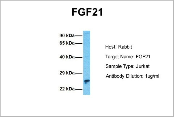
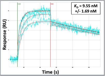 Application Data
(Surface Plasmon Resonance Kinetic Characterization of Polyclonal Antibody Affinity. Purified polyclonal antibodies were immobilized on a Protein A/G coated Carterra LSA sensor chip (PAGH200M) at concentrations of 5, and 50 ug/mL in duplicate. Antibodies on the surface were exposed to interaction with peptides sequentially via microfluidic controlled flow at 333nM peptide concentration for kinetic characterization of the binders for affinity and specificity, followed by curve fitting using the Kinetics software. Kd determinations for both concentrations were averaged and results and standard deviation are shown.)
Application Data
(Surface Plasmon Resonance Kinetic Characterization of Polyclonal Antibody Affinity. Purified polyclonal antibodies were immobilized on a Protein A/G coated Carterra LSA sensor chip (PAGH200M) at concentrations of 5, and 50 ug/mL in duplicate. Antibodies on the surface were exposed to interaction with peptides sequentially via microfluidic controlled flow at 333nM peptide concentration for kinetic characterization of the binders for affinity and specificity, followed by curve fitting using the Kinetics software. Kd determinations for both concentrations were averaged and results and standard deviation are shown.)
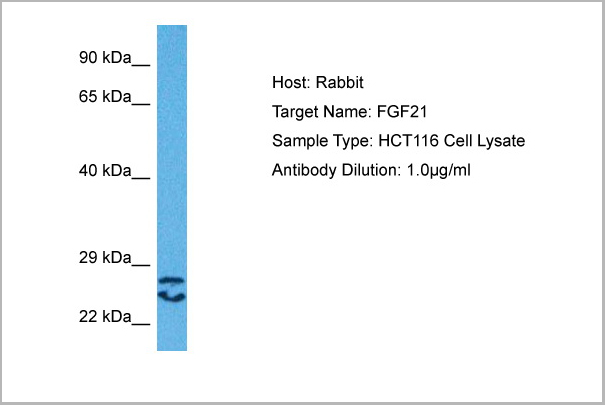
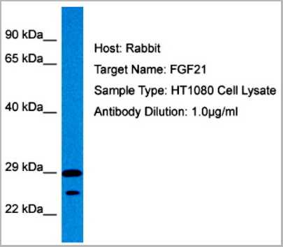
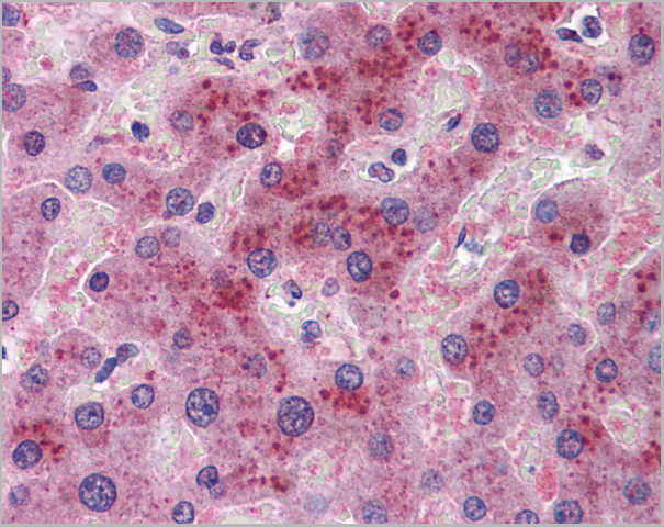
FGF21, Polyclonal Antibody (Cat# AAA23563)
Full Name
FGF21 antibody - N-terminal region
Reactivity
Tested Species Reactivity: Human
Predicted Species Reactivity: Human, Mouse, Rat, Cow, Dog, Guinea Pig, Horse, Rabbit
Predicted Species Reactivity: Human, Mouse, Rat, Cow, Dog, Guinea Pig, Horse, Rabbit
Applications
Immunohistochemistry, Western Blot
Purity
Affinity Purified
Pricing
HBsAg, Polyclonal Antibody (Cat# AAA14317)
Full Name
HBsAg antibody
Reactivity
Specific for all 4 subtypes of Hepatitis B surface Antigens
Purity
HBsAg antibody was purified by affinity chromatography.
Pricing
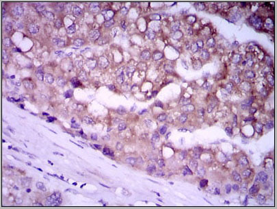
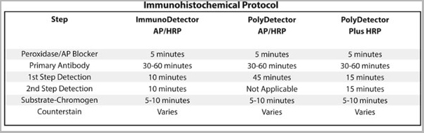
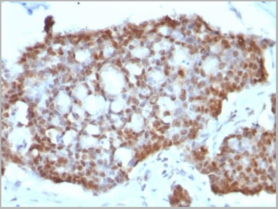
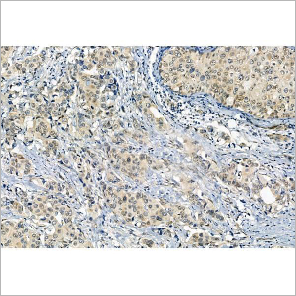
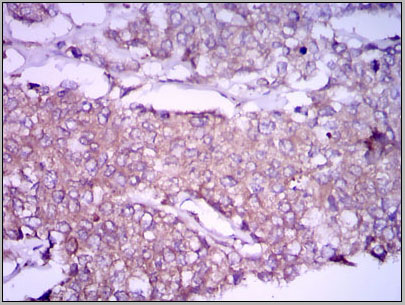 IHC (Immunohistchemistry)
(Immunohistochemical analysis of paraffin-embedded breast cancer tissues using GPC3 mouse mAb with DAB staining.)
IHC (Immunohistchemistry)
(Immunohistochemical analysis of paraffin-embedded breast cancer tissues using GPC3 mouse mAb with DAB staining.)
GPC3, Monoclonal Antibody (Cat# AAA14118)
Full Name
Anti-GPC3 Mouse mAb
Gene Names
GPC3; SGB; DGSX; MXR7; SDYS; SGBS; OCI-5; SGBS1; GTR2-2
Reactivity
Human, Mouse
Applications
Immunohistochemistry, Immunocytochemistry, Immunofluorescence, Flow Cytometry
Purity
Affinity Purified
Pricing
HBcAg, Recombinant Protein (Cat# AAA14357)
Full Name
HBcAg protein
Applications
Western Blot
Purity
>95 % (by SDS-PAGE)
Pricing
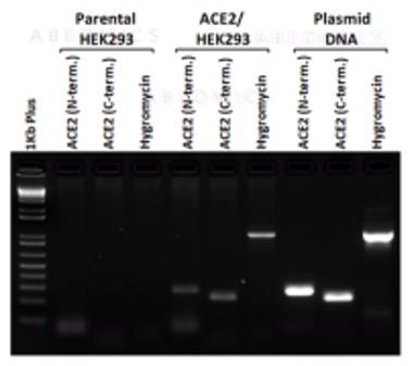
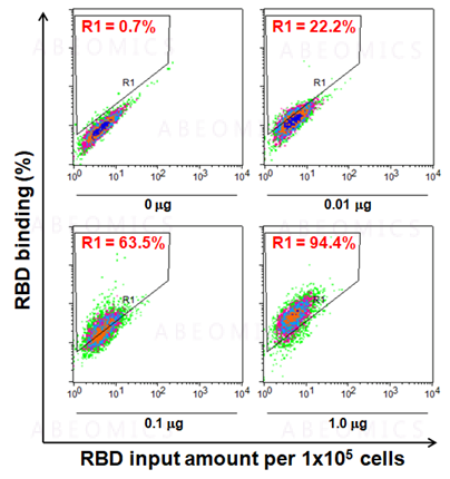
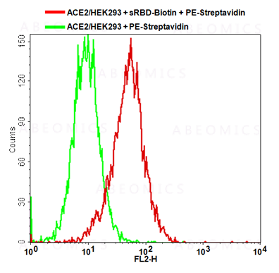
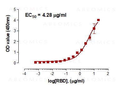
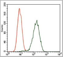
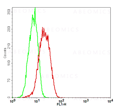 Application Data
(Fig-5: Cell Surface staining of human ACE2 in the ACE2/HEK293 stable cell line. ATTO 488-conjugated anti-hACE2 antibody (clone AC18F; was used at 1ug/ 1x10^6 cells (Red). ATTO 488-conjugated mouse IgG1 was used as isotype control at 1ug/ 1x10^6 cells (Green).)
Application Data
(Fig-5: Cell Surface staining of human ACE2 in the ACE2/HEK293 stable cell line. ATTO 488-conjugated anti-hACE2 antibody (clone AC18F; was used at 1ug/ 1x10^6 cells (Red). ATTO 488-conjugated mouse IgG1 was used as isotype control at 1ug/ 1x10^6 cells (Green).)
ACE2, Cell Line (Cat# AAA14888)
Full Name
ACE2/HEK293 Stable Cell Line
Gene Names
ACE2; ACEH
Applications
Functional Assay
Pricing
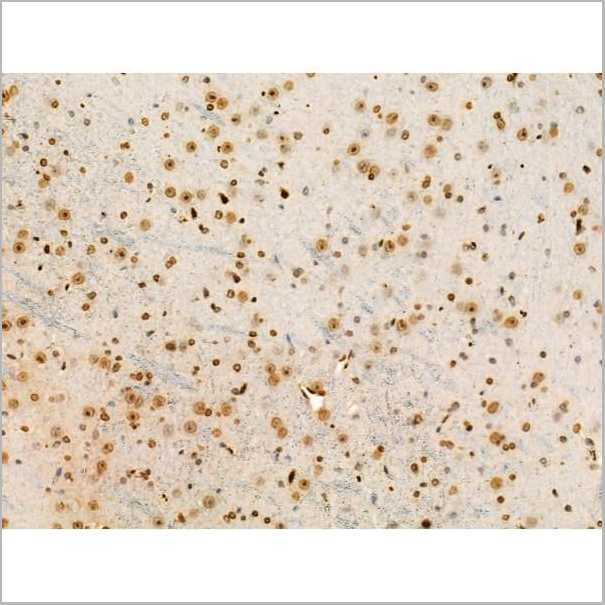
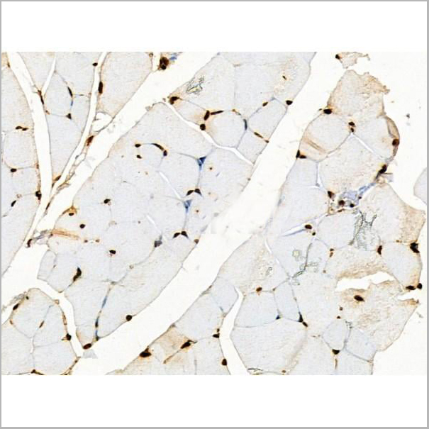
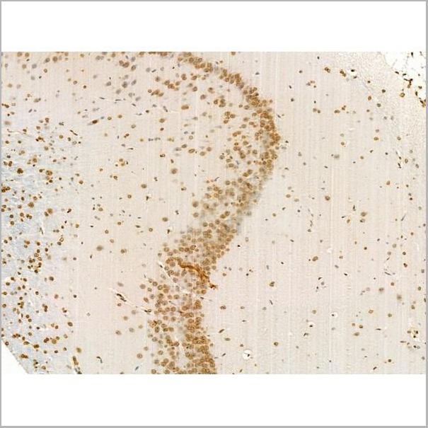
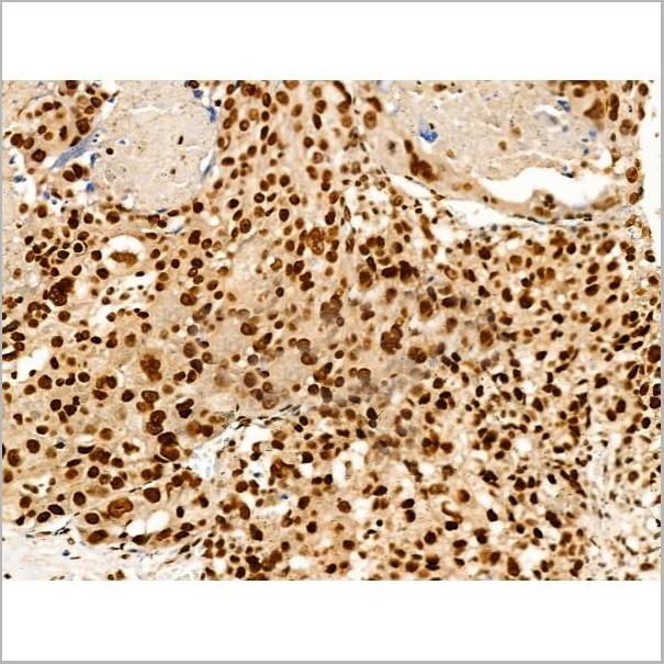
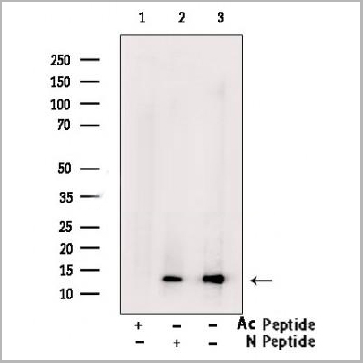
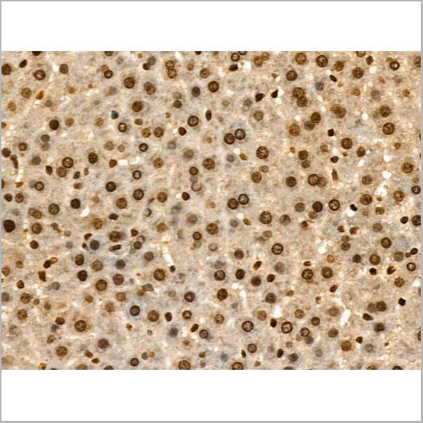
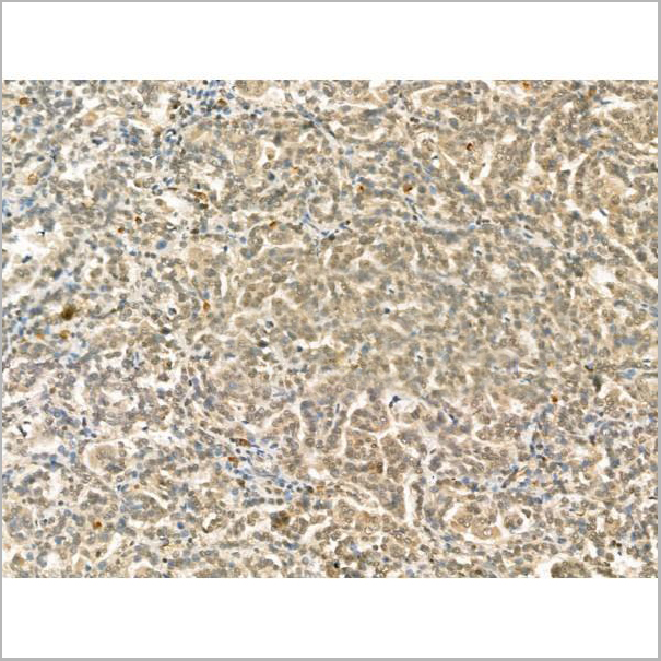
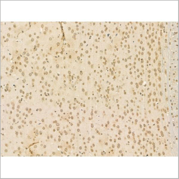
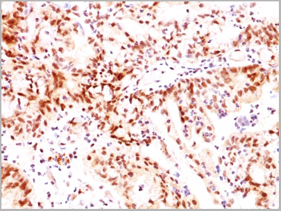
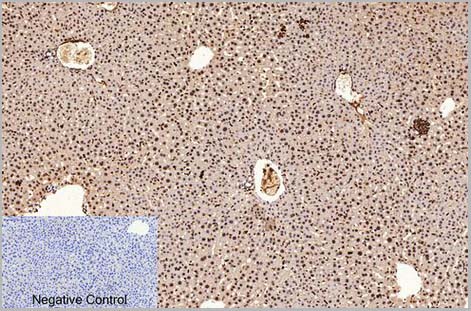
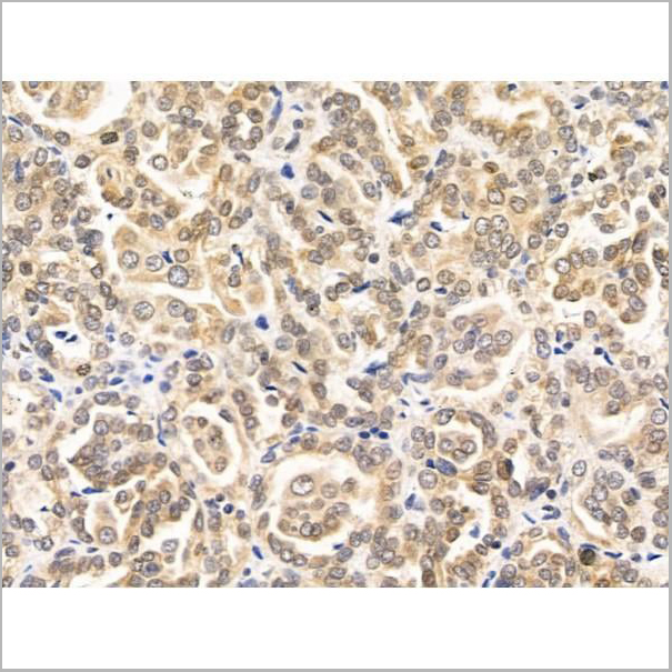
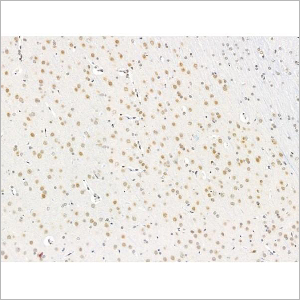
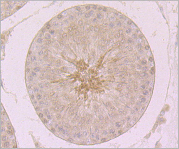
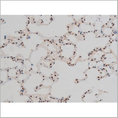
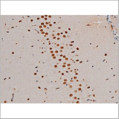
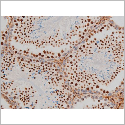
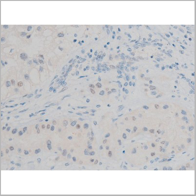
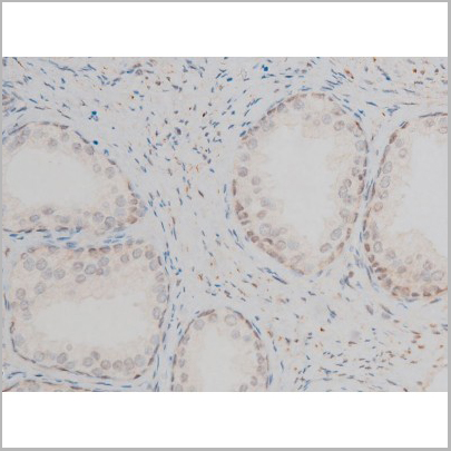
 IHC (Immunohistochemistry)
(At 1/100 staining Mouse liver tissue by IHC-P. The sample was formaldehyde fixed and a heat mediated antigen retrieval step in citrate buffer was performed. The sample was then blocked and incubated with the primary antibody at 4 degree C overnight. An HRP conjugated anti-Rabbit antibody was used as the secondary antibody.)
IHC (Immunohistochemistry)
(At 1/100 staining Mouse liver tissue by IHC-P. The sample was formaldehyde fixed and a heat mediated antigen retrieval step in citrate buffer was performed. The sample was then blocked and incubated with the primary antibody at 4 degree C overnight. An HRP conjugated anti-Rabbit antibody was used as the secondary antibody.)
Histone H2B, Polyclonal Antibody (Cat# AAA31352)
Full Name
Acetyl-Histone H2B (Lys20) Antibody
Reactivity
Human, Mouse, Rat
Applications
Western Blot, Immunohistochemistry, Immunofluorescence, Immunocytochemistry, Peptide ELISA
Purity
The antiserum was purified by peptide affinity chromatography using SulfoLink Coupling Resin
Pricing
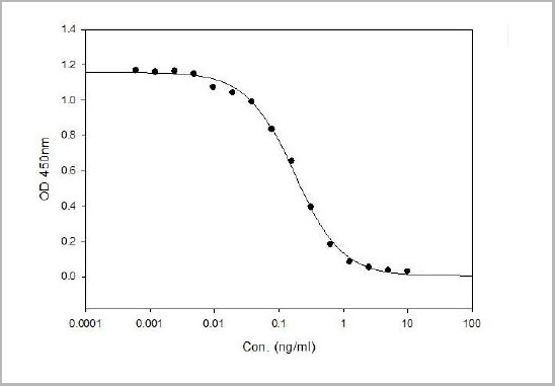
 ELISA
(ELISA Profile of AAA13690)
ELISA
(ELISA Profile of AAA13690)
HA (B/Phuket/3073/2013), Monoclonal Antibody (Cat# AAA13690)
Full Name
Anti-HA (B/Phuket/3073/2013) Monoclonal Antibody
Applications
Western Blot, Immunoprecipitation, Immunofluorescence
Pricing
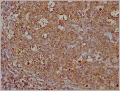
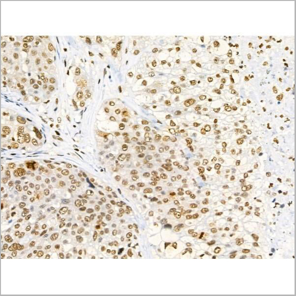
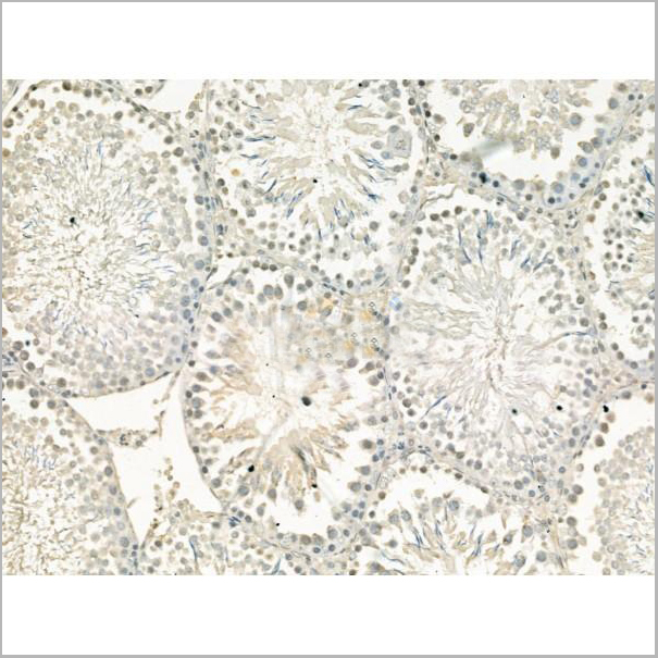
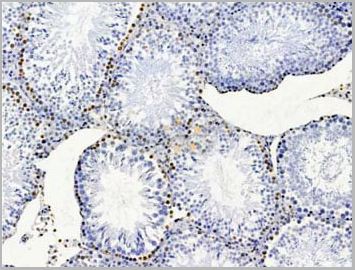
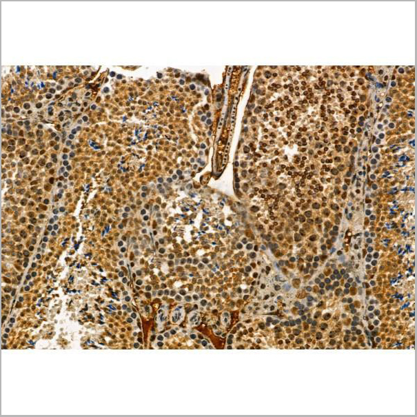
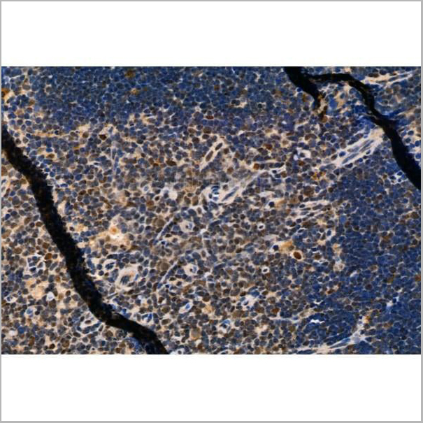
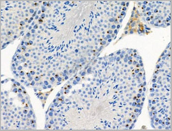
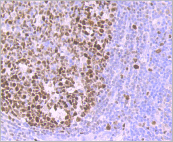
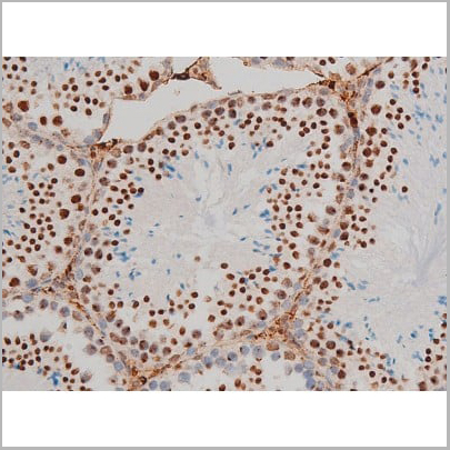
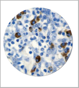
 IHC (Immunohistchemistry)
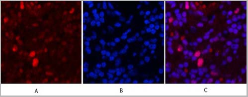
(Fig.6. Immunohistochemical analysis of paraffin-embedded rat testis tissue. 1, PCNA Monoclonal Antibod y(1D7) was diluted at 1:200 (4°C, overnight). 2, Sodium citrate pH 6.0 was used for antibody retrieval(>98°C, 20min). 3, secondary antibody was diluted at 1:200 (room temperature, 30min). Negative control was used by secondary antibody only.)
IHC (Immunohistchemistry)
(Fig.6. Immunohistochemical analysis of paraffin-embedded rat testis tissue. 1, PCNA Monoclonal Antibod y(1D7) was diluted at 1:200 (4°C, overnight). 2, Sodium citrate pH 6.0 was used for antibody retrieval(>98°C, 20min). 3, secondary antibody was diluted at 1:200 (room temperature, 30min). Negative control was used by secondary antibody only.)
PCNA, Monoclonal Antibody (Cat# AAA31541)
Full Name
Anti-PCNA Mouse Monoclonal Antibody (1D7)
Reactivity
Human, Mouse, Rat
Applications
Western Blot, Immunohistochemistry, Immunofluorescence
Purity
The antibody was affinity-purified from mouse ascites by affinity-chromatography using specific immunogen
Pricing
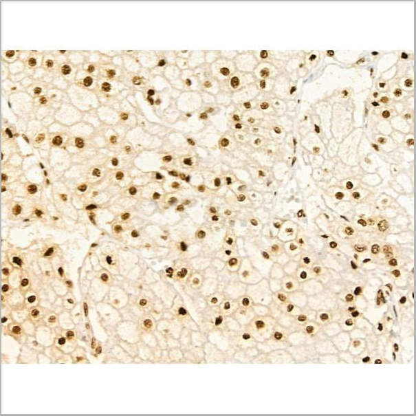
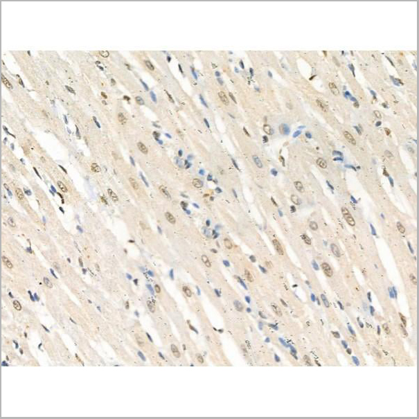
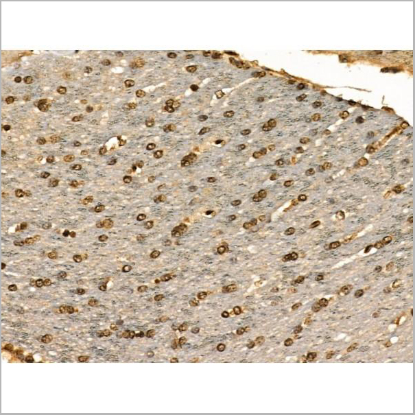
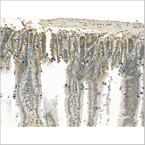
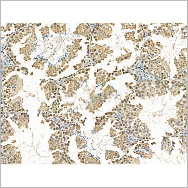
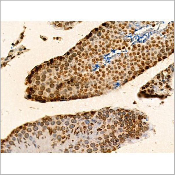
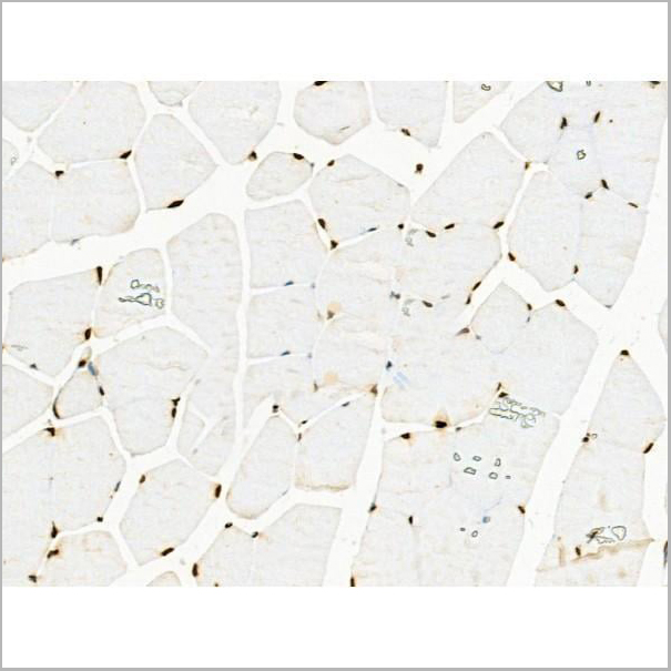
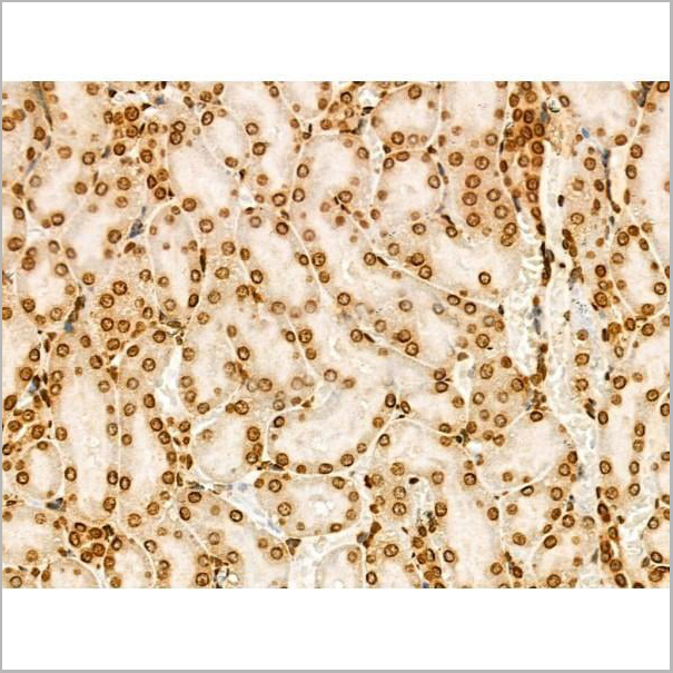
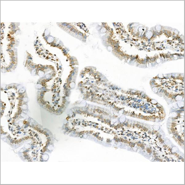
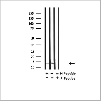
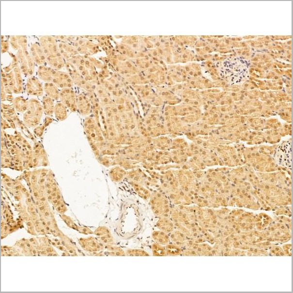
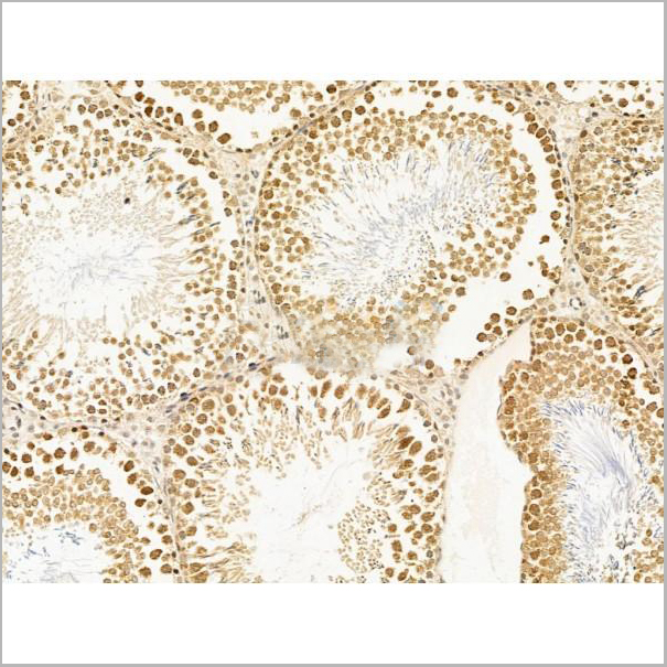
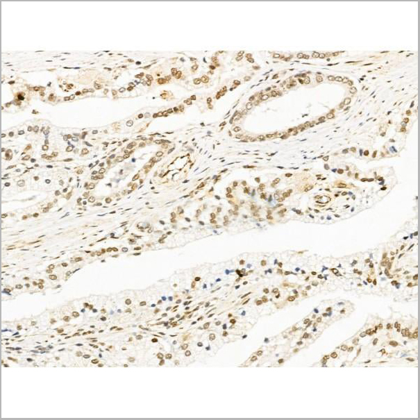
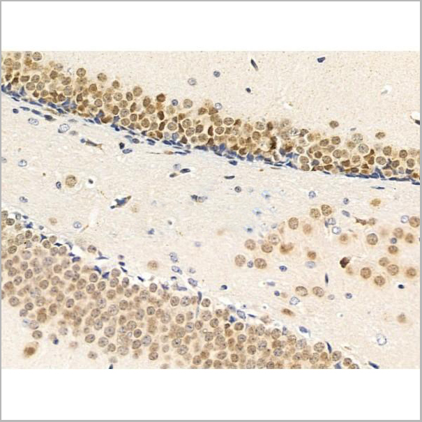
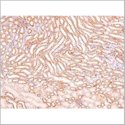
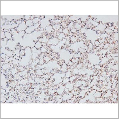
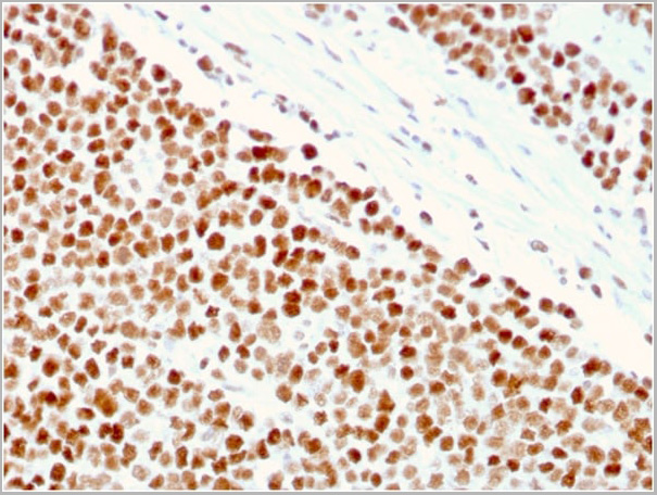
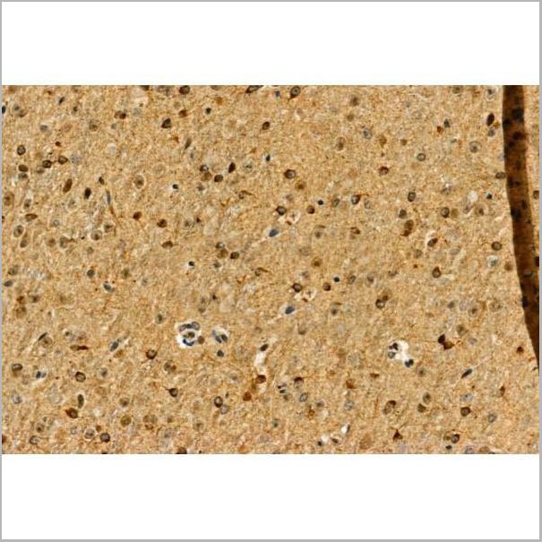
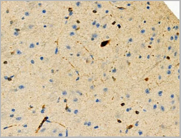
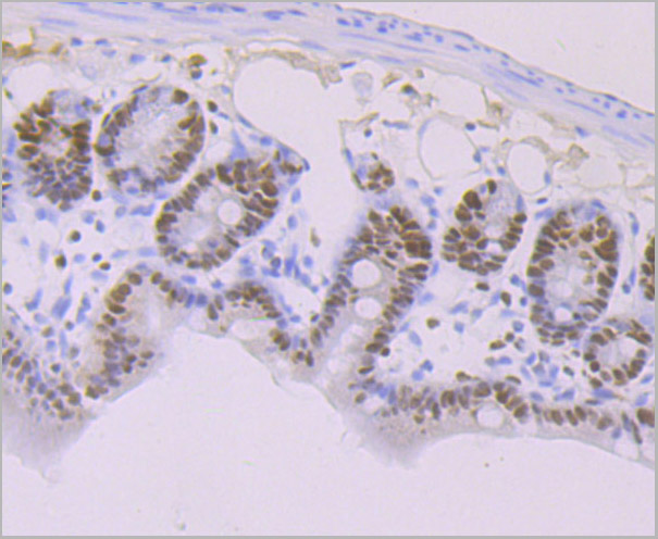
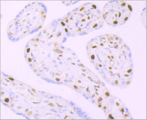
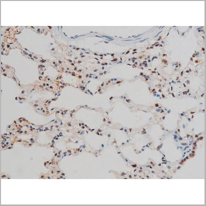
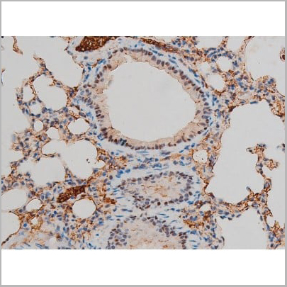
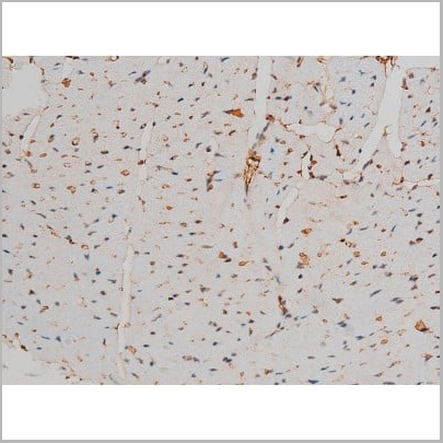
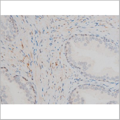
 IHC (Immunohistochemistry-Paraffin)
(AAA31235 at 1/100 staining Rat testis tissue by IHC-P. The sample was formaldehyde fixed and a heat mediated antigen retrieval step in citrate buffer was performed. The sample was then blocked and incubated with the primary antibody at 4 degree C overnight. An HRP conjugated anti-Rabbit antibody was used as the secondary antibody.)
IHC (Immunohistochemistry-Paraffin)
(AAA31235 at 1/100 staining Rat testis tissue by IHC-P. The sample was formaldehyde fixed and a heat mediated antigen retrieval step in citrate buffer was performed. The sample was then blocked and incubated with the primary antibody at 4 degree C overnight. An HRP conjugated anti-Rabbit antibody was used as the secondary antibody.)
DNA Polymerase theta, Polyclonal Antibody (Cat# AAA31235)
Full Name
DNA Polymerase theta Antibody
Gene Names
POLQ; PRO0327
Reactivity
Human, Mouse, Rat
Applications
Western Blot, Immunofluorescence, Immunocytochemistry, Immunohistochemistry
Purity
The antiserum was purified by peptide affinity chromatography using SulfoLink Coupling Resin.
Pricing

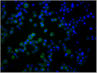
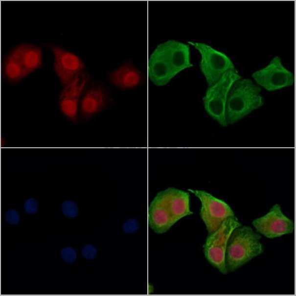
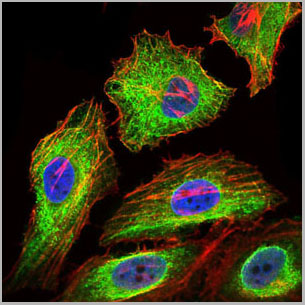
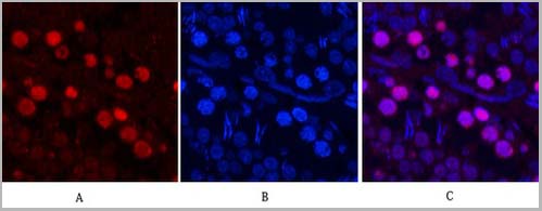
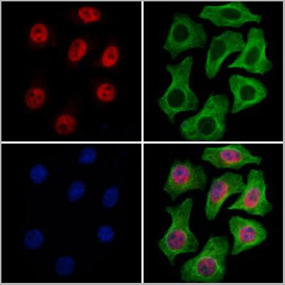
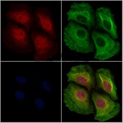
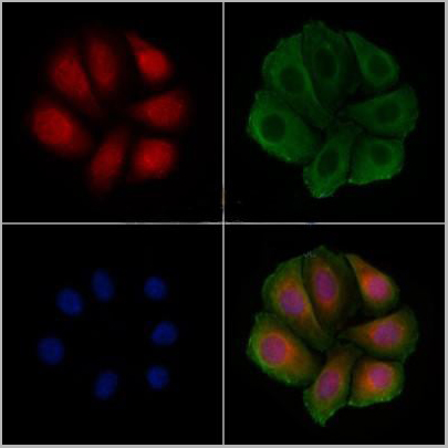
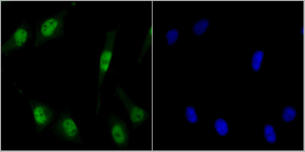
 IF (Immunofluorescence)
(AAA31247 staining HepG2 cells by IF/ICC. The samples were fixed with PFA and permeabilized in 0.1% Triton X-100,then blocked in 10% serum for 45 minutes at 25°C. Samples were then incubated with primary Ab(AAA31247) and mouse anti-beta tubulin Ab(T0023) for 1 hour at 37°C. An AlexaFluor594 conjugated goat anti-rabbit IgG(H+L) Ab(Red) and an AlexaFluor488 conjugated goat anti-mouse IgG(H+L) Ab(Green) were used as the secondary antibody.The nuclear counter stain is DAPI(blue).)
IF (Immunofluorescence)
(AAA31247 staining HepG2 cells by IF/ICC. The samples were fixed with PFA and permeabilized in 0.1% Triton X-100,then blocked in 10% serum for 45 minutes at 25°C. Samples were then incubated with primary Ab(AAA31247) and mouse anti-beta tubulin Ab(T0023) for 1 hour at 37°C. An AlexaFluor594 conjugated goat anti-rabbit IgG(H+L) Ab(Red) and an AlexaFluor488 conjugated goat anti-mouse IgG(H+L) Ab(Green) were used as the secondary antibody.The nuclear counter stain is DAPI(blue).)
MBD4, Polyclonal Antibody (Cat# AAA31247)
Full Name
MBD4 Antibody
Gene Names
MBD4; MED1
Reactivity
Human, Mouse, Rat
Predicted Reactivity: Pig(88%)
Predicted Reactivity: Pig(88%)
Applications
ELISA
Purity
The antiserum was purified by peptide affinity chromatography using SulfoLink Coupling Resin (Thermo Fisher Scientific).
Pricing
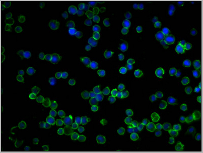
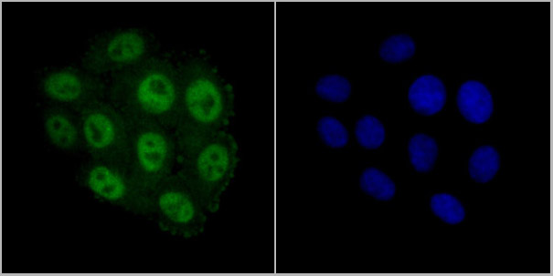
 IF (Immunofluorescence)
(AAA31264 staining Hela cells by IF/ICC. The samples were fixed with PFA and permeabilized in 0.1% Triton X-100,then blocked in 10% serum for 45 minutes at 25°C. Samples were then incubated with primary Ab(AAA31264 1:200) and mouse anti-beta tubulin Ab(T0023 1:200) for 1 hour at 37°C. An AlexaFluor594 conjugated goat anti-rabbit IgG(H+L) Ab(Red) and an AlexaFluor488 conjugated goat anti-mouse IgG(H+L) Ab(Green) were used as the secondary antibody.The nuclear counter stain is DAPI(blue).)
IF (Immunofluorescence)
(AAA31264 staining Hela cells by IF/ICC. The samples were fixed with PFA and permeabilized in 0.1% Triton X-100,then blocked in 10% serum for 45 minutes at 25°C. Samples were then incubated with primary Ab(AAA31264 1:200) and mouse anti-beta tubulin Ab(T0023 1:200) for 1 hour at 37°C. An AlexaFluor594 conjugated goat anti-rabbit IgG(H+L) Ab(Red) and an AlexaFluor488 conjugated goat anti-mouse IgG(H+L) Ab(Green) were used as the secondary antibody.The nuclear counter stain is DAPI(blue).)
RECQL5, Polyclonal Antibody (Cat# AAA31264)
Full Name
RECQL5 Antibody
Gene Names
RECQL5; RECQ5
Reactivity
Human, Mouse, Rat
Predicted Reactivity: Pig(86%)
Predicted Reactivity: Pig(86%)
Applications
ELISA
Purity
The antiserum was purified by peptide affinity chromatography using SulfoLink Coupling Resin (Thermo Fisher Scientific).
Pricing
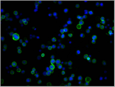
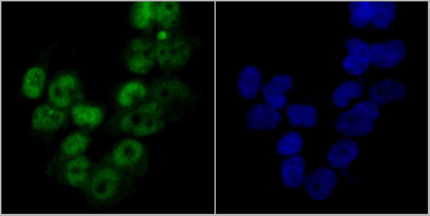
 Application Data
(AAA31382 staining Hela cells(4h of LPS treatment) by IF/ICC. The samples were fixed with PFA and permeabilized in 0.1% Triton X-100,then blocked in 10% serum for 45 minutes at 25°C. Samples were then incubated with primary Ab (AAA31382 1:200) and mouse anti-beta tubulin Ab(T0023 1:200) for 1 hour at 37°C. An AlexaFluor594 conjugated goat anti-rabbit IgG(H+L) Ab(Red) and an AlexaFluor488 conjugated goat anti-mouse IgG(H+L) Ab(Green) were used as the secondary antibody.The nuclear counter stain is DAPI (blue).)
Application Data
(AAA31382 staining Hela cells(4h of LPS treatment) by IF/ICC. The samples were fixed with PFA and permeabilized in 0.1% Triton X-100,then blocked in 10% serum for 45 minutes at 25°C. Samples were then incubated with primary Ab (AAA31382 1:200) and mouse anti-beta tubulin Ab(T0023 1:200) for 1 hour at 37°C. An AlexaFluor594 conjugated goat anti-rabbit IgG(H+L) Ab(Red) and an AlexaFluor488 conjugated goat anti-mouse IgG(H+L) Ab(Green) were used as the secondary antibody.The nuclear counter stain is DAPI (blue).)
NF kappaB p105/p50, Polyclonal Antibody (Cat# AAA31382)
Full Name
Phospho-NF kappaB p105/p50 (Ser923) Antibody
Gene Names
NFKB1; p50; KBF1; p105; EBP-1; NF-kB1; NFKB-p50; NFkappaB; NF-kappaB; NFKB-p105; NF-kappa-B
Reactivity
Human, Mouse, Rat
Applications
Western Blot, Immunohistochemistry, Immunofluorescence, Immunocytochemistry, Peptide ELISA
Purity
Peptide affinity purification
Pricing

 Application Data
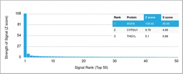
(Analysis of Protein Array containing >19,000 full-length human proteins using MSH6 Mouse Monoclonal Antibody (MSH6/3086) Z- and S- Score: The Z-score represents the strength of a signal that a monoclonal antibody (MAb) (in combination with a fluorescently-tagged anti-IgG secondary antibody) produces when binding to a particular protein on the HuProtTM array. Z-scores are described in units of standard deviations (SD’s) above the mean value of all signals generated on that array. If targets on HuProtTM are arranged in descending order of the Z-score, the S-score is the difference (also in units of SD’s) between the Z-score. S-score therefore represents the relative target specificity of a MAb to its intended target. A MAb is considered to specific to its intended target, if the MAb has an S-score of at least 2.5. For example, if a MAb binds to protein X with a Z-score of 43 and to protein Y with a Z-score of 14, then the S-score for the binding of that MAb to protein X is equal to 29.)
Application Data
(Analysis of Protein Array containing >19,000 full-length human proteins using MSH6 Mouse Monoclonal Antibody (MSH6/3086) Z- and S- Score: The Z-score represents the strength of a signal that a monoclonal antibody (MAb) (in combination with a fluorescently-tagged anti-IgG secondary antibody) produces when binding to a particular protein on the HuProtTM array. Z-scores are described in units of standard deviations (SD’s) above the mean value of all signals generated on that array. If targets on HuProtTM are arranged in descending order of the Z-score, the S-score is the difference (also in units of SD’s) between the Z-score. S-score therefore represents the relative target specificity of a MAb to its intended target. A MAb is considered to specific to its intended target, if the MAb has an S-score of at least 2.5. For example, if a MAb binds to protein X with a Z-score of 43 and to protein Y with a Z-score of 14, then the S-score for the binding of that MAb to protein X is equal to 29.)
MSH6, Monoclonal Antibody (Cat# AAA23917)
Full Name
MSH6 (DNA Mismatch Repair Protein)
Gene Names
MSH6; GTBP; HSAP; p160; GTMBP; HNPCC5
Reactivity
Human
Applications
Flow Cytometry, Immunofluorescence, Western Blot, Immunohistochemistry
Purity
Purified Ab with BSA and Azide at 200ug/ml OR Purified Ab WITHOUT BSA and Azide at 1.0mg/ml
Pricing

 Application Data
(AAA31434 staining Hela cells(4h of LPS treatment) by IF/ICC. The samples were fixed with PFA and permeabilized in 0.1% Triton X-100,then blocked in 10% serum for 45 minutes at 25°C. Samples were then incubated with primary Ab(AAA31434 1:200) and mouse anti-beta tubulin Ab( 1:200) for 1 hour at 37°C. An AlexaFluor594 conjugated goat anti-rabbit IgG(H+L) Ab(Red) and an AlexaFluor488 conjugated goat anti-mouse IgG(H+L) Ab(Green) were used as the secondary antibody. The nuclear counter stain is DAPI(blue).)
Application Data
(AAA31434 staining Hela cells(4h of LPS treatment) by IF/ICC. The samples were fixed with PFA and permeabilized in 0.1% Triton X-100,then blocked in 10% serum for 45 minutes at 25°C. Samples were then incubated with primary Ab(AAA31434 1:200) and mouse anti-beta tubulin Ab( 1:200) for 1 hour at 37°C. An AlexaFluor594 conjugated goat anti-rabbit IgG(H+L) Ab(Red) and an AlexaFluor488 conjugated goat anti-mouse IgG(H+L) Ab(Green) were used as the secondary antibody. The nuclear counter stain is DAPI(blue).)
Smad3, Polyclonal Antibody (Cat# AAA31434)
Full Name
Phospho-Smad3 (Ser423+Ser425) Antibody
Gene Names
SMAD3; LDS3; LDS1C; MADH3; JV15-2; HSPC193; HsT17436
Reactivity
Human, Mouse, Rat
Applications
Western Blot, Immunohistochemistry, Immunofluorescence, Immunocytochemistry
Purity
The antibody is from purified rabbit serum by affinity purification via sequential chromatography on phospho-peptide and non-phospho-peptide affinity columns.
Pricing
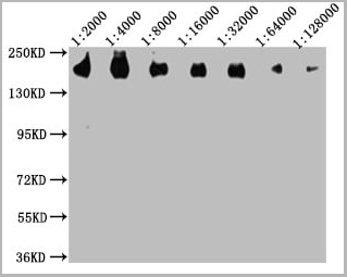
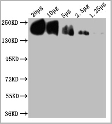
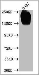
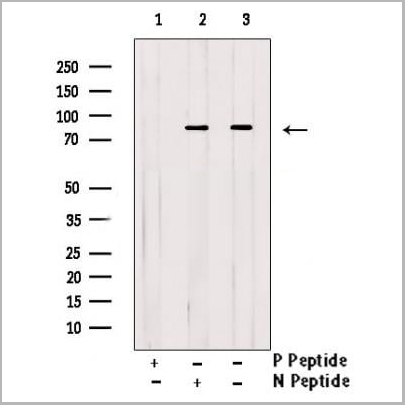
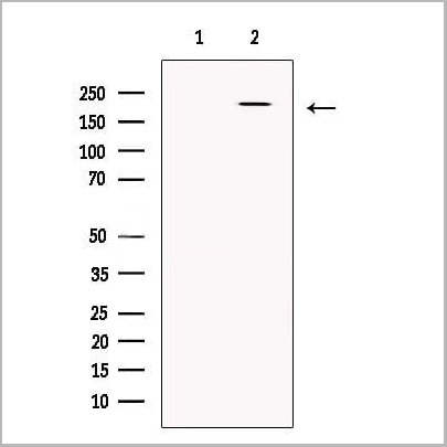
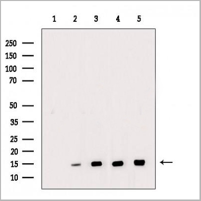
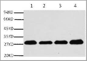
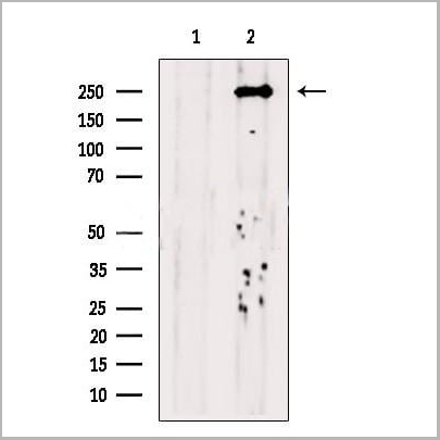
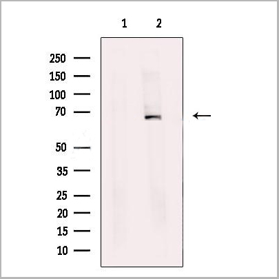
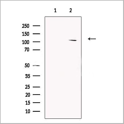
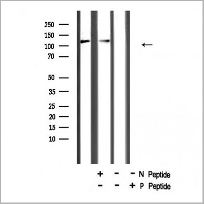
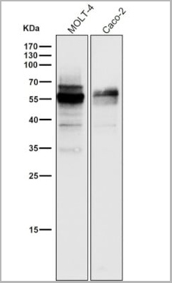
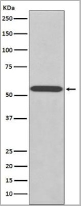
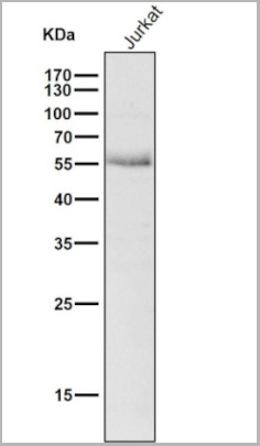
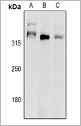
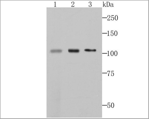
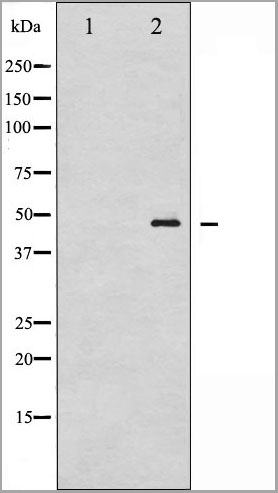
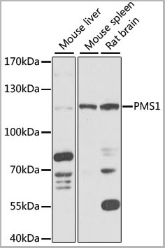 WB (Western Blot)
(Western blot analysis of extracts of various cell lines, using PMS1 antibody (AAA28270) at 1:1000 dilution.Secondary antibody: HRP Goat Anti-Rabbit IgG (H+L) at 1:10000 dilution.Lysates/proteins: 25ug per lane.Blocking buffer: 3% nonfat dry milk in TBST.Detection: ECL Basic Kit.Exposure time: 5s.)
WB (Western Blot)
(Western blot analysis of extracts of various cell lines, using PMS1 antibody (AAA28270) at 1:1000 dilution.Secondary antibody: HRP Goat Anti-Rabbit IgG (H+L) at 1:10000 dilution.Lysates/proteins: 25ug per lane.Blocking buffer: 3% nonfat dry milk in TBST.Detection: ECL Basic Kit.Exposure time: 5s.)
PMS1, Polyclonal Antibody (Cat# AAA28270)
Full Name
PMS1 Polyclonal Antibody
Gene Names
PMS1; MLH2; PMSL1; hPMS1; HNPCC3
Applications
Western Blot
Purity
Affinity Purification
Pricing
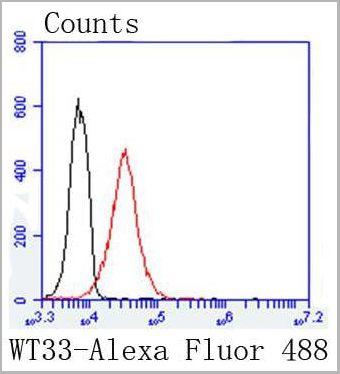 FCM (Flow Cytometry)
(Flow cytometric analysis of K562 cells with Wilms Tumor Protein antibody at 1/50 dilution (red) compared with an unlabelled control (cells without incubation with primary antibody; black). Alexa Fluor 488-conjugated goat anti rabbit IgG was used as the secondary antibody.)
FCM (Flow Cytometry)
(Flow cytometric analysis of K562 cells with Wilms Tumor Protein antibody at 1/50 dilution (red) compared with an unlabelled control (cells without incubation with primary antibody; black). Alexa Fluor 488-conjugated goat anti rabbit IgG was used as the secondary antibody.)
Wilms Tumor Protein, Monoclonal Antibody (Cat# AAA30123)
Full Name
Wilms Tumor Protein Antibody
Gene Names
WT1; GUD; AWT1; WAGR; WT33; NPHS4; WIT-2; EWS-WT1
Reactivity
Human, Mouse
Applications
Western Blot, Immunocytochemistry, Immunofluorescence, Immunohistochemistry, Flow Cytometry
Purity
Affinity-chromatography
Pricing
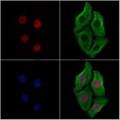
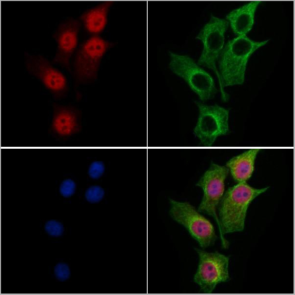
 IF (Immunofluorescence)
(Immunofluorescent analysis of DNA Polymerase zeta staining in HEK293T cells. Formalin-fixed cells were permeabilized with 0.1% Triton X-100 in TBS for 5-10 minutes and blocked with 3% BSA-PBS for 30 minutes at room temperature. Cells were probed with the primary antibody in 3% BSA-PBS and incubated overnight at 4 degree C in a humidified chamber. Cells were washed with PBST and incubated with a DyLight 594-conjugated secondary antibody (red) in PBS at room temperature in the dark.)
IF (Immunofluorescence)
(Immunofluorescent analysis of DNA Polymerase zeta staining in HEK293T cells. Formalin-fixed cells were permeabilized with 0.1% Triton X-100 in TBS for 5-10 minutes and blocked with 3% BSA-PBS for 30 minutes at room temperature. Cells were probed with the primary antibody in 3% BSA-PBS and incubated overnight at 4 degree C in a humidified chamber. Cells were washed with PBST and incubated with a DyLight 594-conjugated secondary antibody (red) in PBS at room temperature in the dark.)
DNA Polymerase zeta, Polyclonal Antibody (Cat# AAA27811)
Full Name
Anti-DNA Polymerase zeta Antibody
Gene Names
REV3L; POLZ; REV3
Reactivity
Human, Mouse, Rat, Bovine, Chicken, Dog, Pig
Applications
Western Blot, Immunohistochemistry, Immunofluorescence, Immunocytochemistry
Purity
The antibody was purified by immunogen affinity chromatography.
Pricing
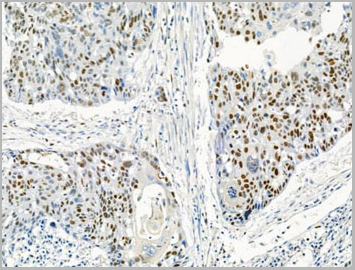
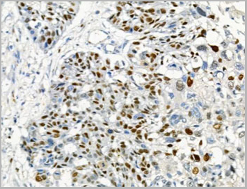
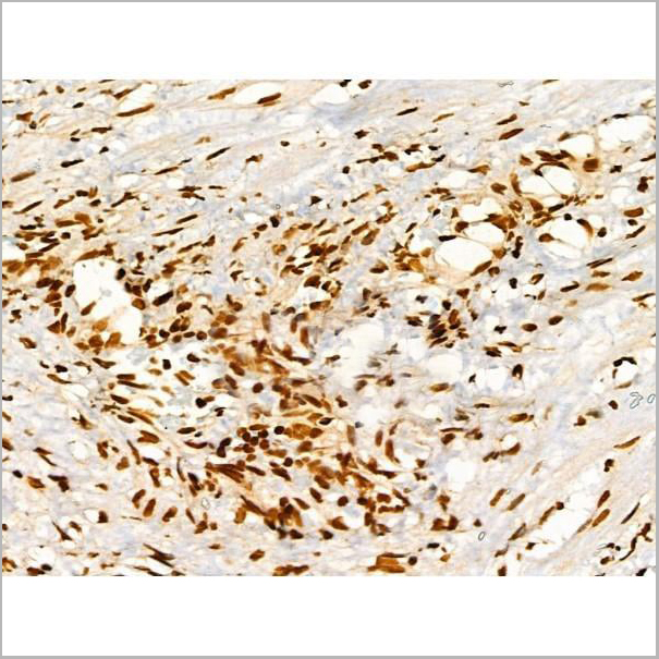
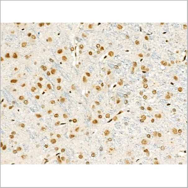
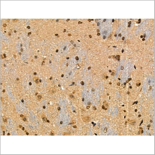
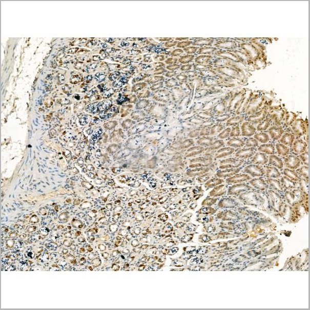
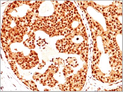
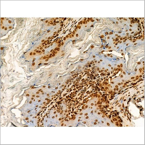
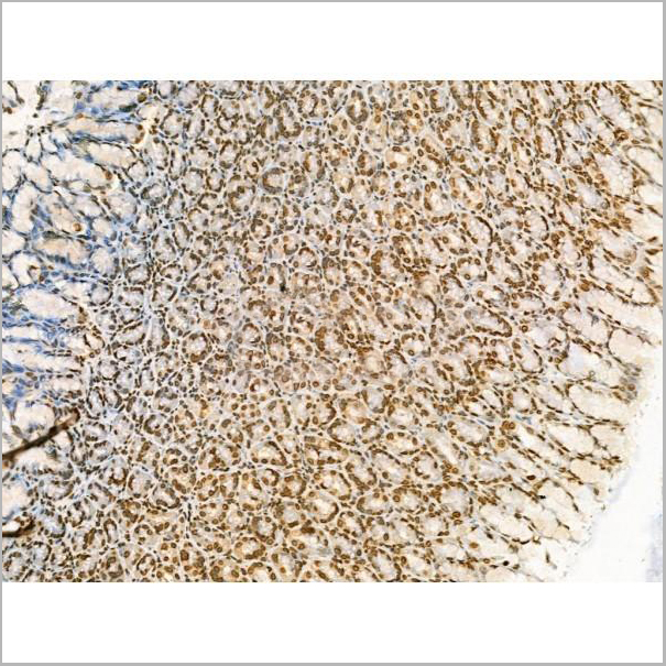
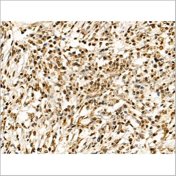
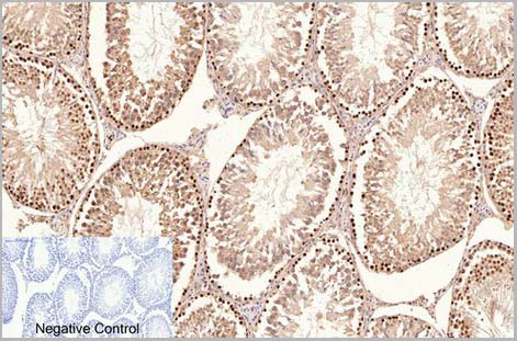
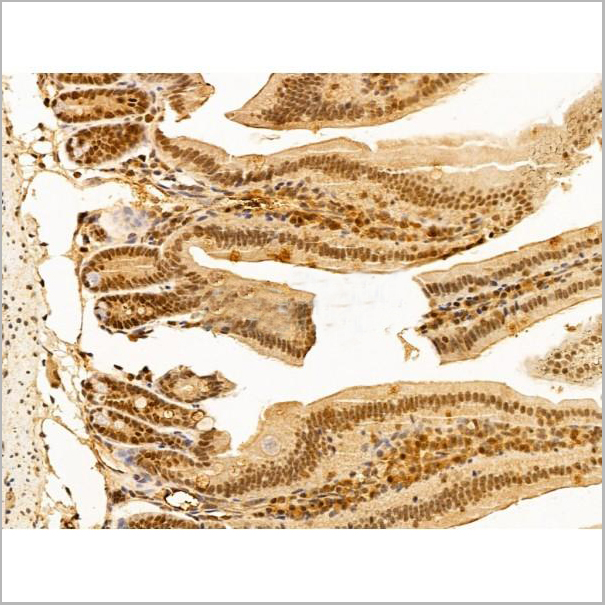
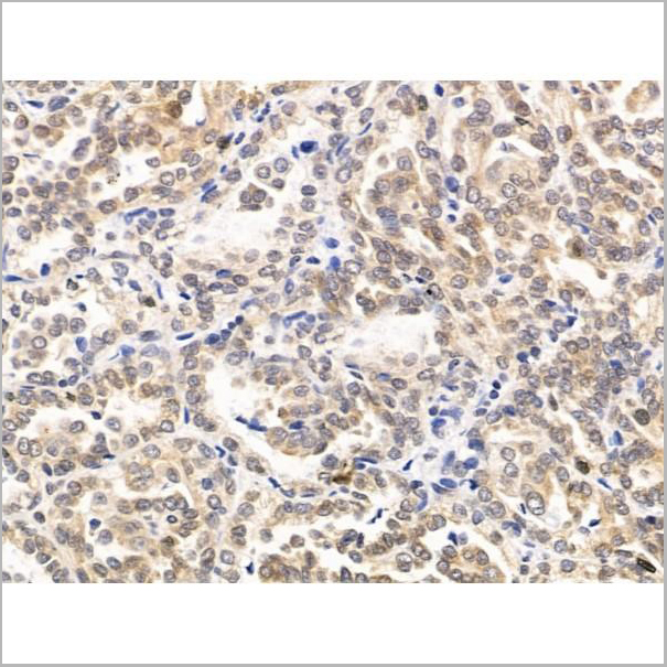
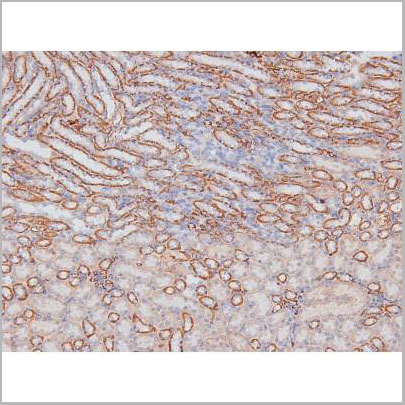
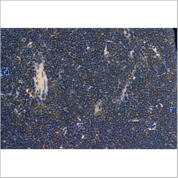
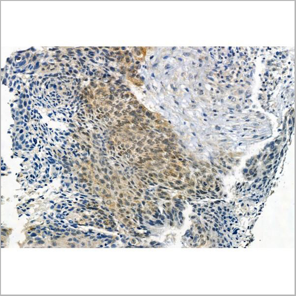
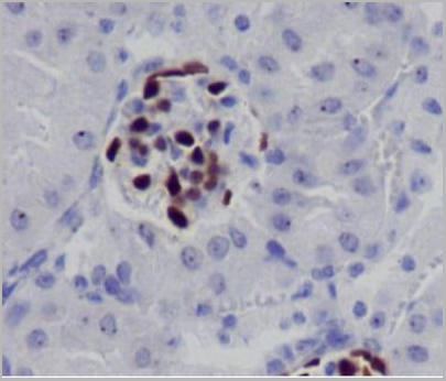
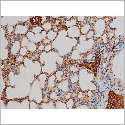
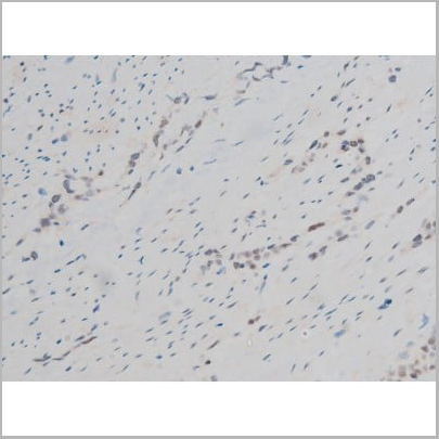
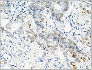 IHC (Immunohistchemistry)
(AAA31461 at 1/100 staining Human gastric cancer by IHC-P. The sample was formaldehyde fixed and a heat mediated antigen retrieval step in citrate buffer was performed. The sample was then blocked and incubated with the primary antibody at 4°C overnight. An HRP conjugated anti-Rabbit antibody was used as the secondary antibody.)
IHC (Immunohistchemistry)
(AAA31461 at 1/100 staining Human gastric cancer by IHC-P. The sample was formaldehyde fixed and a heat mediated antigen retrieval step in citrate buffer was performed. The sample was then blocked and incubated with the primary antibody at 4°C overnight. An HRP conjugated anti-Rabbit antibody was used as the secondary antibody.)
MCM2, Polyclonal Antibody (Cat# AAA31461)
Full Name
Phospho-MCM2 (Ser53) Antibody
Gene Names
MCM2; BM28; CCNL1; CDCL1; cdc19; D3S3194; MITOTIN
Reactivity
Human, Mouse, Rat
Applications
Western Blot, Immunohistochemistry, Peptide ELISA
Purity
Peptide affinity purification.
Pricing
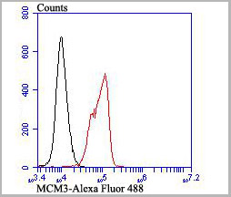 FCM (Flow Cytometry)
(Flow cytometric analysis of K562 cells with MCM3 antibody at 1/100 dilution (red) compared with an unlabelled control (cells without incubation with primary antibody; black). Alexa Fluor 488-conjugated goat anti-rabbit IgG was used as the secondary antibody.)
FCM (Flow Cytometry)
(Flow cytometric analysis of K562 cells with MCM3 antibody at 1/100 dilution (red) compared with an unlabelled control (cells without incubation with primary antibody; black). Alexa Fluor 488-conjugated goat anti-rabbit IgG was used as the secondary antibody.)
MCM3, Monoclonal Antibody (Cat# AAA30464)
Full Name
MCM3 Antibody
Gene Names
MCM3; HCC5; P1.h; RLFB; P1-MCM3
Reactivity
Human, Mouse, Rat
Applications
Western Blot, Immunocytochemistry, Immunofluorescence, Immunohistochemistry, Flow Cytometry
Purity
ProA affinity purified
Pricing
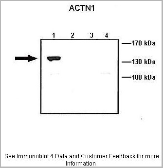
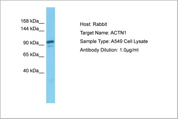
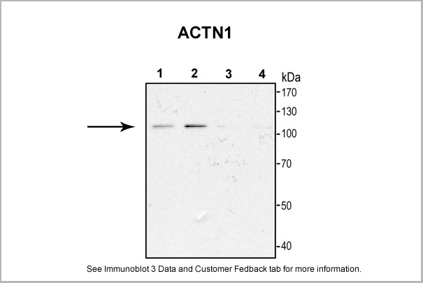
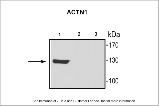
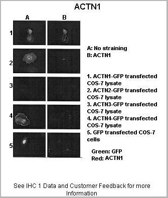
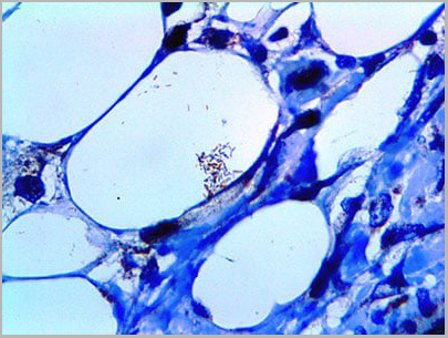
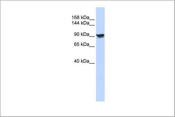 WB (Western Blot)
(WB Suggested Anti-ACTN1 Antibody Titration: 0.2-1 ug/mlELISA Titer: 1:312500Positive Control: 293T cell lysate)
WB (Western Blot)
(WB Suggested Anti-ACTN1 Antibody Titration: 0.2-1 ug/mlELISA Titer: 1:312500Positive Control: 293T cell lysate)
ACTN1, Polyclonal Antibody (Cat# AAA23579)
Full Name
ACTN1 antibody - N-terminal region
Gene Names
ACTN1; BDPLT15
Reactivity
Cow, Dog, Guinea Pig, Horse, Human, Mouse, Rabbit, Rat, Zebrafish
Applications
Immunohistochemistry, Western Blot
Purity
Affinity Purified
Pricing
 QC (Quality Control)
(This Antibody has been quality control tested by immunohistochemistry as follows)
QC (Quality Control)
(This Antibody has been quality control tested by immunohistochemistry as follows)
Mycobacterium tuberculosis, Polyclonal Antibody (Cat# AAA13576)
Full Name
Mycobacterium tuberculosis
Reactivity
Human
Applications
Immunohistochemistry
Pricing
 Quality Control
(This antibody has been quality control tested by Immunohistochemistry as follows.)
Quality Control
(This antibody has been quality control tested by Immunohistochemistry as follows.)
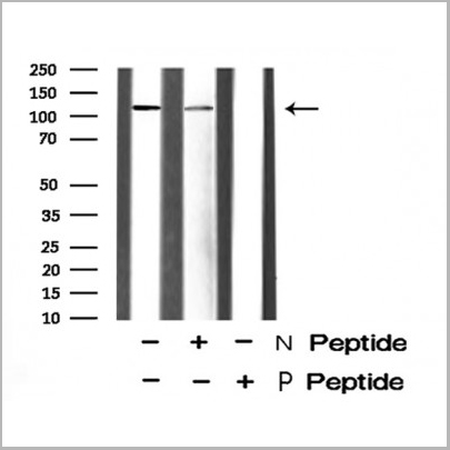
RAD50, Polyclonal Antibody (Cat# AAA31432)
Full Name
Phospho-RAD50 (Ser635) Antibody
Gene Names
RAD50; NBSLD; RAD502; hRad50
Reactivity
Human, Mouse, Rat
Applications
Western Blot, Immunohistochemistry
Purity
The antibody is from purified rabbit serum by affinity purification via sequential chromatography on phospho-peptide and non-phospho-peptide affinity columns.
Pricing

 Application Data
TCR Beta F1, Monoclonal Antibody (Cat# AAA13568)
Full Name
TCR Beta F1
Reactivity
Human
Applications
Immunohistochemistry
Pricing
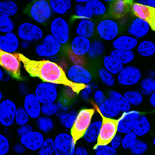
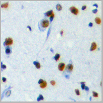
 IS (Immunostaining)
(Immunostaining of cultured E20 rat cortical neurons and glia stained with anti-nestin antibody (AAA14229, red, 1:500) and anti-MAP2 antibody (, green, 1:500). The blue is Hoechst staining for nuclear DNA. The nestin antibody labels developing astrocytes and neuronal stem cells in a clearly filamentous fashion, while the MAP2 antibody stains dendrites and perikarya of mature neurons.)
IS (Immunostaining)
(Immunostaining of cultured E20 rat cortical neurons and glia stained with anti-nestin antibody (AAA14229, red, 1:500) and anti-MAP2 antibody (, green, 1:500). The blue is Hoechst staining for nuclear DNA. The nestin antibody labels developing astrocytes and neuronal stem cells in a clearly filamentous fashion, while the MAP2 antibody stains dendrites and perikarya of mature neurons.)
Nestin, Monoclonal Antibody (Cat# AAA14229)
Full Name
Anti-Nestin
Gene Names
NES; Nbla00170
Applications
Western Blot, Immunofluorescence
Purity
Protein G purified culture supernatant
Pricing
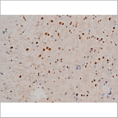
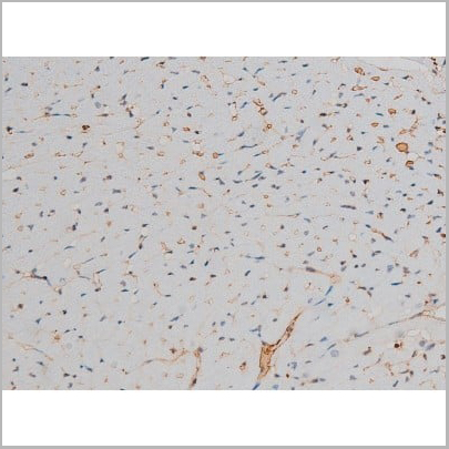
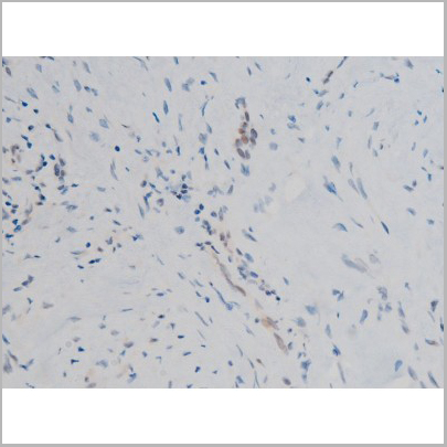
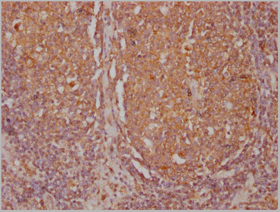
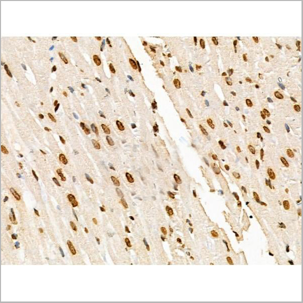
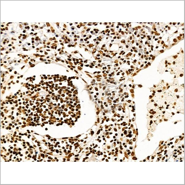
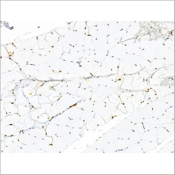
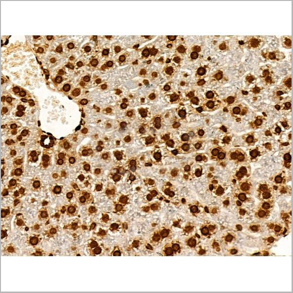
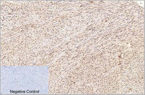
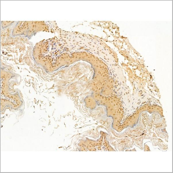
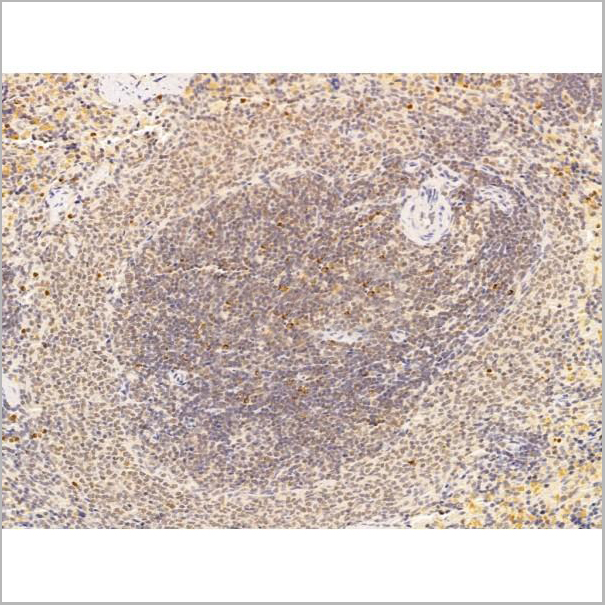
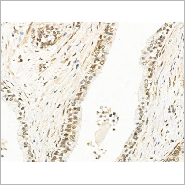
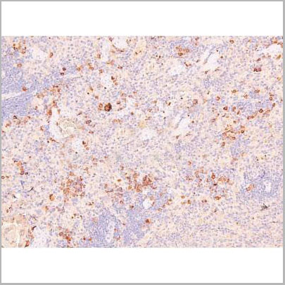
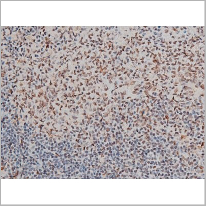 IHC (Immunohistochemistry)
(AAA31013 at 1/200 staining Rat spleen tissue sections by IHC-P. The tissue was formaldehyde fixed and a heat mediated antigen retrieval step in citrate buffer was performed. The tissue was then blocked and incubated with the antibody for 1.5 hours at 22 degree C. An HRP conjugated goat anti-rabbit antibody was used as the secondary.)
IHC (Immunohistochemistry)
(AAA31013 at 1/200 staining Rat spleen tissue sections by IHC-P. The tissue was formaldehyde fixed and a heat mediated antigen retrieval step in citrate buffer was performed. The tissue was then blocked and incubated with the antibody for 1.5 hours at 22 degree C. An HRP conjugated goat anti-rabbit antibody was used as the secondary.)
GATA1, Polyclonal Antibody (Cat# AAA31013)
Full Name
Phospho-GATA1 (Ser142) Antibody
Gene Names
GATA1; GF1; GF-1; NFE1; XLTT; ERYF1; NF-E1; XLANP; XLTDA; GATA-1
Reactivity
Human, Mouse, Rat
Applications
Western Blot, Immunohistochemistry, Immunofluorescence, Immunocytochemistry, Immunoprecipitation
Purity
From purified rabbit serum by affinity purification via sequential chromatography on phospho-and non-phospho-peptide affinity columns.
Pricing
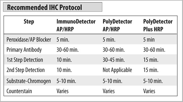
 IHC Protocol
CD7, Monoclonal Antibody (Cat# AAA13566)
Full Name
CD7
Gene Names
CD7; GP40; TP41; Tp40; LEU-9
Reactivity
Human
Applications
Immunohistochemistry
Pricing


 Application Data
(Published customer image: Mouse anti V5 tag antibody, clone SV5-Pk1 used for the detection of V5 tagged WEEV_nsP3 protein by western blotting and immunofluorescenceImage caption: WEEV nsP3 interaction with host IKKbeta. A) U87MGs were transfected in a 6-well plate with 5 ug of pUC19 and WEEV_nsP3_HA for 24 hours. Cell lysates were resolved using SDS-PAGE and subsequently immunoblotted with V5 antibody and beta-actin served as a loading control. B) U87MGs were transfected with WEEV_nsP3_V5; cells were fixed after 24 hours and stained with antibodies against the endogenous IKKbeta and the V5 tag. Cells were incubated with appropriate secondary Alexa Fluor antibodies and the nuclei stained with DAPI. Co-localization of IKKbeta with WEEV_nsP3_V5 (yellow) was observed as shown by the arrows. B) Panels E -H serve as an example of transfected cells in a given field of view that show co-localization of IKKbeta and WEEV_nsP3_V5 24 hours post transfection. Panels I-L represent magnified images of other cells showing co-localization of IKKbeta and WEEV_nsP3_V5. Panel M is a magnified image of panel L. The co-localization was confirmed by Z-stack analysis. Co-localization was calculated to be approximately in 61% of cells (163 cells were counted of which 44% demonstrated expression of nsP3. Of those cells that expressed nsP3, 61% showed co-localization of both proteins). Images were taken using Nikon Eclipse TE2000-U at 60x magnification and are representative of 2 independent experiments.From: Amaya M, Voss K, Sampey G, Senina S, de la Fuente C, et al. (2014) The Role of IKKbeta in Venezuelan Equine Encephalitis Virus Infection. PLoS ONE 9(2): e86745.)
Application Data
(Published customer image: Mouse anti V5 tag antibody, clone SV5-Pk1 used for the detection of V5 tagged WEEV_nsP3 protein by western blotting and immunofluorescenceImage caption: WEEV nsP3 interaction with host IKKbeta. A) U87MGs were transfected in a 6-well plate with 5 ug of pUC19 and WEEV_nsP3_HA for 24 hours. Cell lysates were resolved using SDS-PAGE and subsequently immunoblotted with V5 antibody and beta-actin served as a loading control. B) U87MGs were transfected with WEEV_nsP3_V5; cells were fixed after 24 hours and stained with antibodies against the endogenous IKKbeta and the V5 tag. Cells were incubated with appropriate secondary Alexa Fluor antibodies and the nuclei stained with DAPI. Co-localization of IKKbeta with WEEV_nsP3_V5 (yellow) was observed as shown by the arrows. B) Panels E -H serve as an example of transfected cells in a given field of view that show co-localization of IKKbeta and WEEV_nsP3_V5 24 hours post transfection. Panels I-L represent magnified images of other cells showing co-localization of IKKbeta and WEEV_nsP3_V5. Panel M is a magnified image of panel L. The co-localization was confirmed by Z-stack analysis. Co-localization was calculated to be approximately in 61% of cells (163 cells were counted of which 44% demonstrated expression of nsP3. Of those cells that expressed nsP3, 61% showed co-localization of both proteins). Images were taken using Nikon Eclipse TE2000-U at 60x magnification and are representative of 2 independent experiments.From: Amaya M, Voss K, Sampey G, Senina S, de la Fuente C, et al. (2014) The Role of IKKbeta in Venezuelan Equine Encephalitis Virus Infection. PLoS ONE 9(2): e86745.)
V5-TAG, Monoclonal Antibody (Cat# AAA11930)
Full Name
MOUSE ANTI V5-TAG
Applications
Immunohistochemistry, Flow Cytometry, Immunofluorescence, Immunoprecipitation, Western Blot, Radioimmunoassay
Pricing


 Application Data
(Published customer image: Mouse anti V5 tag antibody, clone SV5-Pk1 used for the detection of V5 tagged WEEV_nsP3 protein by western blotting and immunofluorescenceImage caption: WEEV nsP3 interaction with host IKKbeta. A) U87MGs were transfected in a 6-well plate with 5 ug of pUC19 and WEEV_nsP3_HA for 24 hours. Cell lysates were resolved using SDS-PAGE and subsequently immunoblotted with V5 antibody and beta-actin served as a loading control. B) U87MGs were transfected with WEEV_nsP3_V5; cells were fixed after 24 hours and stained with antibodies against the endogenous IKKbeta and the V5 tag. Cells were incubated with appropriate secondary Alexa Fluor antibodies and the nuclei stained with DAPI. Co-localization of IKKbeta with WEEV_nsP3_V5 (yellow) was observed as shown by the arrows. B) Panels E -H serve as an example of transfected cells in a given field of view that show co-localization of IKKbeta and WEEV_nsP3_V5 24 hours post transfection. Panels I-L represent magnified images of other cells showing co-localization of IKKbeta and WEEV_nsP3_V5. Panel M is a magnified image of panel L. The co-localization was confirmed by Z-stack analysis. Co-localization was calculated to be approximately in 61% of cells (163 cells were counted of which 44% demonstrated expression of nsP3. Of those cells that expressed nsP3, 61% showed co-localization of both proteins). Images were taken using Nikon Eclipse TE2000-U at 60x magnification and are representative of 2 independent experiments.From: Amaya M, Voss K, Sampey G, Senina S, de la Fuente C, et al. (2014) The Role of IKKbeta in Venezuelan Equine Encephalitis Virus Infection. PLoS ONE 9(2): e86745.)
Application Data
(Published customer image: Mouse anti V5 tag antibody, clone SV5-Pk1 used for the detection of V5 tagged WEEV_nsP3 protein by western blotting and immunofluorescenceImage caption: WEEV nsP3 interaction with host IKKbeta. A) U87MGs were transfected in a 6-well plate with 5 ug of pUC19 and WEEV_nsP3_HA for 24 hours. Cell lysates were resolved using SDS-PAGE and subsequently immunoblotted with V5 antibody and beta-actin served as a loading control. B) U87MGs were transfected with WEEV_nsP3_V5; cells were fixed after 24 hours and stained with antibodies against the endogenous IKKbeta and the V5 tag. Cells were incubated with appropriate secondary Alexa Fluor antibodies and the nuclei stained with DAPI. Co-localization of IKKbeta with WEEV_nsP3_V5 (yellow) was observed as shown by the arrows. B) Panels E -H serve as an example of transfected cells in a given field of view that show co-localization of IKKbeta and WEEV_nsP3_V5 24 hours post transfection. Panels I-L represent magnified images of other cells showing co-localization of IKKbeta and WEEV_nsP3_V5. Panel M is a magnified image of panel L. The co-localization was confirmed by Z-stack analysis. Co-localization was calculated to be approximately in 61% of cells (163 cells were counted of which 44% demonstrated expression of nsP3. Of those cells that expressed nsP3, 61% showed co-localization of both proteins). Images were taken using Nikon Eclipse TE2000-U at 60x magnification and are representative of 2 independent experiments.From: Amaya M, Voss K, Sampey G, Senina S, de la Fuente C, et al. (2014) The Role of IKKbeta in Venezuelan Equine Encephalitis Virus Infection. PLoS ONE 9(2): e86745.)
V5-TAG, Monoclonal Antibody (Cat# AAA11864)
Full Name
MOUSE ANTI V5-TAG:FITC
Applications
Immunofluorescence
Pricing
DNA (Double Stranded) (dsDNA), Antibody (Cat# AAA13410)
Full Name
Human anti dsDNA
Applications
Immunofluorescence
Purity
Delipidized and defibrinated, then the immunoglobulin fraction is precipitated and collected.
Pricing
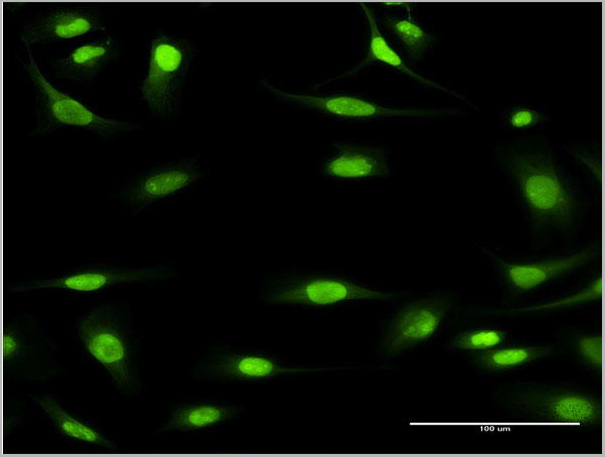
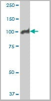
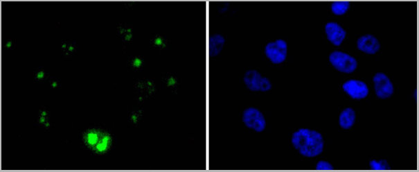
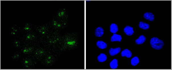
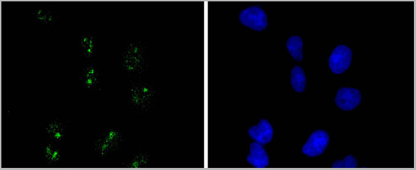
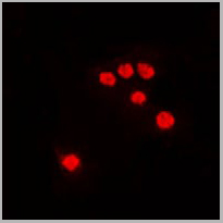
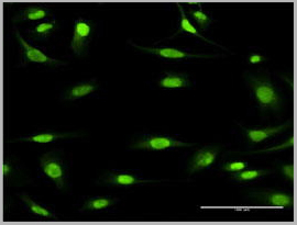 IF (Immunofluorescence)
(Immunofluorescence of monoclonal antibody to MCM3 on HeLa cell. [antibody concentration 10 ug/ml])
IF (Immunofluorescence)
(Immunofluorescence of monoclonal antibody to MCM3 on HeLa cell. [antibody concentration 10 ug/ml])
MCM3, Monoclonal Antibody (Cat# AAA26113)
Full Name
MCM3 (Minichromosome Maintenance Complex Component 3, HCC5, MGC1157, P1-MCM3, P1.h, RLFB) (APC)
Gene Names
MCM3; HCC5; P1.h; RLFB; P1-MCM3
Applications
Immunofluorescence, Western Blot
Purity
Purified
Pricing