Filters
▼Clonality
▼Type
▼Reactivity
▼Gene Name
▼Isotype
▼Host
▼Application
▼Clone
▼Monoclonal Antibodies
Get accurate results in your research with our Monoclonal Antibodies, which are specially made to target exactly what you require for your research, and will produce consistent, reliable performance in lab tests.
Viewing 1-50 of 27560 product results


 Application Data
(Detection limit for recombinant GST tagged ROCK2 is 0.3 ng/ml as a capture antibody.)
Application Data
(Detection limit for recombinant GST tagged ROCK2 is 0.3 ng/ml as a capture antibody.)
ROCK2, Monoclonal Antibody (Cat# AAA26357)


 Application Data
(Detection limit for recombinant GST tagged ROCK2 is 0.3 ng/ml as a capture antibody.)
Application Data
(Detection limit for recombinant GST tagged ROCK2 is 0.3 ng/ml as a capture antibody.)
ROCK2, Monoclonal Antibody (Cat# AAA26091)


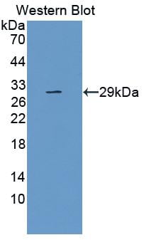
 Application Data
(Detection limit for recombinant GST tagged TCEA3 is approximately 0.03ng/ml as a capture antibody.)
Application Data
(Detection limit for recombinant GST tagged TCEA3 is approximately 0.03ng/ml as a capture antibody.)
TCEA3, Monoclonal Antibody (Cat# AAA26076)
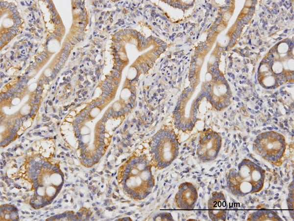
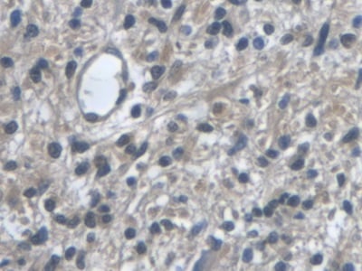
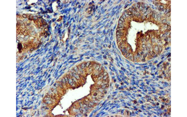
 IHC (Immunohiostchemistry)
(Immunochemical staining of human BCL2L1 in human gallbladder with mouse monoclonal antibody at 1:200 dilution, formalin-fixed paraffin embedded sections.)
IHC (Immunohiostchemistry)
(Immunochemical staining of human BCL2L1 in human gallbladder with mouse monoclonal antibody at 1:200 dilution, formalin-fixed paraffin embedded sections.)
BCL-xL/BCL2L1, Monoclonal Antibody (Cat# AAA258963)
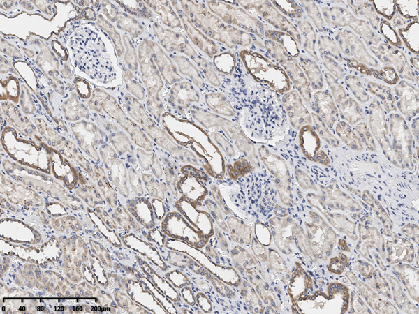
 IHC (Immunohiostchemistry)
(Immunochemical staining of human LCN2 in human lung with rabbit monoclonal antibody at 1:1000 dilution, formalin-fixed paraffin embedded sections.)
IHC (Immunohiostchemistry)
(Immunochemical staining of human LCN2 in human lung with rabbit monoclonal antibody at 1:1000 dilution, formalin-fixed paraffin embedded sections.)
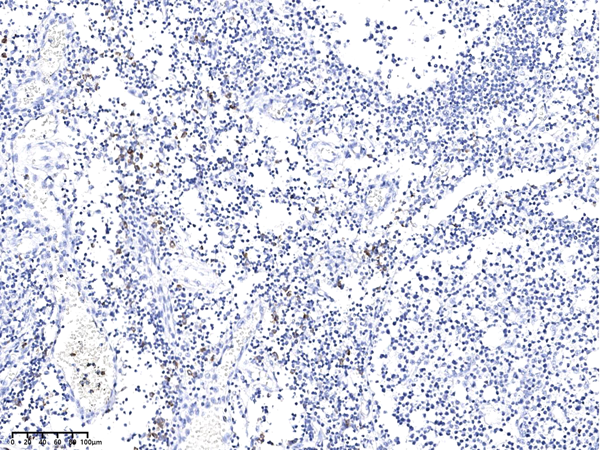
Lipocalin-2/NGAL, Monoclonal Recombinant Antibody (Cat# AAA258781)
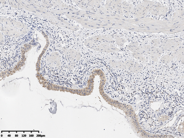
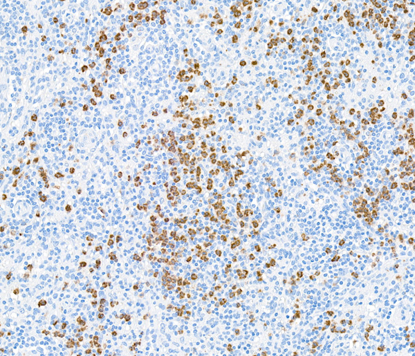
 IHC (Immunohiostchemistry)
(Immunochemical staining of human Granzyme B in human tonsil with mouse monoclonal antibody at 1:200 dilution, formalin-fixed paraffin embedded sections.)
IHC (Immunohiostchemistry)
(Immunochemical staining of human Granzyme B in human tonsil with mouse monoclonal antibody at 1:200 dilution, formalin-fixed paraffin embedded sections.)
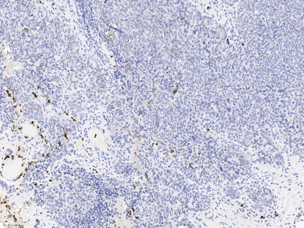
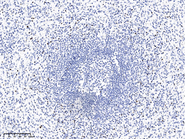
Granzyme B, Monoclonal Antibody (Cat# AAA258794)
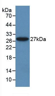
 IP (Immunoprecipitation)
(IL-33 was immunoprecipitated using:Lane A:0.5 mg HeLa, K562, jurkat Whole Cell Lysate2 uL anti-IL-33 mouse monoclonal antibody and 60 ug of Immunomagnetic beads Protein A/G.Primary antibody:Anti-IL-33 mouse monoclonal antibody,at 1:100 dilutionSecondary antibody:Rabbit Anti-Mouse IgG F(ab)2/HRP at 1/10000 dilutionDeveloped using the ECL technique.Performed under reducing conditions.Predicted band size: 31 kDaObserved band size :25 kDa)
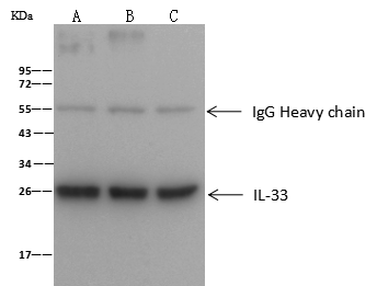
IP (Immunoprecipitation)
(IL-33 was immunoprecipitated using:Lane A:0.5 mg HeLa, K562, jurkat Whole Cell Lysate2 uL anti-IL-33 mouse monoclonal antibody and 60 ug of Immunomagnetic beads Protein A/G.Primary antibody:Anti-IL-33 mouse monoclonal antibody,at 1:100 dilutionSecondary antibody:Rabbit Anti-Mouse IgG F(ab)2/HRP at 1/10000 dilutionDeveloped using the ECL technique.Performed under reducing conditions.Predicted band size: 31 kDaObserved band size :25 kDa)
IL-33, Monoclonal Antibody (Cat# AAA258795)


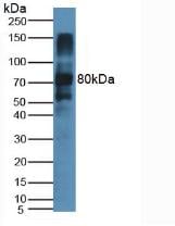
 IP (Immunoprecipitation)
(MME was immunoprecipitated using:Lane A:0.5 mg Raji Whole Cell Lysate4 uL anti-MME mouse monoclonal antibody and 60 ug of Immunomagnetic beads Protein A/G.Primary antibody:Anti-MME mouse monoclonal antibody,at 1:100 dilutionSecondary antibody:Rabbit Anti-Mouse IgG F(ab)2/HRP at 1/10000 dilutionDeveloped using the ECL technique.Performed under reducing conditions.Predicted band size: 85 kDaObserved band size :100 kDa)
IP (Immunoprecipitation)
(MME was immunoprecipitated using:Lane A:0.5 mg Raji Whole Cell Lysate4 uL anti-MME mouse monoclonal antibody and 60 ug of Immunomagnetic beads Protein A/G.Primary antibody:Anti-MME mouse monoclonal antibody,at 1:100 dilutionSecondary antibody:Rabbit Anti-Mouse IgG F(ab)2/HRP at 1/10000 dilutionDeveloped using the ECL technique.Performed under reducing conditions.Predicted band size: 85 kDaObserved band size :100 kDa)
CD10/Neprilysin, Monoclonal Antibody (Cat# AAA258847)
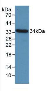
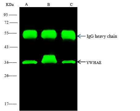 IP (Immunoprecipitation)
(YWHAE was immunoprecipitated using:Lane A:0.5 mg Jurkat Whole Cell LysateLane B:0.5 mg HepG2 Whole Cell LysateLane C:0.5 mg SH-SY5Y Whole Cell Lysate4 uL anti-YWHAE mouse monoclonal antibody and 60 ug of Immunomagnetic beads Protein A/G.Primary antibody:Anti-YWHAE mouse monoclonal antibody,at 1:100 dilutionSecondary antibody:Rabbit Anti-Mouse IgG F(ab)2/HRP at 1/10000 dilutionDeveloped using the ECL technique.Performed under reducing conditions.Predicted band size: 34 kDaObserved band size :34 kDa)
IP (Immunoprecipitation)
(YWHAE was immunoprecipitated using:Lane A:0.5 mg Jurkat Whole Cell LysateLane B:0.5 mg HepG2 Whole Cell LysateLane C:0.5 mg SH-SY5Y Whole Cell Lysate4 uL anti-YWHAE mouse monoclonal antibody and 60 ug of Immunomagnetic beads Protein A/G.Primary antibody:Anti-YWHAE mouse monoclonal antibody,at 1:100 dilutionSecondary antibody:Rabbit Anti-Mouse IgG F(ab)2/HRP at 1/10000 dilutionDeveloped using the ECL technique.Performed under reducing conditions.Predicted band size: 34 kDaObserved band size :34 kDa)
14-3-3 epsilon, Monoclonal Antibody (Cat# AAA258853)
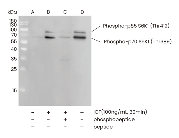
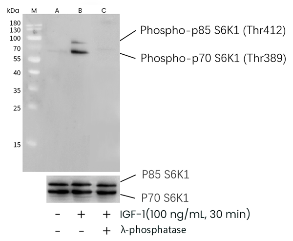
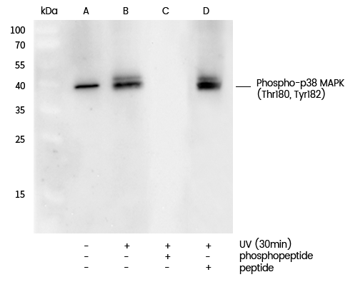
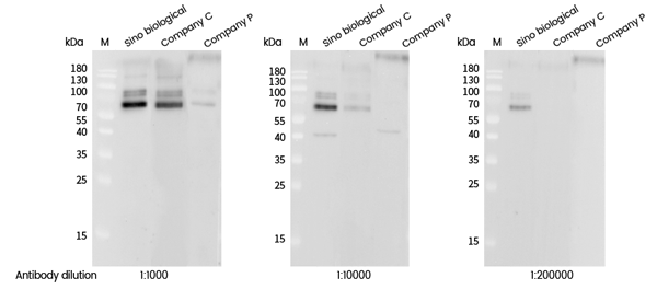 WB (Western Blot)
(Western blot analysis of extracts from serum-starved MCF7, treated with IGF-1 (100 ng/mL, 30 min), using Phospho-S6K1 (Thr389) rabbit monoclonal Antibody (#AAA258873) and other brands’ antibodies (company C, company P) at dilution of 1:1000, 1:10000 and 1:200000.)
WB (Western Blot)
(Western blot analysis of extracts from serum-starved MCF7, treated with IGF-1 (100 ng/mL, 30 min), using Phospho-S6K1 (Thr389) rabbit monoclonal Antibody (#AAA258873) and other brands’ antibodies (company C, company P) at dilution of 1:1000, 1:10000 and 1:200000.)
p70 S6 Kinase 1, Monoclonal Recombinant Antibody (Cat# AAA258873)
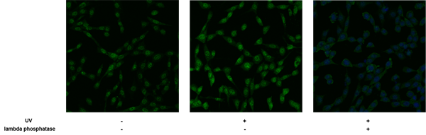 IF (Immunofluorescence)
(Immunofluorescence staining of Phospho-p38 MAPK (Thr180, Tyr182) in serum-starved NIH-3T3 cells, untreated (left), treated with UV (30mins) (middle) or treated with UV (30mins) and lambda phosphatase (right). Cells were fixed with 4% PFA, permeabilzed with 0.1% Triton X-100 in PBS, blocked with 10% serum, and incubated with rabbit anti-Mouse Phospho-p38 MAPK (Thr180, Tyr182) monoclonal antibody (dilution ratio 1:60) at 4? overnight. Then cells were stained with the Alexa Fluor488-conjugated Goat Anti-rabbit IgG secondary antibody (green). Positive staining was mainly localized to Nucleus.)
IF (Immunofluorescence)
(Immunofluorescence staining of Phospho-p38 MAPK (Thr180, Tyr182) in serum-starved NIH-3T3 cells, untreated (left), treated with UV (30mins) (middle) or treated with UV (30mins) and lambda phosphatase (right). Cells were fixed with 4% PFA, permeabilzed with 0.1% Triton X-100 in PBS, blocked with 10% serum, and incubated with rabbit anti-Mouse Phospho-p38 MAPK (Thr180, Tyr182) monoclonal antibody (dilution ratio 1:60) at 4? overnight. Then cells were stained with the Alexa Fluor488-conjugated Goat Anti-rabbit IgG secondary antibody (green). Positive staining was mainly localized to Nucleus.)
p38 MAPK, Monoclonal Recombinant Antibody (Cat# AAA258875)
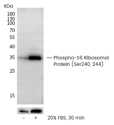
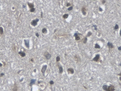
 IHC (Immunohiostchemistry)
(Immunohistochemical analysis of paraffin-embedded human carcinoma of sigmoid tissue,untreated (left) or lambda phosphatase-treated (right), using Recombinant Phospho- S6 Ribosomal Protein (Ser240/244) Antibody, Rabbit Monoclonal (#AAA258880) at 1:2000 dilution.)
IHC (Immunohiostchemistry)
(Immunohistochemical analysis of paraffin-embedded human carcinoma of sigmoid tissue,untreated (left) or lambda phosphatase-treated (right), using Recombinant Phospho- S6 Ribosomal Protein (Ser240/244) Antibody, Rabbit Monoclonal (#AAA258880) at 1:2000 dilution.)
S6 Ribosomal, Monoclonal Antibody (Cat# AAA258880)
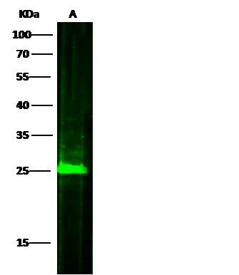
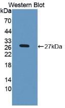
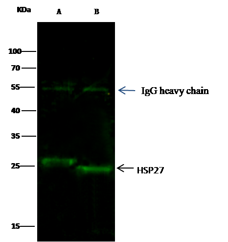 IP (Immunoprecipitation)
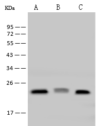
(HSP27 was immunoprecipitated using:Lane A:0.5 mg HepG2 Whole Cell LysateLane B:0.5 mg Hela Whole Cell Lysate2 uL anti-HSP27 mouse monoclonal antibody and 15 ul of 50 % Protein G agarose.Primary antibody:Anti-HSP27 mouse monoclonal antibody,at 1:200 dilutionSecondary antibody:Dylight 800-labeled antibody to Mouse IgG (H+L), at 1:7500 dilutionDeveloped using the odssey technique.Performed under reducing conditions.Predicted band size: 25 kDaObserved band size: 25 kDa)
IP (Immunoprecipitation)
(HSP27 was immunoprecipitated using:Lane A:0.5 mg HepG2 Whole Cell LysateLane B:0.5 mg Hela Whole Cell Lysate2 uL anti-HSP27 mouse monoclonal antibody and 15 ul of 50 % Protein G agarose.Primary antibody:Anti-HSP27 mouse monoclonal antibody,at 1:200 dilutionSecondary antibody:Dylight 800-labeled antibody to Mouse IgG (H+L), at 1:7500 dilutionDeveloped using the odssey technique.Performed under reducing conditions.Predicted band size: 25 kDaObserved band size: 25 kDa)
Hsp27/HSPB1, Monoclonal Antibody (Cat# AAA254856)
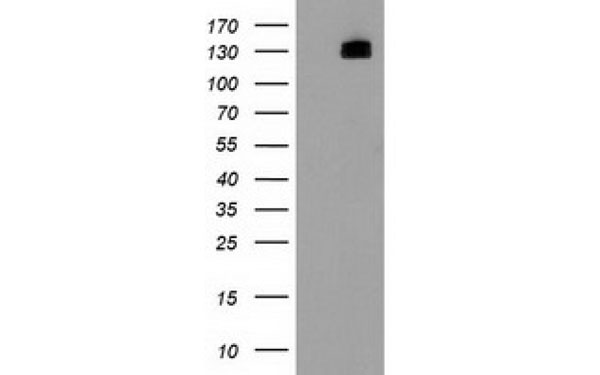
 WB (Western Blot)
(Western Blot: Sample: Recombinant CCND1, Gallus.)
WB (Western Blot)
(Western Blot: Sample: Recombinant CCND1, Gallus.)
Cyclin D1 (CCND1), Monoclonal Antibody (Cat# AAA134814)
 WB (Western Blot)
(Western Blot: Sample: Recombinant ALT, Rat.)
WB (Western Blot)
(Western Blot: Sample: Recombinant ALT, Rat.)
Alanine Aminotransferase (ALT), Monoclonal Antibody (Cat# AAA134829)
CD158d, Monoclonal Antibody (Cat# AAA128699)
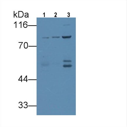 WB (Western Blot)
(Western Blot; Sample: Lane1: Rat Cerebrum lysate; Lane2: Porcine Cerebrum lysate; Lane3: Rat Placenta lysate Primary Ab: 3ug/ml Mouse AntiHuman VGF Antibody Second Ab: 0.2ug/mL HRPLinked Caprine AntiMouse IgG Polyclonal Antibody (Catalog: SAA544Mu19))
WB (Western Blot)
(Western Blot; Sample: Lane1: Rat Cerebrum lysate; Lane2: Porcine Cerebrum lysate; Lane3: Rat Placenta lysate Primary Ab: 3ug/ml Mouse AntiHuman VGF Antibody Second Ab: 0.2ug/mL HRPLinked Caprine AntiMouse IgG Polyclonal Antibody (Catalog: SAA544Mu19))
VGF Nerve Growth Factor Inducible (VGF), Monoclonal Antibody (Cat# AAA151684)
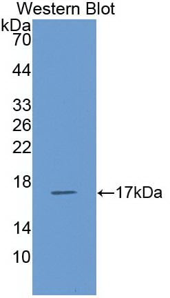 WB (Western Blot)
(Western Blot: Sample: Recombinant protein.)
WB (Western Blot)
(Western Blot: Sample: Recombinant protein.)
Regulated On Activation In Normal T-Cell Expressed And Secreted (RANTES), Monoclonal Antibody (Cat# AAA130636)

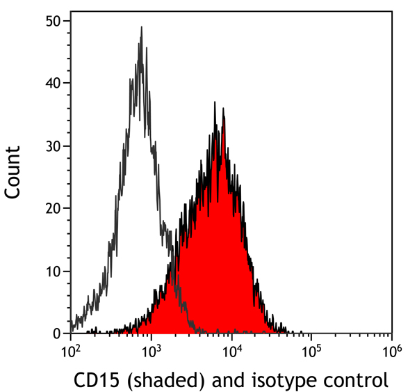
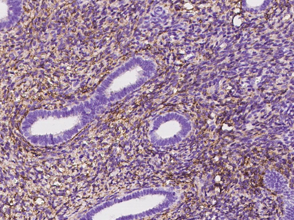
 IHC (Immunohistochemisry)
(Detection of human CD15 by immunohistochemistry. Sample: FFPE section of spleen. Antibody: Mouse anti-CD15 monoclonal antibody [MMA (Leu-M1)] (AAA213511 lot 1). Secondary: HRP-conjugated goat anti-mouse IgM .)
IHC (Immunohistochemisry)
(Detection of human CD15 by immunohistochemistry. Sample: FFPE section of spleen. Antibody: Mouse anti-CD15 monoclonal antibody [MMA (Leu-M1)] (AAA213511 lot 1). Secondary: HRP-conjugated goat anti-mouse IgM .)
CD15, Monoclonal Antibody (Cat# AAA213511)
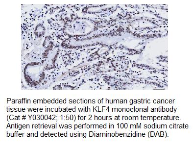
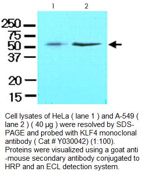 Application Data
KLF4, Monoclonal Antibody (Cat# AAA214202)
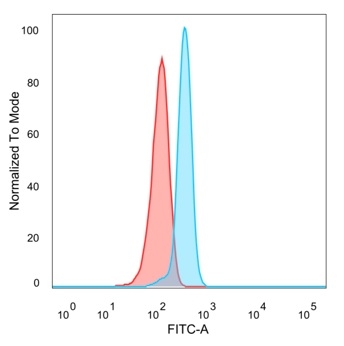 FCM/FACS (Flow Cytometry)
(Flow Cytometric Analysis of PFA-fixed HeLa cells. SPIC Mouse Monoclonal Antibody (PCRP-SPIC-2C5) followed by goat anti-mouse IgG-CF488 (blue); unstained cells (red).)
FCM/FACS (Flow Cytometry)
(Flow Cytometric Analysis of PFA-fixed HeLa cells. SPIC Mouse Monoclonal Antibody (PCRP-SPIC-2C5) followed by goat anti-mouse IgG-CF488 (blue); unstained cells (red).)
SPI-C/Transcription factor Spi-C, Monoclonal Antibody (Cat# AAA215557)
 SDS-PAGE
(SDS-PAGE Analysis Purified GPX4/MCSP Mouse Monoclonal Antibody (LHM 2). Confirmation of Purity and Integrity of Antibody.)
SDS-PAGE
(SDS-PAGE Analysis Purified GPX4/MCSP Mouse Monoclonal Antibody (LHM 2). Confirmation of Purity and Integrity of Antibody.)
GPX4/MCSP, Monoclonal Antibody (Cat# AAA214995)
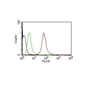 FCM/FACS (Flow Cytometry)
(Flow Cytometry of human Pan-Cytokeratins on HeLa Cells. Black: Cells alone; Green: Isotype Control; Red: PE-labeled Pan-Cytokeratin Monoclonal Antibody (SPM115 + SPM116).)
FCM/FACS (Flow Cytometry)
(Flow Cytometry of human Pan-Cytokeratins on HeLa Cells. Black: Cells alone; Green: Isotype Control; Red: PE-labeled Pan-Cytokeratin Monoclonal Antibody (SPM115 + SPM116).)
Cytokeratin, pan, Monoclonal Antibody (Cat# AAA214629)
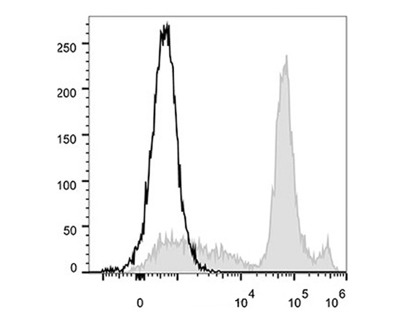
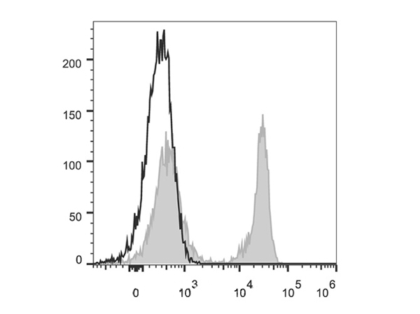
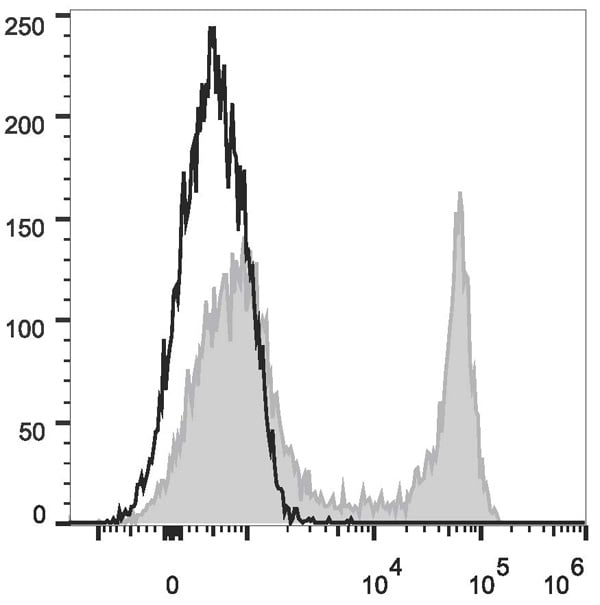 FCM/FACS (Flow Cytometry)
(C57BL/6 murine splenocytes are stained with Anti-Mouse CD45R/B220 Monoclonal Antibody(FITC Conjugated)(filled gray histogram). Unstained splenocytes (empty black histogram) are used as control.)
FCM/FACS (Flow Cytometry)
(C57BL/6 murine splenocytes are stained with Anti-Mouse CD45R/B220 Monoclonal Antibody(FITC Conjugated)(filled gray histogram). Unstained splenocytes (empty black histogram) are used as control.)
CD45R/B220, Monoclonal Antibody (Cat# AAA174748)

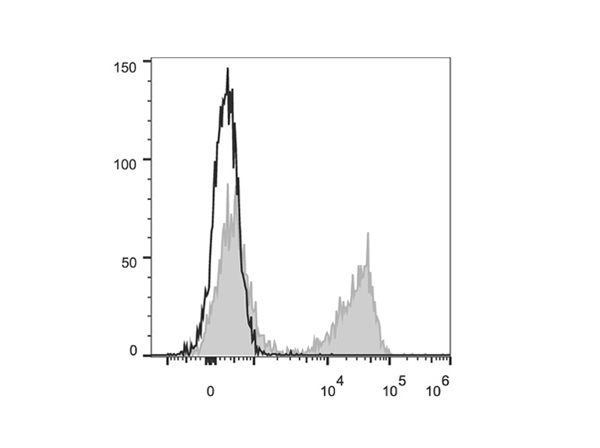
 FCM/FACS (Flow Cytometry)
(C57BL/6 murine bone marrow cells are stained with Anti-Mouse Ly6C Monoclonal Antibody(Alexa Fluor 488 Conjugated)(filled gray histogram). Unstained bone marrow cells (empty black histogram) are used as control.)
FCM/FACS (Flow Cytometry)
(C57BL/6 murine bone marrow cells are stained with Anti-Mouse Ly6C Monoclonal Antibody(Alexa Fluor 488 Conjugated)(filled gray histogram). Unstained bone marrow cells (empty black histogram) are used as control.)
Ly6C, Monoclonal Antibody (Cat# AAA174648)
 FCM/FACS (Flow Cytometry)
(C57BL/6 murine bone marrow cells are stained with Anti-Mouse Ly6C Monoclonal Antibody(Alexa Fluor 647 Conjugated)(filled gray histogram). Unstained bone marrow cells (empty black histogram) are used as control.)
FCM/FACS (Flow Cytometry)
(C57BL/6 murine bone marrow cells are stained with Anti-Mouse Ly6C Monoclonal Antibody(Alexa Fluor 647 Conjugated)(filled gray histogram). Unstained bone marrow cells (empty black histogram) are used as control.)
Ly6C, Monoclonal Antibody (Cat# AAA174649)
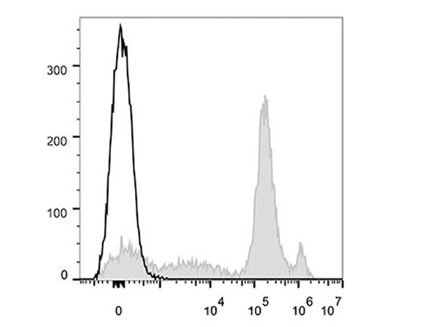
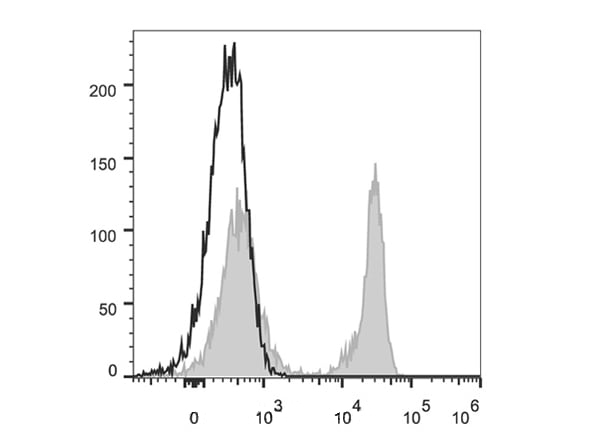
 FCM/FACS (Flow Cytometry)
(C57BL/6 murine splenocytes are stained with Anti-Mouse TCRbeta Monoclonal Antibody(PE Conjugated)(filled gray histogram). Unstained splenocytes (empty black histogram) are used as control.)
FCM/FACS (Flow Cytometry)
(C57BL/6 murine splenocytes are stained with Anti-Mouse TCRbeta Monoclonal Antibody(PE Conjugated)(filled gray histogram). Unstained splenocytes (empty black histogram) are used as control.)
TCRbeta, Monoclonal Antibody (Cat# AAA174655)

 FCM/FACS (Flow Cytometry)
(C57BL/6 murine splenocytes are stained with Anti-Mouse TCRbeta Monoclonal Antibody(PerCP/Cy5.5 Conjugated)(filled gray histogram). Unstained splenocytes (empty black histogram) are used as control.)
FCM/FACS (Flow Cytometry)
(C57BL/6 murine splenocytes are stained with Anti-Mouse TCRbeta Monoclonal Antibody(PerCP/Cy5.5 Conjugated)(filled gray histogram). Unstained splenocytes (empty black histogram) are used as control.)
TCRbeta, Monoclonal Antibody (Cat# AAA174656)
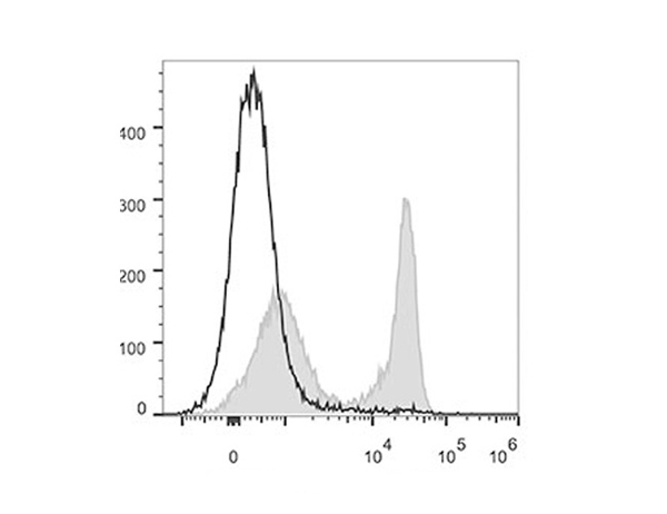
 FCM/FACS (Flow Cytometry)
(C57BL/6 murine splenocytes are stained with Anti-Mouse TCRbeta Monoclonal Antibody(Alexa Fluor 488 Conjugated)(filled gray histogram). Unstained splenocytes (empty black histogram) are used as control.)
FCM/FACS (Flow Cytometry)
(C57BL/6 murine splenocytes are stained with Anti-Mouse TCRbeta Monoclonal Antibody(Alexa Fluor 488 Conjugated)(filled gray histogram). Unstained splenocytes (empty black histogram) are used as control.)
TCRbeta, Monoclonal Antibody (Cat# AAA174657)
NHBA/Dextranase, Monoclonal Recombinant Antibody (Cat# AAA120371)
CA9, Monoclonal Recombinant Antibody (Cat# AAA120730)
Protein A/G purified from cell culture supernatant.
MUC1-Tn/STn, Monoclonal Antibody (Cat# AAA120745)
 WB (Western Blot)
(Western Blot: Sample: Recombinant LPO, Rat.)
WB (Western Blot)
(Western Blot: Sample: Recombinant LPO, Rat.)
Lactoperoxidase, Monoclonal Antibody (Cat# AAA144665)
Klotho Beta (KLb), Monoclonal Antibody (Cat# AAA146432)

 WB (Western Blot)
(Western Blot: Sample: Recombinant protein.)
WB (Western Blot)
(Western Blot: Sample: Recombinant protein.)
Toll Like Receptor 5, Monoclonal Antibody (Cat# AAA141275)
 WB (Western Blot)
(Western Blot: Sample: Recombinant protein.)
WB (Western Blot)
(Western Blot: Sample: Recombinant protein.)
Interleukin 1 Beta, Monoclonal Antibody (Cat# AAA141285)
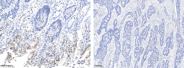
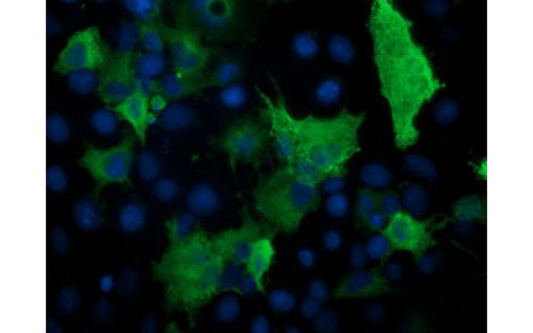
 IHC (Immunohistochemistry)
(Formalin-fixed, paraffin-embedded human Melanoma stained with Melanoma Marker Mouse Monoclonal Antibody (DT101+ BC199 + HMB45).)
IHC (Immunohistochemistry)
(Formalin-fixed, paraffin-embedded human Melanoma stained with Melanoma Marker Mouse Monoclonal Antibody (DT101+ BC199 + HMB45).)
Melanoma Marker (MART-1 + gp100), Monoclonal Antibody (Cat# AAA216087)
 SDS-PAGE
(SDS-PAGE Analysis Purified CD51/Integrin ?V Rat Monoclonal Antibody (AvB6 53a. 2). Confirmation of Purity and Integrity of Antibody.)
SDS-PAGE
(SDS-PAGE Analysis Purified CD51/Integrin ?V Rat Monoclonal Antibody (AvB6 53a. 2). Confirmation of Purity and Integrity of Antibody.)
Integrin alpha V + beta 6, Monoclonal Antibody (Cat# AAA216098)
Cyclin D1 (CCND1), Monoclonal Antibody (Cat# AAA147996)
Interleukin 4 (IL4), Monoclonal Antibody (Cat# AAA149420)
Immunoglobulin G1 (IgG1), Monoclonal Antibody (Cat# AAA149851)
Anti-Mullerian Hormone (AMH), Monoclonal Antibody (Cat# AAA149783)
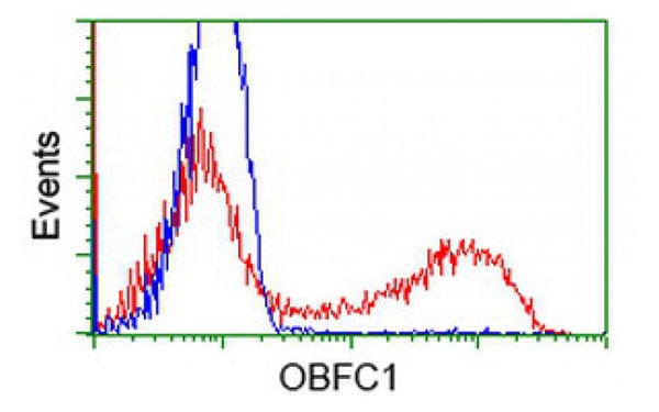
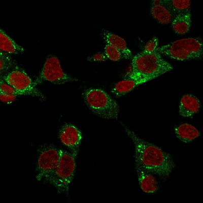
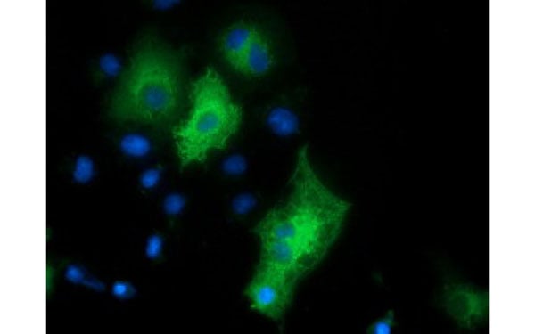
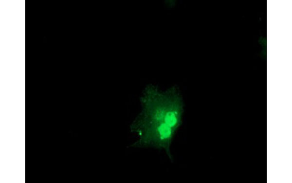 IF (Immunofluorescence)
(Immunofluorescent staining of COS7 cells transiently transfected with recombinant OBFC1 protein using OBFC1 antibody)
IF (Immunofluorescence)
(Immunofluorescent staining of COS7 cells transiently transfected with recombinant OBFC1 protein using OBFC1 antibody)
OBFC1, Monoclonal Antibody (Cat# AAA108147)
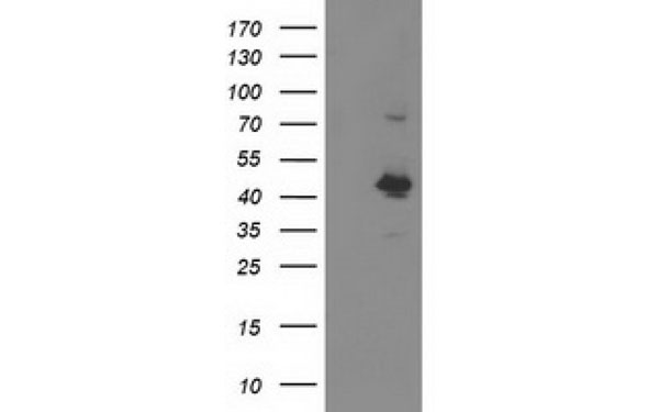
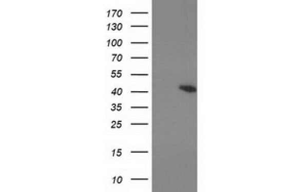 WB (Western Blot)
(Western Blot analysis of HEK293T cell lysates (5 ug) transfected with either recombinant PRKAR1B protein (Right) or empty vector (Left) detected with PRKAR1B antibody)
WB (Western Blot)
(Western Blot analysis of HEK293T cell lysates (5 ug) transfected with either recombinant PRKAR1B protein (Right) or empty vector (Left) detected with PRKAR1B antibody)
PRKAR1B, Monoclonal Antibody (Cat# AAA107361)
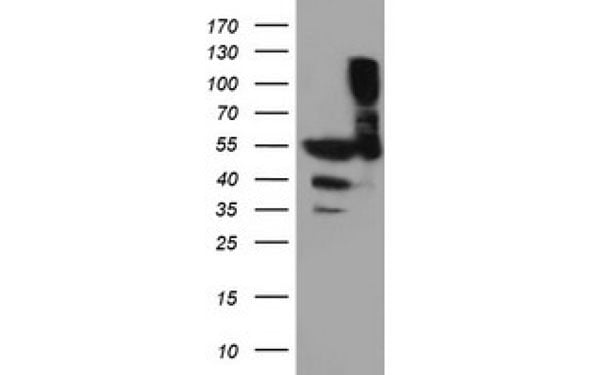
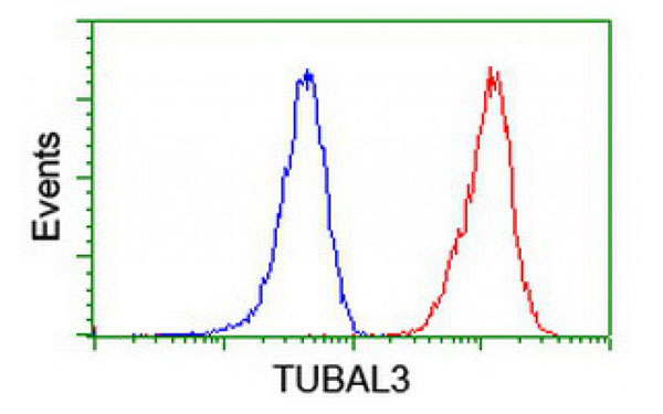
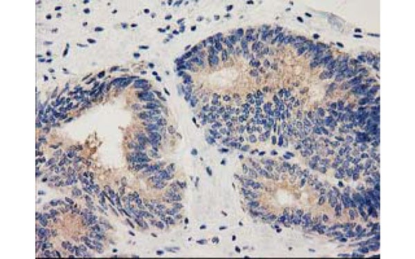
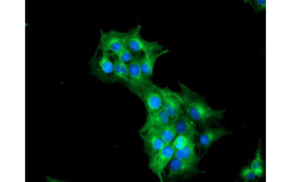 IF (Immunofluorescence)
(Immunofluorescent staining of COS7 cells transiently transfected with recombinant TUBAL3 protein using TUBAL3 antibody)
IF (Immunofluorescence)
(Immunofluorescent staining of COS7 cells transiently transfected with recombinant TUBAL3 protein using TUBAL3 antibody)
TUBAL3, Monoclonal Antibody (Cat# AAA106813)
 IF (Immunofluorescence)
(Immunofluorescent staining of COS7 cells transiently transfected with recombinant HDAC6 protein using HDAC6 antibody)
IF (Immunofluorescence)
(Immunofluorescent staining of COS7 cells transiently transfected with recombinant HDAC6 protein using HDAC6 antibody)
HDAC6, Monoclonal Antibody (Cat# AAA106422)
Mouse anti TRITC, Monoclonal Antibody (Cat# AAA77518)
Mouse anti Human IgG2 (Fc subclass specific), Monoclonal Secondary Antibody (Cat# AAA77535)
N-Terminal proBNP (NT-proBNP), Monoclonal Antibody (Cat# AAA78097)
Purification: Protein A chromatography


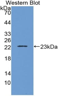
 Application Data
(Detection limit for recombinant GST tagged TCEA3 is approximately 0.03ng/ml as a capture antibody.)
Application Data
(Detection limit for recombinant GST tagged TCEA3 is approximately 0.03ng/ml as a capture antibody.)
TCEA3, Monoclonal Antibody (Cat# AAA26342)
What are Monoclonal Antibodies?
Monoclonal antibodies are specialized laboratory-produced proteins developed for binding to specific biological antigens or other molecular targets. Since they come from a single cell (or clone), they are especially consistent and accurate in the data they are involved in producing.
This type of antibody material has been shown to be a powerful tool in finding and subsequently destroying harmful cells in an organism, such as those found in cancers or various autoimmune diseases. This makes them excellent aids in medical testing and research, which is why they are so widely used.
AAA Biotech offers a comprehensive range of high-quality monoclonal antibodies that perform effectively in various laboratory tests, including (amongst others) ELISA, western blotting, immunohistochemistry, and flow cytometry. All of the products in our catalog are thoroughly quality tested to make sure that they are reliable and will consistently perform well in your research.
What Are The Uses of Monoclonal Antibodies
Monoclonal antibodies are used in many lab tests, including (amongst others) ELISA, western blotting, immunohistochemistry, and flow cytometry.
ELISA is a test that helps detect a specific substance/analyte in a sample. It uses antibodies (often monoclonal) bound to a solid surface (such as the well of a microplate) to “capture” the substance/analyte in the sample and immobilize it so that the detection antibody component can then bind to it and produce a signal, which can then be measured.
Western blotting identifies specific proteins in a sample. The sample is first separated on a gel, and then antibodies are applied that will typically bind to the target, which will all be localized to a single band in a lane.
Immunohistochemistry helps locate specific proteins in cells or tissue samples using antibodies.
Flow cytometry looks at and sorts cells. It uses antibodies that are conjugated to reporter molecules called “fluorophores”, which, under special lights, emit light themselves, which can then be measured by a detector instrument.
How Monoclonal Antibodies Are Used as Medicine?
Please note that all of the products listed in AAA Biotech’s also known as AAA Bio or AAABio catalog are strictly for research-use only (RUO).
Monoclonal antibodies can also be used as therapeutic/medical treatments, particularly in the context of cancers. They are designed to find and bind to specific cells or proteins, helping the immune system recognize and attack the cancer. These treatments work in different ways, such as:
- Radioimmunotherapy attaches a small amount of radioactive molecule to the antibody, so it delivers the radiation directly to the cancer cells that the antibody is specifically binding to.
- Antibody-directed enzyme prodrug therapy uses antibodies that are specifically bound to special enzymes. These enzymes activate a harmless drug in the body and turn it into a cancer-killing drug only near the cancer cells—this helps avoid harming healthy cells.
- Immunoliposomes are tiny “bubbles” filled with medicine/drug and coated with antibodies. They carry the drug straight to the cancer cells.
Why Buy Monoclonal Antibodies From Us?
At AAA Biotech, we provide high-performance monoclonal antibodies designed to support a wide range of research needs.
1. Validated for Versatile Applications
The antibodies in our catalog are extensively validated and compatible with multiple techniques, including (but not limited to) ELISA, flow cytometry (FC), immunocytochemistry (ICC), immunofluorescence (IF), immunohistochemistry (IHC), immunoprecipitation (IP), and western blotting (WB).
2. Wide Selection & Specialized Options
We offer antibodies for common and rare species, that are available in various conjugated forms, and also in recombinant formats. Essentially, there is almost anything one might need to meet their experimental model’s requirements.
3. High-Quality Proteins
Our proteins meet high purity standards—90% or more as confirmed by SDS-PAGE. Many are available with tags like His, Flag, GST, or MBP, and we also supply native and biologically active proteins for functional studies.
Frequently Asked Questions
1. Are your monoclonal antibodies validated for specific applications?
Yes, our antibodies are tested and validated for use in methods such as ELISA, western blot, IHC, flow cytometry, and more. Refer to specific product pages or datasheets for individual product information.
2. How do I choose the right monoclonal antibody for my application?
Review the product details directly for application validation, species reactivity, and target information. You may also contact our support team at any time for help.
3. How quickly can I receive my order?
Most orders are processed and shipped within 1–3 business days, depending on product availability and your shipping location.