Filters
Clonality
Type
Reactivity
Gene Name
Isotype
Host
Application
Clone
666 results for "ECL" - showing 400-450


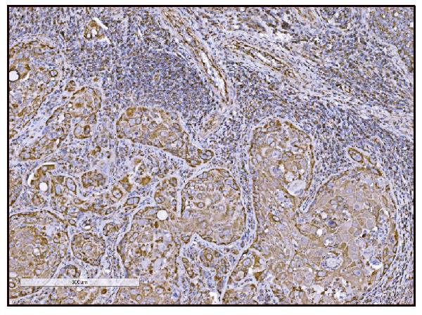


 IHC (Immunohistochemistry)
(Figure 14. IHC analysis of FABP-1/GOT2 using anti-FABP-1/GOT2 antibody (AAA19309).FABP-1/GOT2 was detected in paraffin-embedded section of rat lymph node tissue. Heat mediated antigen retrieval was performed in EDTA buffer (pH8. 0, epitope retrieval solution). The tissue section was blocked with 10% goat serum. The tissue section was then incubated with 2μg/ml rabbit anti-FABP-1/GOT2 Antibody (AAA19309) overnight at 4 degree C. Biotinylated goat anti-rabbit IgG was used as secondary antibody and incubated for 30 minutes at 37 degree C. The tissue section was developed using Strepavidin-Biotin-Complex (SABC) (Catalog # with DAB as the chromogen.)
IHC (Immunohistochemistry)
(Figure 14. IHC analysis of FABP-1/GOT2 using anti-FABP-1/GOT2 antibody (AAA19309).FABP-1/GOT2 was detected in paraffin-embedded section of rat lymph node tissue. Heat mediated antigen retrieval was performed in EDTA buffer (pH8. 0, epitope retrieval solution). The tissue section was blocked with 10% goat serum. The tissue section was then incubated with 2μg/ml rabbit anti-FABP-1/GOT2 Antibody (AAA19309) overnight at 4 degree C. Biotinylated goat anti-rabbit IgG was used as secondary antibody and incubated for 30 minutes at 37 degree C. The tissue section was developed using Strepavidin-Biotin-Complex (SABC) (Catalog # with DAB as the chromogen.)
FABP-1/GOT2, Polyclonal Antibody (Cat# AAA19309)
Full Name
Anti-FABP-1/GOT2 Antibody
Gene Names
GOT2; KAT4; KATIV; mitAAT
Reactivity
Human, Mouse, Rat
Applications
WB, IHC-P, ICC, IF, FC/FACS/FCM, EIA
Purity
Immunogen affinity purified.
Pricing


 IHC (Immunohistchemistry)
(Immunohistochemistry of paraffin-embedded mouse liver using KIAA1456 antibody at dilution of 1:200 (40x lens).)
IHC (Immunohistchemistry)
(Immunohistochemistry of paraffin-embedded mouse liver using KIAA1456 antibody at dilution of 1:200 (40x lens).)
KIAA1456, Polyclonal Antibody (Cat# AAA28126)
Full Name
KIAA1456 Polyclonal Antibody
Gene Names
KIAA1456; TRM9L; C8orf79
Reactivity
Human
Applications
WB, IHC, IF
Purity
Affinity Purification
Pricing


 IF (Immunofluorescence)
(Immunofluorescence analysis of HeLa cells using CSNK1E antibody. Blue: DAPI for nuclear staining.)
IF (Immunofluorescence)
(Immunofluorescence analysis of HeLa cells using CSNK1E antibody. Blue: DAPI for nuclear staining.)
CSNK1E, Polyclonal Antibody (Cat# AAA28143)
Full Name
CSNK1E Polyclonal Antibody
Gene Names
CSNK1E; HCKIE; CKIepsilon
Reactivity
Human, Mouse, Rat
Applications
WB, IHC, IF
Purity
Affinity Purification
Pricing


 IHC (Immunohistchemistry)
(Immunohistochemistry of paraffin-embedded mouse brain using JMJD6 Antibody at dilution of 1:100 (40x lens).)
IHC (Immunohistchemistry)
(Immunohistochemistry of paraffin-embedded mouse brain using JMJD6 Antibody at dilution of 1:100 (40x lens).)
JMJD6, Polyclonal Antibody (Cat# AAA28145)
Full Name
JMJD6 Polyclonal Antibody
Gene Names
JMJD6; PSR; PTDSR; PTDSR1
Reactivity
Human, Mouse, Rat
Applications
WB, IHC, IF, IP
Purity
Affinity Purification
Pricing


 IHC (Immunohistchemistry)
(Figure 6. IHC analysis of CPI17 alpha using anti- CPI17 alpha antibody (AAA19176).CPI17 alpha was detected in paraffin-embedded section of human placenta tissues. Heat mediated antigen retrieval was performed in citrate buffer (pH6, epitope retrieval solution) for 20 mins. The tissue section was blocked with 10% goat serum. The tissue section was then incubated with 1ug/ml rabbit anti- CPI17 alpha Antibody (AAA19176) overnight at 4 degree C. Biotinylated goat anti-rabbit IgG was used as secondary antibody and incubated for 30 minutes at 37 degree C. The tissue section was developed using Strepavidin-Biotin-Complex (SABC) with DAB as the chromogen.)
IHC (Immunohistchemistry)
(Figure 6. IHC analysis of CPI17 alpha using anti- CPI17 alpha antibody (AAA19176).CPI17 alpha was detected in paraffin-embedded section of human placenta tissues. Heat mediated antigen retrieval was performed in citrate buffer (pH6, epitope retrieval solution) for 20 mins. The tissue section was blocked with 10% goat serum. The tissue section was then incubated with 1ug/ml rabbit anti- CPI17 alpha Antibody (AAA19176) overnight at 4 degree C. Biotinylated goat anti-rabbit IgG was used as secondary antibody and incubated for 30 minutes at 37 degree C. The tissue section was developed using Strepavidin-Biotin-Complex (SABC) with DAB as the chromogen.)
CPI17 alpha, Polyclonal Antibody (Cat# AAA19176)
Full Name
Anti-CPI17 alpha Picoband Antibody
Gene Names
PPP1R14A; CPI17; CPI-17; PPP1INL
Reactivity
Human, Mouse, Rat
No cross reactivity with other proteins
No cross reactivity with other proteins
Applications
IHC, WB
Purity
Immunogen affinity purified
Pricing


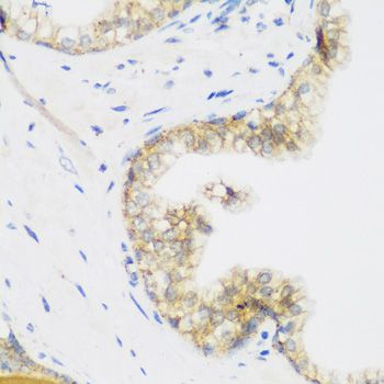
 IHC (Immunohistochemistry)
(Figure 8. IHC analysis of PRDM1/Blimp1 using anti-PRDM1/Blimp1 antibody (AAA19133).PRDM1/Blimp1 was detected in paraffin-embedded section of rat small intestine tissue. Heat mediated antigen retrieval was performed in citrate buffer (pH6, epitope retrieval solution) for 20 mins. The tissue section was blocked with 10% goat serum. The tissue section was then incubated with 1ug/ml rabbit anti-PRDM1/Blimp1 Antibody (AAA19133) overnight at 4 degree C. Biotinylated goat anti-rabbit IgG was used as secondary antibody and incubated for 30 minutes at 37 degree C. The tissue section was developed using Strepavidin-Biotin-Complex (SABC) with DAB as the chromogen.)
IHC (Immunohistochemistry)
(Figure 8. IHC analysis of PRDM1/Blimp1 using anti-PRDM1/Blimp1 antibody (AAA19133).PRDM1/Blimp1 was detected in paraffin-embedded section of rat small intestine tissue. Heat mediated antigen retrieval was performed in citrate buffer (pH6, epitope retrieval solution) for 20 mins. The tissue section was blocked with 10% goat serum. The tissue section was then incubated with 1ug/ml rabbit anti-PRDM1/Blimp1 Antibody (AAA19133) overnight at 4 degree C. Biotinylated goat anti-rabbit IgG was used as secondary antibody and incubated for 30 minutes at 37 degree C. The tissue section was developed using Strepavidin-Biotin-Complex (SABC) with DAB as the chromogen.)
PRDM1/Blimp1, Polyclonal Antibody (Cat# AAA19133)
Full Name
Anti-PRDM1/Blimp1 Picoband antibody
Gene Names
PRDM1; BLIMP1; PRDI-BF1
Reactivity
Human, Mouse, Rat
No cross reactivity with other proteins.
No cross reactivity with other proteins.
Applications
EIA, IHC, WB
Pricing


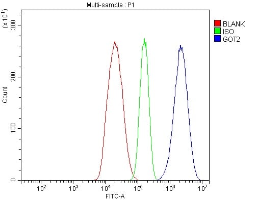
 FCM (Flow Cytometry)
(Figure 7. Flow Cytometry analysis of 293T cells using anti-OTULIN antibody (AAA19319).Overlay histogram showing 293T cells stained with AAA19319 (Blue line). The cells were blocked with 10% normal goat serum. And then incubated with rabbit anti-OTULIN Antibody (AAA19319, 1μg/1x106 cells) for 30 min at 20 degree C. DyLight®488 conjugated goat anti-rabbit IgG (5-10μg/1x106 cells) was used as secondary antibody for 30 minutes at 20 degree C. Isotype control antibody (Green line) was rabbit IgG (1μg/1x106) used under the same conditions. Unlabelled sample (Red line) was also used as a control.)
FCM (Flow Cytometry)
(Figure 7. Flow Cytometry analysis of 293T cells using anti-OTULIN antibody (AAA19319).Overlay histogram showing 293T cells stained with AAA19319 (Blue line). The cells were blocked with 10% normal goat serum. And then incubated with rabbit anti-OTULIN Antibody (AAA19319, 1μg/1x106 cells) for 30 min at 20 degree C. DyLight®488 conjugated goat anti-rabbit IgG (5-10μg/1x106 cells) was used as secondary antibody for 30 minutes at 20 degree C. Isotype control antibody (Green line) was rabbit IgG (1μg/1x106) used under the same conditions. Unlabelled sample (Red line) was also used as a control.)
OTULIN, Polyclonal Antibody (Cat# AAA19319)
Full Name
Anti-OTULIN Antibody
Gene Names
OTULIN; GUM; FAM105B
Reactivity
Human, Mouse, Rat
Applications
WB, IHC-P, ICC, IF, FC/FACS/FCM, EIA
Purity
Immunogen affinity purified.
Pricing


 FCM (Flow Cytometry)
(Figure 13. Flow Cytometry analysis of Hela cells using anti-TAF4 antibody (AAA19307).Overlay histogram showing Hela cells stained with AAA19307 (Blue line). The cells were blocked with 10% normal goat serum. And then incubated with rabbit anti-TAF4 Antibody (AAA19307, 1μg/1x106 cells) for 30 min at 20 degree C. DyLight®488 conjugated goat anti-rabbit IgG (5-10μg/1x106 cells) was used as secondary antibody for 30 minutes at 20 degree C. Isotype control antibody (Green line) was rabbit IgG (1μg/1x106) used under the same conditions. Unlabelled sample (Red line) was also used as a control.)
FCM (Flow Cytometry)
(Figure 13. Flow Cytometry analysis of Hela cells using anti-TAF4 antibody (AAA19307).Overlay histogram showing Hela cells stained with AAA19307 (Blue line). The cells were blocked with 10% normal goat serum. And then incubated with rabbit anti-TAF4 Antibody (AAA19307, 1μg/1x106 cells) for 30 min at 20 degree C. DyLight®488 conjugated goat anti-rabbit IgG (5-10μg/1x106 cells) was used as secondary antibody for 30 minutes at 20 degree C. Isotype control antibody (Green line) was rabbit IgG (1μg/1x106) used under the same conditions. Unlabelled sample (Red line) was also used as a control.)
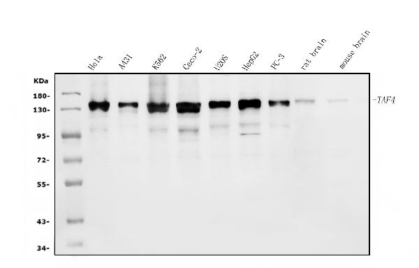
TAF4, Polyclonal Antibody (Cat# AAA19307)
Full Name
Anti-TAF4 Antibody
Gene Names
TAF4; TAF2C; TAF4A; TAF2C1; TAFII130; TAFII135
Reactivity
Human, Mouse, Rat
Applications
WB, IHC-P, ICC, IF, EIA
Purity
Immunogen affinity purified.
Pricing


 IHC (Immunohistchemistry)
(Immunohistochemistry of paraffin-embedded mouse brain using IRAK4 antibody at dilution of 1:100 (40x lens).)
IHC (Immunohistchemistry)
(Immunohistochemistry of paraffin-embedded mouse brain using IRAK4 antibody at dilution of 1:100 (40x lens).)
IRAK4, Polyclonal Antibody (Cat# AAA28120)
Full Name
IRAK4 Polyclonal Antibody
Gene Names
IRAK4; IPD1; REN64; IRAK-4; NY-REN-64
Reactivity
Human, Mouse, Rat
Applications
WB, IHC
Purity
Affinity Purification
Pricing


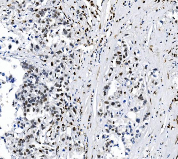
 IHC (Immunohistchemistry)
(Figure 6. IHC analysis of Cdc20 using anti-Cdc20 antibody (AAA19132).Cdc20 was detected in paraffin-embedded section of human mammary cancer tissue. Heat mediated antigen retrieval was performed in citrate buffer (pH6, epitope retrieval solution) for 20 mins. The tissue section was blocked with 10% goat serum. The tissue section was then incubated with 1ug/ml rabbit anti-Cdc20 Antibody (AAA19132) overnight at 4 degree C. Biotinylated goat anti-rabbit IgG was used as secondary antibody and incubated for 30 minutes at 37 degree C. The tissue section was developed using Strepavidin-Biotin-Complex (SABC) with DAB as the chromogen.)
IHC (Immunohistchemistry)
(Figure 6. IHC analysis of Cdc20 using anti-Cdc20 antibody (AAA19132).Cdc20 was detected in paraffin-embedded section of human mammary cancer tissue. Heat mediated antigen retrieval was performed in citrate buffer (pH6, epitope retrieval solution) for 20 mins. The tissue section was blocked with 10% goat serum. The tissue section was then incubated with 1ug/ml rabbit anti-Cdc20 Antibody (AAA19132) overnight at 4 degree C. Biotinylated goat anti-rabbit IgG was used as secondary antibody and incubated for 30 minutes at 37 degree C. The tissue section was developed using Strepavidin-Biotin-Complex (SABC) with DAB as the chromogen.)
Cdc20/P55 Cdc, Polyclonal Antibody (Cat# AAA19132)
Full Name
Anti-Cdc20/P55 Cdc Picoband Antibody
Gene Names
CDC20; CDC20A; p55CDC; bA276H19.3
Reactivity
Human, Mouse, Rat
No cross reactivity with other proteins.
No cross reactivity with other proteins.
Applications
IHC, WB
Purity
Immunogen affinity purified
Pricing


 FCM (Flow Cytometry)
(Figure 11. Flow Cytometry analysis of A431 cells using anti-SAMHD1 antibody (AAA19355).Overlay histogram showing A431 cells stained with AAA19355 (Blue line). The cells were blocked with 10% normal goat serum. And then incubated with mouse anti- SAMHD1 Antibody (AAA19355, 1μg/1x106 cells) for 30 min at 20 degree C. DyLight®488 conjugated goat anti-mouse IgG (BA1126, 5-10μg/1x106 cells) was used as secondary antibody for 30 minutes at 20 degree C. Isotype control antibody (Green line) was mouse IgG (1μg/1x106) used under the same conditions. Unlabelled sample (Red line) was also used as a control.)
FCM (Flow Cytometry)
(Figure 11. Flow Cytometry analysis of A431 cells using anti-SAMHD1 antibody (AAA19355).Overlay histogram showing A431 cells stained with AAA19355 (Blue line). The cells were blocked with 10% normal goat serum. And then incubated with mouse anti- SAMHD1 Antibody (AAA19355, 1μg/1x106 cells) for 30 min at 20 degree C. DyLight®488 conjugated goat anti-mouse IgG (BA1126, 5-10μg/1x106 cells) was used as secondary antibody for 30 minutes at 20 degree C. Isotype control antibody (Green line) was mouse IgG (1μg/1x106) used under the same conditions. Unlabelled sample (Red line) was also used as a control.)
SAMHD1, Monoclonal Antibody (Cat# AAA19355)
Full Name
Anti-SAMHD1 Antibody (monoclonal, 3B9)
Gene Names
SAMHD1; DCIP; CHBL2; HDDC1; MOP-5; SBBI88
Reactivity
Human
Applications
WB, IHC-P, ICC, IF, FC/FACS/FCM
Purity
Immunogen affinity purified.
Pricing


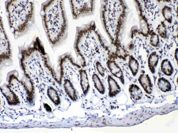

 IHC (Immunohistchemistry)
(Figure 6. IHC analysis of Musashi 1/Msi1 using anti- Musashi 1/Msi1 antibody (AAA19172).Musashi 1/Msi1 was detected in paraffin-embedded section of human mammary cancer tissues. Heat mediated antigen retrieval was performed in citrate buffer (pH6, epitope retrieval solution) for 20 mins. The tissue section was blocked with 10% goat serum. The tissue section was then incubated with 1ug/ml rabbit anti- Musashi 1/Msi1 Antibody (AAA19172) overnight at 4 degree C. Biotinylated goat anti-rabbit IgG was used as secondary antibody and incubated for 30 minutes at 37 degree C. The tissue section was developed using Strepavidin-Biotin-Complex (SABC) with DAB as the chromogen.)
IHC (Immunohistchemistry)
(Figure 6. IHC analysis of Musashi 1/Msi1 using anti- Musashi 1/Msi1 antibody (AAA19172).Musashi 1/Msi1 was detected in paraffin-embedded section of human mammary cancer tissues. Heat mediated antigen retrieval was performed in citrate buffer (pH6, epitope retrieval solution) for 20 mins. The tissue section was blocked with 10% goat serum. The tissue section was then incubated with 1ug/ml rabbit anti- Musashi 1/Msi1 Antibody (AAA19172) overnight at 4 degree C. Biotinylated goat anti-rabbit IgG was used as secondary antibody and incubated for 30 minutes at 37 degree C. The tissue section was developed using Strepavidin-Biotin-Complex (SABC) with DAB as the chromogen.)
Musashi 1/Msi1, Polyclonal Antibody (Cat# AAA19172)
Full Name
Anti-Musashi 1/Msi1 Picoband Antibody
Reactivity
Human, Mouse, Rat
No cross reactivity with other proteins
No cross reactivity with other proteins
Applications
IHC, WB
Purity
Immunogen affinity purified
Pricing


 FCM (Flow Cytometry)
(Figure 6. Flow Cytometry analysis of HEPA1-6 cells using anti-PAR4/Pawr antibody (AAA19278).Overlay histogram showing HEPA1-6 cells stained with AAA19278 (Blue line). The cells were blocked with 10% normal goat serum. And then incubated with rabbit anti-PAR4/Pawr Antibody (AAA19278, 1μg/1x106 cells) for 30 min at 20 degree C. DyLight®488 conjugated goat anti-rabbit IgG (5-10μg/1x106 cells) was used as secondary antibody for 30 minutes at 20 degree C. Isotype control antibody (Green line) was rabbit IgG (1μg/1x106) used under the same conditions. Unlabelled sample (Red line) was also used as a control.)
FCM (Flow Cytometry)
(Figure 6. Flow Cytometry analysis of HEPA1-6 cells using anti-PAR4/Pawr antibody (AAA19278).Overlay histogram showing HEPA1-6 cells stained with AAA19278 (Blue line). The cells were blocked with 10% normal goat serum. And then incubated with rabbit anti-PAR4/Pawr Antibody (AAA19278, 1μg/1x106 cells) for 30 min at 20 degree C. DyLight®488 conjugated goat anti-rabbit IgG (5-10μg/1x106 cells) was used as secondary antibody for 30 minutes at 20 degree C. Isotype control antibody (Green line) was rabbit IgG (1μg/1x106) used under the same conditions. Unlabelled sample (Red line) was also used as a control.)
PAR4/Pawr, Polyclonal Antibody (Cat# AAA19278)
Full Name
Anti-PAR4/Pawr Antibody
Gene Names
Pawr; PAR4; Par-4; 2310001G03Rik
Reactivity
Mouse, Rat
Applications
WB, IHC-P, ICC, IF, FC/FACS/FCM, EIA
Purity
Immunogen affinity purified.
Pricing


 FCM (Flow Cytometry)
(Figure 10. Flow Cytometry analysis of A431 cells using anti-CDC27 antibody (AAA19285).Overlay histogram showing A431 cells stained with AAA19285 (Blue line). The cells were blocked with 10% normal goat serum. And then incubated with rabbit anti-CDC27 Antibody (AAA19285, 1μg/1x106 cells) for 30 min at 20 degree C. DyLight®488 conjugated goat anti-rabbit IgG (5-10μg/1x106 cells) was used as secondary antibody for 30 minutes at 20 degree C. Isotype control antibody (Green line) was rabbit IgG (1μg/1x106) used under the same conditions. Unlabelled sample (Red line) was also used as a control.)
FCM (Flow Cytometry)
(Figure 10. Flow Cytometry analysis of A431 cells using anti-CDC27 antibody (AAA19285).Overlay histogram showing A431 cells stained with AAA19285 (Blue line). The cells were blocked with 10% normal goat serum. And then incubated with rabbit anti-CDC27 Antibody (AAA19285, 1μg/1x106 cells) for 30 min at 20 degree C. DyLight®488 conjugated goat anti-rabbit IgG (5-10μg/1x106 cells) was used as secondary antibody for 30 minutes at 20 degree C. Isotype control antibody (Green line) was rabbit IgG (1μg/1x106) used under the same conditions. Unlabelled sample (Red line) was also used as a control.)
CDC27, Polyclonal Antibody (Cat# AAA19285)
Full Name
Anti-CDC27 Antibody
Gene Names
CDC27; APC3; HNUC; NUC2; ANAPC3; CDC27Hs; D0S1430E; D17S978E
Reactivity
Human, Mouse, Rat
Applications
WB, IHC-P, ICC, IF, FC/FACS/FCM, EIA
Purity
Immunogen affinity purified.
Pricing


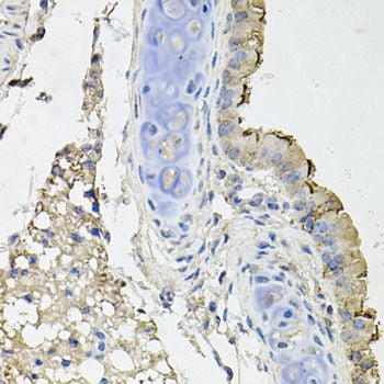
 IHC (Immunohistochemistry)
(Figure 8. IHC analysis of TAGLN/Transgelin using anti-TAGLN/Transgelin antibody (AAA19286).TAGLN/Transgelin was detected in paraffin-embedded section of rat testis tissue. Heat mediated antigen retrieval was performed in EDTA buffer (pH8. 0, epitope retrieval solution). The tissue section was blocked with 10% goat serum. The tissue section was then incubated with 2μg/ml rabbit anti-TAGLN/Transgelin Antibody (AAA19286) overnight at 4 degree C. Biotinylated goat anti-rabbit IgG was used as secondary antibody and incubated for 30 minutes at 37 degree C. The tissue section was developed using Strepavidin-Biotin-Complex (SABC) (Catalog # with DAB as the chromogen.)
IHC (Immunohistochemistry)
(Figure 8. IHC analysis of TAGLN/Transgelin using anti-TAGLN/Transgelin antibody (AAA19286).TAGLN/Transgelin was detected in paraffin-embedded section of rat testis tissue. Heat mediated antigen retrieval was performed in EDTA buffer (pH8. 0, epitope retrieval solution). The tissue section was blocked with 10% goat serum. The tissue section was then incubated with 2μg/ml rabbit anti-TAGLN/Transgelin Antibody (AAA19286) overnight at 4 degree C. Biotinylated goat anti-rabbit IgG was used as secondary antibody and incubated for 30 minutes at 37 degree C. The tissue section was developed using Strepavidin-Biotin-Complex (SABC) (Catalog # with DAB as the chromogen.)
TAGLN/Transgelin, Polyclonal Antibody (Cat# AAA19286)
Full Name
Anti-TAGLN/Transgelin Antibody
Gene Names
TAGLN; SM22; SMCC; TAGLN1; WS3-10
Reactivity
Human, Mouse, Rat
Applications
WB, IHC-P, ICC, IF, FC/FACS/FCM, EIA
Purity
Immunogen affinity purified.
Pricing


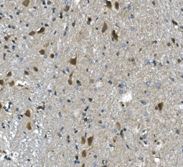
 IHC (Immunohistchemistry)
(Figure 6. IHC analysis of PP2A-alpha/PPP2CA using anti-PP2A-alpha/PPP2CA antibody (AAA19366).PP2A-alpha/PPP2CA was detected in paraffin-embedded section of rat brain tissue. Heat mediated antigen retrieval was performed in EDTA buffer (pH8. 0, epitope retrieval solution). The tissue section was blocked with 10% goat serum. The tissue section was then incubated with 1μg/ml mouse anti-PP2A-alpha/PPP2CA Antibody (AAA19366) overnight at 4 degree C. Biotinylated goat anti-mouse IgG was used as secondary antibody and incubated for 30 minutes at 37 degree C. The tissue section was developed using Strepavidin-Biotin-Complex (SABC) (Catalog # with DAB as the chromogen.)
IHC (Immunohistchemistry)
(Figure 6. IHC analysis of PP2A-alpha/PPP2CA using anti-PP2A-alpha/PPP2CA antibody (AAA19366).PP2A-alpha/PPP2CA was detected in paraffin-embedded section of rat brain tissue. Heat mediated antigen retrieval was performed in EDTA buffer (pH8. 0, epitope retrieval solution). The tissue section was blocked with 10% goat serum. The tissue section was then incubated with 1μg/ml mouse anti-PP2A-alpha/PPP2CA Antibody (AAA19366) overnight at 4 degree C. Biotinylated goat anti-mouse IgG was used as secondary antibody and incubated for 30 minutes at 37 degree C. The tissue section was developed using Strepavidin-Biotin-Complex (SABC) (Catalog # with DAB as the chromogen.)
PP2A-alpha/PPP2CA, Monoclonal Antibody (Cat# AAA19366)
Full Name
Anti-PP2A-alpha/PPP2CA Antibody (monoclonal, 3B6)
Gene Names
PPP2CA; RP-C; PP2Ac; PP2CA; PP2Calpha
Reactivity
Human, Monkey, Mouse, Rat
Applications
WB, IHC-P, ICC, IF, FC/FACS/FCM
Purity
Immunogen affinity purified.
Pricing


 IF (Immunofluorescence)
(Figure 9. IF analysis of ApoER2/LRP8 using anti- ApoER2/LRP8 antibody (AAA19275).ApoER2/LRP8 was detected in immunocytochemical section of HepG2 cells. Enzyme antigen retrieval was performed using IHC enzyme antigen retrieval reagent for 15 mins. The cells were blocked with 10% goat serum. And then incubated with 4μg/mL rabbit anti- ApoER2/LRP8 Antibody (AAA19275) overnight at 4 degree C. DyLight®488 Conjugated Goat Anti-Rabbit IgG was used as secondary antibody at 1:100 dilution and incubated for 30 minutes at 37 degree C. The section was counterstained with DAPI. Visualize using a fluorescence microscope and filter sets appropriate for the label used.)
IF (Immunofluorescence)
(Figure 9. IF analysis of ApoER2/LRP8 using anti- ApoER2/LRP8 antibody (AAA19275).ApoER2/LRP8 was detected in immunocytochemical section of HepG2 cells. Enzyme antigen retrieval was performed using IHC enzyme antigen retrieval reagent for 15 mins. The cells were blocked with 10% goat serum. And then incubated with 4μg/mL rabbit anti- ApoER2/LRP8 Antibody (AAA19275) overnight at 4 degree C. DyLight®488 Conjugated Goat Anti-Rabbit IgG was used as secondary antibody at 1:100 dilution and incubated for 30 minutes at 37 degree C. The section was counterstained with DAPI. Visualize using a fluorescence microscope and filter sets appropriate for the label used.)
ApoER2/LRP8, Polyclonal Antibody (Cat# AAA19275)
Full Name
Anti-ApoER2/LRP8 Antibody
Gene Names
LRP8; MCI1; LRP-8; APOER2; HSZ75190
Reactivity
Human, Mouse, Rat
Applications
WB, IHC-P, ICC, IF, FC/FACS/FCM, EIA
Purity
Immunogen affinity purified.
Pricing


 IF (Immunofluorescence)
(Figure 15. IF analysis of LSM2 using anti- LSM2 antibody (AAA19316).LSM2 was detected in immunocytochemical section of MCF-7 cells. Enzyme antigen retrieval was performed using IHC enzyme antigen retrieval reagent for 15 mins. The cells were blocked with 10% goat serum. And then incubated with 5μg/mL rabbit anti- LSM2 Antibody (AAA19316) overnight at 4 degree C. DyLight®488 Conjugated Goat Anti-Rabbit IgG was used as secondary antibody at 1:100 dilution and incubated for 30 minutes at 37 degree C. The section was counterstained with DAPI. Visualize using a fluorescence microscope and filter sets appropriate for the label used.)
IF (Immunofluorescence)
(Figure 15. IF analysis of LSM2 using anti- LSM2 antibody (AAA19316).LSM2 was detected in immunocytochemical section of MCF-7 cells. Enzyme antigen retrieval was performed using IHC enzyme antigen retrieval reagent for 15 mins. The cells were blocked with 10% goat serum. And then incubated with 5μg/mL rabbit anti- LSM2 Antibody (AAA19316) overnight at 4 degree C. DyLight®488 Conjugated Goat Anti-Rabbit IgG was used as secondary antibody at 1:100 dilution and incubated for 30 minutes at 37 degree C. The section was counterstained with DAPI. Visualize using a fluorescence microscope and filter sets appropriate for the label used.)
LSM2, Polyclonal Antibody (Cat# AAA19316)
Full Name
Anti-LSM2 Antibody
Gene Names
LSM2; G7B; snRNP; C6orf28; YBL026W
Reactivity
Human, Mouse, Rat, Monkey
Applications
WB, IHC-P, ICC, IF, FC/FACS/FCM
Purity
Immunogen affinity purified.
Pricing


 FCM (Flow Cytometry)
(Figure 6. Flow Cytometry analysis of THP-1 cells using anti-TLR1 antibody (AAA19134).Overlay histogram showing THP-1 cells stained with AAA19134 (Blue line).The cells were blocked with 10% normal goat serum. And then incubated with rabbit anti-TLR1 Antibody (AAA19134,1ug/1x10^6 cells) for 30 min at 20 degree C. DyLight®488 conjugated goat anti-rabbit IgG (5-10ug/1x10^6 cells) was used as secondary antibody for 30 minutes at 20 degree C. Isotype control antibody (Green line) was rabbit IgG (1ug/1x106) used under the same conditions. Unlabelled sample (Red line) was also used as a control.)
FCM (Flow Cytometry)
(Figure 6. Flow Cytometry analysis of THP-1 cells using anti-TLR1 antibody (AAA19134).Overlay histogram showing THP-1 cells stained with AAA19134 (Blue line).The cells were blocked with 10% normal goat serum. And then incubated with rabbit anti-TLR1 Antibody (AAA19134,1ug/1x10^6 cells) for 30 min at 20 degree C. DyLight®488 conjugated goat anti-rabbit IgG (5-10ug/1x10^6 cells) was used as secondary antibody for 30 minutes at 20 degree C. Isotype control antibody (Green line) was rabbit IgG (1ug/1x106) used under the same conditions. Unlabelled sample (Red line) was also used as a control.)
TLR1, Polyclonal Antibody (Cat# AAA19134)
Full Name
Anti-TLR1 Picoband antibody
Gene Names
TLR1; TIL; CD281; rsc786; TIL. LPRS5
Reactivity
Human, Mouse, Rat
No cross reactivity with other proteins.
No cross reactivity with other proteins.
Applications
EIA, FC/FACS, IHC, ICC, WB
Pricing


 IHC (Immunohistochemistry)
(Figure 11. IHC analysis of U2AF65/U2AF2 using anti U2AF65/U2AF2 antibody (AAA19376).U2AF65/U2AF2 was detected in paraffin-embedded section of human gallbladder adenocarcinoma tissue. Heat mediated antigen retrieval was performed in EDTA buffer (pH8. 0, epitope retrieval solution). The tissue section was blocked with 10% goat serum. The tissue section was then incubated with 2μg/ml mouse anti-U2AF65/U2AF2 Antibody (AAA19376) overnight at 4 degree C. Biotinylated goat anti-mouse IgG was used as secondary antibody and incubated for 30 minutes at 37 degree C. The tissue section was developed using Strepavidin-Biotin-Complex (SABC) (Catalog # with DAB as the chromogen.)
IHC (Immunohistochemistry)
(Figure 11. IHC analysis of U2AF65/U2AF2 using anti U2AF65/U2AF2 antibody (AAA19376).U2AF65/U2AF2 was detected in paraffin-embedded section of human gallbladder adenocarcinoma tissue. Heat mediated antigen retrieval was performed in EDTA buffer (pH8. 0, epitope retrieval solution). The tissue section was blocked with 10% goat serum. The tissue section was then incubated with 2μg/ml mouse anti-U2AF65/U2AF2 Antibody (AAA19376) overnight at 4 degree C. Biotinylated goat anti-mouse IgG was used as secondary antibody and incubated for 30 minutes at 37 degree C. The tissue section was developed using Strepavidin-Biotin-Complex (SABC) (Catalog # with DAB as the chromogen.)
U2AF65/U2AF2, Monoclonal Antibody (Cat# AAA19376)
Full Name
Anti-U2AF65/U2AF2 Antibody (monoclonal, 3G9)
Gene Names
U2AF2; U2AF65
Reactivity
Human, Mouse, Rat
Applications
WB, IHC-P, ICC, IF, FC/FACS/FCM
Purity
Immunogen affinity purified.
Pricing


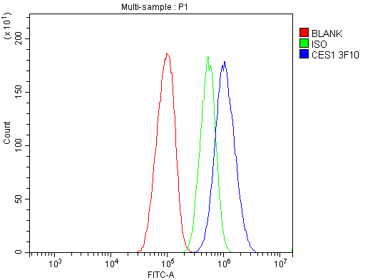


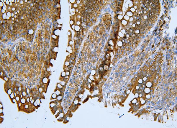
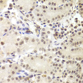
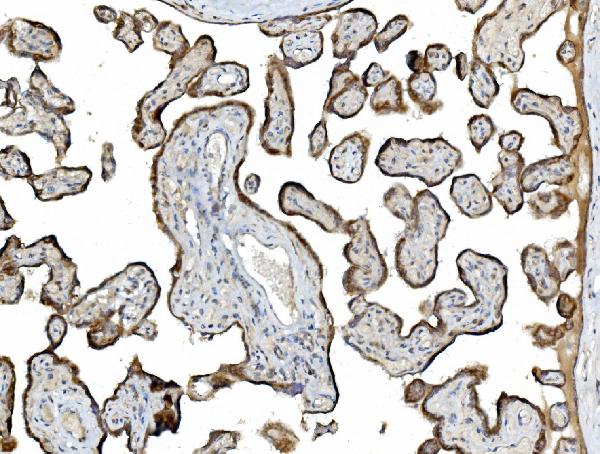
 IHC (Immunohistochemistry)
(Figure 8. IHC analysis of Liver Carboxylesterase 1/CES1 using anti-Liver Carboxylesterase 1/CES1 antibody (AAA19363).Liver Carboxylesterase 1/CES1 was detected in paraffin-embedded section of rat liver tissue. Heat mediated antigen retrieval was performed in EDTA buffer (pH8. 0, epitope retrieval solution). The tissue section was blocked with 10% goat serum. The tissue section was then incubated with 2μg/ml mouse anti-Liver Carboxylesterase 1/CES1 Antibody (AAA19363) overnight at 4 degree C. Biotinylated goat anti-mouse IgG was used as secondary antibody and incubated for 30 minutes at 37 degree C. The tissue section was developed using Strepavidin-Biotin-Complex (SABC) (Catalog # with DAB as the chromogen.)
IHC (Immunohistochemistry)
(Figure 8. IHC analysis of Liver Carboxylesterase 1/CES1 using anti-Liver Carboxylesterase 1/CES1 antibody (AAA19363).Liver Carboxylesterase 1/CES1 was detected in paraffin-embedded section of rat liver tissue. Heat mediated antigen retrieval was performed in EDTA buffer (pH8. 0, epitope retrieval solution). The tissue section was blocked with 10% goat serum. The tissue section was then incubated with 2μg/ml mouse anti-Liver Carboxylesterase 1/CES1 Antibody (AAA19363) overnight at 4 degree C. Biotinylated goat anti-mouse IgG was used as secondary antibody and incubated for 30 minutes at 37 degree C. The tissue section was developed using Strepavidin-Biotin-Complex (SABC) (Catalog # with DAB as the chromogen.)
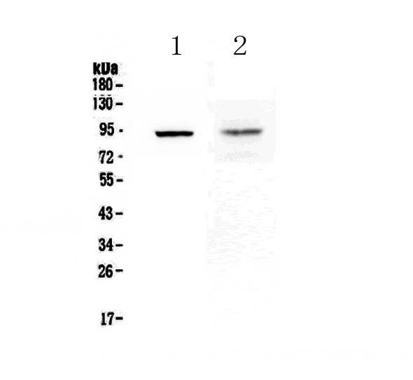
Liver Carboxylesterase 1/CES1, Monoclonal Antibody (Cat# AAA19363)
Full Name
Anti-Liver Carboxylesterase 1/CES1 Antibody (monoclonal, 3F10)
Gene Names
CES1; CEH; REH; TGH; ACAT; CE-1; CES2; HMSE; SES1; HMSE1; PCE-1; hCE-1
Reactivity
Human, Mouse, Rat
Applications
WB, IHC-P, ICC, IF, FC/FACS/FCM
Purity
Immunogen affinity purified.
Pricing


 IF (Immunofluorescence)
(Immunofluorescence analysis of NIH-3T3 cells using PKM1-specific Rabbit pAb at dilution of 1:100 (40x lens). Blue: DAPI for nuclear staining.)
IF (Immunofluorescence)
(Immunofluorescence analysis of NIH-3T3 cells using PKM1-specific Rabbit pAb at dilution of 1:100 (40x lens). Blue: DAPI for nuclear staining.)
PKM1-specific, Polyclonal Antibody (Cat# AAA28375)
Full Name
PKM1-specific Rabbit pAb
Gene Names
PKM; PK3; TCB; OIP3; PKM2; CTHBP; THBP1
Reactivity
Human, Mouse, Rat
Applications
WB, IHC, IF
Purity
Affinity purification
Pricing


 FCM (Flow Cytometry)
(Flow cytometry analysis of Hsp105alpha in Hep3B cell line, staining at 2-5ug for 1x10⁶cells (red line). The secondary antibody used goat anti-mouse IgG Alexa fluor 488 conjugate. Isotype control antibody was mouse IgG (black line).)
FCM (Flow Cytometry)
(Flow cytometry analysis of Hsp105alpha in Hep3B cell line, staining at 2-5ug for 1x10⁶cells (red line). The secondary antibody used goat anti-mouse IgG Alexa fluor 488 conjugate. Isotype control antibody was mouse IgG (black line).)
HSP105alpha, Monoclonal Antibody (Cat# AAA11727)
Full Name
HSP105alpha antibody
Gene Names
HSPH1; HSP105; HSP105A; HSP105B; NY-CO-25
Reactivity
Human
Applications
Western Blot, Flow Cytometry, Immunocytochemistry
Purity
By protein-G affinity chromatography
Pricing


 FCM (Flow Cytometry)
(Figure 8. Flow Cytometry analysis of SiHa cells using anti-Astrin/Deepest/SPAG5 antibody (AAA19313).Overlay histogram showing SiHa cells stained with AAA19313 (Blue line). The cells were blocked with 10% normal goat serum. And then incubated with rabbit anti-Astrin/Deepest/SPAG5 Antibody (AAA19313, 1μg/1x106 cells) for 30 min at 20 degree C. DyLight®488 conjugated goat anti-rabbit IgG (5-10μg/1x106 cells) was used as secondary antibody for 30 minutes at 20 degree C. Isotype control antibody (Green line) was rabbit IgG (1μg/1x106) used under the same conditions. Unlabelled sample (Red line) was also used as a control.)
FCM (Flow Cytometry)
(Figure 8. Flow Cytometry analysis of SiHa cells using anti-Astrin/Deepest/SPAG5 antibody (AAA19313).Overlay histogram showing SiHa cells stained with AAA19313 (Blue line). The cells were blocked with 10% normal goat serum. And then incubated with rabbit anti-Astrin/Deepest/SPAG5 Antibody (AAA19313, 1μg/1x106 cells) for 30 min at 20 degree C. DyLight®488 conjugated goat anti-rabbit IgG (5-10μg/1x106 cells) was used as secondary antibody for 30 minutes at 20 degree C. Isotype control antibody (Green line) was rabbit IgG (1μg/1x106) used under the same conditions. Unlabelled sample (Red line) was also used as a control.)
Astrin/Deepest/SPAG5, Polyclonal Antibody (Cat# AAA19313)
Full Name
Anti-Astrin/Deepest/SPAG5 Antibody
Gene Names
SPAG5; MAP126; DEEPEST; hMAP126
Reactivity
Human
Applications
WB, IHC-P, ICC, IF, FC/FACS/FCM, EIA
Purity
Immunogen affinity purified.
Pricing


 IHC (Immunohistochemistry)
(Immunohistochemistry of paraffin-embedded mouse brain using SRP19 antibody at dilution of 1:100 (40x lens).)
IHC (Immunohistochemistry)
(Immunohistochemistry of paraffin-embedded mouse brain using SRP19 antibody at dilution of 1:100 (40x lens).)
SRP19, Polyclonal Antibody (Cat# AAA28153)
Full Name
SRP19 Polyclonal Antibody
Reactivity
Human, Mouse, Rat
Applications
WB, IHC, IF
Purity
Affinity Purification
Pricing


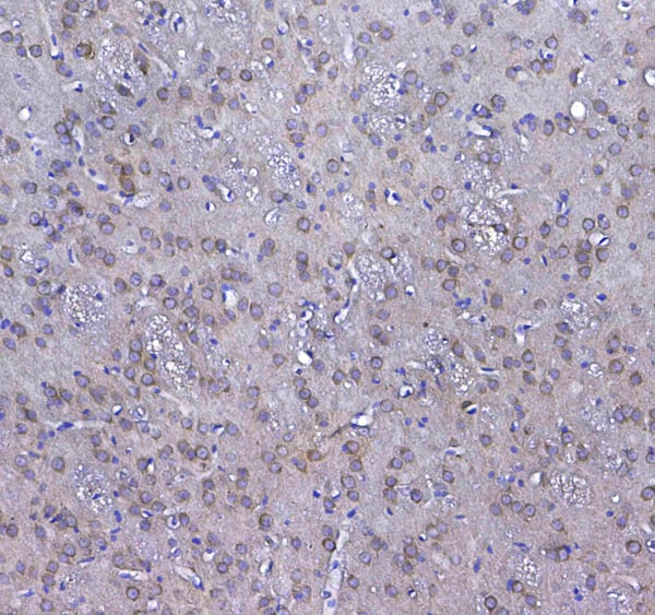
 IHC (Immunohistchemistry)
(Figure 6. IHC analysis of GLO1 using anti-GLO1 antibody (AAA19155).GLO1 was detected in paraffin-embedded section of rat spleen tissue. Heat mediated antigen retrieval was performed in citrate buffer (pH6, epitope retrieval solution) for 20 mins. The tissue section was blocked with 10% goat serum. The tissue section was then incubated with 1ug/ml rabbit anti-GLO1 Antibody (AAA19155) overnight at 4 degree C. Biotinylated goat anti-rabbit IgG was used as secondary antibody and incubated for 30 minutes at 37 degree C. The tissue section was developed using Strepavidin-Biotin-Complex (SABC) with DAB as the chromogen.)
IHC (Immunohistchemistry)
(Figure 6. IHC analysis of GLO1 using anti-GLO1 antibody (AAA19155).GLO1 was detected in paraffin-embedded section of rat spleen tissue. Heat mediated antigen retrieval was performed in citrate buffer (pH6, epitope retrieval solution) for 20 mins. The tissue section was blocked with 10% goat serum. The tissue section was then incubated with 1ug/ml rabbit anti-GLO1 Antibody (AAA19155) overnight at 4 degree C. Biotinylated goat anti-rabbit IgG was used as secondary antibody and incubated for 30 minutes at 37 degree C. The tissue section was developed using Strepavidin-Biotin-Complex (SABC) with DAB as the chromogen.)
GLO1/Glyoxalase I, Polyclonal Antibody (Cat# AAA19155)
Full Name
Anti-GLO1/Glyoxalase I Picoband antibody
Gene Names
GLO1; GLYI; GLOD1; HEL-S-74
Reactivity
Human, Mouse, Rat
No cross reactivity with other proteins.
No cross reactivity with other proteins.
Applications
EIA, IHC, WB
Pricing


 IHC (Immunohistchemistry)
(Figure 6. IHC analysis of Synaptopodin/SYNPO using anti-Synaptopodin/SYNPO antibody (AAA19271).Synaptopodin/SYNPO was detected in paraffin-embedded section of mouse brain tissue. Heat mediated antigen retrieval was performed in EDTA buffer (pH8. 0, epitope retrieval solution). The tissue section was blocked with 10% goat serum. The tissue section was then incubated with 2μg/ml rabbit anti-Synaptopodin/SYNPO Antibody (AAA19271) overnight at 4 degree C. Biotinylated goat anti-rabbit IgG was used as secondary antibody and incubated for 30 minutes at 37 degree C. The tissue section was developed using Strepavidin-Biotin-Complex (SABC) (Catalog # with DAB as the chromogen.)
IHC (Immunohistchemistry)
(Figure 6. IHC analysis of Synaptopodin/SYNPO using anti-Synaptopodin/SYNPO antibody (AAA19271).Synaptopodin/SYNPO was detected in paraffin-embedded section of mouse brain tissue. Heat mediated antigen retrieval was performed in EDTA buffer (pH8. 0, epitope retrieval solution). The tissue section was blocked with 10% goat serum. The tissue section was then incubated with 2μg/ml rabbit anti-Synaptopodin/SYNPO Antibody (AAA19271) overnight at 4 degree C. Biotinylated goat anti-rabbit IgG was used as secondary antibody and incubated for 30 minutes at 37 degree C. The tissue section was developed using Strepavidin-Biotin-Complex (SABC) (Catalog # with DAB as the chromogen.)
Synaptopodin/SYNPO, Polyclonal Antibody (Cat# AAA19271)
Full Name
Anti-Synaptopodin/SYNPO Antibody
Reactivity
Human, Mouse, Rat
Applications
WB, IHC-P, ICC, IF, FC/FACS/FCM
Purity
Immunogen affinity purified.
Pricing


 FCM (Flow Cytometry)
(Figure 7. Flow Cytometry analysis of THP-1 cells using anti-PNPT1 antibody (AAA19306).Overlay histogram showing THP-1 cells stained with AAA19306 (Blue line). The cells were blocked with 10% normal goat serum. And then incubated with rabbit anti-PNPT1 Antibody (AAA19306, 1μg/1x106 cells) for 30 min at 20 degree C. DyLight®488 conjugated goat anti-rabbit IgG (5-10μg/1x106 cells) was used as secondary antibody for 30 minutes at 20 degree C. Isotype control antibody (Green line) was rabbit IgG (1μg/1x106) used under the same conditions. Unlabelled sample (Red line) was also used as a control.)
FCM (Flow Cytometry)
(Figure 7. Flow Cytometry analysis of THP-1 cells using anti-PNPT1 antibody (AAA19306).Overlay histogram showing THP-1 cells stained with AAA19306 (Blue line). The cells were blocked with 10% normal goat serum. And then incubated with rabbit anti-PNPT1 Antibody (AAA19306, 1μg/1x106 cells) for 30 min at 20 degree C. DyLight®488 conjugated goat anti-rabbit IgG (5-10μg/1x106 cells) was used as secondary antibody for 30 minutes at 20 degree C. Isotype control antibody (Green line) was rabbit IgG (1μg/1x106) used under the same conditions. Unlabelled sample (Red line) was also used as a control.)
PNPT1, Polyclonal Antibody (Cat# AAA19306)
Full Name
Anti-PNPT1 Antibody
Gene Names
PNPT1; OLD35; DFNB70; PNPASE; old-35; COXPD13
Reactivity
Human, Mouse, Rat
Applications
WB, IHC-P, ICC, IF, FC/FACS/FCM, EIA
Purity
Immunogen affinity purified.
Pricing
 IHC (Immunohistchemistry)
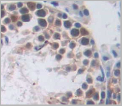
(DAB staining on IHC-P.Samples: Human Tissue)
IHC (Immunohistchemistry)
(DAB staining on IHC-P.Samples: Human Tissue)
RAD51 Homolog, Polyclonal Antibody (Cat# AAA21011)
Full Name
RAD51 Homolog (RAD51) Polyclonal Antibody
Gene Names
RAD51; RECA; BRCC5; FANCR; MRMV2; HRAD51; RAD51A; HsRad51; HsT16930
Reactivity
Human, Mouse, Rat
Applications
Westen Blot, Immunohistochemistry, Immunocytochemistry, Immunoprecipitation
Purity
Antigen-specific affinity chromatography followed by Protein A affinity chromatography
Pricing


 IP (Immunoprecipitation)
(Immunoprecipitation analysis of 200ug extracts of HeLa cells using 3ug NF-kB p65/RelA antibody . Western blot was performed from the immunoprecipitate using NF-kB p65/RelA antibody at a dilition of 1:1000.)
IP (Immunoprecipitation)
(Immunoprecipitation analysis of 200ug extracts of HeLa cells using 3ug NF-kB p65/RelA antibody . Western blot was performed from the immunoprecipitate using NF-kB p65/RelA antibody at a dilition of 1:1000.)
NF-kB p65/RelA, Monoclonal Antibody (Cat# AAA28381)
Full Name
[KO Validated] NF-kB p65/RelA Rabbit mAb
Gene Names
RELA; p65; NFKB3
Reactivity
Human, Mouse, Rat
Applications
WB, IHC, IP
Purity
Affinity purification
Pricing


 IHC (Immunohistochemistry)
(Figure 8. IHC analysis of GSTM3 using anti-GSTM3 antibody (AAA19168).GSTM3 was detected in paraffin-embedded section of rat testis tissue. Heat mediated antigen retrieval was performed in citrate buffer (pH6, epitope retrieval solution) for 20 mins. The tissue section was blocked with 10% goat serum. The tissue section was then incubated with 1ug/ml rabbit anti-GSTM3 Antibody (AAA19168) overnight at 4 degree C. Biotinylated goat anti-rabbit IgG was used as secondary antibody and incubated for 30 minutes at 37 degree C. The tissue section was developed using Strepavidin-Biotin-Complex (SABC) with DAB as the chromogen.)
IHC (Immunohistochemistry)
(Figure 8. IHC analysis of GSTM3 using anti-GSTM3 antibody (AAA19168).GSTM3 was detected in paraffin-embedded section of rat testis tissue. Heat mediated antigen retrieval was performed in citrate buffer (pH6, epitope retrieval solution) for 20 mins. The tissue section was blocked with 10% goat serum. The tissue section was then incubated with 1ug/ml rabbit anti-GSTM3 Antibody (AAA19168) overnight at 4 degree C. Biotinylated goat anti-rabbit IgG was used as secondary antibody and incubated for 30 minutes at 37 degree C. The tissue section was developed using Strepavidin-Biotin-Complex (SABC) with DAB as the chromogen.)
GSTM3, Polyclonal Antibody (Cat# AAA19168)
Full Name
Anti-GSTM3 Picoband antibody
Gene Names
GSTM3; GST5; GSTB; GTM3; GSTM3-3
Reactivity
Human, Mouse, Rat
No cross reactivity with other proteins.
No cross reactivity with other proteins.
Applications
EIA, IHC, WB
Pricing


 IHC (Immunohistchemistry)
(Immunohistochemistry of paraffin-embedded human breast using PRPF3 Antibody at dilution of 1:100 (40x lens).)
IHC (Immunohistchemistry)
(Immunohistochemistry of paraffin-embedded human breast using PRPF3 Antibody at dilution of 1:100 (40x lens).)
PRPF3, Polyclonal Antibody (Cat# AAA28150)
Full Name
PRPF3 Polyclonal Antibody
Gene Names
PRPF3; PRP3; RP18; HPRP3; Prp3p; HPRP3P; SNRNP90
Reactivity
Human, Mouse, Rat
Applications
WB, IHC, IF
Purity
Affinity Purification
Pricing


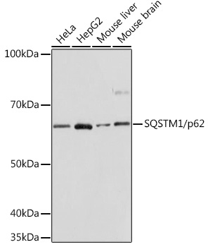

 IP (Immunoprecipitation)
(Immunoprecipitation analysis of 300ug extracts of HeLa cells using 3ug SQSTM1/p62 antibody . Western blot was performed from the immunoprecipitate using SQSTM1/p62 antibody at a dilition of 1:1000.)
IP (Immunoprecipitation)
(Immunoprecipitation analysis of 300ug extracts of HeLa cells using 3ug SQSTM1/p62 antibody . Western blot was performed from the immunoprecipitate using SQSTM1/p62 antibody at a dilition of 1:1000.)
SQSTM1/p62, Monoclonal Antibody (Cat# AAA28383)
Full Name
[KO Validated] SQSTM1/p62 Rabbit mAb
Gene Names
SQSTM1; p60; p62; A170; OSIL; PDB3; ZIP3; p62B
Reactivity
Human, Mouse, Rat
Applications
WB, IHC, IF, IP
Purity
Affinity purification
Pricing


 FCM (Flow Cytometry)
(Figure 9. Flow Cytometry analysis of U937 cells using anti-GNG7 antibody (AAA19335).Overlay histogram showing U937 cells stained with AAA19335 (Blue line). The cells were blocked with 10% normal goat serum. And then incubated with rabbit anti-GNG7 Antibody (AAA19335, 1μg/1x106 cells) for 30 min at 20 degree C. DyLight®488 conjugated goat anti-rabbit IgG (5-10μg/1x106 cells) was used as secondary antibody for 30 minutes at 20 degree C. Isotype control antibody (Green line) was rabbit IgG (1μg/1x106) used under the same conditions. Unlabelled sample (Red line) was also used as a control.)
FCM (Flow Cytometry)
(Figure 9. Flow Cytometry analysis of U937 cells using anti-GNG7 antibody (AAA19335).Overlay histogram showing U937 cells stained with AAA19335 (Blue line). The cells were blocked with 10% normal goat serum. And then incubated with rabbit anti-GNG7 Antibody (AAA19335, 1μg/1x106 cells) for 30 min at 20 degree C. DyLight®488 conjugated goat anti-rabbit IgG (5-10μg/1x106 cells) was used as secondary antibody for 30 minutes at 20 degree C. Isotype control antibody (Green line) was rabbit IgG (1μg/1x106) used under the same conditions. Unlabelled sample (Red line) was also used as a control.)
GNG7, Polyclonal Antibody (Cat# AAA19335)
Full Name
Anti-GNG7 Antibody
Reactivity
Human, Mouse, Rat
Applications
WB, IHC-P, FC/FACS/FCM, EIA
Purity
Immunogen affinity purified.
Pricing


 FCM (Flow Cytometry)
(Figure 7. Flow Cytometry analysis of CACO-2 cells using anti-Carbonic Anhydrase 13/CA13 antibody (AAA19326).Overlay histogram showing CACO-2 cells stained with AAA19326 (Blue line). The cells were blocked with 10% normal goat serum. And then incubated with rabbit anti-Carbonic Anhydrase 13/CA13 Antibody (AAA19326, 1μg/1x106 cells) for 30 min at 20 degree C. DyLight®488 conjugated goat anti-rabbit IgG (5-10μg/1x106 cells) was used as secondary antibody for 30 minutes at 20 degree C. Isotype control antibody (Green line) was rabbit IgG (1μg/1x106) used under the same conditions. Unlabelled sample (Red line) was also used as a control.)
FCM (Flow Cytometry)
(Figure 7. Flow Cytometry analysis of CACO-2 cells using anti-Carbonic Anhydrase 13/CA13 antibody (AAA19326).Overlay histogram showing CACO-2 cells stained with AAA19326 (Blue line). The cells were blocked with 10% normal goat serum. And then incubated with rabbit anti-Carbonic Anhydrase 13/CA13 Antibody (AAA19326, 1μg/1x106 cells) for 30 min at 20 degree C. DyLight®488 conjugated goat anti-rabbit IgG (5-10μg/1x106 cells) was used as secondary antibody for 30 minutes at 20 degree C. Isotype control antibody (Green line) was rabbit IgG (1μg/1x106) used under the same conditions. Unlabelled sample (Red line) was also used as a control.)
Carbonic Anhydrase 13/CA13, Polyclonal Antibody (Cat# AAA19326)
Full Name
Anti-Carbonic Anhydrase 13/CA13 Antibody
Gene Names
CA13; CAXIII
Reactivity
Human, Mouse, Rat
Applications
WB, IHC-P, ICC, IF, FC/FACS/FCM
Purity
Immunogen affinity purified.
Pricing


 FCM (Flow Cytometry)
(Figure 8. Flow Cytometry analysis of SiHa cells using anti- ASS1 antibody (AAA19370).Overlay histogram showing SiHa cells stained with AAA19370 (Blue line). The cells were blocked with 10% normal goat serum. And then incubated with mouse anti-ASS1 Antibody (AAA19370, 1μg/1x106 cells) for 30 min at 20 degree C. DyLight®488 conjugated goat anti-mouse IgG (BA1126, 5-10μg/1x106 cells) was used as secondary antibody for 30 minutes at 20 degree C. Isotype control antibody (Green line) was mouse IgG (1μg/1x106) used under the same conditions. Unlabelled sample (Red line) was also used as a control.)
FCM (Flow Cytometry)
(Figure 8. Flow Cytometry analysis of SiHa cells using anti- ASS1 antibody (AAA19370).Overlay histogram showing SiHa cells stained with AAA19370 (Blue line). The cells were blocked with 10% normal goat serum. And then incubated with mouse anti-ASS1 Antibody (AAA19370, 1μg/1x106 cells) for 30 min at 20 degree C. DyLight®488 conjugated goat anti-mouse IgG (BA1126, 5-10μg/1x106 cells) was used as secondary antibody for 30 minutes at 20 degree C. Isotype control antibody (Green line) was mouse IgG (1μg/1x106) used under the same conditions. Unlabelled sample (Red line) was also used as a control.)
ASS1, Monoclonal Antibody (Cat# AAA19370)
Full Name
Anti-ASS1 Antibody (monoclonal, 7I9)
Gene Names
ASS1; ASS; CTLN1
Reactivity
Human, Mouse, Rat, Monkey
Applications
WB, IHC-P, ICC, IF, FC/FACS/FCM
Purity
Immunogen affinity purified.
Pricing


 FCM (Flow Cytometry)
(Figure 6. Flow Cytometry analysis of MCF-7 cells using anti-DDX1 antibody (AAA19379).Overlay histogram showing MCF-7 cells stained with AAA19379 (Blue line). The cells were blocked with 10% normal goat serum. And then incubated with mouse anti- DDX1 Antibody (AAA19379, 1μg/1x106 cells) for 30 min at 20 degree C. DyLight®488 conjugated goat anti-mouse IgG (BA1126, 5-10μg/1x106 cells) was used as secondary antibody for 30 minutes at 20 degree C. Isotype control antibody (Green line) was mouse IgG (1μg/1x106) used under the same conditions. Unlabelled sample (Red line) was also used as a control.)
FCM (Flow Cytometry)
(Figure 6. Flow Cytometry analysis of MCF-7 cells using anti-DDX1 antibody (AAA19379).Overlay histogram showing MCF-7 cells stained with AAA19379 (Blue line). The cells were blocked with 10% normal goat serum. And then incubated with mouse anti- DDX1 Antibody (AAA19379, 1μg/1x106 cells) for 30 min at 20 degree C. DyLight®488 conjugated goat anti-mouse IgG (BA1126, 5-10μg/1x106 cells) was used as secondary antibody for 30 minutes at 20 degree C. Isotype control antibody (Green line) was mouse IgG (1μg/1x106) used under the same conditions. Unlabelled sample (Red line) was also used as a control.)
DDX1, Monoclonal Antibody (Cat# AAA19379)
Full Name
Anti-DDX1 Antibody (monoclonal, 2D4)
Gene Names
DDX1; DBP-RB; UKVH5d
Reactivity
Human, Mouse, Rat
Applications
WB, IHC-P, FC/FACS/FCM
Purity
Immunogen affinity purified.
Pricing


 FCM (Flow Cytometry)
(Figure 12. Flow Cytometry analysis of U251 cells using anti-HP1 alpha/CBX5 antibody (AAA19373).Overlay histogram showing U251 cells stained with AAA19373 (Blue line). The cells were blocked with 10% normal goat serum. And then incubated with mouse anti- HP1 alpha/CBX5 Antibody (AAA19373, 1μg/1x106 cells) for 30 min at 20 degree C. DyLight®488 conjugated goat anti-mouse IgG (BA1126, 5-10μg/1x106 cells) was used as secondary antibody for 30 minutes at 20 degree C. Isotype control antibody (Green line) was mouse IgG (1μg/1x106) used under the same conditions. Unlabelled sample (Red line) was also used as a control.)
FCM (Flow Cytometry)
(Figure 12. Flow Cytometry analysis of U251 cells using anti-HP1 alpha/CBX5 antibody (AAA19373).Overlay histogram showing U251 cells stained with AAA19373 (Blue line). The cells were blocked with 10% normal goat serum. And then incubated with mouse anti- HP1 alpha/CBX5 Antibody (AAA19373, 1μg/1x106 cells) for 30 min at 20 degree C. DyLight®488 conjugated goat anti-mouse IgG (BA1126, 5-10μg/1x106 cells) was used as secondary antibody for 30 minutes at 20 degree C. Isotype control antibody (Green line) was mouse IgG (1μg/1x106) used under the same conditions. Unlabelled sample (Red line) was also used as a control.)
HP1 alpha/CBX5, Monoclonal Antibody (Cat# AAA19373)
Full Name
Anti-HP1 alpha/CBX5 Antibody (monoclonal, 8G6)
Gene Names
CBX5; HP1; HP1A
Reactivity
Human, Mouse, Rat
Applications
WB, IHC-P, ICC, IF, FC/FACS/FCM
Purity
Immunogen affinity purified.
Pricing

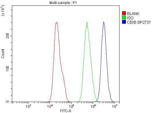


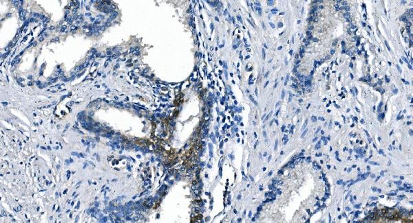
 IHC (Immunohistochemistry)
(Figure 8. IHC analysis of Cbx8 using anti-Cbx8 antibody (AAA19304).Cbx8 was detected in paraffin-embedded section of rat small intestine tissue. Heat mediated antigen retrieval was performed in EDTA buffer (pH8. 0, epitope retrieval solution). The tissue section was blocked with 10% goat serum. The tissue section was then incubated with 1μg/ml rabbit anti-Cbx8 Antibody (AAA19304) overnight at 4 degree C. Biotinylated goat anti-rabbit IgG was used as secondary antibody and incubated for 30 minutes at 37 degree C. The tissue section was developed using Strepavidin-Biotin-Complex (SABC) (Catalog # with DAB as the chromogen.)
IHC (Immunohistochemistry)
(Figure 8. IHC analysis of Cbx8 using anti-Cbx8 antibody (AAA19304).Cbx8 was detected in paraffin-embedded section of rat small intestine tissue. Heat mediated antigen retrieval was performed in EDTA buffer (pH8. 0, epitope retrieval solution). The tissue section was blocked with 10% goat serum. The tissue section was then incubated with 1μg/ml rabbit anti-Cbx8 Antibody (AAA19304) overnight at 4 degree C. Biotinylated goat anti-rabbit IgG was used as secondary antibody and incubated for 30 minutes at 37 degree C. The tissue section was developed using Strepavidin-Biotin-Complex (SABC) (Catalog # with DAB as the chromogen.)
Cbx8, Polyclonal Antibody (Cat# AAA19304)
Full Name
Anti-Cbx8 Antibody
Gene Names
CBX8; PC3; RC1
Reactivity
Human, Mouse, Rat
Applications
WB, IHC-P, ICC, IF, FC/FACS/FCM, EIA
Purity
Immunogen affinity purified.
Pricing


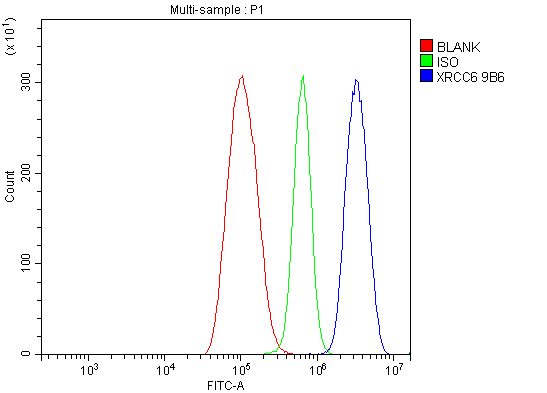 FCM (Flow Cytometry)
(Figure 7. Flow Cytometry analysis of THP-1 cells using anti-Ku70 antibody (AAA19361).Overlay histogram showing THP-1 cells stained with AAA19361 (Blue line). The cells were blocked with 10% normal goat serum. And then incubated with mouse anti- Ku70 Antibody (AAA19361, 1μg/1x106 cells) for 30 min at 20 degree C. DyLight®488 conjugated goat anti-mouse IgG (BA1126, 5-10μg/1x106 cells) was used as secondary antibody for 30 minutes at 20 degree C. Isotype control antibody (Green line) was mouse IgG (1μg/1x106) used under the same conditions. Unlabelled sample (Red line) was also used as a control.)
FCM (Flow Cytometry)
(Figure 7. Flow Cytometry analysis of THP-1 cells using anti-Ku70 antibody (AAA19361).Overlay histogram showing THP-1 cells stained with AAA19361 (Blue line). The cells were blocked with 10% normal goat serum. And then incubated with mouse anti- Ku70 Antibody (AAA19361, 1μg/1x106 cells) for 30 min at 20 degree C. DyLight®488 conjugated goat anti-mouse IgG (BA1126, 5-10μg/1x106 cells) was used as secondary antibody for 30 minutes at 20 degree C. Isotype control antibody (Green line) was mouse IgG (1μg/1x106) used under the same conditions. Unlabelled sample (Red line) was also used as a control.)
Ku70, Monoclonal Antibody (Cat# AAA19361)
Full Name
Anti-Ku70 Antibody (monoclonal, 9B6)
Gene Names
XRCC6; ML8; KU70; TLAA; CTC75; CTCBF; G22P1
Reactivity
Human
Applications
WB, IHC-P, ICC, IF, FC/FACS/FCM
Purity
Immunogen affinity purified.
Pricing


 IHC (Immunohistochemistry)
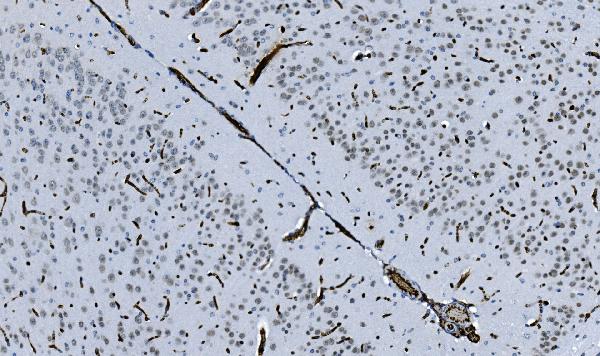
(Immunohistochemistry of paraffin-embedded mouse lung using ATP1B1 antibody at dilution of 1:100 (40x lens).)
IHC (Immunohistochemistry)
(Immunohistochemistry of paraffin-embedded mouse lung using ATP1B1 antibody at dilution of 1:100 (40x lens).)
ATP1B1, Polyclonal Antibody (Cat# AAA28138)
Full Name
ATP1B1 Polyclonal Antibody
Gene Names
ATP1B1; ATP1B
Reactivity
Human, Mouse, Rat
Applications
WB, IHC
Purity
Affinity Purification
Pricing


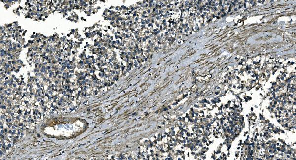
 IHC (Immunohistchemistry)
(Figure 6. IHC analysis of CD13/ANPEP using anti-CD13/ANPEP antibody (AAA19371).CD13/ANPEP was detected in paraffin-embedded section of rat kidney tissue. Heat mediated antigen retrieval was performed in EDTA buffer (pH8. 0, epitope retrieval solution). The tissue section was blocked with 10% goat serum. The tissue section was then incubated with 2μg/ml mouse anti-CD13/ANPEP Antibody (AAA19371) overnight at 4 degree C. Biotinylated goat anti-mouse IgG was used as secondary antibody and incubated for 30 minutes at 37 degree C. The tissue section was developed using Strepavidin-Biotin-Complex (SABC) (Catalog # with DAB as the chromogen.)
IHC (Immunohistchemistry)
(Figure 6. IHC analysis of CD13/ANPEP using anti-CD13/ANPEP antibody (AAA19371).CD13/ANPEP was detected in paraffin-embedded section of rat kidney tissue. Heat mediated antigen retrieval was performed in EDTA buffer (pH8. 0, epitope retrieval solution). The tissue section was blocked with 10% goat serum. The tissue section was then incubated with 2μg/ml mouse anti-CD13/ANPEP Antibody (AAA19371) overnight at 4 degree C. Biotinylated goat anti-mouse IgG was used as secondary antibody and incubated for 30 minutes at 37 degree C. The tissue section was developed using Strepavidin-Biotin-Complex (SABC) (Catalog # with DAB as the chromogen.)
CD13/ANPEP, Monoclonal Antibody (Cat# AAA19371)
Full Name
Anti-CD13/ANPEP Antibody (monoclonal, 5B9)
Gene Names
ANPEP; APN; CD13; LAP1; P150; PEPN; GP150
Reactivity
Human, Rat, Monkey
Applications
WB, IHC-P
Purity
Immunogen affinity purified.
Pricing


 IHC (Immunohistochemistry)
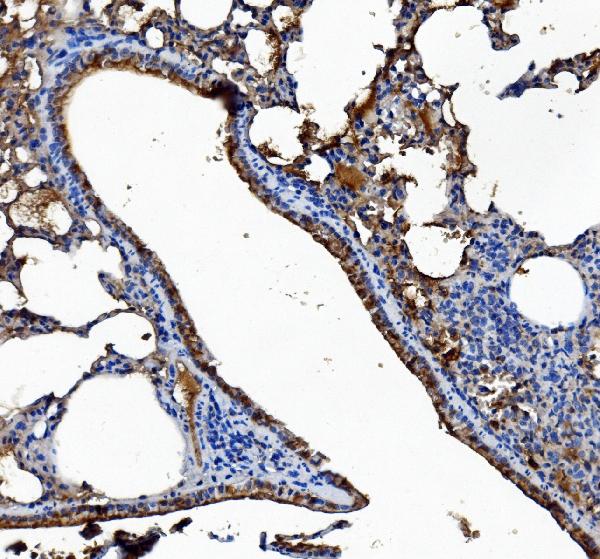
(Figure 7. IHC analysis of MYLK using anti-MYLK antibody (AAA19154).MYLK was detected in paraffin-embedded section of rat lung tissue. Heat mediated antigen retrieval was performed in citrate buffer (pH6, epitope retrieval solution) for 20 mins. The tissue section was blocked with 10% goat serum. The tissue section was then incubated with 1ug/ml rabbit anti-MYLK Antibody (AAA19154) overnight at 4 degree C. Biotinylated goat anti-rabbit IgG was used as secondary antibody and incubated for 30 minutes at 37 degree C. The tissue section was developed using Strepavidin-Biotin-Complex (SABC) with DAB as the chromogen. )
IHC (Immunohistochemistry)
(Figure 7. IHC analysis of MYLK using anti-MYLK antibody (AAA19154).MYLK was detected in paraffin-embedded section of rat lung tissue. Heat mediated antigen retrieval was performed in citrate buffer (pH6, epitope retrieval solution) for 20 mins. The tissue section was blocked with 10% goat serum. The tissue section was then incubated with 1ug/ml rabbit anti-MYLK Antibody (AAA19154) overnight at 4 degree C. Biotinylated goat anti-rabbit IgG was used as secondary antibody and incubated for 30 minutes at 37 degree C. The tissue section was developed using Strepavidin-Biotin-Complex (SABC) with DAB as the chromogen. )
MYLK, Polyclonal Antibody (Cat# AAA19154)
Full Name
Anti-MYLK Picoband antibody
Gene Names
MYLK; KRP; AAT7; MLCK; MLCK1; MYLK1; smMLCK; MLCK108; MLCK210; MSTP083
Reactivity
Human, Mouse, Rat
No cross reactivity with other proteins.
No cross reactivity with other proteins.
Applications
EIA, IHC, WB
Pricing


 FCM (Flow Cytometry)
(Figure 10. Flow Cytometry analysis of U20S cells using anti-PDIA5 antibody (AAA19331).Overlay histogram showing U20S cells stained with AAA19331 (Blue line). The cells were blocked with 10% normal goat serum. And then incubated with rabbit anti-PDIA5 Antibody (AAA19331, 1μg/1x106 cells) for 30 min at 20 degree C. DyLight®488 conjugated goat anti-rabbit IgG (5-10μg/1x106 cells) was used as secondary antibody for 30 minutes at 20 degree C. Isotype control antibody (Green line) was rabbit IgG (1μg/1x106) used under the same conditions. Unlabelled sample (Red line) was also used as a control.)
FCM (Flow Cytometry)
(Figure 10. Flow Cytometry analysis of U20S cells using anti-PDIA5 antibody (AAA19331).Overlay histogram showing U20S cells stained with AAA19331 (Blue line). The cells were blocked with 10% normal goat serum. And then incubated with rabbit anti-PDIA5 Antibody (AAA19331, 1μg/1x106 cells) for 30 min at 20 degree C. DyLight®488 conjugated goat anti-rabbit IgG (5-10μg/1x106 cells) was used as secondary antibody for 30 minutes at 20 degree C. Isotype control antibody (Green line) was rabbit IgG (1μg/1x106) used under the same conditions. Unlabelled sample (Red line) was also used as a control.)
PDIA5, Polyclonal Antibody (Cat# AAA19331)
Full Name
Anti-PDIA5 Antibody
Gene Names
PDIA5; PDIR
Reactivity
Human, Mouse, Rat
Applications
WB, IHC-P, ICC, IF, FC/FACS/FCM, EIA
Purity
Immunogen affinity purified.
Pricing


 FCM (Flow Cytometry)
(Figure 9. Flow Cytometry analysis of A549 cells using anti-JAB1 antibody (AAA19365).Overlay histogram showing A549 cells stained with AAA19365 (Blue line). The cells were blocked with 10% normal goat serum. And then incubated with mouse anti-JAB1 Antibody (AAA19365, 1μg/1x106 cells) for 30 min at 20 degree C. DyLight®488 conjugated goat anti-mouse IgG (BA1126, 5-10μg/1x106 cells) was used as secondary antibody for 30 minutes at 20 degree C. Isotype control antibody (Green line) was mouse IgG (1μg/1x106) used under the same conditions. Unlabelled sample (Red line) was also used as a control.)
FCM (Flow Cytometry)
(Figure 9. Flow Cytometry analysis of A549 cells using anti-JAB1 antibody (AAA19365).Overlay histogram showing A549 cells stained with AAA19365 (Blue line). The cells were blocked with 10% normal goat serum. And then incubated with mouse anti-JAB1 Antibody (AAA19365, 1μg/1x106 cells) for 30 min at 20 degree C. DyLight®488 conjugated goat anti-mouse IgG (BA1126, 5-10μg/1x106 cells) was used as secondary antibody for 30 minutes at 20 degree C. Isotype control antibody (Green line) was mouse IgG (1μg/1x106) used under the same conditions. Unlabelled sample (Red line) was also used as a control.)
JAB1, Monoclonal Antibody (Cat# AAA19365)
Full Name
Anti-JAB1 Antibody (monoclonal, 4G9)
Gene Names
COPS5; CSN5; JAB1; SGN5; MOV-34
Reactivity
Human, Mouse, Rat
Applications
WB, IHC-P, ICC, IF, FC/FACS/FCM
Purity
Immunogen affinity purified.
Pricing


 FCM (Flow Cytometry)
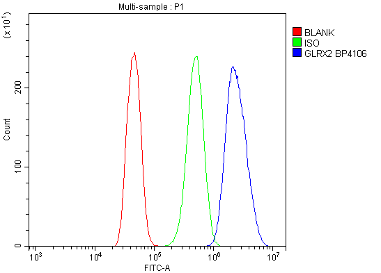
(Figure 11. Flow Cytometry analysis of CACO-2 cells using anti-GRX2/GLRX2 antibody (AAA19305).Overlay histogram showing CACO-2 cells stained with AAA19305 (Blue line). The cells were blocked with 10% normal goat serum. And then incubated with rabbit anti-GRX2/GLRX2 Antibody (AAA19305, 1μg/1x106 cells) for 30 min at 20 degree C. DyLight®488 conjugated goat anti-rabbit IgG (5-10μg/1x106 cells) was used as secondary antibody for 30 minutes at 20 degree C. Isotype control antibody (Green line) was rabbit IgG (1μg/1x106) used under the same conditions. Unlabelled sample (Red line) was also used as a control.)
FCM (Flow Cytometry)
(Figure 11. Flow Cytometry analysis of CACO-2 cells using anti-GRX2/GLRX2 antibody (AAA19305).Overlay histogram showing CACO-2 cells stained with AAA19305 (Blue line). The cells were blocked with 10% normal goat serum. And then incubated with rabbit anti-GRX2/GLRX2 Antibody (AAA19305, 1μg/1x106 cells) for 30 min at 20 degree C. DyLight®488 conjugated goat anti-rabbit IgG (5-10μg/1x106 cells) was used as secondary antibody for 30 minutes at 20 degree C. Isotype control antibody (Green line) was rabbit IgG (1μg/1x106) used under the same conditions. Unlabelled sample (Red line) was also used as a control.)
GRX2/GLRX2, Polyclonal Antibody (Cat# AAA19305)
Full Name
Anti-GRX2/GLRX2 Antibody
Gene Names
GLRX2; GRX2; CGI-133
Reactivity
Human, Mouse
Applications
WB, IHC-P, ICC, IF, FC/FACS/FCM, EIA
Purity
Immunogen affinity purified.
Pricing


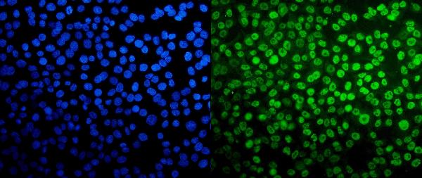
 IF (Immunofluorescence)
(Immunofluorescence analysis of NIH-3T3 cells using Phospho-Histone H2AX-S139 antibody . Blue: DAPI for nuclear staining.)
IF (Immunofluorescence)
(Immunofluorescence analysis of NIH-3T3 cells using Phospho-Histone H2AX-S139 antibody . Blue: DAPI for nuclear staining.)
Phospho-Histone H2AX-S139, Monoclonal Antibody (Cat# AAA28390)
Full Name
Phospho-Histone H2AX-S139 Rabbit mAb
Gene Names
H2AFX; H2AX; H2A.X; H2A/X
Reactivity
Human, Mouse, Rat
Applications
WB, IHC, IF
Purity
Affinity purification
Pricing


 IF (Immunofluorescence)
(Immunofluorescence analysis of U2OS cells using CUL4B antibody.)
IF (Immunofluorescence)
(Immunofluorescence analysis of U2OS cells using CUL4B antibody.)
CUL4B, Polyclonal Antibody (Cat# AAA28142)
Full Name
CUL4B Polyclonal Antibody
Gene Names
CUL4B; SFM2; MRXSC; CUL-4B; MRXHF2; MRXS15
Reactivity
Human, Mouse, Rat
Applications
WB, IHC, IF
Purity
Affinity Purification
Pricing


 FCM (Flow Cytometry)
(Figure 6. Flow Cytometry analysis of CACO-2 cells using anti-Galectin 2/LGALS2 antibody (AAA19287).Overlay histogram showing CACO-2 cells stained with AAA19287 (Blue line). The cells were blocked with 10% normal goat serum. And then incubated with rabbit anti-Galectin 2/LGALS2 Antibody (AAA19287, 1μg/1x106 cells) for 30 min at 20 degree C. DyLight®488 conjugated goat anti-rabbit IgG (5-10μg/1x106 cells) was used as secondary antibody for 30 minutes at 20 degree C. Isotype control antibody (Green line) was rabbit IgG (1μg/1x106) used under the same conditions. Unlabelled sample (Red line) was also used as a control.)
FCM (Flow Cytometry)
(Figure 6. Flow Cytometry analysis of CACO-2 cells using anti-Galectin 2/LGALS2 antibody (AAA19287).Overlay histogram showing CACO-2 cells stained with AAA19287 (Blue line). The cells were blocked with 10% normal goat serum. And then incubated with rabbit anti-Galectin 2/LGALS2 Antibody (AAA19287, 1μg/1x106 cells) for 30 min at 20 degree C. DyLight®488 conjugated goat anti-rabbit IgG (5-10μg/1x106 cells) was used as secondary antibody for 30 minutes at 20 degree C. Isotype control antibody (Green line) was rabbit IgG (1μg/1x106) used under the same conditions. Unlabelled sample (Red line) was also used as a control.)
Galectin 2/LGALS2, Polyclonal Antibody (Cat# AAA19287)
Full Name
Anti-Galectin 2/LGALS2 Antibody
Gene Names
LGALS2; HL14
Reactivity
Human
Applications
WB, IHC-P, ICC, IF, FC/FACS/FCM, EIA
Purity
Immunogen affinity purified.
Pricing


 FCM (Flow Cytometry)
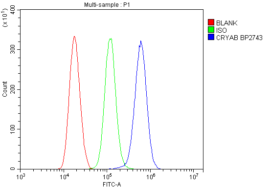
(Figure 13. Flow Cytometry analysis of THP-1 cells using anti-Alpha B Crystallin/CRYAB antibody (AAA19277).Overlay histogram showing THP-1 cells stained with AAA19277 (Blue line). The cells were blocked with 10% normal goat serum. And then incubated with rabbit anti-Alpha B Crystallin/CRYAB Antibody (AAA19277, 1μg/1x106 cells) for 30 min at 20 degree C. DyLight®488 conjugated goat anti-rabbit IgG (5-10μg/1x106 cells) was used as secondary antibody for 30 minutes at 20 degree C. Isotype control antibody (Green line) was rabbit IgG (1μg/1x106) used under the same conditions. Unlabelled sample (Red line) was also used as a control.)
FCM (Flow Cytometry)
(Figure 13. Flow Cytometry analysis of THP-1 cells using anti-Alpha B Crystallin/CRYAB antibody (AAA19277).Overlay histogram showing THP-1 cells stained with AAA19277 (Blue line). The cells were blocked with 10% normal goat serum. And then incubated with rabbit anti-Alpha B Crystallin/CRYAB Antibody (AAA19277, 1μg/1x106 cells) for 30 min at 20 degree C. DyLight®488 conjugated goat anti-rabbit IgG (5-10μg/1x106 cells) was used as secondary antibody for 30 minutes at 20 degree C. Isotype control antibody (Green line) was rabbit IgG (1μg/1x106) used under the same conditions. Unlabelled sample (Red line) was also used as a control.)
Alpha B Crystallin/CRYAB, Polyclonal Antibody (Cat# AAA19277)
Full Name
Anti-Alpha B Crystallin/CRYAB Antibody
Gene Names
CRYAB; CRYA2; CTPP2; HSPB5; CMD1II
Reactivity
Human, Mouse, Rat, Monkey
Applications
WB, IHC-P, ICC, IF, FC/FACS/FCM, EIA
Purity
Immunogen affinity purified.
Pricing