Filters
Clonality
Type
Reactivity
Gene Name
Isotype
Host
Application
Clone
537 results for " Normal" - showing 400-450
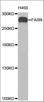
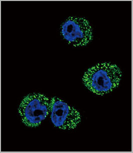
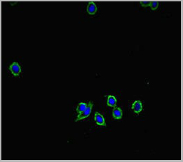
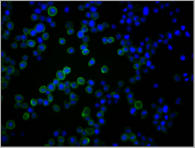
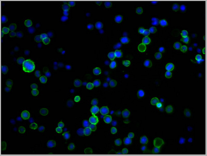
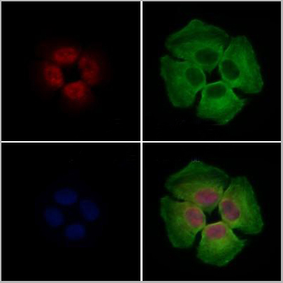
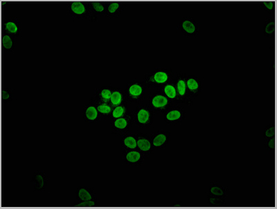
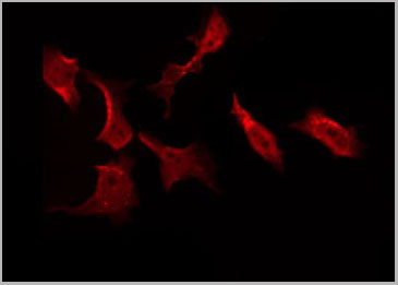
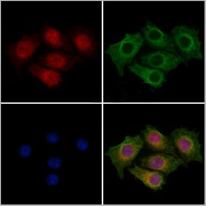
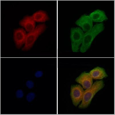
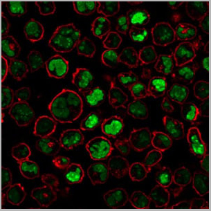
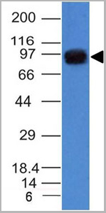
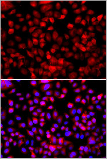 IF (Immunofluorescence)
(Immunofluorescence analysis of A549 cellsusing Fatty Acid Synthase antibody(AAA10626). Blue: DAPI for nuclear staining)
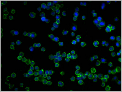
IF (Immunofluorescence)
(Immunofluorescence analysis of A549 cellsusing Fatty Acid Synthase antibody(AAA10626). Blue: DAPI for nuclear staining)
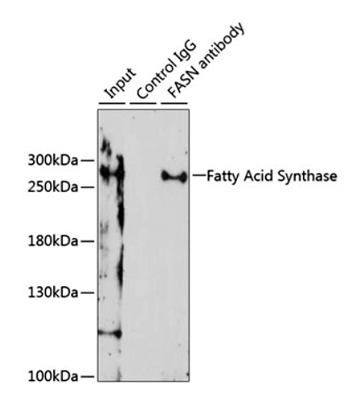
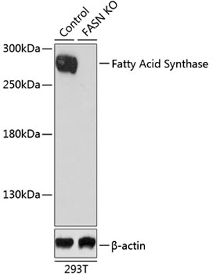
Fatty Acid Synthase, Polyclonal Antibody (Cat# AAA10626)
Full Name
FASN Polyclonal Antibody
Gene Names
FASN; FAS; OA-519; SDR27X1
Applications
Western Blot, Immunohistochemistry, Immunoprecipitation, Immunofluorescence
Purity
Affinity purification
Pricing
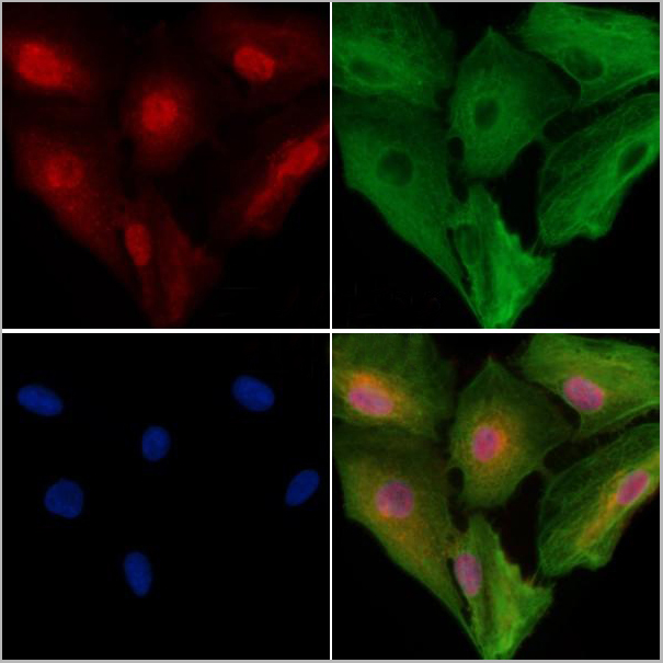
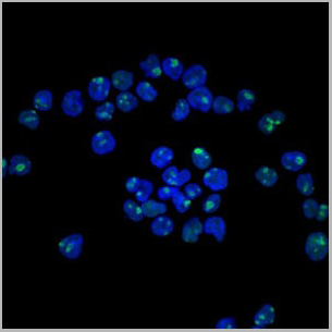
 Application Data
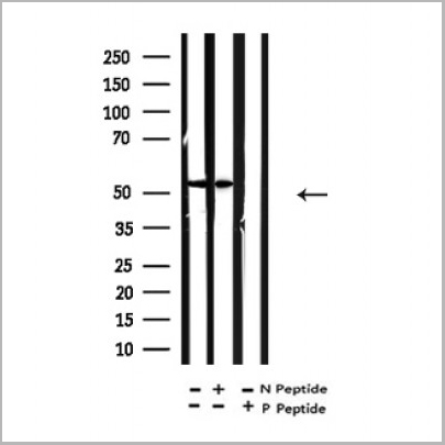
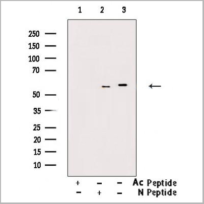
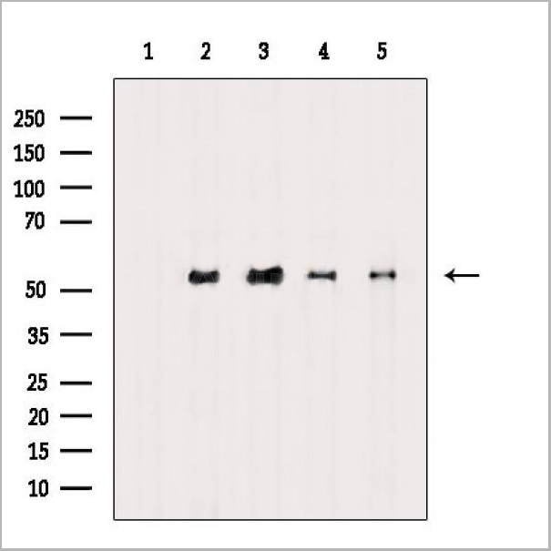
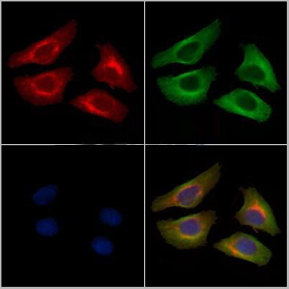
(At 25 degree C. Samples were then incubated with primary Ab(At 37 degree C. An AlexaFluor594 conjugated goat anti-rabbit IgG(H+L) Ab(Red) and an AlexaFluor488 conjugated goat anti-mouse IgG(H+L) Ab(Green) were used as the secondary antibody.The nuclear counter stain is DAPI(blue).)
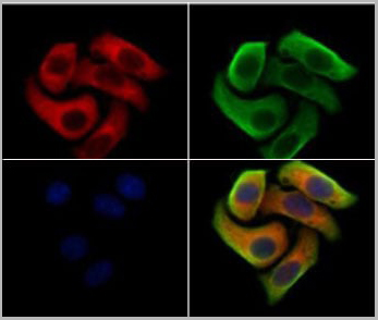
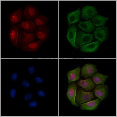
Application Data
(At 25 degree C. Samples were then incubated with primary Ab(At 37 degree C. An AlexaFluor594 conjugated goat anti-rabbit IgG(H+L) Ab(Red) and an AlexaFluor488 conjugated goat anti-mouse IgG(H+L) Ab(Green) were used as the secondary antibody.The nuclear counter stain is DAPI(blue).)
alpha Tubulin, Polyclonal Antibody (Cat# AAA31344)
Full Name
Acetyl-alpha Tubulin (Lys40) Antibody
Gene Names
TUBA1B; K-ALPHA-1
Reactivity
Human, Mouse, Rat
Applications
Western Blot, Immunohistochemistry, Immunofluorescence, Immunocytochemistry
Purity
The antiserum was purified by peptide affinity chromatography using SulfoLink Coupling Resin (Thermo Fisher Scientific).
Pricing
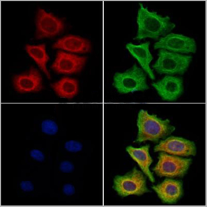
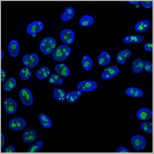
 IF (Immunofluorescence)
(Staining Hela cells by IF/ICC. The samples were fixedwith PFA and permeabilized in 0.1% Triton X-100,then blockedin 10% serum for 45 minutes at 25°C. Samples were thenincubated with primary Ab(DF13215 1:200) and mouse anti-beta tubulin Ab(T0023 1:200) for 1 hour at 37°C. An AlexaFluor594 conjugated goat anti-rabbit IgG(H+L) Ab(Red)and an AlexaFluor488 conjugated goat anti-mouse IgG(H+L)Ab(Green) were used as the secondary antibody.)
IF (Immunofluorescence)
(Staining Hela cells by IF/ICC. The samples were fixedwith PFA and permeabilized in 0.1% Triton X-100,then blockedin 10% serum for 45 minutes at 25°C. Samples were thenincubated with primary Ab(DF13215 1:200) and mouse anti-beta tubulin Ab(T0023 1:200) for 1 hour at 37°C. An AlexaFluor594 conjugated goat anti-rabbit IgG(H+L) Ab(Red)and an AlexaFluor488 conjugated goat anti-mouse IgG(H+L)Ab(Green) were used as the secondary antibody.)
Clathrin, Polyclonal Antibody (Cat# AAA31165)
Full Name
Clathrin Antibody
Gene Names
CLTC; Hc; CHC; CHC17; MRD56; CLH-17; CLTCL2
Reactivity
Human, Mouse, Rat
Applications
Western Blot, Immunofluorescence, Immunocytochemistry
Purity
Peptide affinity purifcation
Pricing
 ELISA
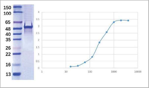
(ELISA plate was coated by N protein, 100 uL/cell, at various concentrations. The direct ELISA analysis was performed by loading 100 uL per well of a anti C-ter His tag HRP conjugated mAb at a concentration of 1:2000. The plate was incubated for 1 hours at 37 degree C, then washed 5 time. Detection was performed using TMB substrate for 10 minutes at room temperature in the dark. The plate was stopped with 2M sulfuric acid. Absorbances were read on a spectrophotometer at 450 nm.)
ELISA
(ELISA plate was coated by N protein, 100 uL/cell, at various concentrations. The direct ELISA analysis was performed by loading 100 uL per well of a anti C-ter His tag HRP conjugated mAb at a concentration of 1:2000. The plate was incubated for 1 hours at 37 degree C, then washed 5 time. Detection was performed using TMB substrate for 10 minutes at room temperature in the dark. The plate was stopped with 2M sulfuric acid. Absorbances were read on a spectrophotometer at 450 nm.)
COVID 19 Nucleocapsid (NP) N3 fragment (C-ter domain) Coronavirus, Recombinant Protein (Cat# AAA27949)
Full Name
2019-nCov Nucleocapsid Recombinant Protein N3 fragment (C-ter domain)
Applications
ELISA
Purity
Purity: >95% by SDS-page
Purification: Ni column
Purification: Ni column
Pricing


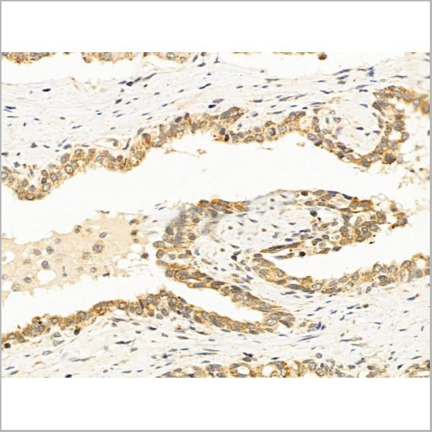
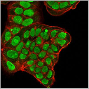
 IHC (Immunohistochemistry)
(Immunofluorescence staining of MCF-7 cells with AAA27006 at 1:133, counter-stained with DAPI. The cells were fixed in 4% formaldehyde, permeabilized using 0.2% Triton X-100 and blocked in 10% normal Goat Serum. The cells were then incubated with the antibody overnight at 4 degree C.The secondary antibody was Alexa Fluor 488-congugated AffiniPure Goat Anti-Rabbit IgG (H+L).)
IHC (Immunohistochemistry)
(Immunofluorescence staining of MCF-7 cells with AAA27006 at 1:133, counter-stained with DAPI. The cells were fixed in 4% formaldehyde, permeabilized using 0.2% Triton X-100 and blocked in 10% normal Goat Serum. The cells were then incubated with the antibody overnight at 4 degree C.The secondary antibody was Alexa Fluor 488-congugated AffiniPure Goat Anti-Rabbit IgG (H+L).)
NECTIN4, Polyclonal Antibody (Cat# AAA27006)
Full Name
NECTIN4 Antibody
Gene Names
NECTIN4; LNIR; PRR4; EDSS1; PVRL4; nectin-4
Reactivity
Human, Mouse
Applications
Western Blot, Immunohistochemistry, Immunofluorescence
Purity
>95%, Protein G purified
Pricing
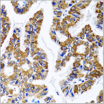
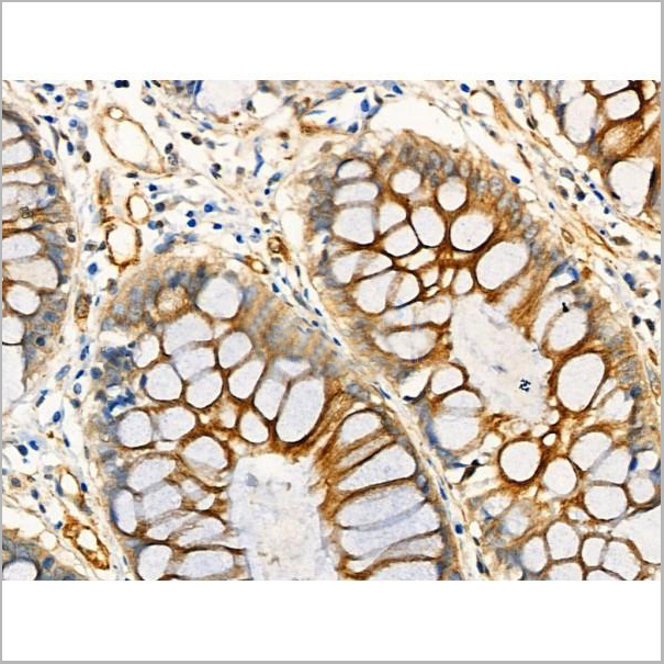
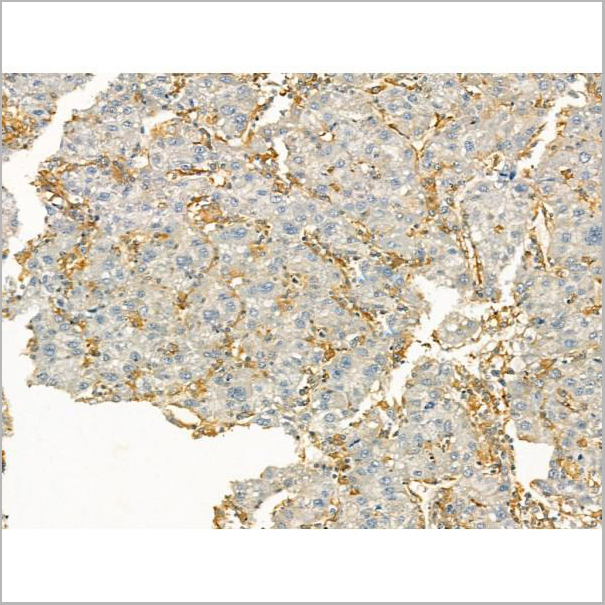
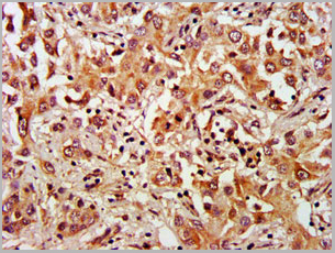
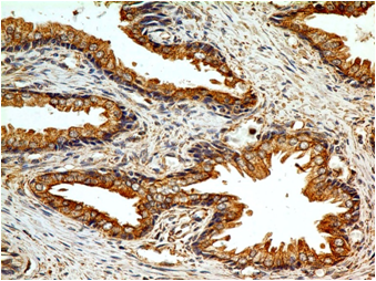
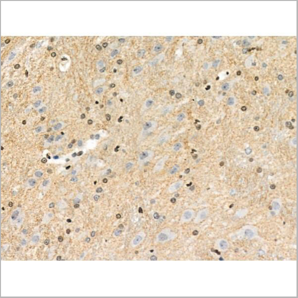
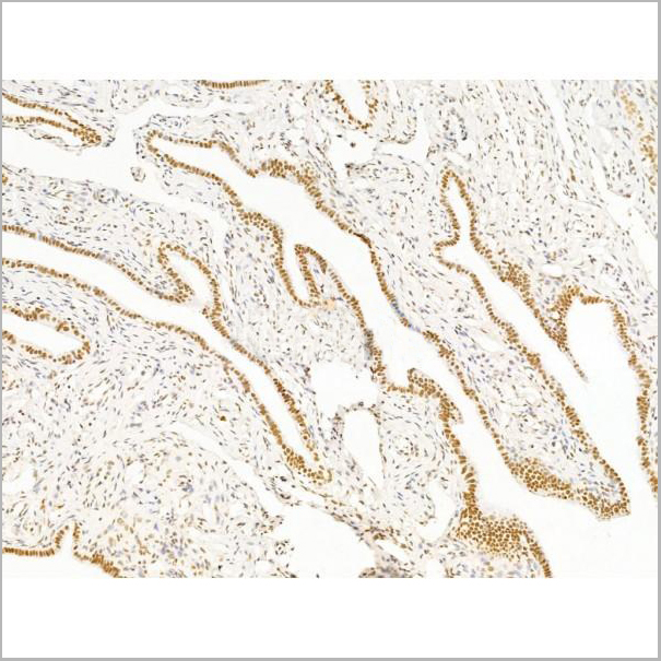
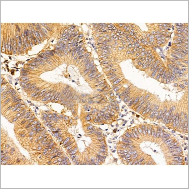
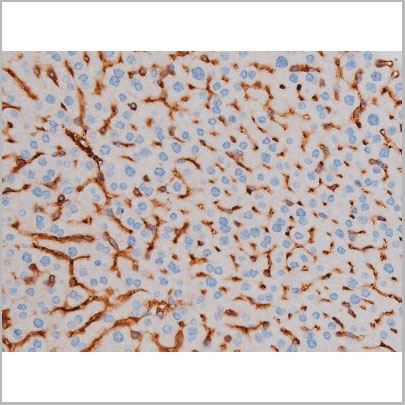
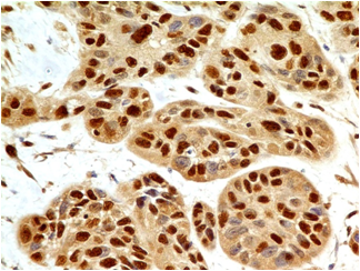
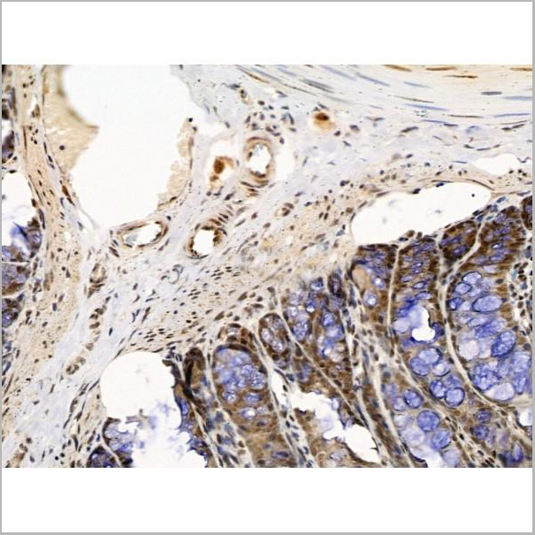
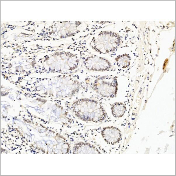
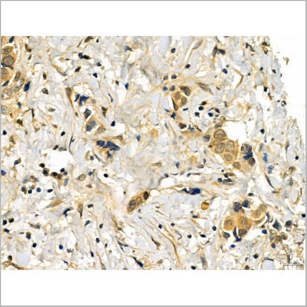
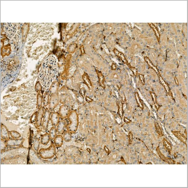
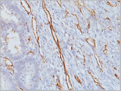
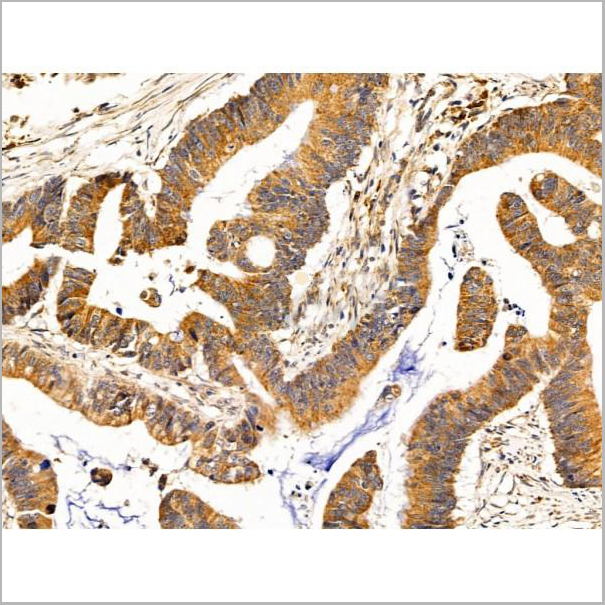
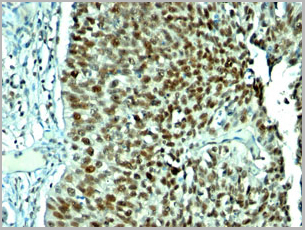
 IHC (Immunohistochemistry-Paraffin)
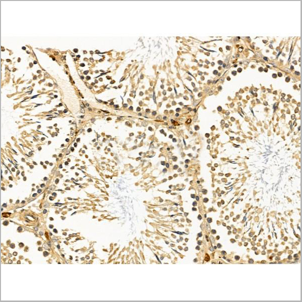
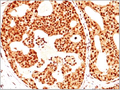
(AAA31251 at 1/100 staining Human colorectal cancer and adjacent normal tissues by IHC-P. The sample was formaldehyde fixed and a heat mediated antigen retrieval step in citrate buffer was performed. The sample was then blocked and incubated with the primary antibody at 4°C overnight. An HRP conjugated anti-Rabbit antibody was used as the secondary antibody.)
IHC (Immunohistochemistry-Paraffin)
(AAA31251 at 1/100 staining Human colorectal cancer and adjacent normal tissues by IHC-P. The sample was formaldehyde fixed and a heat mediated antigen retrieval step in citrate buffer was performed. The sample was then blocked and incubated with the primary antibody at 4°C overnight. An HRP conjugated anti-Rabbit antibody was used as the secondary antibody.)
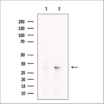
Dcp2, Polyclonal Antibody (Cat# AAA31251)
Full Name
Dcp2 Antibody
Gene Names
DCP2; NUDT20
Reactivity
Human, Mouse, Rat
Predicted Reactivity: Pig(100%), Horse(100%), Sheep(100%), Rabbit(100%), Dog(100%), Xenopus(88%)
Predicted Reactivity: Pig(100%), Horse(100%), Sheep(100%), Rabbit(100%), Dog(100%), Xenopus(88%)
Applications
ELISA
Purity
The antiserum was purified by peptide affinity chromatography using SulfoLink Coupling Resin (Thermo Fisher Scientific).
Pricing
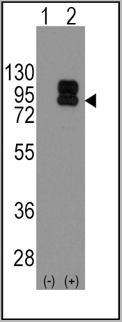

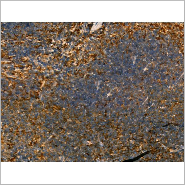
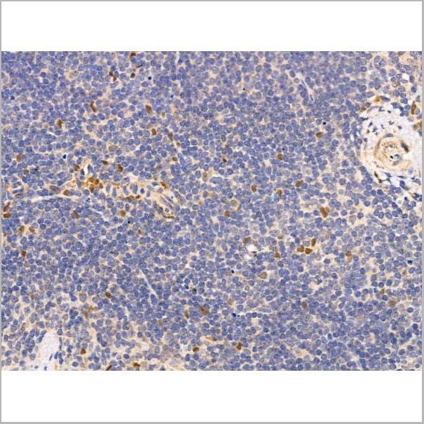
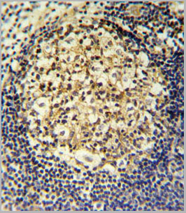
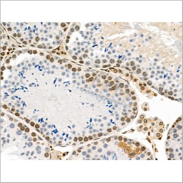
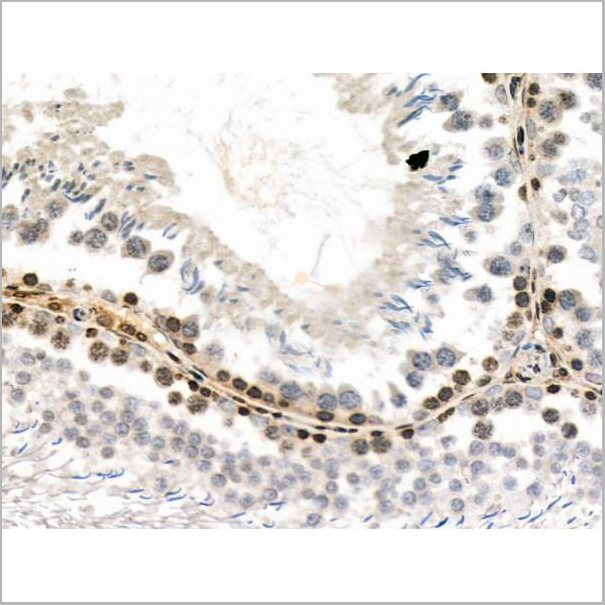
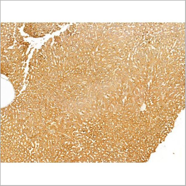
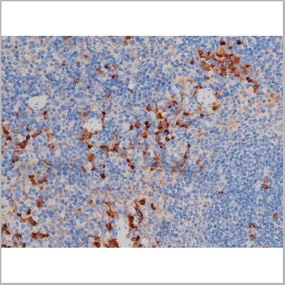
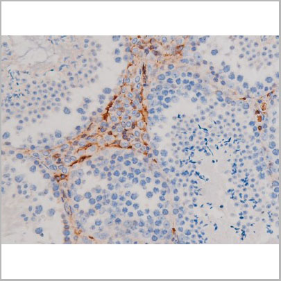
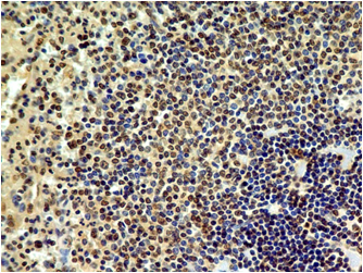
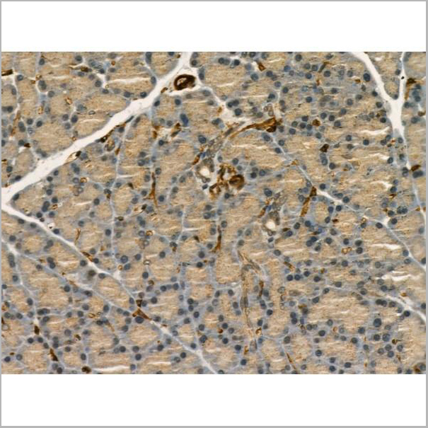
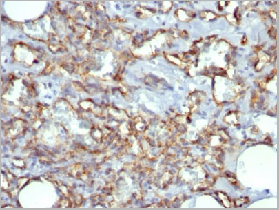
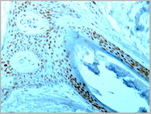
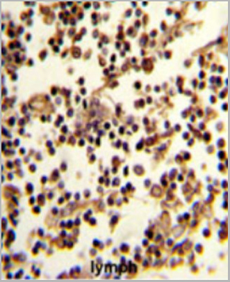 IHC (Immunohistchemistry)
(Formalin-fixed and paraffin-embedded human lymph reacted with CD19 Antibody (N-term), which was peroxidase-conjugated to the secondary antibody, followed by DAB staining. This data demonstrates the use of this antibody for immunohistochemistry; clinical relevance has not been evaluated.)
IHC (Immunohistchemistry)
(Formalin-fixed and paraffin-embedded human lymph reacted with CD19 Antibody (N-term), which was peroxidase-conjugated to the secondary antibody, followed by DAB staining. This data demonstrates the use of this antibody for immunohistochemistry; clinical relevance has not been evaluated.)
CD19, Polyclonal Antibody (Cat# AAA28685)
Full Name
CD19 Antibody (N-term)
Gene Names
CD19; B4; CVID3
Reactivity
Human
Applications
Western Blot, Immunofluorescence, Flow Cytometry, Immunohistochemistry
Purity
This antibody is purified through a protein A column, followed by peptide affinity purification.
Pricing


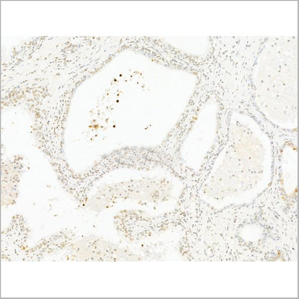
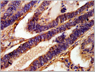
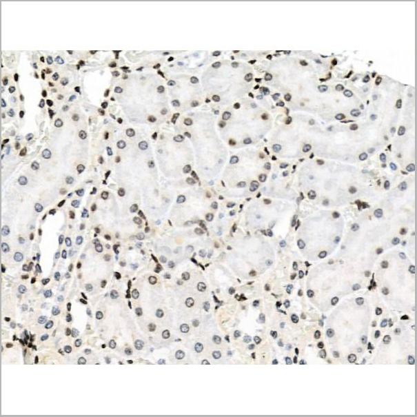
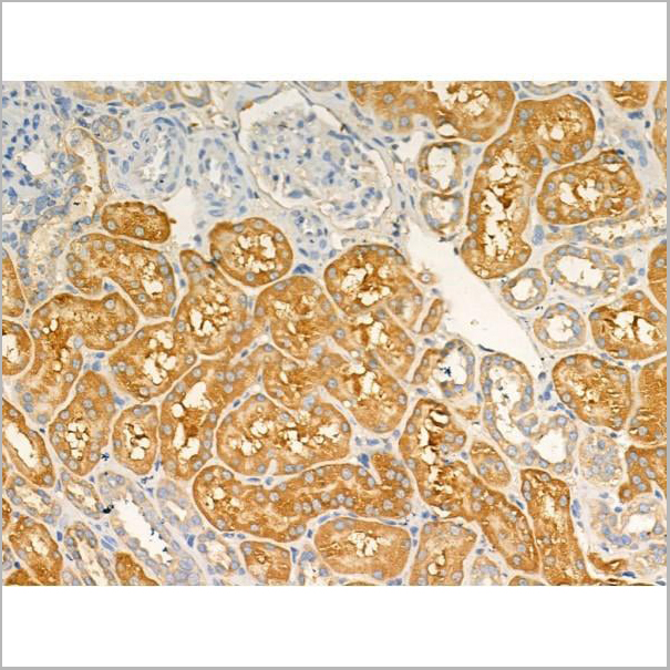
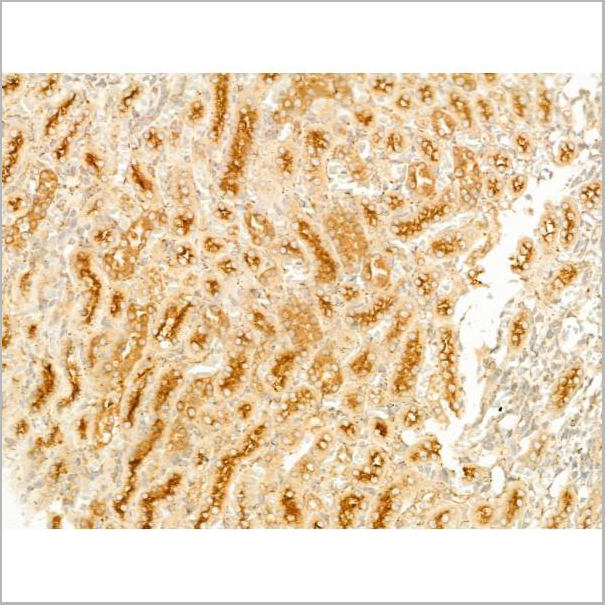
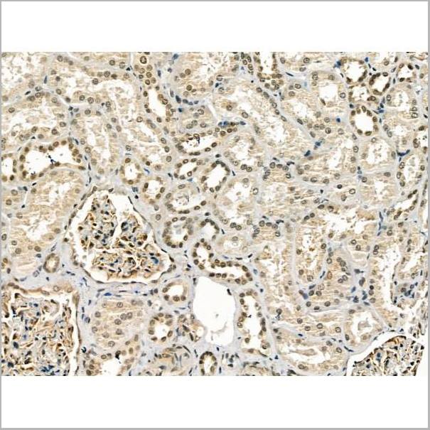
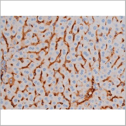
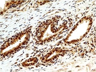
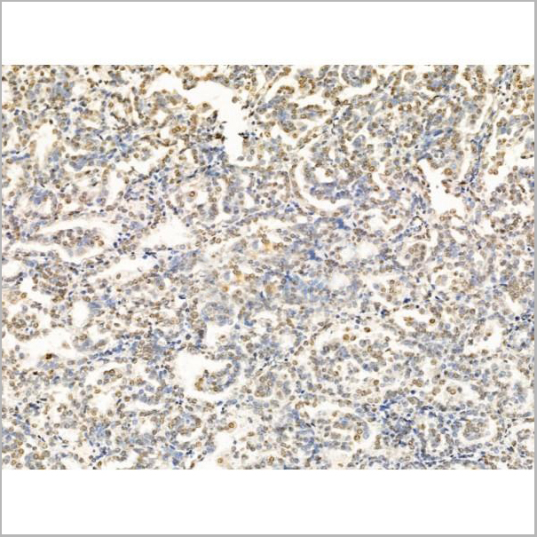
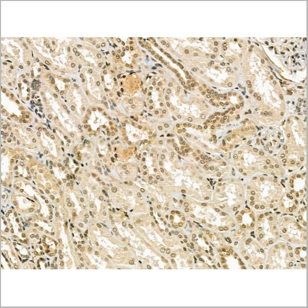
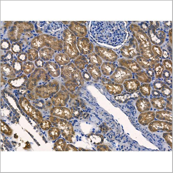
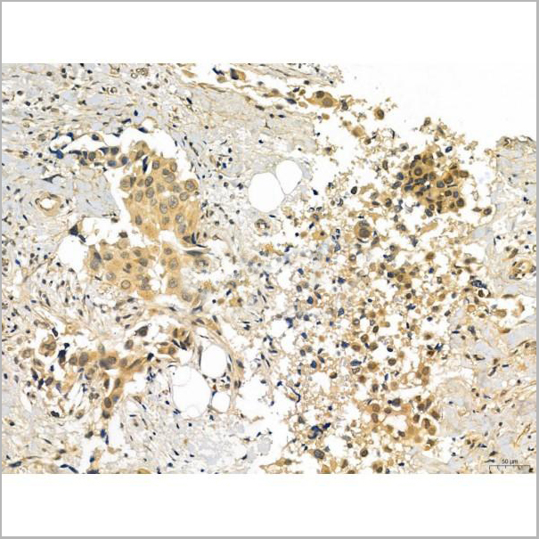
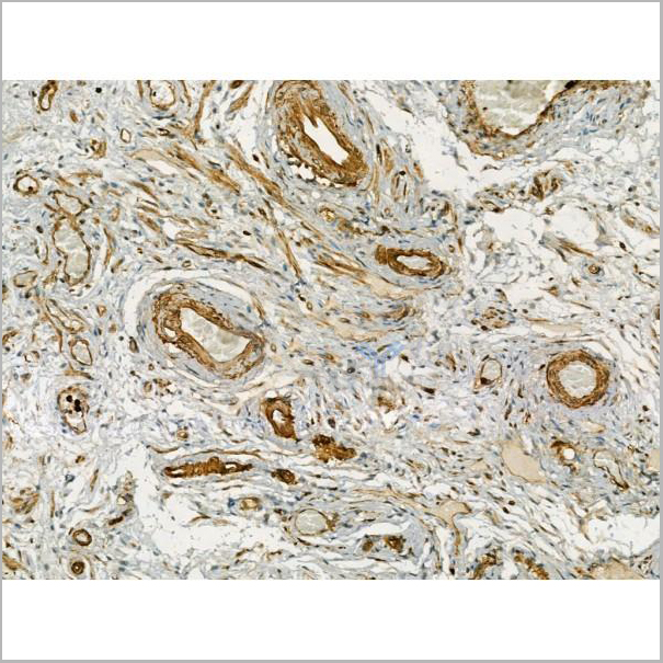
 IHC (Immunohistchemistry-Paraffin)
(AAA31217 at 1/100 staining Human kidney cancer and adjacent normal tissues by IHC-P. The sample was formaldehyde fixed and a heat mediated antigen retrieval step in citrate buffer was performed. The sample was then blocked and incubated with the primary antibody at 4°C overnight. An HRP conjugated anti-Rabbit antibody was used as the secondary antibody.)
IHC (Immunohistchemistry-Paraffin)
(AAA31217 at 1/100 staining Human kidney cancer and adjacent normal tissues by IHC-P. The sample was formaldehyde fixed and a heat mediated antigen retrieval step in citrate buffer was performed. The sample was then blocked and incubated with the primary antibody at 4°C overnight. An HRP conjugated anti-Rabbit antibody was used as the secondary antibody.)
TBLR1, Polyclonal Antibody (Cat# AAA31217)
Full Name
TBLR1 Antibody
Gene Names
TBL1XR1; C21; DC42; IRA1; MRD41; TBLR1
Reactivity
Human, Mouse, Rat
Predicted Reactivity: Pig(100%), Bovine(100%), Sheep(100%), Rabbit(100%), Dog(100%), Chicken(100%), Xenopus(88%)
Predicted Reactivity: Pig(100%), Bovine(100%), Sheep(100%), Rabbit(100%), Dog(100%), Chicken(100%), Xenopus(88%)
Applications
ELISA
Purity
The antiserum was purified by peptide affinity chromatography using SulfoLink Coupling Resin (Thermo Fisher Scientific).
Pricing
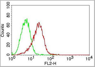
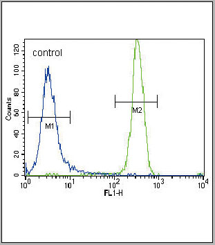
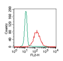
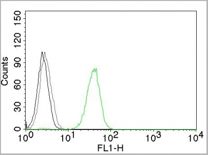
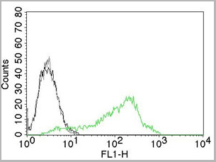
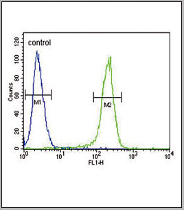 FCM (Flow Cytometry)
(CCR7 Antibody (N-term) (AAA28701) flow cytometric analysis of 293 cells (right histogram) compared to a negative controlcell (left histogram). FITC-conjugated goat-anti-rabbit secondary antibodies were used for the analysis.)
FCM (Flow Cytometry)
(CCR7 Antibody (N-term) (AAA28701) flow cytometric analysis of 293 cells (right histogram) compared to a negative controlcell (left histogram). FITC-conjugated goat-anti-rabbit secondary antibodies were used for the analysis.)
CCR7, Polyclonal Antibody (Cat# AAA28701)
Full Name
CCR7 Antibody (N-term)
Gene Names
CCR7; BLR2; EBI1; CCR-7; CD197; CDw197; CMKBR7; CC-CKR-7
Reactivity
Human, mouse
Applications
Western Blot, Immunohistochemistry, Immunofluorescence, Flow Cytometry
Purity
Peptide Affinity Purified Rabbit Polyclonal Antibody (Pab)
Pricing
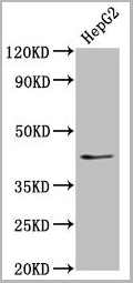
 IF (Immunofluorescence)
(Immunofluorescence staining of HepG2 cells with AAA26952 at 1:333, counter- stained with DAPI. The cells were fixed in 4% formaldehyde, permeabilized using 0.2% Triton X-100 and blocked in 10% normal Goat Serum. The cells were then incubated with the antibody overnight at 4°C. The secondary antibody was Alexa Fluor 488-congugated AffiniPure Goat Anti-Rabbit IgG(H+L)
IF (Immunofluorescence)
(Immunofluorescence staining of HepG2 cells with AAA26952 at 1:333, counter- stained with DAPI. The cells were fixed in 4% formaldehyde, permeabilized using 0.2% Triton X-100 and blocked in 10% normal Goat Serum. The cells were then incubated with the antibody overnight at 4°C. The secondary antibody was Alexa Fluor 488-congugated AffiniPure Goat Anti-Rabbit IgG(H+L)
XKR8, Polyclonal Antibody (Cat# AAA26952)
Full Name
XKR8 Antibody
Gene Names
XKR8; XRG8; hXkr8
Reactivity
Human
Applications
Western Blot, Immunohistochemistry, Immunofluorescence
Purity
>95%, Protein G purified
Pricing

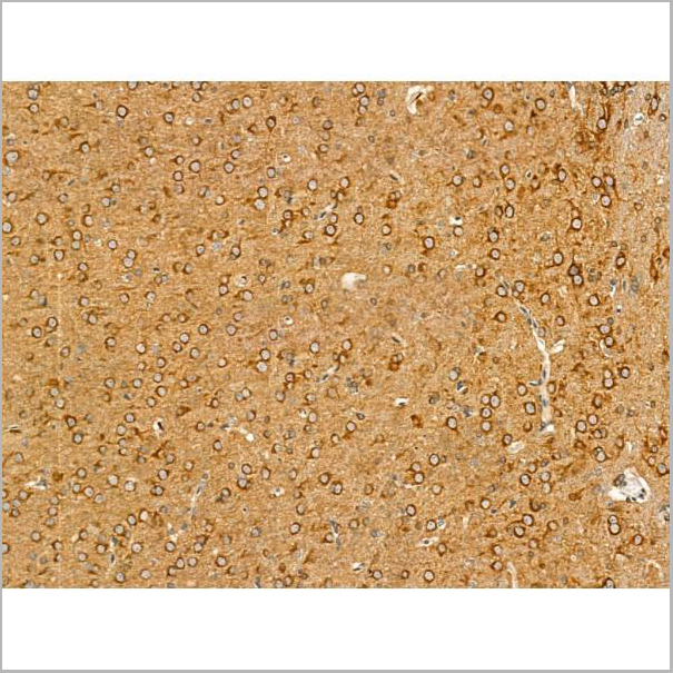
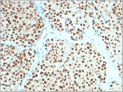
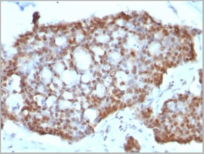
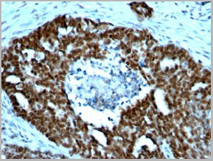
 IHC (Immunohistochemistry)
(Immunohistochemical analysis of TLR2 in normal human prostate tissue using TLR2 antibody (Clone: ABM3A87) at 5 ug/ml.)
IHC (Immunohistochemistry)
(Immunohistochemical analysis of TLR2 in normal human prostate tissue using TLR2 antibody (Clone: ABM3A87) at 5 ug/ml.)
TLR2, Monoclonal Antibody (Cat# AAA14864)
Full Name
TLR2 Monoclonal Antibody
Gene Names
TLR2; TIL4; CD282
Reactivity
Mouse, Human
Applications
Immunohistochemistry, Flow Cytometry, Western Blot, Immunofluorescence
Purity
Protein G Chromatography
Pricing
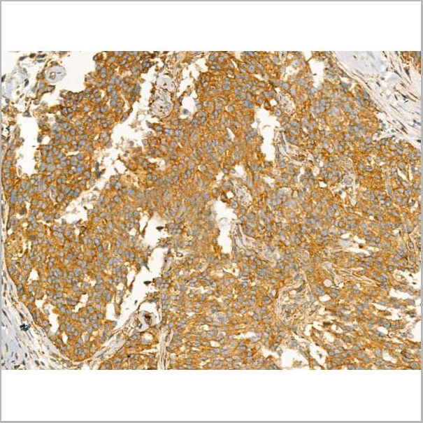
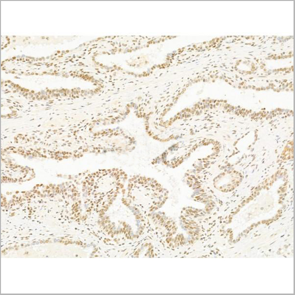
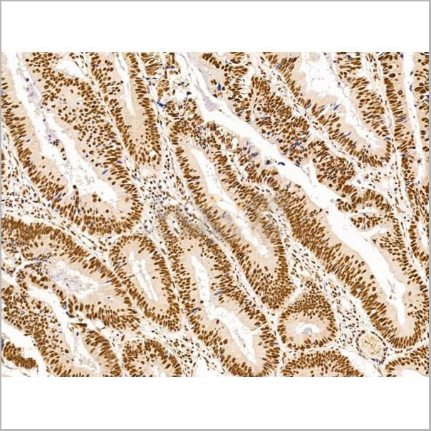
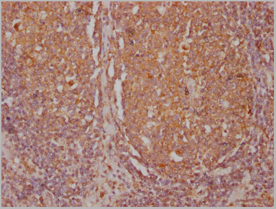
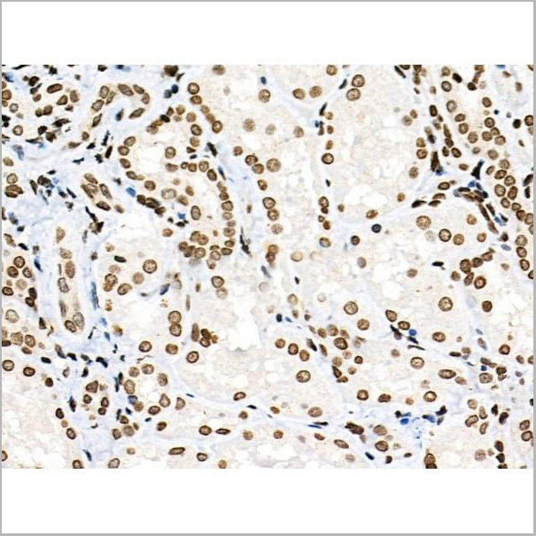
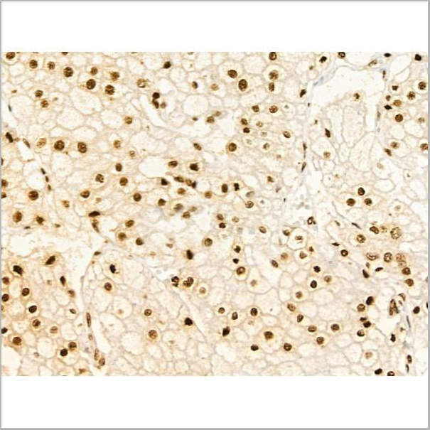
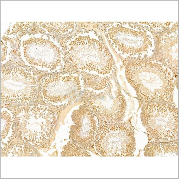
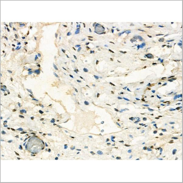
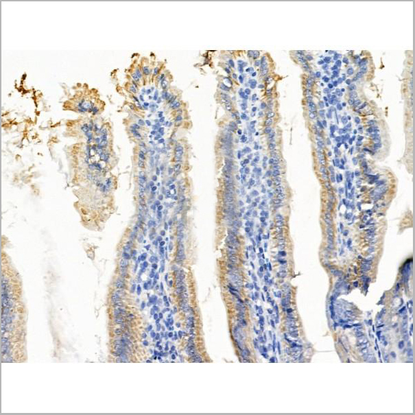
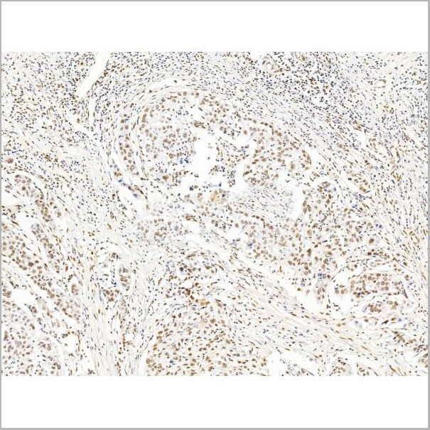
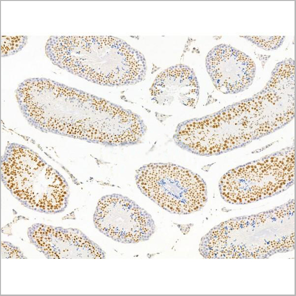
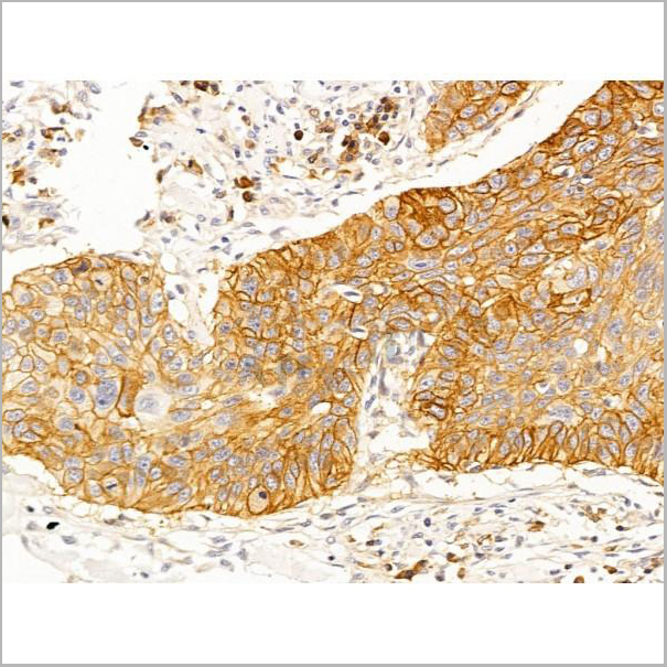
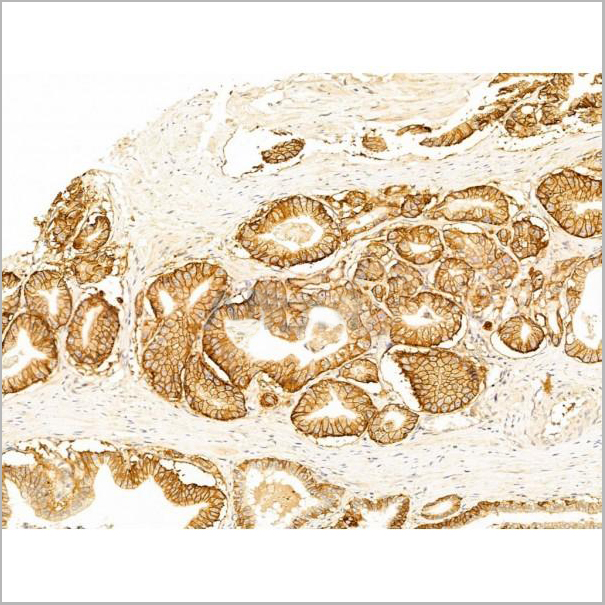
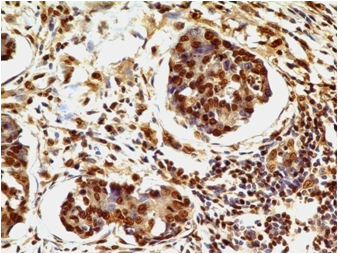
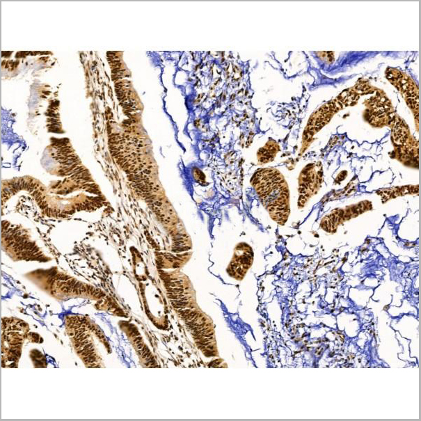
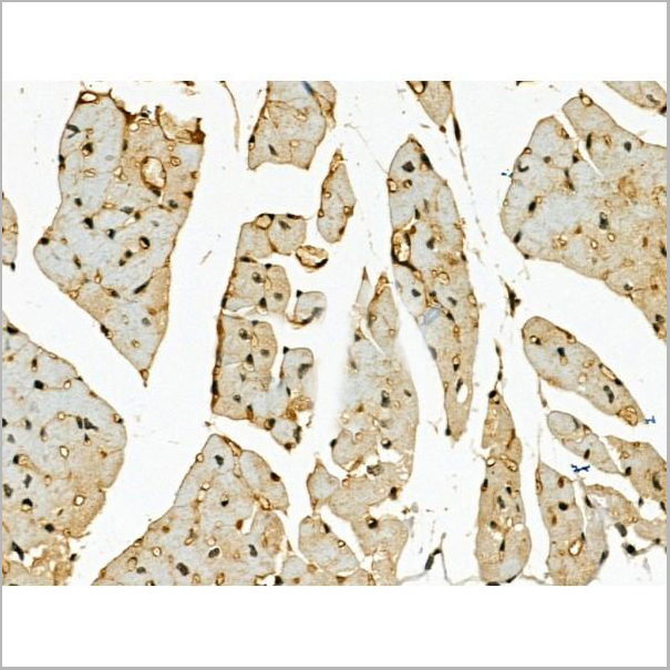
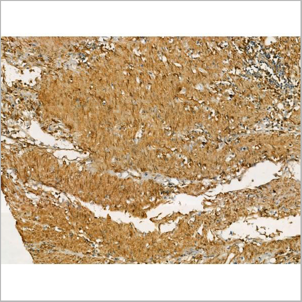
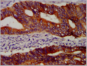
 IHC (Immunohistchemistry-Paraffin)
(AAA31276 at 1/100 staining Human colorectal cancer and adjacent normal tissues by IHC-P. The sample was formaldehyde fixed and a heat mediated antigen retrieval step in citrate buffer was performed. The sample was then blocked and incubated with the primary antibody at 4°C overnight. An HRP conjugated anti-Rabbit antibody was used as the secondary antibody.)
IHC (Immunohistchemistry-Paraffin)
(AAA31276 at 1/100 staining Human colorectal cancer and adjacent normal tissues by IHC-P. The sample was formaldehyde fixed and a heat mediated antigen retrieval step in citrate buffer was performed. The sample was then blocked and incubated with the primary antibody at 4°C overnight. An HRP conjugated anti-Rabbit antibody was used as the secondary antibody.)
SCAMP2, Polyclonal Antibody (Cat# AAA31276)
Full Name
SCAMP2 Antibody
Reactivity
Human, Mouse, Rat, Monkey
Predicted Reactivity: Pig(100%), Zebrafish(100%), Bovine(100%), Horse(100%), Sheep(100%), Rabbit(100%), Dog(100%), Chicken(100%), Xenopus(100%)
Predicted Reactivity: Pig(100%), Zebrafish(100%), Bovine(100%), Horse(100%), Sheep(100%), Rabbit(100%), Dog(100%), Chicken(100%), Xenopus(100%)
Applications
ELISA
Purity
The antiserum was purified by peptide affinity chromatography using SulfoLink Coupling Resin (Thermo Fisher Scientific).
Pricing
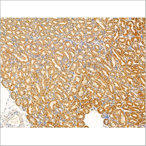
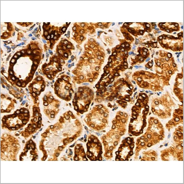
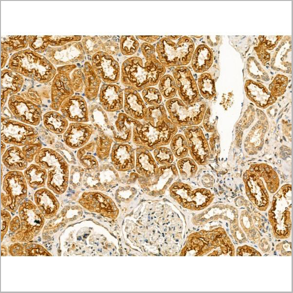
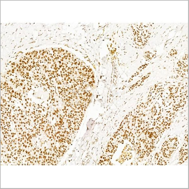
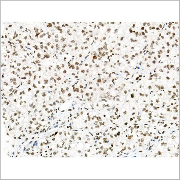
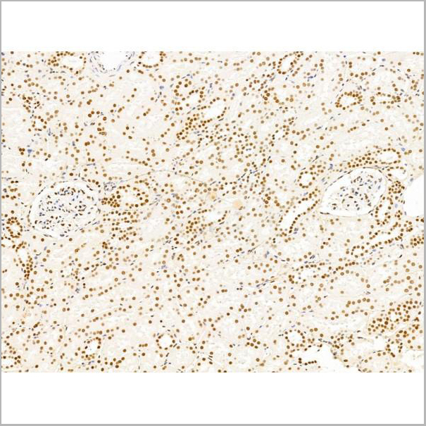
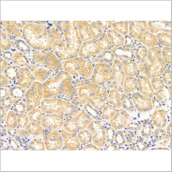
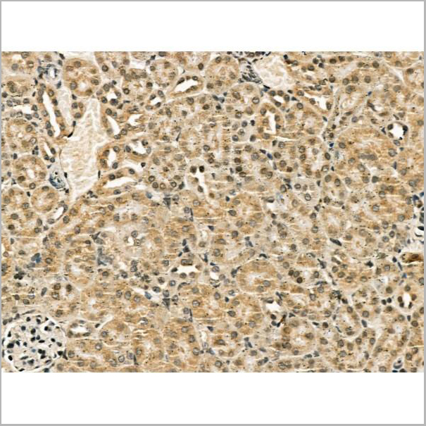
 IHC (Immunohistchemistry-Paraffin)
(AAA31208 at 1/100 staining Human kidney cancer and adjacent normal tissues by IHC-P. The sample was formaldehyde fixed and a heat mediated antigen retrieval step in citrate buffer was performed. The sample was then blocked and incubated with the primary antibody at 4°C overnight. An HRP conjugated anti-Rabbit antibody was used as the secondary antibody.)
IHC (Immunohistchemistry-Paraffin)
(AAA31208 at 1/100 staining Human kidney cancer and adjacent normal tissues by IHC-P. The sample was formaldehyde fixed and a heat mediated antigen retrieval step in citrate buffer was performed. The sample was then blocked and incubated with the primary antibody at 4°C overnight. An HRP conjugated anti-Rabbit antibody was used as the secondary antibody.)
SV2A, Polyclonal Antibody (Cat# AAA31208)
Full Name
SV2A Antibody
Gene Names
SV2A; SV2
Reactivity
Human, Mouse, Rat
Predicted Reactivity: Pig(100%), Bovine(100%), Horse(100%), Sheep(100%), Rabbit(100%)
Predicted Reactivity: Pig(100%), Bovine(100%), Horse(100%), Sheep(100%), Rabbit(100%)
Applications
ELISA
Purity
Peptide affinity purification
Pricing
 Application Data
(Z- and S- Score: The Z-score represents the strength of a signal that a monoclonal antibody (MAb) (in combination with a fluorescently-tagged anti-IgG secondary antibody) produces when binding to a particular protein on the HuProtTM array. Z-scores are described in units of standard deviations (SD's) above the mean value of all signals generated on that array. If targets on HuProtTM are arranged in descending order of the Z-score, the S-score is the difference (also in units of SD's) between the Z-score. S-score therefore represents the relative target specificity of a MAb to its intended target. A MAb is considered to specific to its intended target, if the MAb has an S-score of at least 2.5. For example, if a MAb binds to protein X with a Z-score of 43 and to protein Y with a Z-score of 14, then the S-score for the binding of that MAb to protein X is equal to 29.)
Application Data
(Z- and S- Score: The Z-score represents the strength of a signal that a monoclonal antibody (MAb) (in combination with a fluorescently-tagged anti-IgG secondary antibody) produces when binding to a particular protein on the HuProtTM array. Z-scores are described in units of standard deviations (SD's) above the mean value of all signals generated on that array. If targets on HuProtTM are arranged in descending order of the Z-score, the S-score is the difference (also in units of SD's) between the Z-score. S-score therefore represents the relative target specificity of a MAb to its intended target. A MAb is considered to specific to its intended target, if the MAb has an S-score of at least 2.5. For example, if a MAb binds to protein X with a Z-score of 43 and to protein Y with a Z-score of 14, then the S-score for the binding of that MAb to protein X is equal to 29.)
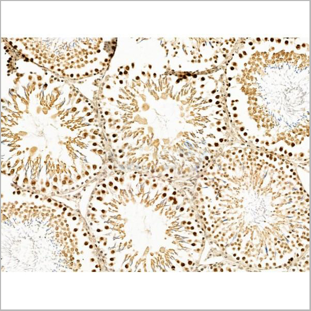
SOX10, Monoclonal Antibody (Cat# AAA13845)
Full Name
SOX10 (Melanoma Marker) Mouse Monoclonal Antibody
Gene Names
SOX10; DOM; WS4; PCWH; WS2E; WS4C
Reactivity
Human, Mouse
Applications
Flow Cytometry, Immunofluorescence, Western Blot, Immunohistochemistry
Pricing
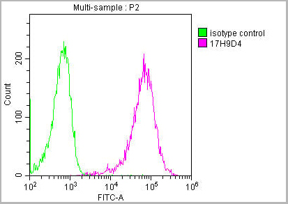
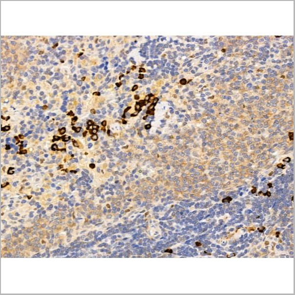
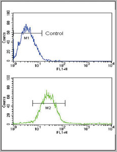
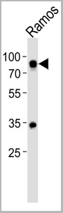
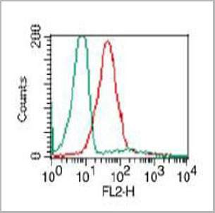
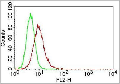
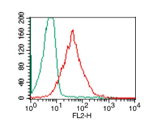
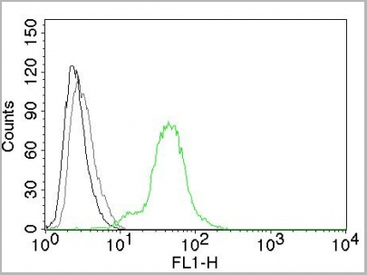
 FCM (Flow Cytometry)
(Overlay histogram showing Raji cells stained with (red line) at 1:500. The cells were incubated in 10% normal goat serum to block non-specific protein-protein interactions followed by the antibody (1ug/1*106cells) for 1 h at 4 degree C. The secondary antibody used was FITC-conjugated Goat Anti-Mouse IgG(H+L) at 1/100 dilution for 30min at 4 degree C. Isotype control antibody (green line) was mouse IgG2b (1ug/1*106cells) used under the same conditions. Acquisition of >10,000 events was performed.)
FCM (Flow Cytometry)
(Overlay histogram showing Raji cells stained with (red line) at 1:500. The cells were incubated in 10% normal goat serum to block non-specific protein-protein interactions followed by the antibody (1ug/1*106cells) for 1 h at 4 degree C. The secondary antibody used was FITC-conjugated Goat Anti-Mouse IgG(H+L) at 1/100 dilution for 30min at 4 degree C. Isotype control antibody (green line) was mouse IgG2b (1ug/1*106cells) used under the same conditions. Acquisition of >10,000 events was performed.)
CD45, Monoclonal Antibody (Cat# AAA27049)
Full Name
CD45 Monoclonal Antibody
Gene Names
PTPRC; LCA; LY5; B220; CD45; L-CA; T200; CD45R; GP180
Reactivity
Human
Applications
Western Blot, Immunohistochemistry, Immunofluorescence, Flow Cytometry
Purity
>95%, Protein A purified
Pricing
 Application Data
(At 25 degree C. Samples were then incubated with primary Ab(At 37 degree C. An AlexaFluor594 conjugated goat anti-rabbit IgG(H+L) Ab(Red) and an AlexaFluor488 conjugated goat anti-mouse IgG(H+L) Ab(Green) were used as the secondary antibody.The nuclear counter stain is DAPI(blue).)
Application Data
(At 25 degree C. Samples were then incubated with primary Ab(At 37 degree C. An AlexaFluor594 conjugated goat anti-rabbit IgG(H+L) Ab(Red) and an AlexaFluor488 conjugated goat anti-mouse IgG(H+L) Ab(Green) were used as the secondary antibody.The nuclear counter stain is DAPI(blue).)
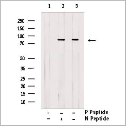
PRC1, Polyclonal Antibody (Cat# AAA31450)
Full Name
Phospho-PRC1 (Thr481) Antibody
Gene Names
PRC1; ASE1
Reactivity
Human, Mouse, Rat
Predicted Reactivity: Pig (80%), Horse (90%)
Predicted Reactivity: Pig (80%), Horse (90%)
Applications
Western Blot, Immunohistochemistry, Immunofluorescence, Immunocytochemistry, Peptide ELISA
Purity
The antibody is from purified rabbit serum by affinity purification via sequential chromatography on phospho-peptide and non-phospho-peptide affinity columns.
Pricing
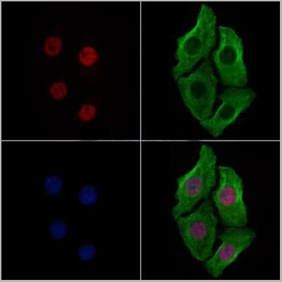 Application Data
(AAA31369 staining Hela cells(4h of LPS treatment) by IF/ICC. The samples were fixed with PFA and permeabilized in 0.1% Triton X-100,then blocked in 10% serum for 45 minutes at 25°C. Samples were then incubated with primary Ab(AAA31369 1:200) and mouse anti-beta tubulin Ab(T0023 1:200) for 1 hour at 37°C. An AlexaFluor594 conjugated goat anti-rabbit IgG(H+L) Ab(Red) and an AlexaFluor488 conjugated goat anti-mouse IgG(H+L) Ab(Green) were used as the secondary Ab. The nuclear counter stain is DAPI(blue).)
Application Data
(AAA31369 staining Hela cells(4h of LPS treatment) by IF/ICC. The samples were fixed with PFA and permeabilized in 0.1% Triton X-100,then blocked in 10% serum for 45 minutes at 25°C. Samples were then incubated with primary Ab(AAA31369 1:200) and mouse anti-beta tubulin Ab(T0023 1:200) for 1 hour at 37°C. An AlexaFluor594 conjugated goat anti-rabbit IgG(H+L) Ab(Red) and an AlexaFluor488 conjugated goat anti-mouse IgG(H+L) Ab(Green) were used as the secondary Ab. The nuclear counter stain is DAPI(blue).)
FOXM1, Polyclonal Antibody (Cat# AAA31369)
Full Name
Phospho-FOXM1 (Ser35) Antibody
Gene Names
FOXM1; MPP2; TGT3; HFH11; HNF-3; INS-1; MPP-2; PIG29; FKHL16; FOXM1B; HFH-11; TRIDENT; MPHOSPH2
Reactivity
Human, Mouse, Rat
Applications
Western Blot, Immunohistochemistry, Immunofluorescence, Immunocytochemistry
Purity
The antibody is from purified rabbit serum by affinity purification via sequential chromatography on phospho-peptide and non-phospho-peptide affinity columns.
Pricing
 FCM (Flow Cytometry)
(WB - MCSF Receptor (CSF1R) Antibody (C-term) AAA28757 detail IHC-P - MCSF Receptor (CSF1R) Antibody (C-term) AAA28757 detail IHC-P - MCSF Receptor (CSF1R) Antibody (C-term) AAA28757 detail FC - MCSF Receptor (CSF1R) Antibody (C-term) AAA28757 detail Overlay histogram showing HepG2 cells stained with AAA28757(green line). The cells were fixed with 2% paraformaldehyde (10 min) and then permeabilized with 90% methanol for 10 min. The cells were then icubated in 2% bovine serum albumin to block non-specific protein-protein interactions followed by the antibody (AAA28757, 1:25 dilution) for 60 min at 37ºC. The secondary antibody used was Goat-Anti-Rabbit IgG, DyLight® 488 Conjugated Highly Cross-Adsorbed(1583138) at 1/200 dilution for 40 min at 37ºC. Isotype control antibody (blue line) was rabbit IgG1 (1ug/1x10^6 cells) used under the same conditions. Acquisition of >10, 000 events was performed.)
FCM (Flow Cytometry)
(WB - MCSF Receptor (CSF1R) Antibody (C-term) AAA28757 detail IHC-P - MCSF Receptor (CSF1R) Antibody (C-term) AAA28757 detail IHC-P - MCSF Receptor (CSF1R) Antibody (C-term) AAA28757 detail FC - MCSF Receptor (CSF1R) Antibody (C-term) AAA28757 detail Overlay histogram showing HepG2 cells stained with AAA28757(green line). The cells were fixed with 2% paraformaldehyde (10 min) and then permeabilized with 90% methanol for 10 min. The cells were then icubated in 2% bovine serum albumin to block non-specific protein-protein interactions followed by the antibody (AAA28757, 1:25 dilution) for 60 min at 37ºC. The secondary antibody used was Goat-Anti-Rabbit IgG, DyLight® 488 Conjugated Highly Cross-Adsorbed(1583138) at 1/200 dilution for 40 min at 37ºC. Isotype control antibody (blue line) was rabbit IgG1 (1ug/1x10^6 cells) used under the same conditions. Acquisition of >10, 000 events was performed.)
MCSF Receptor (CSF1R), Polyclonal Antibody (Cat# AAA28757)
Full Name
MCSF Receptor (CSF1R) Antibody (C-term)
Gene Names
CSF1R; FMS; CSFR; FIM2; HDLS; C-FMS; CD115; CSF-1R; M-CSF-R
Reactivity
Human
Applications
Western Blot, Flow Cytometry, Immunohistochemistry
Purity
Purified Rabbit Polyclonal Antibody (Pab)
Pricing
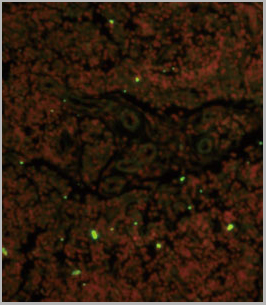
 IF (Immunofluorescence)
(AAA31188 staining A549 cells by IF/ICC. The samples were fixed with PFA and permeabilized in 0.1% Triton X-100,then blocked in 10% serum for 45 minutes at 25°C. Samples were then incubated with primary Ab(AAA31188) and mouse anti-beta tubulin Ab(T0023) for 1 hour at 37°C. An AlexaFluor594 conjugated goat anti-rabbit IgG(H+L) Ab(Red) and an AlexaFluor488 conjugated goat anti-mouse IgG(H+L) Ab(Green) were used as the secondary antibody.The nuclear counter stain is DAPI (blue).)
IF (Immunofluorescence)
(AAA31188 staining A549 cells by IF/ICC. The samples were fixed with PFA and permeabilized in 0.1% Triton X-100,then blocked in 10% serum for 45 minutes at 25°C. Samples were then incubated with primary Ab(AAA31188) and mouse anti-beta tubulin Ab(T0023) for 1 hour at 37°C. An AlexaFluor594 conjugated goat anti-rabbit IgG(H+L) Ab(Red) and an AlexaFluor488 conjugated goat anti-mouse IgG(H+L) Ab(Green) were used as the secondary antibody.The nuclear counter stain is DAPI (blue).)
ARF3, Polyclonal Antibody (Cat# AAA31188)
Full Name
ARF3 Antibody
Reactivity
Human, Rat
Predicted Reactivity: Pig(100%), Bovine(100%), Horse(100%), Sheep(100%), Chicken(100%), Xenopus(100%)
Predicted Reactivity: Pig(100%), Bovine(100%), Horse(100%), Sheep(100%), Chicken(100%), Xenopus(100%)
Applications
ELISA
Purity
The antiserum was purified by peptide affinity chromatography using SulfoLink Coupling Resin (Thermo Fisher Scientific).
Pricing
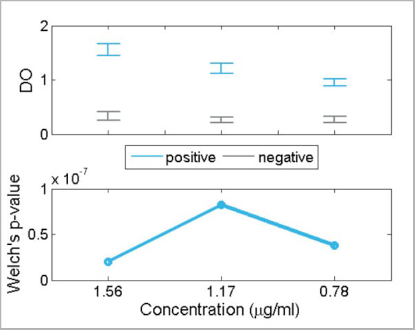 Application Data
(In this plot, the optical density at 450/620 nm for positive (blue) and negative (gray) IgM sera are compared for each concentration of the recombinant antigen. An appropriate statistical test of significance for the comparison of means between both groups, the Welch's test, is employed. Eligible concentrations for the use of the antigen should present statistically significant differences between positive and negative sera. This happens when the intervals at the top do not overlap and, equivalently, when the p-value at the bottom is below 0.05. In the present figure, all p-values are below 0.05 and thus the intervals do not overlap. Therefore, any of the showed concentrations can be used to distinguish between positive and negative sera.)
Application Data
(In this plot, the optical density at 450/620 nm for positive (blue) and negative (gray) IgM sera are compared for each concentration of the recombinant antigen. An appropriate statistical test of significance for the comparison of means between both groups, the Welch's test, is employed. Eligible concentrations for the use of the antigen should present statistically significant differences between positive and negative sera. This happens when the intervals at the top do not overlap and, equivalently, when the p-value at the bottom is below 0.05. In the present figure, all p-values are below 0.05 and thus the intervals do not overlap. Therefore, any of the showed concentrations can be used to distinguish between positive and negative sera.)
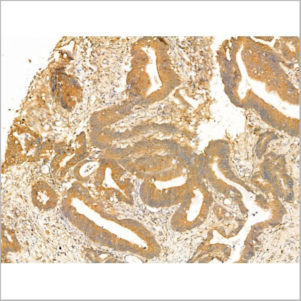
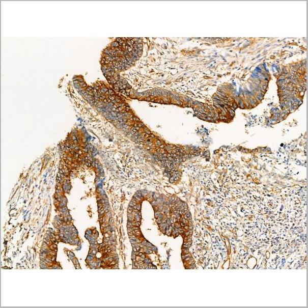
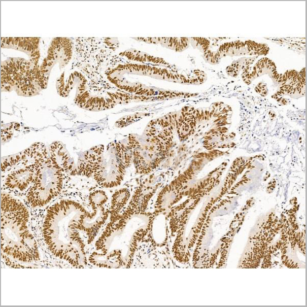
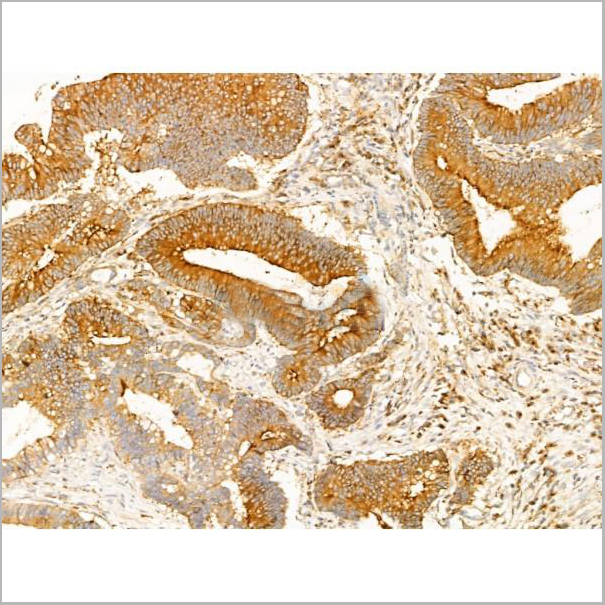
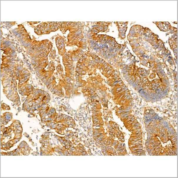
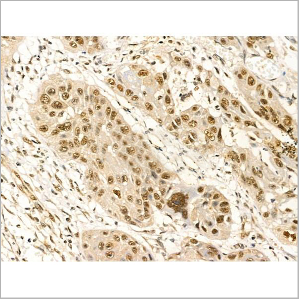
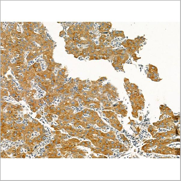
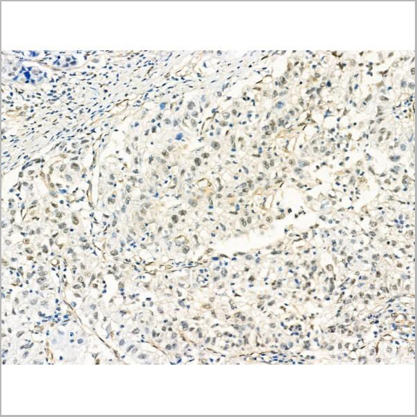
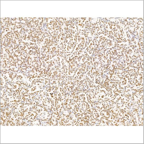
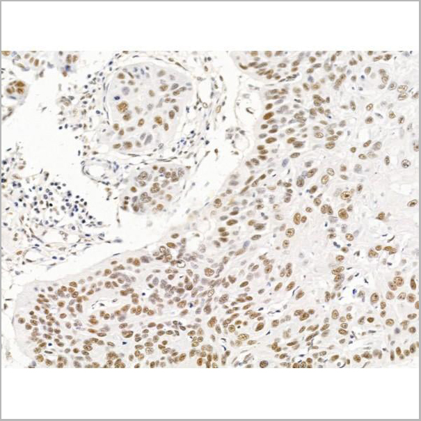
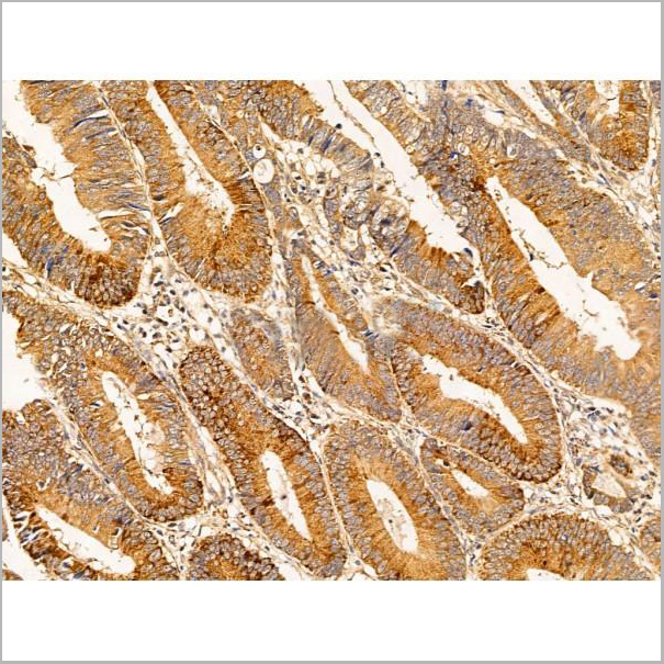
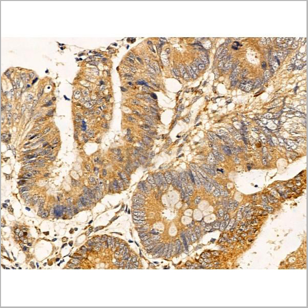
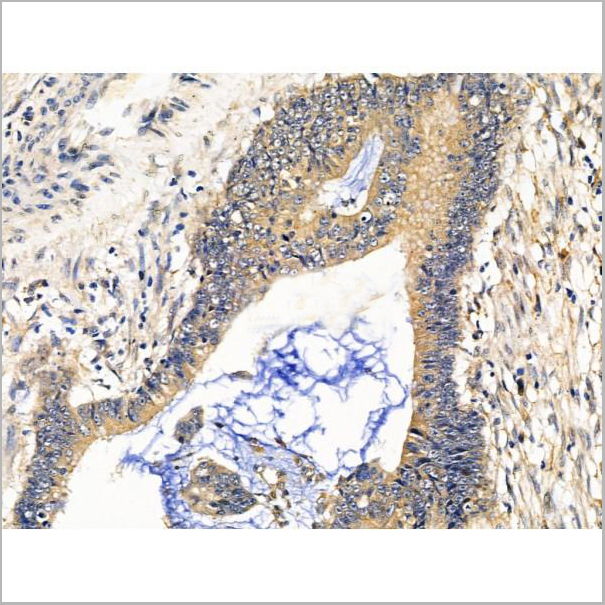
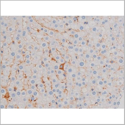
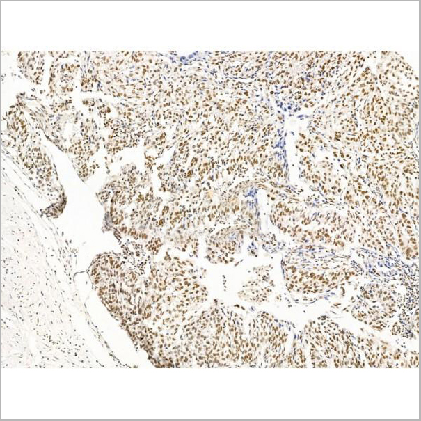
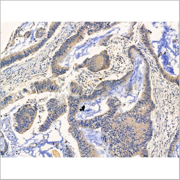
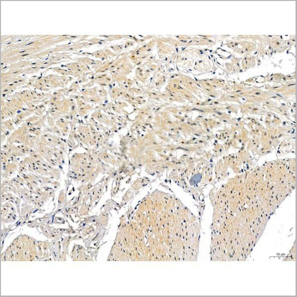
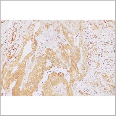
 IHC (Immunohistochemistry-Paraffin)
(AAA31273 at 1/100 staining Human colorectal cancer and adjacent normal tissues by IHC-P. The sample was formaldehyde fixed and a heat mediated antigen retrieval step in citrate buffer was performed. The sample was then blocked and incubated with the primary antibody at 4°C overnight. An HRP conjugated anti-Rabbit antibody was used as the secondary antibody.)
IHC (Immunohistochemistry-Paraffin)
(AAA31273 at 1/100 staining Human colorectal cancer and adjacent normal tissues by IHC-P. The sample was formaldehyde fixed and a heat mediated antigen retrieval step in citrate buffer was performed. The sample was then blocked and incubated with the primary antibody at 4°C overnight. An HRP conjugated anti-Rabbit antibody was used as the secondary antibody.)
GCHFR, Polyclonal Antibody (Cat# AAA31273)
Full Name
GCHFR Antibody
Gene Names
GCHFR; P35; GFRP; HsT16933
Reactivity
Human, Mouse, Rat
Predicted Reactivity: Bovine(88%), Horse(88%), Dog(88%)
Predicted Reactivity: Bovine(88%), Horse(88%), Dog(88%)
Applications
ELISA
Purity
The antiserum was purified by peptide affinity chromatography using SulfoLink Coupling Resin (Thermo Fisher Scientific).
Pricing

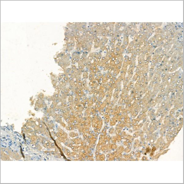
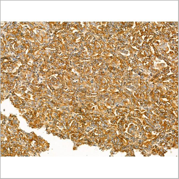
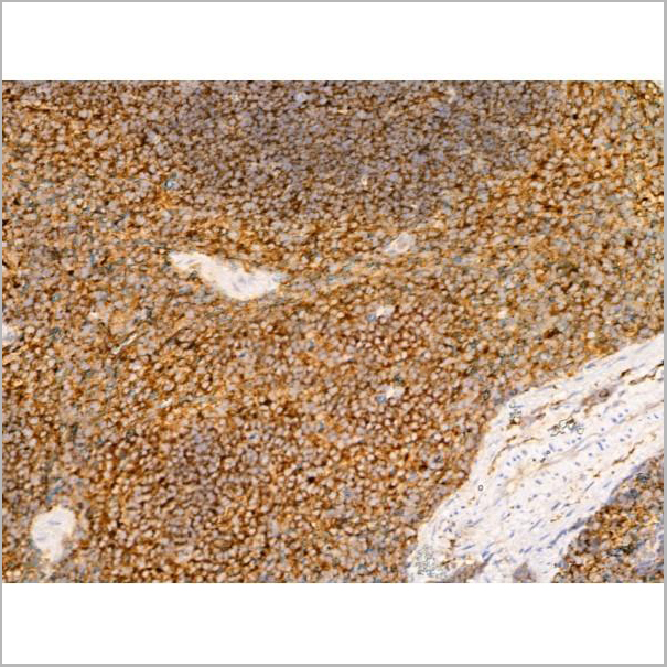
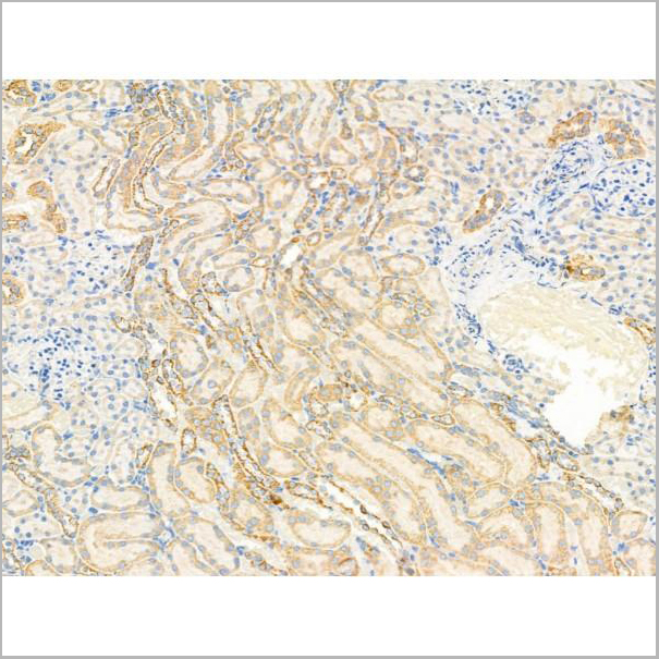
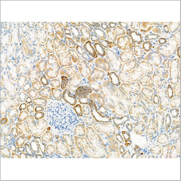
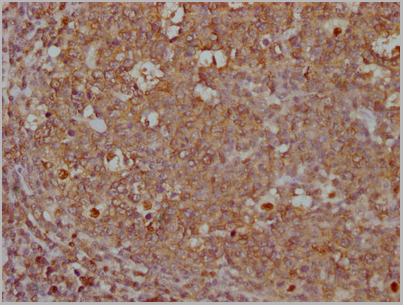
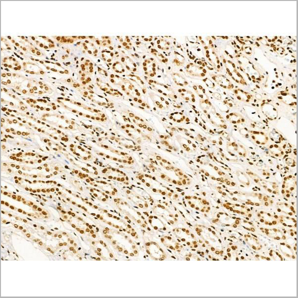
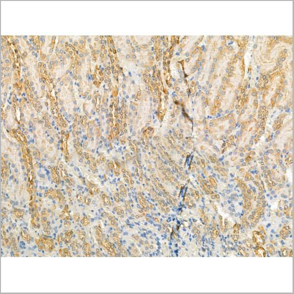
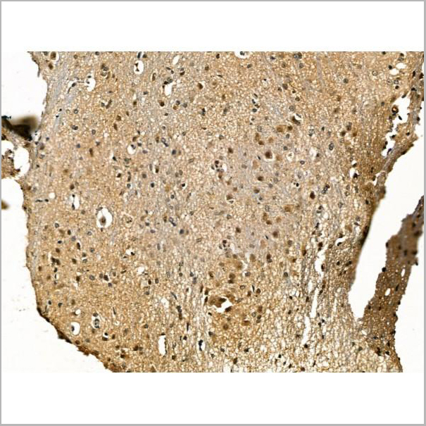
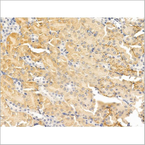
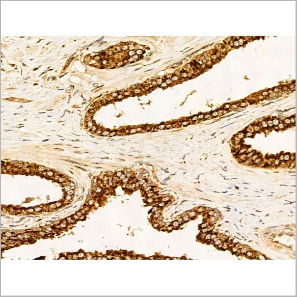
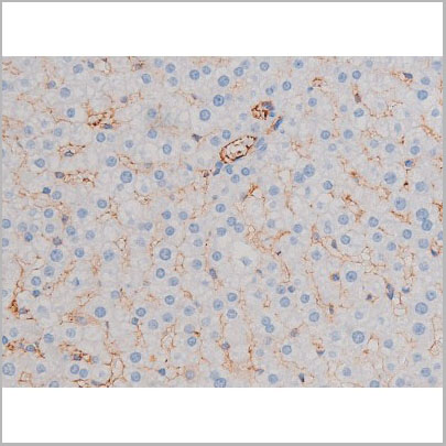
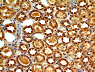
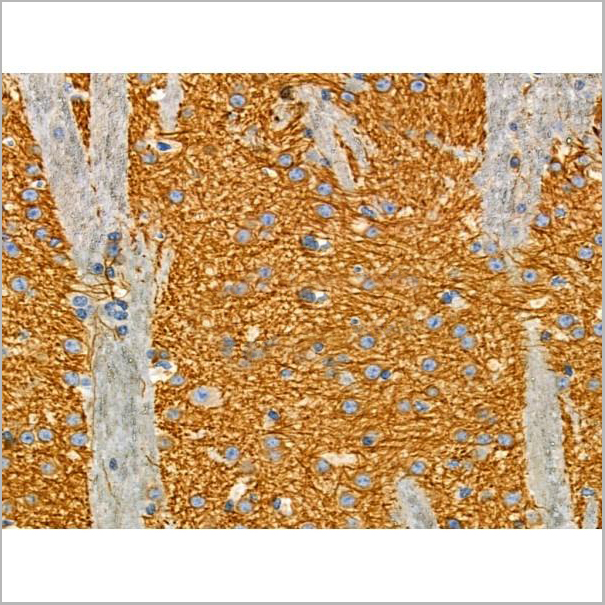
 IHC (Immunohistchemistry-Paraffin)
(AAA31183 at 1/100 staining Human kidney cancer by IHC-P. The sample was formaldehyde fixed and a heat mediated antigen retrieval step in citrate buffer was performed. The sample was then blocked and incubated with the primary antibody at 4°C overnight. An HRP conjugated anti-Rabbit antibody was used as the secondary antibody.)
IHC (Immunohistchemistry-Paraffin)
(AAA31183 at 1/100 staining Human kidney cancer by IHC-P. The sample was formaldehyde fixed and a heat mediated antigen retrieval step in citrate buffer was performed. The sample was then blocked and incubated with the primary antibody at 4°C overnight. An HRP conjugated anti-Rabbit antibody was used as the secondary antibody.)
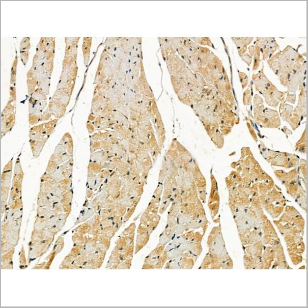
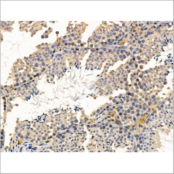
GDF8/Myostatin, Polyclonal Antibody (Cat# AAA31183)
Full Name
GDF8/Myostatin Antibody
Gene Names
MSTN; GDF8; MSLHP
Reactivity
Human, Mouse, Rat, Sheep
Applications
Western Blot, Immunohistochemistry
Purity
Peptide affinity purification
Pricing
 IF (Immunofluorescence)
(AAA31176 staining Hela cells by IF/ICC. The samples were fixed with PFA and permeabilized in 0.1% Triton X-100,then blocked in 10% serum for 45 minutes at 25°C. Samples were then incubated with primary Ab(AAA31176 1:200) and mouse anti-beta tubulin Ab(T0023 1:200) for 1 hour at 37°C. An AlexaFluor594 conjugated goat anti-rabbit IgG(H+L) Ab(Red) and an AlexaFluor488 conjugated goat anti-mouse IgG(H+L) Ab(Green) were used as the secondary antibody.The nuclear counter stain is DAPI(blue).)
IF (Immunofluorescence)
(AAA31176 staining Hela cells by IF/ICC. The samples were fixed with PFA and permeabilized in 0.1% Triton X-100,then blocked in 10% serum for 45 minutes at 25°C. Samples were then incubated with primary Ab(AAA31176 1:200) and mouse anti-beta tubulin Ab(T0023 1:200) for 1 hour at 37°C. An AlexaFluor594 conjugated goat anti-rabbit IgG(H+L) Ab(Red) and an AlexaFluor488 conjugated goat anti-mouse IgG(H+L) Ab(Green) were used as the secondary antibody.The nuclear counter stain is DAPI(blue).)
cbx7, Polyclonal Antibody (Cat# AAA31176)
Full Name
cbx7 Antibody
Reactivity
Human, Mouse, Rat
Predicted Reactivity: Pig(100%), Bovine(100%), Sheep(100%), Rabbit(100%), Dog(100%), Chicken(88%), Xenopus(100%)
Predicted Reactivity: Pig(100%), Bovine(100%), Sheep(100%), Rabbit(100%), Dog(100%), Chicken(88%), Xenopus(100%)
Applications
Immunofluorescence, Immunocytochemistry
Purity
The antiserum was purified by peptide affinity chromatography using SulfoLink Coupling Resin (Thermo Fisher Scientific).
Pricing
 IF (Immunofluorescence)
(AAA31205 staining Hela cells by IF/ICC. The samples were fixed with PFA and permeabilized in 0.1% Triton X-100,then blocked in 10% serum for 45 minutes at 25°C. Samples were then incubated with primary Ab(AAA31205 1:200) and mouse anti-beta tubulin Ab(T0023 1:200) for 1 hour at 37°C. An AlexaFluor594 conjugated goat anti-rabbit IgG(H+L) Ab(Red) and an AlexaFluor488 conjugated goat anti-mouse IgG(H+L) Ab(Green) were used as the secondary antibody.The nuclear counter stain is DAPI(blue).)
IF (Immunofluorescence)
(AAA31205 staining Hela cells by IF/ICC. The samples were fixed with PFA and permeabilized in 0.1% Triton X-100,then blocked in 10% serum for 45 minutes at 25°C. Samples were then incubated with primary Ab(AAA31205 1:200) and mouse anti-beta tubulin Ab(T0023 1:200) for 1 hour at 37°C. An AlexaFluor594 conjugated goat anti-rabbit IgG(H+L) Ab(Red) and an AlexaFluor488 conjugated goat anti-mouse IgG(H+L) Ab(Green) were used as the secondary antibody.The nuclear counter stain is DAPI(blue).)
CBX1, Polyclonal Antibody (Cat# AAA31205)
Full Name
CBX1/HP1 beta Antibody
Gene Names
CBX1; CBX; M31; MOD1; p25beta; HP1-BETA; HP1Hsbeta; HP1Hs-beta
Reactivity
Human, Mouse, Rat
Predicted Reactivity: Pig(100%), Bovine(100%), Horse(100%), Sheep(100%), Rabbit(100%), Dog(100%), Chicken(100%)
Predicted Reactivity: Pig(100%), Bovine(100%), Horse(100%), Sheep(100%), Rabbit(100%), Dog(100%), Chicken(100%)
Applications
ELISA
Purity
The antiserum was purified by peptide affinity chromatography using SulfoLink Coupling Resin (Thermo Fisher Scientific).
Pricing


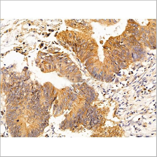
 IHC (Immunohistochemistry-Paraffin)
(AAA31239 at 1/100 staining Human colorectal cancer and adjacent normal tissues by IHC-P. The sample was formaldehyde fixed and a heat mediated antigen retrieval step in citrate buffer was performed. The sample was then blocked and incubated with the primary antibody at 4°C overnight. An HRP conjugated anti-Rabbit antibody was used as the secondary antibody.)
IHC (Immunohistochemistry-Paraffin)
(AAA31239 at 1/100 staining Human colorectal cancer and adjacent normal tissues by IHC-P. The sample was formaldehyde fixed and a heat mediated antigen retrieval step in citrate buffer was performed. The sample was then blocked and incubated with the primary antibody at 4°C overnight. An HRP conjugated anti-Rabbit antibody was used as the secondary antibody.)
WIPI1, Polyclonal Antibody (Cat# AAA31239)
Full Name
WIPI1 Antibody
Gene Names
WIPI1; ATG18; ATG18A; WIPI49
Reactivity
Human, Mouse, Rat
Predicted Reactivity: Pig(100%), Bovine(88%), Horse(100%), Rabbit(100%), Dog(88%), Chicken(86%)
Predicted Reactivity: Pig(100%), Bovine(88%), Horse(100%), Rabbit(100%), Dog(88%), Chicken(86%)
Applications
ELISA
Purity
The antiserum was purified by peptide affinity chromatography using SulfoLink Coupling Resin (Thermo Fisher Scientific).
Pricing
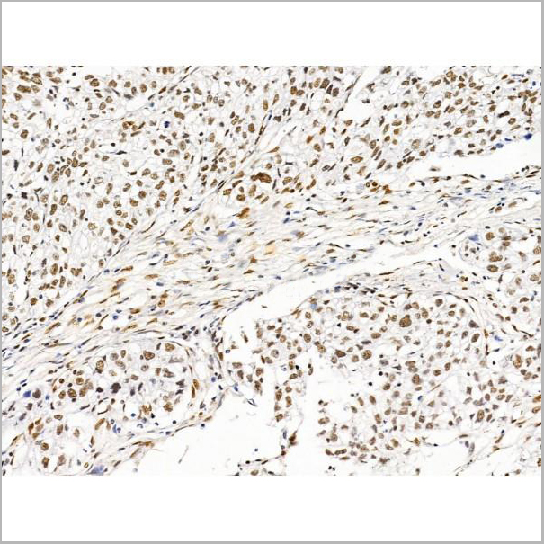
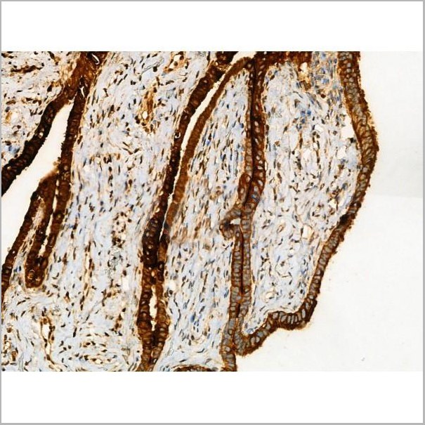
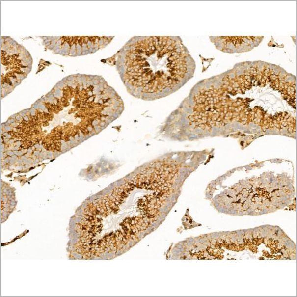
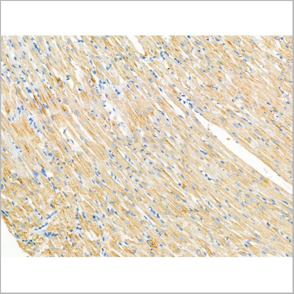
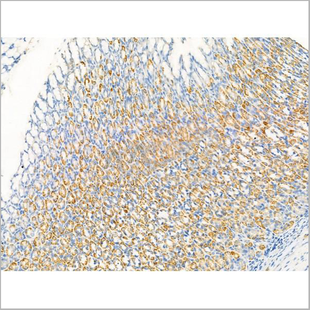
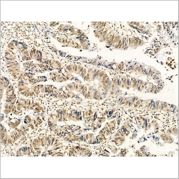
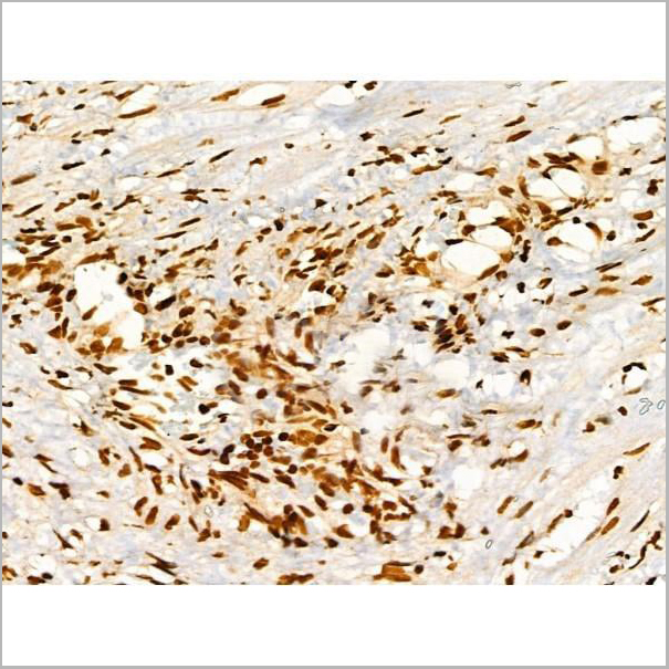
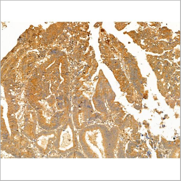
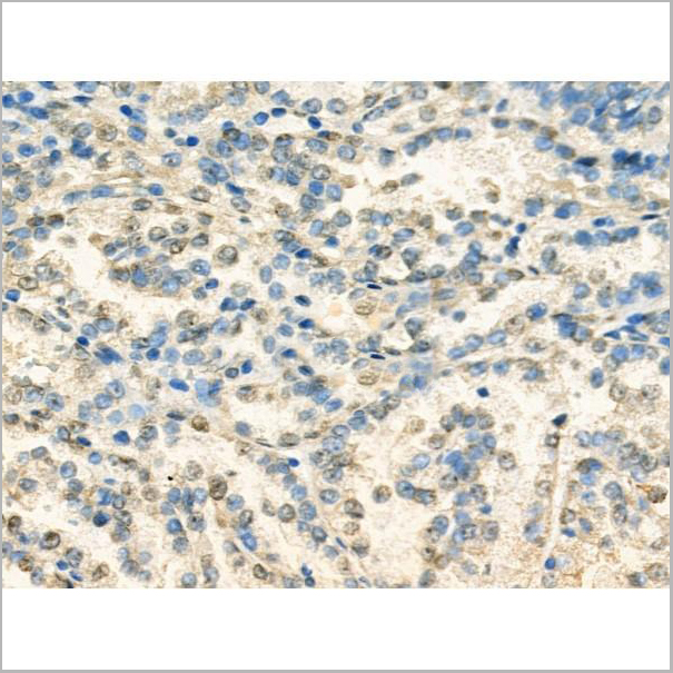
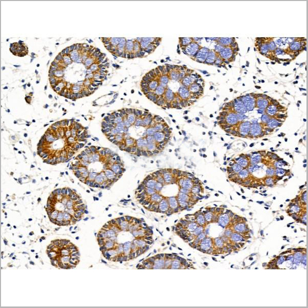
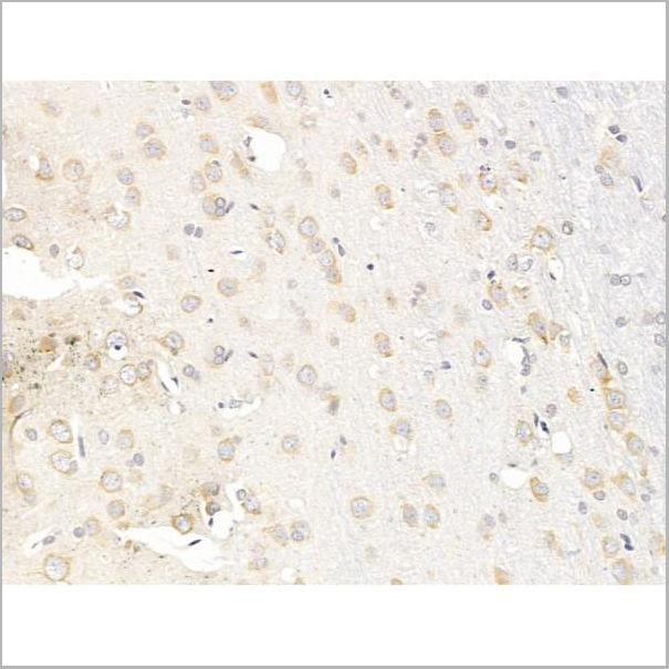
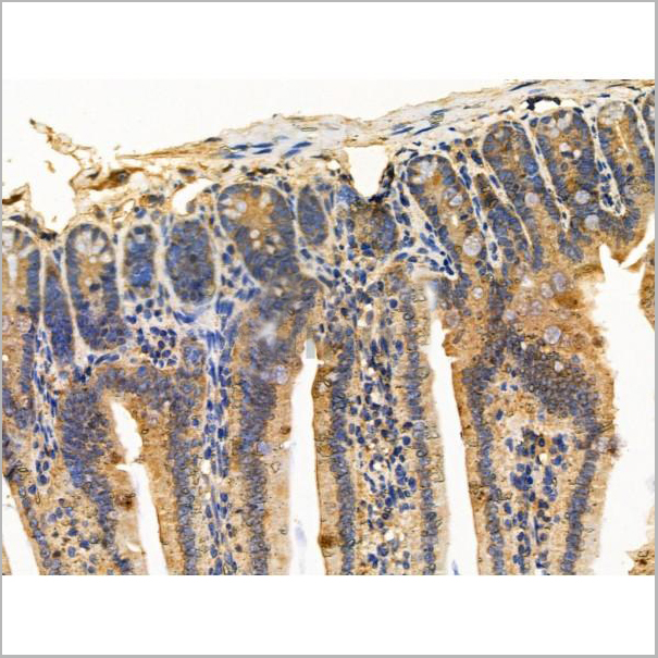
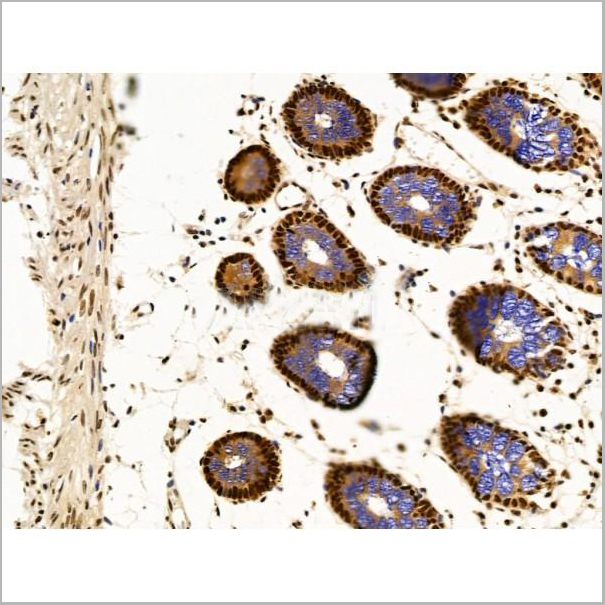
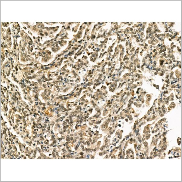
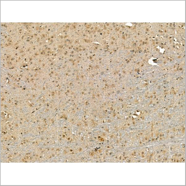
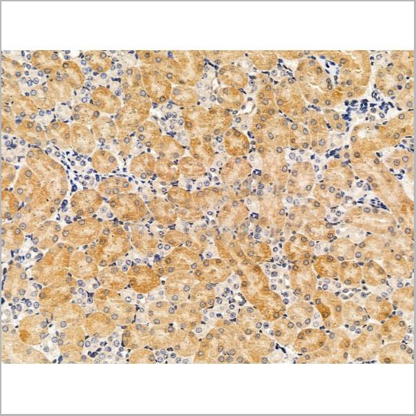
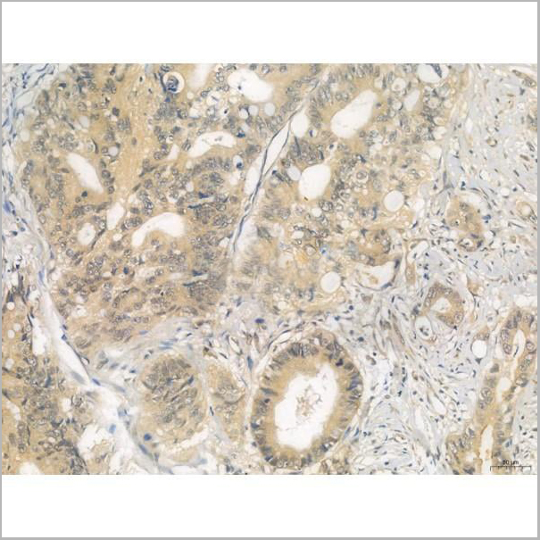
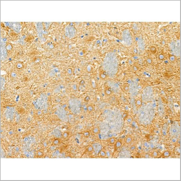
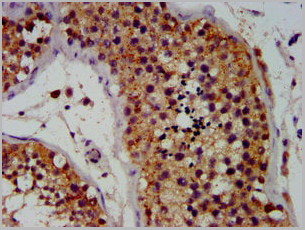
 IHC (Immunohistochemistry-Paraffin)
(AAA31257 at 1/100 staining Human esophageal cancer by IHC-P. The sample was formaldehyde fixed and a heat mediated antigen retrieval step in citrate buffer was performed. The sample was then blocked and incubated with the primary antibody at 4°C overnight. An HRP conjugated anti-Rabbit antibody was used as the secondary antibody.)
IHC (Immunohistochemistry-Paraffin)
(AAA31257 at 1/100 staining Human esophageal cancer by IHC-P. The sample was formaldehyde fixed and a heat mediated antigen retrieval step in citrate buffer was performed. The sample was then blocked and incubated with the primary antibody at 4°C overnight. An HRP conjugated anti-Rabbit antibody was used as the secondary antibody.)
ILF1, Polyclonal Antibody (Cat# AAA31257)
Full Name
ILF1 Antibody
Gene Names
FOXK2; ILF; ILF1; ILF-1; nGTBP
Reactivity
Human, Mouse, Rat
Predicted Reactivity: Horse(100%)
Predicted Reactivity: Horse(100%)
Applications
ELISA
Purity
The antiserum was purified by peptide affinity chromatography using SulfoLink Coupling Resin (Thermo Fisher Scientific).
Pricing
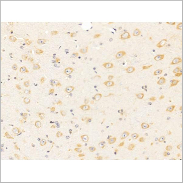
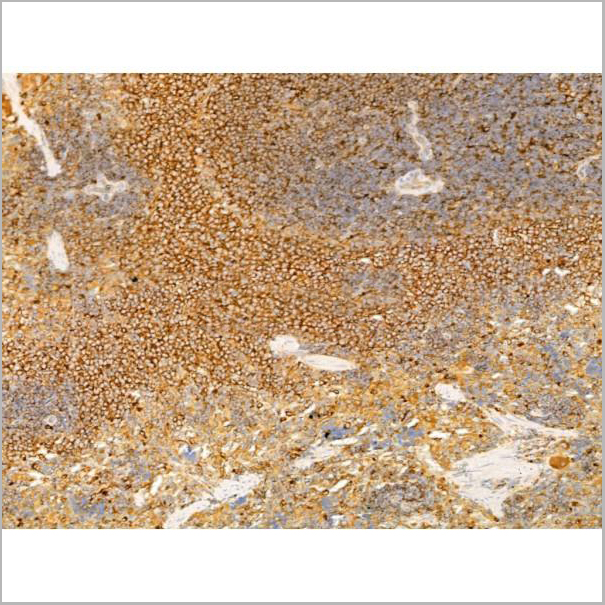
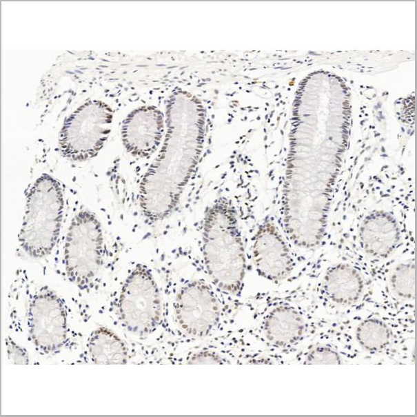
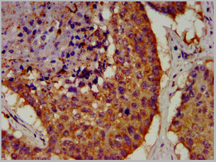
 IHC (Immunohistochemistry-Paraffin)
(AAA31259 at 1/100 staining Human esophageal cancer by IHC-P. The sample was formaldehyde fixed and a heat mediated antigen retrieval step in citrate buffer was performed. The sample was then blocked and incubated with the primary antibody at 4°C overnight. An HRP conjugated anti-Rabbit antibody was used as the secondary antibody.)
IHC (Immunohistochemistry-Paraffin)
(AAA31259 at 1/100 staining Human esophageal cancer by IHC-P. The sample was formaldehyde fixed and a heat mediated antigen retrieval step in citrate buffer was performed. The sample was then blocked and incubated with the primary antibody at 4°C overnight. An HRP conjugated anti-Rabbit antibody was used as the secondary antibody.)
SLC1A5, Polyclonal Antibody (Cat# AAA31259)
Full Name
SLC1A5 Antibody
Gene Names
SLC1A5; R16; AAAT; ATBO; M7V1; RDRC; ASCT2; M7VS1
Reactivity
Human, Mouse, Rat
Predicted Reactivity: Pig(100%), Bovine(88%), Horse(88%), Sheep(88%), Rabbit(88%), Dog(100%)
Predicted Reactivity: Pig(100%), Bovine(88%), Horse(88%), Sheep(88%), Rabbit(88%), Dog(100%)
Applications
ELISA
Purity
The antiserum was purified by peptide affinity chromatography using SulfoLink Coupling Resin (Thermo Fisher Scientific).
Pricing
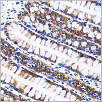
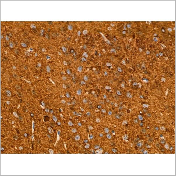
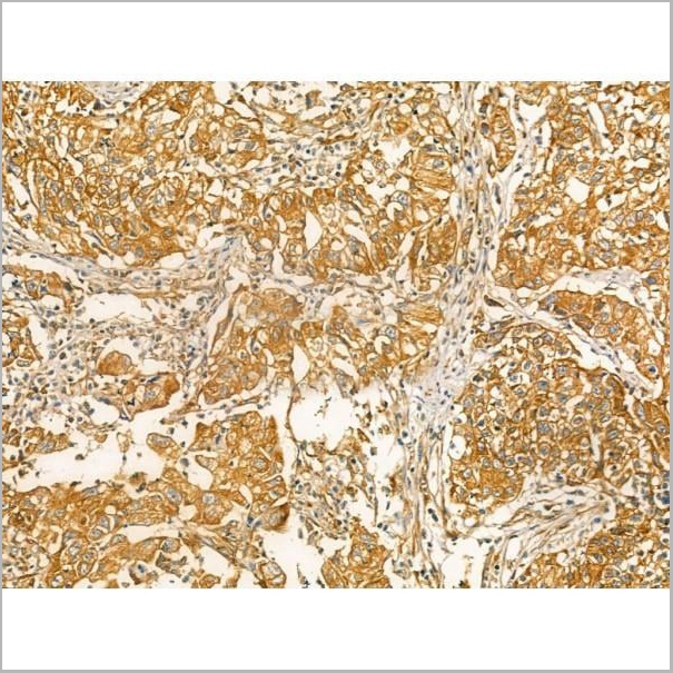
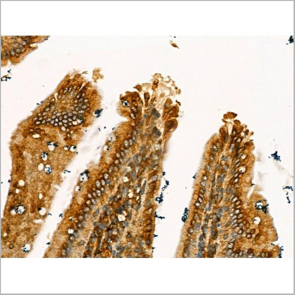
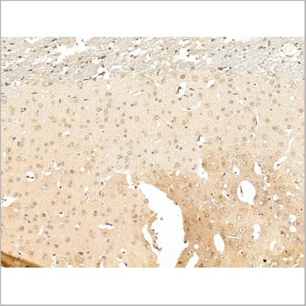
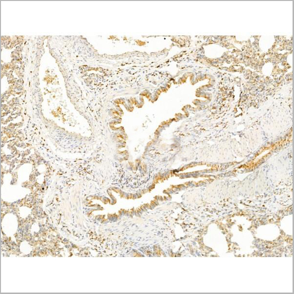
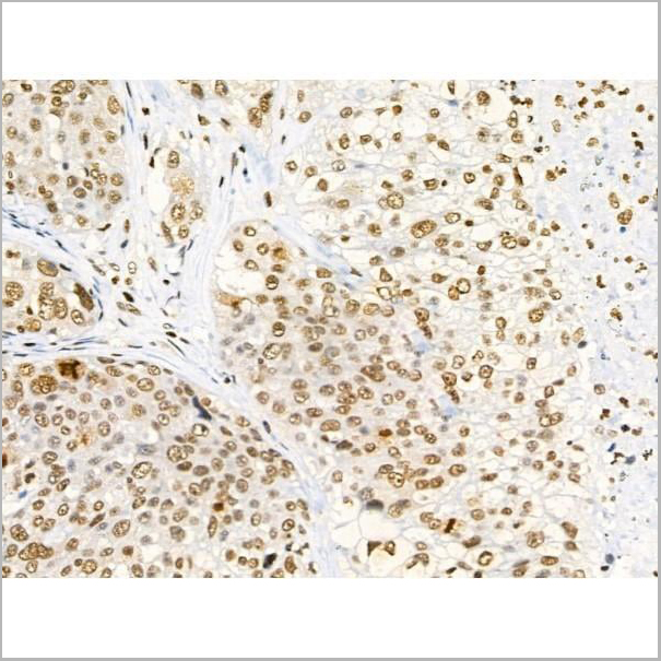
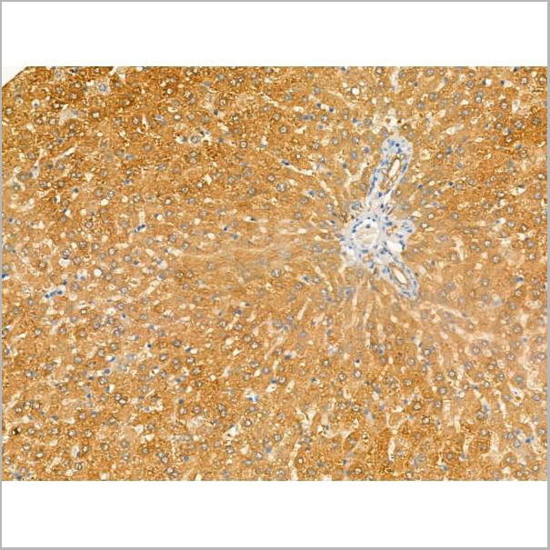
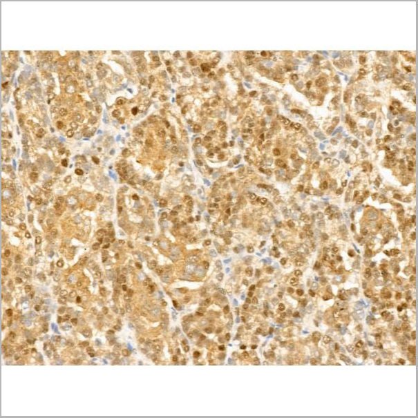
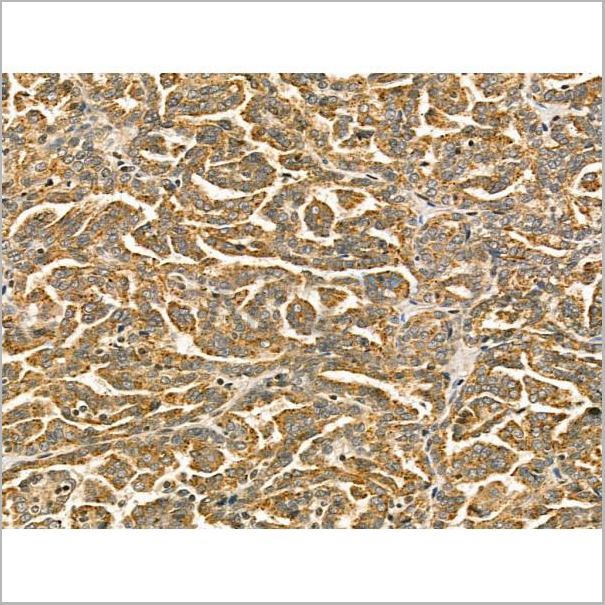
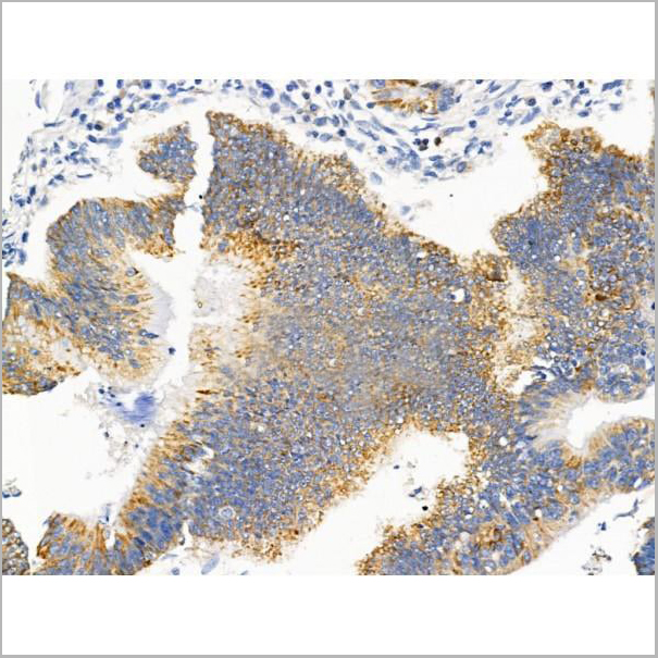
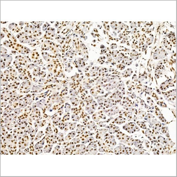
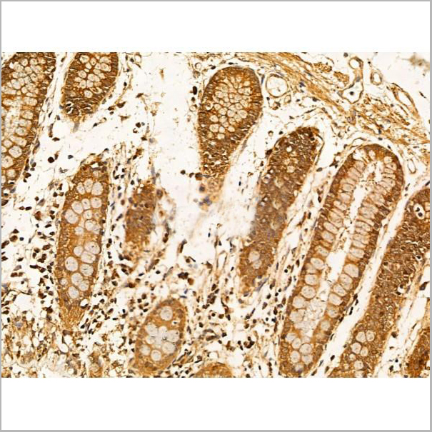
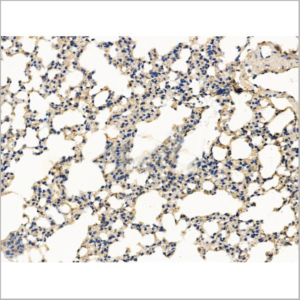
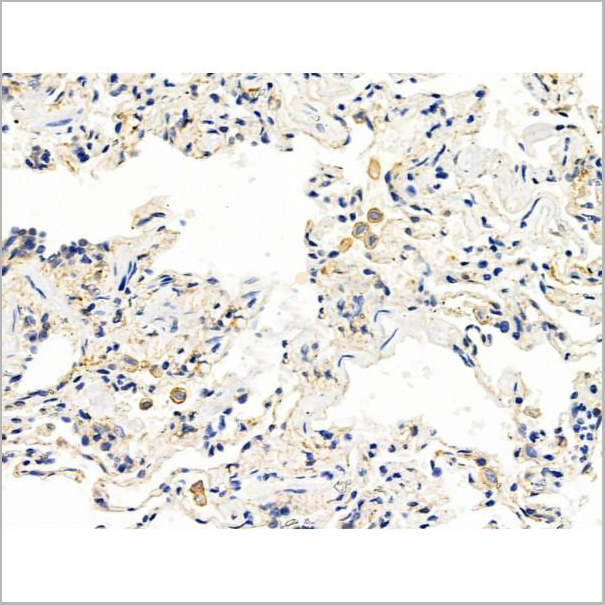
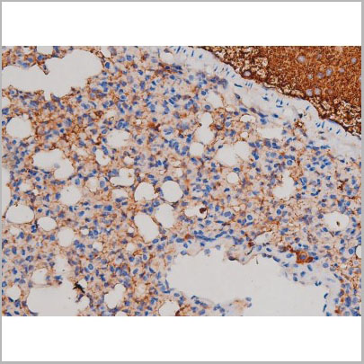
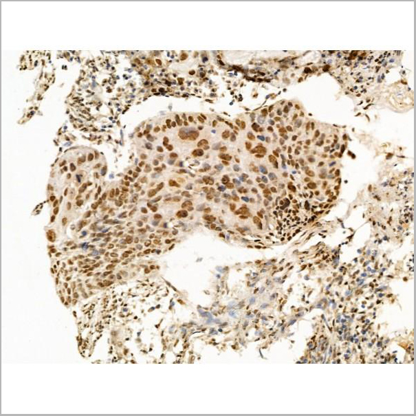
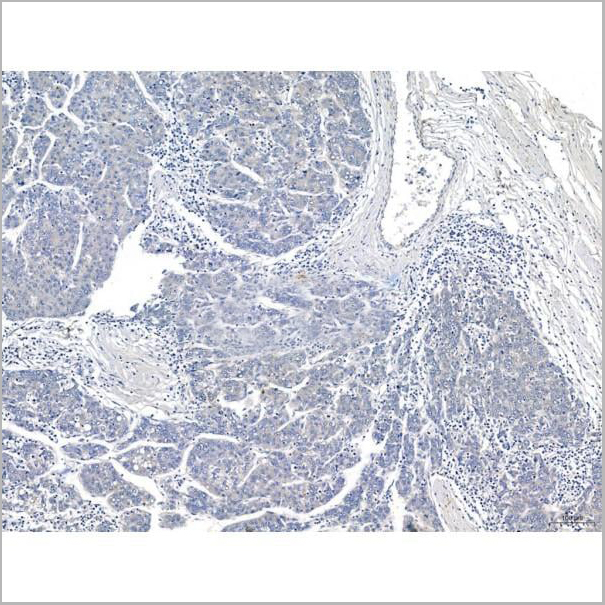
 IHC (Immunohistochemistry)
(AAA31135 at 1/100 staining Human lung cancer and adjacent normal tissues by IHC-P. The sample was formaldehyde fixed and a heat mediated antigen retrieval step in citrate buffer was performed. The sample was then blocked and incubated with the primary antibody at 4°C overnight. An HRP conjugated anti-Rabbit antibody was used as the secondary antibody.)
IHC (Immunohistochemistry)
(AAA31135 at 1/100 staining Human lung cancer and adjacent normal tissues by IHC-P. The sample was formaldehyde fixed and a heat mediated antigen retrieval step in citrate buffer was performed. The sample was then blocked and incubated with the primary antibody at 4°C overnight. An HRP conjugated anti-Rabbit antibody was used as the secondary antibody.)
PIP, Polyclonal Antibody (Cat# AAA31135)
Full Name
PIP Antibody
Gene Names
PIP; GPIP4; GCDFP15; GCDFP-15
Reactivity
Human, Mouse, Rat
Applications
Western Blot, Immunohistochemistry, Immunofluorescence
Purity
Peptide affinity purification
Pricing
 IF (Immunofluorescence)
(AAA31261 staining Hela cells by IF/ICC. The samples were fixed with PFA and permeabilized in 0.1% Triton X-100,then blocked in 10% serum for 45 minutes at 25°C. Samples were then incubated with primary Ab(AAA31261 1:200) and mouse anti-beta tubulin Ab(T0023 1:200) for 1 hour at 37°C. An AlexaFluor594 conjugated goat anti-rabbit IgG(H+L) Ab(Red) and an AlexaFluor488 conjugated goat anti-mouse IgG(H+L) Ab(Green) were used as the secondary antibody.The nuclear counter stain is DAPI(blue).)
IF (Immunofluorescence)
(AAA31261 staining Hela cells by IF/ICC. The samples were fixed with PFA and permeabilized in 0.1% Triton X-100,then blocked in 10% serum for 45 minutes at 25°C. Samples were then incubated with primary Ab(AAA31261 1:200) and mouse anti-beta tubulin Ab(T0023 1:200) for 1 hour at 37°C. An AlexaFluor594 conjugated goat anti-rabbit IgG(H+L) Ab(Red) and an AlexaFluor488 conjugated goat anti-mouse IgG(H+L) Ab(Green) were used as the secondary antibody.The nuclear counter stain is DAPI(blue).)
Filaggrin, Polyclonal Antibody (Cat# AAA31261)
Full Name
Filaggrin Antibody
Gene Names
FLI1; EWSR2; SIC-1; BDPLT21
Reactivity
Human, Rat
Applications
ELISA
Purity
The antiserum was purified by peptide affinity chromatography using SulfoLink Coupling Resin (Thermo Fisher Scientific).
Pricing
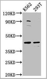
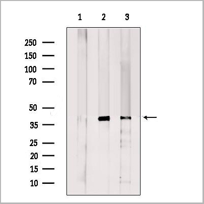
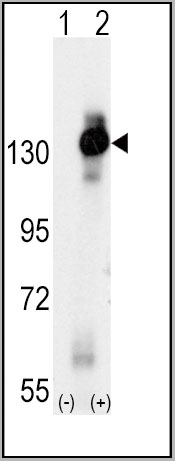
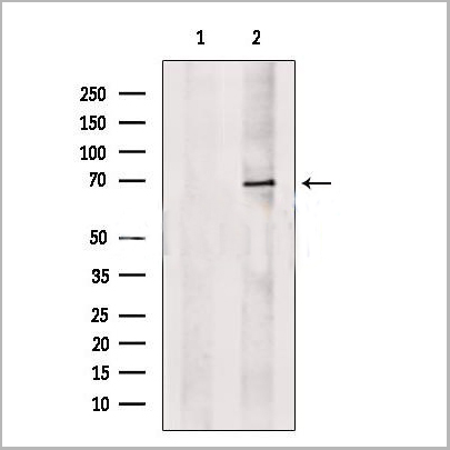
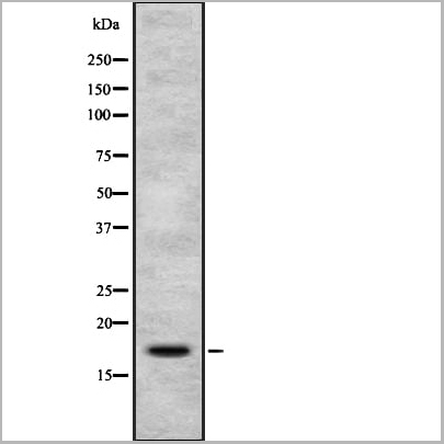
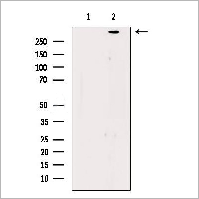
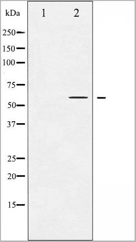
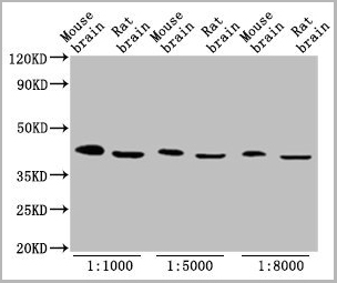
 WB (Western Blot)
(Western Blot analysis of 100ng of PEGylated BSA using AAA24071 at 1:30,000 dilution. A: Normal BSA. B: PEGylated BSA.)
WB (Western Blot)
(Western Blot analysis of 100ng of PEGylated BSA using AAA24071 at 1:30,000 dilution. A: Normal BSA. B: PEGylated BSA.)
Polyethylene Glycol, Polyclonal Antibody (Cat# AAA24071)
Full Name
Polyethylene Glycol (PEG)
Applications
Western Blot
Purity
Affinity Purified
Purified by immunoaffinity chromatography
Purified by immunoaffinity chromatography
Pricing
 IF (Immunofluorescence)
(Immunofluorescence staining of HepG2 cells with AAA27019 at 1:133, counter-stained with DAPI. The cells were fixed in 4% formaldehyde, permeabilized using 0.2% Triton X-100 and blocked in 10% normal Goat Serum. The cells were then incubated with the antibody overnight at 4 degree C. The secondary antibody was Alexa Fluor 488-congugated AffiniPure Goat Anti-Rabbit IgG(H+L).)
IF (Immunofluorescence)
(Immunofluorescence staining of HepG2 cells with AAA27019 at 1:133, counter-stained with DAPI. The cells were fixed in 4% formaldehyde, permeabilized using 0.2% Triton X-100 and blocked in 10% normal Goat Serum. The cells were then incubated with the antibody overnight at 4 degree C. The secondary antibody was Alexa Fluor 488-congugated AffiniPure Goat Anti-Rabbit IgG(H+L).)
ETV2, Polyclonal Antibody (Cat# AAA27019)
Full Name
ETV2 Antibody
Gene Names
ETV2; ER71; ETSRP71
Reactivity
Human
Applications
Immunofluorescence
Purity
>95%, Protein G purified
Pricing
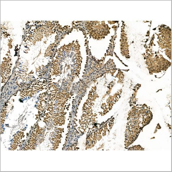
 IHC (Immunohistochemistry-Paraffin)
(AAA31201 at 1/100 staining Human lung cancer and adjacent normal tissues by IHC-P. The sample was formaldehyde fixed and a heat mediated antigen retrieval step in citrate buffer was performed. The sample was then blocked and incubated with the primary antibody at 4°C overnight. An HRP conjugated anti-Rabbit antibody was used as the secondary antibody.)
IHC (Immunohistochemistry-Paraffin)
(AAA31201 at 1/100 staining Human lung cancer and adjacent normal tissues by IHC-P. The sample was formaldehyde fixed and a heat mediated antigen retrieval step in citrate buffer was performed. The sample was then blocked and incubated with the primary antibody at 4°C overnight. An HRP conjugated anti-Rabbit antibody was used as the secondary antibody.)
PICALM, Polyclonal Antibody (Cat# AAA31201)
Full Name
PICALM Antibody
Gene Names
PICALM; LAP; CALM; CLTH
Reactivity
Human, Mouse, Rat
Predicted Reactivity: Pig(100%), Bovine(100%), Horse(100%), Sheep(100%), Rabbit(100%), Dog(100%)
Predicted Reactivity: Pig(100%), Bovine(100%), Horse(100%), Sheep(100%), Rabbit(100%), Dog(100%)
Applications
ELISA
Purity
The antiserum was purified by peptide affinity chromatography using SulfoLink Coupling Resin (Thermo Fisher Scientific).
Pricing
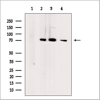
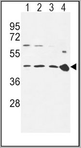
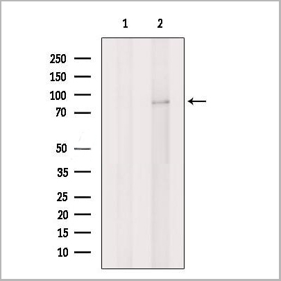
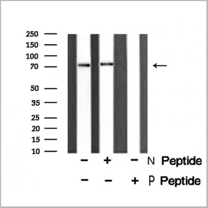
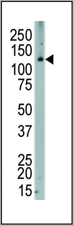
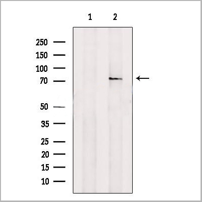
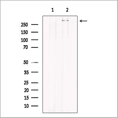
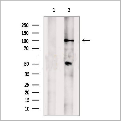
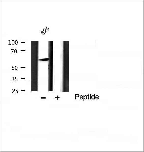 WB (Western Blot)
(Western blot analysis of Akt phosphorylation expression in B2C Cell line lysates, The lane on the right is treated with the antigen-specific peptide.)
WB (Western Blot)
(Western blot analysis of Akt phosphorylation expression in B2C Cell line lysates, The lane on the right is treated with the antigen-specific peptide.)
Akt, Polyclonal Antibody (Cat# AAA31035)
Full Name
Phospho-Akt (Ser124) Antibody
Gene Names
AKT1; AKT; PKB; RAC; CWS6; PRKBA; PKB-ALPHA; RAC-ALPHA
Reactivity
Human, Mouse, Rat
Applications
Western Blot, Immunohistochemistry, Immunofluorescence, Immunocytochemistry
Purity
From purified rabbit serum by affinity purification via sequential chromatography on phospho-and non-phospho-peptide affinity columns.
Pricing
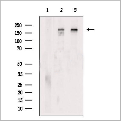
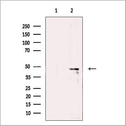
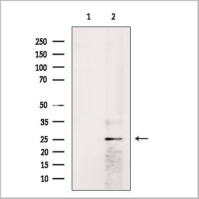
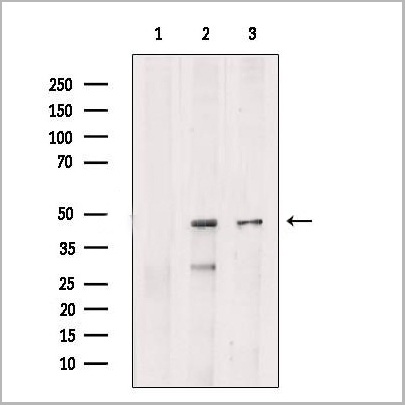
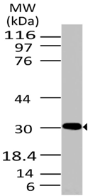
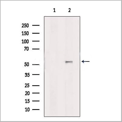
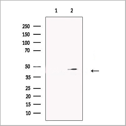
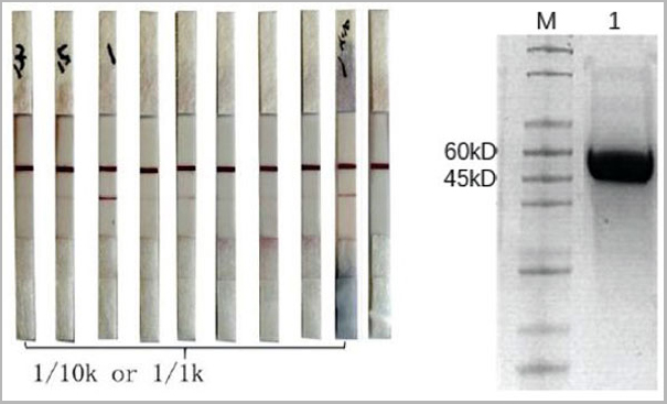 SDS-PAGE
(Under DTT-reducing SDS-PAGE conditions, the 2019-nCov Nucleocapsid protein NX fragment migrates as a 50 kDa protein (Lane 1).)
SDS-PAGE
(Under DTT-reducing SDS-PAGE conditions, the 2019-nCov Nucleocapsid protein NX fragment migrates as a 50 kDa protein (Lane 1).)
COVID 19 Nucleocapsid (NP) N fragment protein (NX) Coronavirus, Recombinant Protein (Cat# AAA27950)
Full Name
2019-nCov Nucleocapsid protein N fragment protein (NX)
Applications
SDS-Page
Purity
Purity: >95% by SDS-page
Purification: Ni column
Purification: Ni column
Pricing
 FCM (Flow Cytometry)
(Fig-4: Intracellular flow analysis of TLR7 in Raji cells using 0.5 ug/10^6 cells of TLR7 antibody (Clone: ABM2C27). Green represents isotype control; red represents anti-TLR7 antibody. Goat anti-mouse PE conjugate was used as secondary antibody.)
FCM (Flow Cytometry)
(Fig-4: Intracellular flow analysis of TLR7 in Raji cells using 0.5 ug/10^6 cells of TLR7 antibody (Clone: ABM2C27). Green represents isotype control; red represents anti-TLR7 antibody. Goat anti-mouse PE conjugate was used as secondary antibody.)
TLR7, Monoclonal Antibody (Cat# AAA14863)
Full Name
TLR7 Monoclonal Antibody
Gene Names
TLR7; TLR7-like
Reactivity
Human
Applications
Immunohistochemistry, Flow Cytometry
Purity
Purified; Protein G Chromatography
Pricing
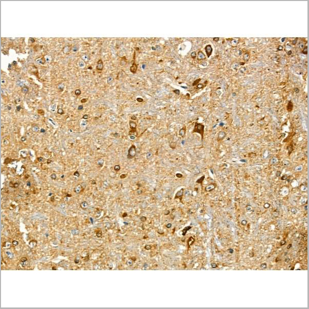
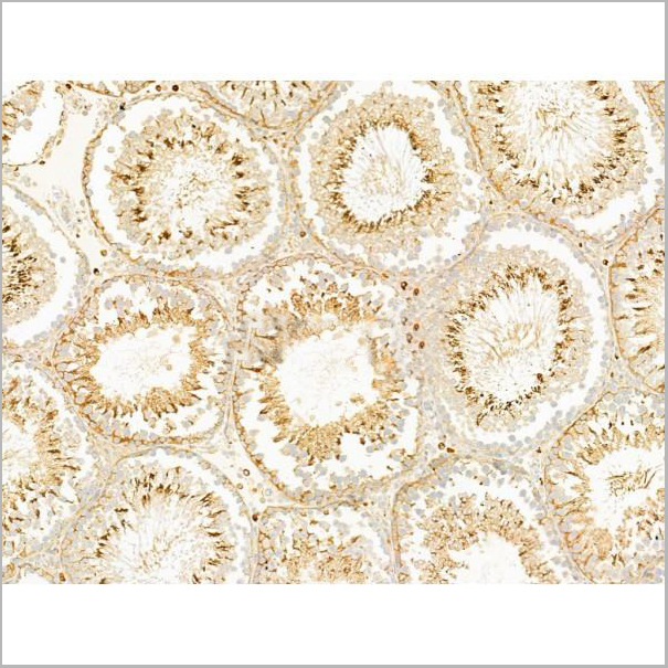
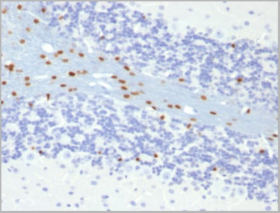
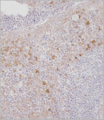
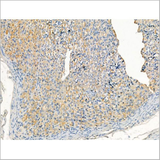
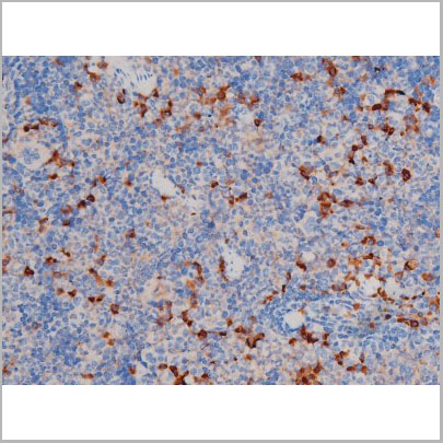
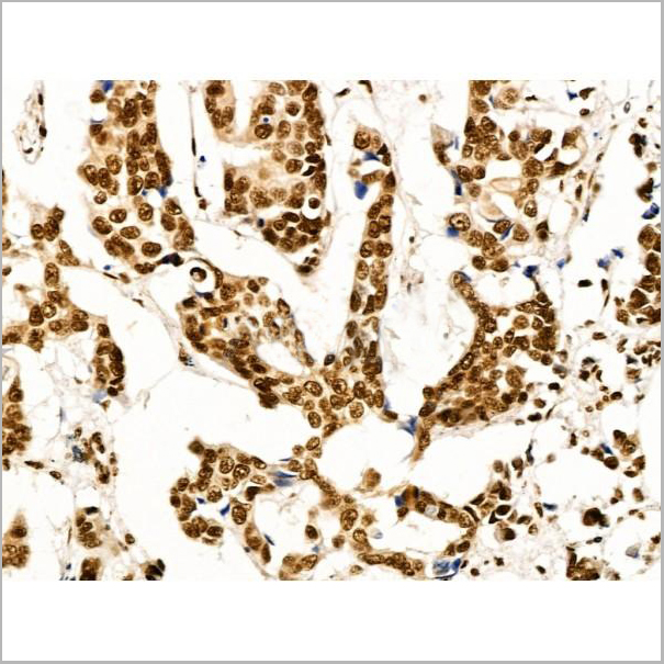
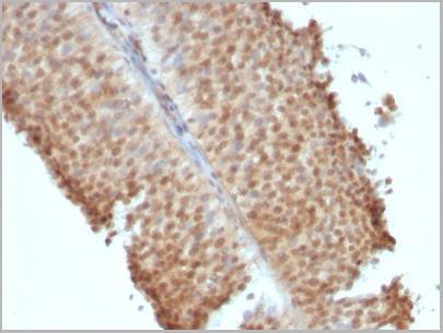
 IHC (Immunohistochemistry)
(Immunohistochemical analysis of HMGB1 in normal human spleen tissue using HMGB1 antibody (Clone: ABM24D3) at 1 ug/ml.)
IHC (Immunohistochemistry)
(Immunohistochemical analysis of HMGB1 in normal human spleen tissue using HMGB1 antibody (Clone: ABM24D3) at 1 ug/ml.)
HMGB1, Monoclonal Antibody (Cat# AAA14866)
Full Name
HMGB1 Monoclonal Antibody
Gene Names
HMGB1; HMG1; HMG3; SBP-1
Reactivity
Human (Predicted Reactivity: Mouse
Applications
Western Blot, Immunohistochemistry, Flow Cytometry
Pricing
 IHC (Immunohistochemistry-Paraffin)
(AAA31236 at 1/100 staining Human gastric cancer and adjacent normal tissues by IHC-P. The sample was formaldehyde fixed and a heat mediated antigen retrieval step in citrate buffer was performed. The sample was then blocked and incubated with the primary antibody at 4°C overnight. An HRP conjugated anti-Rabbit antibody was used as the secondary antibody.)
IHC (Immunohistochemistry-Paraffin)
(AAA31236 at 1/100 staining Human gastric cancer and adjacent normal tissues by IHC-P. The sample was formaldehyde fixed and a heat mediated antigen retrieval step in citrate buffer was performed. The sample was then blocked and incubated with the primary antibody at 4°C overnight. An HRP conjugated anti-Rabbit antibody was used as the secondary antibody.)
OVOL2, Polyclonal Antibody (Cat# AAA31236)
Full Name
OVOL2 Antibody
Gene Names
OVOL2; CHED; CHED1; CHED2; PPCD1; ZNF339; EUROIMAGE566589
Reactivity
Human, Mouse, Rat
Predicted Reactivity: Pig(100%), Bovine(100%), Horse(100%), Sheep(100%), Rabbit(100%), Dog(100%), Chicken(100%)
Predicted Reactivity: Pig(100%), Bovine(100%), Horse(100%), Sheep(100%), Rabbit(100%), Dog(100%), Chicken(100%)
Applications
ELISA
Purity
The antiserum was purified by peptide affinity chromatography using SulfoLink Coupling Resin (Thermo Fisher Scientific).
Pricing

 IF (Immunofluorescence)
(AAA31178 staining HepG2 cells by IF/ICC. The samples were fixed with PFA and permeabilized in 0.1% Triton X-100,then blocked in 10% serum for 45 minutes at 25°C. Samples were then incubated with primary Ab(AAA31178) and mouse anti-beta tubulin Ab(T0023) for 1 hour at 37°C. An AlexaFluor594 conjugated goat anti-rabbit IgG(H+L) Ab(Red) and an AlexaFluor488 conjugated goat anti-mouse IgG(H+L) Ab(Green) were used as the secondary antibody.The nuclear counter stain is DAPI(blue).)
IF (Immunofluorescence)
(AAA31178 staining HepG2 cells by IF/ICC. The samples were fixed with PFA and permeabilized in 0.1% Triton X-100,then blocked in 10% serum for 45 minutes at 25°C. Samples were then incubated with primary Ab(AAA31178) and mouse anti-beta tubulin Ab(T0023) for 1 hour at 37°C. An AlexaFluor594 conjugated goat anti-rabbit IgG(H+L) Ab(Red) and an AlexaFluor488 conjugated goat anti-mouse IgG(H+L) Ab(Green) were used as the secondary antibody.The nuclear counter stain is DAPI(blue).)
CENPB, Polyclonal Antibody (Cat# AAA31178)
Full Name
CENPB Antibody
Reactivity
Human, Mouse, Rat
Predicted Reactivity: Pig(100%), Bovine(100%)
Predicted Reactivity: Pig(100%), Bovine(100%)
Applications
ELISA
Purity
The antiserum was purified by peptide affinity chromatography using SulfoLink Coupling Resin (Thermo Fisher Scientific).
Pricing
 IF (Immunofluorescence)
(AAA31215 staining Hela cells by IF/ICC. The samples were fixed with PFA and permeabilized in 0.1% Triton X-100,then blocked in 10% serum for 45 minutes at 25°C. Samples were then incubated with primary Ab(AAA31215 1:200) and mouse anti-beta tubulin Ab(T0023 1:200) for 1 hour at 37°C. An AlexaFluor594 conjugated goat anti-rabbit IgG(H+L) Ab(Red) and an AlexaFluor488 conjugated goat anti-mouse IgG(H+L) Ab(Green) were used as the secondary antibody.The nuclear counter stain is DAPI(blue).)
IF (Immunofluorescence)
(AAA31215 staining Hela cells by IF/ICC. The samples were fixed with PFA and permeabilized in 0.1% Triton X-100,then blocked in 10% serum for 45 minutes at 25°C. Samples were then incubated with primary Ab(AAA31215 1:200) and mouse anti-beta tubulin Ab(T0023 1:200) for 1 hour at 37°C. An AlexaFluor594 conjugated goat anti-rabbit IgG(H+L) Ab(Red) and an AlexaFluor488 conjugated goat anti-mouse IgG(H+L) Ab(Green) were used as the secondary antibody.The nuclear counter stain is DAPI(blue).)
PDE11A, Polyclonal Antibody (Cat# AAA31215)
Full Name
PDE11A Antibody
Gene Names
PDE11A; PPNAD2
Reactivity
Human, Mouse, Rat
Predicted Reactivity: Pig(100%), Zebrafish(86%), Bovine(100%), Sheep(100%), Rabbit(100%), Dog(100%), Chicken(100%)
Predicted Reactivity: Pig(100%), Zebrafish(86%), Bovine(100%), Sheep(100%), Rabbit(100%), Dog(100%), Chicken(100%)
Applications
ELISA
Purity
The antiserum was purified by peptide affinity chromatography using SulfoLink Coupling Resin (Thermo Fisher Scientific).
Pricing
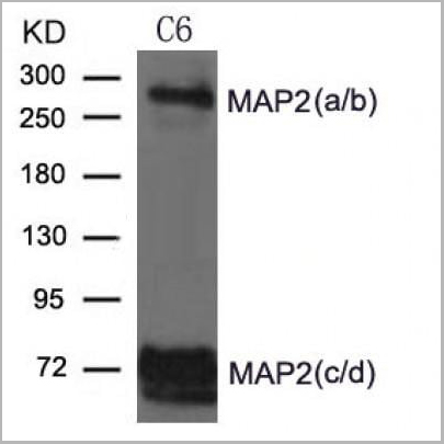
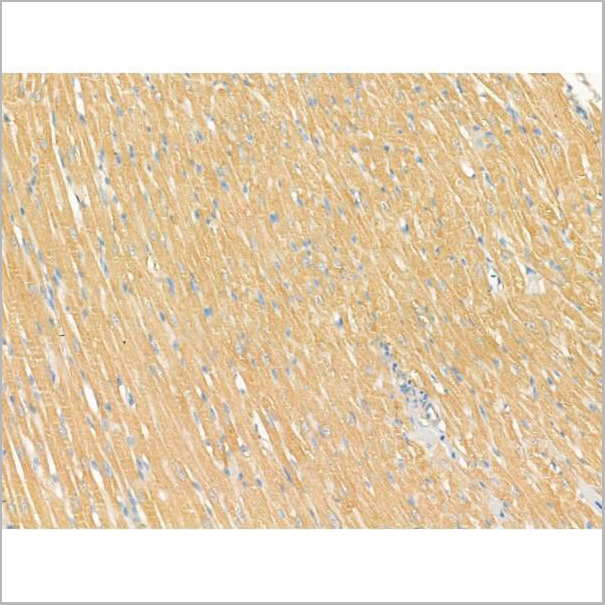
 Application Data
(At 25 degree C. Samples were then incubated with primary Ab(At 37 degree C. An AlexaFluor594 conjugated goat anti-rabbit IgG(H+L) Ab(Red) and an AlexaFluor488 conjugated goat anti-mouse IgG(H+L) Ab(Green) were used as the secondary antibody.The nuclear counter stain is DAPI (blue).)
Application Data
(At 25 degree C. Samples were then incubated with primary Ab(At 37 degree C. An AlexaFluor594 conjugated goat anti-rabbit IgG(H+L) Ab(Red) and an AlexaFluor488 conjugated goat anti-mouse IgG(H+L) Ab(Green) were used as the secondary antibody.The nuclear counter stain is DAPI (blue).)
MAP2, Polyclonal Antibody (Cat# AAA31338)
Full Name
MAP2 Antibody
Gene Names
MAP2; MAP2A; MAP2B; MAP2C
Reactivity
Human, Mouse, Rat
Applications
Western Blot, Immunohistochemistry, Immunofluorescence, Immunocytochemistry, Peptide ELISA
Purity
Peptide affinity purification
Pricing
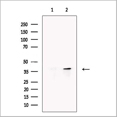
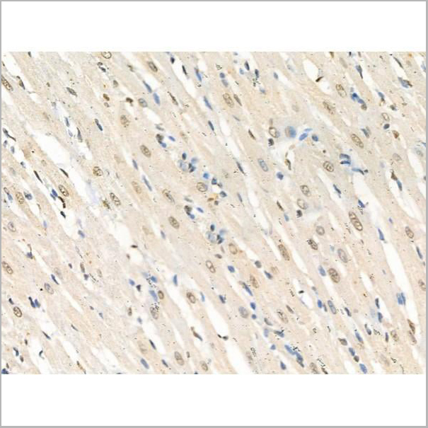
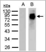
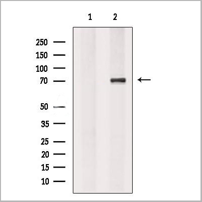
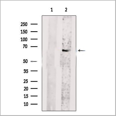
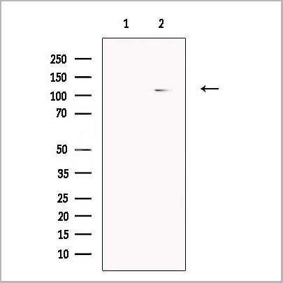
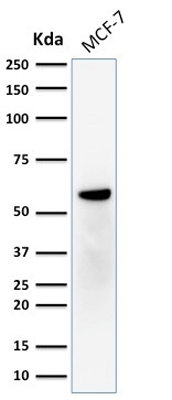 WB (Western Blot)
(Western Blot Analysis of human MCF-7 cell lysate using ER-beta1 Mouse Monoclonal Antibody (ERb455).)
WB (Western Blot)
(Western Blot Analysis of human MCF-7 cell lysate using ER-beta1 Mouse Monoclonal Antibody (ERb455).)
ER-beta1 (Estrogen Receptor beta-1), Monoclonal Antibody (Cat# AAA13814)
Full Name
ER-beta1 (Estrogen Receptor beta-1) Mouse Monoclonal Antibody
Gene Names
ESR2; Erb; ESRB; ESTRB; NR3A2; ER-BETA; ESR-BETA
Reactivity
Human
Applications
Flow Cytometry, Immunofluorescence, Western Blot, Immunohistochemistry
Pricing
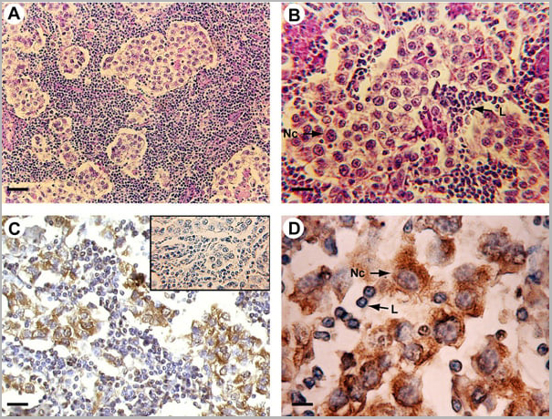 Application Data
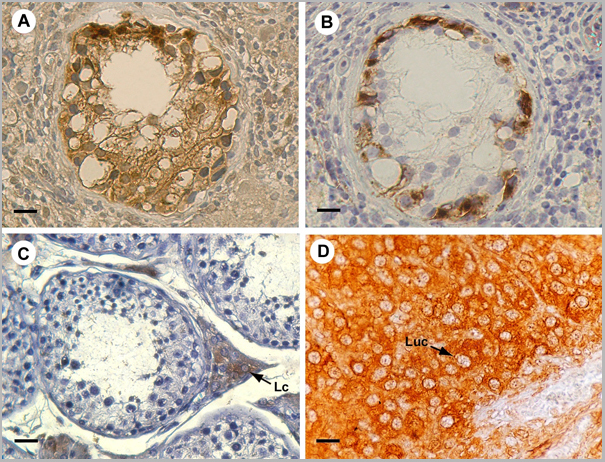
(Published customer image: Mouse anti Human cytochrome p450 aromatase antibody, clone H4 used for the detaction of aromatase in human tissues by Immunohistochemistry on paraffin sectionsImage caption:Morphology and P450 arom immunoreactivity of tumoral region in human testis with seminoma. A-B: Haematoxylin-eosin staining. C-D: Strong P450 arom immunoreactivity in cytoplasm of neoplastic cells (Nc) and unstained lymphocytes (L). Insert: absorption control. Scale bars: A, 20 um; B-C, 12.5 um; D, 5 um.From: Rago V, Romeo F, Aquila S, Montanaro D, And S, Carpino A. Cytochrome P450 aromatase expression in human seminoma. Reprod Biol Endocrinol. 2005 Dec 22;3:72.)
Application Data
(Published customer image: Mouse anti Human cytochrome p450 aromatase antibody, clone H4 used for the detaction of aromatase in human tissues by Immunohistochemistry on paraffin sectionsImage caption:Morphology and P450 arom immunoreactivity of tumoral region in human testis with seminoma. A-B: Haematoxylin-eosin staining. C-D: Strong P450 arom immunoreactivity in cytoplasm of neoplastic cells (Nc) and unstained lymphocytes (L). Insert: absorption control. Scale bars: A, 20 um; B-C, 12.5 um; D, 5 um.From: Rago V, Romeo F, Aquila S, Montanaro D, And S, Carpino A. Cytochrome P450 aromatase expression in human seminoma. Reprod Biol Endocrinol. 2005 Dec 22;3:72.)
CYTOCHROME P450 AROMATASE, Monoclonal Antibody (Cat# AAA12113)
Full Name
MOUSE ANTI HUMAN CYTOCHROME P450 AROMATASE
Gene Names
CYP19A1; ARO; ARO1; CPV1; CYAR; CYP19; CYPXIX; P-450AROM
Applications
Immunofluorescence, Immunohistochemistry, Western Blot
Pricing
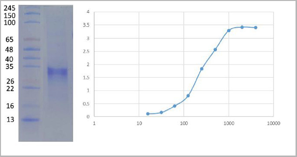
 Application Data
(ELISA plate was coated by N protein, 100 uL/cell, at various concentrations. The direct ELISA analysis was performed by loading 100 uL per well of a anti C-ter His tag HRP conjugated mAb at a concentration of 1:2000. The plate was incubated for 1 hours at 37 degree C, then washed 5 time. Detection was performed using TMB substrate for 10 minutes at room temperature in the dark. The plate was stopped with 2M sulfuric acid. Absorbances were read on a spectrophotometer at 450 nm.)
Application Data
(ELISA plate was coated by N protein, 100 uL/cell, at various concentrations. The direct ELISA analysis was performed by loading 100 uL per well of a anti C-ter His tag HRP conjugated mAb at a concentration of 1:2000. The plate was incubated for 1 hours at 37 degree C, then washed 5 time. Detection was performed using TMB substrate for 10 minutes at room temperature in the dark. The plate was stopped with 2M sulfuric acid. Absorbances were read on a spectrophotometer at 450 nm.)
COVID 19 Nucleocapsid (NP) Coronavirus, Recombinant Protein (Cat# AAA27948)
Full Name
2019-nCov Nucleocapsid Recombinant Protein (full length)
Applications
ELISA
Purity
Purity: >95% by SDS-page
Purification: Ni column
Purification: Ni column
Pricing

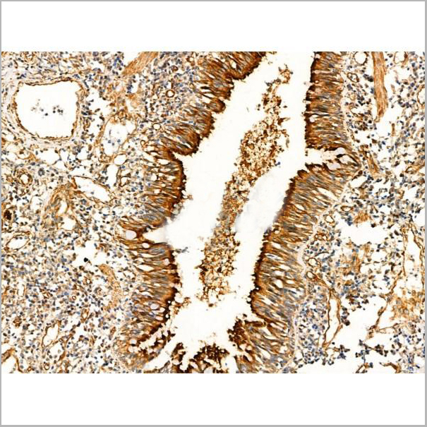
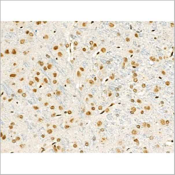
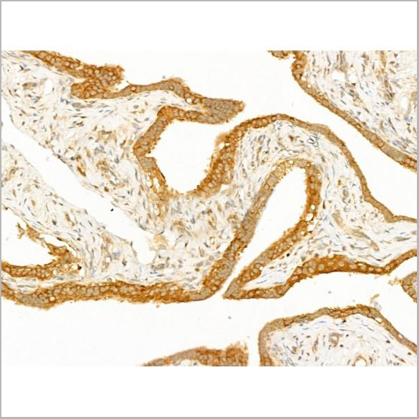
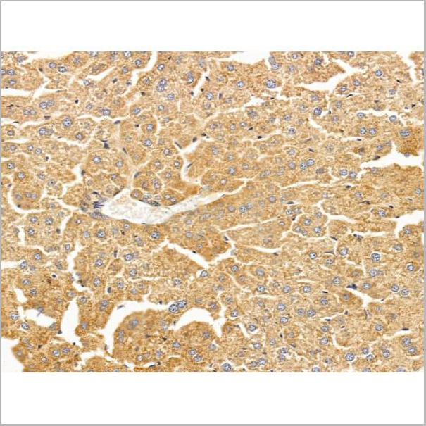
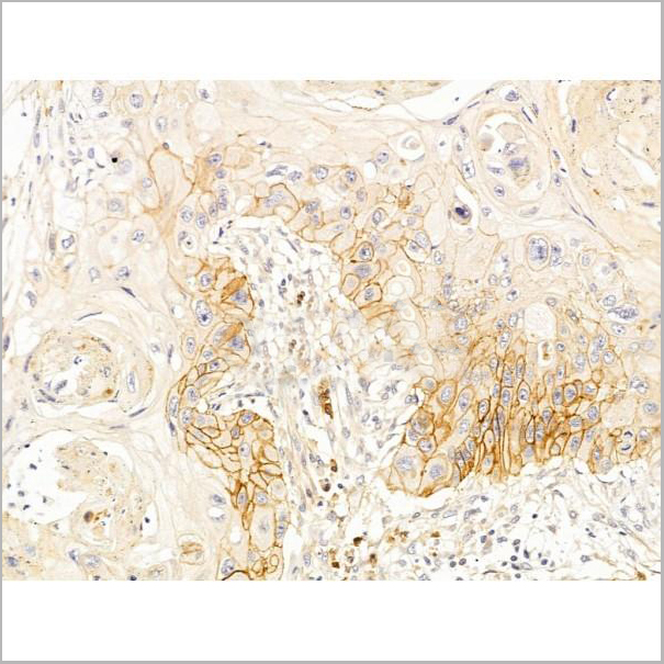
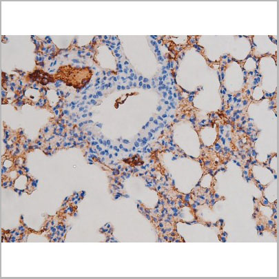
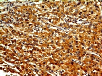
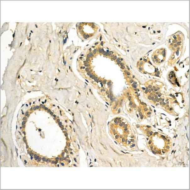
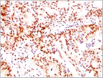
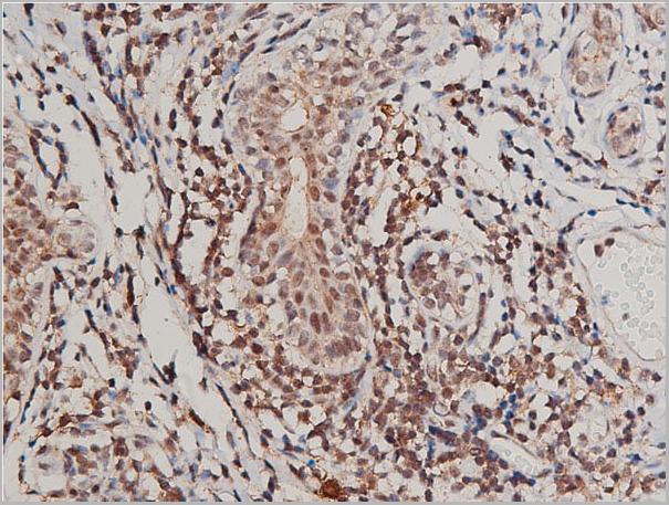
 IHC (Immunohistochemistry-Paraffin)
(AAA31179 at 1/100 staining human breast carcinoma tissue sections by IHC-P. The tissue was formaldehyde fixed and a heat mediated antigen retrieval step in citrate buffer was performed. The tissue was then blocked and incubated with the antibody for 1.5 hours at 22°C. An HRP conjugated goat anti-rabbit antibody was used as the secondary antibody.)
IHC (Immunohistochemistry-Paraffin)
(AAA31179 at 1/100 staining human breast carcinoma tissue sections by IHC-P. The tissue was formaldehyde fixed and a heat mediated antigen retrieval step in citrate buffer was performed. The tissue was then blocked and incubated with the antibody for 1.5 hours at 22°C. An HRP conjugated goat anti-rabbit antibody was used as the secondary antibody.)
CD36, Polyclonal Antibody (Cat# AAA31179)
Full Name
CD36 Antibody
Gene Names
CD36; FAT; GP4; GP3B; GPIV; CHDS7; PASIV; SCARB3; BDPLT10
Reactivity
Human, Mouse, Rat
Applications
Western Blot, Immunohistochemistry
Purity
Peptide affinity purification
Pricing
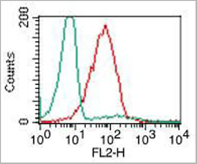
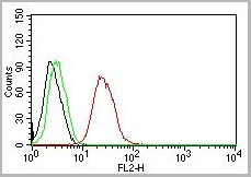
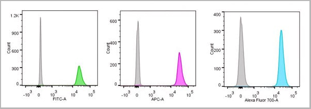
 FCM (Flow Cytometry)
(Flow Cytometric Analysis of paraformaldehyde-fixed Jurkat cells using CD31 Mouse Monoclonal Antibody (JC/70A) followed by goat anti- Mouse- IgG-CF488 (Blue); Isotype Control (Red).)
FCM (Flow Cytometry)
(Flow Cytometric Analysis of paraformaldehyde-fixed Jurkat cells using CD31 Mouse Monoclonal Antibody (JC/70A) followed by goat anti- Mouse- IgG-CF488 (Blue); Isotype Control (Red).)
CD31 / PECAM-1, Monoclonal Antibody (Cat# AAA13818)
Full Name
CD31 / PECAM-1 (Endothelial Cell Marker) Mouse Monoclonal Antibody
Gene Names
PECAM1; CD31; PECA1; GPIIA'; PECAM-1; endoCAM; CD31/EndoCAM
Reactivity
Human, Cynomolgus Monkey, Rabbit.
Does not react with Rat and Pig
Does not react with Rat and Pig
Applications
Flow Cytometry, Immunofluorescence, Western Blot, Immunohistochemistry
Pricing
 IF (Immunofluorescence)
(AAA31246 staining A549 cells by IF/ICC. The samples were fixed with PFA and permeabilized in 0.1% Triton X-100,then blocked in 10% serum for 45 minutes at 25°C. Samples were then incubated with primary Ab(AAA31246) and mouse anti-beta tubulin Ab(T0023) for 1 hour at 37°C. An AlexaFluor594 conjugated goat anti-rabbit IgG(H+L) Ab(Red) and an AlexaFluor488 conjugated goat anti-mouse IgG(H+L) Ab(Green) were used as the secondary antibody.The nuclear counter stain is DAPI (blue).)
IF (Immunofluorescence)
(AAA31246 staining A549 cells by IF/ICC. The samples were fixed with PFA and permeabilized in 0.1% Triton X-100,then blocked in 10% serum for 45 minutes at 25°C. Samples were then incubated with primary Ab(AAA31246) and mouse anti-beta tubulin Ab(T0023) for 1 hour at 37°C. An AlexaFluor594 conjugated goat anti-rabbit IgG(H+L) Ab(Red) and an AlexaFluor488 conjugated goat anti-mouse IgG(H+L) Ab(Green) were used as the secondary antibody.The nuclear counter stain is DAPI (blue).)
TROY, Polyclonal Antibody (Cat# AAA31246)
Full Name
TROY Antibody
Gene Names
TNFRSF19; TAJ; TROY; TRADE; TAJ-alpha
Reactivity
Human, Mouse, Rat
Predicted Reactivity: Horse(88%), Sheep(86%)
Predicted Reactivity: Horse(88%), Sheep(86%)
Applications
ELISA
Purity
The antiserum was purified by peptide affinity chromatography using SulfoLink Coupling Resin (Thermo Fisher Scientific).
Pricing


 IF (Immunofluorescence)
(AAA31213 staining Hela cells by IF/ICC. The samples were fixed with PFA and permeabilized in 0.1% Triton X-100,then blocked in 10% serum for 45 minutes at 25°C. Samples were then incubated with primary Ab(AAA31213 1:200) and mouse anti-beta tubulin Ab(T0023 1:200) for 1 hour at 37°C. An AlexaFluor594 conjugated goat anti-rabbit IgG(H+L) Ab(Red) and an AlexaFluor488 conjugated goat anti-mouse IgG(H+L) Ab(Green) were used as the secondary antibody.The nuclear counter stain is DAPI(blue).)
IF (Immunofluorescence)
(AAA31213 staining Hela cells by IF/ICC. The samples were fixed with PFA and permeabilized in 0.1% Triton X-100,then blocked in 10% serum for 45 minutes at 25°C. Samples were then incubated with primary Ab(AAA31213 1:200) and mouse anti-beta tubulin Ab(T0023 1:200) for 1 hour at 37°C. An AlexaFluor594 conjugated goat anti-rabbit IgG(H+L) Ab(Red) and an AlexaFluor488 conjugated goat anti-mouse IgG(H+L) Ab(Green) were used as the secondary antibody.The nuclear counter stain is DAPI(blue).)
Exportin-5, Polyclonal Antibody (Cat# AAA31213)
Full Name
Exportin-5 Antibody
Gene Names
XPO5; exp5
Reactivity
Human, Mouse, Rat
Predicted Reactivity: Pig(88%), Bovine(100%), Horse(100%), Sheep(100%), Rabbit(88%), Dog(100%), Chicken(100%)
Predicted Reactivity: Pig(88%), Bovine(100%), Horse(100%), Sheep(100%), Rabbit(88%), Dog(100%), Chicken(100%)
Applications
ELISA
Purity
The antiserum was purified by peptide affinity chromatography using SulfoLink Coupling Resin (Thermo Fisher Scientific).
Pricing
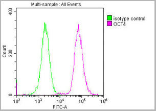
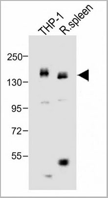
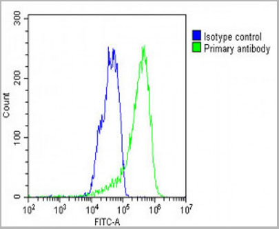
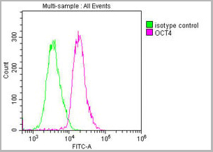 FCM (Flow Cytometry)
(Overlay histogram showing Ntera-2 cells stained with CSB-MA018403A0m (red line) at 1:100. The cells were incubated in 1x PBS /10% normal goat serum to block non-specific protein-protein interactions followed by primary antibody for 1 h at 4 degree C. The secondary antibody used was FITC goat anti-mouse IgG(H+L) at 1/200 dilution for 1 h at 4 degree C. Isotype control antibody (green line) was used under the same conditions. Acquisition of >10,000 events was performed.)
FCM (Flow Cytometry)
(Overlay histogram showing Ntera-2 cells stained with CSB-MA018403A0m (red line) at 1:100. The cells were incubated in 1x PBS /10% normal goat serum to block non-specific protein-protein interactions followed by primary antibody for 1 h at 4 degree C. The secondary antibody used was FITC goat anti-mouse IgG(H+L) at 1/200 dilution for 1 h at 4 degree C. Isotype control antibody (green line) was used under the same conditions. Acquisition of >10,000 events was performed.)
OCT4, Monoclonal Antibody (Cat# AAA27015)
Full Name
OCT4 Monoclonal Antibody
Gene Names
POU5F1; OCT3; OCT4; OTF3; OTF4; OTF-3; Oct-3; Oct-4
Reactivity
Human, Mouse, Rat
Applications
Western Blot, Immunohistochemistry, Immunofluorescence, Flow Cytometry
Purity
>95%
Protein G Purified
Protein G Purified
Pricing

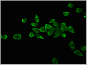
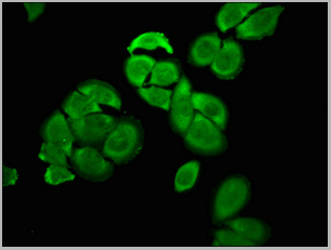
 IF (Immunofluorescence)
Nucleolin, Monoclonal Antibody (Cat# AAA13823)
Full Name
Nucleolin (Marker of Human Cells) Mouse Monoclonal Antibody
Gene Names
NCL; C23
Reactivity
Human.
Does not react with Mouse, Rat and Cow
Does not react with Mouse, Rat and Cow
Applications
Flow Cytometry, Immunofluorescence, Western Blot, Immunohistochemistry
Pricing
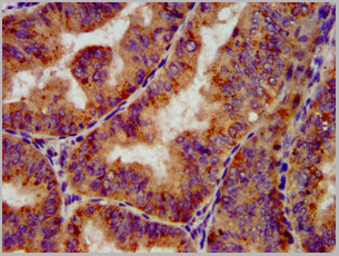
 Application Data
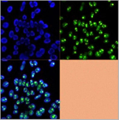
(AAA31020 staining HepG2 cells(30min of 4uM Forskolin treatment) by IF/ICC. The samples were fixed with PFA and permeabilized in 0.1% Triton X-100,then blocked in 10% serum for 45 minutes at 25°C. Samples were then incubated with primary Ab(AAA31020) and mouse anti-beta tubulin Ab for 1 hour at 37°C. An AlexaFluor594 conjugated goat anti-rabbit IgG(H+L) Ab(Red) and an AlexaFluor488 conjugated goat anti-mouse IgG(H+L) Ab(Green) were used as the secondary Ab. The nuclear counter stain is DAPI(blue))
Application Data
(AAA31020 staining HepG2 cells(30min of 4uM Forskolin treatment) by IF/ICC. The samples were fixed with PFA and permeabilized in 0.1% Triton X-100,then blocked in 10% serum for 45 minutes at 25°C. Samples were then incubated with primary Ab(AAA31020) and mouse anti-beta tubulin Ab for 1 hour at 37°C. An AlexaFluor594 conjugated goat anti-rabbit IgG(H+L) Ab(Red) and an AlexaFluor488 conjugated goat anti-mouse IgG(H+L) Ab(Green) were used as the secondary Ab. The nuclear counter stain is DAPI(blue))
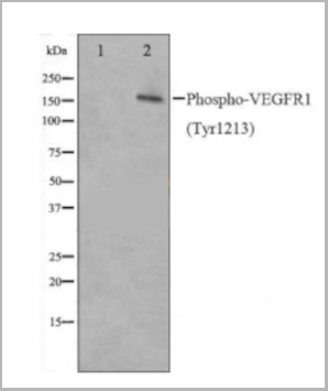
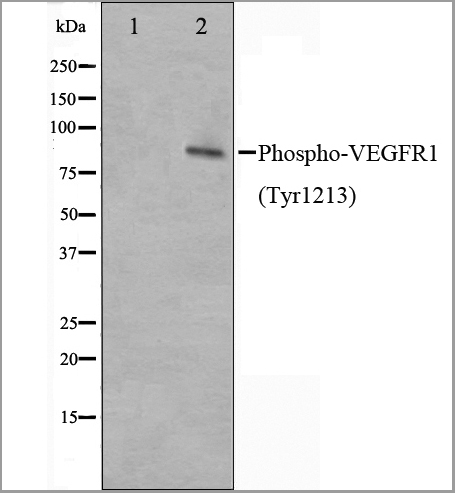
VEGFR1, Polyclonal Antibody (Cat# AAA31020)
Full Name
Phospho-VEGFR1 (Tyr1213) Antibody
Gene Names
FLT1; FLT; FLT-1; VEGFR1; VEGFR-1
Reactivity
Human, Mouse, Rat
Applications
Western Blot, Immunofluorescence, Immunocytochemistry, Immunohistochemistry
Purity
The Ab is from purified rabbit serum by affinity purification via sequential chromatography on phospho-peptide and non-phospho-peptide affinity columns.
Pricing