Filters
Clonality
Type
Reactivity
Gene Name
Isotype
Host
Application
Clone
7408 results for " Serum" - showing 300-350
 Standard Curve (Sample)
Regenerating Islet Derived Protein 3 Beta (REG3b), RTU ELISA Kit (Cat# AAA23980)
Full Name
Mouse Regenerating Islet Derived Protein 3 Beta (REG3b) ELISA Kit
Gene Names
Reg3b; HIP; Pap; PAP1; REG-III
Reactivity
Mouse
Pricing


 Application Data
(Detection of Mouse anti Bovine CD86 in a flow cytometric analysis of bovine peripheral blood lymphocytes using Goat anti Mouse IgG:FITC (AAA12304))
Application Data
(Detection of Mouse anti Bovine CD86 in a flow cytometric analysis of bovine peripheral blood lymphocytes using Goat anti Mouse IgG:FITC (AAA12304))
IgG, Polyclonal Antibody (Cat# AAA12304)
Full Name
GOAT ANTI MOUSE IgG:FITC (RAT ADSORBED)
Applications
Flow Cytometry
Pricing


 Application Data
(Staining of mouse peritoneal macrophages with Rat anti Mouse CD11b)
Application Data
(Staining of mouse peritoneal macrophages with Rat anti Mouse CD11b)
CD11b, Monoclonal Antibody (Cat# AAA11889)
Full Name
RAT ANTI MOUSE CD11b:FITC
Gene Names
Itgam; CR3; CR3A; MAC1; Cd11b; Ly-40; Mac-1; Mac-1a; CD11b/CD18; F730045J24Rik
Applications
Flow Cytometry
Pricing


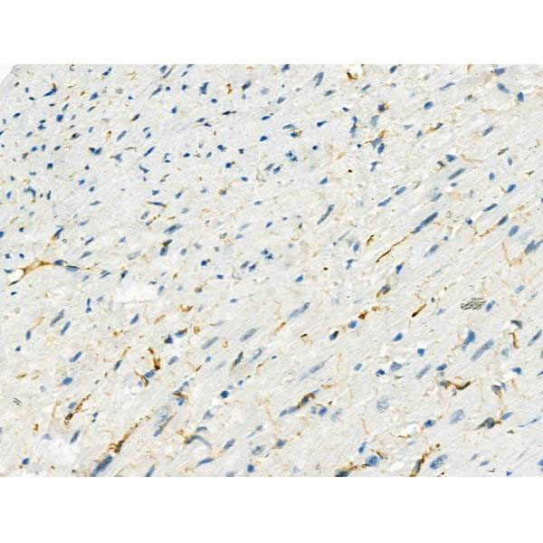
 IHC (Immunohistchemistry)
(At 1/100 staining Rat heart tissue by IHC-P. The sample was formaldehyde fixed and a heat mediated antigen retrieval step in citrate buffer was performed. The sample was then blocked and incubated with the primary antibody at 4 degree C overnight. An HRP conjugated anti-Rabbit antibody was used as the secondary antibody.)
IHC (Immunohistchemistry)
(At 1/100 staining Rat heart tissue by IHC-P. The sample was formaldehyde fixed and a heat mediated antigen retrieval step in citrate buffer was performed. The sample was then blocked and incubated with the primary antibody at 4 degree C overnight. An HRP conjugated anti-Rabbit antibody was used as the secondary antibody.)
VR1, Polyclonal Antibody (Cat# AAA31468)
Full Name
Phospho-VR1 (Ser502) Antibody
Gene Names
TRPV1; VR1
Reactivity
Human, Mouse, Rat
Predicted Reactivity: Pig (91%), Bovine (91%), Sheep (91%), Rabbit (82%), Dog (91%), Chicken (82%), Xenopus (82%)
Predicted Reactivity: Pig (91%), Bovine (91%), Sheep (91%), Rabbit (82%), Dog (91%), Chicken (82%), Xenopus (82%)
Applications
Western Blot, Immunohistochemistry, Peptide ELISA
Purity
The antibody is from purified rabbit serum by affinity purification via sequential chromatography on phospho-peptide and non-phospho-peptide affinity columns.
Pricing
 Standard Curve (Sample)
Triglyceride, Assay Kit (Cat# AAA27848)
Full Name
Triglyceride Microplate Assay Kit
Reactivity
General
Applications
Functional Assay
Pricing
 Standard Curve (Sample)
IL-17, ELISA Kit (Cat# AAA13490)
Full Name
IL-17 Human ELISA Kit
Reactivity
Human
Applications
ELISA
Pricing
 Standard Curve (Sample)
Protein phosphatase 1 regulatory subunit 1B (PPP1R1B), ELISA Kit (Cat# AAA27322)
Full Name
Rat Protein phosphatase 1 regulatory subunit 1B (PPP1R1B) ELISA Kit
Reactivity
Rat
Pricing
 Standard Curve (Sample)
Lipopolysaccharides, LPS, ELISA Kit (Cat# AAA15327)
Full Name
Human Lipopolysaccharides, LPS ELISA Kit
Reactivity
Human
Pricing
 Standard Curve (Sample)
Ca2+/Mg2+ ATPase, Assay Kit (Cat# AAA27824)
Full Name
Ca2+/Mg2+ ATPase Microplate Assay Kit
Gene Names
ATP2B1; PMCA1
Reactivity
General
Applications
Functional Assay
Pricing
 Standard Curve (Sample)
lipopolysaccharide binding protein, ELISA Kit (Cat# AAA18400)
Full Name
Mouse Lipopolysaccharide-binding protein, LBP ELISA Kit
Gene Names
Lbp; Ly88; Bpifd2
Reactivity
Mouse
Pricing


 Application Data
(Detection of Mouse anti Human CD300a in a flow cytometric analysis of human peripheral blood monocytes using Rabbit F(ab')2 anti Mouse IgG:FITC (AAA12305))
Application Data
(Detection of Mouse anti Human CD300a in a flow cytometric analysis of human peripheral blood monocytes using Rabbit F(ab')2 anti Mouse IgG:FITC (AAA12305))
IgG, Polyclonal Antibody (Cat# AAA12305)
Full Name
RABBIT F(ab')2 ANTI MOUSE IgG:FITC
Applications
Flow Cytometry
Pricing


 WB (Western Blot)
(Western blot analysis of Catenin 1 Antibody expression in MCF-7 cells lysates.The lane on the left is treated with the antigen-specific peptide..The lane on the left is treated with the antigen-specific peptide.)
WB (Western Blot)
(Western blot analysis of Catenin 1 Antibody expression in MCF-7 cells lysates.The lane on the left is treated with the antigen-specific peptide..The lane on the left is treated with the antigen-specific peptide.)
Catenin alpha1, Polyclonal Antibody (Cat# AAA31080)
Full Name
Catenin alpha1 Antibody
Gene Names
CTNNA1; MDPT2; CAP102
Reactivity
Human, Mouse, Rat
Applications
Western Blot
Purity
The antiserum was purified by peptide affinity chromatography using SulfoLink Coupling Resin.
Pricing
 Standard Curve (Sample)
platelet-derived growth factor-BB (PDGF-BB), ELISA Kit (Cat# AAA18066)
Full Name
Horse platelet-derived growth factor-BB, PDGF-BB ELISA Kit
Reactivity
Horse
Pricing
 Standard Curve (Sample)
Angiotensin II type 1 receptor antibody (ATIIR1 Ab), ELISA Kit (Cat# AAA12969)
Full Name
Mouse Angiotensin II type 1 receptor antibody (ATIIR1 Ab) ELISA Kit
Gene Names
Scd1; ab; Scd; Scd-1; AA589638; AI265570
Reactivity
Mouse
Pricing
 Standard Curve (Sample)
Angiopoietin 2, ELISA Kit (Cat# AAA16720)
Full Name
Monkey Angiopoietin 2 ELISA Kit
Gene Names
ANGPT2; ANG2; AGPT2
Reactivity
Monkey
Pricing
 Standard Curve (Sample)
S100 calcium binding protein B, ELISA Kit (Cat# AAA18039)
Full Name
Mouse Protein S100-B, S100B ELISA Kit
Gene Names
S100b; Bpb; AI850290
Reactivity
Mouse
Pricing
 WB (Western Blot)
(Positive WB detected in: recombinant proteinAll lanes: MELT Antibody at 1:1000Secondary: Goat polyclonal to rabbit IgG at 1/50000 dilutionPredicted band size: 17 kDaObserved band size: 17 kDa)
WB (Western Blot)
(Positive WB detected in: recombinant proteinAll lanes: MELT Antibody at 1:1000Secondary: Goat polyclonal to rabbit IgG at 1/50000 dilutionPredicted band size: 17 kDaObserved band size: 17 kDa)
MELT, Polyclonal Antibody (Cat# AAA27057)
Full Name
Rabbit anti-Apis mellifera (Honeybee) MELT Polyclonal Antibody
Gene Names
Melt; GB10355
Reactivity
Apis mellifera
Applications
Western Blot
Purity
>95%, Protein G purified
Pricing


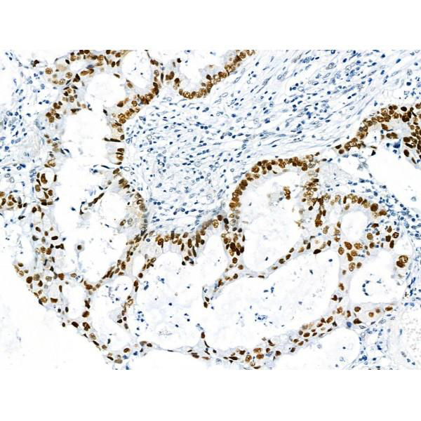
 IHC (Immunohistchemistry)
(AAA31084 at 1/200 staining human liver cancer tissue sections by IHC-P. The tissue was formaldehyde fixed and a heat mediated antigen retrieval step in citrate buffer was performed. The tissue was then blocked and incubated with the antibody for 1.5 hours at 22 degree C. An HRP conjugated goat anti-rabbit antibody was used as the secondary.)
IHC (Immunohistchemistry)
(AAA31084 at 1/200 staining human liver cancer tissue sections by IHC-P. The tissue was formaldehyde fixed and a heat mediated antigen retrieval step in citrate buffer was performed. The tissue was then blocked and incubated with the antibody for 1.5 hours at 22 degree C. An HRP conjugated goat anti-rabbit antibody was used as the secondary.)
HSP90B, Polyclonal Antibody (Cat# AAA31084)
Full Name
HSP90B Antibody
Gene Names
HSP90AB1; HSP84; HSPC2; HSPCB; D6S182; HSP90B
Reactivity
Human, Mouse, Rat
Applications
Western Blot, Immunohistochemistry, Immunofluorescence, Immunocytochemistry, Immunoprecipitation
Purity
The antiserum was purified by peptide affinity chromatography using SulfoLink Coupling Resin.
Pricing


 IF (Immunofluorescence)
(Figure 11. IF analysis of H Cadherin/CDH13 using anti- H Cadherin/CDH13 antibody (AAA19252).H Cadherin/CDH13 was detected in immunocytochemical section of SIHA cells. Enzyme antigen retrieval was performed using IHC enzyme antigen retrieval reagent for 15 mins. The cells were blocked with 10% goat serum. And then incubated with 5μg/mL rabbit anti- H Cadherin/CDH13 Antibody (AAA19252) overnight at 4 degree C. DyLight®488 Conjugated Goat Anti-Rabbit IgG was used as secondary antibody at 1:100 dilution and incubated for 30 minutes at 37 degree C. The section was counterstained with DAPI. Visualize using a fluorescence microscope and filter sets appropriate for the label used.)
IF (Immunofluorescence)
(Figure 11. IF analysis of H Cadherin/CDH13 using anti- H Cadherin/CDH13 antibody (AAA19252).H Cadherin/CDH13 was detected in immunocytochemical section of SIHA cells. Enzyme antigen retrieval was performed using IHC enzyme antigen retrieval reagent for 15 mins. The cells were blocked with 10% goat serum. And then incubated with 5μg/mL rabbit anti- H Cadherin/CDH13 Antibody (AAA19252) overnight at 4 degree C. DyLight®488 Conjugated Goat Anti-Rabbit IgG was used as secondary antibody at 1:100 dilution and incubated for 30 minutes at 37 degree C. The section was counterstained with DAPI. Visualize using a fluorescence microscope and filter sets appropriate for the label used.)
H Cadherin/CDH13, Polyclonal Antibody (Cat# AAA19252)
Full Name
Anti-H Cadherin/CDH13 Antibody
Gene Names
CDH13; CDHH; P105
Reactivity
Human, Rat
Applications
Western Blot, Immunohistochemistry, Immunocytochemistry, Immunofluorescence, Flow Cytometry, Direct ELISA
Purity
Immunogen affinity purified.
Pricing


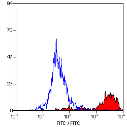
 FCM (Flow Cytometry)
(Figure 6. Flow Cytometry analysis of CACO-2 cells using anti-PLTP antibody (AAA19255).Overlay histogram showing CACO-2 cells stained with AAA19255 (Blue line). The cells were blocked with 10% normal goat serum. And then incubated with rabbit anti-PLTP Antibody (AAA19255, 1μg/1x106 cells) for 30 min at 20 degree C. DyLight®488 conjugated goat anti-rabbit IgG (5-10μg/1x106 cells) was used as secondary antibody for 30 minutes at 20 degree C. Isotype control antibody (Green line) was rabbit IgG (1μg/1x106) used under the same conditions. Unlabelled sample (Red line) was also used as a control.)
FCM (Flow Cytometry)
(Figure 6. Flow Cytometry analysis of CACO-2 cells using anti-PLTP antibody (AAA19255).Overlay histogram showing CACO-2 cells stained with AAA19255 (Blue line). The cells were blocked with 10% normal goat serum. And then incubated with rabbit anti-PLTP Antibody (AAA19255, 1μg/1x106 cells) for 30 min at 20 degree C. DyLight®488 conjugated goat anti-rabbit IgG (5-10μg/1x106 cells) was used as secondary antibody for 30 minutes at 20 degree C. Isotype control antibody (Green line) was rabbit IgG (1μg/1x106) used under the same conditions. Unlabelled sample (Red line) was also used as a control.)
PLTP, Polyclonal Antibody (Cat# AAA19255)
Full Name
Anti-PLTP Antibody
Gene Names
PLTP; BPIFE; HDLCQ9
Reactivity
Human
Applications
Western Blot, Immunohistochemistry, Immunocytochemistry, Immunofluorescence, Flow Cytometry, Direct ELISA
Purity
Immunogen affinity purified.
Pricing


 Application Data
(Published customer image Infiltration of GFP+ BM-cells in infarct and peri-infarct regions. (A-B) Dot plots of viable macrophages/granulocytes (CD11b+CD45high, top right quadrants) and microglia (CD11b+CD45dim, bottom right quadrants) in cortex from BM-chimeric unmanipulated mice and mice exposed to pMCAO. (C) Bar graph showing mean numbers of CD11b+CD45dim microglia and CD11b+CD45high macrophages/granulocytes in BM-chimeric mice 24 hours after pMCAO, subdivided based on expression of GFP (n = 5). Approximately 92% of of the CD45high population were GFP+. (D) Estimation and comparison of mean numbers of CD11b+CD45dim microglia in non-chimeric (n = 10) versus BM-chimeric mice (n = 5) 24 hours after of pMCAO shows significantly fewer CD11b+CD45dim microglial cells in irradiated mice. (E) Overview, showing distribution of infiltrating GFP+ BM-derived cells into infarct (IF) and peri-infarct (P-IF) regions 24 hours after pMCAO. (E-G) By 24 hours, GFP+ single cells (F) and vessel-associated aggregates of GFP+ cells (arrows in G) were observed in infarct and peri-infarct regions. Some of the vessel-associated cells were round, leukocyte-like cells (arrows) while others were elongated cells lining the vasculature (arrow heads in G and in insert). (H) Bar graph showing mean numbers of single GFP+ cells and vessel-associated aggregates of GFP+ cells in ipsi- and contralateral cortex 24 hours after surgery (n = 10). (I-P) Immunohistochemical staining of CD45.1 (I, K), CD45.2 (J, L), IgG2a (M, O) and CD45 (N, P) in ischemic tissue in BM-chimeric (I, J, M, N) and non-chimeric mice (K, L, O, P) 24 hours after pMCAO. N.D, none detected. Scale bars: 200 um (A), 10 um (B, C). 50 um (I-P) *P < 0.05, **P < 0.01, and ***P < 0.001.From: Clausen BH, Lambertsen KL, Babcock AA, Holm TH, Dagnaes-Hansen F, Finsen B. Interleukin-1beta and tumor necrosis factor-alpha are expressed by different subsets of microglia and macrophages after ischemic stroke in mice. J Neuroinflammation. 2008 Oct 23;5:46.)
Application Data
(Published customer image Infiltration of GFP+ BM-cells in infarct and peri-infarct regions. (A-B) Dot plots of viable macrophages/granulocytes (CD11b+CD45high, top right quadrants) and microglia (CD11b+CD45dim, bottom right quadrants) in cortex from BM-chimeric unmanipulated mice and mice exposed to pMCAO. (C) Bar graph showing mean numbers of CD11b+CD45dim microglia and CD11b+CD45high macrophages/granulocytes in BM-chimeric mice 24 hours after pMCAO, subdivided based on expression of GFP (n = 5). Approximately 92% of of the CD45high population were GFP+. (D) Estimation and comparison of mean numbers of CD11b+CD45dim microglia in non-chimeric (n = 10) versus BM-chimeric mice (n = 5) 24 hours after of pMCAO shows significantly fewer CD11b+CD45dim microglial cells in irradiated mice. (E) Overview, showing distribution of infiltrating GFP+ BM-derived cells into infarct (IF) and peri-infarct (P-IF) regions 24 hours after pMCAO. (E-G) By 24 hours, GFP+ single cells (F) and vessel-associated aggregates of GFP+ cells (arrows in G) were observed in infarct and peri-infarct regions. Some of the vessel-associated cells were round, leukocyte-like cells (arrows) while others were elongated cells lining the vasculature (arrow heads in G and in insert). (H) Bar graph showing mean numbers of single GFP+ cells and vessel-associated aggregates of GFP+ cells in ipsi- and contralateral cortex 24 hours after surgery (n = 10). (I-P) Immunohistochemical staining of CD45.1 (I, K), CD45.2 (J, L), IgG2a (M, O) and CD45 (N, P) in ischemic tissue in BM-chimeric (I, J, M, N) and non-chimeric mice (K, L, O, P) 24 hours after pMCAO. N.D, none detected. Scale bars: 200 um (A), 10 um (B, C). 50 um (I-P) *P < 0.05, **P < 0.01, and ***P < 0.001.From: Clausen BH, Lambertsen KL, Babcock AA, Holm TH, Dagnaes-Hansen F, Finsen B. Interleukin-1beta and tumor necrosis factor-alpha are expressed by different subsets of microglia and macrophages after ischemic stroke in mice. J Neuroinflammation. 2008 Oct 23;5:46.)
CD11b, Monoclonal Antibody (Cat# AAA12182)
Full Name
RAT ANTI MOUSE CD11b:FITC
Gene Names
Itgam; CR3; CR3A; MAC1; Cd11b; Ly-40; Mac-1; Mac-1a; CD11b/CD18; F730045J24Rik
Applications
Flow Cytometry
Pricing
 Standard Curve (Sample)
Pancreastatin, ELISA Kit (Cat# AAA16466)
Full Name
Human Pancreastatin ELISA Kit
Reactivity
Human
Pricing
 Standard Curve (Sample)
IL-10, ELISA Kit (Cat# AAA13482)
Full Name
IL-10 Rat ELISA Kit
Reactivity
Rat
Applications
ELISA
Pricing


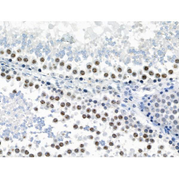
 IHC (Immunohistochemistry)
(At 1/100 staining Human prostate cancer and adjacent normal tissues by IHC-P. The sample was formaldehyde fixed and a heat mediated antigen retrieval step in citrate buffer was performed. The sample was then blocked and incubated with the primary antibody at 4 degree C overnight. An HRP conjugated anti-Rabbit antibody was used as the secondary antibody.)
IHC (Immunohistochemistry)
(At 1/100 staining Human prostate cancer and adjacent normal tissues by IHC-P. The sample was formaldehyde fixed and a heat mediated antigen retrieval step in citrate buffer was performed. The sample was then blocked and incubated with the primary antibody at 4 degree C overnight. An HRP conjugated anti-Rabbit antibody was used as the secondary antibody.)
PAK5/6, Polyclonal Antibody (Cat# AAA31281)
Full Name
Phospho-PAK5/6 (Ser602/Ser560) Antibody
Gene Names
PAK7; PAK5
Reactivity
Human, Mouse, Rat
Applications
Immunohistochemistry, Peptide ELISA
Purity
The antibody is from purified rabbit serum by affinity purification via sequential chromatography on phospho-peptide and non-phospho-peptide affinity columns.
Pricing


 IHC (Immunohistochemistry)
(At 1/100 staining Human mammary cancer and adjacent normal tissues by IHC-P. The sample was formaldehyde fixed and a heat mediated antigen retrieval step in citrate buffer was performed. The sample was then blocked and incubated with the primary antibody at 4 degree C overnight. An HRP conjugated anti-Rabbit antibody was used as the secondary antibody.)
IHC (Immunohistochemistry)
(At 1/100 staining Human mammary cancer and adjacent normal tissues by IHC-P. The sample was formaldehyde fixed and a heat mediated antigen retrieval step in citrate buffer was performed. The sample was then blocked and incubated with the primary antibody at 4 degree C overnight. An HRP conjugated anti-Rabbit antibody was used as the secondary antibody.)
MCM2, Polyclonal Antibody (Cat# AAA31460)
Full Name
Phospho-MCM2 (Ser27) Antibody
Gene Names
MCM2; BM28; CCNL1; CDCL1; cdc19; D3S3194; MITOTIN
Reactivity
Human, Mouse, Rat
Predicted Reactivity: Pig (100%), Zebrafish (100%), Bovine (100%), Horse (100%), Rabbit (100%), Dog (100%), Xenopus (100%)
Predicted Reactivity: Pig (100%), Zebrafish (100%), Bovine (100%), Horse (100%), Rabbit (100%), Dog (100%), Xenopus (100%)
Applications
Western Blot, Immunohistochemistry, Peptide ELISA
Purity
The antibody is from purified rabbit serum by affinity purification via sequential chromatography on phospho-peptide and non-phospho-peptide affinity columns.
Pricing


 Application Data
(At 25 degree C. Samples were then incubated with primary Ab(At 37 degree C. An AlexaFluor594 conjugated goat anti-rabbit IgG(H+L) Ab(Red) and an AlexaFluor488 conjugated goat anti-mouse IgG(H+L) Ab(Green) were used as the secondary antibody.The nuclear counter stain is DAPI (blue).)
Application Data
(At 25 degree C. Samples were then incubated with primary Ab(At 37 degree C. An AlexaFluor594 conjugated goat anti-rabbit IgG(H+L) Ab(Red) and an AlexaFluor488 conjugated goat anti-mouse IgG(H+L) Ab(Green) were used as the secondary antibody.The nuclear counter stain is DAPI (blue).)
JIP1, Polyclonal Antibody (Cat# AAA31317)
Full Name
Phospho-JIP1 (Thr205) Antibody
Gene Names
MAPK8IP1; IB1; JIP1; JIP-1; PRKM8IP
Reactivity
Human, Mouse, Rat
Applications
Western Blot, Immunohistochemistry, Immunofluorescence, Immunocytochemistry, Peptide ELISA
Purity
The antibody is from purified rabbit serum by affinity purification via sequential chromatography on phospho-peptide and non-phospho-peptide affinity columns.
Pricing


 FCM (Flow Cytometry)
(Figure 8. Flow Cytometry analysis of A431 cells using anti-EGFR antibody (AAA19214).Overlay histogram showing A431 cells stained with AAA19214 (Blue line). The cells were blocked with 10% normal goat serum. And then incubated with rabbit anti-EGFR Antibody (AAA19214,1μg/1x106 cells) for 30 min at 20 degree C. DyLight®488 conjugated goat anti-rabbit IgG (5-10μg/1x106 cells) was used as secondary antibody for 30 minutes at 20 degree C. Isotype control antibody (Green line) was rabbit IgG (1μg/1x106) used under the same conditions. Unlabelled sample (Red line) was also used as a control.)
FCM (Flow Cytometry)
(Figure 8. Flow Cytometry analysis of A431 cells using anti-EGFR antibody (AAA19214).Overlay histogram showing A431 cells stained with AAA19214 (Blue line). The cells were blocked with 10% normal goat serum. And then incubated with rabbit anti-EGFR Antibody (AAA19214,1μg/1x106 cells) for 30 min at 20 degree C. DyLight®488 conjugated goat anti-rabbit IgG (5-10μg/1x106 cells) was used as secondary antibody for 30 minutes at 20 degree C. Isotype control antibody (Green line) was rabbit IgG (1μg/1x106) used under the same conditions. Unlabelled sample (Red line) was also used as a control.)
EGFR, Polyclonal Antibody (Cat# AAA19214)
Full Name
Anti-EGFR Antibody
Gene Names
EGFR; ERBB; HER1; mENA; ERBB1; PIG61
Reactivity
Human, Mouse, Rat
Applications
Western Blot, Immunohistochemistry, Immunocytochemistry, Immunofluorescence, Flow Cytometry, Direct ELISA
Purity
Immunogen affinity purified.
Pricing


 Application Data
(At 25 degree C. Samples were then incubated with primary Ab(At 37 degree C. An AlexaFluor594 conjugated goat anti-rabbit IgG(H+L) Ab(Red) and an AlexaFluor488 conjugated goat anti-mouse IgG(H+L) Ab(Green) were used as the secondary antibody.The nuclear counter stain is DAPI (blue).)
Application Data
(At 25 degree C. Samples were then incubated with primary Ab(At 37 degree C. An AlexaFluor594 conjugated goat anti-rabbit IgG(H+L) Ab(Red) and an AlexaFluor488 conjugated goat anti-mouse IgG(H+L) Ab(Green) were used as the secondary antibody.The nuclear counter stain is DAPI (blue).)
LRRK2, Polyclonal Antibody (Cat# AAA31287)
Full Name
Phospho-LRRK2 (Ser935) Antibody
Gene Names
LRRK2; PARK8; RIPK7; ROCO2; AURA17; DARDARIN
Reactivity
Human, Mouse, Rat
Applications
Western Blot, Immunohistochemistry, Immunofluorescence, Immunocytochemistry, Peptide ELISA
Purity
The antibody is from purified rabbit serum by affinity purification via sequential chromatography on phospho-peptide and non-phospho-peptide affinity columns.
Pricing
 Standard Curve (Sample)
activity-regulated cytoskeleton-associated protein, ELISA Kit (Cat# AAA18135)
Full Name
Human Activity-regulated cytoskeleton-associated protein, ARC ELISA Kit
Gene Names
ARC; Arg3.1
Reactivity
Human
Pricing
 WB (Western Blot)
((0.1ug/ml) staining of Human Brain lysate (35ug protein in RIPA buffer). Primary incubation was 1 hour. Detected by chemiluminescence.)
WB (Western Blot)
((0.1ug/ml) staining of Human Brain lysate (35ug protein in RIPA buffer). Primary incubation was 1 hour. Detected by chemiluminescence.)
Orexin receptor 1, Polyclonal Antibody (Cat# AAA13669)
Full Name
Goat anti-Orexin receptor 1 Antibody
Gene Names
HCRTR1; OX1R
Reactivity
Tested: Human; Expected from sequence similarity: Human, Mouse, Rat, Dog, Sheep
Applications
Peptide ELISA, Western Blot
Purity
Purified from goat serum by ammonium sulphate precipitation followed by antigen affinity chromatography using the immunizing peptide.
Pricing


 Application Data
(Immunoperoxidase staining of mouse lymph node cryosection stained with Rat antii Mouse CD8alpha antibody, clone KT15 followed by horseradish peroxidase conjugatedGoat anti Rat IgG s a detection reagent. High power)
Application Data
(Immunoperoxidase staining of mouse lymph node cryosection stained with Rat antii Mouse CD8alpha antibody, clone KT15 followed by horseradish peroxidase conjugatedGoat anti Rat IgG s a detection reagent. High power)
CD8 ALPHA, Monoclonal Antibody (Cat# AAA12055)
Full Name
RAT ANTI MOUSE CD8 ALPHA:RPE
Gene Names
Cd8a; Ly-2; Ly-B; Ly-35; Lyt-2; BB154331
Applications
Flow Cytometry
Pricing


 ICC (Immunocytochemistry)
(Immunostaining analysis in HeLa cells. HeLa cells were fixed with 4% paraformaldehyde and permeabilized with 0.1% Triton X-100 in PBS. The cells were immunostained with anti-TPX2 mAb. Nuclear was stained with Hoechst (blue fluorescence). [Lot No. 3164C6a-1])
ICC (Immunocytochemistry)
(Immunostaining analysis in HeLa cells. HeLa cells were fixed with 4% paraformaldehyde and permeabilized with 0.1% Triton X-100 in PBS. The cells were immunostained with anti-TPX2 mAb. Nuclear was stained with Hoechst (blue fluorescence). [Lot No. 3164C6a-1])
TPX2, Monoclonal Antibody (Cat# AAA10610)
Full Name
Mouse monoclonal antibody Anti-Human TPX2
Gene Names
TPX2; DIL2; p100; DIL-2; HCTP4; FLS353; HCA519; REPP86; C20orf1; C20orf2; GD:C20orf1
Reactivity
Human
Applications
Dot Blot, Immunocytochemistry, Immunoprecipitation, Western Blot, Flow Cytometry
Pricing


 Application Data
(At 25 degree C. The primary antibody was diluted at 1/200 and incubated with the sample for 1 hour at 37 degree C. An Alexa Fluor 594 conjugated goat anti-rabbit IgG (H+L) Ab, diluted at 1/600, was used as the secondary antibody.)
Application Data
(At 25 degree C. The primary antibody was diluted at 1/200 and incubated with the sample for 1 hour at 37 degree C. An Alexa Fluor 594 conjugated goat anti-rabbit IgG (H+L) Ab, diluted at 1/600, was used as the secondary antibody.)
CD22, Polyclonal Antibody (Cat# AAA31418)
Full Name
Phospho-CD22 (Tyr842) Antibody
Gene Names
CD22; SIGLEC2; SIGLEC-2
Reactivity
Human, Mouse, Rat
Predicted Reactivity: Horse (83%)
Predicted Reactivity: Horse (83%)
Applications
Western Blot, Immunohistochemistry, Immunofluorescence, Immunocytochemistry, Peptide ELISA
Purity
The antibody is from purified rabbit serum by affinity purification via sequential chromatography on phospho-peptide and non-phospho-peptide affinity columns.
Pricing


 Application Data
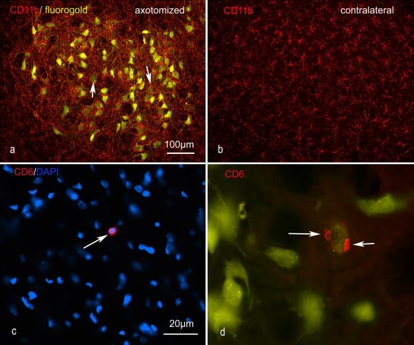
(Published customer image: Representative images of the inflammatory changes in the facial nucleus during axonal regeneration, one week following facial nerve transaction. a, b: CD11b immunoreactivity for microglia is increased in the axotomized facial nucleus, and microglia enwrap the facial motor neurons, e.g. at arrows. The regenerating neurons were retrogradely labelled with fluorogold. c, d: CD6- positive T-cells accumulated in the injured motor nucleus (arrows). They had little cytoplasm but dense nuclei (c) and were sometimes clustered around neurons retrogradely labelled with fluorogold (d). The scale bar in (a) also applies to (b) and that in (c) also applies to (d).From: Shokouhi et al. BMC Neuroscience 2010 11:13.)
Application Data
(Published customer image: Representative images of the inflammatory changes in the facial nucleus during axonal regeneration, one week following facial nerve transaction. a, b: CD11b immunoreactivity for microglia is increased in the axotomized facial nucleus, and microglia enwrap the facial motor neurons, e.g. at arrows. The regenerating neurons were retrogradely labelled with fluorogold. c, d: CD6- positive T-cells accumulated in the injured motor nucleus (arrows). They had little cytoplasm but dense nuclei (c) and were sometimes clustered around neurons retrogradely labelled with fluorogold (d). The scale bar in (a) also applies to (b) and that in (c) also applies to (d).From: Shokouhi et al. BMC Neuroscience 2010 11:13.)
CD11b, Monoclonal Antibody (Cat# AAA12146)
Full Name
MOUSE ANTI RAT CD11b:FITC
Gene Names
ITGAM; CD11B
Applications
Flow Cytometry
Pricing


 Application Data
(At 25 degree C. The primary antibody was diluted at 1/200 and incubated with the sample for 1 hour at 37 degree C. An Alexa Fluor 594 conjugated goat anti-rabbit IgG (H+L) Ab, diluted at 1/600, was used as the secondary antibody.)
Application Data
(At 25 degree C. The primary antibody was diluted at 1/200 and incubated with the sample for 1 hour at 37 degree C. An Alexa Fluor 594 conjugated goat anti-rabbit IgG (H+L) Ab, diluted at 1/600, was used as the secondary antibody.)
p53, Polyclonal Antibody (Cat# AAA31429)
Full Name
Phospho-p53 (Ser392) Antibody
Gene Names
TP53; P53; BCC7; LFS1; TRP53
Reactivity
Human, Mouse
Predicted Reactivity: Pig (88%), Bovine (88%), Sheep (88%), Rabbit (88%)
Predicted Reactivity: Pig (88%), Bovine (88%), Sheep (88%), Rabbit (88%)
Applications
Western Blot, Immunohistochemistry, Immunofluorescence, Immunocytochemistry, Peptide ELISA
Purity
The antibody is from purified rabbit serum by affinity purification via sequential chromatography on phospho-peptide and non-phospho-peptide affinity columns.
Pricing
 Standard Curve (Sample)
IL-6, ELISA Kit (Cat# AAA13517)
Full Name
IL-6 Rat ELISA Kit
Gene Names
Il6; ILg6; Ifnb2
Reactivity
Rat
Applications
ELISA
Pricing
 Standard Curve (Sample)
Folic Acid/Vitamin B9, ELISA Kit (Cat# AAA17669)
Full Name
Folic Acid/Vitamin B9 ELISA Kit
Reactivity
General
Pricing
 Standard Curve (Sample)
Leptin, ELISA Kit (Cat# AAA13511)
Full Name
Leptin Human ELISA Kit
Gene Names
LEP; OB; OBS; LEPD
Reactivity
Human
Applications
ELISA
Pricing


 FCM (Flow Cytometry)
(Overlay histogram showing MCF-7 cells stained with CSB-MA013563A0m (red line) at 1:400. The cells were incubated in 1x PBS /10% normal goat serum to block non-specific protein-protein interactions followed by primary antibody for 1 h at 4 degree C. The secondary antibody used was FITC goat anti-mouse IgG(H+L) at 1/200 dilution for 1 h at 4 degree C. Isotype control antibody (green line) was used under the same conditions. Acquisition of >10,000 events was performed.)
FCM (Flow Cytometry)
(Overlay histogram showing MCF-7 cells stained with CSB-MA013563A0m (red line) at 1:400. The cells were incubated in 1x PBS /10% normal goat serum to block non-specific protein-protein interactions followed by primary antibody for 1 h at 4 degree C. The secondary antibody used was FITC goat anti-mouse IgG(H+L) at 1/200 dilution for 1 h at 4 degree C. Isotype control antibody (green line) was used under the same conditions. Acquisition of >10,000 events was performed.)
CD146, Monoclonal Antibody (Cat# AAA27014)
Full Name
CD146 Monoclonal Antibody
Gene Names
MCAM; CD146; MUC18; HEMCAM; METCAM; MelCAM
Reactivity
Human
Applications
Western Blot, Immunofluorescence, Flow Cytometry
Purity
>95%
Protein G Purified
Protein G Purified
Pricing


 IHC (Immunohistochemistry)
(At 1/100 staining Mouse kidney tissue by IHC-P. The sample was formaldehyde fixed and a heat mediated antigen retrieval step in citrate buffer was performed. The sample was then blocked and incubated with the primary antibody at 4 degree C overnight. An HRP conjugated anti-Rabbit antibody was used as the secondary antibody.)
IHC (Immunohistochemistry)
(At 1/100 staining Mouse kidney tissue by IHC-P. The sample was formaldehyde fixed and a heat mediated antigen retrieval step in citrate buffer was performed. The sample was then blocked and incubated with the primary antibody at 4 degree C overnight. An HRP conjugated anti-Rabbit antibody was used as the secondary antibody.)
p38 MAPK, Polyclonal Antibody (Cat# AAA31333)
Full Name
Phospho-p38 MAPK (Thr180/Tyr182) Antibody
Gene Names
MAPK14; RK; p38; CSBP; EXIP; Mxi2; CSBP1; CSBP2; CSPB1; PRKM14; PRKM15; SAPK2A; p38ALPHA
Reactivity
Human, Mouse, Rat
Predicted Reactivity: Pig (100%), Bovine (100%), Horse (100%), Sheep (100%), Rabbit (100%), Dog (100%), Xenopus (91%)
Predicted Reactivity: Pig (100%), Bovine (100%), Horse (100%), Sheep (100%), Rabbit (100%), Dog (100%), Xenopus (91%)
Applications
Western Blot, Immunohistochemistry, Immunofluorescence, Immunocytochemistry, Immunoprecipitation, Peptide ELISA
Purity
The antibody is from purified rabbit serum by affinity purification via sequential chromatography on phospho-peptide and non-phospho-peptide affinity columns.
Pricing


 IHC (Immunohistchemistry)
(At 1/200 staining Human kidney tissue sections by IHC-P. The tissue was formaldehyde fixed and a heat mediated antigen retrieval step in citrate buffer was performed. The tissue was then blocked and incubated with the antibody for 1.5 hours at 22 degree C. An HRP conjugated goat anti-rabbit antibody was used as the secondary antibody.)
IHC (Immunohistchemistry)
(At 1/200 staining Human kidney tissue sections by IHC-P. The tissue was formaldehyde fixed and a heat mediated antigen retrieval step in citrate buffer was performed. The tissue was then blocked and incubated with the antibody for 1.5 hours at 22 degree C. An HRP conjugated goat anti-rabbit antibody was used as the secondary antibody.)
FGFR1/2/3/4, Polyclonal Antibody (Cat# AAA31417)
Full Name
Phospho-FGFR1/2/3/4 (Tyr653+Tyr654) Antibody
Gene Names
FGFR1; CEK; FLG; HH2; OGD; FLT2; KAL2; BFGFR; CD331; FGFBR; FLT-2; HBGFR; N-SAM; FGFR-1; bFGF-R-1
Reactivity
Human, Mouse, Rat, Monkey
Predicted Reactivity: Bovine (100%), Horse (100%), Sheep (100%), Rabbit (100%), Dog (100%), Chicken (100%), Xenopus (100%)
Predicted Reactivity: Bovine (100%), Horse (100%), Sheep (100%), Rabbit (100%), Dog (100%), Chicken (100%), Xenopus (100%)
Applications
Western Blot, Immunohistochemistry, Peptide ELISA
Purity
The antibody is from purified rabbit serum by affinity purification via sequential chromatography on phospho-peptide and non-phospho-peptide affinity columns.
Pricing
 Standard Curve (Sample)
Serum Phosphorus, Assay Kit (Cat# AAA27828)
Full Name
Serum Phosphorus Microplate Assay Kit
Reactivity
General
Applications
Functional Assay
Pricing
 SDS-PAGE
IgG Fc fragment, Native Protein (Cat# AAA15929)
Full Name
Mouse IgG Fc fragment
Applications
Western Blot
Purity
Greater or equal to 85% purity as determined by SDS-PAGE.
Pricing
 Standard Curve (Sample)
Catalase, Assay Kit (Cat# AAA27850)
Full Name
Catalase Microplate Assay Kit
Reactivity
General
Applications
Functional Assay
Pricing


 Application Data
(Staining of human peripheral blood granulocytes with Mouse anti Human CD45: Pacific Blue)
Application Data
(Staining of human peripheral blood granulocytes with Mouse anti Human CD45: Pacific Blue)
CD45, Monoclonal Antibody (Cat# AAA12202)
Full Name
MOUSE ANTI HUMAN CD45:FITC
Gene Names
PTPRC; LCA; LY5; B220; CD45; L-CA; T200; CD45R; GP180
Applications
Flow Cytometry
Pricing


 Application Data
(Immunoperoxidase staining of mouse lymph node cryosection stained with Rat antii Mouse CD8alpha antibody, clone KT15 followed by horseradish peroxidase conjugatedGoat anti Rat IgG s a detection reagent. High power)
Application Data
(Immunoperoxidase staining of mouse lymph node cryosection stained with Rat antii Mouse CD8alpha antibody, clone KT15 followed by horseradish peroxidase conjugatedGoat anti Rat IgG s a detection reagent. High power)
CD8 ALPHA, Monoclonal Antibody (Cat# AAA11885)
Full Name
RAT ANTI MOUSE CD8 ALPHA:FITC
Gene Names
Cd8a; Ly-2; Ly-B; Ly-35; Lyt-2; BB154331
Applications
Flow Cytometry
Pricing


 Application Data
(Overlay histogram showing HL-60 cells stained with AAA27009 (red line) at 1:500. The cells were incubated in 1x PBS /10% normal goat serum to block non-specific protein-protein interactions followed by primary antibody for 1 h at 4 degree C.The secondary antibody used was FITC goat anti-mouse IgG (H+L) at 1/200 dilution for 1 h at 4 degree C. Isotype control antibody (green line) was used under the same conditions. Acquisition of >10, 000 events was performed.)
Application Data
(Overlay histogram showing HL-60 cells stained with AAA27009 (red line) at 1:500. The cells were incubated in 1x PBS /10% normal goat serum to block non-specific protein-protein interactions followed by primary antibody for 1 h at 4 degree C.The secondary antibody used was FITC goat anti-mouse IgG (H+L) at 1/200 dilution for 1 h at 4 degree C. Isotype control antibody (green line) was used under the same conditions. Acquisition of >10, 000 events was performed.)
CD31, Monoclonal Antibody (Cat# AAA27009)
Full Name
CD31 Monoclonal Antibody
Gene Names
PECAM1; CD31; PECA1; GPIIA'; PECAM-1; endoCAM; CD31/EndoCAM
Reactivity
Human
Applications
Western Blot, Immunohistochemistry, Immunofluorescence, Flow Cytometry
Purity
>95%, Protein G purified
Pricing


 IHC (Immunohistochemistry)
(At 1/100 staining Human gastric cancer by IHC-P. The sample was formaldehyde fixed and a heat mediated antigen retrieval step in citrate buffer was performed. The sample was then blocked and incubated with the primary antibody at 4 degree C overnight. An HRP conjugated anti-Rabbit antibody was used as the secondary antibody.)
IHC (Immunohistochemistry)
(At 1/100 staining Human gastric cancer by IHC-P. The sample was formaldehyde fixed and a heat mediated antigen retrieval step in citrate buffer was performed. The sample was then blocked and incubated with the primary antibody at 4 degree C overnight. An HRP conjugated anti-Rabbit antibody was used as the secondary antibody.)
Smad1/9, Polyclonal Antibody (Cat# AAA31453)
Full Name
Phospho-Smad1/9 (Ser463/Ser465) Antibody
Gene Names
SMAD1; BSP1; JV41; BSP-1; JV4-1; MADH1; MADR1
Reactivity
Human, Mouse, Rat
Predicted Reactivity: Pig (100%), Zebrafish (100%), Bovine (100%), Sheep (100%), Rabbit (100%), Dog (100%), Chicken (100%), Xenopus (100%)
Predicted Reactivity: Pig (100%), Zebrafish (100%), Bovine (100%), Sheep (100%), Rabbit (100%), Dog (100%), Chicken (100%), Xenopus (100%)
Applications
Western Blot, Immunohistochemistry, Peptide ELISA
Purity
The antibody is from purified rabbit serum by affinity purification via sequential chromatography on phospho-peptide and non-phospho-peptide affinity columns.
Pricing


 Application Data
(At 25 degree C. The primary antibody was diluted at 1/200 and incubated with the sample for 1 hour at 37 degree C. An Alexa Fluor 594 conjugated goat anti-rabbit IgG (H+L) Ab, diluted at 1/600, was used as the secondary antibody.)
Application Data
(At 25 degree C. The primary antibody was diluted at 1/200 and incubated with the sample for 1 hour at 37 degree C. An Alexa Fluor 594 conjugated goat anti-rabbit IgG (H+L) Ab, diluted at 1/600, was used as the secondary antibody.)
SRF, Polyclonal Antibody (Cat# AAA31435)
Full Name
Phospho-SRF (Thr159) Antibody
Gene Names
SRF; MCM1
Reactivity
Human, Mouse, Rat
Predicted Reactivity: Pig (100%), Bovine (100%), Horse (100%), Rabbit (100%), Dog (100%), Chicken (100%), Xenopus (100%)
Predicted Reactivity: Pig (100%), Bovine (100%), Horse (100%), Rabbit (100%), Dog (100%), Chicken (100%), Xenopus (100%)
Applications
Western Blot, Immunohistochemistry, Immunofluorescence, Immunocytochemistry, Peptide ELISA
Purity
The antibody is from purified rabbit serum by affinity purification via sequential chromatography on phospho-peptide and non-phospho-peptide affinity columns.
Pricing


 FCM (Flow Cytometry)
(Overlay histogram showing Hela cells stained with (red line) at 1:100. The cells were fixed in 4% formaldehyde and permeated by 0.2% TritonX-100. Then 10% normal goat serum was Incubated to block non-specific protein-protein interactions followed by the antibody (1ug/1*106cells) for 1 h at 4 degree C. The secondary antibody used was FITC-conjugated Goat Anti-Mouse IgG(H+L) at 1/100 dilution for 30min at 4 degree C. Isotype control antibody (green line) was mouse IgG2b (1ug/1*106cells) used under the same conditions. Acquisition of >10,000 events was performed.)
FCM (Flow Cytometry)
(Overlay histogram showing Hela cells stained with (red line) at 1:100. The cells were fixed in 4% formaldehyde and permeated by 0.2% TritonX-100. Then 10% normal goat serum was Incubated to block non-specific protein-protein interactions followed by the antibody (1ug/1*106cells) for 1 h at 4 degree C. The secondary antibody used was FITC-conjugated Goat Anti-Mouse IgG(H+L) at 1/100 dilution for 30min at 4 degree C. Isotype control antibody (green line) was mouse IgG2b (1ug/1*106cells) used under the same conditions. Acquisition of >10,000 events was performed.)
YWHAZ, Monoclonal Antibody (Cat# AAA27050)
Full Name
YWHAZ Monoclonal Antibody
Gene Names
YWHAZ; YWHAD; KCIP-1; 14-3-3-zeta
Reactivity
Human, Mouse, Rat
Applications
Western Blot, Immunohistochemistry, Immunofluorescence, Flow Cytometry
Purity
>95%, Protein A purified
Pricing