Filters
Clonality
Type
Reactivity
Gene Name
Isotype
Host
Application
Clone
1005 results for " HRP conjugated" - showing 300-350


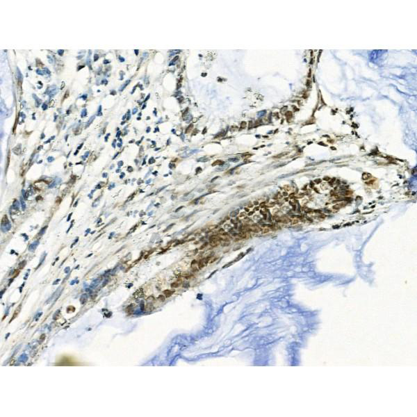
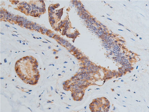
 IHC (Immunohistochemistry)
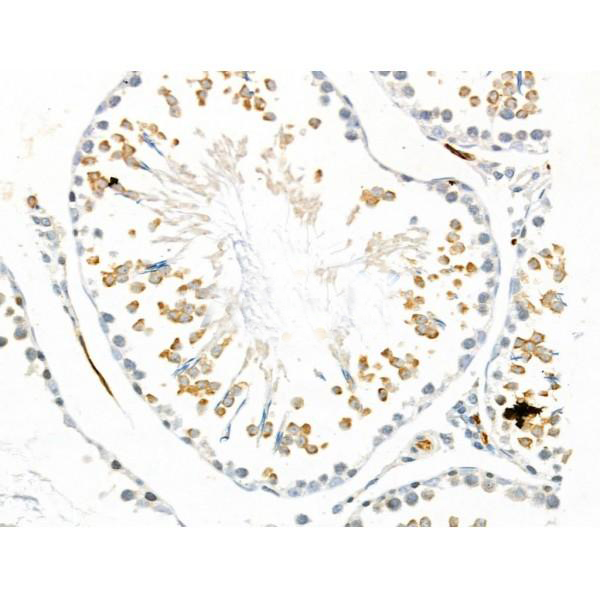
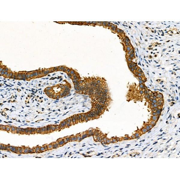
(At 1/100 staining Mouse testis tissue by IHC-P. The sample was formaldehyde fixed and a heat mediated antigen retrieval step in citrate buffer was performed. The sample was then blocked and incubated with the primary antibody at 4 degree C overnight. An HRP conjugated anti-Rabbit antibody was used as the secondary antibody.)
IHC (Immunohistochemistry)
(At 1/100 staining Mouse testis tissue by IHC-P. The sample was formaldehyde fixed and a heat mediated antigen retrieval step in citrate buffer was performed. The sample was then blocked and incubated with the primary antibody at 4 degree C overnight. An HRP conjugated anti-Rabbit antibody was used as the secondary antibody.)
IRS1, Polyclonal Antibody (Cat# AAA31366)
Full Name
Phospho-IRS1 (Tyr896) Antibody
Gene Names
IRS1; HIRS-1
Reactivity
Human, Mouse, Rat
Predicted Reactivity: Pig (100%), Zebrafish (100%), Bovine (100%), Horse (100%), Sheep (100%), Rabbit (100%), Dog (100%)
Predicted Reactivity: Pig (100%), Zebrafish (100%), Bovine (100%), Horse (100%), Sheep (100%), Rabbit (100%), Dog (100%)
Applications
Western Blot, Immunohistochemistry, Peptide ELISA
Purity
The antibody is from purified rabbit serum by affinity purification via sequential chromatography on phospho-peptide and non-phospho-peptide affinity columns.
Pricing


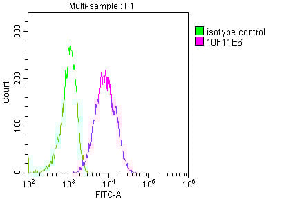
 FCM (Flow Cytometry)
(Overlay histogram showing MCF-7 cells stained with (red line). The cells were fixed with 70% Ethylalcohol (18h) and then incubated in 10% normal goat serum to block non-specific protein-protein interactions followed by the primary antibody at 1/200 for 1 h at 4 degree C. The secondary antibody used was FITC goat anti-mouse IgG(H+L) at 1/100 dilution for 30min at 4 degree C. Isotype control antibody (green line) was mouse IgG1 used under the same conditions. Acquisition of >10,000 events was performed.)
FCM (Flow Cytometry)
(Overlay histogram showing MCF-7 cells stained with (red line). The cells were fixed with 70% Ethylalcohol (18h) and then incubated in 10% normal goat serum to block non-specific protein-protein interactions followed by the primary antibody at 1/200 for 1 h at 4 degree C. The secondary antibody used was FITC goat anti-mouse IgG(H+L) at 1/100 dilution for 30min at 4 degree C. Isotype control antibody (green line) was mouse IgG1 used under the same conditions. Acquisition of >10,000 events was performed.)
HSPA8, Monoclonal Antibody (Cat# AAA27046)
Full Name
HSPA8 Monoclonal Antibody
Gene Names
HSPA8; LAP1; HSC54; HSC70; HSC71; HSP71; HSP73; NIP71; HSPA10
Reactivity
Human
Applications
Western Blot, Immunohistochemistry, Immunofluorescence, Flow Cytometry
Purity
>95%, Protein G purified
Pricing


 FCM (Flow Cytometry)
(Figure 9. Flow Cytometry analysis of SiHa cells using anti-DCTN1/p150-glued antibody (AAA19253).Overlay histogram showing SiHa cells stained with AAA19253 (Blue line). The cells were blocked with 10% normal goat serum. And then incubated with rabbit anti-DCTN1/p150-glued Antibody (AAA19253, 1μg/1x106 cells) for 30 min at 20 degree C. DyLight®488 conjugated goat anti-rabbit IgG (5-10μg/1x106 cells) was used as secondary antibody for 30 minutes at 20 degree C. Isotype control antibody (Green line) was rabbit IgG (1μg/1x106) used under the same conditions. Unlabelled sample (Red line) was also used as a control.)
FCM (Flow Cytometry)
(Figure 9. Flow Cytometry analysis of SiHa cells using anti-DCTN1/p150-glued antibody (AAA19253).Overlay histogram showing SiHa cells stained with AAA19253 (Blue line). The cells were blocked with 10% normal goat serum. And then incubated with rabbit anti-DCTN1/p150-glued Antibody (AAA19253, 1μg/1x106 cells) for 30 min at 20 degree C. DyLight®488 conjugated goat anti-rabbit IgG (5-10μg/1x106 cells) was used as secondary antibody for 30 minutes at 20 degree C. Isotype control antibody (Green line) was rabbit IgG (1μg/1x106) used under the same conditions. Unlabelled sample (Red line) was also used as a control.)
DCTN1/p150-glued, Polyclonal Antibody (Cat# AAA19253)
Full Name
Anti-DCTN1/p150-glued Antibody
Gene Names
DCTN1; P135; DP-150; DAP-150
Reactivity
Human, Mouse, Rat
Applications
Western Blot, Immunohistochemistry, Immunocytochemistry, Immunofluorescence, Flow Cytometry, Direct ELISA
Purity
Immunogen affinity purified.
Pricing


 Application Data
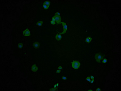
(At 25 degree C. Samples were then incubated with primary Ab(At 37 degree C. An AlexaFluor594 conjugated goat anti-rabbit IgG(H+L) Ab(Red) and an AlexaFluor488 conjugated goat anti-mouse IgG(H+L) Ab(Green) were used as the secondary antibody.The nuclear counter stain is DAPI(blue).)
Application Data
(At 25 degree C. Samples were then incubated with primary Ab(At 37 degree C. An AlexaFluor594 conjugated goat anti-rabbit IgG(H+L) Ab(Red) and an AlexaFluor488 conjugated goat anti-mouse IgG(H+L) Ab(Green) were used as the secondary antibody.The nuclear counter stain is DAPI(blue).)
Bcl-2, Polyclonal Antibody (Cat# AAA31360)
Full Name
Phospho-Bcl-2 (Thr56) Antibody
Gene Names
BCL2; Bcl-2; PPP1R50
Reactivity
Human, Mouse, Rat
Predicted Reactivity: Pig (86%), Bovine (83%)
Predicted Reactivity: Pig (86%), Bovine (83%)
Applications
Western Blot, Immunohistochemistry, Immunofluorescence, Immunocytochemistry, Peptide ELISA
Purity
The antibody is from purified rabbit serum by affinity purification via sequential chromatography on phospho-peptide and non-phospho-peptide affinity columns.
Pricing


 IP (Immunoprecipitation)
(Immunoprecipitating ENO1 in HepG2 whole cell lysate Lane 1: Mouse control IgG (1ug) instead of in HepG2 whole cell lysate. For western blotting, a HRP-conjugated Protein G antibody was used as the secondary antibody (1/5000) Lane 2: (1ul) + HepG2 whole cell lysate (500ug) Lane 3: HepG2 whole cell lysate (10ug))
IP (Immunoprecipitation)
(Immunoprecipitating ENO1 in HepG2 whole cell lysate Lane 1: Mouse control IgG (1ug) instead of in HepG2 whole cell lysate. For western blotting, a HRP-conjugated Protein G antibody was used as the secondary antibody (1/5000) Lane 2: (1ul) + HepG2 whole cell lysate (500ug) Lane 3: HepG2 whole cell lysate (10ug))
ENO1, Monoclonal Antibody (Cat# AAA27044)
Full Name
ENO1 Monoclonal Antibody
Gene Names
ENO1; NNE; PPH; MPB1; ENO1L1
Reactivity
Human, Mouse, Rat, Rabbit
Applications
Western Blot, Immunofluorescence, Flow Cytometry, Immunoprecipitation
Purity
>95%, Protein G purified
Pricing


 FCM (Flow Cytometry)
(Figure 8. Flow Cytometry analysis of U251 cells using anti-GABARAP antibody (AAA19250).Overlay histogram showing U251 cells stained with AAA19250 (Blue line). The cells were blocked with 10% normal goat serum. And then incubated with rabbit anti-GABARAP Antibody (AAA19250, 1μg/1x106 cells) for 30 min at 20 degree C. DyLight®488 conjugated goat anti-rabbit IgG (5-10μg/1x106 cells) was used as secondary antibody for 30 minutes at 20 degree C. Isotype control antibody (Green line) was rabbit IgG (1μg/1x106) used under the same conditions. Unlabelled sample (Red line) was also used as a control.)
FCM (Flow Cytometry)
(Figure 8. Flow Cytometry analysis of U251 cells using anti-GABARAP antibody (AAA19250).Overlay histogram showing U251 cells stained with AAA19250 (Blue line). The cells were blocked with 10% normal goat serum. And then incubated with rabbit anti-GABARAP Antibody (AAA19250, 1μg/1x106 cells) for 30 min at 20 degree C. DyLight®488 conjugated goat anti-rabbit IgG (5-10μg/1x106 cells) was used as secondary antibody for 30 minutes at 20 degree C. Isotype control antibody (Green line) was rabbit IgG (1μg/1x106) used under the same conditions. Unlabelled sample (Red line) was also used as a control.)
GABARAP, Polyclonal Antibody (Cat# AAA19250)
Full Name
Anti-GABARAP Antibody
Gene Names
GABARAP; MM46; ATG8A; GABARAP-a
Reactivity
Human, Mouse, Rat
Applications
Western Blot, Immunohistochemistry, Flow Cytometry
Purity
Immunogen affinity purified.
Pricing


 Application Data
(Staining of human peripheral blood lymphocytes with Mouse anti Human HLA ABC:Alexa Fluor 647)
Application Data
(Staining of human peripheral blood lymphocytes with Mouse anti Human HLA ABC:Alexa Fluor 647)
HLA ABC, Monoclonal Antibody (Cat# AAA11895)
Full Name
MOUSE ANTI HUMAN HLA ABC:HRP
Reactivity
Macaque, Bovine, Cynomolgus Monkey, Baboon, Rhesus Monkey, Chimpanzee, Gorilla, Shrew
Applications
Immunohistochemistry
Pricing


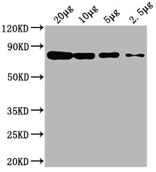
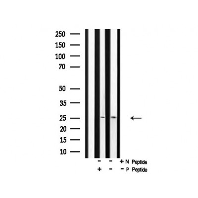
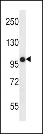
 WB (Western Blot)
(Western blot analysis of Phospho-MOB1A (Tyr26) in lysates of 293 IFN 2500U/ml 30', using Phospho-MOB1A (Tyr26) Antibody.)
WB (Western Blot)
(Western blot analysis of Phospho-MOB1A (Tyr26) in lysates of 293 IFN 2500U/ml 30', using Phospho-MOB1A (Tyr26) Antibody.)
MOB1A, Polyclonal Antibody (Cat# AAA31161)
Full Name
Phospho-MOB1A (Tyr26) Antibody
Gene Names
MOB1B; MATS2; MOB4A; MOBKL1A
Reactivity
Human, Mouse, Rat
Predicted: Bovine, Horse, Sheep, Rabbit, Dog, Xenopus
Predicted: Bovine, Horse, Sheep, Rabbit, Dog, Xenopus
Applications
Western Blot, Immunohistochemistry, Peptide ELISA
Purity
Purified rabbit serum by affinity purification via sequential chromatography on phospho- and non-phospho-peptide affinity columns.
Pricing


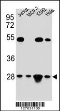 WB (Western Blot)
(TFAM Antibody (C-term) western blot analysis in Hela,Jurkat,K562,MCF-7 cell line lysates (35ug/lane).This demonstrates the TFAM antibody detected the TFAM protein (arrow).)
WB (Western Blot)
(TFAM Antibody (C-term) western blot analysis in Hela,Jurkat,K562,MCF-7 cell line lysates (35ug/lane).This demonstrates the TFAM antibody detected the TFAM protein (arrow).)
TFAM, Polyclonal Antibody (Cat# AAA28696)
Full Name
TFAM Antibody (C-term)
Gene Names
TFAM; TCF6; MTTF1; MTTFA; TCF6L1; TCF6L2; TCF6L3
Reactivity
Human
Applications
Immunohistochemistry, Flow Cytometry, Immunofluorescence, Western Blot
Purity
Peptide Affinity Purified Rabbit Polyclonal Antibody (Pab)
Pricing


 IHC (Immunohistochemistry)
(At 1/100 staining Human gastric cancer and adjacent normal tissues by IHC-P. The sample was formaldehyde fixed and a heat mediated antigen retrieval step in citrate buffer was performed. The sample was then blocked and incubated with the primary antibody at 4 degree C overnight. An HRP conjugated anti-Rabbit antibody was used as the secondary antibody.)
IHC (Immunohistochemistry)
(At 1/100 staining Human gastric cancer and adjacent normal tissues by IHC-P. The sample was formaldehyde fixed and a heat mediated antigen retrieval step in citrate buffer was performed. The sample was then blocked and incubated with the primary antibody at 4 degree C overnight. An HRP conjugated anti-Rabbit antibody was used as the secondary antibody.)
CAMKK1/2, Polyclonal Antibody (Cat# AAA31402)
Full Name
Phospho-CAMKK1/2 (Ser458/Ser495) Antibody
Gene Names
CAMKK1; CAMKKA
Reactivity
Human, Mouse, Rat
Predicted Reactivity: Pig (100%), Bovine (100%), Sheep (100%), Rabbit (100%), Dog (100%), Chicken (100%), Xenopus (100%)
Predicted Reactivity: Pig (100%), Bovine (100%), Sheep (100%), Rabbit (100%), Dog (100%), Chicken (100%), Xenopus (100%)
Applications
Western Blot, Immunohistochemistry, Peptide ELISA
Purity
The antibody is from purified rabbit serum by affinity purification via sequential chromatography on phospho-peptide and non-phospho-peptide affinity columns.
Pricing


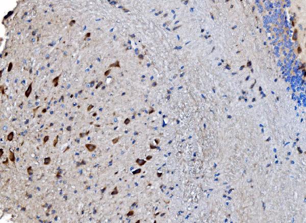
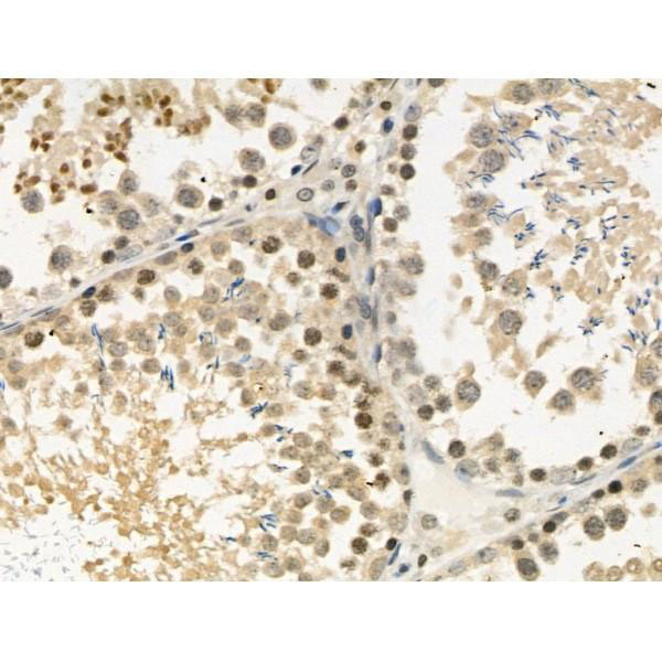
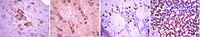 IHC (Immunohistchemistry)
(Immunohistochemistry Analysis: Representative lot data. (Fig. 1 and 2) Paraffin-embedded mouse and human brain tissue was prepared using heat-induced epitope retrieval in citrate buffer, pH 6.0. Immunostaining was performed using a 1:100 dilution. Reactivity was detected using the IHC-Select Detection Kit. Staining pattern appears as cytoplasmic. (Fig. 3 and 4) Paraffin-embedded mouse and mouse olfactory lobe and cerebellum brain tissue was prepared using heat-induced epitope retrieval in citrate buffer, pH 6.0. Immunostaining was performed using a Chicken IgY Antibody 1:100 dilution of Cat. No. AB15894, anti-Tbr2. Reactivity was detected using the IHC-Select Detection Kit. Immunoreactivity seen here is mostly nuclear.)
IHC (Immunohistchemistry)
(Immunohistochemistry Analysis: Representative lot data. (Fig. 1 and 2) Paraffin-embedded mouse and human brain tissue was prepared using heat-induced epitope retrieval in citrate buffer, pH 6.0. Immunostaining was performed using a 1:100 dilution. Reactivity was detected using the IHC-Select Detection Kit. Staining pattern appears as cytoplasmic. (Fig. 3 and 4) Paraffin-embedded mouse and mouse olfactory lobe and cerebellum brain tissue was prepared using heat-induced epitope retrieval in citrate buffer, pH 6.0. Immunostaining was performed using a Chicken IgY Antibody 1:100 dilution of Cat. No. AB15894, anti-Tbr2. Reactivity was detected using the IHC-Select Detection Kit. Immunoreactivity seen here is mostly nuclear.)
EOMES, Polyclonal Antibody (Cat# AAA26893)
Full Name
EOMES (Eomesodermin Homolog, T-box Brain Protein 2, TBR2, T-brain-2, TBR-2)
Gene Names
Eomes; Tbr2; TBR-2; C77258
Reactivity
Mouse, Human, Rat
Applications
Immunohistochemistry, Western Blot
Purity
Purified by affinity chromatography.
Pricing


 FCM (Flow Cytometry)
(Overlay histogram showing HepG2 cells stained with AAA28062 (red line) at 1:100. The cells were fixed in 4% formaldehyde and permeated by 0.2% TritonX-100. Then 10% normal goat serum was Incubated to block non-specific protein-protein interactions followed by the antibody (1ug/1*106cells) for 1 h at 4 degree C. The secondary antibody used was FITC-conjugated Goat Anti-Mouse IgG(H+L) at 1/100 dilution for 30min at 4 degree C. Isotype control antibody (green line) was mouse IgG2b (1ug/1*106cells) used under the same conditions. Acquisition of >10,000 events was performed.)
FCM (Flow Cytometry)
(Overlay histogram showing HepG2 cells stained with AAA28062 (red line) at 1:100. The cells were fixed in 4% formaldehyde and permeated by 0.2% TritonX-100. Then 10% normal goat serum was Incubated to block non-specific protein-protein interactions followed by the antibody (1ug/1*106cells) for 1 h at 4 degree C. The secondary antibody used was FITC-conjugated Goat Anti-Mouse IgG(H+L) at 1/100 dilution for 30min at 4 degree C. Isotype control antibody (green line) was mouse IgG2b (1ug/1*106cells) used under the same conditions. Acquisition of >10,000 events was performed.)
CD63, Monoclonal Antibody (Cat# AAA28062)
Full Name
CD63 Monoclonal Antibody
Gene Names
Cd63; ME491; C75951; Tspan30
Reactivity
Human, Rabbit
Applications
Western Blot, Immunohistochemistry, Immunofluorescence, Flow Cytometry
Purity
>95%, Protein G purified
Pricing
 WB (Western Blot)
(Western blot analysis of lysates from HT-29 cell line and mouse spleen tissue lysate(from left to right), using JAK1 Antibody (C-term). AAA28692 was diluted at 1:1000 at each lane. A goat anti-rabbit IgG H&L(HRP) at 1:5000 dilution was used as the secondary antibody. Lysates at 35ug per lane.)
WB (Western Blot)
(Western blot analysis of lysates from HT-29 cell line and mouse spleen tissue lysate(from left to right), using JAK1 Antibody (C-term). AAA28692 was diluted at 1:1000 at each lane. A goat anti-rabbit IgG H&L(HRP) at 1:5000 dilution was used as the secondary antibody. Lysates at 35ug per lane.)
JAK1, Polyclonal Antibody (Cat# AAA28692)
Full Name
JAK1 Antibody (C-term)
Gene Names
JAK1; JTK3; JAK1A; JAK1B
Reactivity
Human, mouse
Applications
Western Blot
Purity
Purified Rabbit Polyclonal Antibody (Pab)
Pricing


 IHC (Immunohistchemistry)
(Immunohistochemistry Analysis: Representative lot data. (Fig. 1 and 2) Paraffin-embedded mouse and human brain tissue was prepared using heat-induced epitope retrieval in citrate buffer, pH 6.0. Immunostaining was performed using a 1:100 dilution. Reactivity was detected using the IHC-Select Detection Kit. Staining pattern appears as cytoplasmic. (Fig. 3 and 4) Paraffin-embedded mouse and mouse olfactory lobe and cerebellum brain tissue was prepared using heat-induced epitope retrieval in citrate buffer, pH 6.0. Immunostaining was performed using a Chicken IgY Antibody 1:100 dilution of Cat. No. AB15894, anti-Tbr2. Reactivity was detected using the IHC-Select Detection Kit. Immunoreactivity seen here is mostly nuclear.)
IHC (Immunohistchemistry)
(Immunohistochemistry Analysis: Representative lot data. (Fig. 1 and 2) Paraffin-embedded mouse and human brain tissue was prepared using heat-induced epitope retrieval in citrate buffer, pH 6.0. Immunostaining was performed using a 1:100 dilution. Reactivity was detected using the IHC-Select Detection Kit. Staining pattern appears as cytoplasmic. (Fig. 3 and 4) Paraffin-embedded mouse and mouse olfactory lobe and cerebellum brain tissue was prepared using heat-induced epitope retrieval in citrate buffer, pH 6.0. Immunostaining was performed using a Chicken IgY Antibody 1:100 dilution of Cat. No. AB15894, anti-Tbr2. Reactivity was detected using the IHC-Select Detection Kit. Immunoreactivity seen here is mostly nuclear.)
EOMES, Polyclonal Antibody (Cat# AAA26883)
Full Name
EOMES (Eomesodermin Homolog, T-box Brain Protein 2, TBR2, T-brain-2, TBR-2)
Gene Names
Eomes; Tbr2; TBR-2; C77258
Reactivity
Mouse, Human, Rat
Applications
Immunohistochemistry, Western Blot
Purity
Purified by affinity chromatography.
Pricing


 FCM (Flow Cytometry)
(Figure 6. Flow Cytometry analysis of PC-3 cells using anti-ATG14L antibody (AAA11658).Overlay histogram showing PC-3 cells stained with AAA11658 (Blue line).The cells were blocked with 10% normal goat serum. And then incubated with rabbit anti-ATG14L Antibody (AAA11658,1ug/1x10^6 cells) for 30 min at 20 degree C. DyLight®488 conjugated goat anti-rabbit IgG (5-10ug/1x10^6 cells) was used as secondary antibody for 30 minutes at 20 degree C. Isotype control antibody (Green line) was rabbit IgG (1ug/1x106) used under the same conditions. Unlabelled sample (Red line) was also used as a control.)
FCM (Flow Cytometry)
(Figure 6. Flow Cytometry analysis of PC-3 cells using anti-ATG14L antibody (AAA11658).Overlay histogram showing PC-3 cells stained with AAA11658 (Blue line).The cells were blocked with 10% normal goat serum. And then incubated with rabbit anti-ATG14L Antibody (AAA11658,1ug/1x10^6 cells) for 30 min at 20 degree C. DyLight®488 conjugated goat anti-rabbit IgG (5-10ug/1x10^6 cells) was used as secondary antibody for 30 minutes at 20 degree C. Isotype control antibody (Green line) was rabbit IgG (1ug/1x106) used under the same conditions. Unlabelled sample (Red line) was also used as a control.)
ATG14L, Polyclonal Antibody (Cat# AAA11658)
Full Name
Anti-ATG14L Antibody
Gene Names
ATG14; ATG14L; BARKOR; KIAA0831
Reactivity
Human, Rat
Applications
Western Blot, Immunohistochemistry
Purity
Immunogen Affinity Purified
Pricing


 Application Data
(At 25 degree C. The primary antibody was diluted at 1/200 and incubated with the sample for 1 hour at 37 degree C. An Alexa Fluor 594 conjugated goat anti-rabbit IgG (H+L) antibody(Red), diluted at 1/600, was used as secondary antibody.)
Application Data
(At 25 degree C. The primary antibody was diluted at 1/200 and incubated with the sample for 1 hour at 37 degree C. An Alexa Fluor 594 conjugated goat anti-rabbit IgG (H+L) antibody(Red), diluted at 1/600, was used as secondary antibody.)
JAK3, Polyclonal Antibody (Cat# AAA31404)
Full Name
Phospho-JAK3 (Tyr904) Antibody
Gene Names
JAK3; JAKL; LJAK; JAK-3; L-JAK; JAK3_HUMAN
Reactivity
Human, Mouse, Rat
Predicted Reactivity: Bovine (100%), Sheep (100%), Dog (100%)
Predicted Reactivity: Bovine (100%), Sheep (100%), Dog (100%)
Applications
Western Blot, Immunohistochemistry, Immunofluorescence, Immunocytochemistry, Peptide ELISA
Purity
The antibody is from purified rabbit serum by affinity purification via sequential chromatography on phospho-peptide and non-phospho-peptide affinity columns.
Pricing


 IHC (Immunohistochemistry)
(At 1/100 staining Human prostate cancer and adjacent normal tissues by IHC-P. The sample was formaldehyde fixed and a heat mediated antigen retrieval step in citrate buffer was performed. The sample was then blocked and incubated with the primary antibody at 4 degree C overnight. An HRP conjugated anti-Rabbit antibody was used as the secondary antibody.)
IHC (Immunohistochemistry)
(At 1/100 staining Human prostate cancer and adjacent normal tissues by IHC-P. The sample was formaldehyde fixed and a heat mediated antigen retrieval step in citrate buffer was performed. The sample was then blocked and incubated with the primary antibody at 4 degree C overnight. An HRP conjugated anti-Rabbit antibody was used as the secondary antibody.)
EEF1A2, Polyclonal Antibody (Cat# AAA31289)
Full Name
Phospho-EEF1A2 (Ser358) Antibody
Gene Names
EEF1A2; HS1; STN; EF1A; STNL; EEF1AL; EF-1-alpha-2
Reactivity
Human, Mouse, Rat
Applications
Immunohistochemistry, Peptide ELISA
Purity
The antibody is from purified rabbit serum by affinity purification via sequential chromatography on phospho-peptide and non-phospho-peptide affinity columns.
Pricing
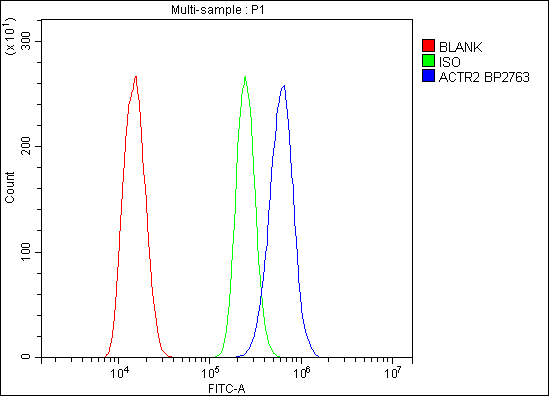


 FCM (Flow Cytometry)
(Figure 6. Flow Cytometry analysis of C6 cells using anti-Arp2/ACTR2 antibody (AAA19284).Overlay histogram showing C6 cells stained with AAA19284 (Blue line). The cells were blocked with 10% normal goat serum. And then incubated with rabbit anti-Arp2/ACTR2 Antibody (AAA19284,1μg/1x106 cells) for 30 min at 20 degree C. DyLight®488 conjugated goat anti-rabbit IgG (5-10μg/1x106 cells) was used as secondary antibody for 30 minutes at 20 degree C. Isotype control antibody (Green line) was rabbit IgG (1μg/1x106) used under the same conditions. Unlabelled sample (Red line) was also used as a control.)
FCM (Flow Cytometry)
(Figure 6. Flow Cytometry analysis of C6 cells using anti-Arp2/ACTR2 antibody (AAA19284).Overlay histogram showing C6 cells stained with AAA19284 (Blue line). The cells were blocked with 10% normal goat serum. And then incubated with rabbit anti-Arp2/ACTR2 Antibody (AAA19284,1μg/1x106 cells) for 30 min at 20 degree C. DyLight®488 conjugated goat anti-rabbit IgG (5-10μg/1x106 cells) was used as secondary antibody for 30 minutes at 20 degree C. Isotype control antibody (Green line) was rabbit IgG (1μg/1x106) used under the same conditions. Unlabelled sample (Red line) was also used as a control.)
Arp2/ACTR2, Polyclonal Antibody (Cat# AAA19284)
Full Name
Anti-Arp2/ACTR2 Antibody
Gene Names
ACTR2; ARP2
Reactivity
Human, Mouse, Monkey, Rat
Applications
WB, IHC-P, ICC, IF, FC/FACS/FCM, EIA
Purity
Immunogen affinity purified.
Pricing
 ELISA
(ELISA plate was coated by 2019-nCoV NP, 100 uL/cell at 5ug/ml. The indirect ELISA analysis was performed by loading 100 uL per well of the anti-2019-nCoV N mAb (BN9) at various concentrations. The plate was incubated for 1 hours at 37 degree C, then washed 5 times. An anti hFc HRP conjugated mAb at a concentration of 1:2000, 100uL/well was used as the secondary antibody. Again, the plate was incubated for 1 hours at 37 degree C, then washed 5 times. Detection was performed using TMB substrate for 10 minutes at room temperature in the dark. The plate was stopped with 2M sulfuric acid. Signal was read on a spectrophotometer at 450 nm.)
ELISA
(ELISA plate was coated by 2019-nCoV NP, 100 uL/cell at 5ug/ml. The indirect ELISA analysis was performed by loading 100 uL per well of the anti-2019-nCoV N mAb (BN9) at various concentrations. The plate was incubated for 1 hours at 37 degree C, then washed 5 times. An anti hFc HRP conjugated mAb at a concentration of 1:2000, 100uL/well was used as the secondary antibody. Again, the plate was incubated for 1 hours at 37 degree C, then washed 5 times. Detection was performed using TMB substrate for 10 minutes at room temperature in the dark. The plate was stopped with 2M sulfuric acid. Signal was read on a spectrophotometer at 450 nm.)
COVID 19 Nucleocapsid (NP) (BN9) Coronavirus, Monoclonal Antibody (Cat# AAA27957)
Full Name
Human anti-2019-nCoV N mAb (BN9)
Applications
ELISA
Purity
Purity: > = 95% as determined by SDS-PAGE
Purification: Protein A
Purification: Protein A
Pricing


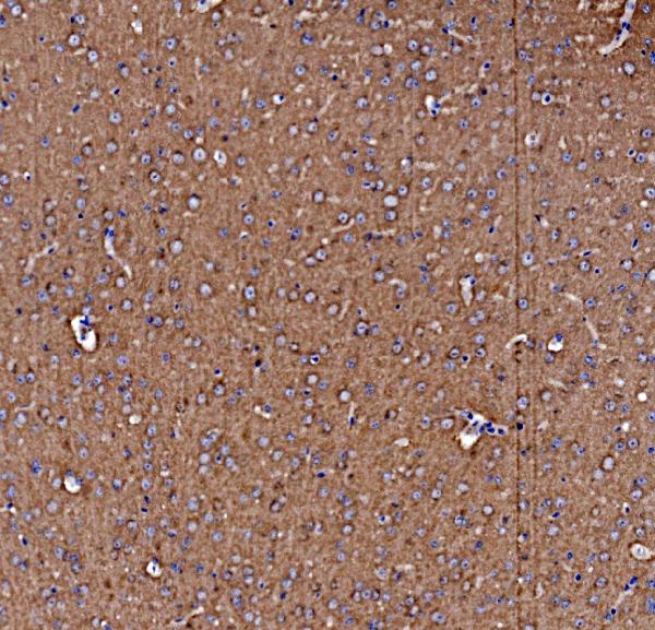
 WB (Western Blot)
(Formalin-fixed and paraffin-embedded human brain tissue reacted with OLR1 Antibody (Center), which was peroxidase-conjugated to the secondary antibody, followed by DAB staining. This data demonstrates the use of this antibody for immunohistochemistry; clinical relevance has not been evaluated.)
WB (Western Blot)
(Formalin-fixed and paraffin-embedded human brain tissue reacted with OLR1 Antibody (Center), which was peroxidase-conjugated to the secondary antibody, followed by DAB staining. This data demonstrates the use of this antibody for immunohistochemistry; clinical relevance has not been evaluated.)
OLR1, Polyclonal Antibody (Cat# AAA28737)
Full Name
OLR1 Antibody (Center)
Gene Names
OLR1; LOX1; LOXIN; SLOX1; CLEC8A; SCARE1
Reactivity
Human
Applications
Western Blot, Immunohistochemistry
Purity
Peptide Affinity Purified Rabbit Polyclonal Antibody (Pab)
Pricing


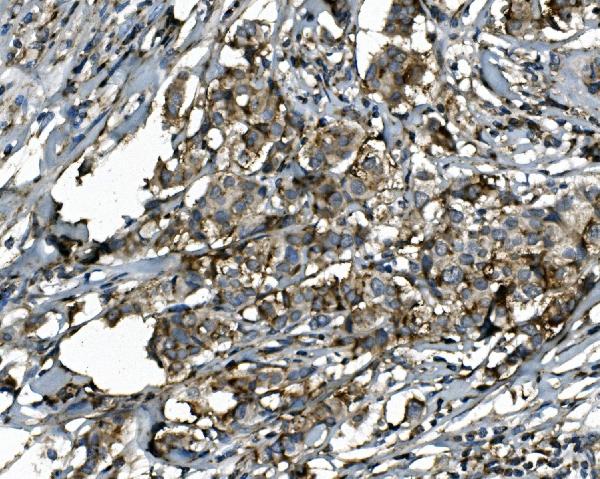
 IHC (Immunohistochemistry)
(At 1/100 staining Human esophageal cancer and adjacent normal tissues by IHC-P. The sample was formaldehyde fixed and a heat mediated antigen retrieval step in citrate buffer was performed. The sample was then blocked and incubated with the primary antibody at 4 degree C overnight. An HRP conjugated anti-Rabbit antibody was used as the secondary antibody.)
IHC (Immunohistochemistry)
(At 1/100 staining Human esophageal cancer and adjacent normal tissues by IHC-P. The sample was formaldehyde fixed and a heat mediated antigen retrieval step in citrate buffer was performed. The sample was then blocked and incubated with the primary antibody at 4 degree C overnight. An HRP conjugated anti-Rabbit antibody was used as the secondary antibody.)
EIF2S2, Polyclonal Antibody (Cat# AAA31456)
Full Name
Phospho-EIF2S2 (Ser2) Antibody
Gene Names
EIF2S2; EIF2; EIF2B; PPP1R67; EIF2beta; eIF-2-beta
Reactivity
Human, Mouse, Rat
Predicted Reactivity: Pig (100%), Zebrafish (100%), Bovine (100%), Horse (100%), Sheep (83%), Rabbit (100%), Dog (83%), Xenopus (100%)
Predicted Reactivity: Pig (100%), Zebrafish (100%), Bovine (100%), Horse (100%), Sheep (83%), Rabbit (100%), Dog (83%), Xenopus (100%)
Applications
Western Blot, Immunohistochemistry, Peptide ELISA
Purity
The antibody is from purified rabbit serum by affinity purification via sequential chromatography on phospho-peptide and non-phospho-peptide affinity columns.
Pricing


 Application Data
(At 25 degree C. Samples were then incubated with primary Ab(At 37 degree C. An AlexaFluor594 conjugated goat anti-rabbit IgG(H+L) Ab(Red) and an AlexaFluor488 conjugated goat anti-mouse IgG(H+L) Ab(Green) were used as the secondary antibody.The nuclear counter stain is DAPI (blue).)
Application Data
(At 25 degree C. Samples were then incubated with primary Ab(At 37 degree C. An AlexaFluor594 conjugated goat anti-rabbit IgG(H+L) Ab(Red) and an AlexaFluor488 conjugated goat anti-mouse IgG(H+L) Ab(Green) were used as the secondary antibody.The nuclear counter stain is DAPI (blue).)
eIF4B, Polyclonal Antibody (Cat# AAA31302)
Full Name
Phospho-eIF4B (Ser504) Antibody
Gene Names
EIF4B; EIF-4B; PRO1843
Reactivity
Human, Mouse, Rat
Applications
Western Blot, Immunohistochemistry, Immunofluorescence, Immunocytochemistry, Peptide ELISA
Purity
The antibody is from purified rabbit serum by affinity purification via sequential chromatography on phospho-peptide and non-phospho-peptide affinity columns.
Pricing


 FCM (Flow Cytometry)
(Figure 7. Flow Cytometry analysis of Hela cells using anti-Aquaporin 3 antibody (AAA11516).Overlay histogram showing Hela cells stained with AAA11516 (Blue line).The cells were blocked with 10% normal goat serum. And then incubated with rabbit anti-Aquaporin 3 Antibody (AAA11516,1ug/1x10^6 cells) for 30 min at 20 degree C. DyLight®488 conjugated goat anti-rabbit IgG (5-10ug/1x10^6 cells) was used as secondary antibody for 30 minutes at 20 degree C. Isotype control antibody (Green line) was rabbit IgG (1ug/1x106) used under the same conditions. Unlabelled sample (Red line) was also used as a control.)
FCM (Flow Cytometry)
(Figure 7. Flow Cytometry analysis of Hela cells using anti-Aquaporin 3 antibody (AAA11516).Overlay histogram showing Hela cells stained with AAA11516 (Blue line).The cells were blocked with 10% normal goat serum. And then incubated with rabbit anti-Aquaporin 3 Antibody (AAA11516,1ug/1x10^6 cells) for 30 min at 20 degree C. DyLight®488 conjugated goat anti-rabbit IgG (5-10ug/1x10^6 cells) was used as secondary antibody for 30 minutes at 20 degree C. Isotype control antibody (Green line) was rabbit IgG (1ug/1x106) used under the same conditions. Unlabelled sample (Red line) was also used as a control.)
Aquaporin 3, Polyclonal Antibody (Cat# AAA11516)
Full Name
Anti-Aquaporin 3 antibody
Gene Names
AQP3; GIL; AQP-3
Reactivity
Human, Mouse, Rat
Applications
Western Blot, Immunohistochemistry, Immunohistochemistry, Immunocytochemistry
Purity
Immunogen affinity purified.
Pricing


 IF (Immunofluorescence)
(Figure 6. IF analysis of SGK1 using anti- SGK1 antibody (AAA19225).SGK1 was detected in immunocytochemical section of A549 cells. Enzyme antigen retrieval was performed using IHC enzyme antigen retrieval reagent for 15 mins. The cells were blocked with 10% goat serum. And then incubated with 4μg/mL rabbit anti- SGK1 Antibody (AAA19225) overnight at 4 degree C. DyLight®488 Conjugated Goat Anti-Rabbit IgG was used as secondary antibody at 1:100 dilution and incubated for 30 minutes at 37 degree C. The section was counterstained with DAPI. Visualize using a fluorescence microscope and filter sets appropriate for the label used.)
IF (Immunofluorescence)
(Figure 6. IF analysis of SGK1 using anti- SGK1 antibody (AAA19225).SGK1 was detected in immunocytochemical section of A549 cells. Enzyme antigen retrieval was performed using IHC enzyme antigen retrieval reagent for 15 mins. The cells were blocked with 10% goat serum. And then incubated with 4μg/mL rabbit anti- SGK1 Antibody (AAA19225) overnight at 4 degree C. DyLight®488 Conjugated Goat Anti-Rabbit IgG was used as secondary antibody at 1:100 dilution and incubated for 30 minutes at 37 degree C. The section was counterstained with DAPI. Visualize using a fluorescence microscope and filter sets appropriate for the label used.)
SGK1, Polyclonal Antibody (Cat# AAA19225)
Full Name
Anti-SGK1 Antibody
Gene Names
SGK1; SGK
Reactivity
Human
Applications
Western Blot, Immunohistochemistry, Immunocytochemistry, Immunofluorescence, Flow Cytometry
Purity
Immunogen affinity purified.
Pricing


 IHC (Immunohistchemistry)
(At 1/100 staining Human prostate cancer and adjacent normal tissues by IHC-P. The sample was formaldehyde fixed and a heat mediated antigen retrieval step in citrate buffer was performed. The sample was then blocked and incubated with the primary antibody at 4 degree C overnight. An HRP conjugated anti-Rabbit antibody was used as the secondary antibody.)
IHC (Immunohistchemistry)
(At 1/100 staining Human prostate cancer and adjacent normal tissues by IHC-P. The sample was formaldehyde fixed and a heat mediated antigen retrieval step in citrate buffer was performed. The sample was then blocked and incubated with the primary antibody at 4 degree C overnight. An HRP conjugated anti-Rabbit antibody was used as the secondary antibody.)
TOPBP1, Polyclonal Antibody (Cat# AAA31467)
Full Name
Phospho-TOPBP1 (Ser1159) Antibody
Gene Names
TOPBP1; TOP2BP1
Reactivity
Human, Mouse, Rat
Predicted Reactivity: Pig (100%), Zebrafish (80%), Bovine (100%), Horse (100%), Sheep (100%), Rabbit (100%), Dog (100%), Chicken (80%), Xenopus (80%)
Predicted Reactivity: Pig (100%), Zebrafish (80%), Bovine (100%), Horse (100%), Sheep (100%), Rabbit (100%), Dog (100%), Chicken (80%), Xenopus (80%)
Applications
Western Blot, Immunohistochemistry, Peptide ELISA
Purity
The antibody is from purified rabbit serum by affinity purification via sequential chromatography on phospho-peptide and non-phospho-peptide affinity columns.
Pricing


 FCM (Flow Cytometry)
(Figure 8. Flow Cytometry analysis of HEPA1-6 cells using anti-PI-16/PI16 antibody (AAA19330).Overlay histogram showing HEPA1-6 cells stained with AAA19330 (Blue line). The cells were blocked with 10% normal goat serum. And then incubated with rabbit anti-PI-16/PI16 Antibody (AAA19330, 1μg/1x106 cells) for 30 min at 20 degree C. DyLight®488 conjugated goat anti-rabbit IgG (5-10μg/1x106 cells) was used as secondary antibody for 30 minutes at 20 degree C. Isotype control antibody (Green line) was rabbit IgG (1μg/1x106) used under the same conditions. Unlabelled sample (Red line) was also used as a control.)
FCM (Flow Cytometry)
(Figure 8. Flow Cytometry analysis of HEPA1-6 cells using anti-PI-16/PI16 antibody (AAA19330).Overlay histogram showing HEPA1-6 cells stained with AAA19330 (Blue line). The cells were blocked with 10% normal goat serum. And then incubated with rabbit anti-PI-16/PI16 Antibody (AAA19330, 1μg/1x106 cells) for 30 min at 20 degree C. DyLight®488 conjugated goat anti-rabbit IgG (5-10μg/1x106 cells) was used as secondary antibody for 30 minutes at 20 degree C. Isotype control antibody (Green line) was rabbit IgG (1μg/1x106) used under the same conditions. Unlabelled sample (Red line) was also used as a control.)
PI-16/PI16, Polyclonal Antibody (Cat# AAA19330)
Full Name
Anti-PI-16/PI16 Antibody
Gene Names
PI16; PSPBP; CRISP9; MSMBBP
Reactivity
Human, Mouse, Rat, Monkey
Applications
WB, IHC-P, FC/FACS/FCM, EIA
Purity
Immunogen affinity purified.
Pricing


 Application Data
(At 25 degree C. The primary antibody was diluted at 1/200 and incubated with the sample for 1 hour at 37 degree C. An Alexa Fluor 594 conjugated goat anti-rabbit IgG (H+L) Ab, diluted at 1/600, was used as the secondary antibody.)
Application Data
(At 25 degree C. The primary antibody was diluted at 1/200 and incubated with the sample for 1 hour at 37 degree C. An Alexa Fluor 594 conjugated goat anti-rabbit IgG (H+L) Ab, diluted at 1/600, was used as the secondary antibody.)
p27 Kip1, Polyclonal Antibody (Cat# AAA31387)
Full Name
Phospho-p27 Kip1 (Thr157) Antibody
Gene Names
CDKN1B; KIP1; MEN4; CDKN4; MEN1B; P27KIP1
Reactivity
Human, Mouse, Rat
Predicted Reactivity: Bovine (89%), Sheep (89%), Rabbit (100%), Dog (100%), Chicken (89%)
Predicted Reactivity: Bovine (89%), Sheep (89%), Rabbit (100%), Dog (100%), Chicken (89%)
Applications
Western Blot, Immunohistochemistry, Immunofluorescence, Immunocytochemistry, Peptide ELISA
Purity
The antibody is from purified rabbit serum by affinity purification via sequential chromatography on phospho-peptide and non-phospho-peptide affinity columns.
Pricing


 WB (Western Blot)
(FOLR2 Antibody (N-term) IHC analysis in formalin fixed and paraffin embedded human spleen followed by peroxidase conjugation of the secondary antibody and DAB staining. This data demonstrates the use of the FOLR2 Antibody (N-term) for immunohistochemistry. Clinical relevance has not been evaluated.)
WB (Western Blot)
(FOLR2 Antibody (N-term) IHC analysis in formalin fixed and paraffin embedded human spleen followed by peroxidase conjugation of the secondary antibody and DAB staining. This data demonstrates the use of the FOLR2 Antibody (N-term) for immunohistochemistry. Clinical relevance has not been evaluated.)
FOLR2, Polyclonal Antibody (Cat# AAA28783)
Full Name
FOLR2 Antibody (N-term)
Gene Names
FOLR2; FBP; FR-P3; FR-BETA; BETA-HFR; FBP/PL-1
Reactivity
Human, mouse
Applications
Western Blot, Immunohistochemistry, Flow Cytometry
Purity
Peptide Affinity Purified Rabbit Polyclonal Antibody (Pab)
Pricing


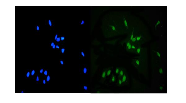
 IF (Immunofluorescence)
(ICC/IF analysis of FABP7 in PC3 cells. The cell was stained with AAA11731 (1:100). The secondary antibody (green) was used Alexa Fluor 488. DAPI was stained the cell nucleus (blue).)
IF (Immunofluorescence)
(ICC/IF analysis of FABP7 in PC3 cells. The cell was stained with AAA11731 (1:100). The secondary antibody (green) was used Alexa Fluor 488. DAPI was stained the cell nucleus (blue).)
FABP7, Monoclonal Antibody (Cat# AAA11731)
Full Name
FABP7 antibody
Gene Names
FABP7; MRG; BLBP; FABPB; B-FABP
Reactivity
Human, Mouse
Applications
Western Blot, Immunocytochemistry, Immunofluorescence
Purity
By protein-G affinity chromatography
Pricing


 IHC (Immunohistchemistry)
(Immunohistochemistry Analysis: Representative lot data. (Fig. 1 and 2) Paraffin-embedded mouse and human brain tissue was prepared using heat-induced epitope retrieval in citrate buffer, pH 6.0. Immunostaining was performed using a 1:100 dilution. Reactivity was detected using the IHC-Select Detection Kit. Staining pattern appears as cytoplasmic. (Fig. 3 and 4) Paraffin-embedded mouse and mouse olfactory lobe and cerebellum brain tissue was prepared using heat-induced epitope retrieval in citrate buffer, pH 6.0. Immunostaining was performed using a Chicken IgY Antibody 1:100 dilution of Cat. No. AB15894, anti-Tbr2. Reactivity was detected using the IHC-Select Detection Kit. Immunoreactivity seen here is mostly nuclear.)
IHC (Immunohistchemistry)
(Immunohistochemistry Analysis: Representative lot data. (Fig. 1 and 2) Paraffin-embedded mouse and human brain tissue was prepared using heat-induced epitope retrieval in citrate buffer, pH 6.0. Immunostaining was performed using a 1:100 dilution. Reactivity was detected using the IHC-Select Detection Kit. Staining pattern appears as cytoplasmic. (Fig. 3 and 4) Paraffin-embedded mouse and mouse olfactory lobe and cerebellum brain tissue was prepared using heat-induced epitope retrieval in citrate buffer, pH 6.0. Immunostaining was performed using a Chicken IgY Antibody 1:100 dilution of Cat. No. AB15894, anti-Tbr2. Reactivity was detected using the IHC-Select Detection Kit. Immunoreactivity seen here is mostly nuclear.)
EOMES, Polyclonal Antibody (Cat# AAA26884)
Full Name
EOMES (Eomesodermin Homolog, T-box Brain Protein 2, TBR2, T-brain-2, TBR-2)
Gene Names
Eomes; Tbr2; TBR-2; C77258
Reactivity
Mouse, Human, Rat
Applications
Immunohistochemistry, Western Blot
Purity
Purified by affinity chromatography.
Pricing
 FCM (Flow Cytometry)
(Figure 2. Flow Cytometry analysis of A549 cells using anti-WEE1 antibody (AAA19240).Overlay histogram showing A549 cells stained with AAA19240 (Blue line). The cells were blocked with 10% normal goat serum. And then incubated with rabbit anti-WEE1 Antibody (AAA19240, 1μg/1x106 cells) for 30 min at 20 degree C. DyLight®488 conjugated goat anti-rabbit IgG (5-10μg/1x106 cells) was used as secondary antibody for 30 minutes at 20 degree C. Isotype control antibody (Green line) was rabbit IgG (1μg/1x106) used under the same conditions. Unlabelled sample (Red line) was also used as a control.)
FCM (Flow Cytometry)
(Figure 2. Flow Cytometry analysis of A549 cells using anti-WEE1 antibody (AAA19240).Overlay histogram showing A549 cells stained with AAA19240 (Blue line). The cells were blocked with 10% normal goat serum. And then incubated with rabbit anti-WEE1 Antibody (AAA19240, 1μg/1x106 cells) for 30 min at 20 degree C. DyLight®488 conjugated goat anti-rabbit IgG (5-10μg/1x106 cells) was used as secondary antibody for 30 minutes at 20 degree C. Isotype control antibody (Green line) was rabbit IgG (1μg/1x106) used under the same conditions. Unlabelled sample (Red line) was also used as a control.)
WEE1, Polyclonal Antibody (Cat# AAA19240)
Full Name
Anti-WEE1 Antibody
Gene Names
WEE1; WEE1A; WEE1hu
Reactivity
Human, Mouse
Applications
Western Blot, Flow Cytometry, Direct ELISA
Purity
Immunogen affinity purified.
Pricing


 Application Data
(At 25 degree C. Samples were then incubated with primary Ab(At 37 degree C. An AlexaFluor594 conjugated goat anti-rabbit IgG(H+L) Ab(Red) and an AlexaFluor488 conjugated goat anti-mouse IgG(H+L) Ab(Green) were used as the secondary antibody.The nuclear counter stain is DAPI (blue).)
Application Data
(At 25 degree C. Samples were then incubated with primary Ab(At 37 degree C. An AlexaFluor594 conjugated goat anti-rabbit IgG(H+L) Ab(Red) and an AlexaFluor488 conjugated goat anti-mouse IgG(H+L) Ab(Green) were used as the secondary antibody.The nuclear counter stain is DAPI (blue).)
JIP1, Polyclonal Antibody (Cat# AAA31318)
Full Name
Phospho-JIP1 (Thr103) Antibody
Gene Names
MAPK8IP1; IB1; JIP1; JIP-1; PRKM8IP
Reactivity
Human, Mouse, Rat
Applications
Immunohistochemistry, Immunofluorescence, Immunocytochemistry, Peptide ELISA
Purity
The antibody is from purified rabbit serum by affinity purification via sequential chromatography on phospho-peptide and non-phospho-peptide affinity columns.
Pricing


 WB (Western Blot)
(Western blot analysis of CARD6 Antibody (Center) in MDA-MB231 cell line lysates (35ug/lane). CARD6 (arrow) was detected using the purified Pab.)
WB (Western Blot)
(Western blot analysis of CARD6 Antibody (Center) in MDA-MB231 cell line lysates (35ug/lane). CARD6 (arrow) was detected using the purified Pab.)
CARD6, Polyclonal Antibody (Cat# AAA28742)
Full Name
CARD6 Antibody (Center)
Gene Names
CARD6; CINCIN1
Reactivity
Human
Applications
Immunohistochemistry, Western Blot
Purity
Peptide Affinity Purified Rabbit Polyclonal Antibody (Pab)
Pricing


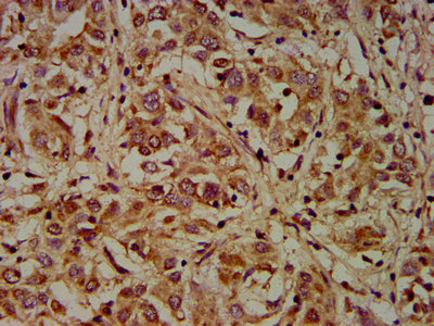
 IHC (Immunohistchemistry)
(Formalin-fixed and paraffin-embedded human cancer tissue reacted with the primary antibody, which was peroxidase-conjugated to the secondary antibody, followed by DAB staining. This data demonstrates the use of this antibody for immunohistochemistry; clinical relevance has not been evaluated. BC = breast carcinoma; HC = hepatocarcinoma.)
IHC (Immunohistchemistry)
(Formalin-fixed and paraffin-embedded human cancer tissue reacted with the primary antibody, which was peroxidase-conjugated to the secondary antibody, followed by DAB staining. This data demonstrates the use of this antibody for immunohistochemistry; clinical relevance has not been evaluated. BC = breast carcinoma; HC = hepatocarcinoma.)
EphA4, Polyclonal Antibody (Cat# AAA28736)
Full Name
EphA4 Antibody (C-term)
Gene Names
EPHA4; SEK; HEK8; TYRO1
Reactivity
Human, mouse
Applications
Western Blot, Immunohistochemistry
Purity
Purified Rabbit Polyclonal Antibody (Pab)
Pricing

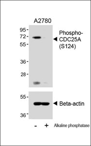


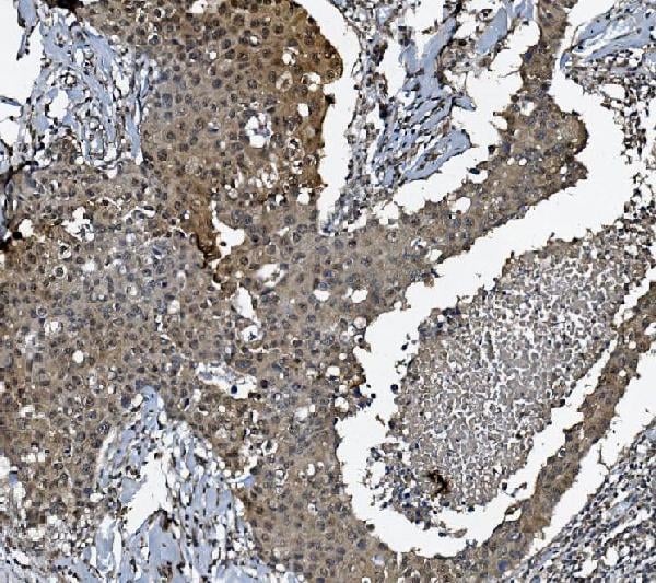
 IHC (Immunohistchemistry)
(Immunohistochemical analysis of (AAA28653) on paraffin-embedded Human breastcarcinoma tissue. Tissue was fixed withformaldehyde at room temperature. Heatinduced epitope retrieval was performed byEDTA buffer (pH9. 0). Samples wereincubated with primary antibody(1:100) for 1hour at room temperature. Undiluted CRFAnti-Polyvalent HRP Polymer antibody wasused as the secondary antibody.)
IHC (Immunohistchemistry)
(Immunohistochemical analysis of (AAA28653) on paraffin-embedded Human breastcarcinoma tissue. Tissue was fixed withformaldehyde at room temperature. Heatinduced epitope retrieval was performed byEDTA buffer (pH9. 0). Samples wereincubated with primary antibody(1:100) for 1hour at room temperature. Undiluted CRFAnti-Polyvalent HRP Polymer antibody wasused as the secondary antibody.)
Phospho-CDC25A (S124), Polyclonal Antibody (Cat# AAA28653)
Full Name
Phospho-CDC25A (S124) Antibody
Gene Names
CDC25A; CDC25A2
Reactivity
Human (Predicted Reactivity: Rat)
Applications
WB, EIA, IHC
Purity
Peptide Affinity Purified Rabbit Polyclonal Antibody (Pab)
Pricing


 IHC (Immunohistchemistry)
(AAA31091 at 1/200 staining human breast cancer tissue sections by IHC-P. The tissue was formaldehyde fixed and a heat mediated antigen retrieval step in citrate buffer was performed. The tissue was then blocked and incubated with the antibody for 1.5 hours at 22 degree C. An HRP conjugated goat anti-rabbit antibody was used as the secondary.)
IHC (Immunohistchemistry)
(AAA31091 at 1/200 staining human breast cancer tissue sections by IHC-P. The tissue was formaldehyde fixed and a heat mediated antigen retrieval step in citrate buffer was performed. The tissue was then blocked and incubated with the antibody for 1.5 hours at 22 degree C. An HRP conjugated goat anti-rabbit antibody was used as the secondary.)
VEGFR2, Polyclonal Antibody (Cat# AAA31091)
Full Name
VEGFR2 Antibody
Gene Names
KDR; FLK1; CD309; VEGFR; VEGFR2
Reactivity
Human, Mouse, Rat
Applications
Western Blot, Immunohistochemistry, Immunofluorescence, Immunocytochemistry
Purity
The antiserum was purified by peptide affinity chromatography using SulfoLink Coupling Resin.
Pricing


 FCM (Flow Cytometry)
(Figure 10. Flow Cytometry analysis of K562 cells using anti-RCC1 antibody (AAA19263).Overlay histogram showing K562 cells stained with AAA19263 (Blue line). The cells were blocked with 10% normal goat serum. And then incubated with rabbit anti-RCC1 Antibody (AAA19263,1μg/1x106 cells) for 30 min at 20 degree C. DyLight®488 conjugated goat anti-rabbit IgG (5-10μg/1x106 cells) was used as secondary antibody for 30 minutes at 20 degree C. Isotype control antibody (Green line) was rabbit IgG (1μg/1x106) used under the same conditions. Unlabelled sample (Red line) was also used as a control.)
FCM (Flow Cytometry)
(Figure 10. Flow Cytometry analysis of K562 cells using anti-RCC1 antibody (AAA19263).Overlay histogram showing K562 cells stained with AAA19263 (Blue line). The cells were blocked with 10% normal goat serum. And then incubated with rabbit anti-RCC1 Antibody (AAA19263,1μg/1x106 cells) for 30 min at 20 degree C. DyLight®488 conjugated goat anti-rabbit IgG (5-10μg/1x106 cells) was used as secondary antibody for 30 minutes at 20 degree C. Isotype control antibody (Green line) was rabbit IgG (1μg/1x106) used under the same conditions. Unlabelled sample (Red line) was also used as a control.)
RCC1, Polyclonal Antibody (Cat# AAA19263)
Full Name
Anti-RCC1 Antibody
Gene Names
RCC1; CHC1; RCC1-I; SNHG3-RCC1
Reactivity
Human, Mouse, Rat
Applications
Western Blot, Immunohistochemistry, Immunohistochemistry, Immunocytochemistry, Immunofluorescence, Flow Cytometry, Direct ELISA
Purity
Immunogen affinity purified.
Pricing


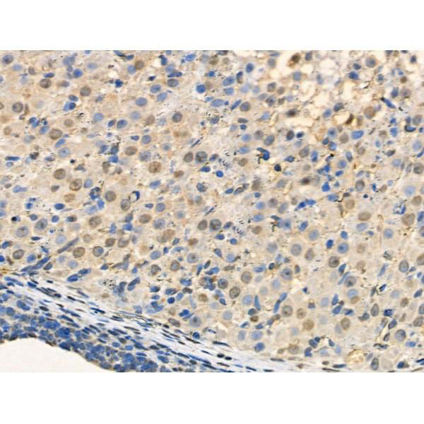
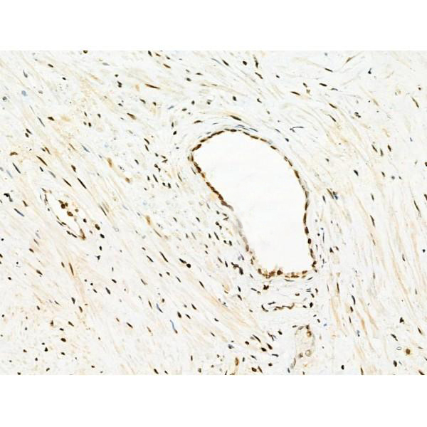
 IHC (Immunohistochemistry)
(At 1/100 staining Mouse kidney tissue by IHC-P. The sample was formaldehyde fixed and a heat mediated antigen retrieval step in citrate buffer was performed. The sample was then blocked and incubated with the primary antibody at 4 degree C overnight. An HRP conjugated anti-Rabbit antibody was used as the secondary antibody.)
IHC (Immunohistochemistry)
(At 1/100 staining Mouse kidney tissue by IHC-P. The sample was formaldehyde fixed and a heat mediated antigen retrieval step in citrate buffer was performed. The sample was then blocked and incubated with the primary antibody at 4 degree C overnight. An HRP conjugated anti-Rabbit antibody was used as the secondary antibody.)
YB1, Polyclonal Antibody (Cat# AAA31294)
Full Name
Phospho-YB1 (Ser102) Antibody
Gene Names
YBX1; YB1; BP-8; CSDB; DBPB; YB-1; CSDA2; NSEP1; NSEP-1; MDR-NF1
Reactivity
Human, Mouse, Rat
Applications
Immunohistochemistry, Peptide ELISA
Purity
The antibody is from purified rabbit serum by affinity purification via sequential chromatography on phospho-peptide and non-phospho-peptide affinity columns.
Pricing


 FCM (Flow Cytometry)
(Figure 8. Flow Cytometry analysis of SiHa cells using anti- ASS1 antibody (AAA19369).Overlay histogram showing SiHa cells stained with AAA19369 (Blue line). The cells were blocked with 10% normal goat serum. And then incubated with mouse anti-ASS1 Antibody (AAA19369, 1μg/1x106 cells) for 30 min at 20 degree C. DyLight®488 conjugated goat anti-mouse IgG (BA1126, 5-10μg/1x106 cells) was used as secondary antibody for 30 minutes at 20 degree C. Isotype control antibody (Green line) was mouse IgG (1μg/1x106) used under the same conditions. Unlabelled sample (Red line) was also used as a control.)
FCM (Flow Cytometry)
(Figure 8. Flow Cytometry analysis of SiHa cells using anti- ASS1 antibody (AAA19369).Overlay histogram showing SiHa cells stained with AAA19369 (Blue line). The cells were blocked with 10% normal goat serum. And then incubated with mouse anti-ASS1 Antibody (AAA19369, 1μg/1x106 cells) for 30 min at 20 degree C. DyLight®488 conjugated goat anti-mouse IgG (BA1126, 5-10μg/1x106 cells) was used as secondary antibody for 30 minutes at 20 degree C. Isotype control antibody (Green line) was mouse IgG (1μg/1x106) used under the same conditions. Unlabelled sample (Red line) was also used as a control.)
ASS1, Monoclonal Antibody (Cat# AAA19369)
Full Name
Anti-ASS1 Antibody (monoclonal, 5I5)
Gene Names
ASS1; ASS; CTLN1
Reactivity
Human, Mouse, Rat, Monkey
Applications
WB, IHC-P, ICC, IF, FC/FACS/FCM
Purity
Immunogen affinity purified.
Pricing


 IHC (Immunohistchemistry)
(Immunohistochemistry Analysis: Representative lot data. (Fig. 1 and 2) Paraffin-embedded mouse and human brain tissue was prepared using heat-induced epitope retrieval in citrate buffer, pH 6.0. Immunostaining was performed using a 1:100 dilution. Reactivity was detected using the IHC-Select Detection Kit. Staining pattern appears as cytoplasmic. (Fig. 3 and 4) Paraffin-embedded mouse and mouse olfactory lobe and cerebellum brain tissue was prepared using heat-induced epitope retrieval in citrate buffer, pH 6.0. Immunostaining was performed using a Chicken IgY Antibody 1:100 dilution of Cat. No. AB15894, anti-Tbr2. Reactivity was detected using the IHC-Select Detection Kit. Immunoreactivity seen here is mostly nuclear.)
IHC (Immunohistchemistry)
(Immunohistochemistry Analysis: Representative lot data. (Fig. 1 and 2) Paraffin-embedded mouse and human brain tissue was prepared using heat-induced epitope retrieval in citrate buffer, pH 6.0. Immunostaining was performed using a 1:100 dilution. Reactivity was detected using the IHC-Select Detection Kit. Staining pattern appears as cytoplasmic. (Fig. 3 and 4) Paraffin-embedded mouse and mouse olfactory lobe and cerebellum brain tissue was prepared using heat-induced epitope retrieval in citrate buffer, pH 6.0. Immunostaining was performed using a Chicken IgY Antibody 1:100 dilution of Cat. No. AB15894, anti-Tbr2. Reactivity was detected using the IHC-Select Detection Kit. Immunoreactivity seen here is mostly nuclear.)
EOMES, Polyclonal Antibody (Cat# AAA26891)
Full Name
EOMES (Eomesodermin Homolog, T-box Brain Protein 2, TBR2, T-brain-2, TBR-2)
Gene Names
Eomes; Tbr2; TBR-2; C77258
Reactivity
Mouse, Human, Rat
Applications
Immunohistochemistry, Western Blot
Purity
Purified by affinity chromatography.
Pricing


 IHC (Immunohistchemistry)
(Immunohistochemistry Analysis: Representative lot data. (Fig. 1 and 2) Paraffin-embedded mouse and human brain tissue was prepared using heat-induced epitope retrieval in citrate buffer, pH 6.0. Immunostaining was performed using a 1:100 dilution. Reactivity was detected using the IHC-Select Detection Kit. Staining pattern appears as cytoplasmic. (Fig. 3 and 4) Paraffin-embedded mouse and mouse olfactory lobe and cerebellum brain tissue was prepared using heat-induced epitope retrieval in citrate buffer, pH 6.0. Immunostaining was performed using a Chicken IgY Antibody 1:100 dilution of Cat. No. AB15894, anti-Tbr2. Reactivity was detected using the IHC-Select Detection Kit. Immunoreactivity seen here is mostly nuclear.)
IHC (Immunohistchemistry)
(Immunohistochemistry Analysis: Representative lot data. (Fig. 1 and 2) Paraffin-embedded mouse and human brain tissue was prepared using heat-induced epitope retrieval in citrate buffer, pH 6.0. Immunostaining was performed using a 1:100 dilution. Reactivity was detected using the IHC-Select Detection Kit. Staining pattern appears as cytoplasmic. (Fig. 3 and 4) Paraffin-embedded mouse and mouse olfactory lobe and cerebellum brain tissue was prepared using heat-induced epitope retrieval in citrate buffer, pH 6.0. Immunostaining was performed using a Chicken IgY Antibody 1:100 dilution of Cat. No. AB15894, anti-Tbr2. Reactivity was detected using the IHC-Select Detection Kit. Immunoreactivity seen here is mostly nuclear.)
EOMES, Polyclonal Antibody (Cat# AAA26892)
Full Name
EOMES (Eomesodermin Homolog, T-box Brain Protein 2, TBR2, T-brain-2, TBR-2)
Gene Names
Eomes; Tbr2; TBR-2; C77258
Reactivity
Mouse, Human, Rat
Applications
Immunohistochemistry, Western Blot
Purity
Purified by affinity chromatography.
Pricing


 IHC (Immunohistochemistry)
(AAA31132 at 1/100 staining rat testicular tissue sections by IHC-P. The tissue was formaldehyde fixed and a heat mediated antigen retrieval step in citrate buffer was performed. The tissue was then blocked and incubated with the antibody for 1.5 hours at 22 degree C. An HRP conjugated goat anti-rabbit antibody was used as the secondary.)
IHC (Immunohistochemistry)
(AAA31132 at 1/100 staining rat testicular tissue sections by IHC-P. The tissue was formaldehyde fixed and a heat mediated antigen retrieval step in citrate buffer was performed. The tissue was then blocked and incubated with the antibody for 1.5 hours at 22 degree C. An HRP conjugated goat anti-rabbit antibody was used as the secondary.)
EFNB2, Polyclonal Antibody (Cat# AAA31132)
Full Name
EFNB2 Antibody
Reactivity
Human, Mouse, Rat
Applications
Western Blot, Immunohistochemistry, Immunofluorescence, Immunocytochemistry
Purity
The antiserum was purified by peptide affinity chromatography using SulfoLink Coupling Resin.
Pricing


 Application Data
(C:FGFR2/isolectinB4 (C) and FGFR1/isolectinB4 (D) staining of apparent mesenchymal cells and the subpopulation of endothelial cells. Virtually all other dispersed apparent mesenchymal cells express FGFR1 and FGFR2 (merged image in E). F: FGFR2 (F) and FGFR1 (G) staining in clustered cells of epithelial origin (inferred by morphology here) demonstrating that epithelial cells express both FGFR1 and FGFR2 (merged image with DAPI staining in H).)
Application Data
(C:FGFR2/isolectinB4 (C) and FGFR1/isolectinB4 (D) staining of apparent mesenchymal cells and the subpopulation of endothelial cells. Virtually all other dispersed apparent mesenchymal cells express FGFR1 and FGFR2 (merged image in E). F: FGFR2 (F) and FGFR1 (G) staining in clustered cells of epithelial origin (inferred by morphology here) demonstrating that epithelial cells express both FGFR1 and FGFR2 (merged image with DAPI staining in H).)
FGFR2, Polyclonal Antibody (Cat# AAA26855)
Full Name
FGFR2, NT (FGFR2, BEK, KGFR, KSAM, Fibroblast growth factor receptor 2, K-sam, Keratinocyte growth factor receptor, CD332) (Azide free) (HRP)
Gene Names
FGFR2; BEK; JWS; BBDS; CEK3; CFD1; ECT1; KGFR; TK14; TK25; BFR-1; CD332; K-SAM
Reactivity
Human, Monkey, Mouse, Rat
Applications
IHC, EIA, WB
Purity
Purified by Protein G Affinity Chromatography.
Pricing


 FCM (Flow Cytometry)
(Figure 11. Flow Cytometry analysis of U937 cells using anti-NSF antibody (AAA19222).Overlay histogram showing U937 cells stained with AAA19222 (Blue line). The cells were blocked with 10% normal goat serum. And then incubated with rabbit anti-NSF Antibody (AAA19222, 1μg/1x106 cells) for 30 min at 20 degree C. DyLight®488 conjugated goat anti-rabbit IgG (5-10μg/1x106 cells) was used as secondary antibody for 30 minutes at 20 degree C. Isotype control antibody (Green line) was rabbit IgG (1μg/1x106) used under the same conditions. Unlabelled sample (Red line) was also used as a control.)
FCM (Flow Cytometry)
(Figure 11. Flow Cytometry analysis of U937 cells using anti-NSF antibody (AAA19222).Overlay histogram showing U937 cells stained with AAA19222 (Blue line). The cells were blocked with 10% normal goat serum. And then incubated with rabbit anti-NSF Antibody (AAA19222, 1μg/1x106 cells) for 30 min at 20 degree C. DyLight®488 conjugated goat anti-rabbit IgG (5-10μg/1x106 cells) was used as secondary antibody for 30 minutes at 20 degree C. Isotype control antibody (Green line) was rabbit IgG (1μg/1x106) used under the same conditions. Unlabelled sample (Red line) was also used as a control.)
NSF, Polyclonal Antibody (Cat# AAA19222)
Full Name
Anti-NSF Antibody
Gene Names
NSF; SKD2
Reactivity
Human, Mouse, Rat
Applications
Western Blot, Immunohistochemistry, Immunocytochemistry, Immunofluorescence, Flow Cytometry, Direct ELISA
Purity
Immunogen affinity purified.
Pricing


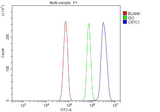 FCM (Flow Cytometry)
(Figure 8. Flow Cytometry analysis of RH35 cells using anti-TORC1/CRTC1 antibody (AAA19251).Overlay histogram showing RH35 cells stained with AAA19251 (Blue line). The cells were blocked with 10% normal goat serum. And then incubated with rabbit anti-TORC1/CRTC1 Antibody (AAA19251, 1μg/1x106 cells) for 30 min at 20 degree C. DyLight®488 conjugated goat anti-rabbit IgG (5-10μg/1x106 cells) was used as secondary antibody for 30 minutes at 20 degree C. Isotype control antibody (Green line) was rabbit IgG (1μg/1x106) used under the same conditions. Unlabelled sample (Red line) was also used as a control.)
FCM (Flow Cytometry)
(Figure 8. Flow Cytometry analysis of RH35 cells using anti-TORC1/CRTC1 antibody (AAA19251).Overlay histogram showing RH35 cells stained with AAA19251 (Blue line). The cells were blocked with 10% normal goat serum. And then incubated with rabbit anti-TORC1/CRTC1 Antibody (AAA19251, 1μg/1x106 cells) for 30 min at 20 degree C. DyLight®488 conjugated goat anti-rabbit IgG (5-10μg/1x106 cells) was used as secondary antibody for 30 minutes at 20 degree C. Isotype control antibody (Green line) was rabbit IgG (1μg/1x106) used under the same conditions. Unlabelled sample (Red line) was also used as a control.)
TORC1/CRTC1, Polyclonal Antibody (Cat# AAA19251)
Full Name
Anti-TORC1/CRTC1 Antibody
Gene Names
CRTC1; MECT1; TORC1; TORC-1; WAMTP1
Reactivity
Human, Mouse, Rat
Applications
Western Blot, Immunohistochemistry, Immunocytochemistry, Immunofluorescence, Flow Cytometry
Purity
Immunogen affinity purified.
Pricing
 WB (Western Blot)
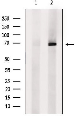
(Western blot analysis of extracts from Hela cells, using SLC5A5 Ab. The lane on the left was treated with blocking peptide.)
WB (Western Blot)
(Western blot analysis of extracts from Hela cells, using SLC5A5 Ab. The lane on the left was treated with blocking peptide.)
SLC5A5, Polyclonal Antibody (Cat# AAA31104)
Full Name
SLC5A5 Antibody
Gene Names
SLC5A5; NIS; TDH1
Reactivity
Human, Mouse
Applications
Western Blot, Immunohistochemistry
Purity
The antiserum was purified by peptide affinity chromatography using SulfoLink Coupling Resin (Thermo Fisher Scientific).
Pricing


 FCM (Flow Cytometry)
(Figure 6. Flow Cytometry analysis of HL-60 cells using anti-MCM6 antibody (AAA19264).Overlay histogram showing HL-60 cells stained with AAA19264 (Blue line). The cells were blocked with 10% normal goat serum. And then incubated with rabbit anti-MCM6 Antibody (AAA19264,1μg/1x106 cells) for 30 min at 20 degree C. DyLight®488 conjugated goat anti-rabbit IgG (5-10μg/1x106 cells) was used as secondary antibody for 30 minutes at 20 degree C. Isotype control antibody (Green line) was rabbit IgG (1μg/1x106) used under the same conditions. Unlabelled sample (Red line) was also used as a control.)
FCM (Flow Cytometry)
(Figure 6. Flow Cytometry analysis of HL-60 cells using anti-MCM6 antibody (AAA19264).Overlay histogram showing HL-60 cells stained with AAA19264 (Blue line). The cells were blocked with 10% normal goat serum. And then incubated with rabbit anti-MCM6 Antibody (AAA19264,1μg/1x106 cells) for 30 min at 20 degree C. DyLight®488 conjugated goat anti-rabbit IgG (5-10μg/1x106 cells) was used as secondary antibody for 30 minutes at 20 degree C. Isotype control antibody (Green line) was rabbit IgG (1μg/1x106) used under the same conditions. Unlabelled sample (Red line) was also used as a control.)
MCM6, Polyclonal Antibody (Cat# AAA19264)
Full Name
Anti-MCM6 Antibody
Gene Names
MCM6; Mis5; P105MCM; MCG40308
Reactivity
Human, Mouse, Rat, Monkey
Applications
Western Blot, Immunohistochemistry, Immunocytochemistry, Immunofluorescence, Flow Cytometry, Direct ELISA
Purity
Immunogen affinity purified.
Pricing
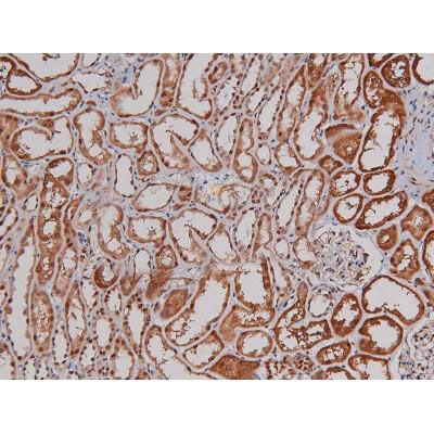
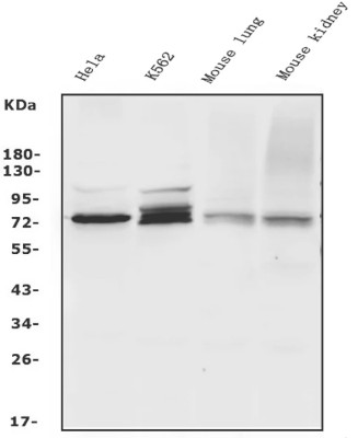
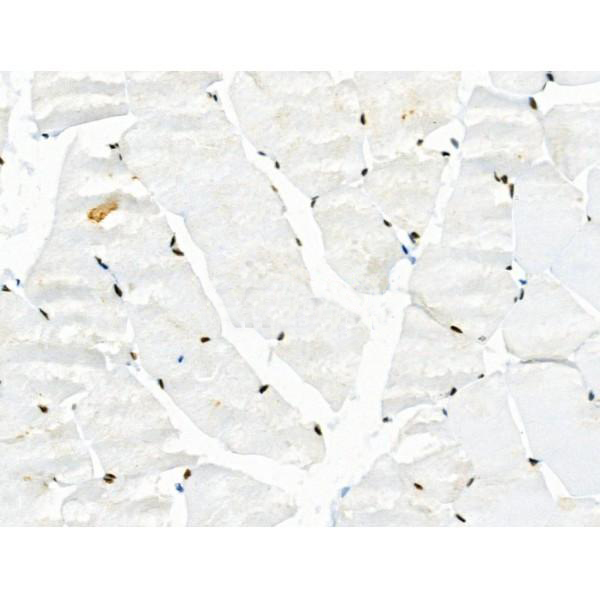
 IHC (Immunohistochemistry)
(IHC image of AAA27003 diluted at 1:200 and staining in paraffin-embedded human skeletal muscle tissue performed on a Leica BondTM system. After dewaxing and hydration, antigen retrieval was mediated by high pressure in a citrate buffer (pH 6.0). Section was blocked with 10% normal goat serum 30min at RT. Then primary antibody (1% BSA) was incubated at 4 degree C overnight. The primary is detected by a biotinylated secondary antibody and visualized using an HRP conjugated SP system.)
IHC (Immunohistochemistry)
(IHC image of AAA27003 diluted at 1:200 and staining in paraffin-embedded human skeletal muscle tissue performed on a Leica BondTM system. After dewaxing and hydration, antigen retrieval was mediated by high pressure in a citrate buffer (pH 6.0). Section was blocked with 10% normal goat serum 30min at RT. Then primary antibody (1% BSA) was incubated at 4 degree C overnight. The primary is detected by a biotinylated secondary antibody and visualized using an HRP conjugated SP system.)
DMD, Polyclonal Antibody (Cat# AAA27003)
Full Name
DMD Antibody
Gene Names
DMD; BMD; CMD3B; MRX85; DXS142; DXS164; DXS206; DXS230; DXS239; DXS268; DXS269; DXS270; DXS272
Reactivity
Human
Applications
Immunohistochemistry
Purity
>95%, Protein G purified
Pricing


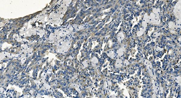
 IHC (Immunohistochemistry)
(At 1/100 staining Human gastric cancer by IHC-P. The sample was formaldehyde fixed and a heat mediated antigen retrieval step in citrate buffer was performed. The sample was then blocked and incubated with the primary antibody at 4 degree C overnight. An HRP conjugated anti-Rabbit antibody was used as the secondary antibody.)
IHC (Immunohistochemistry)
(At 1/100 staining Human gastric cancer by IHC-P. The sample was formaldehyde fixed and a heat mediated antigen retrieval step in citrate buffer was performed. The sample was then blocked and incubated with the primary antibody at 4 degree C overnight. An HRP conjugated anti-Rabbit antibody was used as the secondary antibody.)
alpha 1 Catenin, Polyclonal Antibody (Cat# AAA31379)
Full Name
alpha 1 Catenin Antibody
Gene Names
CTNNA1; CAP102
Reactivity
Human, Mouse, Rat
Applications
Western Blot, Immunohistochemistry, Peptide ELISA
Purity
The antiserum was purified by peptide affinity chromatography using SulfoLink Coupling Resin
Pricing


 FCM (Flow Cytometry)
(Figure 7. Flow Cytometry analysis of Hela cells using anti-ATG9A antibody (AAA19280).Overlay histogram showing Hela cells stained with AAA19280 (Blue line). The cells were blocked with 10% normal goat serum. And then incubated with rabbit anti-ATG9A Antibody (AAA19280, 1μg/1x106 cells) for 30 min at 20 degree C. DyLight®488 conjugated goat anti-rabbit IgG (5-10μg/1x106 cells) was used as secondary antibody for 30 minutes at 20 degree C. Isotype control antibody (Green line) was rabbit IgG (1μg/1x106) used under the same conditions. Unlabelled sample (Red line) was also used as a control.)
FCM (Flow Cytometry)
(Figure 7. Flow Cytometry analysis of Hela cells using anti-ATG9A antibody (AAA19280).Overlay histogram showing Hela cells stained with AAA19280 (Blue line). The cells were blocked with 10% normal goat serum. And then incubated with rabbit anti-ATG9A Antibody (AAA19280, 1μg/1x106 cells) for 30 min at 20 degree C. DyLight®488 conjugated goat anti-rabbit IgG (5-10μg/1x106 cells) was used as secondary antibody for 30 minutes at 20 degree C. Isotype control antibody (Green line) was rabbit IgG (1μg/1x106) used under the same conditions. Unlabelled sample (Red line) was also used as a control.)
ATG9A, Polyclonal Antibody (Cat# AAA19280)
Full Name
Anti-ATG9A Antibody
Gene Names
ATG9A; mATG9; APG9L1; MGD3208
Reactivity
Human, Mouse, Rat
Applications
WB, IHC-P, FC/FACS/FCM, EIA
Purity
Immunogen affinity purified.
Pricing