Filters
Clonality
Type
Reactivity
Gene Name
Isotype
Host
Application
Clone
2811 results for " C" - showing 300-350


 WB (Western Blot)
(Anti-GST Tag mouse monoclonal antibody at 1:25000, 1:1250, 1:25000Lane A: GST-mBID transfected E.coli cell lysate (0.2ug)Lane B: GST-YWHAB transfected E.coli cell lysate (0.2ug)Lane C: Empty vecter (0.2ug)SecondaryGoat Anti-Mouse IgG H&L (Dylight800) at 1/15000 dilution.Developed using the Odyssey technique. Performed under reducing conditions.)
WB (Western Blot)
(Anti-GST Tag mouse monoclonal antibody at 1:25000, 1:1250, 1:25000Lane A: GST-mBID transfected E.coli cell lysate (0.2ug)Lane B: GST-YWHAB transfected E.coli cell lysate (0.2ug)Lane C: Empty vecter (0.2ug)SecondaryGoat Anti-Mouse IgG H&L (Dylight800) at 1/15000 dilution.Developed using the Odyssey technique. Performed under reducing conditions.)
GST, Monoclonal Antibody (Cat# AAA27744)
Full Name
Anti-GST tag Antibody, Mouse Monoclonal
Reactivity
Schistosoma japonicum
Applications
Western Blot, Immunoprecipitation
Purity
Protein A
Pricing


 FCM (Flow Cytometry)
(Flow cytometric analysis of A549 cells with AKT1/2/3 antibody at 1/50 dilution (blue) compared with an unlabelled control (cells without incubation with primary antibody; red). Alexa Fluor 488-conjugated goat anti rabbit IgG was used as the secondary antibody.)
FCM (Flow Cytometry)
(Flow cytometric analysis of A549 cells with AKT1/2/3 antibody at 1/50 dilution (blue) compared with an unlabelled control (cells without incubation with primary antibody; red). Alexa Fluor 488-conjugated goat anti rabbit IgG was used as the secondary antibody.)
AKT1/2/3, Monoclonal Antibody (Cat# AAA30101)
Full Name
AKT1/2/3 Antibody
Gene Names
AKT1; AKT; PKB; RAC; CWS6; PRKBA; PKB-ALPHA; RAC-ALPHA
Reactivity
Human, Mouse, Rat
Applications
Western Blot, Immunocytochemistry, Immunofluorescence, Immunohistochemistry, Immunoprecipitation, Flow Cytometry
Purity
ProA affinity purified
Pricing


 SDS-PAGE
(Purified ER-beta Mouse Monoclonal Antibody (ESR2/686). Confirmation of Integrity and Purity of Antibody.)
SDS-PAGE
(Purified ER-beta Mouse Monoclonal Antibody (ESR2/686). Confirmation of Integrity and Purity of Antibody.)
ER-beta1 (Estrogen Receptor beta-1), Monoclonal Antibody (Cat# AAA13846)
Full Name
ER-beta1 (Estrogen Receptor beta-1) Mouse Monoclonal Antibody
Gene Names
ESR2; Erb; ESRB; ESTRB; NR3A2; ER-BETA; ESR-BETA
Reactivity
Human
Applications
Flow Cytometry, Immunofluorescence, Western Blot, Immunohistochemistry
Pricing


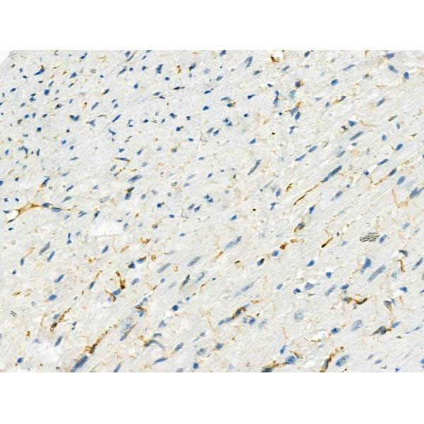
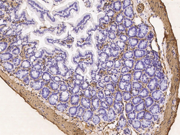
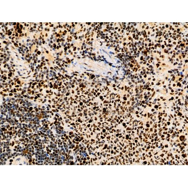
 IHC (Immunohistchemistry)
(AAA31084 at 1/200 staining human liver cancer tissue sections by IHC-P. The tissue was formaldehyde fixed and a heat mediated antigen retrieval step in citrate buffer was performed. The tissue was then blocked and incubated with the antibody for 1.5 hours at 22 degree C. An HRP conjugated goat anti-rabbit antibody was used as the secondary.)
IHC (Immunohistchemistry)
(AAA31084 at 1/200 staining human liver cancer tissue sections by IHC-P. The tissue was formaldehyde fixed and a heat mediated antigen retrieval step in citrate buffer was performed. The tissue was then blocked and incubated with the antibody for 1.5 hours at 22 degree C. An HRP conjugated goat anti-rabbit antibody was used as the secondary.)
HSP90B, Polyclonal Antibody (Cat# AAA31084)
Full Name
HSP90B Antibody
Gene Names
HSP90AB1; HSP84; HSPC2; HSPCB; D6S182; HSP90B
Reactivity
Human, Mouse, Rat
Applications
Western Blot, Immunohistochemistry, Immunofluorescence, Immunocytochemistry, Immunoprecipitation
Purity
The antiserum was purified by peptide affinity chromatography using SulfoLink Coupling Resin.
Pricing


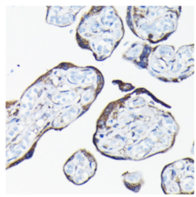
 Application Data
(Publised customer image:Mouse anti Human CD163 antibody, clone EDHu-1 used for the identification of perivascular macrophages in human brain by immunofluorescence.Image caption:Images demonstrating immunohistological stainings of amylin and double immunofluorescence staining against NG2/amylin, laminin/amylin and CD163/amylin in the hippocampus of the patient with AD and T2D.Amylin cell inclusions are indicated with arrows in (a) and shown in a higher magnification in (b). Pericytes with round cell bodies and NG2-positive coverage of the microvessel surface (green in c), without amylin cell inclusions (red in d), displayed round DAPI-positive cell nuclei (blue in e). The images in (c), (d) and (e) are merged in (f). Cells with more diffuse and weak NG2 staining (indicated by the arrowhead, green in g) and cytosolic amylin cell inclusions (red in h) showed altered cell nuclei (indicated with arrow, blue in i).The adjacent unaffected NG2-positive cell is indicated with an arrowhead in (i). The images in (g), (h) and (i) are merged in (j). Cells enclosed by laminin (green in K) contained amylin grains (red in l) and fragmented DAPI-positive cell nuclei (indicated with an arrow blue in m). The images in (k), (l) and (m) are merged in (n).Loss of NG2 coverage (green in o) was associated with polarized amylin cell inclusion (red in p) and fragmented DAPI-positive cell nuclei (indicated with an arrow, blue in q). The images in (o), (p) and (q) are merged in (r). Staining against macrophage marker CD163 (green in s) did not co-localize with amylin cell inclusions (red in t). The cell nucleus was stained with DAPI (blue in U). The images in (s), (t) and (u) are merged in (v). Scale bars: (a) 50 mum, (b), (c) to (v) 5 mum.From: Schultz, N. et al. (2016).Amylin alters human brain pericyte viability and NG2 expression.J Cereb Blood Flow & Metab. Jun 28 [Epub ahead of print]This is from an open access article distributed under the terms of the Creative Commons Attribution License.)
Application Data
(Publised customer image:Mouse anti Human CD163 antibody, clone EDHu-1 used for the identification of perivascular macrophages in human brain by immunofluorescence.Image caption:Images demonstrating immunohistological stainings of amylin and double immunofluorescence staining against NG2/amylin, laminin/amylin and CD163/amylin in the hippocampus of the patient with AD and T2D.Amylin cell inclusions are indicated with arrows in (a) and shown in a higher magnification in (b). Pericytes with round cell bodies and NG2-positive coverage of the microvessel surface (green in c), without amylin cell inclusions (red in d), displayed round DAPI-positive cell nuclei (blue in e). The images in (c), (d) and (e) are merged in (f). Cells with more diffuse and weak NG2 staining (indicated by the arrowhead, green in g) and cytosolic amylin cell inclusions (red in h) showed altered cell nuclei (indicated with arrow, blue in i).The adjacent unaffected NG2-positive cell is indicated with an arrowhead in (i). The images in (g), (h) and (i) are merged in (j). Cells enclosed by laminin (green in K) contained amylin grains (red in l) and fragmented DAPI-positive cell nuclei (indicated with an arrow blue in m). The images in (k), (l) and (m) are merged in (n).Loss of NG2 coverage (green in o) was associated with polarized amylin cell inclusion (red in p) and fragmented DAPI-positive cell nuclei (indicated with an arrow, blue in q). The images in (o), (p) and (q) are merged in (r). Staining against macrophage marker CD163 (green in s) did not co-localize with amylin cell inclusions (red in t). The cell nucleus was stained with DAPI (blue in U). The images in (s), (t) and (u) are merged in (v). Scale bars: (a) 50 mum, (b), (c) to (v) 5 mum.From: Schultz, N. et al. (2016).Amylin alters human brain pericyte viability and NG2 expression.J Cereb Blood Flow & Metab. Jun 28 [Epub ahead of print]This is from an open access article distributed under the terms of the Creative Commons Attribution License.)
CD163, Monoclonal Antibody (Cat# AAA12256)
Full Name
Mouse Anti Human CD163: RPE
Gene Names
CD163; M130; MM130; SCARI1
Reactivity
Human
Applications
Flow Cytometry
Pricing


 IF (Immunofluorescence)
(Figure 11. IF analysis of H Cadherin/CDH13 using anti- H Cadherin/CDH13 antibody (AAA19252).H Cadherin/CDH13 was detected in immunocytochemical section of SIHA cells. Enzyme antigen retrieval was performed using IHC enzyme antigen retrieval reagent for 15 mins. The cells were blocked with 10% goat serum. And then incubated with 5μg/mL rabbit anti- H Cadherin/CDH13 Antibody (AAA19252) overnight at 4 degree C. DyLight®488 Conjugated Goat Anti-Rabbit IgG was used as secondary antibody at 1:100 dilution and incubated for 30 minutes at 37 degree C. The section was counterstained with DAPI. Visualize using a fluorescence microscope and filter sets appropriate for the label used.)
IF (Immunofluorescence)
(Figure 11. IF analysis of H Cadherin/CDH13 using anti- H Cadherin/CDH13 antibody (AAA19252).H Cadherin/CDH13 was detected in immunocytochemical section of SIHA cells. Enzyme antigen retrieval was performed using IHC enzyme antigen retrieval reagent for 15 mins. The cells were blocked with 10% goat serum. And then incubated with 5μg/mL rabbit anti- H Cadherin/CDH13 Antibody (AAA19252) overnight at 4 degree C. DyLight®488 Conjugated Goat Anti-Rabbit IgG was used as secondary antibody at 1:100 dilution and incubated for 30 minutes at 37 degree C. The section was counterstained with DAPI. Visualize using a fluorescence microscope and filter sets appropriate for the label used.)
H Cadherin/CDH13, Polyclonal Antibody (Cat# AAA19252)
Full Name
Anti-H Cadherin/CDH13 Antibody
Gene Names
CDH13; CDHH; P105
Reactivity
Human, Rat
Applications
Western Blot, Immunohistochemistry, Immunocytochemistry, Immunofluorescence, Flow Cytometry, Direct ELISA
Purity
Immunogen affinity purified.
Pricing


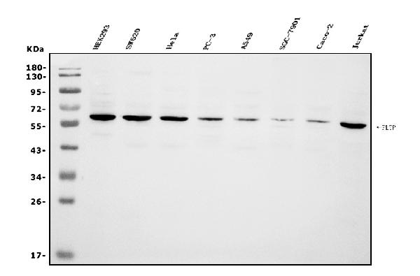
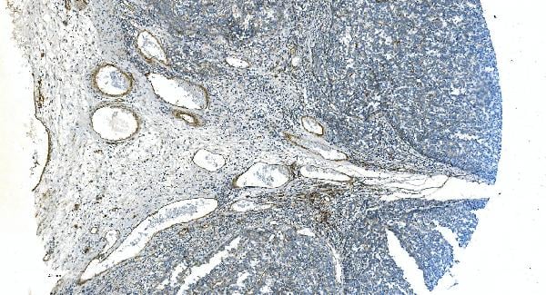
 FCM (Flow Cytometry)
(Figure 6. Flow Cytometry analysis of CACO-2 cells using anti-PLTP antibody (AAA19255).Overlay histogram showing CACO-2 cells stained with AAA19255 (Blue line). The cells were blocked with 10% normal goat serum. And then incubated with rabbit anti-PLTP Antibody (AAA19255, 1μg/1x106 cells) for 30 min at 20 degree C. DyLight®488 conjugated goat anti-rabbit IgG (5-10μg/1x106 cells) was used as secondary antibody for 30 minutes at 20 degree C. Isotype control antibody (Green line) was rabbit IgG (1μg/1x106) used under the same conditions. Unlabelled sample (Red line) was also used as a control.)
FCM (Flow Cytometry)
(Figure 6. Flow Cytometry analysis of CACO-2 cells using anti-PLTP antibody (AAA19255).Overlay histogram showing CACO-2 cells stained with AAA19255 (Blue line). The cells were blocked with 10% normal goat serum. And then incubated with rabbit anti-PLTP Antibody (AAA19255, 1μg/1x106 cells) for 30 min at 20 degree C. DyLight®488 conjugated goat anti-rabbit IgG (5-10μg/1x106 cells) was used as secondary antibody for 30 minutes at 20 degree C. Isotype control antibody (Green line) was rabbit IgG (1μg/1x106) used under the same conditions. Unlabelled sample (Red line) was also used as a control.)
PLTP, Polyclonal Antibody (Cat# AAA19255)
Full Name
Anti-PLTP Antibody
Gene Names
PLTP; BPIFE; HDLCQ9
Reactivity
Human
Applications
Western Blot, Immunohistochemistry, Immunocytochemistry, Immunofluorescence, Flow Cytometry, Direct ELISA
Purity
Immunogen affinity purified.
Pricing


 Application Data
(Published customer image Infiltration of GFP+ BM-cells in infarct and peri-infarct regions. (A-B) Dot plots of viable macrophages/granulocytes (CD11b+CD45high, top right quadrants) and microglia (CD11b+CD45dim, bottom right quadrants) in cortex from BM-chimeric unmanipulated mice and mice exposed to pMCAO. (C) Bar graph showing mean numbers of CD11b+CD45dim microglia and CD11b+CD45high macrophages/granulocytes in BM-chimeric mice 24 hours after pMCAO, subdivided based on expression of GFP (n = 5). Approximately 92% of of the CD45high population were GFP+. (D) Estimation and comparison of mean numbers of CD11b+CD45dim microglia in non-chimeric (n = 10) versus BM-chimeric mice (n = 5) 24 hours after of pMCAO shows significantly fewer CD11b+CD45dim microglial cells in irradiated mice. (E) Overview, showing distribution of infiltrating GFP+ BM-derived cells into infarct (IF) and peri-infarct (P-IF) regions 24 hours after pMCAO. (E-G) By 24 hours, GFP+ single cells (F) and vessel-associated aggregates of GFP+ cells (arrows in G) were observed in infarct and peri-infarct regions. Some of the vessel-associated cells were round, leukocyte-like cells (arrows) while others were elongated cells lining the vasculature (arrow heads in G and in insert). (H) Bar graph showing mean numbers of single GFP+ cells and vessel-associated aggregates of GFP+ cells in ipsi- and contralateral cortex 24 hours after surgery (n = 10). (I-P) Immunohistochemical staining of CD45.1 (I, K), CD45.2 (J, L), IgG2a (M, O) and CD45 (N, P) in ischemic tissue in BM-chimeric (I, J, M, N) and non-chimeric mice (K, L, O, P) 24 hours after pMCAO. N.D, none detected. Scale bars: 200 um (A), 10 um (B, C). 50 um (I-P) *P < 0.05, **P < 0.01, and ***P < 0.001.From: Clausen BH, Lambertsen KL, Babcock AA, Holm TH, Dagnaes-Hansen F, Finsen B. Interleukin-1beta and tumor necrosis factor-alpha are expressed by different subsets of microglia and macrophages after ischemic stroke in mice. J Neuroinflammation. 2008 Oct 23;5:46.)
Application Data
(Published customer image Infiltration of GFP+ BM-cells in infarct and peri-infarct regions. (A-B) Dot plots of viable macrophages/granulocytes (CD11b+CD45high, top right quadrants) and microglia (CD11b+CD45dim, bottom right quadrants) in cortex from BM-chimeric unmanipulated mice and mice exposed to pMCAO. (C) Bar graph showing mean numbers of CD11b+CD45dim microglia and CD11b+CD45high macrophages/granulocytes in BM-chimeric mice 24 hours after pMCAO, subdivided based on expression of GFP (n = 5). Approximately 92% of of the CD45high population were GFP+. (D) Estimation and comparison of mean numbers of CD11b+CD45dim microglia in non-chimeric (n = 10) versus BM-chimeric mice (n = 5) 24 hours after of pMCAO shows significantly fewer CD11b+CD45dim microglial cells in irradiated mice. (E) Overview, showing distribution of infiltrating GFP+ BM-derived cells into infarct (IF) and peri-infarct (P-IF) regions 24 hours after pMCAO. (E-G) By 24 hours, GFP+ single cells (F) and vessel-associated aggregates of GFP+ cells (arrows in G) were observed in infarct and peri-infarct regions. Some of the vessel-associated cells were round, leukocyte-like cells (arrows) while others were elongated cells lining the vasculature (arrow heads in G and in insert). (H) Bar graph showing mean numbers of single GFP+ cells and vessel-associated aggregates of GFP+ cells in ipsi- and contralateral cortex 24 hours after surgery (n = 10). (I-P) Immunohistochemical staining of CD45.1 (I, K), CD45.2 (J, L), IgG2a (M, O) and CD45 (N, P) in ischemic tissue in BM-chimeric (I, J, M, N) and non-chimeric mice (K, L, O, P) 24 hours after pMCAO. N.D, none detected. Scale bars: 200 um (A), 10 um (B, C). 50 um (I-P) *P < 0.05, **P < 0.01, and ***P < 0.001.From: Clausen BH, Lambertsen KL, Babcock AA, Holm TH, Dagnaes-Hansen F, Finsen B. Interleukin-1beta and tumor necrosis factor-alpha are expressed by different subsets of microglia and macrophages after ischemic stroke in mice. J Neuroinflammation. 2008 Oct 23;5:46.)
CD11b, Monoclonal Antibody (Cat# AAA12182)
Full Name
RAT ANTI MOUSE CD11b:FITC
Gene Names
Itgam; CR3; CR3A; MAC1; Cd11b; Ly-40; Mac-1; Mac-1a; CD11b/CD18; F730045J24Rik
Applications
Flow Cytometry
Pricing


 WB (Western Blot)
(Anti-CD63 Antibody (C-term)at 1:2000 dilution + human plasma lysatesLysates/proteins at 20 ug per lane.SecondaryGoat Anti-Rabbit IgG, (H+L), Peroxidase conjugated at 1/10000 dilutionPredicted band size : 25 kDaBlocking/Dilution buffer: 5% NFDM/TBST.)
WB (Western Blot)
(Anti-CD63 Antibody (C-term)at 1:2000 dilution + human plasma lysatesLysates/proteins at 20 ug per lane.SecondaryGoat Anti-Rabbit IgG, (H+L), Peroxidase conjugated at 1/10000 dilutionPredicted band size : 25 kDaBlocking/Dilution buffer: 5% NFDM/TBST.)
CD63, Polyclonal Antibody (Cat# AAA28725)
Full Name
CD63 Antibody (C-term)
Gene Names
CD63; MLA1; ME491; LAMP-3; OMA81H; TSPAN30
Reactivity
Human
Applications
Western Blot, Immunohistochemistry, Flow Cytometry
Purity
Peptide Affinity Purified Rabbit Polyclonal Antibody (Pab)
Pricing


 IF (Immunofluorescence)
(Immunofluorescence analysis of U2OS cells using CYP1A1 Rabbit pAb (AAA10736) at dilution of 1:150 (40x lens). Blue: DAPI for nuclear staining.)
IF (Immunofluorescence)
(Immunofluorescence analysis of U2OS cells using CYP1A1 Rabbit pAb (AAA10736) at dilution of 1:150 (40x lens). Blue: DAPI for nuclear staining.)
CYP1A1, Polyclonal Antibody (Cat# AAA10736)
Full Name
CYP1A1 Polyclonal Antibody
Gene Names
CYP1A1; AHH; AHRR; CP11; CYP1; P1-450; P450-C; P450DX
Applications
Western Blot, Immunofluorescence
Purity
Affinity Purification
Pricing


 IHC (Immunohistochemistry)
(At 1/100 staining Human prostate cancer and adjacent normal tissues by IHC-P. The sample was formaldehyde fixed and a heat mediated antigen retrieval step in citrate buffer was performed. The sample was then blocked and incubated with the primary antibody at 4 degree C overnight. An HRP conjugated anti-Rabbit antibody was used as the secondary antibody.)
IHC (Immunohistochemistry)
(At 1/100 staining Human prostate cancer and adjacent normal tissues by IHC-P. The sample was formaldehyde fixed and a heat mediated antigen retrieval step in citrate buffer was performed. The sample was then blocked and incubated with the primary antibody at 4 degree C overnight. An HRP conjugated anti-Rabbit antibody was used as the secondary antibody.)
PAK5/6, Polyclonal Antibody (Cat# AAA31281)
Full Name
Phospho-PAK5/6 (Ser602/Ser560) Antibody
Gene Names
PAK7; PAK5
Reactivity
Human, Mouse, Rat
Applications
Immunohistochemistry, Peptide ELISA
Purity
The antibody is from purified rabbit serum by affinity purification via sequential chromatography on phospho-peptide and non-phospho-peptide affinity columns.
Pricing


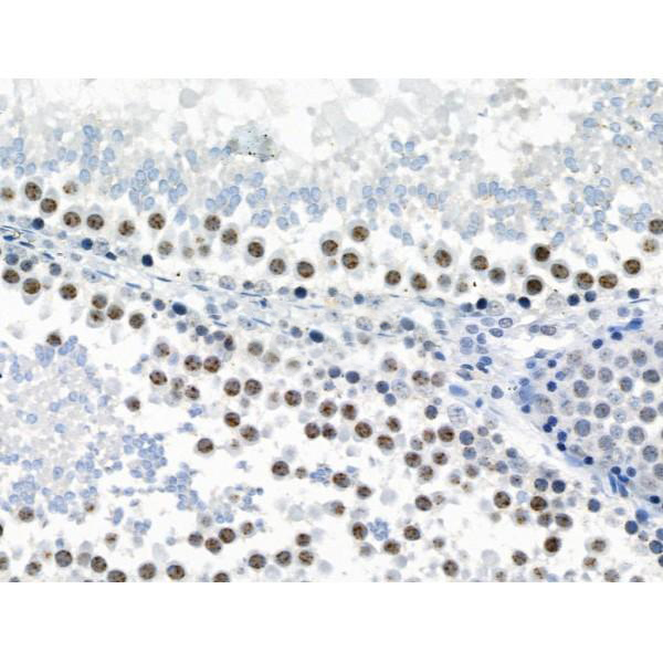
 IHC (Immunohistochemistry)
(At 1/100 staining Human mammary cancer and adjacent normal tissues by IHC-P. The sample was formaldehyde fixed and a heat mediated antigen retrieval step in citrate buffer was performed. The sample was then blocked and incubated with the primary antibody at 4 degree C overnight. An HRP conjugated anti-Rabbit antibody was used as the secondary antibody.)
IHC (Immunohistochemistry)
(At 1/100 staining Human mammary cancer and adjacent normal tissues by IHC-P. The sample was formaldehyde fixed and a heat mediated antigen retrieval step in citrate buffer was performed. The sample was then blocked and incubated with the primary antibody at 4 degree C overnight. An HRP conjugated anti-Rabbit antibody was used as the secondary antibody.)
MCM2, Polyclonal Antibody (Cat# AAA31460)
Full Name
Phospho-MCM2 (Ser27) Antibody
Gene Names
MCM2; BM28; CCNL1; CDCL1; cdc19; D3S3194; MITOTIN
Reactivity
Human, Mouse, Rat
Predicted Reactivity: Pig (100%), Zebrafish (100%), Bovine (100%), Horse (100%), Rabbit (100%), Dog (100%), Xenopus (100%)
Predicted Reactivity: Pig (100%), Zebrafish (100%), Bovine (100%), Horse (100%), Rabbit (100%), Dog (100%), Xenopus (100%)
Applications
Western Blot, Immunohistochemistry, Peptide ELISA
Purity
The antibody is from purified rabbit serum by affinity purification via sequential chromatography on phospho-peptide and non-phospho-peptide affinity columns.
Pricing


 Application Data
(Staining of J774 cells with Rat anti Mouse F4/80 antigen Biotin)
Application Data
(Staining of J774 cells with Rat anti Mouse F4/80 antigen Biotin)
F4/80, Monoclonal Antibody (Cat# AAA12173)
Full Name
RAT ANTI MOUSE F4/80
Gene Names
Emr1; Ly71; F4/80; Gpf480; TM7LN3; DD7A5-7; EGF-TM7
Applications
Immunohistochemistry, Fluorescence Microscopy, Flow Cytometry, Immunofluorescence, Immunoprecipitation, Immunohistochemistry, Radioimmunoassay, Immunohistochemistry, Western Blot
Pricing


 Application Data
(At 37 degree C. The primary antibody was diluted 1/200 and incubated with the sample for 1 hour at 37 degree C. A Alexa Fluor 594 conjugated goat polyclonal to rabbit IgG (H+L), diluted 1/600 was used as secondary antibody.)
Application Data
(At 37 degree C. The primary antibody was diluted 1/200 and incubated with the sample for 1 hour at 37 degree C. A Alexa Fluor 594 conjugated goat polyclonal to rabbit IgG (H+L), diluted 1/600 was used as secondary antibody.)
N Cadherin, Polyclonal Antibody (Cat# AAA31337)
Full Name
N Cadherin Antibody
Gene Names
CDH2; CDHN; NCAD; CD325; CDw325
Reactivity
Human, Mouse, Rat
Predicted Reactivity: Pig (100%), Zebrafish (100%), Bovine (100%), Horse (100%), Sheep (100%), Rabbit (100%), Dog (100%), Chicken (100%), Xenopus (100%)
Predicted Reactivity: Pig (100%), Zebrafish (100%), Bovine (100%), Horse (100%), Sheep (100%), Rabbit (100%), Dog (100%), Chicken (100%), Xenopus (100%)
Applications
Western Blot, Immunohistochemistry, Immunofluorescence, Immunocytochemistry, Peptide ELISA, Immunohistochemistry, Immunohistochemistry
Purity
The antiserum was purified by peptide affinity chromatography using SulfoLink Coupling Resin
Pricing


 Application Data
(At 25 degree C. Samples were then incubated with primary Ab(At 37 degree C. An AlexaFluor594 conjugated goat anti-rabbit IgG(H+L) Ab(Red) and an AlexaFluor488 conjugated goat anti-mouse IgG(H+L) Ab(Green) were used as the secondary antibody.The nuclear counter stain is DAPI (blue).)
Application Data
(At 25 degree C. Samples were then incubated with primary Ab(At 37 degree C. An AlexaFluor594 conjugated goat anti-rabbit IgG(H+L) Ab(Red) and an AlexaFluor488 conjugated goat anti-mouse IgG(H+L) Ab(Green) were used as the secondary antibody.The nuclear counter stain is DAPI (blue).)
LRRK2, Polyclonal Antibody (Cat# AAA31287)
Full Name
Phospho-LRRK2 (Ser935) Antibody
Gene Names
LRRK2; PARK8; RIPK7; ROCO2; AURA17; DARDARIN
Reactivity
Human, Mouse, Rat
Applications
Western Blot, Immunohistochemistry, Immunofluorescence, Immunocytochemistry, Peptide ELISA
Purity
The antibody is from purified rabbit serum by affinity purification via sequential chromatography on phospho-peptide and non-phospho-peptide affinity columns.
Pricing


 FCM (Flow Cytometry)
(Figure 8. Flow Cytometry analysis of A431 cells using anti-EGFR antibody (AAA19214).Overlay histogram showing A431 cells stained with AAA19214 (Blue line). The cells were blocked with 10% normal goat serum. And then incubated with rabbit anti-EGFR Antibody (AAA19214,1μg/1x106 cells) for 30 min at 20 degree C. DyLight®488 conjugated goat anti-rabbit IgG (5-10μg/1x106 cells) was used as secondary antibody for 30 minutes at 20 degree C. Isotype control antibody (Green line) was rabbit IgG (1μg/1x106) used under the same conditions. Unlabelled sample (Red line) was also used as a control.)
FCM (Flow Cytometry)
(Figure 8. Flow Cytometry analysis of A431 cells using anti-EGFR antibody (AAA19214).Overlay histogram showing A431 cells stained with AAA19214 (Blue line). The cells were blocked with 10% normal goat serum. And then incubated with rabbit anti-EGFR Antibody (AAA19214,1μg/1x106 cells) for 30 min at 20 degree C. DyLight®488 conjugated goat anti-rabbit IgG (5-10μg/1x106 cells) was used as secondary antibody for 30 minutes at 20 degree C. Isotype control antibody (Green line) was rabbit IgG (1μg/1x106) used under the same conditions. Unlabelled sample (Red line) was also used as a control.)
EGFR, Polyclonal Antibody (Cat# AAA19214)
Full Name
Anti-EGFR Antibody
Gene Names
EGFR; ERBB; HER1; mENA; ERBB1; PIG61
Reactivity
Human, Mouse, Rat
Applications
Western Blot, Immunohistochemistry, Immunocytochemistry, Immunofluorescence, Flow Cytometry, Direct ELISA
Purity
Immunogen affinity purified.
Pricing


 Application Data
(At 25 degree C. Samples were then incubated with primary Ab(At 37 degree C. An AlexaFluor594 conjugated goat anti-rabbit IgG(H+L) Ab(Red) and an AlexaFluor488 conjugated goat anti-mouse IgG(H+L) Ab(Green) were used as the secondary antibody.The nuclear counter stain is DAPI (blue).)
Application Data
(At 25 degree C. Samples were then incubated with primary Ab(At 37 degree C. An AlexaFluor594 conjugated goat anti-rabbit IgG(H+L) Ab(Red) and an AlexaFluor488 conjugated goat anti-mouse IgG(H+L) Ab(Green) were used as the secondary antibody.The nuclear counter stain is DAPI (blue).)
JIP1, Polyclonal Antibody (Cat# AAA31317)
Full Name
Phospho-JIP1 (Thr205) Antibody
Gene Names
MAPK8IP1; IB1; JIP1; JIP-1; PRKM8IP
Reactivity
Human, Mouse, Rat
Applications
Western Blot, Immunohistochemistry, Immunofluorescence, Immunocytochemistry, Peptide ELISA
Purity
The antibody is from purified rabbit serum by affinity purification via sequential chromatography on phospho-peptide and non-phospho-peptide affinity columns.
Pricing


 Application Data
(Staining of CD209 transfected K562 cells with MOUSE ANTI HUMAN CD209: FITC)
Application Data
(Staining of CD209 transfected K562 cells with MOUSE ANTI HUMAN CD209: FITC)
DC-SIGN, Monoclonal Antibody (Cat# AAA26722)
Full Name
DC-SIGN (Dendritic Cell-specific ICAM3 grabbing Non-integrin, C Type Lectin Domain Family 4 Member L, CLEC4L, CD209, CD209 Antigen, CDSIGN, Dendritic Cell-specific ICAM-3-grabbing Non-integrin 1, DC SIGN1, DC-SIGN1, HIV GP120 Binding Protein, MGC129965) (
Reactivity
Human
Applications
Immunofluorescence
Purity
Purified by protein G affinity chromatography from tissue culture supernatant.
Pricing


 FCM (Flow Cytometry)
(Flow cytometric analysis of Hela cells with NADPH oxidase 4 antibody at 1/50 dilution (blue) compared with an unlabelled control (cells without incubation with primary antibody; red). Alexa Fluor 488-conjugated Goat anti rabbit IgG was used as the secondary antibody.)
FCM (Flow Cytometry)
(Flow cytometric analysis of Hela cells with NADPH oxidase 4 antibody at 1/50 dilution (blue) compared with an unlabelled control (cells without incubation with primary antibody; red). Alexa Fluor 488-conjugated Goat anti rabbit IgG was used as the secondary antibody.)
NADPH Oxidase 4, Polyclonal Antibody (Cat# AAA29878)
Full Name
NADPH Oxidase 4 Antibody
Gene Names
NOX4; KOX; KOX-1; RENOX
Reactivity
Human, Mouse, Rat
Applications
Immunocytochemistry, Immunohistochemistry, Flow Cytometry
Purity
Peptide affinity purified
Pricing
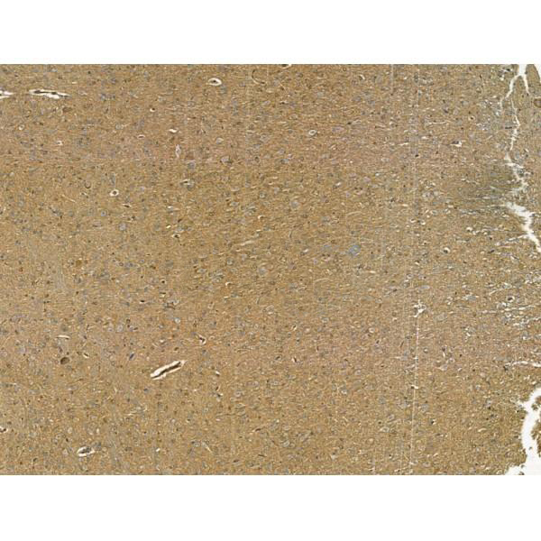
 WB (Western Blot)
((0.1ug/ml) staining of Human Brain lysate (35ug protein in RIPA buffer). Primary incubation was 1 hour. Detected by chemiluminescence.)
WB (Western Blot)
((0.1ug/ml) staining of Human Brain lysate (35ug protein in RIPA buffer). Primary incubation was 1 hour. Detected by chemiluminescence.)
Orexin receptor 1, Polyclonal Antibody (Cat# AAA13669)
Full Name
Goat anti-Orexin receptor 1 Antibody
Gene Names
HCRTR1; OX1R
Reactivity
Tested: Human; Expected from sequence similarity: Human, Mouse, Rat, Dog, Sheep
Applications
Peptide ELISA, Western Blot
Purity
Purified from goat serum by ammonium sulphate precipitation followed by antigen affinity chromatography using the immunizing peptide.
Pricing


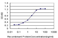 Application Data
(Detection limit for recombinant GST tagged PGR is ~0.3ng/ml as a capture antibody.)
Application Data
(Detection limit for recombinant GST tagged PGR is ~0.3ng/ml as a capture antibody.)
Progesterone Receptor, Monoclonal Antibody (Cat# AAA24623)
Full Name
Progesterone Receptor (PgR, PR, Nuclear Receptor Subfamily 3 Group C Member 3, NR3C3) APC
Gene Names
PGR; PR; NR3C3
Reactivity
Human, Rat
Applications
Immunofluorescence, Immunohistochemistry, Western Blot
Purity
Purified by Protein A Affinity Chromatography.
Pricing


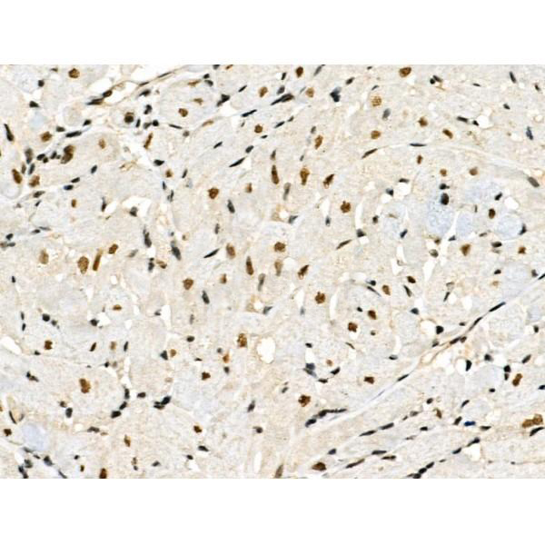
 IHC (Immunohistchemistry)
(Anti-ESRRG / ERR Gamma antibody IHC of human Skin, Melanoma. Immunohistochemistry of formalin-fixed, paraffin-embedded tissue after heat-induced antigen retrieval.)
IHC (Immunohistchemistry)
(Anti-ESRRG / ERR Gamma antibody IHC of human Skin, Melanoma. Immunohistochemistry of formalin-fixed, paraffin-embedded tissue after heat-induced antigen retrieval.)
ESRRG / ERR Gamma, Polyclonal Antibody (Cat# AAA12326)
Full Name
Rabbit Polyclonal to Human ESRRG / ERR Gamma
Gene Names
ESRRG; ERR3; NR3B3; ERRgamma
Reactivity
Gibbon, Bovine, Chicken, Dog, Gorilla, Hamster, Horse, Human, Monkey, Mouse, Orangutan, Pig, Rabbit, Rat
Predicted Reactivity: Bat, Xenopus, Zebrafish (at least 90% immunogen sequence identity)
Predicted Reactivity: Bat, Xenopus, Zebrafish (at least 90% immunogen sequence identity)
Applications
Immunohistochemistry
Purity
Immunoaffinity Purified
Pricing


 IHC (Immunohistochemistry)
(Immunochemical staining of alpha smooth muscle actin in mouse skeletal muscle with rabbit polyclonal antibody at 1:40000 dilution, formalin-fixed paraffin embedded sections.)
IHC (Immunohistochemistry)
(Immunochemical staining of alpha smooth muscle actin in mouse skeletal muscle with rabbit polyclonal antibody at 1:40000 dilution, formalin-fixed paraffin embedded sections.)
ACTA2, Polyclonal Antibody (Cat# AAA27714)
Full Name
Anti-ACTA2 Antibody, Rabbit Polyclonal
Gene Names
ACTA2; AAT6; ACTSA; MYMY5
Reactivity
Human, Mouse, Rat
Applications
Western Blot, Immunohistochemistry
Purity
Protein A & Antigen Affinity
Pricing


 WB (Western Blot)
(CYK18 Antibody (C-term) Western blot of A431 cell line lysates (35 ug/lane). This demonstrates the CYK18 antibody detected the CYK18 protein (arrow) (Kindly offered by Dr. Li).)
WB (Western Blot)
(CYK18 Antibody (C-term) Western blot of A431 cell line lysates (35 ug/lane). This demonstrates the CYK18 antibody detected the CYK18 protein (arrow) (Kindly offered by Dr. Li).)
KRT18 / CK18 / Cytokeratin 18, Polyclonal Antibody (Cat# AAA12370)
Full Name
Rabbit Polyclonal to Human KRT18 / CK18 / Cytokeratin 18
Gene Names
KRT18; K18; CYK18
Reactivity
Human, Mouse
Applications
Immunohistochemistry, Immunofluorescence, Western Blot, Flow Cytometry
Purity
Ammonium sulfate precipitation
Pricing


 WB (Western Blot)
(Anti-TRIM28 rabbit polyclonal antibody at 1:500 dilution Lane A: TRIM28 konckout Hela Whole Cell Lysate Lane B: Hela Whole Cell Lysate Lysates/proteins at 20 ug per lane. Secondary Goat Anti-Rabbit IgG (H+L)/HRP at 1/10000 dilution. Developed using the ECL technique. Performed under reducing conditions. Predicted band size:89 kDa Observed band size:115 kDa (We are unsure as to the identity of these extra bands.))
WB (Western Blot)
(Anti-TRIM28 rabbit polyclonal antibody at 1:500 dilution Lane A: TRIM28 konckout Hela Whole Cell Lysate Lane B: Hela Whole Cell Lysate Lysates/proteins at 20 ug per lane. Secondary Goat Anti-Rabbit IgG (H+L)/HRP at 1/10000 dilution. Developed using the ECL technique. Performed under reducing conditions. Predicted band size:89 kDa Observed band size:115 kDa (We are unsure as to the identity of these extra bands.))
KAP1/TRIM28, Polyclonal Antibody (Cat# AAA27715)
Full Name
Anti-KAP1/TRIM28 Antibody, Rabbit Polyclonal
Gene Names
TRIM28; KAP1; TF1B; RNF96; TIF1B; PPP1R157
Reactivity
Human
Applications
Western Blot, Immunohistochemistry, Immunoprecipitation
Purity
Protein A & Antigen Affinity
Pricing


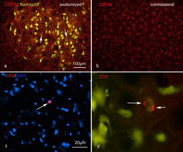 Application Data
(Published customer image: Representative images of the inflammatory changes in the facial nucleus during axonal regeneration, one week following facial nerve transaction. a, b: CD11b immunoreactivity for microglia is increased in the axotomized facial nucleus, and microglia enwrap the facial motor neurons, e.g. at arrows. The regenerating neurons were retrogradely labelled with fluorogold. c, d: CD6- positive T-cells accumulated in the injured motor nucleus (arrows). They had little cytoplasm but dense nuclei (c) and were sometimes clustered around neurons retrogradely labelled with fluorogold (d). The scale bar in (a) also applies to (b) and that in (c) also applies to (d).From: Shokouhi et al. BMC Neuroscience 2010 11:13.)
Application Data
(Published customer image: Representative images of the inflammatory changes in the facial nucleus during axonal regeneration, one week following facial nerve transaction. a, b: CD11b immunoreactivity for microglia is increased in the axotomized facial nucleus, and microglia enwrap the facial motor neurons, e.g. at arrows. The regenerating neurons were retrogradely labelled with fluorogold. c, d: CD6- positive T-cells accumulated in the injured motor nucleus (arrows). They had little cytoplasm but dense nuclei (c) and were sometimes clustered around neurons retrogradely labelled with fluorogold (d). The scale bar in (a) also applies to (b) and that in (c) also applies to (d).From: Shokouhi et al. BMC Neuroscience 2010 11:13.)
CD11b, Monoclonal Antibody (Cat# AAA12146)
Full Name
MOUSE ANTI RAT CD11b:FITC
Gene Names
ITGAM; CD11B
Applications
Flow Cytometry
Pricing


 Application Data
(At 25 degree C. The primary antibody was diluted at 1/200 and incubated with the sample for 1 hour at 37 degree C. An Alexa Fluor 594 conjugated goat anti-rabbit IgG (H+L) Ab, diluted at 1/600, was used as the secondary antibody.)
Application Data
(At 25 degree C. The primary antibody was diluted at 1/200 and incubated with the sample for 1 hour at 37 degree C. An Alexa Fluor 594 conjugated goat anti-rabbit IgG (H+L) Ab, diluted at 1/600, was used as the secondary antibody.)
CD22, Polyclonal Antibody (Cat# AAA31418)
Full Name
Phospho-CD22 (Tyr842) Antibody
Gene Names
CD22; SIGLEC2; SIGLEC-2
Reactivity
Human, Mouse, Rat
Predicted Reactivity: Horse (83%)
Predicted Reactivity: Horse (83%)
Applications
Western Blot, Immunohistochemistry, Immunofluorescence, Immunocytochemistry, Peptide ELISA
Purity
The antibody is from purified rabbit serum by affinity purification via sequential chromatography on phospho-peptide and non-phospho-peptide affinity columns.
Pricing


 Application Data
(At 25 degree C. The primary antibody was diluted at 1/200 and incubated with the sample for 1 hour at 37 degree C. An Alexa Fluor 594 conjugated goat anti-rabbit IgG (H+L) Ab, diluted at 1/600, was used as the secondary antibody.)
Application Data
(At 25 degree C. The primary antibody was diluted at 1/200 and incubated with the sample for 1 hour at 37 degree C. An Alexa Fluor 594 conjugated goat anti-rabbit IgG (H+L) Ab, diluted at 1/600, was used as the secondary antibody.)
p53, Polyclonal Antibody (Cat# AAA31429)
Full Name
Phospho-p53 (Ser392) Antibody
Gene Names
TP53; P53; BCC7; LFS1; TRP53
Reactivity
Human, Mouse
Predicted Reactivity: Pig (88%), Bovine (88%), Sheep (88%), Rabbit (88%)
Predicted Reactivity: Pig (88%), Bovine (88%), Sheep (88%), Rabbit (88%)
Applications
Western Blot, Immunohistochemistry, Immunofluorescence, Immunocytochemistry, Peptide ELISA
Purity
The antibody is from purified rabbit serum by affinity purification via sequential chromatography on phospho-peptide and non-phospho-peptide affinity columns.
Pricing


 IHC (Immunohistochemistry)
(At 1/100 staining Human pancreatic cancer by IHC-P. The sample was formaldehyde fixed and a heat mediated antigen retrieval step in citrate buffer was performed. The sample was then blocked and incubated with the primary antibody at 4 degree C overnight. An HRP conjugated anti-Rabbit antibody was used as the secondary antibody.)
IHC (Immunohistochemistry)
(At 1/100 staining Human pancreatic cancer by IHC-P. The sample was formaldehyde fixed and a heat mediated antigen retrieval step in citrate buffer was performed. The sample was then blocked and incubated with the primary antibody at 4 degree C overnight. An HRP conjugated anti-Rabbit antibody was used as the secondary antibody.)
p53, Polyclonal Antibody (Cat# AAA31355)
Full Name
Acetyl-p53 (Lys381) Antibody
Gene Names
TP53; P53; BCC7; LFS1; TRP53
Reactivity
Human, Mouse, Rat
Predicted Reactivity: Rabbit (90%), Dog (100%)
Predicted Reactivity: Rabbit (90%), Dog (100%)
Applications
Western Blot, Immunohistochemistry, Peptide ELISA
Purity
The antiserum was purified by peptide affinity chromatography using SulfoLink Coupling Resin
Pricing
 Application Data
(SDS - PAGE : purified HA (H5N1) (A/Vietnam/1203/2004) (aa18 - 530) protein.)
Application Data
(SDS - PAGE : purified HA (H5N1) (A/Vietnam/1203/2004) (aa18 - 530) protein.)
H5 (H5N1) (A/Vietnam/1203/2004), Recombinant Protein (Cat# AAA13745)
Full Name
H5 (H5N1) (aa 18-530)(A/Vietnam/1203/2004)
Applications
Western Blot
Purity
≥ 95% (SDS-PAGE)
Pricing


 ELISA
(Titration curve analysis of TIGIT mAbs to detect recombinant TIGIT in ELISA at decreasing concentrations.)
ELISA
(Titration curve analysis of TIGIT mAbs to detect recombinant TIGIT in ELISA at decreasing concentrations.)
TIGIT, Monoclonal Antibody (Cat# AAA11004)
Full Name
TIGIT Antibody [4A10]
Gene Names
TIGIT; VSIG9; VSTM3; WUCAM
Reactivity
Human
Applications
Western Blot, Immunohistochemistry, Immunocytochemistry, Immunofluorescence, Flow Cytometry
Purity
Protein A purified
Pricing


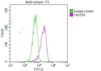
 FCM (Flow Cytometry)
(Overlay histogram showing MCF-7 cells stained with CSB-MA013563A0m (red line) at 1:400. The cells were incubated in 1x PBS /10% normal goat serum to block non-specific protein-protein interactions followed by primary antibody for 1 h at 4 degree C. The secondary antibody used was FITC goat anti-mouse IgG(H+L) at 1/200 dilution for 1 h at 4 degree C. Isotype control antibody (green line) was used under the same conditions. Acquisition of >10,000 events was performed.)
FCM (Flow Cytometry)
(Overlay histogram showing MCF-7 cells stained with CSB-MA013563A0m (red line) at 1:400. The cells were incubated in 1x PBS /10% normal goat serum to block non-specific protein-protein interactions followed by primary antibody for 1 h at 4 degree C. The secondary antibody used was FITC goat anti-mouse IgG(H+L) at 1/200 dilution for 1 h at 4 degree C. Isotype control antibody (green line) was used under the same conditions. Acquisition of >10,000 events was performed.)
CD146, Monoclonal Antibody (Cat# AAA27014)
Full Name
CD146 Monoclonal Antibody
Gene Names
MCAM; CD146; MUC18; HEMCAM; METCAM; MelCAM
Reactivity
Human
Applications
Western Blot, Immunofluorescence, Flow Cytometry
Purity
>95%
Protein G Purified
Protein G Purified
Pricing


 IHC (Immunohistochemistry)
(At 1/100 staining Mouse kidney tissue by IHC-P. The sample was formaldehyde fixed and a heat mediated antigen retrieval step in citrate buffer was performed. The sample was then blocked and incubated with the primary antibody at 4 degree C overnight. An HRP conjugated anti-Rabbit antibody was used as the secondary antibody.)
IHC (Immunohistochemistry)
(At 1/100 staining Mouse kidney tissue by IHC-P. The sample was formaldehyde fixed and a heat mediated antigen retrieval step in citrate buffer was performed. The sample was then blocked and incubated with the primary antibody at 4 degree C overnight. An HRP conjugated anti-Rabbit antibody was used as the secondary antibody.)
p38 MAPK, Polyclonal Antibody (Cat# AAA31333)
Full Name
Phospho-p38 MAPK (Thr180/Tyr182) Antibody
Gene Names
MAPK14; RK; p38; CSBP; EXIP; Mxi2; CSBP1; CSBP2; CSPB1; PRKM14; PRKM15; SAPK2A; p38ALPHA
Reactivity
Human, Mouse, Rat
Predicted Reactivity: Pig (100%), Bovine (100%), Horse (100%), Sheep (100%), Rabbit (100%), Dog (100%), Xenopus (91%)
Predicted Reactivity: Pig (100%), Bovine (100%), Horse (100%), Sheep (100%), Rabbit (100%), Dog (100%), Xenopus (91%)
Applications
Western Blot, Immunohistochemistry, Immunofluorescence, Immunocytochemistry, Immunoprecipitation, Peptide ELISA
Purity
The antibody is from purified rabbit serum by affinity purification via sequential chromatography on phospho-peptide and non-phospho-peptide affinity columns.
Pricing


 IHC (Immunohistchemistry)
(At 1/200 staining Human kidney tissue sections by IHC-P. The tissue was formaldehyde fixed and a heat mediated antigen retrieval step in citrate buffer was performed. The tissue was then blocked and incubated with the antibody for 1.5 hours at 22 degree C. An HRP conjugated goat anti-rabbit antibody was used as the secondary antibody.)
IHC (Immunohistchemistry)
(At 1/200 staining Human kidney tissue sections by IHC-P. The tissue was formaldehyde fixed and a heat mediated antigen retrieval step in citrate buffer was performed. The tissue was then blocked and incubated with the antibody for 1.5 hours at 22 degree C. An HRP conjugated goat anti-rabbit antibody was used as the secondary antibody.)
FGFR1/2/3/4, Polyclonal Antibody (Cat# AAA31417)
Full Name
Phospho-FGFR1/2/3/4 (Tyr653+Tyr654) Antibody
Gene Names
FGFR1; CEK; FLG; HH2; OGD; FLT2; KAL2; BFGFR; CD331; FGFBR; FLT-2; HBGFR; N-SAM; FGFR-1; bFGF-R-1
Reactivity
Human, Mouse, Rat, Monkey
Predicted Reactivity: Bovine (100%), Horse (100%), Sheep (100%), Rabbit (100%), Dog (100%), Chicken (100%), Xenopus (100%)
Predicted Reactivity: Bovine (100%), Horse (100%), Sheep (100%), Rabbit (100%), Dog (100%), Chicken (100%), Xenopus (100%)
Applications
Western Blot, Immunohistochemistry, Peptide ELISA
Purity
The antibody is from purified rabbit serum by affinity purification via sequential chromatography on phospho-peptide and non-phospho-peptide affinity columns.
Pricing


 Application Data
(At 25 degree C. Samples were then incubated with primary Ab(At 37 degree C. An AlexaFluor594 conjugated goat anti-rabbit IgG(H+L) Ab(Red) and an AlexaFluor488 conjugated goat anti-mouse IgG(H+L) Ab(Green) were used as the secondary antibody.The nuclear counter stain is DAPI (blue).)
Application Data
(At 25 degree C. Samples were then incubated with primary Ab(At 37 degree C. An AlexaFluor594 conjugated goat anti-rabbit IgG(H+L) Ab(Red) and an AlexaFluor488 conjugated goat anti-mouse IgG(H+L) Ab(Green) were used as the secondary antibody.The nuclear counter stain is DAPI (blue).)
Lamin A/C, Polyclonal Antibody (Cat# AAA31378)
Full Name
Lamin A/C Antibody
Gene Names
LMNA; FPL; IDC; LFP; CDDC; EMD2; FPLD; HGPS; LDP1; LMN1; LMNC; PRO1; CDCD1; CMD1A; FPLD2; LMNL1; CMT2B1; LGMD1B
Reactivity
Human, Mouse, Rat
Applications
Western Blot, Immunohistochemistry, Immunofluorescence, Immunocytochemistry, Peptide ELISA, Flow Cytometry
Purity
The antiserum was purified by peptide affinity chromatography using SulfoLink Coupling Resin
Pricing


 Application Data
(Staining of human peripheral blood granulocytes with Mouse anti Human CD45: Pacific Blue)
Application Data
(Staining of human peripheral blood granulocytes with Mouse anti Human CD45: Pacific Blue)
CD45, Monoclonal Antibody (Cat# AAA12202)
Full Name
MOUSE ANTI HUMAN CD45:FITC
Gene Names
PTPRC; LCA; LY5; B220; CD45; L-CA; T200; CD45R; GP180
Applications
Flow Cytometry
Pricing


 IHC (Immunohistochemistry)
(At 1/100 staining Human gastric cancer by IHC-P. The sample was formaldehyde fixed and a heat mediated antigen retrieval step in citrate buffer was performed. The sample was then blocked and incubated with the primary antibody at 4 degree C overnight. An HRP conjugated anti-Rabbit antibody was used as the secondary antibody.)
IHC (Immunohistochemistry)
(At 1/100 staining Human gastric cancer by IHC-P. The sample was formaldehyde fixed and a heat mediated antigen retrieval step in citrate buffer was performed. The sample was then blocked and incubated with the primary antibody at 4 degree C overnight. An HRP conjugated anti-Rabbit antibody was used as the secondary antibody.)
Smad1/9, Polyclonal Antibody (Cat# AAA31453)
Full Name
Phospho-Smad1/9 (Ser463/Ser465) Antibody
Gene Names
SMAD1; BSP1; JV41; BSP-1; JV4-1; MADH1; MADR1
Reactivity
Human, Mouse, Rat
Predicted Reactivity: Pig (100%), Zebrafish (100%), Bovine (100%), Sheep (100%), Rabbit (100%), Dog (100%), Chicken (100%), Xenopus (100%)
Predicted Reactivity: Pig (100%), Zebrafish (100%), Bovine (100%), Sheep (100%), Rabbit (100%), Dog (100%), Chicken (100%), Xenopus (100%)
Applications
Western Blot, Immunohistochemistry, Peptide ELISA
Purity
The antibody is from purified rabbit serum by affinity purification via sequential chromatography on phospho-peptide and non-phospho-peptide affinity columns.
Pricing


 Application Data
(Overlay histogram showing HL-60 cells stained with AAA27009 (red line) at 1:500. The cells were incubated in 1x PBS /10% normal goat serum to block non-specific protein-protein interactions followed by primary antibody for 1 h at 4 degree C.The secondary antibody used was FITC goat anti-mouse IgG (H+L) at 1/200 dilution for 1 h at 4 degree C. Isotype control antibody (green line) was used under the same conditions. Acquisition of >10, 000 events was performed.)
Application Data
(Overlay histogram showing HL-60 cells stained with AAA27009 (red line) at 1:500. The cells were incubated in 1x PBS /10% normal goat serum to block non-specific protein-protein interactions followed by primary antibody for 1 h at 4 degree C.The secondary antibody used was FITC goat anti-mouse IgG (H+L) at 1/200 dilution for 1 h at 4 degree C. Isotype control antibody (green line) was used under the same conditions. Acquisition of >10, 000 events was performed.)
CD31, Monoclonal Antibody (Cat# AAA27009)
Full Name
CD31 Monoclonal Antibody
Gene Names
PECAM1; CD31; PECA1; GPIIA'; PECAM-1; endoCAM; CD31/EndoCAM
Reactivity
Human
Applications
Western Blot, Immunohistochemistry, Immunofluorescence, Flow Cytometry
Purity
>95%, Protein G purified
Pricing


 Application Data
(At 25 degree C. The primary antibody was diluted at 1/200 and incubated with the sample for 1 hour at 37 degree C. An Alexa Fluor 594 conjugated goat anti-rabbit IgG (H+L) Ab, diluted at 1/600, was used as the secondary antibody.)
Application Data
(At 25 degree C. The primary antibody was diluted at 1/200 and incubated with the sample for 1 hour at 37 degree C. An Alexa Fluor 594 conjugated goat anti-rabbit IgG (H+L) Ab, diluted at 1/600, was used as the secondary antibody.)
SRF, Polyclonal Antibody (Cat# AAA31435)
Full Name
Phospho-SRF (Thr159) Antibody
Gene Names
SRF; MCM1
Reactivity
Human, Mouse, Rat
Predicted Reactivity: Pig (100%), Bovine (100%), Horse (100%), Rabbit (100%), Dog (100%), Chicken (100%), Xenopus (100%)
Predicted Reactivity: Pig (100%), Bovine (100%), Horse (100%), Rabbit (100%), Dog (100%), Chicken (100%), Xenopus (100%)
Applications
Western Blot, Immunohistochemistry, Immunofluorescence, Immunocytochemistry, Peptide ELISA
Purity
The antibody is from purified rabbit serum by affinity purification via sequential chromatography on phospho-peptide and non-phospho-peptide affinity columns.
Pricing


 FCM (Flow Cytometry)
(Overlay histogram showing Hela cells stained with (red line) at 1:100. The cells were fixed in 4% formaldehyde and permeated by 0.2% TritonX-100. Then 10% normal goat serum was Incubated to block non-specific protein-protein interactions followed by the antibody (1ug/1*106cells) for 1 h at 4 degree C. The secondary antibody used was FITC-conjugated Goat Anti-Mouse IgG(H+L) at 1/100 dilution for 30min at 4 degree C. Isotype control antibody (green line) was mouse IgG2b (1ug/1*106cells) used under the same conditions. Acquisition of >10,000 events was performed.)
FCM (Flow Cytometry)
(Overlay histogram showing Hela cells stained with (red line) at 1:100. The cells were fixed in 4% formaldehyde and permeated by 0.2% TritonX-100. Then 10% normal goat serum was Incubated to block non-specific protein-protein interactions followed by the antibody (1ug/1*106cells) for 1 h at 4 degree C. The secondary antibody used was FITC-conjugated Goat Anti-Mouse IgG(H+L) at 1/100 dilution for 30min at 4 degree C. Isotype control antibody (green line) was mouse IgG2b (1ug/1*106cells) used under the same conditions. Acquisition of >10,000 events was performed.)
YWHAZ, Monoclonal Antibody (Cat# AAA27050)
Full Name
YWHAZ Monoclonal Antibody
Gene Names
YWHAZ; YWHAD; KCIP-1; 14-3-3-zeta
Reactivity
Human, Mouse, Rat
Applications
Western Blot, Immunohistochemistry, Immunofluorescence, Flow Cytometry
Purity
>95%, Protein A purified
Pricing


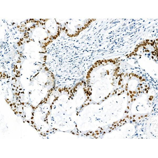
 IHC (Immunohistochemistry)
(At 1/100 staining Human esophageal cancer and adjacent normal tissues by IHC-P. The sample was formaldehyde fixed and a heat mediated antigen retrieval step in citrate buffer was performed. The sample was then blocked and incubated with the primary antibody at 4 degree C overnight. An HRP conjugated anti-Rabbit antibody was used as the secondary antibody.)
IHC (Immunohistochemistry)
(At 1/100 staining Human esophageal cancer and adjacent normal tissues by IHC-P. The sample was formaldehyde fixed and a heat mediated antigen retrieval step in citrate buffer was performed. The sample was then blocked and incubated with the primary antibody at 4 degree C overnight. An HRP conjugated anti-Rabbit antibody was used as the secondary antibody.)
MCM2, Polyclonal Antibody (Cat# AAA31459)
Full Name
Phospho-MCM2 (Ser139) Antibody
Gene Names
MCM2; BM28; CCNL1; CDCL1; cdc19; D3S3194; MITOTIN
Reactivity
Human, Mouse, Rat
Predicted Reactivity: Pig (100%), Zebrafish (80%), Bovine (83%), Horse (100%), Rabbit (100%), Dog (100%), Xenopus (100%)
Predicted Reactivity: Pig (100%), Zebrafish (80%), Bovine (83%), Horse (100%), Rabbit (100%), Dog (100%), Xenopus (100%)
Applications
Western Blot, Immunohistochemistry, Peptide ELISA
Purity
The antibody is from purified rabbit serum by affinity purification via sequential chromatography on phospho-peptide and non-phospho-peptide affinity columns.
Pricing


 WB (Western Blot)
(Western blot analysis of NFIC over-expressed 293 cell line, cotransfected with NFIC Validated Chimera RNAi (Lane 2) or non-transfected control (Lane 1). Blot probed with NFIC monoclonal antibody GAPDH (36.1kD) used as specificity and loading control.)
WB (Western Blot)
(Western blot analysis of NFIC over-expressed 293 cell line, cotransfected with NFIC Validated Chimera RNAi (Lane 2) or non-transfected control (Lane 1). Blot probed with NFIC monoclonal antibody GAPDH (36.1kD) used as specificity and loading control.)
Nuclear Factor I/C, Monoclonal Antibody (Cat# AAA25183)
Full Name
Nuclear Factor I/C (NFIC, CCAAT-binding Transcription Factor, CTF, Nuclear Factor 1 C-Type, CTF5, MGC20153, NF-I, NF1-C, CTF, TGGCA-Binding Protein, NFI) (FITC)
Gene Names
NFIC; CTF; NFI; CTF5; NF-I
Reactivity
Human
Applications
Immunofluorescence, Immunohistochemistry, Western Blot
Purity
Purified by Protein A Affinity Chromatography.
Pricing
 WB (Western Blot)
(Western blot analysis of Podoplanin expression in human placenta lysate (AAA11696).Electrophoresis was performed on a 5-20% SDS-PAGE gel at 70V (Stacking gel) / 90V (Resolving gel) for 2-3 hours. The sample well of each lane was loaded with 50ug of sample under reducing conditions. After Electrophoresis, proteins were transferred to a Nitrocellulose membrane at 150mA for 50-90 minutes. Blocked the membrane with 5% Non-fat Milk/ TBS for 1.5 hour at RT. The membrane was incubated with rabbit anti-PDPN monoclonal antibody overnight at 4 degree C, then washed with TBS-0.1%Tween 3 times with 5 minutes each and probed with a goat anti-rabbit IgG-HRP secondary antibody at a dilution of 1:10000 for 1.5 hour at RT. The signal is developed using an Enhanced Chemiluminescent detection (ECL) kit with Tanon 5200 system. A specific band was detected for PDPN)
WB (Western Blot)
(Western blot analysis of Podoplanin expression in human placenta lysate (AAA11696).Electrophoresis was performed on a 5-20% SDS-PAGE gel at 70V (Stacking gel) / 90V (Resolving gel) for 2-3 hours. The sample well of each lane was loaded with 50ug of sample under reducing conditions. After Electrophoresis, proteins were transferred to a Nitrocellulose membrane at 150mA for 50-90 minutes. Blocked the membrane with 5% Non-fat Milk/ TBS for 1.5 hour at RT. The membrane was incubated with rabbit anti-PDPN monoclonal antibody overnight at 4 degree C, then washed with TBS-0.1%Tween 3 times with 5 minutes each and probed with a goat anti-rabbit IgG-HRP secondary antibody at a dilution of 1:10000 for 1.5 hour at RT. The signal is developed using an Enhanced Chemiluminescent detection (ECL) kit with Tanon 5200 system. A specific band was detected for PDPN)
Podoplanin, Monoclonal Antibody (Cat# AAA11696)
Full Name
Anti-Podoplanin Rabbit Monoclonal Antibody tested for WB in Human, Mouse, Rat.
Gene Names
PDPN; T1A; GP36; GP40; Gp38; OTS8; T1A2; TI1A; T1A-2; AGGRUS; HT1A-1; PA2.26
Reactivity
Human, Mouse, Rat
Applications
Western Blot
Purity
Affinity-chromatography
Pricing


 FCM (Flow Cytometry)
(Flow cytometric analysis of Hela cells with hnRNP C1+C2 antibody at 1/50 dilution (red) compared with an unlabelled control (cells without incubation with primary antibody; black). Alexa Fluor 488-conjugated goat anti rabbit IgG was used as the secondary antibody)
FCM (Flow Cytometry)
(Flow cytometric analysis of Hela cells with hnRNP C1+C2 antibody at 1/50 dilution (red) compared with an unlabelled control (cells without incubation with primary antibody; black). Alexa Fluor 488-conjugated goat anti rabbit IgG was used as the secondary antibody)
hnRNP C1+C2, Monoclonal Antibody (Cat# AAA30156)
Full Name
hnRNP C1+C2 Antibody
Gene Names
HNRNPC; C1; C2; HNRNP; HNRPC; SNRPC
Reactivity
Human, Mouse, Rat, Zebrafish
Applications
Western Blot, Immunocytochemistry, Immunofluorescence, Immunohistochemistry, Immunoprecipitation, Flow Cytometry
Purity
ProA affinity purified
Pricing


 Application Data
(At 25 degree C. The primary antibody was diluted at 1/200 and incubated with the sample for 1 hour at 37 degree C. An Alexa Fluor 594 conjugated goat anti-rabbit IgG (H+L) Ab, diluted at 1/600, was used as the secondary antibody.)
Application Data
(At 25 degree C. The primary antibody was diluted at 1/200 and incubated with the sample for 1 hour at 37 degree C. An Alexa Fluor 594 conjugated goat anti-rabbit IgG (H+L) Ab, diluted at 1/600, was used as the secondary antibody.)
Cyclin E1, Polyclonal Antibody (Cat# AAA31439)
Full Name
Phospho-Cyclin E1 (Ser399) Antibody
Gene Names
CCNE1; CCNE
Reactivity
Human, Mouse, Rat
Predicted Reactivity: Bovine (88%), Horse (88%), Sheep (88%), Chicken (88%), Xenopus (88%)
Predicted Reactivity: Bovine (88%), Horse (88%), Sheep (88%), Chicken (88%), Xenopus (88%)
Applications
Western Blot, Immunohistochemistry, Immunofluorescence, Immunocytochemistry, Peptide ELISA
Purity
The antibody is from purified rabbit serum by affinity purification via sequential chromatography on phospho-peptide and non-phospho-peptide affinity columns.
Pricing


 FCM (Flow Cytometry)
(Figure 8. Flow Cytometry analysis of THP-1 cells using anti-NOX2/gp91phox/CYBB antibody (AAA19218).Overlay histogram showing THP-1 cells stained with AAA19218 (Blue line). The cells were blocked with 10% normal goat serum. And then incubated with rabbit anti-NOX2/gp91phox/CYBB Antibody (AAA19218, 1μg/1x106 cells) for 30 min at 20 degree C. DyLight®488 conjugated goat anti-rabbit IgG (5-10μg/1x106 cells) was used as secondary antibody for 30 minutes at 20 degree C. Isotype control antibody (Green line) was rabbit IgG (1μg/1x106) used under the same conditions. Unlabelled sample (Red line) was also used as a control.)
FCM (Flow Cytometry)
(Figure 8. Flow Cytometry analysis of THP-1 cells using anti-NOX2/gp91phox/CYBB antibody (AAA19218).Overlay histogram showing THP-1 cells stained with AAA19218 (Blue line). The cells were blocked with 10% normal goat serum. And then incubated with rabbit anti-NOX2/gp91phox/CYBB Antibody (AAA19218, 1μg/1x106 cells) for 30 min at 20 degree C. DyLight®488 conjugated goat anti-rabbit IgG (5-10μg/1x106 cells) was used as secondary antibody for 30 minutes at 20 degree C. Isotype control antibody (Green line) was rabbit IgG (1μg/1x106) used under the same conditions. Unlabelled sample (Red line) was also used as a control.)
NOX2/gp91phox/CYBB, Polyclonal Antibody (Cat# AAA19218)
Full Name
Anti-NOX2/gp91phox/CYBB Antibody
Gene Names
CYBB; CGD; NOX2; AMCBX2; GP91-1; GP91PHOX; p91-PHOX; GP91-PHOX
Reactivity
Human
Applications
Western Blot, Immunohistochemistry, Immunocytochemistry, Immunofluorescence, Flow Cytometry, Direct ELISA
Purity
Immunogen affinity purified.
Pricing


 Application Data
(Staining of human peripheral blood granulocytes with Mouse anti Human CD312: Alexa Fluor 488)
Application Data
(Staining of human peripheral blood granulocytes with Mouse anti Human CD312: Alexa Fluor 488)
CD312, Monoclonal Antibody (Cat# AAA12143)
Full Name
MOUSE ANTI HUMAN CD312:RPE
Gene Names
EMR2; CD312
Applications
Flow Cytometry
Pricing


 IHC (Immunohistochemistry)
(At 1/100 staining Human prostate cancer by IHC-P. The sample was formaldehyde fixed and a heat mediated antigen retrieval step in citrate buffer was performed. The sample was then blocked and incubated with the primary antibody at 4 degree C overnight. An HRP conjugated anti-Rabbit antibody was used as the secondary antibody.)
IHC (Immunohistochemistry)
(At 1/100 staining Human prostate cancer by IHC-P. The sample was formaldehyde fixed and a heat mediated antigen retrieval step in citrate buffer was performed. The sample was then blocked and incubated with the primary antibody at 4 degree C overnight. An HRP conjugated anti-Rabbit antibody was used as the secondary antibody.)
hFANCI, Polyclonal Antibody (Cat# AAA31282)
Full Name
Phospho-hFANCI (Ser565) Antibody
Gene Names
FANCI; KIAA1794
Reactivity
Human, Mouse, Rat
Applications
Immunohistochemistry, Peptide ELISA
Purity
The antibody is from purified rabbit serum by affinity purification via sequential chromatography on phospho-peptide and non-phospho-peptide affinity columns.
Pricing


 Application Data
(Staining of J774 cells with Rat anti Mouse F4/80 antigen Biotin)
Application Data
(Staining of J774 cells with Rat anti Mouse F4/80 antigen Biotin)
F4/80, Monoclonal Antibody (Cat# AAA12170)
Full Name
RAT ANTI MOUSE F4/80
Gene Names
Emr1; Ly71; F4/80; Gpf480; TM7LN3; DD7A5-7; EGF-TM7
Applications
Immunohistochemistry, Fluorescence Microscopy, Flow Cytometry, Immunofluorescence, Immunoprecipitation, Immunohistochemistry, Radioimmunoassay, Immunohistochemistry, Western Blot
Pricing


 Application Data
(At 25 degree C. Samples were then incubated with primary Ab(At 37 degree C. An AlexaFluor594 conjugated goat anti-rabbit IgG(H+L) Ab(Red) and an AlexaFluor488 conjugated goat anti-mouse IgG(H+L) Ab(Green) were used as the secondary antibody.The nuclear counter stain is DAPI (blue).)
Application Data
(At 25 degree C. Samples were then incubated with primary Ab(At 37 degree C. An AlexaFluor594 conjugated goat anti-rabbit IgG(H+L) Ab(Red) and an AlexaFluor488 conjugated goat anti-mouse IgG(H+L) Ab(Green) were used as the secondary antibody.The nuclear counter stain is DAPI (blue).)
TOP2A, Polyclonal Antibody (Cat# AAA31306)
Full Name
Phospho-TOP2A (Thr1343) Antibody
Gene Names
TOP2A; TOP2; TP2A
Reactivity
Human, Mouse
Applications
Immunohistochemistry, Immunofluorescence, Immunocytochemistry, Peptide ELISA
Purity
The antibody is from purified rabbit serum by affinity purification via sequential chromatography on phospho-peptide and non-phospho-peptide affinity columns.
Pricing