Filters
Clonality
Type
Reactivity
Gene Name
Isotype
Host
Application
Clone
3873 results for " anti mouse" - showing 3400-3450
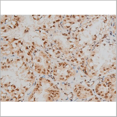
 IHC (Immunohistochemistry)
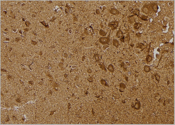
(AAA31036 at 1/200 staining Rat brain tissue sections by IHC-P. The tissue was formaldehyde fixed and a heat mediated antigen retrieval step in citrate buffer was performed. The tissue was then blocked and incubated with the antibody for 1.5 hours at 22 degree C. An HRP conjugated goat anti-rabbit antibody was used as the secondary.)
IHC (Immunohistochemistry)
(AAA31036 at 1/200 staining Rat brain tissue sections by IHC-P. The tissue was formaldehyde fixed and a heat mediated antigen retrieval step in citrate buffer was performed. The tissue was then blocked and incubated with the antibody for 1.5 hours at 22 degree C. An HRP conjugated goat anti-rabbit antibody was used as the secondary.)
IRS-1, Polyclonal Antibody (Cat# AAA31036)
Full Name
Phospho-IRS-1 (Ser312) Antibody
Gene Names
IRS1; HIRS-1
Reactivity
Human, Mouse, Rat, Monkey
Applications
Western Blot, Immunohistochemistry, Immunofluorescence, Immunocytochemistry
Purity
From purified rabbit serum by affinity purification via sequential chromatography on phospho-and non-phospho-peptide affinity columns.
Pricing
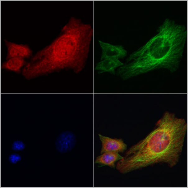
 IF (Immunofluorescence)
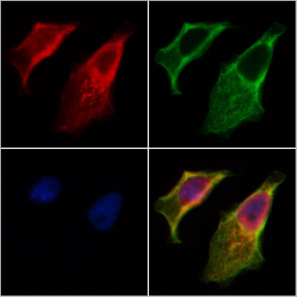
(AAA31266 staining Hela cells by IF/ICC. The samples were fixed with PFA and permeabilized in 0.1% Triton X-100,then blocked in 10% serum for 45 minutes at 25°C. Samples were then incubated with primary Ab(AAA31266 1:200) and mouse anti-beta tubulin Ab(T0023 1:200) for 1 hour at 37°C. An AlexaFluor594 conjugated goat anti-rabbit IgG(H+L) Ab(Red) and an AlexaFluor488 conjugated goat anti-mouse IgG(H+L) Ab(Green) were used as the secondary antibody.The nuclear counter stain is DAPI(blue).)
IF (Immunofluorescence)
(AAA31266 staining Hela cells by IF/ICC. The samples were fixed with PFA and permeabilized in 0.1% Triton X-100,then blocked in 10% serum for 45 minutes at 25°C. Samples were then incubated with primary Ab(AAA31266 1:200) and mouse anti-beta tubulin Ab(T0023 1:200) for 1 hour at 37°C. An AlexaFluor594 conjugated goat anti-rabbit IgG(H+L) Ab(Red) and an AlexaFluor488 conjugated goat anti-mouse IgG(H+L) Ab(Green) were used as the secondary antibody.The nuclear counter stain is DAPI(blue).)
Importin 7, Polyclonal Antibody (Cat# AAA31266)
Full Name
Importin 7 Antibody
Gene Names
IPO7; Imp7; RANBP7
Reactivity
Human, Mouse, Rat
Predicted Reactivity: Pig(100%), Zebrafish(88%), Bovine(100%), Horse(100%), Sheep(100%), Rabbit(100%), Dog(100%), Chicken(100%), Xenopus(88%)
Predicted Reactivity: Pig(100%), Zebrafish(88%), Bovine(100%), Horse(100%), Sheep(100%), Rabbit(100%), Dog(100%), Chicken(100%), Xenopus(88%)
Applications
ELISA
Purity
The antiserum was purified by peptide affinity chromatography using SulfoLink Coupling Resin (Thermo Fisher Scientific).
Pricing
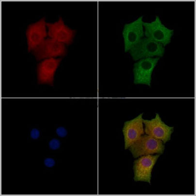
 IF (Immunofluorescence)
(AAA31207 staining HepG2 cells by IF/ICC. The samples were fixed with PFA and permeabilized in 0.1% Triton X-100,then blocked in 10% serum for 45 minutes at 25°C. Samples were then incubated with primary Ab(AAA31207) and mouse anti-beta tubulin Ab for 1 hour at 37°C. An AlexaFluor594 conjugated goat anti-rabbit IgG(H+L) Ab(Red) and an AlexaFluor488 conjugated goat anti-mouse IgG(H+L) Ab(Green) were used as the secondary antibody.The nuclear counter stain is DAPI(blue).)
IF (Immunofluorescence)
(AAA31207 staining HepG2 cells by IF/ICC. The samples were fixed with PFA and permeabilized in 0.1% Triton X-100,then blocked in 10% serum for 45 minutes at 25°C. Samples were then incubated with primary Ab(AAA31207) and mouse anti-beta tubulin Ab for 1 hour at 37°C. An AlexaFluor594 conjugated goat anti-rabbit IgG(H+L) Ab(Red) and an AlexaFluor488 conjugated goat anti-mouse IgG(H+L) Ab(Green) were used as the secondary antibody.The nuclear counter stain is DAPI(blue).)
IL22 Receptor Alpha, Polyclonal Antibody (Cat# AAA31207)
Full Name
IL22 Receptor Alpha Antibody
Gene Names
IL22RA1; IL22R; CRF2-9; IL22R1
Reactivity
Human, Mouse, Rat
Applications
ELISA
Purity
Peptide affinity purification
Pricing
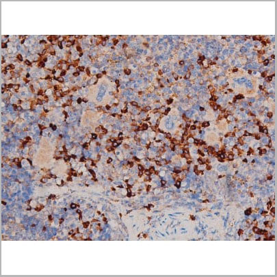
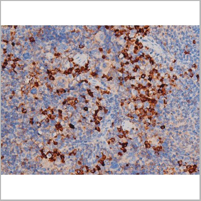
 Application Data
(Staining of human peripheral blood monocytes with Mouse anti Human CD14:RPE)
Application Data
(Staining of human peripheral blood monocytes with Mouse anti Human CD14:RPE)
CD14, Monoclonal Antibody (Cat# AAA11998)
Full Name
MOUSE ANTI HUMAN CD14
Applications
Immunohistochemistry, Flow Cytometry, Immunofluorescence, Immunoprecipitation
Pricing
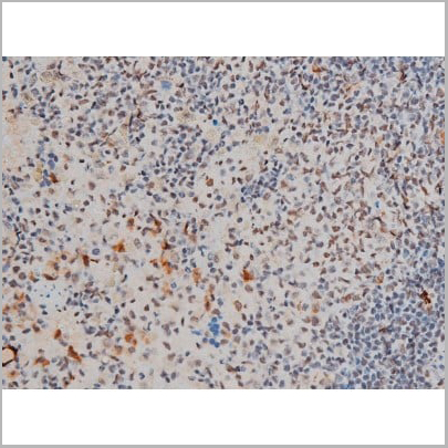
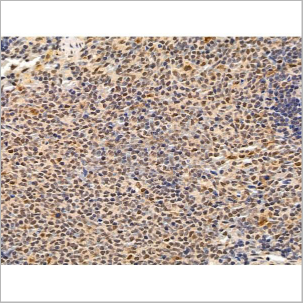
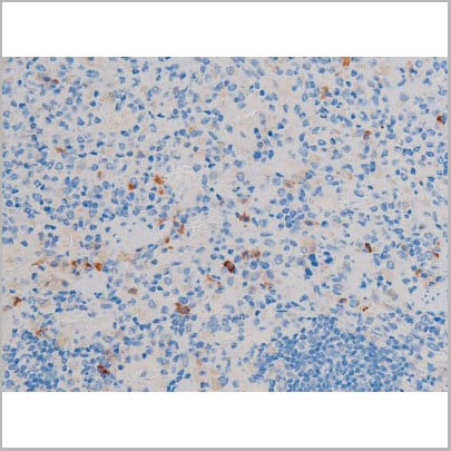
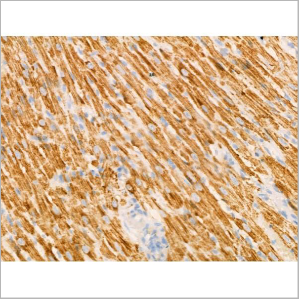
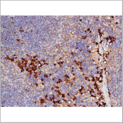
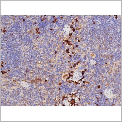
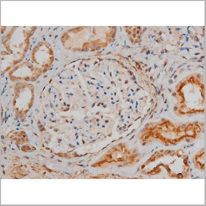
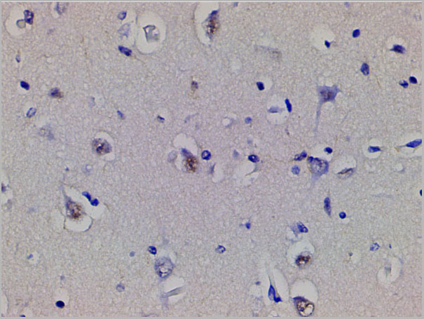
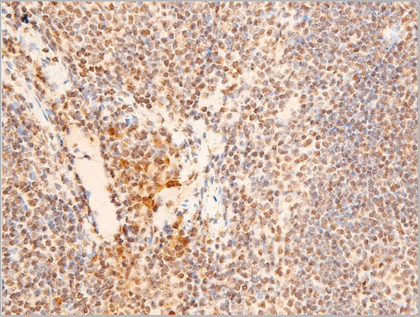
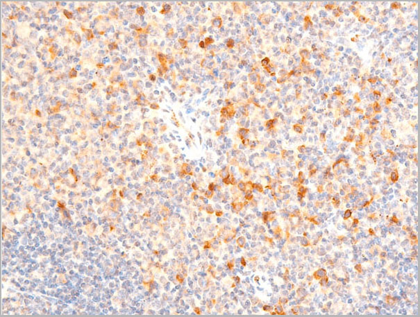
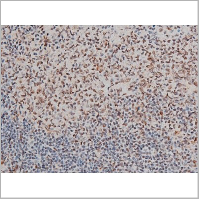 IHC (Immunohistochemistry)
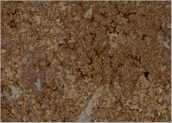
(AAA31013 at 1/200 staining Rat spleen tissue sections by IHC-P. The tissue was formaldehyde fixed and a heat mediated antigen retrieval step in citrate buffer was performed. The tissue was then blocked and incubated with the antibody for 1.5 hours at 22 degree C. An HRP conjugated goat anti-rabbit antibody was used as the secondary.)
IHC (Immunohistochemistry)
(AAA31013 at 1/200 staining Rat spleen tissue sections by IHC-P. The tissue was formaldehyde fixed and a heat mediated antigen retrieval step in citrate buffer was performed. The tissue was then blocked and incubated with the antibody for 1.5 hours at 22 degree C. An HRP conjugated goat anti-rabbit antibody was used as the secondary.)
GATA1, Polyclonal Antibody (Cat# AAA31013)
Full Name
Phospho-GATA1 (Ser142) Antibody
Gene Names
GATA1; GF1; GF-1; NFE1; XLTT; ERYF1; NF-E1; XLANP; XLTDA; GATA-1
Reactivity
Human, Mouse, Rat
Applications
Western Blot, Immunohistochemistry, Immunofluorescence, Immunocytochemistry, Immunoprecipitation
Purity
From purified rabbit serum by affinity purification via sequential chromatography on phospho-and non-phospho-peptide affinity columns.
Pricing
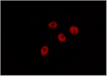
 IF (Immunofluorescence)
(AAA31030 staining HeLa by IF/ICC. The sample were fixed with PFA and permeabilized in 0.1% Triton X-100, then blocked in 10% serum for 45 minutes at 25 degree C. The primary antibody was diluted at 1/200 and incubated with the sample for 1 hour at 37 degree C. An Alexa Fluor 594 conjugated goat anti-rabbit IgG (H+L) Ab, diluted at 1/600, was used as the secondary antibody.)
IF (Immunofluorescence)
(AAA31030 staining HeLa by IF/ICC. The sample were fixed with PFA and permeabilized in 0.1% Triton X-100, then blocked in 10% serum for 45 minutes at 25 degree C. The primary antibody was diluted at 1/200 and incubated with the sample for 1 hour at 37 degree C. An Alexa Fluor 594 conjugated goat anti-rabbit IgG (H+L) Ab, diluted at 1/600, was used as the secondary antibody.)
CDC25A, Polyclonal Antibody (Cat# AAA31030)
Full Name
Phospho-CDC25A (Ser178) Antibody
Gene Names
CDC25A; CDC25A2
Reactivity
Human, Mouse, Rat
Applications
Western Blot, Immunohistochemistry, Immunofluorescence, Immunocytochemistry
Purity
From purified rabbit serum by affinity purification via sequential chromatography on phospho-and non-phospho-peptide affinity columns.
Pricing
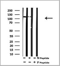
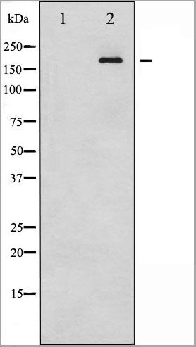
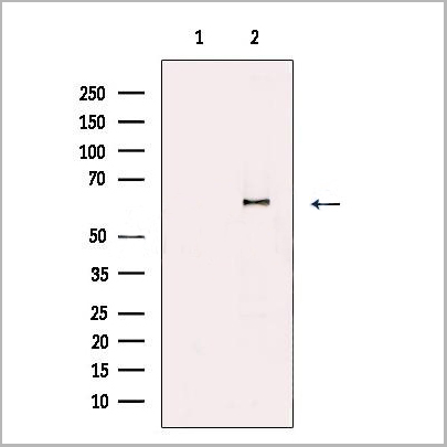
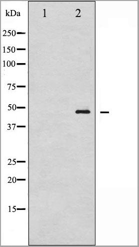
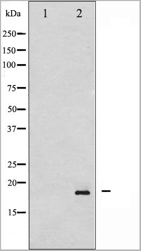
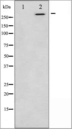
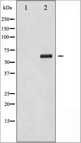
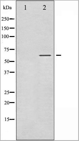
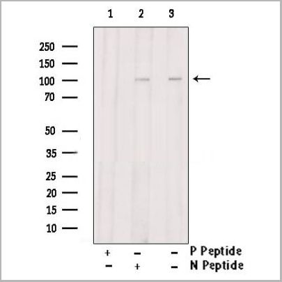
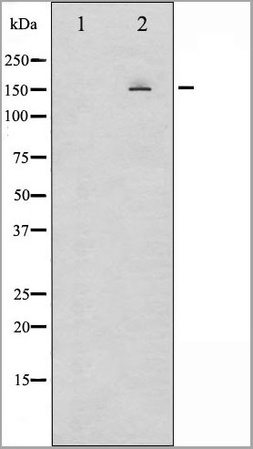
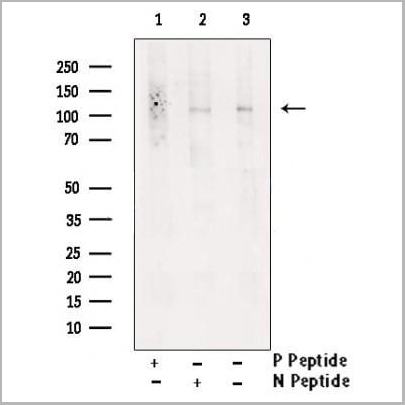 WB (Western Blot)
(Western blot analysis of extracts from K562 , using Phospho-IRE1 (Ser724) Antibody. Lane1 was treated with phospho-blocking peptide, Lane2 was treated with non-phospho-blocking peptide.)
WB (Western Blot)
(Western blot analysis of extracts from K562 , using Phospho-IRE1 (Ser724) Antibody. Lane1 was treated with phospho-blocking peptide, Lane2 was treated with non-phospho-blocking peptide.)
IRE1, Polyclonal Antibody (Cat# AAA31151)
Full Name
Phospho-IRE1 (Ser724) Antibody
Gene Names
ERN1; IRE1; IRE1P; IRE1a; hIRE1p
Reactivity
Human, Mouse, Rat
Applications
Western Blot, Immunohistochemistry, Immunofluorescence, Immunocytochemistry, Peptide ELISA
Purity
Peptide affinity purification
Pricing

 IF (Immunofluorescence)
(AAA31198 staining Hela cells by IF/ICC. The samples were fixed with PFA and permeabilized in 0.1% Triton X-100,then blocked in 10% serum for 45 minutes at 25°C. Samples were then incubated with primary Ab(AAA31198 1:200) and mouse anti-beta tubulin Ab(T0023 1:200) for 1 hour at 37°C. An AlexaFluor594 conjugated goat anti-rabbit IgG(H+L) Ab(Red) and an AlexaFluor488 conjugated goat anti-mouse IgG(H+L) Ab(Green) were used as the secondary antibody.The nuclear counter stain is DAPI(blue).)
IF (Immunofluorescence)
(AAA31198 staining Hela cells by IF/ICC. The samples were fixed with PFA and permeabilized in 0.1% Triton X-100,then blocked in 10% serum for 45 minutes at 25°C. Samples were then incubated with primary Ab(AAA31198 1:200) and mouse anti-beta tubulin Ab(T0023 1:200) for 1 hour at 37°C. An AlexaFluor594 conjugated goat anti-rabbit IgG(H+L) Ab(Red) and an AlexaFluor488 conjugated goat anti-mouse IgG(H+L) Ab(Green) were used as the secondary antibody.The nuclear counter stain is DAPI(blue).)
SNAP23, Polyclonal Antibody (Cat# AAA31198)
Full Name
SNAP23 Antibody
Gene Names
SNAP23; SNAP-23; SNAP23A; SNAP23B; HsT17016
Reactivity
Human, Mouse, Rat
Predicted Reactivity: Pig(100%), Bovine(100%), Horse(100%), Sheep(100%), Rabbit(100%), Dog(100%), Chicken(83%)
Predicted Reactivity: Pig(100%), Bovine(100%), Horse(100%), Sheep(100%), Rabbit(100%), Dog(100%), Chicken(83%)
Applications
Western Blot, Immunofluorescence, Immunocytochemistry
Purity
The antiserum was purified by peptide affinity chromatography using SulfoLink Coupling Resin (Thermo Fisher Scientific).
Pricing
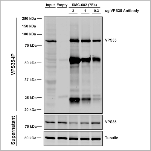
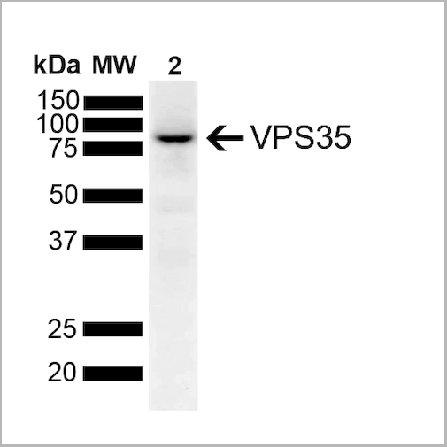
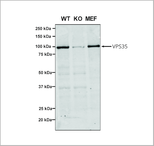
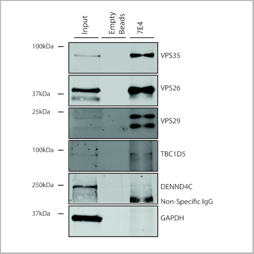
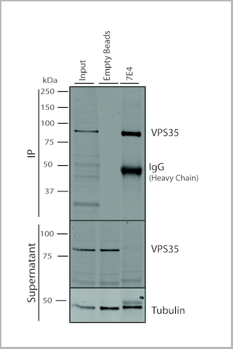
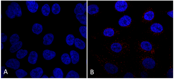
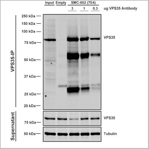 IP (Immunoprecipitation)
(Immunoprecipitation analysis using Mouse Anti-VPS35 Monoclonal Antibody, Clone 7E4. Tissue: embryonic fibroblast. Species: Mouse. Primary Antibody: Mouse Anti-VPS35 Monoclonal Antibody. Three amounts (3, 1 and 0.3 ug) were non-covalently coupled to 10uL of A/G sepharose beads for 1 hour at 4 degree C and next incubated with 250ug of MEF lysate for 2 hours at 4 degree C.)
IP (Immunoprecipitation)
(Immunoprecipitation analysis using Mouse Anti-VPS35 Monoclonal Antibody, Clone 7E4. Tissue: embryonic fibroblast. Species: Mouse. Primary Antibody: Mouse Anti-VPS35 Monoclonal Antibody. Three amounts (3, 1 and 0.3 ug) were non-covalently coupled to 10uL of A/G sepharose beads for 1 hour at 4 degree C and next incubated with 250ug of MEF lysate for 2 hours at 4 degree C.)
VPS35, Monoclonal Antibody (Cat# AAA27671)
Full Name
VPS35 Antibody, Clone 7E4: PerCP
Gene Names
VPS35; MEM3; PARK17
Reactivity
Human, Mouse, Rat
Applications
Western Blot, Immunocytochemistry, Immunofluorescence, Immunoprecipitation
Purity
Protein G Purified
Pricing

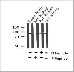 WB (Western Blot)
(Western blot analysis of Phospho-Tau (Thr212) Antibody expression in mouse brain and rat kidney tissues lysates.)
WB (Western Blot)
(Western blot analysis of Phospho-Tau (Thr212) Antibody expression in mouse brain and rat kidney tissues lysates.)
Tau, Polyclonal Antibody (Cat# AAA31001)
Full Name
Phospho-Tau (Thr212) Antibody
Gene Names
MAPT; TAU; MSTD; PPND; DDPAC; MAPTL; MTBT1; MTBT2; FTDP-17; PPP1R103
Reactivity
Human, Mouse, Rat
Applications
Western Blot, Immunohistochemistry
Purity
From purified rabbit serum by affinity purification via sequential chromatography on phospho-and non-phospho-peptide affinity columns.
Pricing
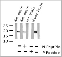

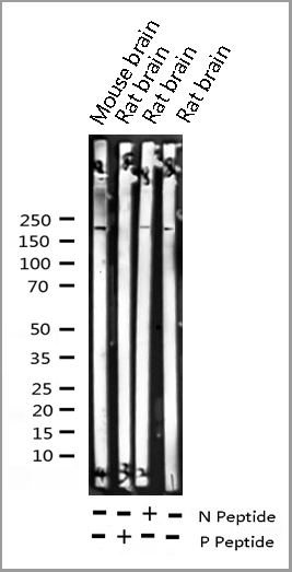
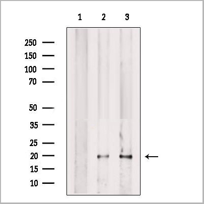
 WB (Western Blot)
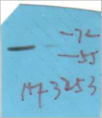
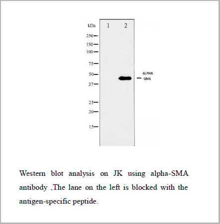
(Western blot analysis of Synuclein phosphorylation expression in Mouse brain tissue lysates, The lane on the left is treated with the antigen-specific peptide.)
WB (Western Blot)
(Western blot analysis of Synuclein phosphorylation expression in Mouse brain tissue lysates, The lane on the left is treated with the antigen-specific peptide.)
Synuclein, Polyclonal Antibody (Cat# AAA31040)
Full Name
Phospho-Synuclein (Ser129) Antibody
Gene Names
SNCA; PD1; NACP; PARK1; PARK4
Reactivity
Human, Mouse, Rat
Applications
Western Blot, Immunohistochemistry
Purity
From purified rabbit serum by affinity purification via sequential chromatography on phospho-and non-phospho-peptide affinity columns.
Pricing
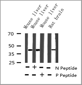
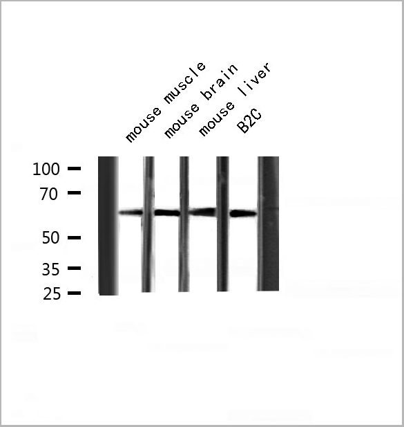
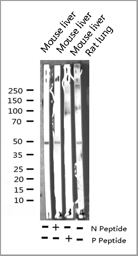
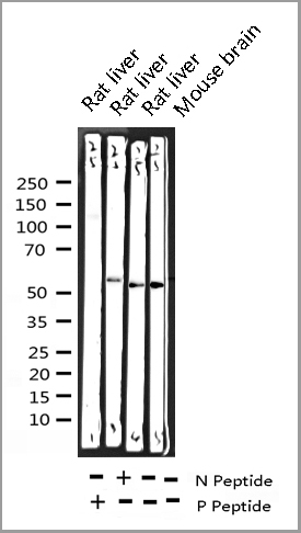
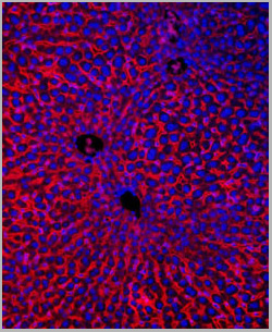
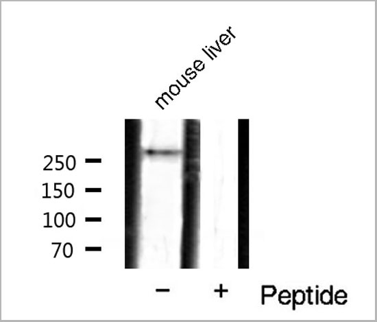 WB (Western Blot)
(Western blot analysis of mTOR phosphorylation expression in mouse liver tissue lysates, The lane on the right is treated with the antigen-specific peptide.)
WB (Western Blot)
(Western blot analysis of mTOR phosphorylation expression in mouse liver tissue lysates, The lane on the right is treated with the antigen-specific peptide.)
mTOR, Polyclonal Antibody (Cat# AAA31048)
Full Name
Phospho-mTOR (Ser2448) Antibody
Gene Names
MTOR; SKS; FRAP; FRAP1; FRAP2; RAFT1; RAPT1
Reactivity
Human, Mouse, Rat, Fish
Applications
Western Blot, Immunohistochemistry
Purity
From purified rabbit serum by affinity purification via sequential chromatography on phospho-and non-phospho-peptide affinity columns.
Pricing
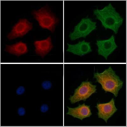
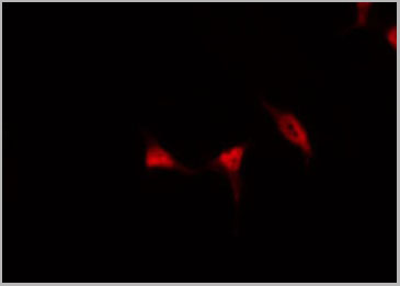
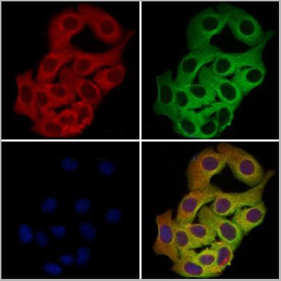
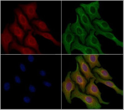
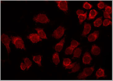
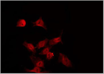
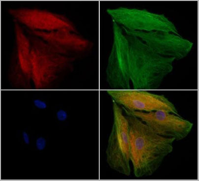 IF (Immunofluorescence)
(AAA31099 staining A549 cells by IF/ICC. The samples were fixed with PFA and permeabilized in 0.1% Triton X-100,then blocked in 10% serum for 45 minutes at 25°C. Samples were then incubated with primary Ab(AAA31099) and mouse antibeta tubulin Ab for 1 hour at 37°C. An AlexaFluor594 conjugated goat anti-rabbit IgG(H+L) Ab(Red) and an AlexaFluor488 conjugated goat anti-mouse IgG(H+L) Ab(Green) were used as the secondary antibody. The nuclear counter stain is DAPI (blue))
IF (Immunofluorescence)
(AAA31099 staining A549 cells by IF/ICC. The samples were fixed with PFA and permeabilized in 0.1% Triton X-100,then blocked in 10% serum for 45 minutes at 25°C. Samples were then incubated with primary Ab(AAA31099) and mouse antibeta tubulin Ab for 1 hour at 37°C. An AlexaFluor594 conjugated goat anti-rabbit IgG(H+L) Ab(Red) and an AlexaFluor488 conjugated goat anti-mouse IgG(H+L) Ab(Green) were used as the secondary antibody. The nuclear counter stain is DAPI (blue))
NOD2, Polyclonal Antibody (Cat# AAA31099)
Full Name
NOD2 Antibody
Gene Names
NOD2; CD; ACUG; BLAU; IBD1; YAOS; BLAUS; NLRC2; NOD2B; CARD15; CLR16.3; PSORAS1
Reactivity
Human, Mouse, Rat
Applications
Western Blot, Immunohistochemistry, Immunofluorescence, Immunocytochemistry
Purity
Peptide affinity chromatography using SulfoLink Coupling Resin (Thermo Fisher Scientific)
Pricing
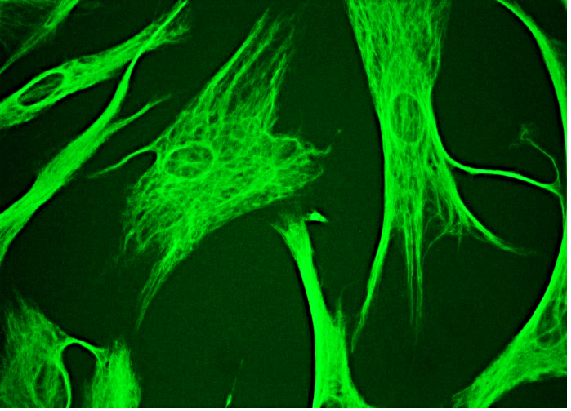
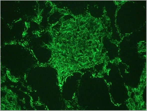 IF (Immunofluorescence)
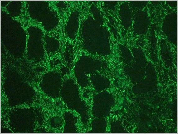
(Figure 2. Indirect immunofluorescence staining of human kidney tissue section with (diluted 1:1000), showing the specific pattern of vimentin in the mesenchymal cell types, such as fibroblasts in the connective tissue, podocytes, and endothelial cells in blood vessels. As expected, no reactivity is seen in the epithelial cell compartment.)
IF (Immunofluorescence)
(Figure 2. Indirect immunofluorescence staining of human kidney tissue section with (diluted 1:1000), showing the specific pattern of vimentin in the mesenchymal cell types, such as fibroblasts in the connective tissue, podocytes, and endothelial cells in blood vessels. As expected, no reactivity is seen in the epithelial cell compartment.)
vimentin, Monoclonal Recombinant Antibody (Cat# AAA14575)
Full Name
Recombinant rabbit anti vimentin
Reactivity
Human, mouse
Applications
Immunocytochemistry, Immunohistochemistry, Immunoblot
Purity
The purity of the antibody is >98% as determined by SDS-Polyacrylamide gel electrophoresis.
Pricing
 IF (Immunofluorescence)
(AAA31103 staining Hela cells by IF/ICC. The samples were fixed with PFA and permeabilized in 0.1% Triton X-100,then blocked in 10% serum for 45 minutes at 25°C. Samples were then incubated with primary Ab(AAA31103) and mouse anti-beta tubulin Ab for 1 hour at 37°C. An AlexaFluor594 conjugated goat anti-rabbit IgG(H+L) Ab(Red) and an AlexaFluor488 conjugated goat anti-mouse IgG(H+L) Ab(Green) were used as the secondary antibody.The nuclear counter stain is DAPI (blue).)
IF (Immunofluorescence)
(AAA31103 staining Hela cells by IF/ICC. The samples were fixed with PFA and permeabilized in 0.1% Triton X-100,then blocked in 10% serum for 45 minutes at 25°C. Samples were then incubated with primary Ab(AAA31103) and mouse anti-beta tubulin Ab for 1 hour at 37°C. An AlexaFluor594 conjugated goat anti-rabbit IgG(H+L) Ab(Red) and an AlexaFluor488 conjugated goat anti-mouse IgG(H+L) Ab(Green) were used as the secondary antibody.The nuclear counter stain is DAPI (blue).)
CCL20, Polyclonal Antibody (Cat# AAA31103)
Full Name
CCL20 Antibody
Gene Names
CCL20; CKb4; LARC; ST38; MIP3A; Exodus; MIP-3a; SCYA20; MIP-3-alpha
Reactivity
Human, Mouse
Applications
Western Blot, Immunohistochemistry, Immunofluorescence, Immunocytochemistry
Purity
Purified by peptide affinity chromatography using SulfoLink Coupling Resin
Pricing
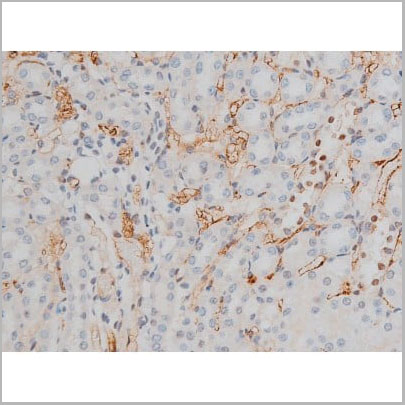
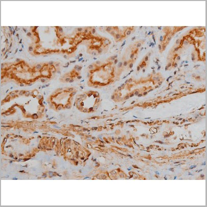

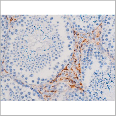
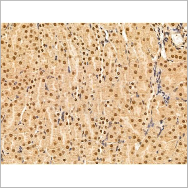
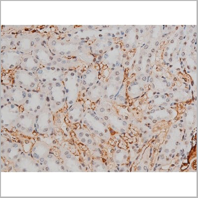
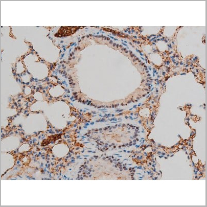
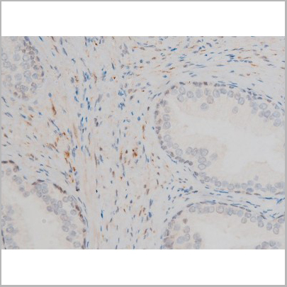
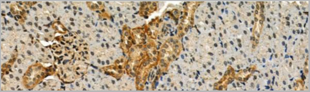
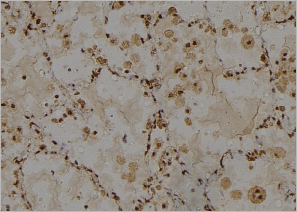
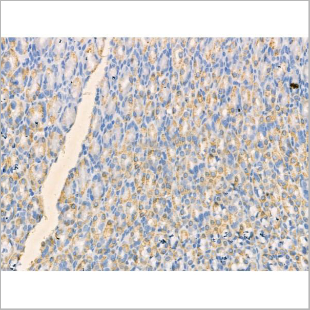
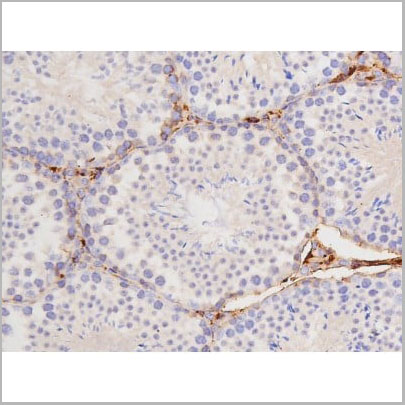
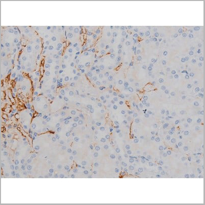
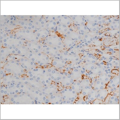
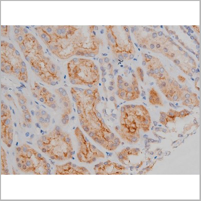
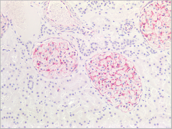
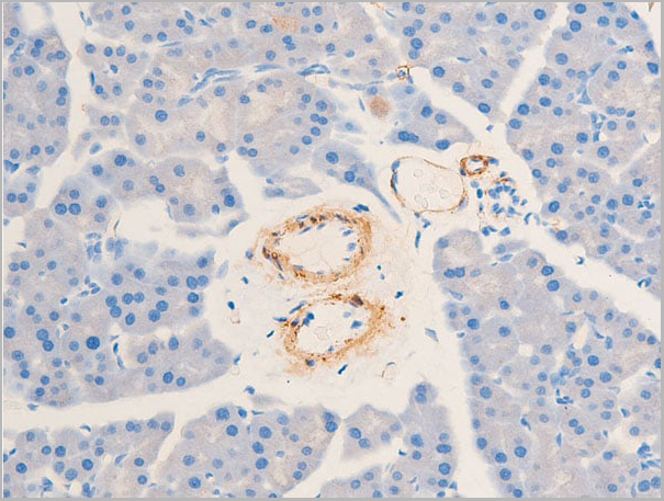
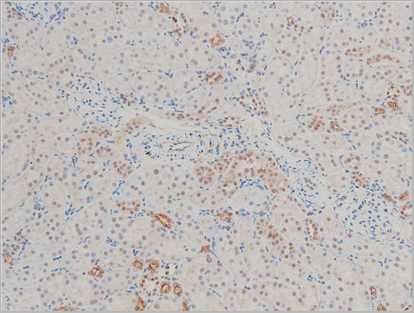
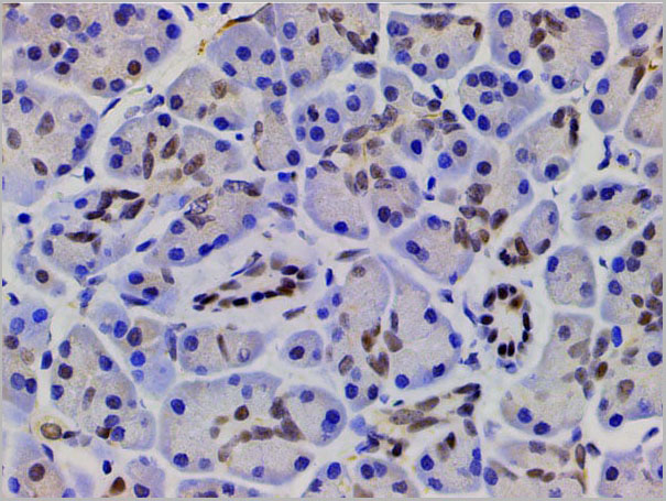
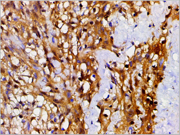
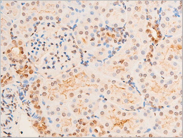
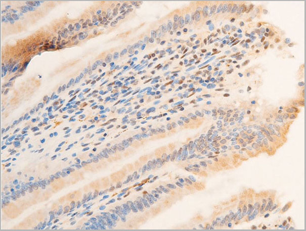
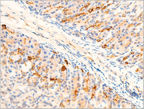
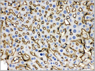
 IHC (Immunohistochemistry)
(AAA31016 at 1/200 staining Rat kidney tissue sections by IHC-P. The tissue was formaldehyde fixed and a heat mediated antigen retrieval step in citrate buffer was performed. The tissue was then blocked and incubated with the antibody for 1.5 hours at 22 degree C. An HRP conjugated goat anti-rabbit antibody was used as the secondary.)
IHC (Immunohistochemistry)
(AAA31016 at 1/200 staining Rat kidney tissue sections by IHC-P. The tissue was formaldehyde fixed and a heat mediated antigen retrieval step in citrate buffer was performed. The tissue was then blocked and incubated with the antibody for 1.5 hours at 22 degree C. An HRP conjugated goat anti-rabbit antibody was used as the secondary.)
CREB, Polyclonal Antibody (Cat# AAA31016)
Full Name
Phospho-CREB (Ser133) Antibody
Gene Names
CREB1; CREB; CREB-1
Reactivity
Human, Mouse, Rat
Applications
Western Blot, Immunohistochemistry, Immunofluorescence, Immunocytochemistry, Immunoprecipitation
Purity
From purified rabbit serum by affinity purification via sequential chromatography on phospho-and non-phospho-peptide affinity columns.
Pricing
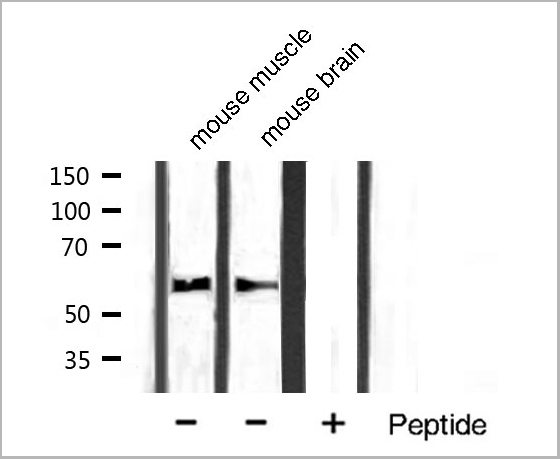
 WB (Western Blot)
(Western blot analysis of extracts of various tissue sample, using Src Antibody.)
WB (Western Blot)
(Western blot analysis of extracts of various tissue sample, using Src Antibody.)
Src, Polyclonal Antibody (Cat# AAA31086)
Full Name
Src Antibody
Gene Names
SRC; ASV; SRC1; THC6; c-SRC; p60-Src
Reactivity
Human, Mouse, Rat, Monkey
Applications
Western Blot, Immunohistochemistry, Immunofluorescence, Immunocytochemistry
Purity
Peptide affinity purification
Pricing
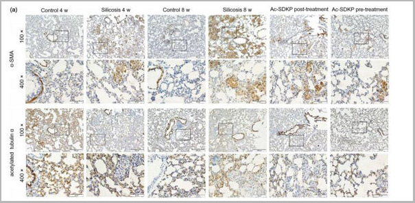
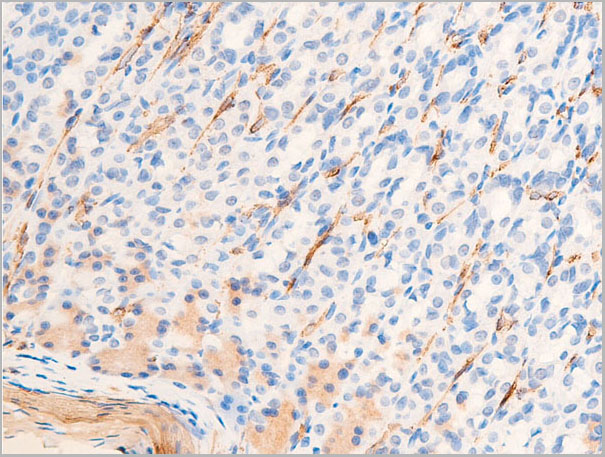
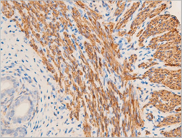
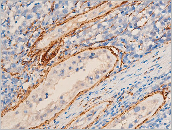
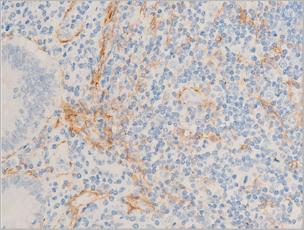
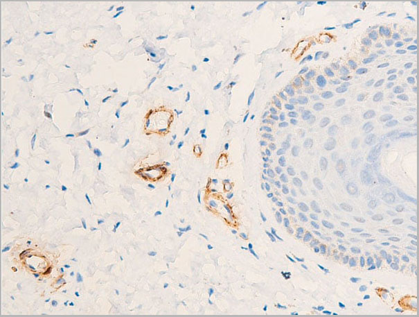
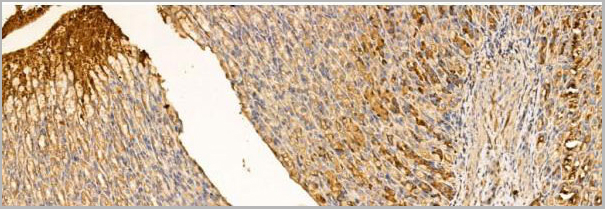
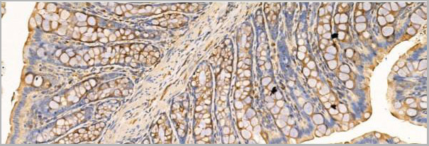
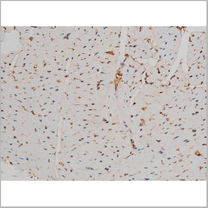
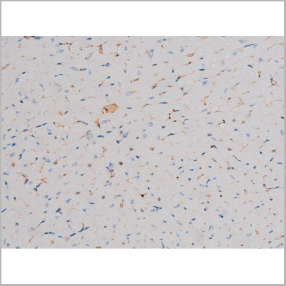
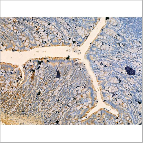
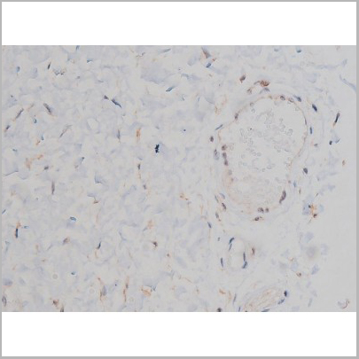
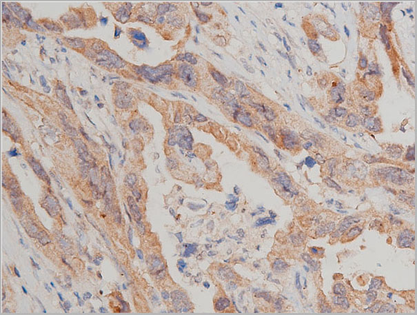
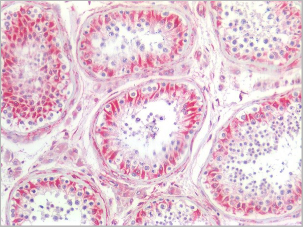
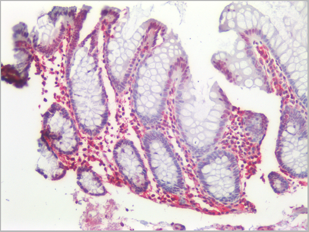
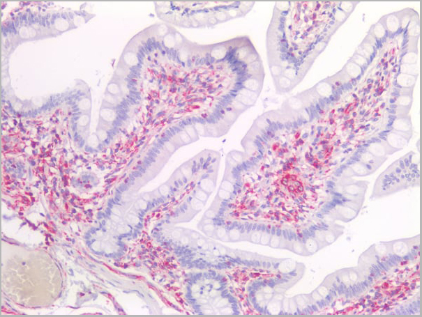
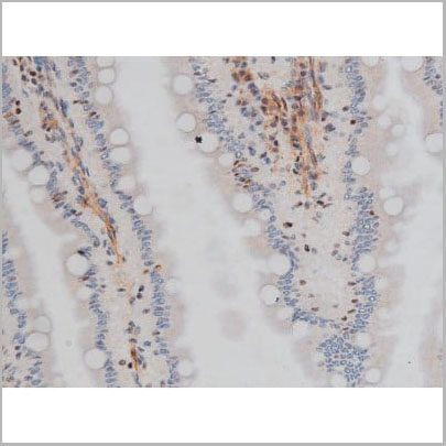
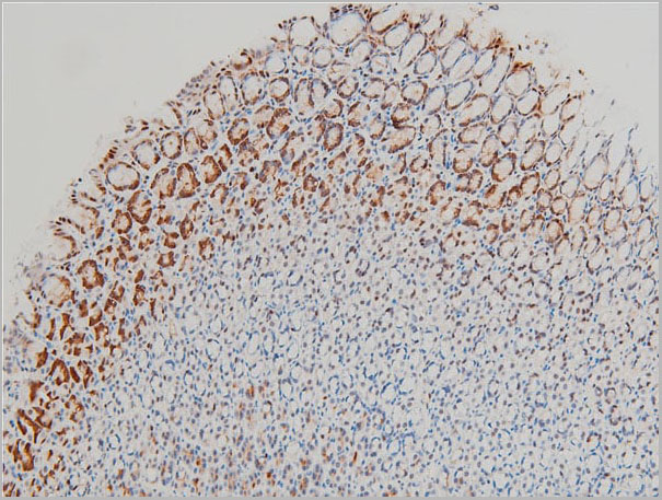
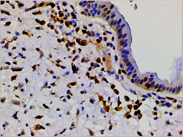
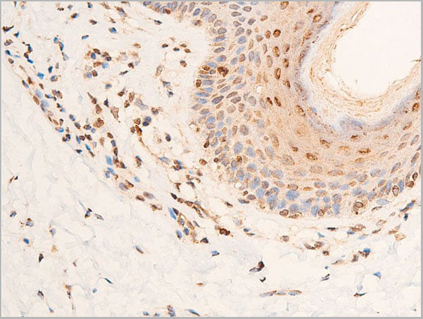
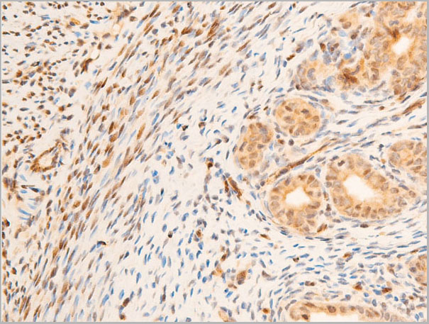
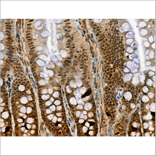
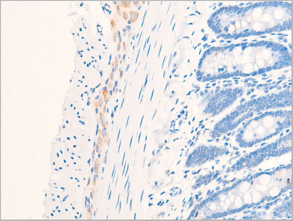
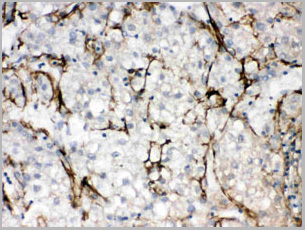
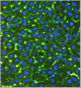
 IHC (Immunohistochemistry)
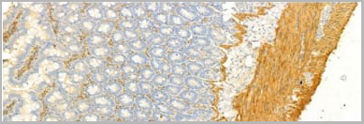
(AF1032 at 1/100 staining Rat colon tissue by IHC-P. The sample was formaldehyde fixed and a heat mediated antigen retrieval step in citrate buffer was performed. The sample was then blocked and incubated with the primary antibody at 4°C overnight. An HRP conjugated anti-Rabbit antibody was used as the secondary antibody.)
IHC (Immunohistochemistry)
(AF1032 at 1/100 staining Rat colon tissue by IHC-P. The sample was formaldehyde fixed and a heat mediated antigen retrieval step in citrate buffer was performed. The sample was then blocked and incubated with the primary antibody at 4°C overnight. An HRP conjugated anti-Rabbit antibody was used as the secondary antibody.)
alpha-SMA, Polyclonal Antibody (Cat# AAA30933)
Full Name
alpha-SMA Antibody
Gene Names
ACTA2; ACTSA
Reactivity
Human, Mouse, Rat, Bovine
Applications
Western Blot, Immunohistochemistry, Immunofluorescence, Immunocytochemistry, Immunoprecipitation
Purity
Peptide affinity purification
Pricing
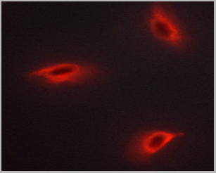
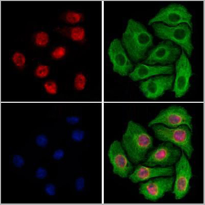
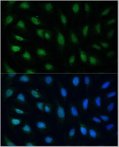
 IF (Immunofluorescence)
(AAA31012 staining Hela by IF/ICC. The sample were fixed with PFA and permeabilized in 0.1% Triton X-100, then blocked in 10% serum for 45 minutes at 25 degree C. The primary antibody was diluted at 1/200 and incubated with the sample for 1 hour at 37 degree C. An Alexa Fluor 594 conjugated goat anti-rabbit IgG (H+L) Ab, diluted at 1/600, was used as the secondary antibody.)
IF (Immunofluorescence)
(AAA31012 staining Hela by IF/ICC. The sample were fixed with PFA and permeabilized in 0.1% Triton X-100, then blocked in 10% serum for 45 minutes at 25 degree C. The primary antibody was diluted at 1/200 and incubated with the sample for 1 hour at 37 degree C. An Alexa Fluor 594 conjugated goat anti-rabbit IgG (H+L) Ab, diluted at 1/600, was used as the secondary antibody.)
ATF2, Polyclonal Antibody (Cat# AAA31012)
Full Name
Phospho-ATF2 (Thr71/53) Antibody
Gene Names
ATF2; HB16; CREB2; TREB7; CREB-2; CRE-BP1
Reactivity
Human, Mouse, Rat
Applications
Western Blot, Immunohistochemistry, Immunofluorescence, Immunocytochemistry, Immunoprecipitation
Purity
From purified rabbit serum by affinity purification via sequential chromatography on phospho-and non-phospho-peptide affinity columns.
Pricing
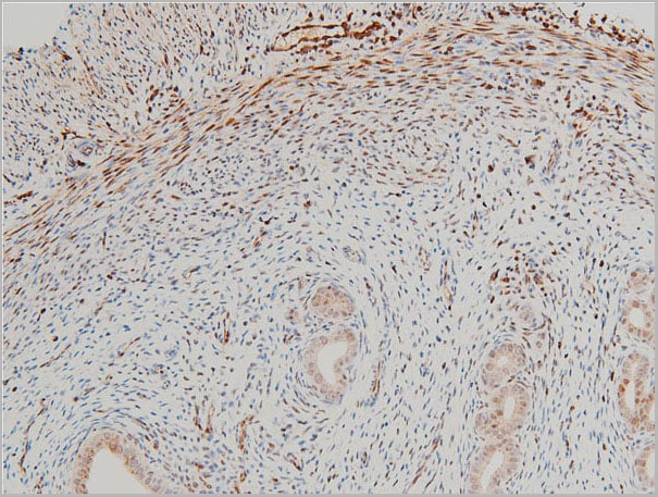


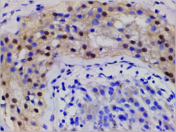
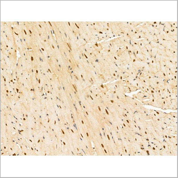
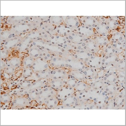
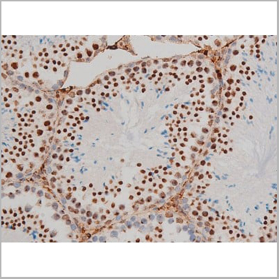
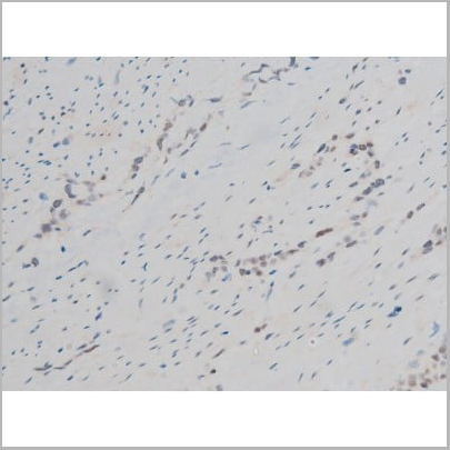
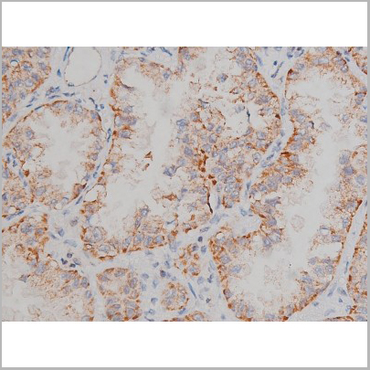
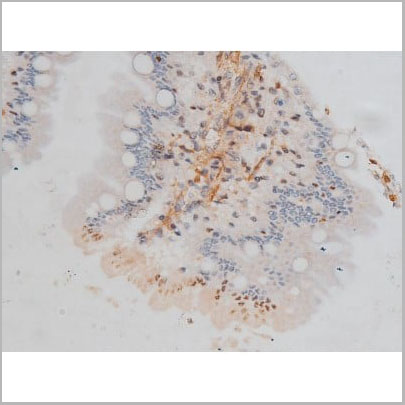
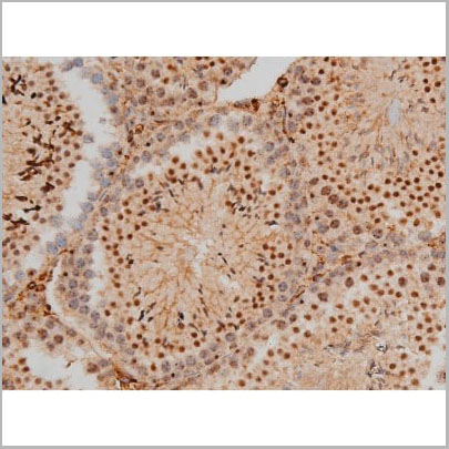
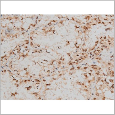
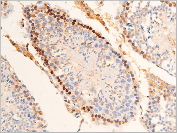
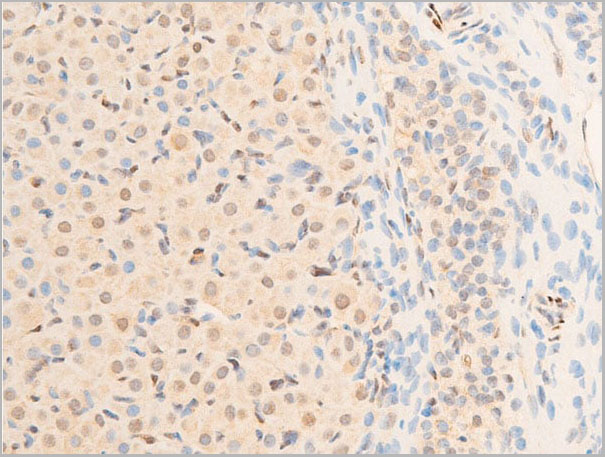
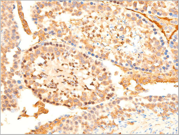
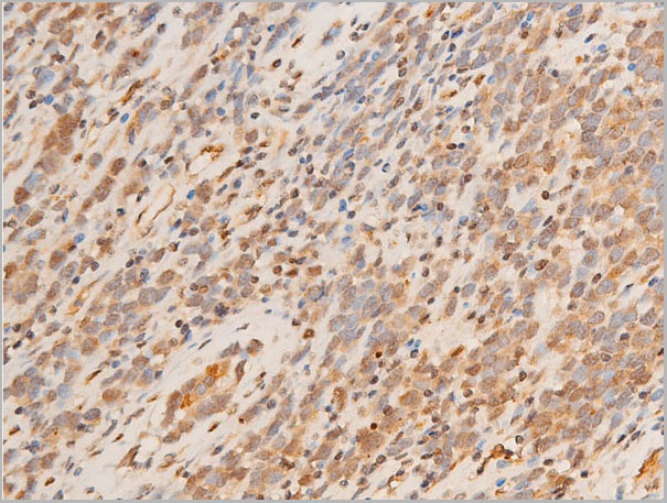
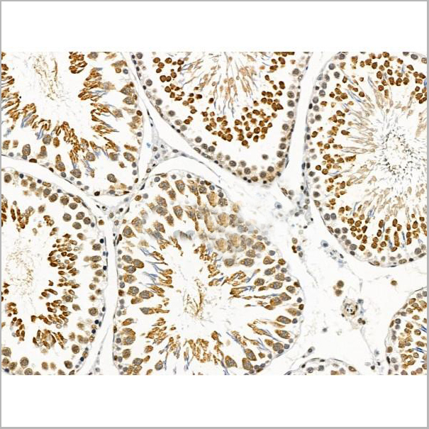
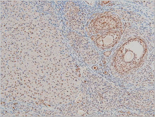 IHC (Immunohistochemistry)
(AAA31025 at 1/100 staining rat ovarian tissue sections by IHC-P. The tissue was formaldehyde fixed and a heat mediated antigen retrieval step in citrate buffer was performed. The tissue was then blocked and incubated with the antibody for 1.5 hours at 22 degree C. An HRP conjugated goat anti-rabbit antibody was used as the secondary.)
IHC (Immunohistochemistry)
(AAA31025 at 1/100 staining rat ovarian tissue sections by IHC-P. The tissue was formaldehyde fixed and a heat mediated antigen retrieval step in citrate buffer was performed. The tissue was then blocked and incubated with the antibody for 1.5 hours at 22 degree C. An HRP conjugated goat anti-rabbit antibody was used as the secondary.)
Cofilin, Polyclonal Antibody (Cat# AAA31025)
Full Name
Phospho-Cofilin (Ser3) Antibody
Gene Names
CFL1; CFL; cofilin; HEL-S-15
Reactivity
Human, Mouse, Rat
Applications
Western Blot, Immunohistochemistry, Immunofluorescence, Immunocytochemistry
Purity
From purified rabbit serum by affinity purification via sequential chromatography on phospho-and non-phospho-peptide affinity columns.
Pricing
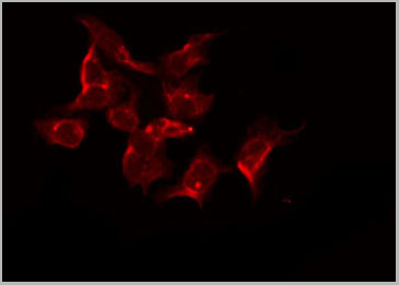
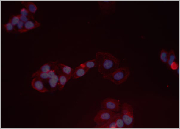
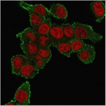 IF (Immunofluorescence)
(Immunofluorescence staining of PFA-fixed HePG2 cells using Tumor Necrosis Factor (TNF alpha) Mouse Monoclonal Antibody (TNF706) followed by goat anti-mouse IgG-CF488 (green). Nuclei stained with RedDot.)
IF (Immunofluorescence)
(Immunofluorescence staining of PFA-fixed HePG2 cells using Tumor Necrosis Factor (TNF alpha) Mouse Monoclonal Antibody (TNF706) followed by goat anti-mouse IgG-CF488 (green). Nuclei stained with RedDot.)
TNF-alpha (Tumor Necrosis Factor alpha), Monoclonal Antibody (Cat# AAA13806)
Full Name
TNF-alpha (Tumor Necrosis Factor alpha) Mouse Monoclonal Antibody
Gene Names
TNF; DIF; TNFA; TNFSF2; TNLG1F; TNF-alpha
Reactivity
Human, Mouse, Rat, Rabbit, Cat, Dog, Zebrafish
Applications
Flow Cytometry, Immunofluorescence, Immunohistochemistry
Pricing
 WB (Western Blot)
(Western blot analysis of Chk2 phosphorylation expression in UV treated COS7 whole cell lysates, The lane on the left is treated with the antigen-specific peptide.)
WB (Western Blot)
(Western blot analysis of Chk2 phosphorylation expression in UV treated COS7 whole cell lysates, The lane on the left is treated with the antigen-specific peptide.)
Chk2, Polyclonal Antibody (Cat# AAA30958)
Full Name
Phospho-Chk2 (Thr383) Antibody
Gene Names
CHEK2; CDS1; CHK2; LFS2; RAD53; hCds1; HuCds1; PP1425
Reactivity
Human, Mouse, Rat
Applications
Western Blot, Immunohistochemistry, Immunofluorescence, Immunocytochemistry
Purity
From purified rabbit serum by affinity purification via sequential chromatography on phospho-and non-phospho-peptide affinity columns.
Pricing
 WB (Western Blot)
(Western blot analysis of SGK phosphorylation expression in Insulin treated HeLa whole cell lysates, The lane on the left is treated with the antigen-specific peptide.)
WB (Western Blot)
(Western blot analysis of SGK phosphorylation expression in Insulin treated HeLa whole cell lysates, The lane on the left is treated with the antigen-specific peptide.)
SGK, Polyclonal Antibody (Cat# AAA30942)
Full Name
Phospho-SGK (Ser422) Antibody
Gene Names
SGK1; SGK
Reactivity
Human, Mouse, Rat
Applications
Western Blot, Immunohistochemistry, Immunofluorescence, Immunocytochemistry
Purity
From purified rabbit serum by affinity purification via sequential chromatography on phospho-and non-phospho-peptide affinity columns.
Pricing
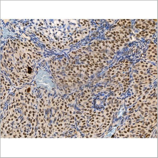
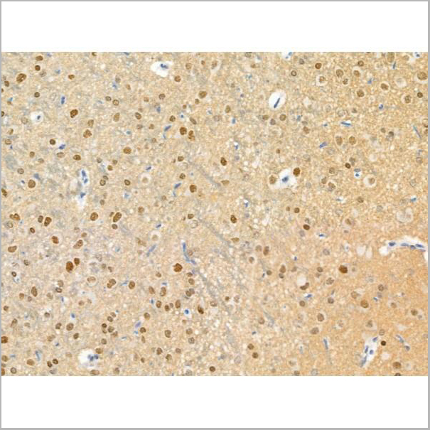
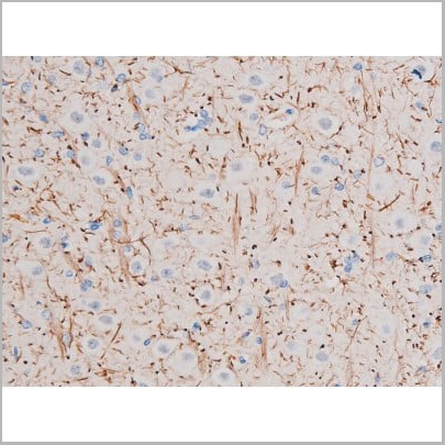
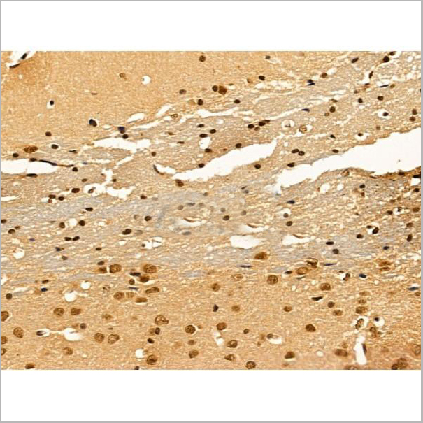
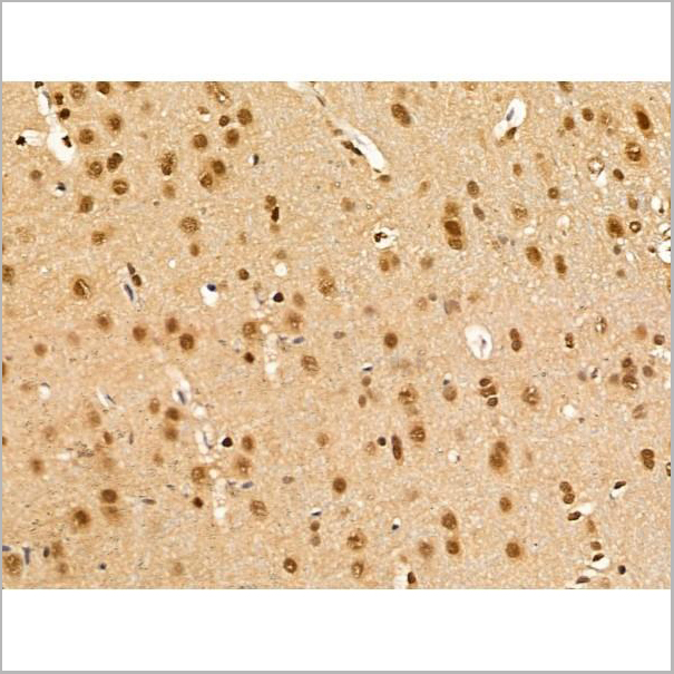
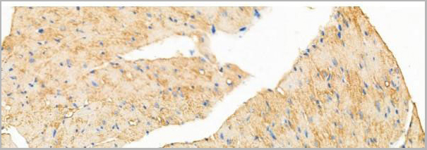
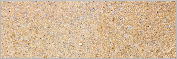
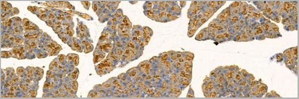
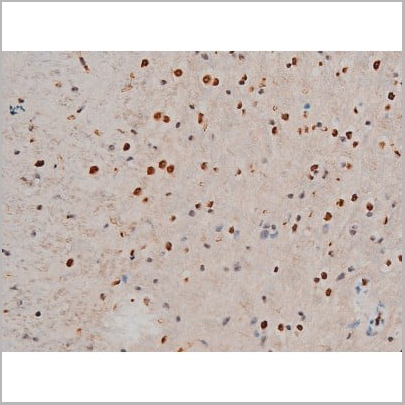
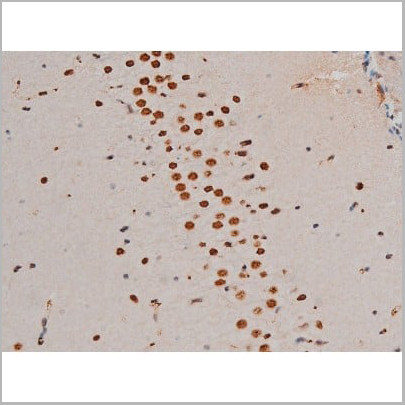
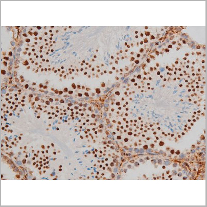
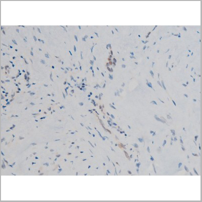
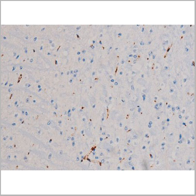
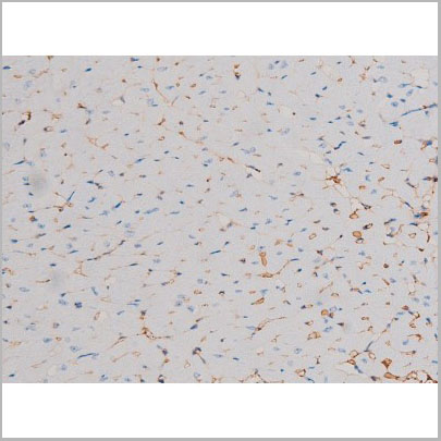
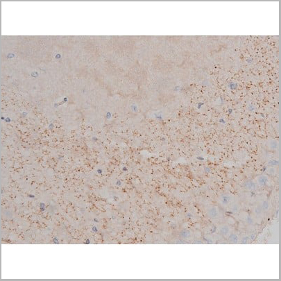
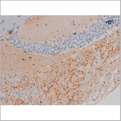
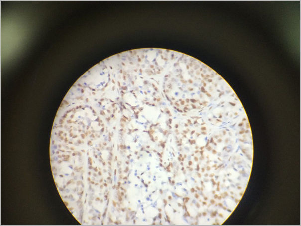
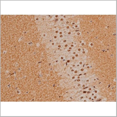
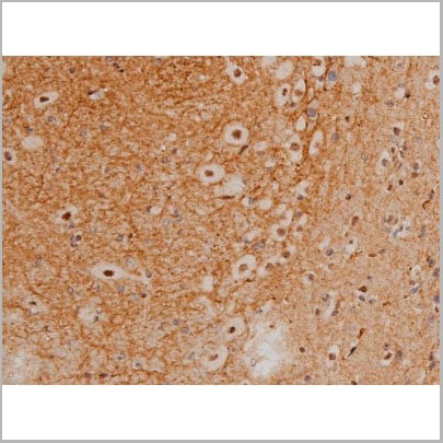
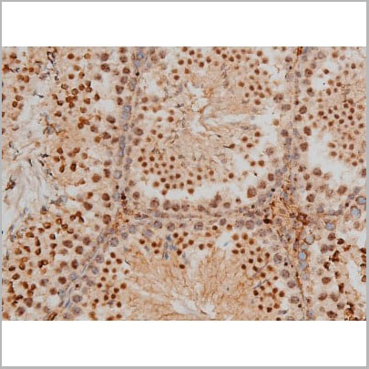
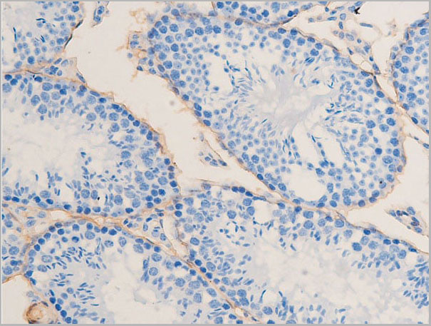
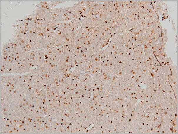
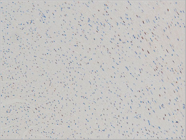
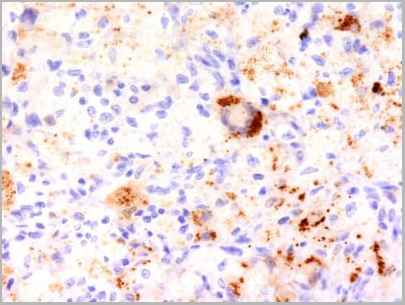
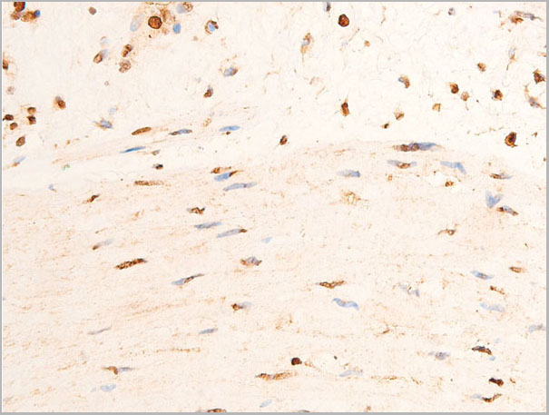
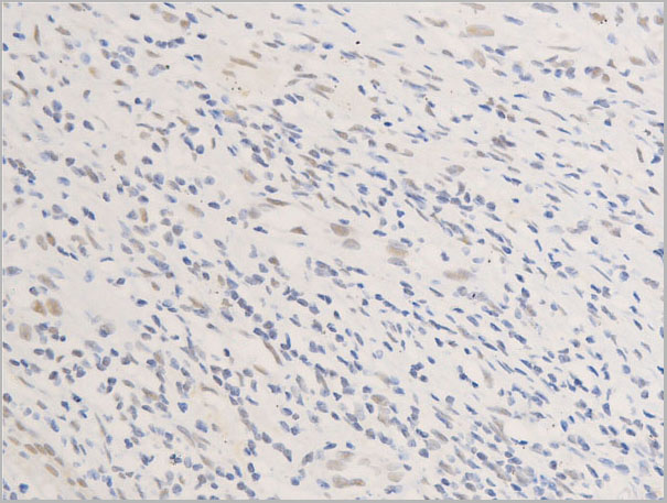
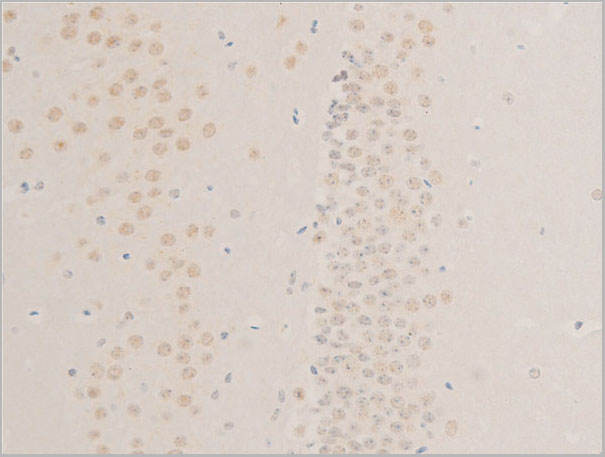
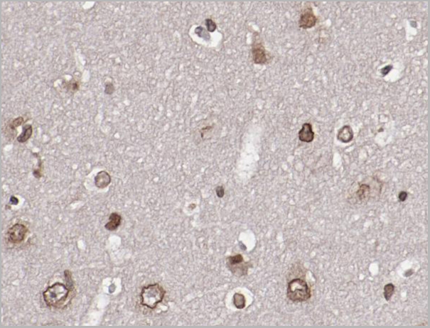
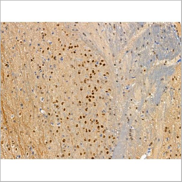 IHC (Immunohistochemistry)
(At 1/100 staining Mouse brain tissue by IHC-P. The sample was formaldehyde fixed and a heat mediated antigen retrieval step in citrate buffer was performed. The sample was then blocked and incubated with the primary antibody at 4 degree C overnight. An HRP conjugated anti-Rabbit antibody was used as the secondary antibody.)
IHC (Immunohistochemistry)
(At 1/100 staining Mouse brain tissue by IHC-P. The sample was formaldehyde fixed and a heat mediated antigen retrieval step in citrate buffer was performed. The sample was then blocked and incubated with the primary antibody at 4 degree C overnight. An HRP conjugated anti-Rabbit antibody was used as the secondary antibody.)
ATF6, Polyclonal Antibody (Cat# AAA31376)
Full Name
Phospho-ATF6 (Thr166) Antibody
Gene Names
ATF6; ATF6A
Reactivity
Human, Mouse, Rat
Predicted Reactivity: Pig (100%), Horse (100%), Rabbit (100%), Dog (100%)
Predicted Reactivity: Pig (100%), Horse (100%), Rabbit (100%), Dog (100%)
Applications
Western Blot, Immunohistochemistry, Peptide ELISA
Purity
The antibody is from purified rabbit serum by affinity purification via sequential chromatography on phospho-peptide and non-phospho-peptide affinity columns.
Pricing
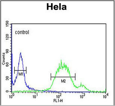
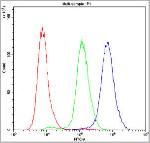
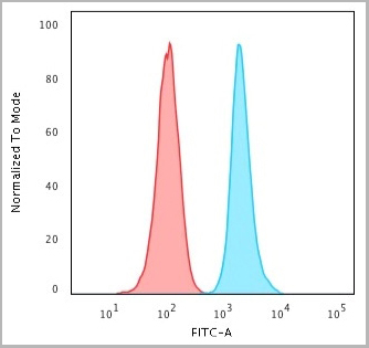
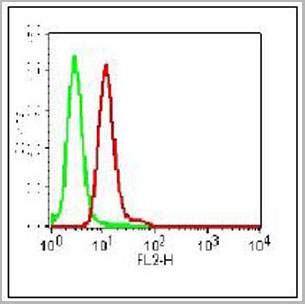
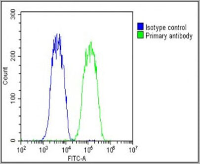 FCM (Flow Cytometry)
(Overlay histogram showing Hela cells stained with AAA28750 (green line). The cells were fixed with 2% paraformaldehyde (10 min) and then permeabilized with 90% methanol for 10 min. The cells were then icubated in 2% bovine serum albumin to block non-specific protein-protein interactions followed by the antibody (AAA28750, 1:25 dilution) for 60 min at 37ºC. The secondary antibody used was Goat-Anti-Rabbit IgG, DyLight® 488 Conjugated Highly Cross-Adsorbed (1583138) at 1/200 dilution for 40 min at 37°C. Isotype control antibody (blue line) was rabbit IgG1 (1ug/1x106 cells) used under the same conditions. Acquisition of >10, 000 events wasperformed.)
FCM (Flow Cytometry)
(Overlay histogram showing Hela cells stained with AAA28750 (green line). The cells were fixed with 2% paraformaldehyde (10 min) and then permeabilized with 90% methanol for 10 min. The cells were then icubated in 2% bovine serum albumin to block non-specific protein-protein interactions followed by the antibody (AAA28750, 1:25 dilution) for 60 min at 37ºC. The secondary antibody used was Goat-Anti-Rabbit IgG, DyLight® 488 Conjugated Highly Cross-Adsorbed (1583138) at 1/200 dilution for 40 min at 37°C. Isotype control antibody (blue line) was rabbit IgG1 (1ug/1x106 cells) used under the same conditions. Acquisition of >10, 000 events wasperformed.)
WNT5A, Polyclonal Antibody (Cat# AAA28750)
Full Name
WNT5A Antibody (Center)
Gene Names
WNT5A; hWNT5A
Reactivity
Human, Mouse, Rat
Predicted: Rabbit
Predicted: Rabbit
Applications
Immunohistochemistry, Immunohistochemistry, Flow Cytometry, Western Blot
Purity
This antibody is purified through a protein A column, followed by peptide affinity purification.
Pricing
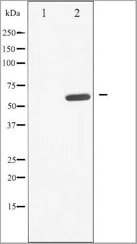
 WB (Western Blot)
(Western blot analysis of EGFR phosphorylation expression in TSA treated HeLa whole cell lysates, The lane on the left is treated with the antigen-specific peptide.)
WB (Western Blot)
(Western blot analysis of EGFR phosphorylation expression in TSA treated HeLa whole cell lysates, The lane on the left is treated with the antigen-specific peptide.)
EGFR, Polyclonal Antibody (Cat# AAA30963)
Full Name
Phospho-EGFR (Ser1026) Antibody
Gene Names
EGFR; ERBB; HER1; mENA; ERBB1; PIG61; NISBD2
Reactivity
Human, Mouse, Rat
Applications
Western Blot, Immunohistochemistry, Immunofluorescence, Immunocytochemistry
Purity
From purified rabbit serum by affinity purification via sequential chromatography on phospho-and non-phospho-peptide affinity columns.
Pricing
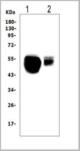
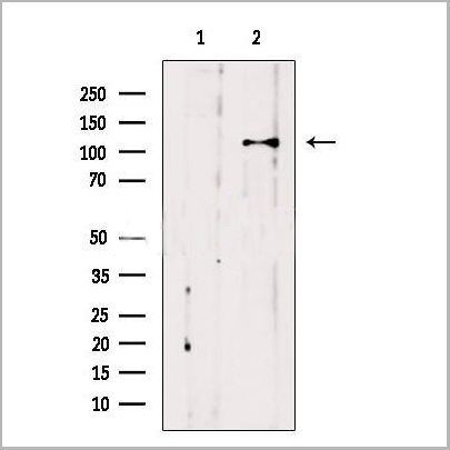
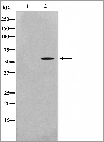
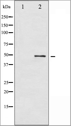
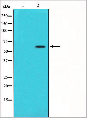
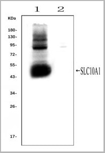 WB (Western Blot)
(Western blot analysis of SLC10A1 using anti- SLC10A1 antibody (AAA11653).Electrophoresis was performed on a 5-20% SDS-PAGE gel at 70V (Stacking gel) / 90V (Resolving gel) for 2-3 hours. The sample well of each lane was loaded with 50ug of sample under reducing conditions.Lane 1: rat liver tissue lysates (positive control), Lane 2: rat kidney tissue lysates, (negative control)After Electrophoresis, proteins were transferred to a Nitrocellulose membrane at 150mA for 50-90 minutes. Blocked the membrane with 5% Non-fat Milk/ TBS for 1.5 hour at RT. The membrane was incubated with rabbit anti-SLC10A1 antigen affinity purified polyclonal antibody (AAA11653) at 0.25ug/mL overnight at 4°C, then washed with TBS-0.1%Tween 3 times with 5 minutes each and probed with a goat anti-rabbit IgG-HRP secondary antibody at a dilution of 1:10000 for 1.5 hour at RT. The signal is developed using an Enhanced Chemiluminescent detection (ECL) kit with Tanon 5200 system. A specific band was detected for SLC10A1 at approximately 50KD. The expected band size for SLC10A1 is at 38KD.)
WB (Western Blot)
(Western blot analysis of SLC10A1 using anti- SLC10A1 antibody (AAA11653).Electrophoresis was performed on a 5-20% SDS-PAGE gel at 70V (Stacking gel) / 90V (Resolving gel) for 2-3 hours. The sample well of each lane was loaded with 50ug of sample under reducing conditions.Lane 1: rat liver tissue lysates (positive control), Lane 2: rat kidney tissue lysates, (negative control)After Electrophoresis, proteins were transferred to a Nitrocellulose membrane at 150mA for 50-90 minutes. Blocked the membrane with 5% Non-fat Milk/ TBS for 1.5 hour at RT. The membrane was incubated with rabbit anti-SLC10A1 antigen affinity purified polyclonal antibody (AAA11653) at 0.25ug/mL overnight at 4°C, then washed with TBS-0.1%Tween 3 times with 5 minutes each and probed with a goat anti-rabbit IgG-HRP secondary antibody at a dilution of 1:10000 for 1.5 hour at RT. The signal is developed using an Enhanced Chemiluminescent detection (ECL) kit with Tanon 5200 system. A specific band was detected for SLC10A1 at approximately 50KD. The expected band size for SLC10A1 is at 38KD.)
SLC10A1, Polyclonal Antibody (Cat# AAA11653)
Full Name
Anti-SLC10A1 Antibody
Gene Names
Slc10a1; Ntcp
Reactivity
Mouse, Rat
Applications
Flow Cytometry, Immunofluorescence, Immunohistochemistry, Immunohistochemistry, Immunocytochemistry, Western Blot
Purity
Immunogen affinity purified.
Pricing

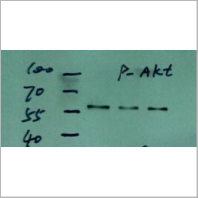
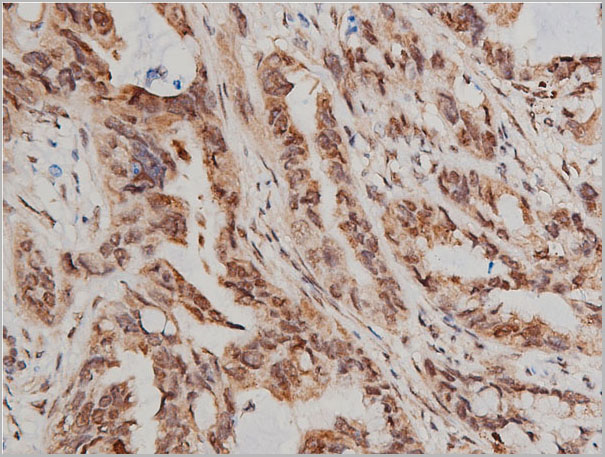
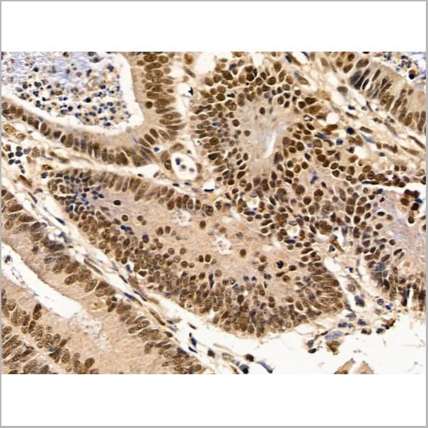
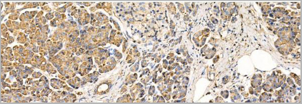
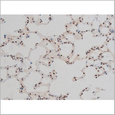
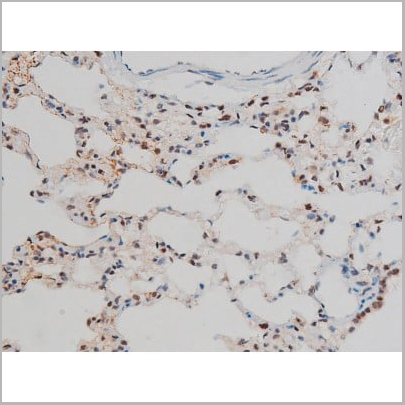
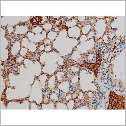
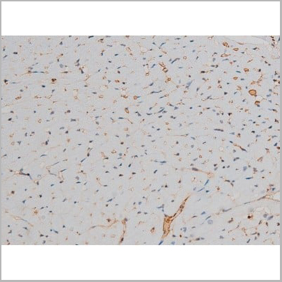
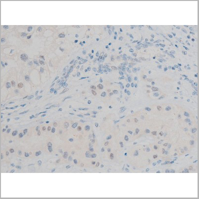
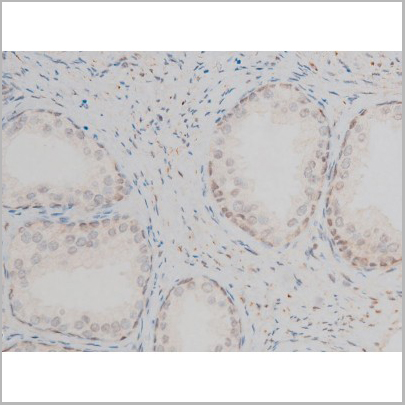
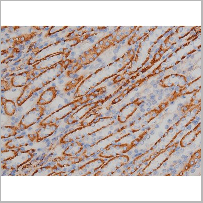
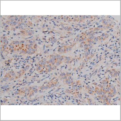
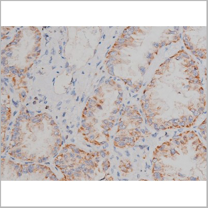
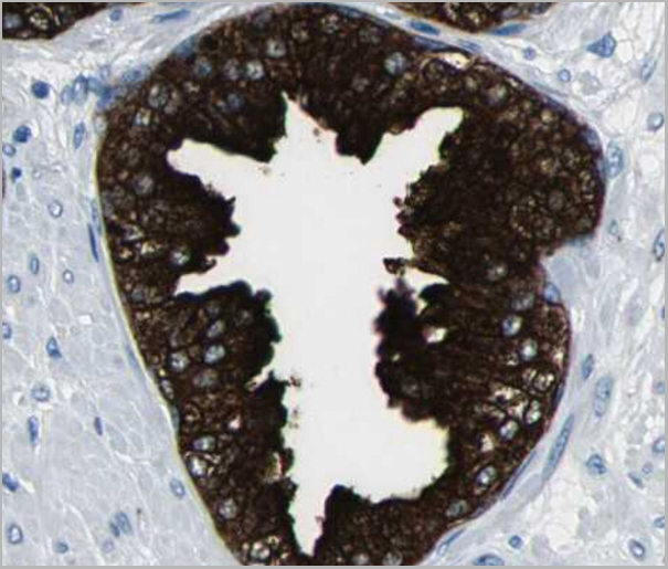
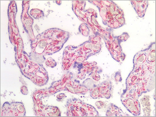
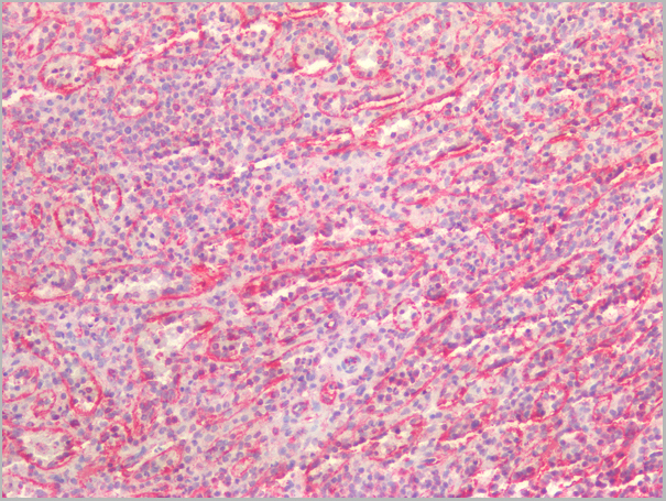
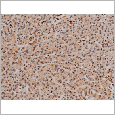
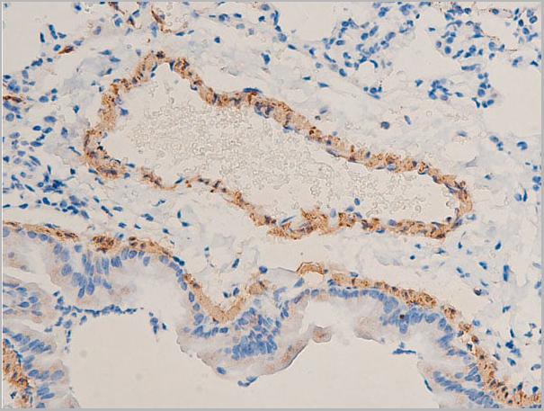
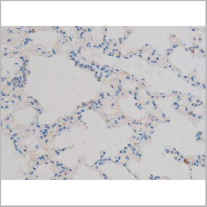
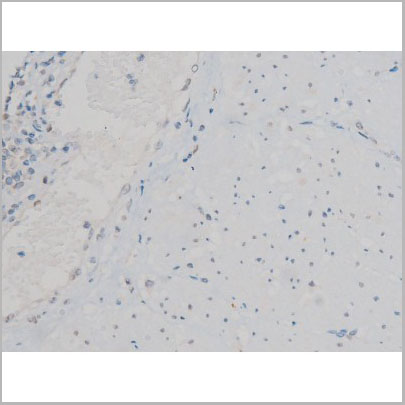
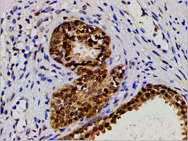
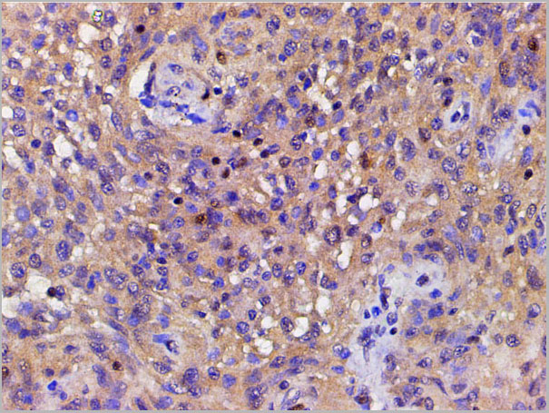
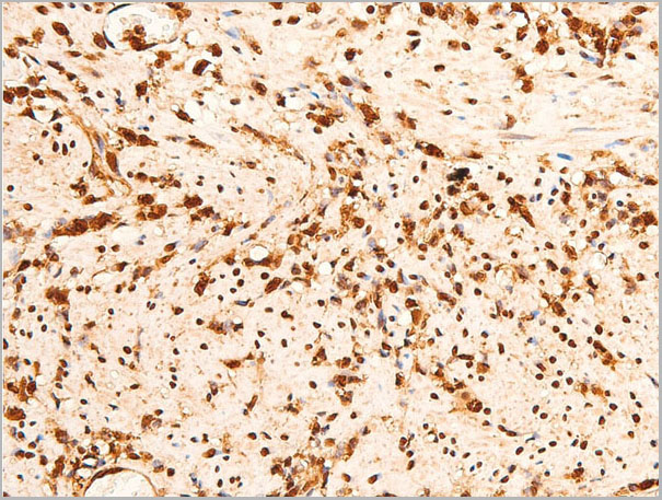
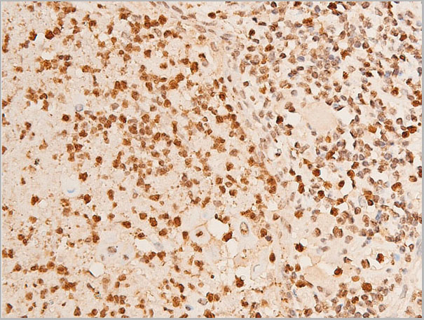
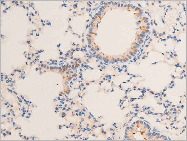
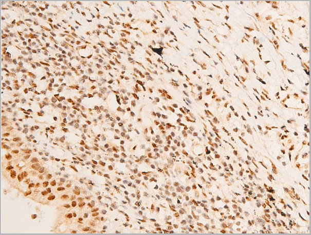
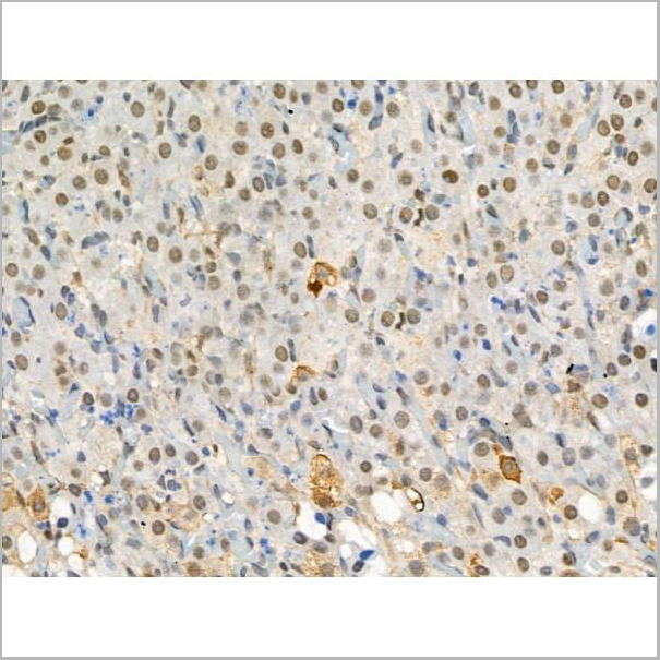
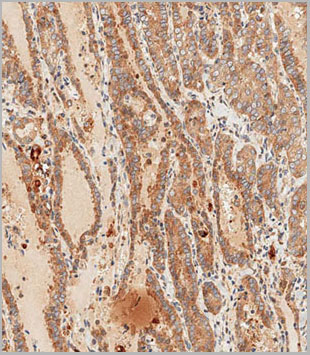
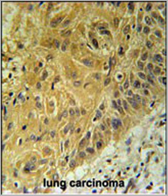
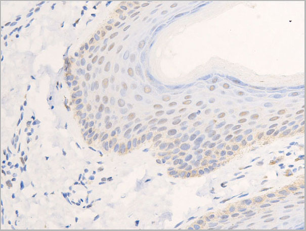
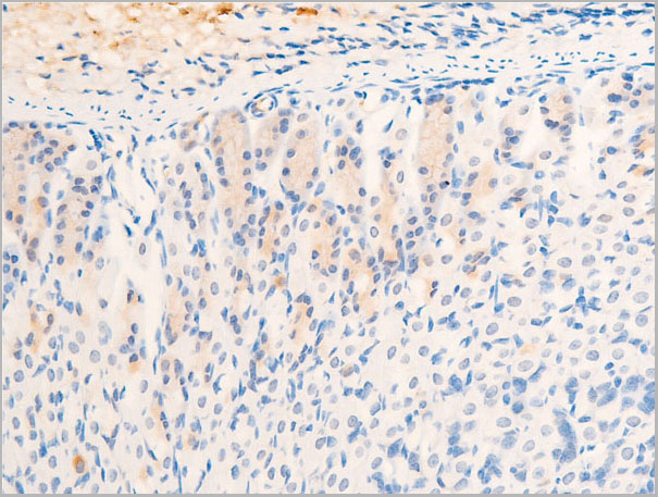
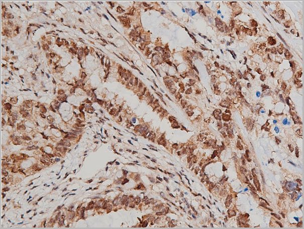 IHC (Immunohistochemistry)
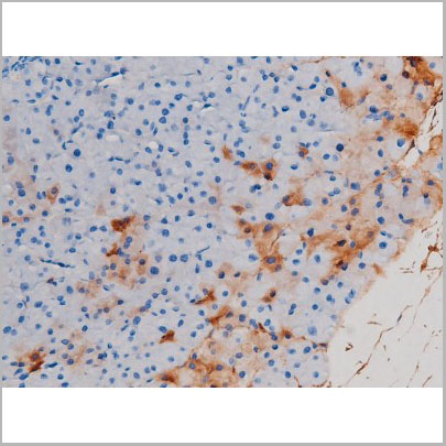
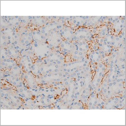
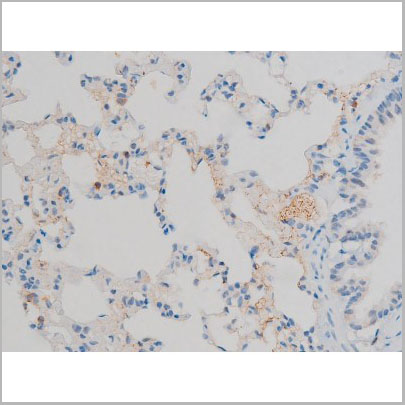
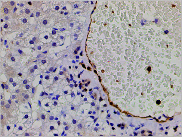
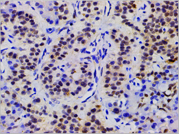
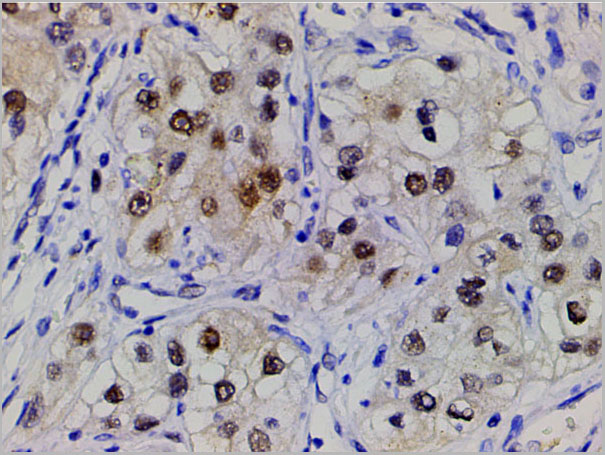
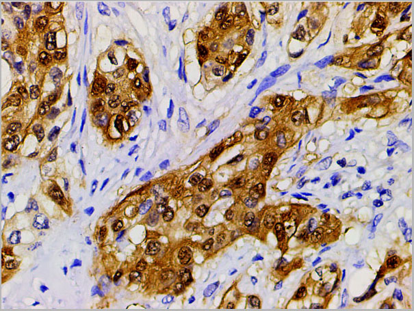
(AAA30920 at 1/200 staining human lung cancer tissue sections by IHC-P. The tissue was formaldehyde fixed and a heat mediated antigen retrieval step in citrate buffer was performed. The tissue was then blocked and incubated with the antibody for 1.5 hours at 22 degree C. An HRP conjugated goat anti-rabbit antibody was used as the secondary.)
IHC (Immunohistochemistry)
(AAA30920 at 1/200 staining human lung cancer tissue sections by IHC-P. The tissue was formaldehyde fixed and a heat mediated antigen retrieval step in citrate buffer was performed. The tissue was then blocked and incubated with the antibody for 1.5 hours at 22 degree C. An HRP conjugated goat anti-rabbit antibody was used as the secondary.)
Akt, Polyclonal Antibody (Cat# AAA30920)
Full Name
Phospho-Akt(Ser473) Antibody
Gene Names
AKT1; AKT; PKB; RAC; CWS6; PRKBA; PKB-ALPHA; RAC-ALPHA
Reactivity
Human, Mouse, Rat
Applications
Western Blot, Immunohistochemistry, Immunofluorescence, Immunocytochemistry
Purity
From purified rabbit serum by affinity purification via sequential chromatography on phospho-and non-phospho-peptide affinity columns.
Pricing
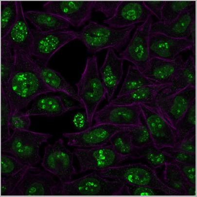
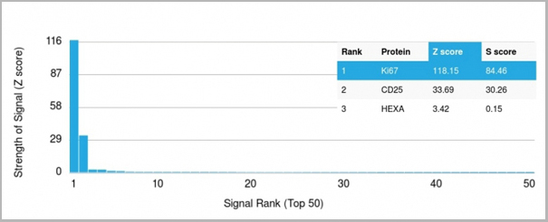 Application Data
(Analysis of Protein Array containing more than 19,000 full-length human proteins using Ki67 Mouse Monoclonal Antibody (MKI67/2466). Z- and S- Score: The Z-score represents the strength of a signal that a monoclonal antibody (MAb) (in combination with a fluorescently-tagged anti-IgG secondary antibody) produces when binding to a particular protein on the HuProtTM array. Z-scores are described in units of standard deviations (SD's) above the mean value of all signals generated on that array. If targets on HuProtTM are arranged in descending order of the Z-score, the S-score is the difference (also in units of SD's) between the Z-score. S-score therefore represents the relative target specificity of a MAb to its intended target. A MAb is considered to specific to its intended target, if the MAb has an S-score of at least 2.5. For example, if a MAb binds to protein X with a Z-score of 43 and to protein Y with a Z-score of 14, then the S-score for the binding of that MAb to protein X is equal to 29.)
Application Data
(Analysis of Protein Array containing more than 19,000 full-length human proteins using Ki67 Mouse Monoclonal Antibody (MKI67/2466). Z- and S- Score: The Z-score represents the strength of a signal that a monoclonal antibody (MAb) (in combination with a fluorescently-tagged anti-IgG secondary antibody) produces when binding to a particular protein on the HuProtTM array. Z-scores are described in units of standard deviations (SD's) above the mean value of all signals generated on that array. If targets on HuProtTM are arranged in descending order of the Z-score, the S-score is the difference (also in units of SD's) between the Z-score. S-score therefore represents the relative target specificity of a MAb to its intended target. A MAb is considered to specific to its intended target, if the MAb has an S-score of at least 2.5. For example, if a MAb binds to protein X with a Z-score of 43 and to protein Y with a Z-score of 14, then the S-score for the binding of that MAb to protein X is equal to 29.)
Ki-67, Monoclonal Antibody (Cat# AAA23909)
Full Name
Ki-67 (Proliferating Cell Marker)
Gene Names
MKI67; KIA; MIB-; MIB-1; PPP1R105
Reactivity
Human. Others not known.
Applications
Flow Cytometry, Immunofluorescence, Immunohistochemistry
Purity
Purified Ab with BSA and Azide at 200ug/ml OR Purified Ab WITHOUT BSA and Azide at 1.0mg/ml
Pricing
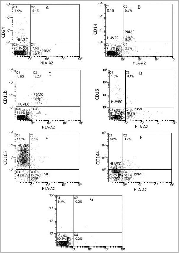
 Application Data
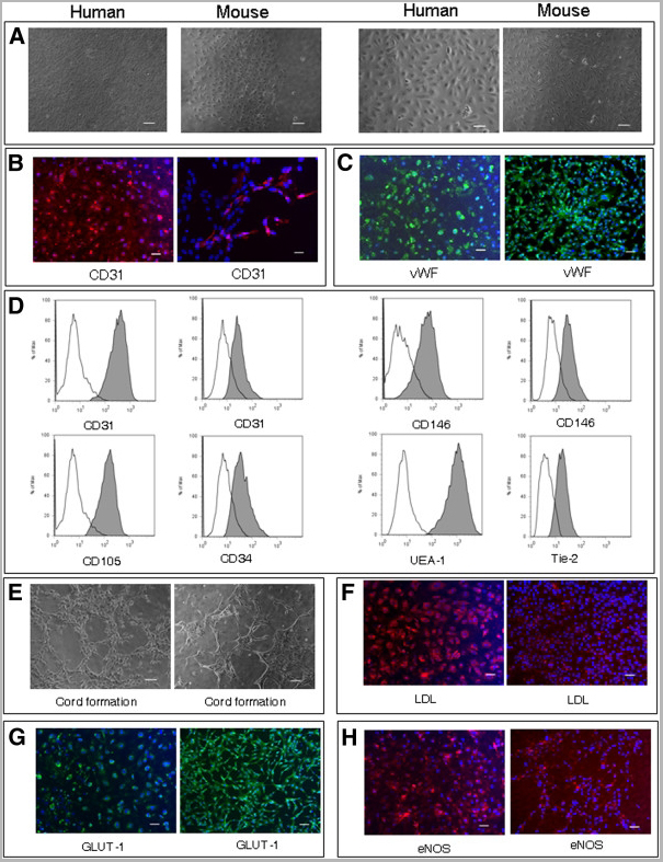
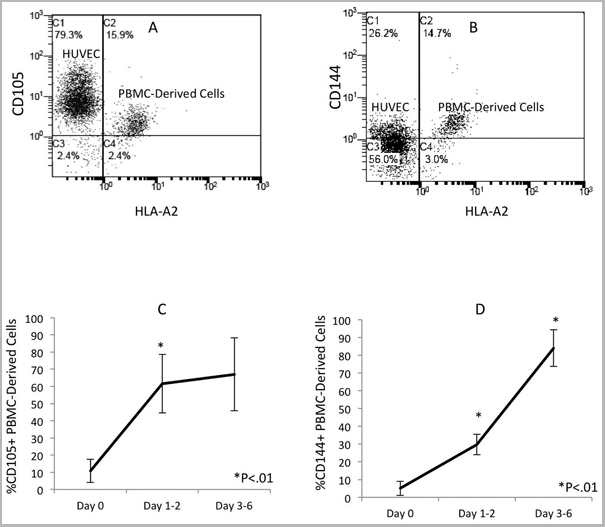
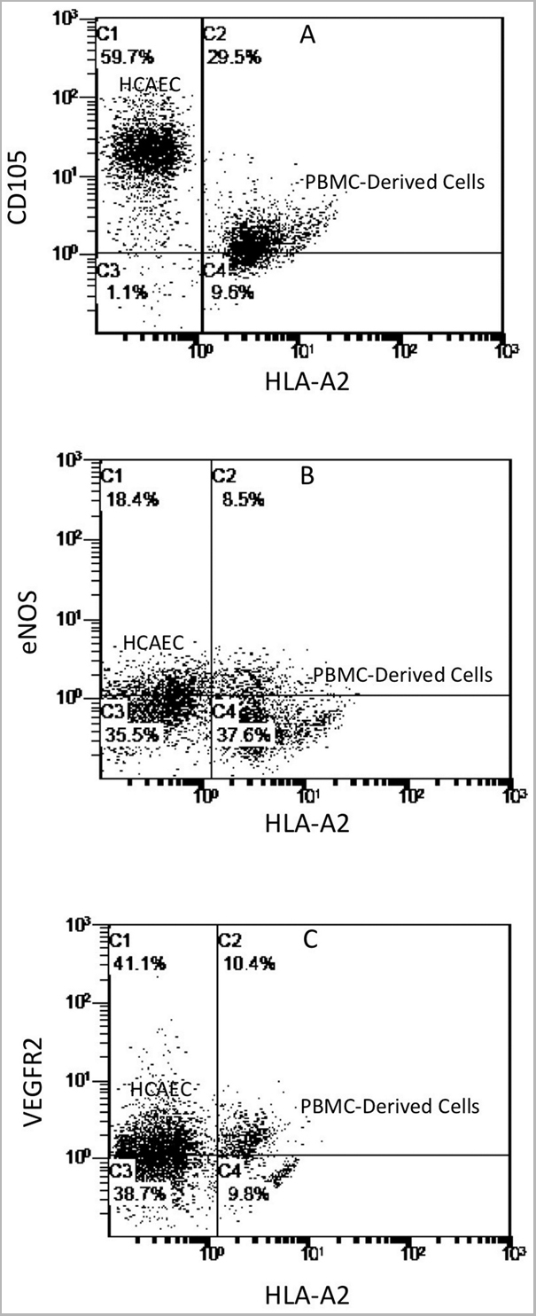
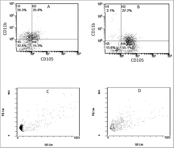
(Published customer image: Mouse anti Human CD105 antibody, clone SN6 used for flow cytometry.Image caption:Phenotype change from HLA-A2+/CD11b+/CD105- to HLA-A2+/CD11b-/CD105+ on endothelium-adherent blood monocyte-derived cells with increase in size and granularity during co-culture. HLA-A2+ PBMCs (1x10⁶ cells/well) were incubated for 2 h (Day 0) with HLA-A2- HUVECs, after which the non-adherent cells were removed by washing. The cell layers were analysed by three-colour flow cytometry staining for HLA-A2, CD11b and CD105 on (A) Day 1 and (B) Day 2. These plots were gated for HLA-A2+ cells. Forward scatter/side scatter dot plots gated for HLA-A2+ cells on Day 0 (C) and Day 2 (D) was shown. These are representative of 2 individual experiments.From: Tso C, Rye K-A, Barter P (2012) Phenotypic and Functional Changes in Blood Monocytes Following Adherence to Endothelium. PLoS ONE 7(5): e37091.)
Application Data
(Published customer image: Mouse anti Human CD105 antibody, clone SN6 used for flow cytometry.Image caption:Phenotype change from HLA-A2+/CD11b+/CD105- to HLA-A2+/CD11b-/CD105+ on endothelium-adherent blood monocyte-derived cells with increase in size and granularity during co-culture. HLA-A2+ PBMCs (1x10⁶ cells/well) were incubated for 2 h (Day 0) with HLA-A2- HUVECs, after which the non-adherent cells were removed by washing. The cell layers were analysed by three-colour flow cytometry staining for HLA-A2, CD11b and CD105 on (A) Day 1 and (B) Day 2. These plots were gated for HLA-A2+ cells. Forward scatter/side scatter dot plots gated for HLA-A2+ cells on Day 0 (C) and Day 2 (D) was shown. These are representative of 2 individual experiments.From: Tso C, Rye K-A, Barter P (2012) Phenotypic and Functional Changes in Blood Monocytes Following Adherence to Endothelium. PLoS ONE 7(5): e37091.)
CD105, Monoclonal Antibody (Cat# AAA12085)
Full Name
MOUSE ANTI HUMAN CD105
Gene Names
ENG; END; HHT1; ORW1
Applications
Immunohistochemistry, Flow Cytometry, Immunoprecipitation, Western Blot
Pricing


 Application Data
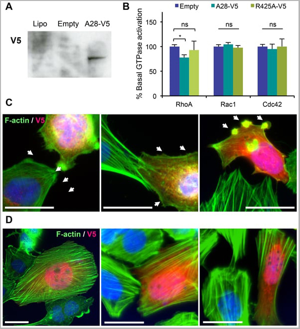
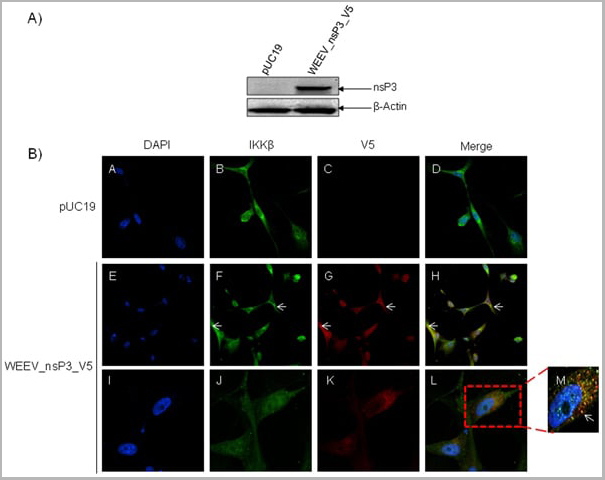
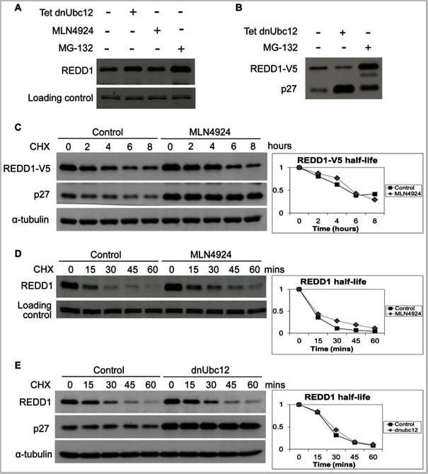
(Published customer image: Mouse anti V5 tag antibody, clone SV5-Pk1 used for the detection of V5 tagged WEEV_nsP3 protein by western blotting and immunofluorescenceImage caption: WEEV nsP3 interaction with host IKKbeta. A) U87MGs were transfected in a 6-well plate with 5 ug of pUC19 and WEEV_nsP3_HA for 24 hours. Cell lysates were resolved using SDS-PAGE and subsequently immunoblotted with V5 antibody and beta-actin served as a loading control. B) U87MGs were transfected with WEEV_nsP3_V5; cells were fixed after 24 hours and stained with antibodies against the endogenous IKKbeta and the V5 tag. Cells were incubated with appropriate secondary Alexa Fluor antibodies and the nuclei stained with DAPI. Co-localization of IKKbeta with WEEV_nsP3_V5 (yellow) was observed as shown by the arrows. B) Panels E -H serve as an example of transfected cells in a given field of view that show co-localization of IKKbeta and WEEV_nsP3_V5 24 hours post transfection. Panels I-L represent magnified images of other cells showing co-localization of IKKbeta and WEEV_nsP3_V5. Panel M is a magnified image of panel L. The co-localization was confirmed by Z-stack analysis. Co-localization was calculated to be approximately in 61% of cells (163 cells were counted of which 44% demonstrated expression of nsP3. Of those cells that expressed nsP3, 61% showed co-localization of both proteins). Images were taken using Nikon Eclipse TE2000-U at 60x magnification and are representative of 2 independent experiments.From: Amaya M, Voss K, Sampey G, Senina S, de la Fuente C, et al. (2014) The Role of IKKbeta in Venezuelan Equine Encephalitis Virus Infection. PLoS ONE 9(2): e86745.)
Application Data
(Published customer image: Mouse anti V5 tag antibody, clone SV5-Pk1 used for the detection of V5 tagged WEEV_nsP3 protein by western blotting and immunofluorescenceImage caption: WEEV nsP3 interaction with host IKKbeta. A) U87MGs were transfected in a 6-well plate with 5 ug of pUC19 and WEEV_nsP3_HA for 24 hours. Cell lysates were resolved using SDS-PAGE and subsequently immunoblotted with V5 antibody and beta-actin served as a loading control. B) U87MGs were transfected with WEEV_nsP3_V5; cells were fixed after 24 hours and stained with antibodies against the endogenous IKKbeta and the V5 tag. Cells were incubated with appropriate secondary Alexa Fluor antibodies and the nuclei stained with DAPI. Co-localization of IKKbeta with WEEV_nsP3_V5 (yellow) was observed as shown by the arrows. B) Panels E -H serve as an example of transfected cells in a given field of view that show co-localization of IKKbeta and WEEV_nsP3_V5 24 hours post transfection. Panels I-L represent magnified images of other cells showing co-localization of IKKbeta and WEEV_nsP3_V5. Panel M is a magnified image of panel L. The co-localization was confirmed by Z-stack analysis. Co-localization was calculated to be approximately in 61% of cells (163 cells were counted of which 44% demonstrated expression of nsP3. Of those cells that expressed nsP3, 61% showed co-localization of both proteins). Images were taken using Nikon Eclipse TE2000-U at 60x magnification and are representative of 2 independent experiments.From: Amaya M, Voss K, Sampey G, Senina S, de la Fuente C, et al. (2014) The Role of IKKbeta in Venezuelan Equine Encephalitis Virus Infection. PLoS ONE 9(2): e86745.)
V5-TAG, Monoclonal Antibody (Cat# AAA11930)
Full Name
MOUSE ANTI V5-TAG
Applications
Immunohistochemistry, Flow Cytometry, Immunofluorescence, Immunoprecipitation, Western Blot, Radioimmunoassay
Pricing


 Application Data
(Published customer image: Mouse anti V5 tag antibody, clone SV5-Pk1 used for the detection of V5 tagged WEEV_nsP3 protein by western blotting and immunofluorescenceImage caption: WEEV nsP3 interaction with host IKKbeta. A) U87MGs were transfected in a 6-well plate with 5 ug of pUC19 and WEEV_nsP3_HA for 24 hours. Cell lysates were resolved using SDS-PAGE and subsequently immunoblotted with V5 antibody and beta-actin served as a loading control. B) U87MGs were transfected with WEEV_nsP3_V5; cells were fixed after 24 hours and stained with antibodies against the endogenous IKKbeta and the V5 tag. Cells were incubated with appropriate secondary Alexa Fluor antibodies and the nuclei stained with DAPI. Co-localization of IKKbeta with WEEV_nsP3_V5 (yellow) was observed as shown by the arrows. B) Panels E -H serve as an example of transfected cells in a given field of view that show co-localization of IKKbeta and WEEV_nsP3_V5 24 hours post transfection. Panels I-L represent magnified images of other cells showing co-localization of IKKbeta and WEEV_nsP3_V5. Panel M is a magnified image of panel L. The co-localization was confirmed by Z-stack analysis. Co-localization was calculated to be approximately in 61% of cells (163 cells were counted of which 44% demonstrated expression of nsP3. Of those cells that expressed nsP3, 61% showed co-localization of both proteins). Images were taken using Nikon Eclipse TE2000-U at 60x magnification and are representative of 2 independent experiments.From: Amaya M, Voss K, Sampey G, Senina S, de la Fuente C, et al. (2014) The Role of IKKbeta in Venezuelan Equine Encephalitis Virus Infection. PLoS ONE 9(2): e86745.)
Application Data
(Published customer image: Mouse anti V5 tag antibody, clone SV5-Pk1 used for the detection of V5 tagged WEEV_nsP3 protein by western blotting and immunofluorescenceImage caption: WEEV nsP3 interaction with host IKKbeta. A) U87MGs were transfected in a 6-well plate with 5 ug of pUC19 and WEEV_nsP3_HA for 24 hours. Cell lysates were resolved using SDS-PAGE and subsequently immunoblotted with V5 antibody and beta-actin served as a loading control. B) U87MGs were transfected with WEEV_nsP3_V5; cells were fixed after 24 hours and stained with antibodies against the endogenous IKKbeta and the V5 tag. Cells were incubated with appropriate secondary Alexa Fluor antibodies and the nuclei stained with DAPI. Co-localization of IKKbeta with WEEV_nsP3_V5 (yellow) was observed as shown by the arrows. B) Panels E -H serve as an example of transfected cells in a given field of view that show co-localization of IKKbeta and WEEV_nsP3_V5 24 hours post transfection. Panels I-L represent magnified images of other cells showing co-localization of IKKbeta and WEEV_nsP3_V5. Panel M is a magnified image of panel L. The co-localization was confirmed by Z-stack analysis. Co-localization was calculated to be approximately in 61% of cells (163 cells were counted of which 44% demonstrated expression of nsP3. Of those cells that expressed nsP3, 61% showed co-localization of both proteins). Images were taken using Nikon Eclipse TE2000-U at 60x magnification and are representative of 2 independent experiments.From: Amaya M, Voss K, Sampey G, Senina S, de la Fuente C, et al. (2014) The Role of IKKbeta in Venezuelan Equine Encephalitis Virus Infection. PLoS ONE 9(2): e86745.)
V5-TAG, Monoclonal Antibody (Cat# AAA11864)
Full Name
MOUSE ANTI V5-TAG:FITC
Applications
Immunofluorescence
Pricing
TCR gamma delta, Monoclonal Antibody (Cat# AAA17922)
Full Name
TCR gamma delta antibody
Reactivity
Recognizes the gamma/delta T-cell receptor (TCR).
Applications
Flow Cytometry, Immunohistochemistry, Immunoprecipitation
Pricing
6xHIS, Monoclonal Antibody (Cat# AAA14456)
Full Name
Purified Mouse anti-6xHIS (CLONE 7B8)-Monoclonal
Applications
Immunofluorescence, Immunoprecipitation, Western Blot
Purity
Purified Mouse anti- 6xHIS Tag .
Folic acid, Monoclonal Antibody (Cat# AAA26945)
Full Name
Mouse anti-folic acid monoclonal Antibody(FA)
Applications
ELISA
Purity
90% ± 5% by SDS-PAGE
Pricing
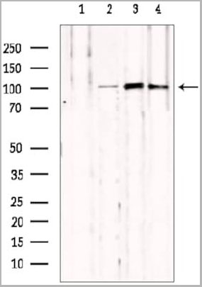
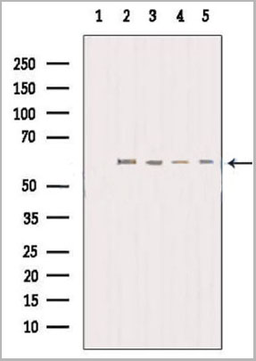
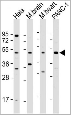
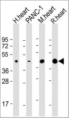
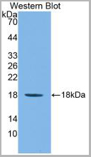
 WB (Western Blot)
(Western blot analysis of CD298 expression in HEK293T (A), Hela (B), A2780 (C), mouse heart (D), mouse liver (E), rat heart (F), rat liver (G) whole cell lysates.(Predicted band size: 31 kD; Observed band size: 40 kD))
WB (Western Blot)
(Western blot analysis of CD298 expression in HEK293T (A), Hela (B), A2780 (C), mouse heart (D), mouse liver (E), rat heart (F), rat liver (G) whole cell lysates.(Predicted band size: 31 kD; Observed band size: 40 kD))
CD298, Polyclonal Antibody (Cat# AAA27791)
Full Name
Anti-CD298 Antibody
Gene Names
ATP1B3; CD298; ATPB-3
Reactivity
Human, Mouse, Rat, Monkey
Applications
Western Blot
Purity
The antibody was purified by immunogen affinity chromatography.
Pricing
T-Cell Receptor gamma delta, Monoclonal Antibody (Cat# AAA14565)
Full Name
Mouse anti T-Cell Receptor gamma delta
Reactivity
Human
Applications
Flow Cytometry, Immunohistochemistry, Immunoprecipitation
Pricing
 WB (Western Blot)
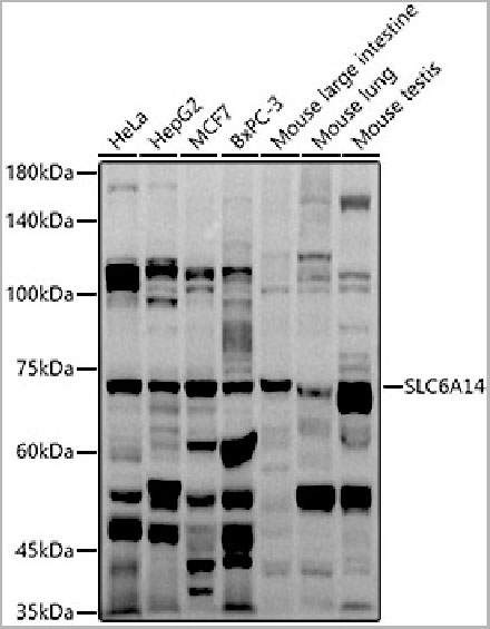
(Western blot analysis of extracts of various cell lines, using SLC6A14 antibody (AAA28230) at 1:1000 dilution.Secondary antibody: HRP Goat Anti-Rabbit IgG (H+L) at 1:10000 dilution. Lysates/proteins: 25ug per lane. Blocking buffer: 3% nonfat dry milk in TBST. Detection: ECL Basic Kit. Exposure time: 90s)
WB (Western Blot)
(Western blot analysis of extracts of various cell lines, using SLC6A14 antibody (AAA28230) at 1:1000 dilution.Secondary antibody: HRP Goat Anti-Rabbit IgG (H+L) at 1:10000 dilution. Lysates/proteins: 25ug per lane. Blocking buffer: 3% nonfat dry milk in TBST. Detection: ECL Basic Kit. Exposure time: 90s)
SLC6A14, Polyclonal Antibody (Cat# AAA28230)
Full Name
SLC6A14 Polyclonal Antibody
Gene Names
SLC6A14; BMIQ11
Reactivity
Human, Mouse
Applications
Western Blot
Purity
Affinity Purification
Pricing
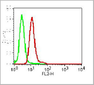
 FCM (Flow Cytometry)
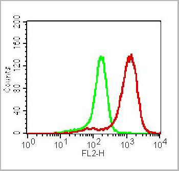
(Figure-1: Cell surface flowcytometry analysis of CLEC1on human PBMC (Monocytes) using 0.5 ug/10^6 Cells of Anti-CLEC1 antibody. Green represent isotype control and red represent Anti human CLEC1 antibody. Goat anti mouse PE conjugated was used as the secondary antibody.)
FCM (Flow Cytometry)
(Figure-1: Cell surface flowcytometry analysis of CLEC1on human PBMC (Monocytes) using 0.5 ug/10^6 Cells of Anti-CLEC1 antibody. Green represent isotype control and red represent Anti human CLEC1 antibody. Goat anti mouse PE conjugated was used as the secondary antibody.)
CLEC1, Monoclonal Antibody (Cat# AAA14882)
Full Name
Monoclonal Antibody to CLEC1 (Clone: ABM2H82)
Gene Names
CLEC1A; CLEC1; CLEC-1
Reactivity
Human
Applications
Flow Cytometry
Purity
Protein G Chromatography Purified
Pricing
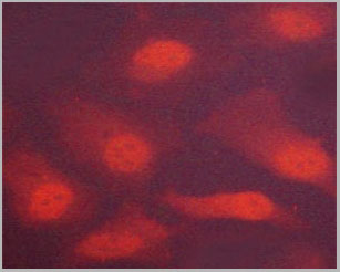
 IF (Immunofluorescence)
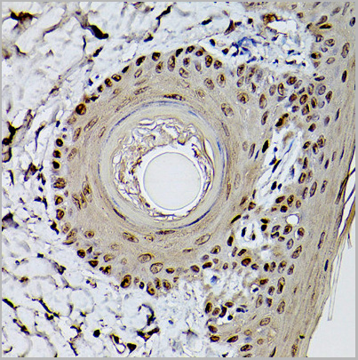
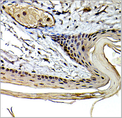
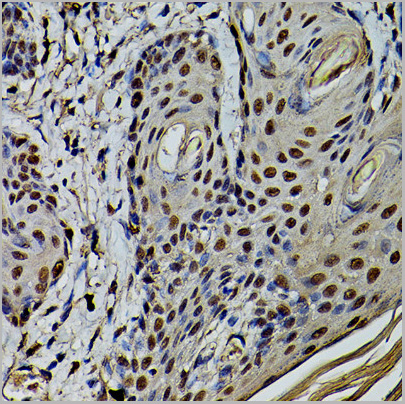
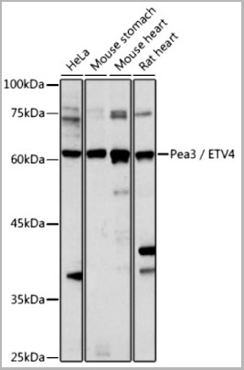
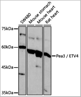
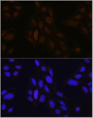
(Immunofluorescence analysis of U-2 OS cells using Pea3 / ETV4 Rabbit pAb (AAA28094) at dilution of 1:100 (40x lens). Blue: DAPI for nuclear staining)
IF (Immunofluorescence)
(Immunofluorescence analysis of U-2 OS cells using Pea3 / ETV4 Rabbit pAb (AAA28094) at dilution of 1:100 (40x lens). Blue: DAPI for nuclear staining)
ETV4, Polyclonal Antibody (Cat# AAA28094)
Full Name
ETV4 Polyclonal Antibody
Gene Names
ETV4; E1AF; PEA3; E1A-F; PEAS3
Applications
Western Blot, Immunohistochemistry, Immunofluorescence
Purity
Affinity Purification
Pricing
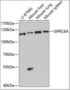 WB (Western Blot)
(Western blot analysis of extracts of various cell lines, using GPRC6A antibody (AAA28200) at 1:1000 dilution.Secondary antibody: HRP Goat Anti-Rabbit IgG (H+L) at 1:10000 dilution.Lysates/proteins: 25ug per lane.Blocking buffer: 3% nonfat dry milk in TBST.Detection: ECL Basic Kit.Exposure time: 90s.)
WB (Western Blot)
(Western blot analysis of extracts of various cell lines, using GPRC6A antibody (AAA28200) at 1:1000 dilution.Secondary antibody: HRP Goat Anti-Rabbit IgG (H+L) at 1:10000 dilution.Lysates/proteins: 25ug per lane.Blocking buffer: 3% nonfat dry milk in TBST.Detection: ECL Basic Kit.Exposure time: 90s.)
GPRC6A, Polyclonal Antibody (Cat# AAA28200)
Full Name
GPRC6A Polyclonal Antibody
Gene Names
GPRC6A; GPCR; bA86F4.3
Reactivity
Human, Mouse
Applications
Western Blot
Purity
Affinity Purification
Pricing
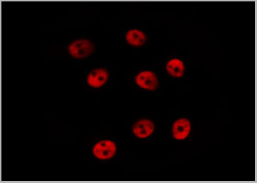
 IF (Immunofluorescence)
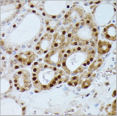
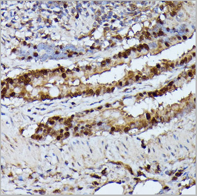
(Immunofluorescence analysis of U2OS cells using GADD45A antibody (AAA28246) at dilution of 1:100. Blue: DAPI for nuclear staining.)
IF (Immunofluorescence)
(Immunofluorescence analysis of U2OS cells using GADD45A antibody (AAA28246) at dilution of 1:100. Blue: DAPI for nuclear staining.)
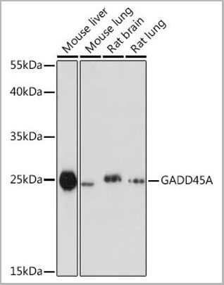
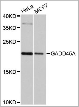
GADD45A, Polyclonal Antibody (Cat# AAA28246)
Full Name
GADD45A Polyclonal Antibody
Gene Names
GADD45A; DDIT1; GADD45
Reactivity
Human, Mouse, Rat
Applications
Western Blot, Immunohistochemistry, Immunofluorescence
Purity
Affinity purification
Pricing
 Application Data
HLA Class 1 Antigen A33,B8, Monoclonal Antibody (Cat# AAA14684)
Full Name
Mouse anti-Human HLA Class 1 Antigen A33,B8
Reactivity
Human
Applications
Cell Typing
Purity
Ascites
Pricing
 FCM (Flow Cytometry)
(Intracellular flow cytometric analysis of RIG-I in K562 Cell line using 0.5 ug/10^6 cells of Anti-RIGI antibody (ABM40B5). Green represent isotype control and red represent Anti-RIG I antibody (AAA14865). Goat anti-mouse PE conjugate was used as secondary.)
FCM (Flow Cytometry)
(Intracellular flow cytometric analysis of RIG-I in K562 Cell line using 0.5 ug/10^6 cells of Anti-RIGI antibody (ABM40B5). Green represent isotype control and red represent Anti-RIG I antibody (AAA14865). Goat anti-mouse PE conjugate was used as secondary.)
RIG-I, Monoclonal Antibody (Cat# AAA14865)
Full Name
RIG-I Monoclonal Antibody
Gene Names
DDX58; RIGI; RIG-I; RLR-1; SGMRT2
Reactivity
Human
Applications
Immunohistochemistry, Flow Cytometry, Western Blot
Purity
Protein G Chromatography
Pricing

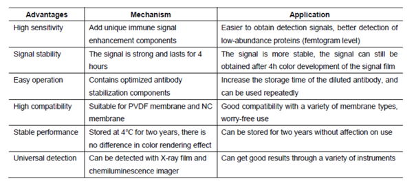 Assay Principle
(After years of development on chemiluminescence technology, there have been a variety of luminescence systems, but the most commonly used one in laboratories is still a technology system based on Luminol or its derivatives (isoluminol, etc.). Western luminescent detection reagent ECL immunoblotting chemiluminescence solution is a non-radioactive (horseradish peroxidase) luminescence system designed to detect femtogram-level trace proteins immobilized on a solid membrane (such as NC, PVDF, etc.). It is an experimental auxiliary reagent for the photosensitive recording of its immunoblotting by X-ray film (radiograph).)
Assay Principle
(After years of development on chemiluminescence technology, there have been a variety of luminescence systems, but the most commonly used one in laboratories is still a technology system based on Luminol or its derivatives (isoluminol, etc.). Western luminescent detection reagent ECL immunoblotting chemiluminescence solution is a non-radioactive (horseradish peroxidase) luminescence system designed to detect femtogram-level trace proteins immobilized on a solid membrane (such as NC, PVDF, etc.). It is an experimental auxiliary reagent for the photosensitive recording of its immunoblotting by X-ray film (radiograph).)
West Femto Maximum Sensitivity Substrate, Reagent (Cat# AAA31545)
Full Name
West Femto Maximum Sensitivity Substrate
Applications
Western Blot, Immunoprecipitation
Pricing
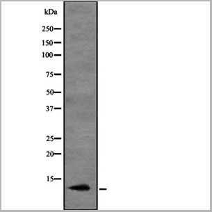
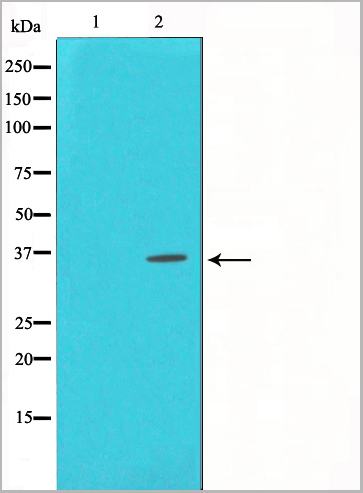
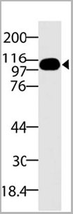
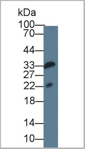
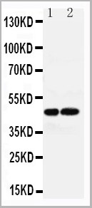 WB (Western Blot)
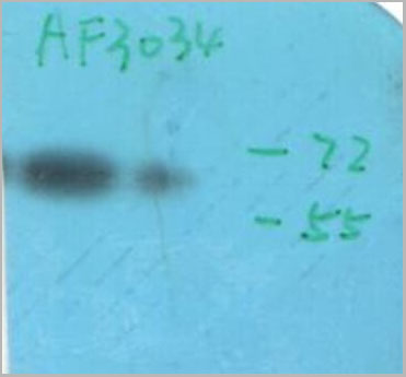
(Anti-AVPR1A antibody, AAA11596, All Western blottingAll lanes: Anti-AVPR1A(AAA11596) at 0.5ug/mlLane 1: Rat Liver Tissue Lysate at 40ugLane 2: Rat Kidney Tissue Lysate at 40ugPredicted bind size: 47KDObserved bind size: 47KD)
WB (Western Blot)
(Anti-AVPR1A antibody, AAA11596, All Western blottingAll lanes: Anti-AVPR1A(AAA11596) at 0.5ug/mlLane 1: Rat Liver Tissue Lysate at 40ugLane 2: Rat Kidney Tissue Lysate at 40ugPredicted bind size: 47KDObserved bind size: 47KD)
AVPR1A, Polyclonal Antibody (Cat# AAA11596)
Full Name
Anti-AVPR1A antibody
Gene Names
Avpr1a; V1a; AVPR; V1aR
Reactivity
Reacts with: Human, Rat
Predicted to work with: Mouse
Predicted to work with: Mouse
Applications
Western Blot
Purity
Immunogen affinity purified.
Pricing
CEA, Monoclonal Antibody (Cat# AAA14296)
Full Name
CEA antibody (in PBS)
Gene Names
CEACAM6; NCA; CEAL; CD66c
Applications
ELISA
Pricing
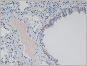
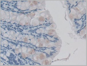
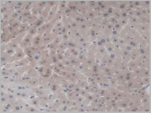
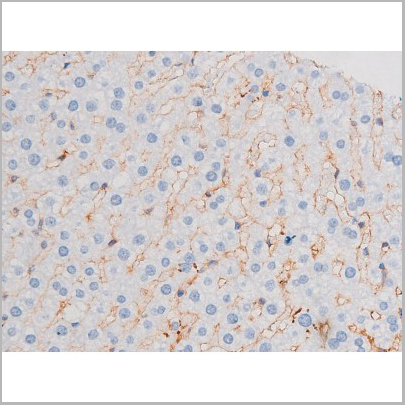
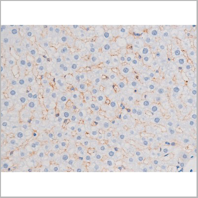
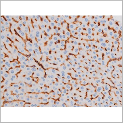
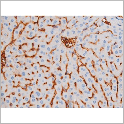
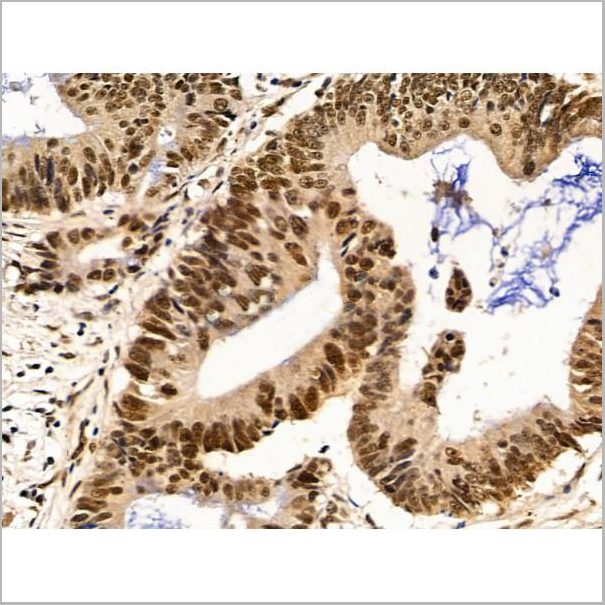
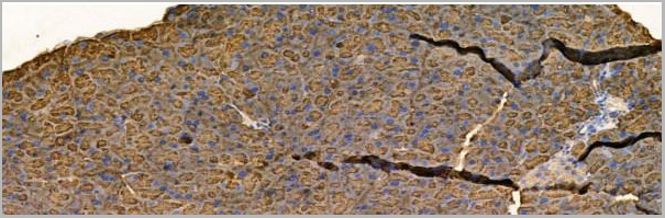
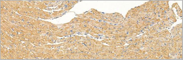
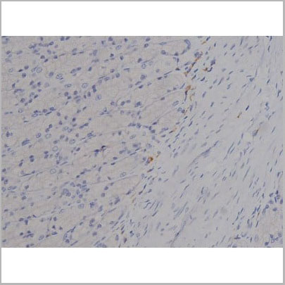
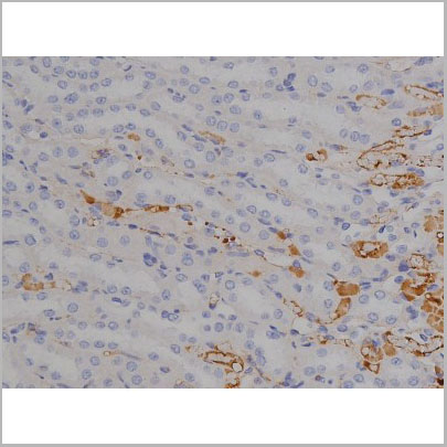
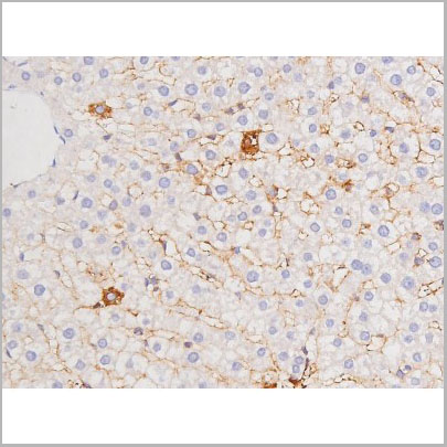
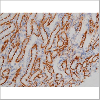
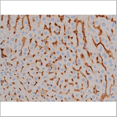
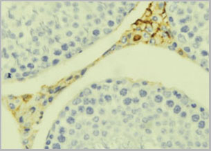
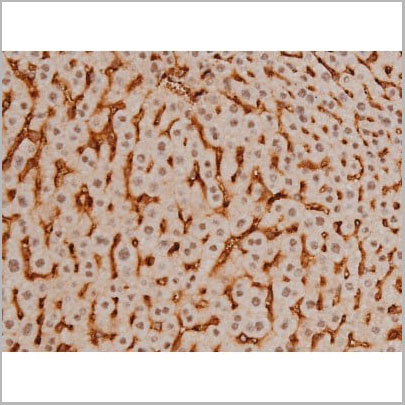
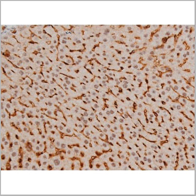
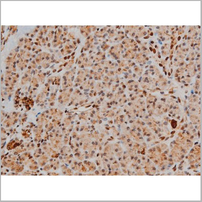
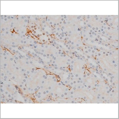
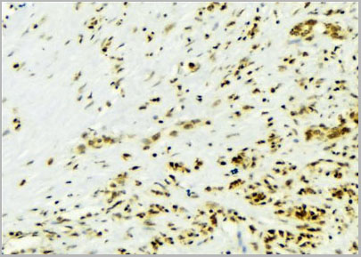
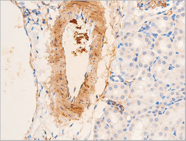
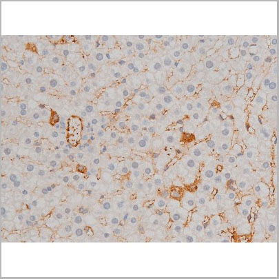
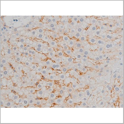
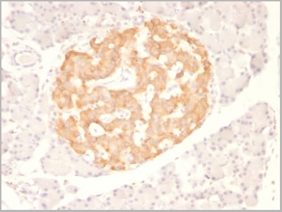
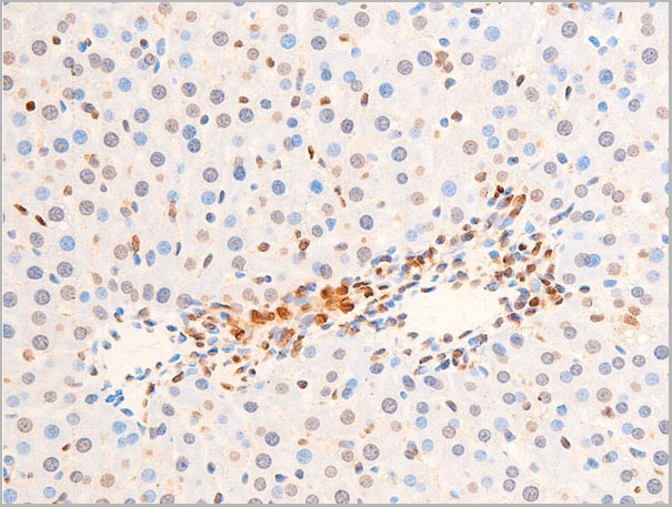
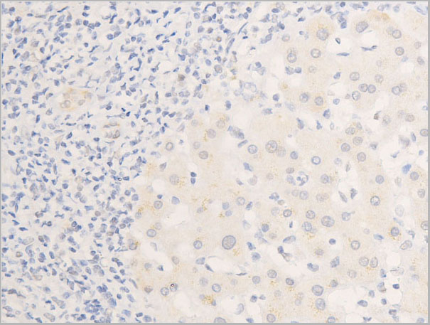
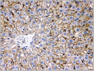
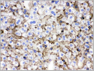
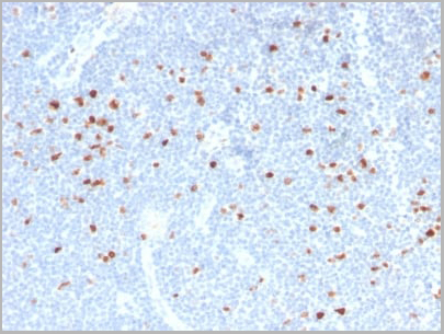
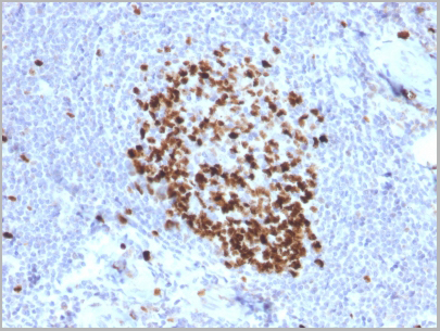
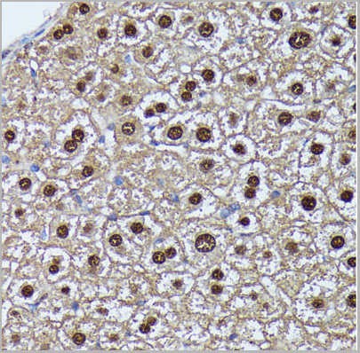
 IHC (Immunohistchemistry)
(Figure. DAB staining on IHC-P; Samples: Mouse Liver Tissue)
IHC (Immunohistchemistry)
(Figure. DAB staining on IHC-P; Samples: Mouse Liver Tissue)
Interleukin 1 Family, Monoclonal Antibody (Cat# AAA21005)
Full Name
Interleukin 1 Family, Member 9 (IL1F9) Monoclonal Antibody
Gene Names
Il1f9; Il36g
Reactivity
Mouse, Rat
Applications
Western Blot, Immunohistochemistry, Immunocytochemistry, Immunoprecipitation
Purity
Protein A + Protein G affinity chromatography
Pricing
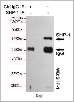 IP (Immunoprecipitation)
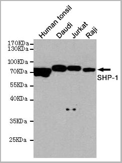
(Immunoprecipitation analysis of Raji cell lysates using SHP-1 mouse mAb.)
IP (Immunoprecipitation)
(Immunoprecipitation analysis of Raji cell lysates using SHP-1 mouse mAb.)
SHP-1, Monoclonal Antibody (Cat# AAA14102)
Full Name
Anti-SHP-1 Mouse mAb
Gene Names
PTPN6; HCP; HCPH; SHP1; SHP-1; HPTP1C; PTP-1C; SHP-1L; SH-PTP1
Reactivity
Human
Applications
Western Blot, Immunoprecipitation
Purity
Affinity purified
Pricing
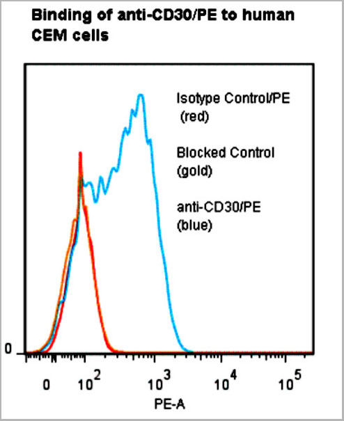 Application Data
CD30, Monoclonal Antibody (Cat# AAA14859)
Full Name
anti-human CD30-R-PE
Reactivity
Human
Applications
Flow Cytometry
Pricing