Filters
Clonality
Type
Reactivity
Gene Name
Isotype
Host
Application
Clone
321 results for " Other Proteins" - showing 250-300
 WB (Western Blot)
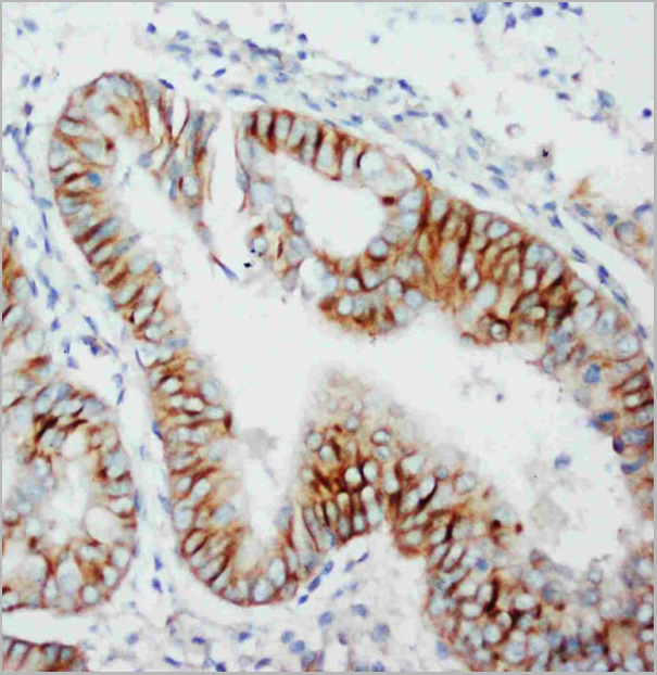
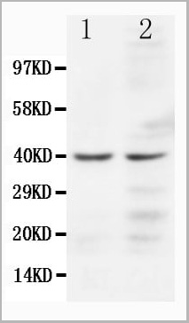
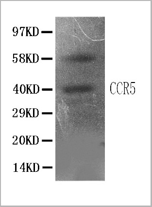
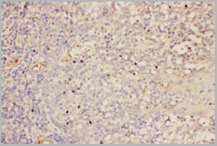
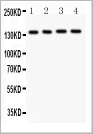
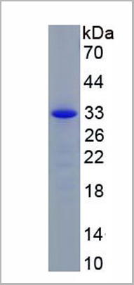
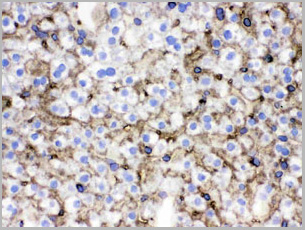
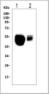
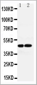
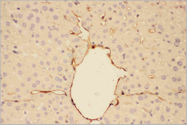
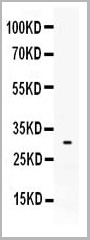
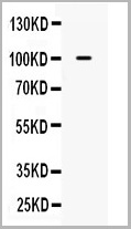
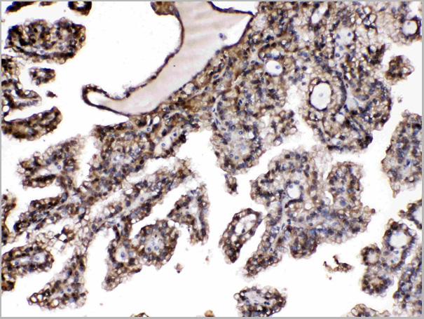
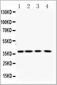
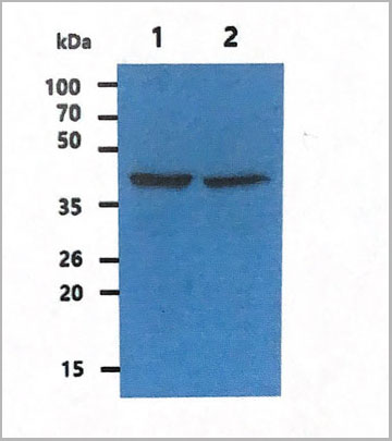
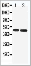
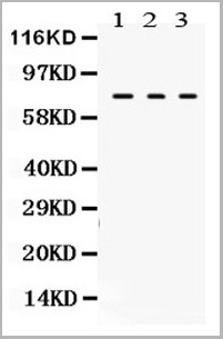
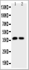
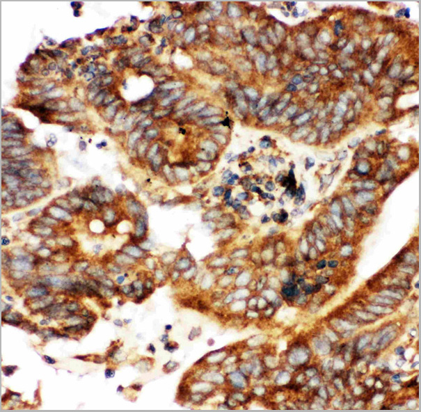
(Anti-CCR5 antibody, AAA11522, IHC(P)All lanes: Anti CCR5 (AAA11522) at 0.5ug/mlWB: Mouse Liver Tissue Lysate at 50ugPredicted bind size: 40KDObserved bind size: 40KD)
WB (Western Blot)
(Anti-CCR5 antibody, AAA11522, IHC(P)All lanes: Anti CCR5 (AAA11522) at 0.5ug/mlWB: Mouse Liver Tissue Lysate at 50ugPredicted bind size: 40KDObserved bind size: 40KD)
CCR5, Polyclonal Antibody (Cat# AAA11522)
Full Name
Anti-CCR5 antibody
Gene Names
CCR5; CKR5; CCR-5; CD195; CKR-5; CCCKR5; CMKBR5; IDDM22; CC-CKR-5
Reactivity
Reacts with: Human, Mouse
Predicted to work with: Rat
Predicted to work with: Rat
Applications
Western Blot, Immunohistochemistry
Purity
Immunogen affinity purified.
Pricing
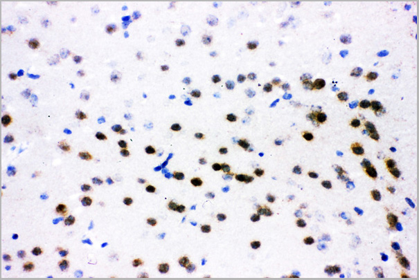
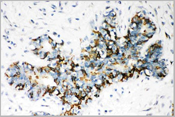
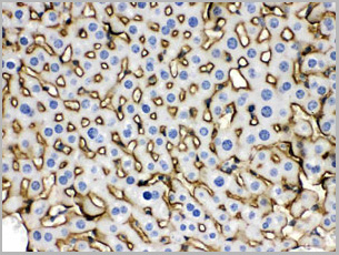
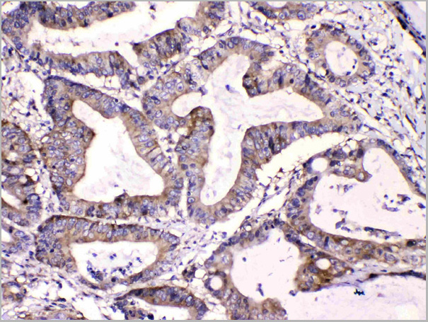
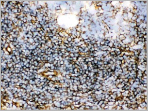
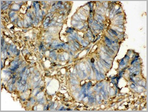
 IHC (Immunohistochemistry)
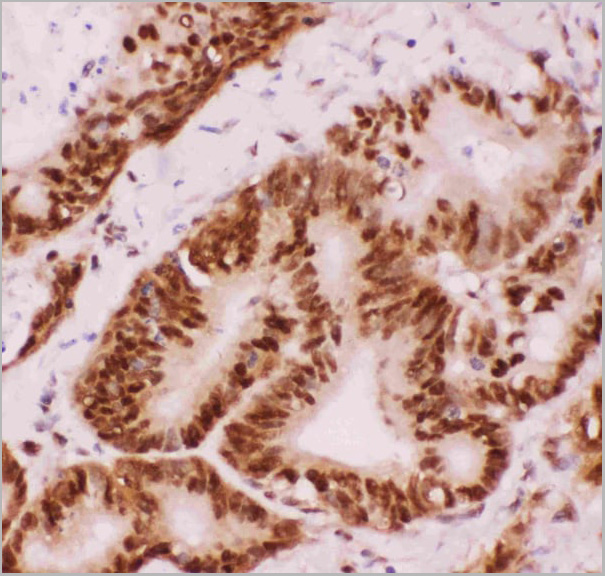
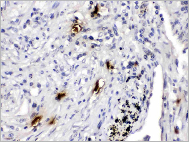
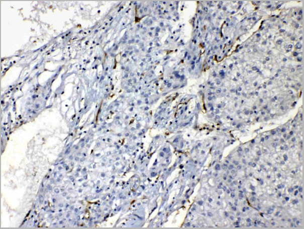
(Anti-NR6A1 / GCNF antibody IHC of human testis. Immunohistochemistry of formalin-fixed, paraffin-embedded tissue after heat-induced antigen retrieval.)
IHC (Immunohistochemistry)
(Anti-NR6A1 / GCNF antibody IHC of human testis. Immunohistochemistry of formalin-fixed, paraffin-embedded tissue after heat-induced antigen retrieval.)
NR6A1 / GCNF, Polyclonal Antibody (Cat# AAA12361)
Full Name
Rabbit Polyclonal to Human NR6A1 / GCNF
Gene Names
NR6A1; RTR; GCNF; NR61; hRTR; CT150; GCNF1; hGCNF
Reactivity
Human, Monkey, Mouse, Rat, Bat, Bovine, Dog, Hamster, Horse, Pig, Rabbit
Applications
Immunohistochemistry
Purity
Immunoaffinity Purified
Pricing
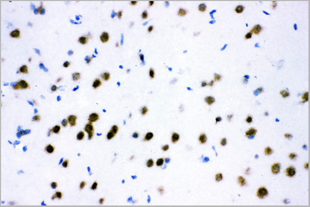
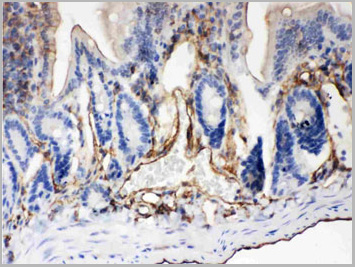
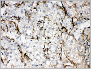
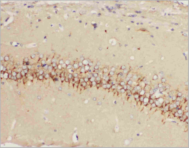
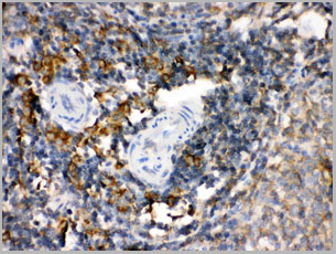
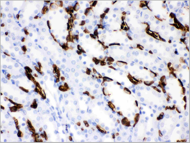
 IHC (Immunohistochemistry)
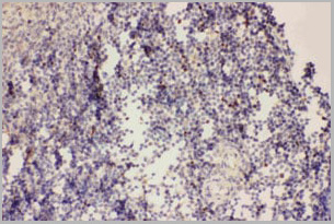
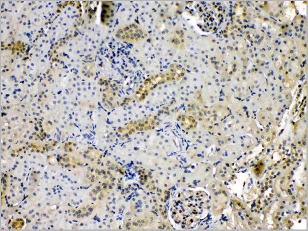
(IHC(P):Rat Spleen Tissue)
IHC (Immunohistochemistry)
(IHC(P):Rat Spleen Tissue)
FOXP3, Polyclonal Antibody (Cat# AAA11651)
Full Name
Anti-FOXP3 Antibody
Gene Names
FOXP3; JM2; AIID; IPEX; PIDX; XPID; DIETER
Reactivity
Human, Mouse, Rat
Applications
Immunohistochemistry
Purity
Immunogen Affinity Purified
Pricing
CIAS1/NALP3, Polyclonal Antibody (Cat# AAA11523)
Full Name
Anti-CIAS1/NALP3 antibody
Gene Names
NLRP3; AII; AVP; FCU; MWS; FCAS; CIAS1; NALP3; C1orf7; CLR1.1; PYPAF1; AGTAVPRL
Reactivity
Human, Rat; Predicted: Mouse
Applications
Western Blot, Immunohistochemistry, Immunocytochemistry, Flow Cytometry
Purity
Immunogen affinity purified
Pricing
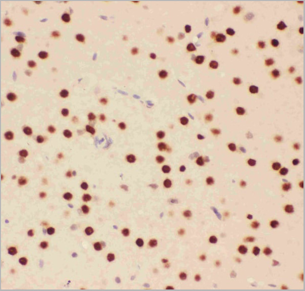
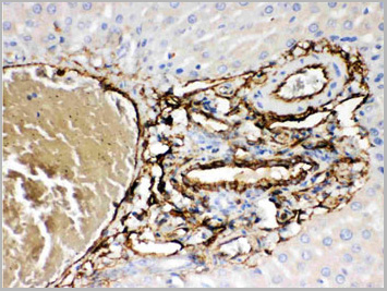
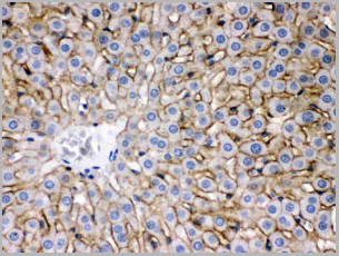
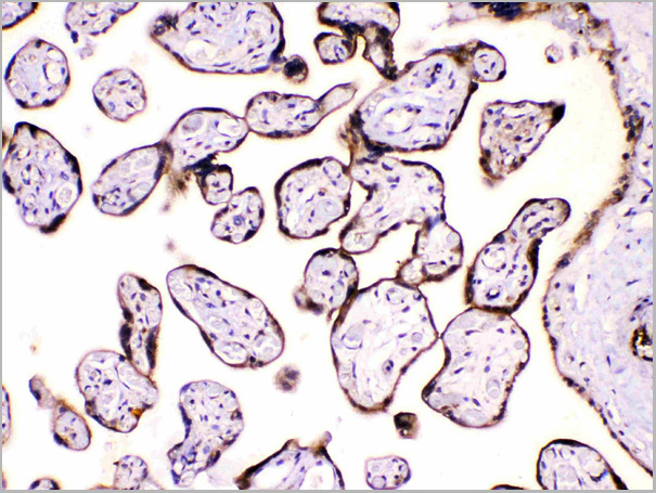
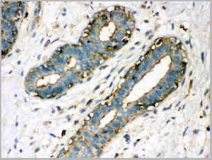
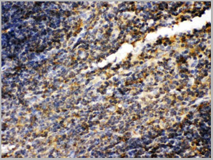
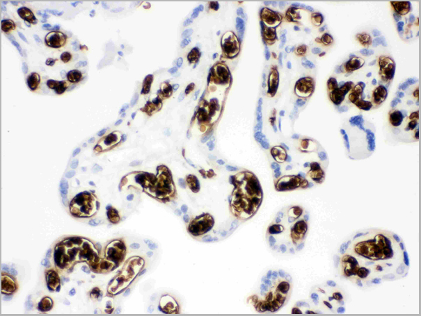
 IHC (Immunohistochemistry)
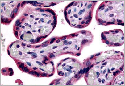
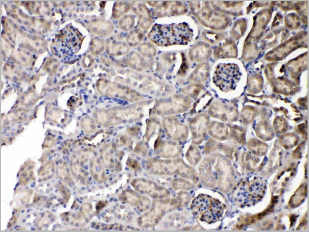
(Anti-SLC7A5 antibody IHC of human placenta, terminal villi. Immunohistochemistry of formalin-fixed, paraffin-embedded tissue after heat-induced antigen retrieval.)
IHC (Immunohistochemistry)
(Anti-SLC7A5 antibody IHC of human placenta, terminal villi. Immunohistochemistry of formalin-fixed, paraffin-embedded tissue after heat-induced antigen retrieval.)
SLC7A5 / CD98, Polyclonal Antibody (Cat# AAA12328)
Full Name
SLC7A5 / CD98 Light Chain
Gene Names
SLC7A5; E16; CD98; LAT1; 4F2LC; MPE16; hLAT1; D16S469E
Reactivity
Human, Monkey, Dog.
Predicted: Bat, Rabbit
Predicted: Bat, Rabbit
Applications
Immunohistochemistry
Purity
Immunoaffinity Purified
Pricing
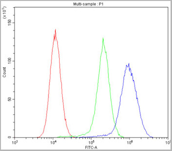
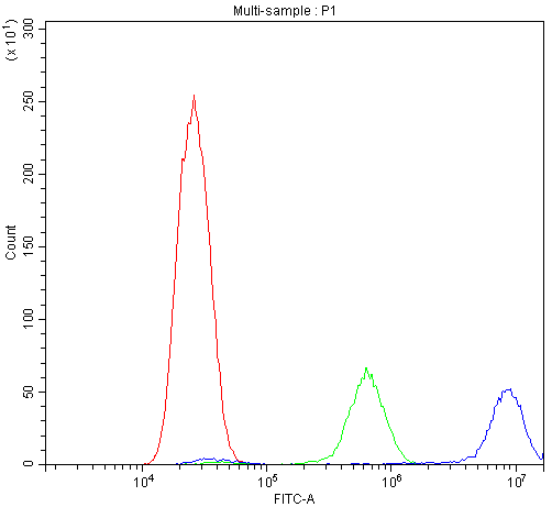
 FCM (Flow Cytometry)
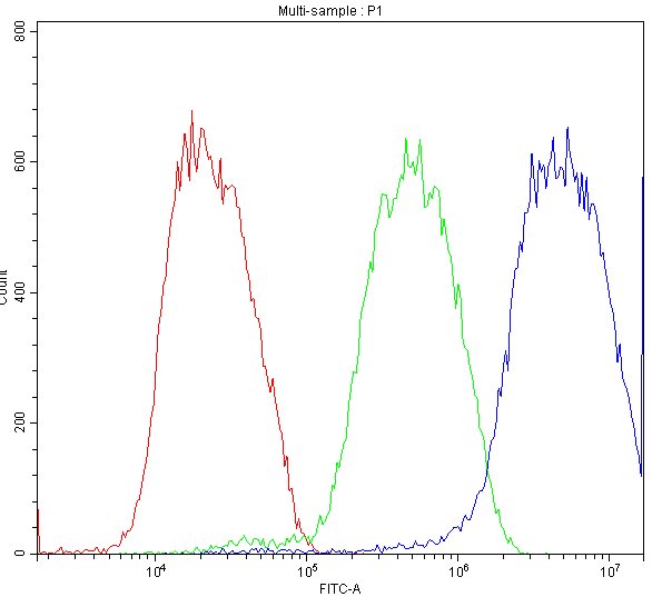
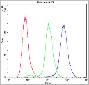
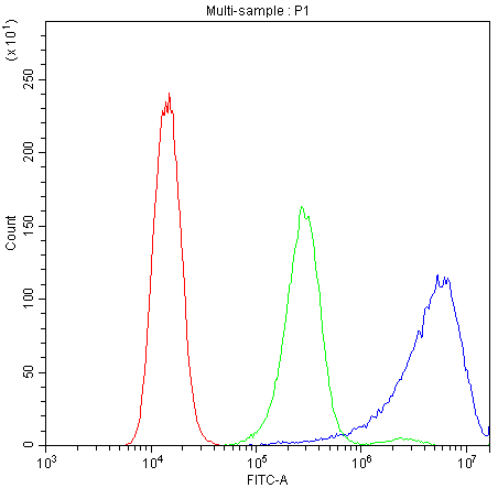
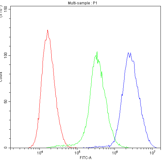
(Figure 8. Flow Cytometry analysis of HepG2 cells using anti-ATF2 antibody (AAA11634).Overlay histogram showing HepG2 cells stained with AAA11634 (Blue line).The cells were blocked with 10% normal goat serum. And then incubated with rabbit anti-ATF2 Antibody (AAA11634,1ug/1x10^6 cells) for 30 min at 20 degree C. DyLight ®488 conjugated goat anti-rabbit IgG (5-10ug/1x10^6 cells) was used as secondary antibody for 30 minutes at 20 degree C. Isotype control antibody (Green line) was rabbit IgG (1ug/1x106) used under the same conditions. Unlabelled sample (Red line) was also used as a control.)
FCM (Flow Cytometry)
(Figure 8. Flow Cytometry analysis of HepG2 cells using anti-ATF2 antibody (AAA11634).Overlay histogram showing HepG2 cells stained with AAA11634 (Blue line).The cells were blocked with 10% normal goat serum. And then incubated with rabbit anti-ATF2 Antibody (AAA11634,1ug/1x10^6 cells) for 30 min at 20 degree C. DyLight ®488 conjugated goat anti-rabbit IgG (5-10ug/1x10^6 cells) was used as secondary antibody for 30 minutes at 20 degree C. Isotype control antibody (Green line) was rabbit IgG (1ug/1x106) used under the same conditions. Unlabelled sample (Red line) was also used as a control.)
Cyclic AMP-dependent transcription factor ATF-2, Polyclonal Antibody (Cat# AAA11634)
Full Name
Anti-ATF2 Antibody
Gene Names
ATF2; HB16; CREB2; TREB7; CREB-2; CRE-BP1
Reactivity
Human, Mouse, Rat. No cross reactivity with other proteins.
Applications
Western Blot, Immunohistochemistry, Immunohistochemistry
Purity
Immunogen affinity purified.
Pricing
 FCM (Flow Cytometry)
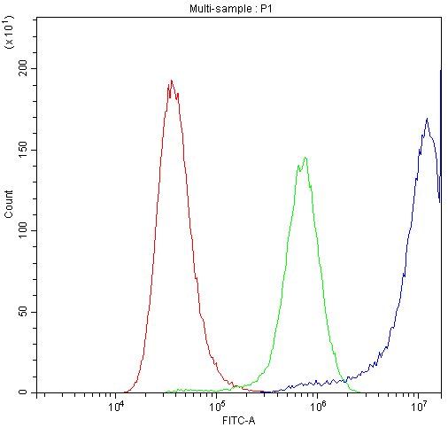
(Figure 6. Flow Cytometry analysis of A549 cells using anti-MDR3 antibody (AAA11652).Overlay histogram showing A549 cells stained with AAA11652 (Blue line).The cells were blocked with 10% normal goat serum. And then incubated with rabbit anti-MDR3 Antibody (AAA11652,1ug/1x106 cells) for 30 min at 20°C. DyLight®488 conjugated goat anti-rabbit IgG (5-10ug/1x106 cells) was used as secondary antibody for 30 minutes at 20°C. Isotype control antibody (Green line) was rabbit IgG (1ug/1x106) used under the same conditions. Unlabelled sample (Red line) was also used as a control.)
FCM (Flow Cytometry)
(Figure 6. Flow Cytometry analysis of A549 cells using anti-MDR3 antibody (AAA11652).Overlay histogram showing A549 cells stained with AAA11652 (Blue line).The cells were blocked with 10% normal goat serum. And then incubated with rabbit anti-MDR3 Antibody (AAA11652,1ug/1x106 cells) for 30 min at 20°C. DyLight®488 conjugated goat anti-rabbit IgG (5-10ug/1x106 cells) was used as secondary antibody for 30 minutes at 20°C. Isotype control antibody (Green line) was rabbit IgG (1ug/1x106) used under the same conditions. Unlabelled sample (Red line) was also used as a control.)
ABCB4, Polyclonal Antibody (Cat# AAA11652)
Full Name
Anti-ABCB4 Antibody
Gene Names
ABCB4; GBD1; ICP3; MDR2; MDR3; PGY3; ABC21; MDR2/3; PFIC-3
Reactivity
Human, Mouse, Rat
Applications
Western Blot, Immunohistochemistry, Immunofluorescence, Flow Cytometry
Purity
Immunogen Affinity Purified
Pricing
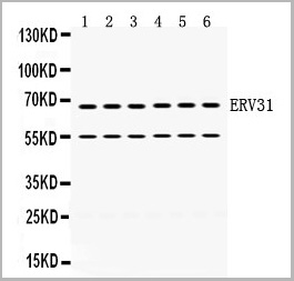 WB (Western Blot)
(Anti- ERV31 antibody, Western blotting)
WB (Western Blot)
(Anti- ERV31 antibody, Western blotting)
ERV31, Polyclonal Antibody (Cat# AAA11665)
Full Name
Anti-ERV31 Antibody
Gene Names
ERV3-1; ERV3; ERVR; envR; ERV-R; HERVR; HERV-R
Reactivity
Human
Applications
Western Blot
Purity
Immunogen Affinity Purified
Pricing
Rhinovirus outer capsid Protein 3, Monoclonal Antibody (Cat# AAA13450)
Full Name
Rhinovirus outer capsid Protein 3 (VP3)
Applications
Immunofluorescence
Purity
> 90% pure. Protein A Chromatography.
Pricing
 WB (Western Blot)
(Anti- FGF2 antibody, AAA11513, Western blottingAll lanes: Anti FGF2(AAA11513) at 0.5ug/mlLane 1: Recombinant Human FGF2 Protein 10ngLane 2: Recombinant Human FGF2 Protein 5ngLane 3: Recombinant Human FGF2 Protein 2.5ngPredicted bind size: 17KDObserved bind size: 17KD)
WB (Western Blot)
(Anti- FGF2 antibody, AAA11513, Western blottingAll lanes: Anti FGF2(AAA11513) at 0.5ug/mlLane 1: Recombinant Human FGF2 Protein 10ngLane 2: Recombinant Human FGF2 Protein 5ngLane 3: Recombinant Human FGF2 Protein 2.5ngPredicted bind size: 17KDObserved bind size: 17KD)
FGF2, Polyclonal Antibody (Cat# AAA11513)
Full Name
Anti-human FGF2 antibody
Gene Names
FGF2; BFGF; FGFB; FGF-2; HBGF-2
Reactivity
Human
Applications
Western Blot, Immunohistochemistry
Purity
Immunogen affinity purified.
Pricing
Mycoplasma hominis, p120, Monoclonal Antibody (Cat# AAA13388)
Full Name
MAb to Mycoplasma hominis
Applications
Immunofluorescence
Purity
>90% pure. Protein A chromatography
Pricing
Rotavirus NCDV (all antigens), Polyclonal Antibody (Cat# AAA13407)
Full Name
Goat anti Rotavirus
Applications
Immunofluorescence
Purity
> 95% pure. Sodium sulfate precipitation and ion exchange chromatography.
Pricing
 SDS-PAGE
Solute Carrier Family 30 Member 8 (SLC30A8), Recombinant Protein (Cat# AAA20271)
Full Name
Recombinant Solute Carrier Family 30 Member 8 (SLC30A8)
Gene Names
SLC30A8; ZNT8; ZnT-8
Applications
Western Blot
Purity
> 95%
Pricing
 WB (Western Blot)
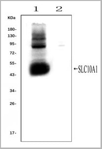
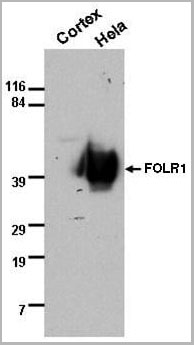
(Western blot analysis of SLC10A1 using anti- SLC10A1 antibody (AAA11653).Electrophoresis was performed on a 5-20% SDS-PAGE gel at 70V (Stacking gel) / 90V (Resolving gel) for 2-3 hours. The sample well of each lane was loaded with 50ug of sample under reducing conditions.Lane 1: rat liver tissue lysates (positive control), Lane 2: rat kidney tissue lysates, (negative control)After Electrophoresis, proteins were transferred to a Nitrocellulose membrane at 150mA for 50-90 minutes. Blocked the membrane with 5% Non-fat Milk/ TBS for 1.5 hour at RT. The membrane was incubated with rabbit anti-SLC10A1 antigen affinity purified polyclonal antibody (AAA11653) at 0.25ug/mL overnight at 4°C, then washed with TBS-0.1%Tween 3 times with 5 minutes each and probed with a goat anti-rabbit IgG-HRP secondary antibody at a dilution of 1:10000 for 1.5 hour at RT. The signal is developed using an Enhanced Chemiluminescent detection (ECL) kit with Tanon 5200 system. A specific band was detected for SLC10A1 at approximately 50KD. The expected band size for SLC10A1 is at 38KD.)
WB (Western Blot)
(Western blot analysis of SLC10A1 using anti- SLC10A1 antibody (AAA11653).Electrophoresis was performed on a 5-20% SDS-PAGE gel at 70V (Stacking gel) / 90V (Resolving gel) for 2-3 hours. The sample well of each lane was loaded with 50ug of sample under reducing conditions.Lane 1: rat liver tissue lysates (positive control), Lane 2: rat kidney tissue lysates, (negative control)After Electrophoresis, proteins were transferred to a Nitrocellulose membrane at 150mA for 50-90 minutes. Blocked the membrane with 5% Non-fat Milk/ TBS for 1.5 hour at RT. The membrane was incubated with rabbit anti-SLC10A1 antigen affinity purified polyclonal antibody (AAA11653) at 0.25ug/mL overnight at 4°C, then washed with TBS-0.1%Tween 3 times with 5 minutes each and probed with a goat anti-rabbit IgG-HRP secondary antibody at a dilution of 1:10000 for 1.5 hour at RT. The signal is developed using an Enhanced Chemiluminescent detection (ECL) kit with Tanon 5200 system. A specific band was detected for SLC10A1 at approximately 50KD. The expected band size for SLC10A1 is at 38KD.)
SLC10A1, Polyclonal Antibody (Cat# AAA11653)
Full Name
Anti-SLC10A1 Antibody
Gene Names
Slc10a1; Ntcp
Reactivity
Mouse, Rat
Applications
Flow Cytometry, Immunofluorescence, Immunohistochemistry, Immunohistochemistry, Immunocytochemistry, Western Blot
Purity
Immunogen affinity purified.
Pricing


 Application Data
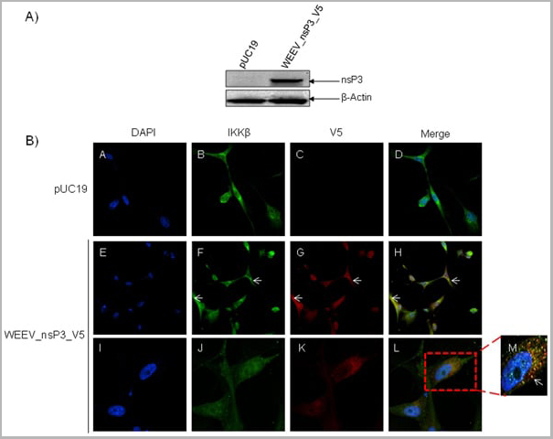
(Published customer image: Mouse anti V5 tag antibody, clone SV5-Pk1 used for the detection of V5 tagged WEEV_nsP3 protein by western blotting and immunofluorescenceImage caption: WEEV nsP3 interaction with host IKKbeta. A) U87MGs were transfected in a 6-well plate with 5 ug of pUC19 and WEEV_nsP3_HA for 24 hours. Cell lysates were resolved using SDS-PAGE and subsequently immunoblotted with V5 antibody and beta-actin served as a loading control. B) U87MGs were transfected with WEEV_nsP3_V5; cells were fixed after 24 hours and stained with antibodies against the endogenous IKKbeta and the V5 tag. Cells were incubated with appropriate secondary Alexa Fluor antibodies and the nuclei stained with DAPI. Co-localization of IKKbeta with WEEV_nsP3_V5 (yellow) was observed as shown by the arrows. B) Panels E -H serve as an example of transfected cells in a given field of view that show co-localization of IKKbeta and WEEV_nsP3_V5 24 hours post transfection. Panels I-L represent magnified images of other cells showing co-localization of IKKbeta and WEEV_nsP3_V5. Panel M is a magnified image of panel L. The co-localization was confirmed by Z-stack analysis. Co-localization was calculated to be approximately in 61% of cells (163 cells were counted of which 44% demonstrated expression of nsP3. Of those cells that expressed nsP3, 61% showed co-localization of both proteins). Images were taken using Nikon Eclipse TE2000-U at 60x magnification and are representative of 2 independent experiments.From: Amaya M, Voss K, Sampey G, Senina S, de la Fuente C, et al. (2014) The Role of IKKbeta in Venezuelan Equine Encephalitis Virus Infection. PLoS ONE 9(2): e86745.)
Application Data
(Published customer image: Mouse anti V5 tag antibody, clone SV5-Pk1 used for the detection of V5 tagged WEEV_nsP3 protein by western blotting and immunofluorescenceImage caption: WEEV nsP3 interaction with host IKKbeta. A) U87MGs were transfected in a 6-well plate with 5 ug of pUC19 and WEEV_nsP3_HA for 24 hours. Cell lysates were resolved using SDS-PAGE and subsequently immunoblotted with V5 antibody and beta-actin served as a loading control. B) U87MGs were transfected with WEEV_nsP3_V5; cells were fixed after 24 hours and stained with antibodies against the endogenous IKKbeta and the V5 tag. Cells were incubated with appropriate secondary Alexa Fluor antibodies and the nuclei stained with DAPI. Co-localization of IKKbeta with WEEV_nsP3_V5 (yellow) was observed as shown by the arrows. B) Panels E -H serve as an example of transfected cells in a given field of view that show co-localization of IKKbeta and WEEV_nsP3_V5 24 hours post transfection. Panels I-L represent magnified images of other cells showing co-localization of IKKbeta and WEEV_nsP3_V5. Panel M is a magnified image of panel L. The co-localization was confirmed by Z-stack analysis. Co-localization was calculated to be approximately in 61% of cells (163 cells were counted of which 44% demonstrated expression of nsP3. Of those cells that expressed nsP3, 61% showed co-localization of both proteins). Images were taken using Nikon Eclipse TE2000-U at 60x magnification and are representative of 2 independent experiments.From: Amaya M, Voss K, Sampey G, Senina S, de la Fuente C, et al. (2014) The Role of IKKbeta in Venezuelan Equine Encephalitis Virus Infection. PLoS ONE 9(2): e86745.)
V5-TAG, Monoclonal Antibody (Cat# AAA11864)
Full Name
MOUSE ANTI V5-TAG:FITC
Applications
Immunofluorescence
Pricing


 Application Data
(Published customer image: Mouse anti V5 tag antibody, clone SV5-Pk1 used for the detection of V5 tagged WEEV_nsP3 protein by western blotting and immunofluorescenceImage caption: WEEV nsP3 interaction with host IKKbeta. A) U87MGs were transfected in a 6-well plate with 5 ug of pUC19 and WEEV_nsP3_HA for 24 hours. Cell lysates were resolved using SDS-PAGE and subsequently immunoblotted with V5 antibody and beta-actin served as a loading control. B) U87MGs were transfected with WEEV_nsP3_V5; cells were fixed after 24 hours and stained with antibodies against the endogenous IKKbeta and the V5 tag. Cells were incubated with appropriate secondary Alexa Fluor antibodies and the nuclei stained with DAPI. Co-localization of IKKbeta with WEEV_nsP3_V5 (yellow) was observed as shown by the arrows. B) Panels E -H serve as an example of transfected cells in a given field of view that show co-localization of IKKbeta and WEEV_nsP3_V5 24 hours post transfection. Panels I-L represent magnified images of other cells showing co-localization of IKKbeta and WEEV_nsP3_V5. Panel M is a magnified image of panel L. The co-localization was confirmed by Z-stack analysis. Co-localization was calculated to be approximately in 61% of cells (163 cells were counted of which 44% demonstrated expression of nsP3. Of those cells that expressed nsP3, 61% showed co-localization of both proteins). Images were taken using Nikon Eclipse TE2000-U at 60x magnification and are representative of 2 independent experiments.From: Amaya M, Voss K, Sampey G, Senina S, de la Fuente C, et al. (2014) The Role of IKKbeta in Venezuelan Equine Encephalitis Virus Infection. PLoS ONE 9(2): e86745.)
Application Data
(Published customer image: Mouse anti V5 tag antibody, clone SV5-Pk1 used for the detection of V5 tagged WEEV_nsP3 protein by western blotting and immunofluorescenceImage caption: WEEV nsP3 interaction with host IKKbeta. A) U87MGs were transfected in a 6-well plate with 5 ug of pUC19 and WEEV_nsP3_HA for 24 hours. Cell lysates were resolved using SDS-PAGE and subsequently immunoblotted with V5 antibody and beta-actin served as a loading control. B) U87MGs were transfected with WEEV_nsP3_V5; cells were fixed after 24 hours and stained with antibodies against the endogenous IKKbeta and the V5 tag. Cells were incubated with appropriate secondary Alexa Fluor antibodies and the nuclei stained with DAPI. Co-localization of IKKbeta with WEEV_nsP3_V5 (yellow) was observed as shown by the arrows. B) Panels E -H serve as an example of transfected cells in a given field of view that show co-localization of IKKbeta and WEEV_nsP3_V5 24 hours post transfection. Panels I-L represent magnified images of other cells showing co-localization of IKKbeta and WEEV_nsP3_V5. Panel M is a magnified image of panel L. The co-localization was confirmed by Z-stack analysis. Co-localization was calculated to be approximately in 61% of cells (163 cells were counted of which 44% demonstrated expression of nsP3. Of those cells that expressed nsP3, 61% showed co-localization of both proteins). Images were taken using Nikon Eclipse TE2000-U at 60x magnification and are representative of 2 independent experiments.From: Amaya M, Voss K, Sampey G, Senina S, de la Fuente C, et al. (2014) The Role of IKKbeta in Venezuelan Equine Encephalitis Virus Infection. PLoS ONE 9(2): e86745.)
V5-TAG, Monoclonal Antibody (Cat# AAA11930)
Full Name
MOUSE ANTI V5-TAG
Applications
Immunohistochemistry, Flow Cytometry, Immunofluorescence, Immunoprecipitation, Western Blot, Radioimmunoassay
Pricing
 Application Data
HA1 (A/chicken/MD/MINHMA/2004)(H7N2), Polyclonal Antibody (Cat# AAA13716)
Full Name
Anti-HA1 (A/chicken/MD/MINHMA/2004)(H7N2)
Applications
Western Blot
Purity
Immunoaffinity chromatography
Pricing
Dengue Virus NS1, Monoclonal Antibody (Cat# AAA13399)
Full Name
MAb to Dengue Virus NS1
Applications
Lateral Flow, Immunofluorescence
Purity
> 90% pure. Protein A Chromatography
Pricing
 WB (Western Blot)
(Anti-AVPR1A antibody, AAA11596, All Western blottingAll lanes: Anti-AVPR1A(AAA11596) at 0.5ug/mlLane 1: Rat Liver Tissue Lysate at 40ugLane 2: Rat Kidney Tissue Lysate at 40ugPredicted bind size: 47KDObserved bind size: 47KD)
WB (Western Blot)
(Anti-AVPR1A antibody, AAA11596, All Western blottingAll lanes: Anti-AVPR1A(AAA11596) at 0.5ug/mlLane 1: Rat Liver Tissue Lysate at 40ugLane 2: Rat Kidney Tissue Lysate at 40ugPredicted bind size: 47KDObserved bind size: 47KD)
AVPR1A, Polyclonal Antibody (Cat# AAA11596)
Full Name
Anti-AVPR1A antibody
Gene Names
Avpr1a; V1a; AVPR; V1aR
Reactivity
Reacts with: Human, Rat
Predicted to work with: Mouse
Predicted to work with: Mouse
Applications
Western Blot
Purity
Immunogen affinity purified.
Pricing
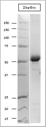 SDS-PAGE
Zika Virus Envelope, Recombinant Protein (Cat# AAA13452)
Full Name
Zika Virus Envelope Recombinant
Applications
Chemiluminescence Immunoassay, Lateral Flow
Purity
> 80% Pure (SDS-PAGE). Affinity Chromatography
Pricing
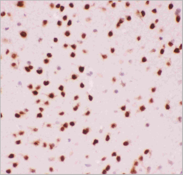
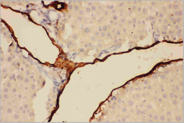
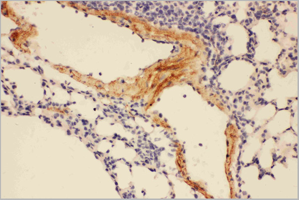
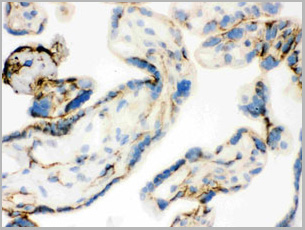
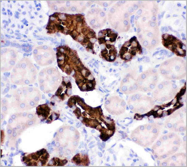
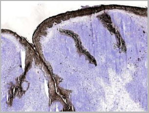
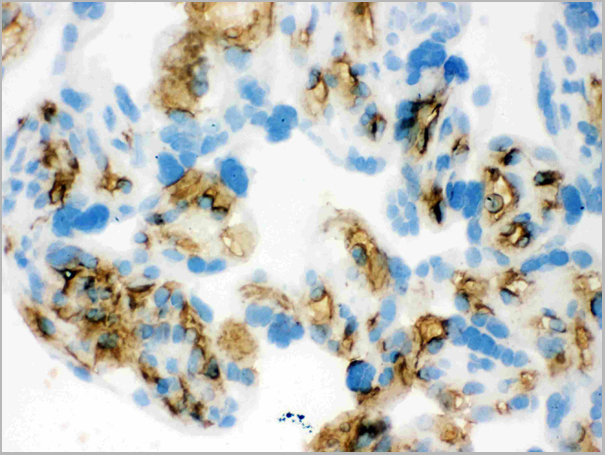
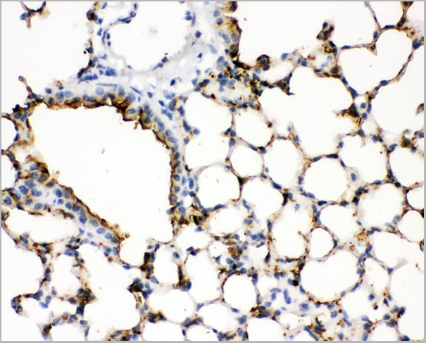 IHC (Immunohistochemistry)
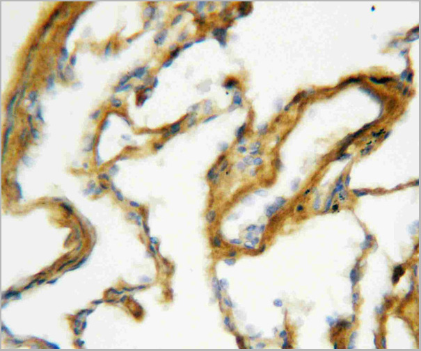
(Figure 3. IHC analysis of SFTPA1 using anti- SFTPA1 antibody (AAA11521).SFTPA1 was detected in paraffin-embedded section of mouse lung tissues. Heat mediated antigen retrieval was performed in citrate buffer (pH6, epitope retrieval solution) for 20 mins. The tissue section was blocked with 10% goat serum. The tissue section was then incubated with 1ug/ml rabbit anti- SFTPA1 Antibody (AAA11521) overnight at 4 degree C. Biotinylated goat anti-rabbit IgG was used as secondary antibody and incubated for 30 minutes at 37 degree C. The tissue section was developed using Strepavidin-Biotin-Complex (SABC) with DAB as the chromogen.)
IHC (Immunohistochemistry)
(Figure 3. IHC analysis of SFTPA1 using anti- SFTPA1 antibody (AAA11521).SFTPA1 was detected in paraffin-embedded section of mouse lung tissues. Heat mediated antigen retrieval was performed in citrate buffer (pH6, epitope retrieval solution) for 20 mins. The tissue section was blocked with 10% goat serum. The tissue section was then incubated with 1ug/ml rabbit anti- SFTPA1 Antibody (AAA11521) overnight at 4 degree C. Biotinylated goat anti-rabbit IgG was used as secondary antibody and incubated for 30 minutes at 37 degree C. The tissue section was developed using Strepavidin-Biotin-Complex (SABC) with DAB as the chromogen.)
Surfactant Protein A, Polyclonal Antibody (Cat# AAA11521)
Full Name
Polyclonal Anti-SFTPA1 Antibody
Gene Names
Sftpa1; SP-A; Sftp1; Sftp-1
Reactivity
Mouse, Rat
Applications
Western Blot, Immunohistochemistry
Purity
Immunogen affinity purified.
Pricing


 Application Data
(Published customer image: Mouse anti V5 tag antibody, clone SV5-Pk1 used for the detection of V5 tagged WEEV_nsP3 protein by western blotting and immunofluorescenceImage caption: WEEV nsP3 interaction with host IKKbeta. A) U87MGs were transfected in a 6-well plate with 5 ug of pUC19 and WEEV_nsP3_HA for 24 hours. Cell lysates were resolved using SDS-PAGE and subsequently immunoblotted with V5 antibody and beta-actin served as a loading control. B) U87MGs were transfected with WEEV_nsP3_V5; cells were fixed after 24 hours and stained with antibodies against the endogenous IKKbeta and the V5 tag. Cells were incubated with appropriate secondary Alexa Fluor antibodies and the nuclei stained with DAPI. Co-localization of IKKbeta with WEEV_nsP3_V5 (yellow) was observed as shown by the arrows. B) Panels E -H serve as an example of transfected cells in a given field of view that show co-localization of IKKbeta and WEEV_nsP3_V5 24 hours post transfection. Panels I-L represent magnified images of other cells showing co-localization of IKKbeta and WEEV_nsP3_V5. Panel M is a magnified image of panel L. The co-localization was confirmed by Z-stack analysis. Co-localization was calculated to be approximately in 61% of cells (163 cells were counted of which 44% demonstrated expression of nsP3. Of those cells that expressed nsP3, 61% showed co-localization of both proteins). Images were taken using Nikon Eclipse TE2000-U at 60x magnification and are representative of 2 independent experiments.From: Amaya M, Voss K, Sampey G, Senina S, de la Fuente C, et al. (2014) The Role of IKKbeta in Venezuelan Equine Encephalitis Virus Infection. PLoS ONE 9(2): e86745.)
Application Data
(Published customer image: Mouse anti V5 tag antibody, clone SV5-Pk1 used for the detection of V5 tagged WEEV_nsP3 protein by western blotting and immunofluorescenceImage caption: WEEV nsP3 interaction with host IKKbeta. A) U87MGs were transfected in a 6-well plate with 5 ug of pUC19 and WEEV_nsP3_HA for 24 hours. Cell lysates were resolved using SDS-PAGE and subsequently immunoblotted with V5 antibody and beta-actin served as a loading control. B) U87MGs were transfected with WEEV_nsP3_V5; cells were fixed after 24 hours and stained with antibodies against the endogenous IKKbeta and the V5 tag. Cells were incubated with appropriate secondary Alexa Fluor antibodies and the nuclei stained with DAPI. Co-localization of IKKbeta with WEEV_nsP3_V5 (yellow) was observed as shown by the arrows. B) Panels E -H serve as an example of transfected cells in a given field of view that show co-localization of IKKbeta and WEEV_nsP3_V5 24 hours post transfection. Panels I-L represent magnified images of other cells showing co-localization of IKKbeta and WEEV_nsP3_V5. Panel M is a magnified image of panel L. The co-localization was confirmed by Z-stack analysis. Co-localization was calculated to be approximately in 61% of cells (163 cells were counted of which 44% demonstrated expression of nsP3. Of those cells that expressed nsP3, 61% showed co-localization of both proteins). Images were taken using Nikon Eclipse TE2000-U at 60x magnification and are representative of 2 independent experiments.From: Amaya M, Voss K, Sampey G, Senina S, de la Fuente C, et al. (2014) The Role of IKKbeta in Venezuelan Equine Encephalitis Virus Infection. PLoS ONE 9(2): e86745.)
V5-TAG, Monoclonal Antibody (Cat# AAA12211)
Full Name
MOUSE ANTI V5-TAG
Applications
Immunohistochemistry, Flow Cytometry, Immunofluorescence, Immunoprecipitation, Western Blot, Radioimmunoassay
Pricing
Flavivirus, Group, Monoclonal Antibody (Cat# AAA14673)
Full Name
Flavivirus, Group
Applications
Immunofluorescence
Purity
Affinity Purified
Purified by Protein A affinity chromatography from ascites.
Purified by Protein A affinity chromatography from ascites.
Pricing
Bacillus cereus/Bacillus subtilis, Spores, Polyclonal Antibody (Cat# AAA14710)
Full Name
Rabbit anti-Bacillus cereus/Bacillus subtilis, Spores
Applications
Immunofluorescence, Fluorescence Microscopy
Purity
Purified IgG fraction to a purity of > 95%.
Pricing
Rhinovirus outer capsid protein 3, Monoclonal Antibody (Cat# AAA13449)
Full Name
Rhinovirus outer capsid protein 3 (VP3)
Applications
Immunofluorescence
Purity
> 90% pure. Protein A Chromatography
Pricing
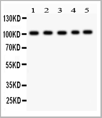
 WB (Western Blot)
(Anti-Caspase-1(P20) antibody, AAA11584, Western blottingLane 1: Rat Brain Tissue LysateLane 2: Rat Spleen Tissue LysateLane 3: Mouse Brain Tissue LysateLane 4: Mouse Spleen Tissue LysateLane 5: Mouse Testis Tissue Lysate)
WB (Western Blot)
(Anti-Caspase-1(P20) antibody, AAA11584, Western blottingLane 1: Rat Brain Tissue LysateLane 2: Rat Spleen Tissue LysateLane 3: Mouse Brain Tissue LysateLane 4: Mouse Spleen Tissue LysateLane 5: Mouse Testis Tissue Lysate)
Caspase-1(P20), Polyclonal Antibody (Cat# AAA11584)
Full Name
Polyclonal Anti-CASP1 (p20) Antibody
Gene Names
Casp1; ICE; Il1bc
Reactivity
Mouse, Rat
Applications
Western Blot
Purity
Immunogen affinity purified.
Pricing
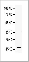
 WB (Western Blot)
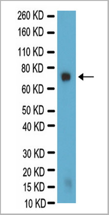
(Western Blot analysis for galactocerebroside in PC12 lysate. PC12 lysate was resolved by electrophoresis, transferred to PVDF membrane and probed with AAA14685 at 1:500 dilution. Proteins were visualized using a goat anti-mouse secondary antibody conjugated to HRP and a chemiluminescence detection system. Arrow indicates protein Galactocerebroside (~75kD).)
WB (Western Blot)
(Western Blot analysis for galactocerebroside in PC12 lysate. PC12 lysate was resolved by electrophoresis, transferred to PVDF membrane and probed with AAA14685 at 1:500 dilution. Proteins were visualized using a goat anti-mouse secondary antibody conjugated to HRP and a chemiluminescence detection system. Arrow indicates protein Galactocerebroside (~75kD).)
Galactocerebroside, Monoclonal Antibody (Cat# AAA14685)
Full Name
Mouse anti-Bovine Galactocerebroside
Applications
Immunohistochemistry, Immunocytochemistry
Purity
Purified by Protein A affinity chromatography.
Pricing
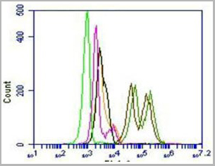
 FCM (Flow Cytometry)
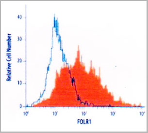
(Flow Cytometry detection of FOLR1 in MCF-7 human cell line using AAA14691. MCF-7 human breast cancer cell line was stained with AAA14691 (filled histogram) or isotype control antibody (open histogram), followed byPhycoerythrin-conjugated Anti-MouseIgG Secondary Antibody.)
FCM (Flow Cytometry)
(Flow Cytometry detection of FOLR1 in MCF-7 human cell line using AAA14691. MCF-7 human breast cancer cell line was stained with AAA14691 (filled histogram) or isotype control antibody (open histogram), followed byPhycoerythrin-conjugated Anti-MouseIgG Secondary Antibody.)
FOLR1, Monoclonal Antibody (Cat# AAA14691)
Full Name
FOLR1 (Folate receptor 1 (adult) Adult folate-binding protein, FBP, Folate receptor, adult, Folate receptor 1, Folate receptor alpha, FOLR, FR-alpha, KB cells FBP, MOv18, Ovarian tumor-associated antigen MOv18)
Gene Names
FOLR1; FBP; FOLR
Reactivity
Human
Applications
Flow Cytometry, Western Blot, Immunocytochemistry
Purity
Purified by Protein G affinity chromatography from hybridoma culture supernatant.
Pricing
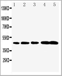
 WB (Western Blot)
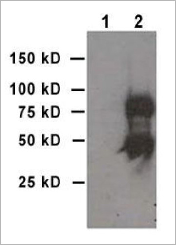
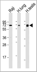
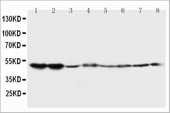
(All lanes : Anti-ERVK-7 Antibody (N-Term) at 1:1000-1:2000 dilutionLane 1: Raji whole cell lysateLane 2: human lung lysateLane 3: human testis lysateLysates/proteins at 20 ug per lane.SecondaryGoat Anti-Rabbit IgG, (H+L), Peroxidase conjugated at 1/10000 dilution.Predicted band size : 67 kDaBlocking/Dilution buffer: 5% NFDM/TBST.)
WB (Western Blot)
(All lanes : Anti-ERVK-7 Antibody (N-Term) at 1:1000-1:2000 dilutionLane 1: Raji whole cell lysateLane 2: human lung lysateLane 3: human testis lysateLysates/proteins at 20 ug per lane.SecondaryGoat Anti-Rabbit IgG, (H+L), Peroxidase conjugated at 1/10000 dilution.Predicted band size : 67 kDaBlocking/Dilution buffer: 5% NFDM/TBST.)
ERVK-7, Polyclonal Antibody (Cat# AAA28797)
Full Name
ERVK-7 Antibody (N-Term)
Reactivity
Human
Applications
Western Blot, Immunohistochemistry
Purity
This antibody is purified through a protein A column, followed by peptide affinity purification.
Pricing
Goat anti Human IgG IgA IgM (Fc specific), Polyclonal Secondary Antibody (Cat# AAA14569)
Full Name
Goat anti Human IgG IgA IgM (Fc specific), conjugated with FITC
Reactivity
Human
Applications
Immunocytochemistry, Immunohistochemistry, Immunofluorescence
Purity
The IgG (7S) fraction is isolated and purified from hyperimmune antisera with strong precipitating activity and contains the bulk of the antibody specificity. It is free of other serum proteins as tested by immunoelectrophoresis and double radial immunodi
Pricing
 WB (Western Blot)
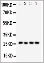
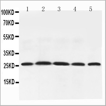
(Figure 6. Western blot analysis of Adiponectin using anti-Adiponectin antibody (AAA11602).Electrophoresis was performed on a 5-20% SDS-PAGE gel at 70V (Stacking gel) / 90V (Resolving gel) for 2-3 hours. The sample well of each lane was loaded with 50ug of sample under reducing conditions.Lane 1: Rat Testis Tissue Lysate,Lane 2: Rat Spleen Tissue Lysate,Lane 3: Rat Skeletal Muscle Tissue Lysate,Lane 4: Rat Cardiac Muscle Tissue Lysate,Lane 5: Rat Liver Tissue Lysate,After Electrophoresis, proteins were transferred to a Nitrocellulose membrane at 150mA for 50-90 minutes. Blocked the membrane with 5% Non-fat Milk/ TBS for 1.5 hour at RT. The membrane was incubated with rabbit anti-Adiponectin antigen affinity purified polyclonal antibody at 0.5ug/mL overnight at 4 degree C, then washed with TBS-0.1%Tween 3 times with 5 minutes each and probed with a goat anti-rabbit IgG-HRP secondary antibody at a dilution of 1:10000 for 1.5 hour at RT. The signal is developed using an Enhanced Chemiluminescent detection (ECL) kit with Tanon 5200 system. A specific band was detected for Adiponectin at approximately 26KD. The expected band size for Adiponectin is at 26KD.)
WB (Western Blot)
(Figure 6. Western blot analysis of Adiponectin using anti-Adiponectin antibody (AAA11602).Electrophoresis was performed on a 5-20% SDS-PAGE gel at 70V (Stacking gel) / 90V (Resolving gel) for 2-3 hours. The sample well of each lane was loaded with 50ug of sample under reducing conditions.Lane 1: Rat Testis Tissue Lysate,Lane 2: Rat Spleen Tissue Lysate,Lane 3: Rat Skeletal Muscle Tissue Lysate,Lane 4: Rat Cardiac Muscle Tissue Lysate,Lane 5: Rat Liver Tissue Lysate,After Electrophoresis, proteins were transferred to a Nitrocellulose membrane at 150mA for 50-90 minutes. Blocked the membrane with 5% Non-fat Milk/ TBS for 1.5 hour at RT. The membrane was incubated with rabbit anti-Adiponectin antigen affinity purified polyclonal antibody at 0.5ug/mL overnight at 4 degree C, then washed with TBS-0.1%Tween 3 times with 5 minutes each and probed with a goat anti-rabbit IgG-HRP secondary antibody at a dilution of 1:10000 for 1.5 hour at RT. The signal is developed using an Enhanced Chemiluminescent detection (ECL) kit with Tanon 5200 system. A specific band was detected for Adiponectin at approximately 26KD. The expected band size for Adiponectin is at 26KD.)
Adiponectin, Polyclonal Antibody (Cat# AAA11602)
Full Name
Anti-Adiponectin antibody
Gene Names
Adipoq; Acdc; Acrp30
Reactivity
Mouse, Rat
Applications
Western Blot, Immunohistochemistry
Purity
Immunogen affinity purified.
Pricing
 WB (Western Blot)
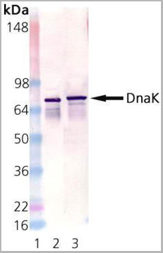
(Western Blot Analysis using AAA14655: Lane 1: MW Marker; Lane 2: DnaK active recombinant protein, Lane 3: E. coli cell lysate.)
WB (Western Blot)
(Western Blot Analysis using AAA14655: Lane 1: MW Marker; Lane 2: DnaK active recombinant protein, Lane 3: E. coli cell lysate.)
DnaK, Monoclonal Antibody (Cat# AAA14655)
Full Name
DnaK
Reactivity
E Coli
Applications
Western Blot
Purity
Affinity Purified
Purified by Protein G affinity chromatography.
Purified by Protein G affinity chromatography.
Pricing
Retinoic Acid Receptor Responder 2, Recombinant Protein (Cat# AAA10868)
Full Name
Recombinant Human Retinoic Acid Receptor Responder 2
Gene Names
RARRES2; TIG2; HP10433
Purity
Greater than 98.0% as determined by: (a) Analysis by RP-HPLC. (b) Analysis by SDS-PAGE.
Pricing
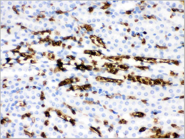
 IHC (Immunohistochemistry)
(Mast Cell Tryptase was detected in paraffin-embedded sections of human lung cancer tissues using rabbit anti- Mast Cell Tryptase Antigen Affinity purified polyclonal antibody at 1ug/mL. The immunohistochemical section was developed using SABC method.)
IHC (Immunohistochemistry)
(Mast Cell Tryptase was detected in paraffin-embedded sections of human lung cancer tissues using rabbit anti- Mast Cell Tryptase Antigen Affinity purified polyclonal antibody at 1ug/mL. The immunohistochemical section was developed using SABC method.)
Mast Cell Tryptase, Polyclonal Antibody (Cat# AAA11674)
Full Name
Anti-Mast Cell Tryptase Antibody
Gene Names
TPSAB1; TPS1; TPS2; TPSB1
Reactivity
Human
Applications
Western Blot, Immunohistochemistry
Purity
Immunogen affinity purified.
Pricing
Gram Positive Bacteria, Monoclonal Antibody (Cat# AAA13365)
Full Name
MAb to Gram Positive Bacteria
Applications
Immunofluorescence
Purity
>90% pure. Protein A chromatography
Pricing
 FCM (Flow Cytometry)
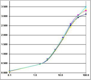
(Figure 6. Flow Cytometry analysis of U20S cells using anti-Calpastatin antibody (AAA11679).Overlay histogram showing U20S cells stained with AAA11679 (Blue line).The cells were blocked with 10% normal goat serum. And then incubated with rabbit anti-Calpastatin Antibody (AAA11679,1ug/1x10^6 cells) for 30 min at 20 degree C. DyLight ®488 conjugated goat anti-rabbit IgG (5-10ug/1x10^6 cells) was used as secondary antibody for 30 minutes at 20 degree C. Isotype control antibody (Green line) was rabbit IgG (1ug/1x106) used under the same conditions. Unlabelled sample (Red line) was also used as a control.)
FCM (Flow Cytometry)
(Figure 6. Flow Cytometry analysis of U20S cells using anti-Calpastatin antibody (AAA11679).Overlay histogram showing U20S cells stained with AAA11679 (Blue line).The cells were blocked with 10% normal goat serum. And then incubated with rabbit anti-Calpastatin Antibody (AAA11679,1ug/1x10^6 cells) for 30 min at 20 degree C. DyLight ®488 conjugated goat anti-rabbit IgG (5-10ug/1x10^6 cells) was used as secondary antibody for 30 minutes at 20 degree C. Isotype control antibody (Green line) was rabbit IgG (1ug/1x106) used under the same conditions. Unlabelled sample (Red line) was also used as a control.)
Calpastatin, Polyclonal Antibody (Cat# AAA11679)
Full Name
Anti-Calpastatin Antibody
Gene Names
CAST; BS-17; PLACK
Reactivity
Human
Applications
Western Blot, Immunohistochemistry
Purity
Immunogen affinity purified.
Pricing

 FCM (Flow Cytometry)
(Figure 7. Flow Cytometry analysis of U251 cells using anti-PNP antibody (AAA11680).Overlay histogram showing U251 cells stained with AAA11680 (Blue line).The cells were blocked with 10% normal goat serum. And then incubated with rabbit anti-PNP Antibody (AAA11680,1ug/1x10^6 cells) for 30 min at 20 degree C. DyLight ®488 conjugated goat anti-rabbit IgG (5-10ug/1x10^6 cells) was used as secondary antibody for 30 minutes at 20 degree C. Isotype control antibody (Green line) was rabbit IgG (1ug/1x106) used under the same conditions. Unlabelled sample (Red line) was also used as a control.)
FCM (Flow Cytometry)
(Figure 7. Flow Cytometry analysis of U251 cells using anti-PNP antibody (AAA11680).Overlay histogram showing U251 cells stained with AAA11680 (Blue line).The cells were blocked with 10% normal goat serum. And then incubated with rabbit anti-PNP Antibody (AAA11680,1ug/1x10^6 cells) for 30 min at 20 degree C. DyLight ®488 conjugated goat anti-rabbit IgG (5-10ug/1x10^6 cells) was used as secondary antibody for 30 minutes at 20 degree C. Isotype control antibody (Green line) was rabbit IgG (1ug/1x106) used under the same conditions. Unlabelled sample (Red line) was also used as a control.)
PNP, Polyclonal Antibody (Cat# AAA11680)
Full Name
Anti-PNP Antibody
Gene Names
PNP; NP; PUNP; PRO1837
Reactivity
Human, Mouse, Rat
Applications
Western Blot, Immunohistochemistry
Purity
Immunogen affinity purified.
Pricing

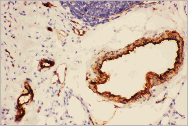
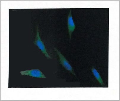
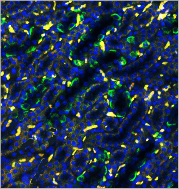
 IF (Immunofluorescence)
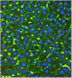
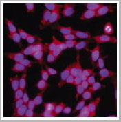
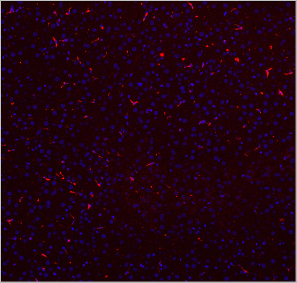
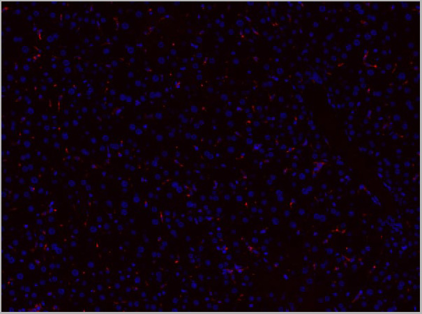
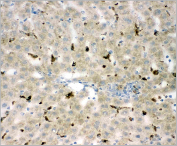
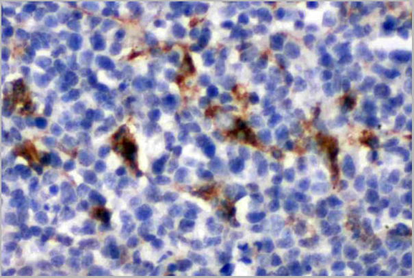
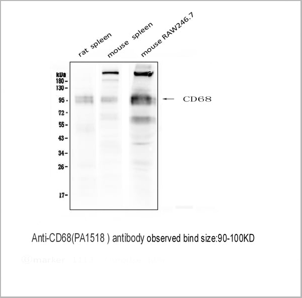
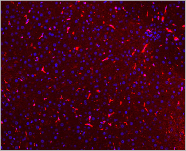
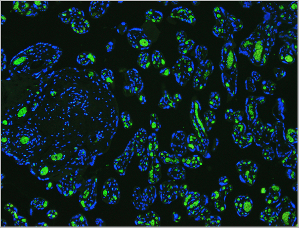
(Figure 7. IF analysis of CD68 using anti- CD68 antibody CD68 was detected in paraffin-embedded section of mouse liver tissues. Heat mediated antigen retrieval was performed in citrate buffer (pH6, epitope retrieval solution) for 20 mins. The tissue section was blocked with 10% goat serum. The tissue section was then incubated with 1ug/mL rabbit anti- CD68 Antibody overnight at 4 degree C. Cy3 Conjugated Goat Anti-Rabbit IgG was used as secondary antibody at 1:100 dilution and incubated for 30 minutes at 37 degree C. The section was counterstained with DAPI. Visualize using a fluorescence microscope and filter sets appropriate for the label used.)
IF (Immunofluorescence)
(Figure 7. IF analysis of CD68 using anti- CD68 antibody CD68 was detected in paraffin-embedded section of mouse liver tissues. Heat mediated antigen retrieval was performed in citrate buffer (pH6, epitope retrieval solution) for 20 mins. The tissue section was blocked with 10% goat serum. The tissue section was then incubated with 1ug/mL rabbit anti- CD68 Antibody overnight at 4 degree C. Cy3 Conjugated Goat Anti-Rabbit IgG was used as secondary antibody at 1:100 dilution and incubated for 30 minutes at 37 degree C. The section was counterstained with DAPI. Visualize using a fluorescence microscope and filter sets appropriate for the label used.)
CD68, Polyclonal Antibody (Cat# AAA11512)
Full Name
Anti-CD68 antibody
Gene Names
Cd68; Lamp4; gp110; Scard1
Reactivity
Mouse, Rat
Applications
Western Blot, Immunohistochemistry, Immunohistochemistry
Purity
Immunogen affinity purified.
Pricing


 Application Data
(Published customer image: Mouse anti V5 tag antibody, clone SV5-Pk1 used for the detection of V5 tagged WEEV_nsP3 protein by western blotting and immunofluorescenceImage caption: WEEV nsP3 interaction with host IKKbeta. A) U87MGs were transfected in a 6-well plate with 5 ug of pUC19 and WEEV_nsP3_HA for 24 hours. Cell lysates were resolved using SDS-PAGE and subsequently immunoblotted with V5 antibody and beta-actin served as a loading control. B) U87MGs were transfected with WEEV_nsP3_V5; cells were fixed after 24 hours and stained with antibodies against the endogenous IKKbeta and the V5 tag. Cells were incubated with appropriate secondary Alexa Fluor antibodies and the nuclei stained with DAPI. Co-localization of IKKbeta with WEEV_nsP3_V5 (yellow) was observed as shown by the arrows. B) Panels E -H serve as an example of transfected cells in a given field of view that show co-localization of IKKbeta and WEEV_nsP3_V5 24 hours post transfection. Panels I-L represent magnified images of other cells showing co-localization of IKKbeta and WEEV_nsP3_V5. Panel M is a magnified image of panel L. The co-localization was confirmed by Z-stack analysis. Co-localization was calculated to be approximately in 61% of cells (163 cells were counted of which 44% demonstrated expression of nsP3. Of those cells that expressed nsP3, 61% showed co-localization of both proteins). Images were taken using Nikon Eclipse TE2000-U at 60x magnification and are representative of 2 independent experiments.From: Amaya M, Voss K, Sampey G, Senina S, de la Fuente C, et al. (2014) The Role of IKKbeta in Venezuelan Equine Encephalitis Virus Infection. PLoS ONE 9(2): e86745.)
Application Data
(Published customer image: Mouse anti V5 tag antibody, clone SV5-Pk1 used for the detection of V5 tagged WEEV_nsP3 protein by western blotting and immunofluorescenceImage caption: WEEV nsP3 interaction with host IKKbeta. A) U87MGs were transfected in a 6-well plate with 5 ug of pUC19 and WEEV_nsP3_HA for 24 hours. Cell lysates were resolved using SDS-PAGE and subsequently immunoblotted with V5 antibody and beta-actin served as a loading control. B) U87MGs were transfected with WEEV_nsP3_V5; cells were fixed after 24 hours and stained with antibodies against the endogenous IKKbeta and the V5 tag. Cells were incubated with appropriate secondary Alexa Fluor antibodies and the nuclei stained with DAPI. Co-localization of IKKbeta with WEEV_nsP3_V5 (yellow) was observed as shown by the arrows. B) Panels E -H serve as an example of transfected cells in a given field of view that show co-localization of IKKbeta and WEEV_nsP3_V5 24 hours post transfection. Panels I-L represent magnified images of other cells showing co-localization of IKKbeta and WEEV_nsP3_V5. Panel M is a magnified image of panel L. The co-localization was confirmed by Z-stack analysis. Co-localization was calculated to be approximately in 61% of cells (163 cells were counted of which 44% demonstrated expression of nsP3. Of those cells that expressed nsP3, 61% showed co-localization of both proteins). Images were taken using Nikon Eclipse TE2000-U at 60x magnification and are representative of 2 independent experiments.From: Amaya M, Voss K, Sampey G, Senina S, de la Fuente C, et al. (2014) The Role of IKKbeta in Venezuelan Equine Encephalitis Virus Infection. PLoS ONE 9(2): e86745.)
V5-TAG, Monoclonal Antibody (Cat# AAA11850)
Full Name
MOUSE ANTI V5-TAG:Biotin
Applications
Immunohistochemistry, Western Blot
Pricing


 ChIP (Chromatin Immunoprecipitation)
(Chromatin immunoprecipitation was performed with cross-linked chromatin from 293T, using Acetyl-Histone H3-K27 Rabbit pAb antibody (AAA28163) and rabbit IgG.The amount of immunoprecipitated DNA was checked by quantitative PCR. Histogram constructed the ratios of the ratio of the immunoprecipitated DNA versus the input.)
ChIP (Chromatin Immunoprecipitation)
(Chromatin immunoprecipitation was performed with cross-linked chromatin from 293T, using Acetyl-Histone H3-K27 Rabbit pAb antibody (AAA28163) and rabbit IgG.The amount of immunoprecipitated DNA was checked by quantitative PCR. Histogram constructed the ratios of the ratio of the immunoprecipitated DNA versus the input.)
H3K27AC, Antibody (Cat# AAA28163)
Full Name
Histone H3K27AC Polyclonal Antibody
Gene Names
HIST3H3; H3t; H3.4; H3/g; H3FT
Applications
Western Blot, Immunohistochemistry, Immunofluorescence, Immunocytochemistry, Immunoprecipitation, Chromatin Immunoprecipitation, Chromatin Immunoprecipitation
Purity
Affinity Purification
Pricing
 IF (Immunofluorescence)
(Immunofluorescence (IFC) analysis of PC3 cells using AAA14697 (1:100). The secondary antibody (green) was used Alexa Fluor 488. DAPI was stained the cell nucleus (blue).)
IF (Immunofluorescence)
(Immunofluorescence (IFC) analysis of PC3 cells using AAA14697 (1:100). The secondary antibody (green) was used Alexa Fluor 488. DAPI was stained the cell nucleus (blue).)
Ribonuclease inhibitor 1, Monoclonal Antibody (Cat# AAA14697)
Full Name
Ribonuclease inhibitor 1 (RNH1)
Gene Names
RNH1; RAI; RNH; MGC4569; MGC18200; MGC54054
Applications
Immunofluorescence, Western Blot
Purity
Affinity Purified
Purified by Protein G affinity chromatography.
Purified by Protein G affinity chromatography.
Pricing
 IHC (Immunohistochemistry)
(Anti-5HT2A Receptor antibody, AAA11510, IHC(P)IHC(P): Rat Brain Tissue)
IHC (Immunohistochemistry)
(Anti-5HT2A Receptor antibody, AAA11510, IHC(P)IHC(P): Rat Brain Tissue)
5HT2A Receptor, Polyclonal Antibody (Cat# AAA11510)
Full Name
Anti-5HT2A Receptor antibody
Gene Names
HTR2A; HTR2; 5-HT2A
Reactivity
Human, Mouse, Rat
Applications
Western Blot, Immunohistochemistry
Purity
Immunogen affinity purified.
Pricing
 IHC (Immunohistochemistry)
(Anti-5HT1A Receptor antibody, AAA11520, IHC(P)IHC(P): Rat Brain Tissue)
IHC (Immunohistochemistry)
(Anti-5HT1A Receptor antibody, AAA11520, IHC(P)IHC(P): Rat Brain Tissue)
5HT1A Receptor, Polyclonal Antibody (Cat# AAA11520)
Full Name
Anti-5HT1A Receptor antibody
Gene Names
HTR1A; G-21; 5HT1a; PFMCD; 5-HT1A; 5-HT-1A; ADRBRL1; ADRB2RL1
Reactivity
Reacts with: Human, Rat
Predicted to work with: Mouse
Predicted to work with: Mouse
Applications
Western Blot, Immunohistochemistry
Purity
Immunogen affinity purified.
Pricing

 IHC (Immunohistochemistry)
(Anti- TGM2 Picoband antibody, AAA11646, IHC(F)IHC(F): Human Placenta Tissue)
IHC (Immunohistochemistry)
(Anti- TGM2 Picoband antibody, AAA11646, IHC(F)IHC(F): Human Placenta Tissue)
TGM2, Polyclonal Antibody (Cat# AAA11646)
Full Name
Anti-TGM2 Antibody
Gene Names
TGM2; TGC; TG(C)
Reactivity
Human, Mouse, Rat
Applications
Western Blot, Immunohistochemistry
Purity
Immunogen Affinity Purified
Pricing
 WB (Western Blot)
(Figure. Western blot analysis of IFN gamma using anti- IFN gamma antibody (AAA11517).Electrophoresis was performed on a 5-20% SDS-PAGE gel at 70V (Stacking gel) / 90V (Resolving gel) for 2-3 hours. The sample well of each lane was loaded with 50ug of sample under reducing conditions.Lane: Recombinant Human IFN gamma Protein 0.5ngAfter Electrophoresis, proteins were transferred to a Nitrocellulose membrane at 150mA for 50-90 minutes. Blocked the membrane with 5% Non-fat Milk/ TBS for 1.5 hour at RT. The membrane was incubated with rabbit anti- IFN gamma antigen affinity purified polyclonal antibody at 0.5ug/mL overnight at 4 degree C, then washed with TBS-0.1%Tween 3 times with 5 minutes each and probed with a goat anti-rabbit IgG-HRP secondary antibody at a dilution of 1:10000 for 1.5 hour at RT. The signal is developed using an Enhanced Chemiluminescent detection (ECL) kit with Tanon 5200 system. A specific band was detected for IFN gamma at approximately 17KD. The expected band size for IFN gamma is at 17KD.)
WB (Western Blot)
(Figure. Western blot analysis of IFN gamma using anti- IFN gamma antibody (AAA11517).Electrophoresis was performed on a 5-20% SDS-PAGE gel at 70V (Stacking gel) / 90V (Resolving gel) for 2-3 hours. The sample well of each lane was loaded with 50ug of sample under reducing conditions.Lane: Recombinant Human IFN gamma Protein 0.5ngAfter Electrophoresis, proteins were transferred to a Nitrocellulose membrane at 150mA for 50-90 minutes. Blocked the membrane with 5% Non-fat Milk/ TBS for 1.5 hour at RT. The membrane was incubated with rabbit anti- IFN gamma antigen affinity purified polyclonal antibody at 0.5ug/mL overnight at 4 degree C, then washed with TBS-0.1%Tween 3 times with 5 minutes each and probed with a goat anti-rabbit IgG-HRP secondary antibody at a dilution of 1:10000 for 1.5 hour at RT. The signal is developed using an Enhanced Chemiluminescent detection (ECL) kit with Tanon 5200 system. A specific band was detected for IFN gamma at approximately 17KD. The expected band size for IFN gamma is at 17KD.)
Interferon gamma, Polyclonal Antibody (Cat# AAA11517)
Full Name
Anti-human Interferon gamma antibody
Gene Names
IFNG; IFG; IFI
Reactivity
Human
Applications
Western Blot, Immunohistochemistry, Neutralization Assay, Immunoprecipitation
Purity
Immunogen affinity purified.
Pricing
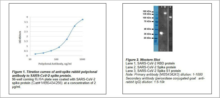 Application Data
COVID 19 Spike Protein Coronavirus, Polyclonal Antibody (Cat# AAA13774)
Full Name
Anti-Spike Protein (SARS-CoV-2) Polyclonal Antibody
Applications
Western Blot
Purity
Protein G immunoaffinity chromatography
Pricing
Hepatitis C Virus (HCV) E2, Monoclonal Antibody (Cat# AAA13364)
Full Name
MAb to HCV E2
Applications
Immunofluorescence, Western Blot
Purity
>90% pure. Protein A chromatography
Pricing
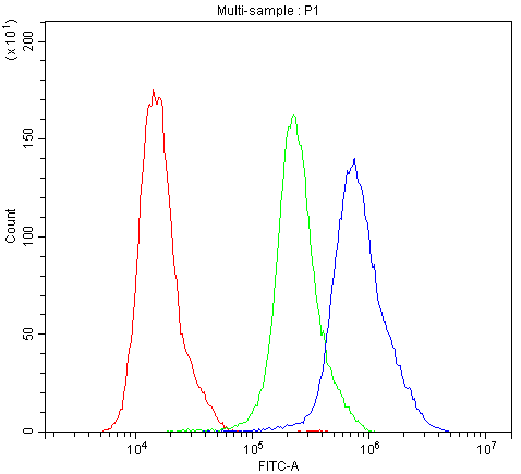
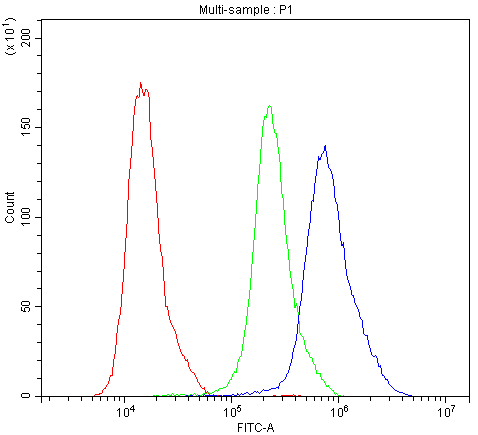 FCM (Flow Cytometry)
(Figure 7. Flow Cytometry analysis of Hela cells using anti-Calbindin antibody (AAA11603).Overlay histogram showing Hela cells stained with AAA11603 (Blue line).The cells were blocked with 10% normal goat serum. And then incubated with rabbit anti-Calbindin Antibody (AAA11603,1ug/1x10^6 cells) for 30 min at 20 degree C. DyLight®488 conjugated goat anti-rabbit IgG (5-10ug/1x10^6 cells) was used as secondary antibody for 30 minutes at 20 degree C. Isotype control antibody (Green line) was rabbit IgG (1ug/1x106) used under the same conditions. Unlabelled sample (Red line) was also used as a control.)
FCM (Flow Cytometry)
(Figure 7. Flow Cytometry analysis of Hela cells using anti-Calbindin antibody (AAA11603).Overlay histogram showing Hela cells stained with AAA11603 (Blue line).The cells were blocked with 10% normal goat serum. And then incubated with rabbit anti-Calbindin Antibody (AAA11603,1ug/1x10^6 cells) for 30 min at 20 degree C. DyLight®488 conjugated goat anti-rabbit IgG (5-10ug/1x10^6 cells) was used as secondary antibody for 30 minutes at 20 degree C. Isotype control antibody (Green line) was rabbit IgG (1ug/1x106) used under the same conditions. Unlabelled sample (Red line) was also used as a control.)
Calbindin, Polyclonal Antibody (Cat# AAA11603)
Full Name
Anti-Calbindin Antibody
Gene Names
CALB1; CALB
Reactivity
Human, Mouse, Rat
Applications
Western Blot, Immunohistochemistry
Purity
Immunogen affinity purified.
Pricing
 IA (Immuno Assay)
(used as capture antibody. Different concentrations were tested (0.5-2 ug/ml).)
IA (Immuno Assay)
(used as capture antibody. Different concentrations were tested (0.5-2 ug/ml).)
Calprotectin, Monoclonal Antibody (Cat# AAA14609)
Full Name
Calprotectin, Human, mAb 27E10, Biotin
Reactivity
Mouse: No; Rhesus Monkey: Yes (subpopulations of macrophages)
Applications
Immunohistochemistry, Immunofluorescence, Flow Cytometry, Immunoassay, Immunoprecipitation, Western Blot
Purity
Protein G
Pricing
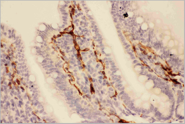
 IF (Immunofluorescence)
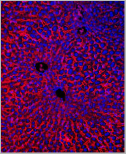
(IF analysis of Band 3 using anti-Band 3 antibody Band 3 was detected in paraffin-embedded section of rat kidney tissues. Heat mediated antigen retrieval was performed in citrate buffer (pH6, epitope retrieval solution ) for 20 mins. The tissue section was blocked with 10% goat serum. The tissue section was then incubated with 1ug/mL rabbit anti-Band 3 Antibody overnight at 4 degree C. DyLight 488 Conjugated Goat Anti-Rabbit IgG was used as secondary antibody at 1:100 dilution and incubated for 30 minutes at 37 degree C. The section was counterstained with DAPI. Visualize using a fluorescence microscope and filter sets appropriate for the label used.)
IF (Immunofluorescence)
(IF analysis of Band 3 using anti-Band 3 antibody Band 3 was detected in paraffin-embedded section of rat kidney tissues. Heat mediated antigen retrieval was performed in citrate buffer (pH6, epitope retrieval solution ) for 20 mins. The tissue section was blocked with 10% goat serum. The tissue section was then incubated with 1ug/mL rabbit anti-Band 3 Antibody overnight at 4 degree C. DyLight 488 Conjugated Goat Anti-Rabbit IgG was used as secondary antibody at 1:100 dilution and incubated for 30 minutes at 37 degree C. The section was counterstained with DAPI. Visualize using a fluorescence microscope and filter sets appropriate for the label used.)
Band 3, Polyclonal Antibody (Cat# AAA11648)
Full Name
Anti-Band 3 Antibody
Gene Names
SLC4A1; DI; FR; SW; WD; WR; AE1; CHC; SAO; WD1; BND3; EPB3; SPH4; CD233; EMPB3; RTA1A
Reactivity
Human, Mouse , Rat.
Applications
Western Blot, Immunohistochemistry
Purity
Immunogen Affinity Purified
Pricing