Filters
Clonality
Type
Reactivity
Gene Name
Isotype
Host
Application
Clone
1009 results for " Cell membrane" - showing 250-300


 WB (Western Blot)
(Western Blot analysis using CD44 antibodyWestern Blot showing using CD44 antibody used against Hela (1) and HUVE-12 (2) cell lysate.)
WB (Western Blot)
(Western Blot analysis using CD44 antibodyWestern Blot showing using CD44 antibody used against Hela (1) and HUVE-12 (2) cell lysate.)
CD44, Monoclonal Antibody (Cat# AAA14314)
Full Name
CD44 antibody
Gene Names
CD44; IN; LHR; MC56; MDU2; MDU3; MIC4; Pgp1; CDW44; CSPG8; HCELL; HUTCH-I; ECMR-III
Applications
Flow Cytometry, Immunofluorescence, Immunohistochemistry, Western Blot
Pricing


 Application Data
(At 25 degree C. Samples were then incubated with primary Ab(At 37 degree C. An AlexaFluor594 conjugated goat anti-rabbit IgG(H+L) Ab(Red) and an AlexaFluor488 conjugated goat anti-mouse IgG(H+L) Ab(Green) were used as the secondary antibody.The nuclear counter stain is DAPI(blue).)
Application Data
(At 25 degree C. Samples were then incubated with primary Ab(At 37 degree C. An AlexaFluor594 conjugated goat anti-rabbit IgG(H+L) Ab(Red) and an AlexaFluor488 conjugated goat anti-mouse IgG(H+L) Ab(Green) were used as the secondary antibody.The nuclear counter stain is DAPI(blue).)
CDK5, Polyclonal Antibody (Cat# AAA31445)
Full Name
Phospho-CDK5 (Ser159) Antibody
Gene Names
CDK5; PSSALRE
Reactivity
Human, Mouse, Rat, Monkey
Predicted Reactivity: Zebrafish (100%), Bovine (100%), Horse (100%), Sheep (100%), Rabbit (100%), Dog (100%), Chicken (100%), Xenopus (100%)
Predicted Reactivity: Zebrafish (100%), Bovine (100%), Horse (100%), Sheep (100%), Rabbit (100%), Dog (100%), Chicken (100%), Xenopus (100%)
Applications
Western Blot, Immunohistochemistry, Immunofluorescence, Immunocytochemistry, Peptide ELISA
Purity
The antibody is from purified rabbit serum by affinity purification via sequential chromatography on phospho-peptide and non-phospho-peptide affinity columns.
Pricing


 WB (Western Blot)
(Applications: Western BlotSample: Rat PFCDetection: Protein Simple, Simple Western size-based assay"We selected this product as a polyclonal ab for our Tg Rat colony. We are interested in adding enzymatic catabolism/clearance of amyloid beta isoforms into our body of research. We purchased this product because it was cheap and we are looking at some 'proof-of-concept' and didn't need remarkable specificity. We still have much to investigate.")
WB (Western Blot)
(Applications: Western BlotSample: Rat PFCDetection: Protein Simple, Simple Western size-based assay"We selected this product as a polyclonal ab for our Tg Rat colony. We are interested in adding enzymatic catabolism/clearance of amyloid beta isoforms into our body of research. We purchased this product because it was cheap and we are looking at some 'proof-of-concept' and didn't need remarkable specificity. We still have much to investigate.")
CD10, Polyclonal Antibody (Cat# AAA11606)
Full Name
Anti-CD10 Antibody
Gene Names
MME; NEP; SFE; CD10; CALLA
Reactivity
Human, Mouse, Rat
Applications
Western Blot, Immunohistochemistry
Purity
Immunogen affinity purified.
Pricing


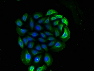
 IF (Immunofluorescence)
(Figure 9. IF analysis of P4HB using anti- P4HB antibody (AAA19258).P4HB was detected in immunocytochemical section of T-47D cells. Enzyme antigen retrieval was performed using IHC enzyme antigen retrieval reagent for 15 mins. The cells were blocked with 10% goat serum. And then incubated with 5μg/mL rabbit anti- P4HB Antibody (AAA19258) overnight at 4 degree C. DyLight®488 Conjugated Goat Anti-Rabbit IgG was used as secondary antibody at 1:100 dilution and incubated for 30 minutes at 37 degree C. The section was counterstained with DAPI. Visualize using a fluorescence microscope and filter sets appropriate for the label used.)
IF (Immunofluorescence)
(Figure 9. IF analysis of P4HB using anti- P4HB antibody (AAA19258).P4HB was detected in immunocytochemical section of T-47D cells. Enzyme antigen retrieval was performed using IHC enzyme antigen retrieval reagent for 15 mins. The cells were blocked with 10% goat serum. And then incubated with 5μg/mL rabbit anti- P4HB Antibody (AAA19258) overnight at 4 degree C. DyLight®488 Conjugated Goat Anti-Rabbit IgG was used as secondary antibody at 1:100 dilution and incubated for 30 minutes at 37 degree C. The section was counterstained with DAPI. Visualize using a fluorescence microscope and filter sets appropriate for the label used.)
P4HB, Polyclonal Antibody (Cat# AAA19258)
Full Name
Anti-P4HB Antibody
Gene Names
P4HB; DSI; GIT; PDI; PHDB; PDIA1; PO4DB; PO4HB; PROHB; ERBA2L; P4Hbeta
Reactivity
Human, Monkey, Mouse, Rat
Applications
Western Blot, Immunohistochemistry, Immunocytochemistry, Immunofluorescence, Flow Cytometry, Direct ELISA
Purity
Immunogen affinity purified.
Pricing


 Application Data
(Immunoperoxidase staining of mouse lymph node cryosection eith Rat anti Mouse antibody clone R3-63 followed by horseradish peroxidase Goat anti Rat IgG antibody . Medium power)
Application Data
(Immunoperoxidase staining of mouse lymph node cryosection eith Rat anti Mouse antibody clone R3-63 followed by horseradish peroxidase Goat anti Rat IgG antibody . Medium power)
CD13, Monoclonal Antibody (Cat# AAA12116)
Full Name
RAT ANTI MOUSE CD13
Gene Names
Anpep; Apn; AP-M; AP-N; Cd13; P150
Applications
Immunohistochemistry, Flow Cytometry, Immunohistochemistry
Pricing


 Application Data
(Staining of rat thymus cells with Mouse anti Rat CD90 (THY1.1):Alexa Fluor 488)
Application Data
(Staining of rat thymus cells with Mouse anti Rat CD90 (THY1.1):Alexa Fluor 488)
CD90, Monoclonal Antibody (Cat# AAA11909)
Full Name
MOUSE ANTI RAT CD90:Low Endotoxin
Gene Names
Thy1; CD7
Applications
Immunohistochemistry, Flow Cytometry, Immunoprecipitation, Western Blot
Pricing
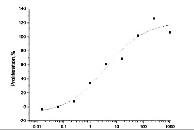 Activity
(The ED(50) was determined by the dose-dependent proliferation of human MCF-7 cells was found to be )
Activity
(The ED(50) was determined by the dose-dependent proliferation of human MCF-7 cells was found to be )
Neuregulin 1 beta, Active Protein (Cat# AAA14425)
Full Name
Human Neuregulin 1 beta Recombinant
Purity
>95%, as determined by SDS-PAGE
Pricing


 Application Data
(Staining of NALM6 cells with MOUSE ANTI HUMAN CD10:ALEXA 488)
Application Data
(Staining of NALM6 cells with MOUSE ANTI HUMAN CD10:ALEXA 488)
CD10, Monoclonal Antibody (Cat# AAA26765)
Full Name
CD10 (CALLA, CD 10, Common Acute Lymphocytic Leukemia Antigen, Enkephalinase, gp100, Membrane Metalloendopeptidase, MME, Neprilysin, Neutral Endopeptidase, Pmel17) (PE)
Gene Names
MME; NEP; SFE; CD10; CALLA
Reactivity
Human
Applications
Flow Cytometry, Immunohistochemistry, Immunoprecipitation, Western Blot
Purity
Purified by protein G affinity chromatography from tissue culture supernatant.
Pricing

 IF (Immunofluorescence)
(ICC/IF analysis of ISG15 in HeLa cells line, stained with DAPI (Blue) for nucleus staining and monoclonal anti-human ISG15 antibody (1:100) with goat anti-mouse IgG-Alexa fluor 488 conjugate (Green).)
IF (Immunofluorescence)
(ICC/IF analysis of ISG15 in HeLa cells line, stained with DAPI (Blue) for nucleus staining and monoclonal anti-human ISG15 antibody (1:100) with goat anti-mouse IgG-Alexa fluor 488 conjugate (Green).)
ISG15, Monoclonal Antibody (Cat# AAA11725)
Full Name
ISG15 antibody
Gene Names
ISG15; G1P2; IP17; UCRP; IFI15; IMD38; hUCRP
Reactivity
Human
Applications
Western Blot, Immunocytochemistry, Immunofluorescence, Flow Cytometry
Purity
By protein-A affinity chromatography
Pricing


 Application Data
(At 25 degree C. Samples were then incubated with primary Ab(At 37 degree C. An AlexaFluor594 conjugated goat anti-rabbit IgG(H+L) Ab(Red) and an AlexaFluor488 conjugated goat anti-mouse IgG(H+L) Ab(Green) were used as the secondary antibody.The nuclear counter stain is DAPI(blue).)
Application Data
(At 25 degree C. Samples were then incubated with primary Ab(At 37 degree C. An AlexaFluor594 conjugated goat anti-rabbit IgG(H+L) Ab(Red) and an AlexaFluor488 conjugated goat anti-mouse IgG(H+L) Ab(Green) were used as the secondary antibody.The nuclear counter stain is DAPI(blue).)
Bcl-2, Polyclonal Antibody (Cat# AAA31360)
Full Name
Phospho-Bcl-2 (Thr56) Antibody
Gene Names
BCL2; Bcl-2; PPP1R50
Reactivity
Human, Mouse, Rat
Predicted Reactivity: Pig (86%), Bovine (83%)
Predicted Reactivity: Pig (86%), Bovine (83%)
Applications
Western Blot, Immunohistochemistry, Immunofluorescence, Immunocytochemistry, Peptide ELISA
Purity
The antibody is from purified rabbit serum by affinity purification via sequential chromatography on phospho-peptide and non-phospho-peptide affinity columns.
Pricing


 IF (Immunofluorescence)
(Immunofluorescence staining of Hela cells with AAA27028 at 1:90, counter-stained with DAPI. The cells were blocked in 10% normal Goat Serum and then incubated with the primary antibody overnight at 4 degree C. The secondary antibody was Alexa Fluor 488-congugated AffiniPure Goat Anti-Mouse IgG(H+L).)
IF (Immunofluorescence)
(Immunofluorescence staining of Hela cells with AAA27028 at 1:90, counter-stained with DAPI. The cells were blocked in 10% normal Goat Serum and then incubated with the primary antibody overnight at 4 degree C. The secondary antibody was Alexa Fluor 488-congugated AffiniPure Goat Anti-Mouse IgG(H+L).)
CD14, Monoclonal Antibody (Cat# AAA27028)
Full Name
CD14 Monoclonal Antibody
Reactivity
Human, Mouse, Rabbit
Applications
Western Blot, Immunohistochemistry, Immunofluorescence, Flow Cytometry
Purity
>95%, Protein A Purified
Pricing
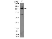


 WB (Western Blot)
(Western Blot Analysis: Representative lot data. Lysate from HEK-293 cells was resolved by electrophoresis, transferred to PVDF and probed with anti-PI3 Kinase, p110beta (0.05 ug/mL). Proteins were visualized using donkey anti-rabbit secondary antibody conjugated to HRP and chemiluminescence detection. Arrow indicates PI3 Kinase, p110beta (~110kD).)
WB (Western Blot)
(Western Blot Analysis: Representative lot data. Lysate from HEK-293 cells was resolved by electrophoresis, transferred to PVDF and probed with anti-PI3 Kinase, p110beta (0.05 ug/mL). Proteins were visualized using donkey anti-rabbit secondary antibody conjugated to HRP and chemiluminescence detection. Arrow indicates PI3 Kinase, p110beta (~110kD).)
Phosphoinositide 3 Kinase, p110 beta, Polyclonal Antibody (Cat# AAA26903)
Full Name
Phosphoinositide 3 Kinase, p110 beta (DKFZp779K1237, MGC133043, p110beta, Phosphatidylinositol 3 Kinase Catalytic beta Polypeptide, Phosphatidylinositol-4,5-bisphosphate 3-kinase Catalytic Subunit beta Isoform, Phosphoinositide 3 Kinase Catalytic beta Pol
Gene Names
PIK3CA; MCM; CWS5; MCAP; PI3K; CLOVE; MCMTC; p110-alpha
Reactivity
Human
Applications
Immunocytochemistry, Western Blot, Immunoprecipitation, Immunohistochemistry
Purity
Purified by Immunoaffinity chromatography.
Pricing


 FCM (Flow Cytometry)
(Figure 9. Flow Cytometry analysis of SiHa cells using anti-DCTN1/p150-glued antibody (AAA19253).Overlay histogram showing SiHa cells stained with AAA19253 (Blue line). The cells were blocked with 10% normal goat serum. And then incubated with rabbit anti-DCTN1/p150-glued Antibody (AAA19253, 1μg/1x106 cells) for 30 min at 20 degree C. DyLight®488 conjugated goat anti-rabbit IgG (5-10μg/1x106 cells) was used as secondary antibody for 30 minutes at 20 degree C. Isotype control antibody (Green line) was rabbit IgG (1μg/1x106) used under the same conditions. Unlabelled sample (Red line) was also used as a control.)
FCM (Flow Cytometry)
(Figure 9. Flow Cytometry analysis of SiHa cells using anti-DCTN1/p150-glued antibody (AAA19253).Overlay histogram showing SiHa cells stained with AAA19253 (Blue line). The cells were blocked with 10% normal goat serum. And then incubated with rabbit anti-DCTN1/p150-glued Antibody (AAA19253, 1μg/1x106 cells) for 30 min at 20 degree C. DyLight®488 conjugated goat anti-rabbit IgG (5-10μg/1x106 cells) was used as secondary antibody for 30 minutes at 20 degree C. Isotype control antibody (Green line) was rabbit IgG (1μg/1x106) used under the same conditions. Unlabelled sample (Red line) was also used as a control.)
DCTN1/p150-glued, Polyclonal Antibody (Cat# AAA19253)
Full Name
Anti-DCTN1/p150-glued Antibody
Gene Names
DCTN1; P135; DP-150; DAP-150
Reactivity
Human, Mouse, Rat
Applications
Western Blot, Immunohistochemistry, Immunocytochemistry, Immunofluorescence, Flow Cytometry, Direct ELISA
Purity
Immunogen affinity purified.
Pricing


 ICC (Immunocytochemistry)
(ICC staining CD14 in LO2 cells (green). The nuclear counter stain is DAPI (blue). Cells were fixed in paraformaldehyde, permeabilised with 0.25% Triton X100/PBS.)
ICC (Immunocytochemistry)
(ICC staining CD14 in LO2 cells (green). The nuclear counter stain is DAPI (blue). Cells were fixed in paraformaldehyde, permeabilised with 0.25% Triton X100/PBS.)
CD14, Monoclonal Antibody (Cat# AAA30146)
Full Name
CD14 Antibody
Reactivity
Human
Applications
Western Blot, Immunocytochemistry, Immunohistochemistry
Purity
ProA affinity purified
Pricing


 FCM (Flow Cytometry)
(Flow cytometric analysis of Siha cells with Cellubrevin antibody at 1/100 dilution (yellow) compared with an unlabelled control (cells without incubation with primary antibody; purple).Alexa Fluor 488-conjugated goat anti-rabbit IgG was used as the secondary antibody.)
FCM (Flow Cytometry)
(Flow cytometric analysis of Siha cells with Cellubrevin antibody at 1/100 dilution (yellow) compared with an unlabelled control (cells without incubation with primary antibody; purple).Alexa Fluor 488-conjugated goat anti-rabbit IgG was used as the secondary antibody.)
Cellubrevin, Monoclonal Antibody (Cat# AAA30492)
Full Name
Cellubrevin Antibody
Gene Names
VAMP3; CEB
Reactivity
Human, Mouse, Rat
Applications
Western Blot, Immunocytochemistry, Immunofluorescence, Immunohistochemistry, Flow Cytometry
Purity
ProA affinity purified
Pricing

 Application Data
(N.B. Appropriate control samples should always be included in any labeling studies.* For optimal results in various applications, it is recommended that each investigator determine dilutions appropriate for individual use.)
Application Data
(N.B. Appropriate control samples should always be included in any labeling studies.* For optimal results in various applications, it is recommended that each investigator determine dilutions appropriate for individual use.)
CD59, Monoclonal Antibody (Cat# AAA14271)
Full Name
Anti-Human CD59, (FITC) (Clone 1F5) (mouse IgG1)
Gene Names
CD59; 1F5; EJ16; EJ30; EL32; G344; MIN1; MIN2; MIN3; MIRL; HRF20; MACIF; MEM43; MIC11; MSK21; 16.3A5; HRF-20; MAC-IP; p18-20
Reactivity
Human
Applications
Immunohistochemistry
Pricing


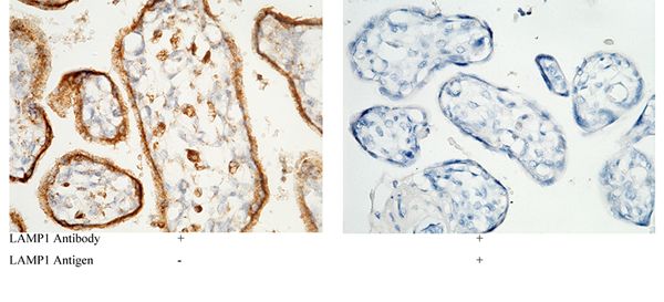
 IP (Immunoprecipitation)
(LAMP1 was immunoprecipitated using:Lane A:0.5 mg Jurkat Whole Cell Lysate1 uL anti-LAMP1 rabbit monoclonal antibody and 15 ul of 50 % Protein G agarose.Primary antibody:Anti-LAMP1 rabbit monoclonal antibody,at 1:500 dilution Secondary antibody:Dylight 800-labeled antibody to rabbit IgG (H+L), at 1:5000 dilution Developed using the odssey technique.Performed under reducing conditions.Predicted band size: 45 kDaObserved band size: 113 kDa)
IP (Immunoprecipitation)
(LAMP1 was immunoprecipitated using:Lane A:0.5 mg Jurkat Whole Cell Lysate1 uL anti-LAMP1 rabbit monoclonal antibody and 15 ul of 50 % Protein G agarose.Primary antibody:Anti-LAMP1 rabbit monoclonal antibody,at 1:500 dilution Secondary antibody:Dylight 800-labeled antibody to rabbit IgG (H+L), at 1:5000 dilution Developed using the odssey technique.Performed under reducing conditions.Predicted band size: 45 kDaObserved band size: 113 kDa)
LAMP1, Monoclonal Antibody (Cat# AAA27745)
Full Name
Recombinant Anti-LAMP1 Antibody, Rabbit Monoclonal
Gene Names
LAMP1; LAMPA; CD107a; LGP120
Reactivity
Human
Applications
Western Blot, Immunohistochemistry, Flow Cytometry, Immunocytochemistry, Immunofluorescence, Immunoprecipitation
Purity
Protein A
Pricing


 FCM (Flow Cytometry)
(Figure 8. Flow Cytometry analysis of U251 cells using anti-GABARAP antibody (AAA19250).Overlay histogram showing U251 cells stained with AAA19250 (Blue line). The cells were blocked with 10% normal goat serum. And then incubated with rabbit anti-GABARAP Antibody (AAA19250, 1μg/1x106 cells) for 30 min at 20 degree C. DyLight®488 conjugated goat anti-rabbit IgG (5-10μg/1x106 cells) was used as secondary antibody for 30 minutes at 20 degree C. Isotype control antibody (Green line) was rabbit IgG (1μg/1x106) used under the same conditions. Unlabelled sample (Red line) was also used as a control.)
FCM (Flow Cytometry)
(Figure 8. Flow Cytometry analysis of U251 cells using anti-GABARAP antibody (AAA19250).Overlay histogram showing U251 cells stained with AAA19250 (Blue line). The cells were blocked with 10% normal goat serum. And then incubated with rabbit anti-GABARAP Antibody (AAA19250, 1μg/1x106 cells) for 30 min at 20 degree C. DyLight®488 conjugated goat anti-rabbit IgG (5-10μg/1x106 cells) was used as secondary antibody for 30 minutes at 20 degree C. Isotype control antibody (Green line) was rabbit IgG (1μg/1x106) used under the same conditions. Unlabelled sample (Red line) was also used as a control.)
GABARAP, Polyclonal Antibody (Cat# AAA19250)
Full Name
Anti-GABARAP Antibody
Gene Names
GABARAP; MM46; ATG8A; GABARAP-a
Reactivity
Human, Mouse, Rat
Applications
Western Blot, Immunohistochemistry, Flow Cytometry
Purity
Immunogen affinity purified.
Pricing


 ICC (Immunocytochemistry)
(Figure 7. IHC analysis of ERAB using anti-ERAB antibody (AAA11668).ERAB was detected in immunocytochemical section of SMMC-7721 cell. Heat mediated antigen retrieval was performed in citrate buffer (pH6, epitope retrieval solution) for 20 mins. The tissue section was blocked with 10% goat serum. The tissue section was then incubated with 1ug/ml rabbit anti-ERAB Antibody (AAA11668) overnight at 4 degree C. Biotinylated goat anti-rabbit IgG was used as secondary antibody and incubated for 30 minutes at 37 degree C. The tissue section was developed using Strepavidin-Biotin-Complex (SABC) with DAB as the chromogen.)
ICC (Immunocytochemistry)
(Figure 7. IHC analysis of ERAB using anti-ERAB antibody (AAA11668).ERAB was detected in immunocytochemical section of SMMC-7721 cell. Heat mediated antigen retrieval was performed in citrate buffer (pH6, epitope retrieval solution) for 20 mins. The tissue section was blocked with 10% goat serum. The tissue section was then incubated with 1ug/ml rabbit anti-ERAB Antibody (AAA11668) overnight at 4 degree C. Biotinylated goat anti-rabbit IgG was used as secondary antibody and incubated for 30 minutes at 37 degree C. The tissue section was developed using Strepavidin-Biotin-Complex (SABC) with DAB as the chromogen.)
ERAB, Polyclonal Antibody (Cat# AAA11668)
Full Name
Anti-ERAB Antibody
Gene Names
HSD17B10; ABAD; CAMR; ERAB; HCD2; MHBD; HADH2; MRPP2; MRX17; MRX31; SCHAD; MRXS10; SDR5C1; 17b-HSD10; DUPXp11.22
Reactivity
Human, Mouse
Applications
Western Blot, Immunohistochemistry
Purity
Immunogen Affinity Purified
Pricing


 Application Data
(Published customer image: Increased accumulation of repair-associated macrophages surrounding collaterals in ischemic hind limbs is PAR2-dependent. (A) Stainings of CD206-positive macrophages (green) and SMA-positive vessels (red) in non-ischemic (control) and ischemic (ligated) hind limbs of WT, PAR1-/- and PAR2-/- mice are shown. Nuclei were visualized with DAPI (blue). Arrows indicate single macrophages in the non-ischemic adductor. Quantification of the average number of repair-associated macrophages per vessel is indicated on the right. (B) Correlation between the number of CD206-positive macrophages in the ischemic tissues and the expression of CD11b and (C) CD115 on monocytes. ** p)
Application Data
(Published customer image: Increased accumulation of repair-associated macrophages surrounding collaterals in ischemic hind limbs is PAR2-dependent. (A) Stainings of CD206-positive macrophages (green) and SMA-positive vessels (red) in non-ischemic (control) and ischemic (ligated) hind limbs of WT, PAR1-/- and PAR2-/- mice are shown. Nuclei were visualized with DAPI (blue). Arrows indicate single macrophages in the non-ischemic adductor. Quantification of the average number of repair-associated macrophages per vessel is indicated on the right. (B) Correlation between the number of CD206-positive macrophages in the ischemic tissues and the expression of CD11b and (C) CD115 on monocytes. ** p)
CD206, Monoclonal Antibody (Cat# AAA12124)
Full Name
RAT ANTI MOUSE CD206:RPE
Gene Names
Mrc1; MR; CD206; AW259686
Applications
Flow Cytometry
Pricing


 Application Data
(Published customer image: Leukocyte infiltration in COX-2-M/-M and COX-2+/+ mice. MPO enzymatic activity (panel A) was statistically similar in COX-2-M/-M and COX-2+/+ livers at 6 h and 24 h post-IRI. Ly-6G+ neutrophil (panel B) and granulocyte (panel C) infiltration were also comparable in COX-2-M/-M and COX-2+/+ livers after IRI. Mac-1+ (panel D) and CD68 (panel E) infiltrating macrophages were significantly reduced in COX-2-M/-M livers at 24 h post-reperfusion, but were statistically indistinguishable in COX-2-M/-M and COX-2+/+ livers at 6 h after IRI. No statistical differences in MMP-9 expression (panel F) could be demonstrated in livers of COX-2-M/-M and COX-2+/+ mice post-IRI. Representative immunostaining (panel G) of infiltrating Ly-6G+ (a,b,e,f) and Mac-1+ (c,d,g,h) leukocytes in livers of COX-2+/+ (a,c,e,g) and COX-2-M/-M (b,d,f,h) mice at 6 h (a to d) and 24 h (e to h) post IRI; (n = 5 -6/group; * indicates p)
Application Data
(Published customer image: Leukocyte infiltration in COX-2-M/-M and COX-2+/+ mice. MPO enzymatic activity (panel A) was statistically similar in COX-2-M/-M and COX-2+/+ livers at 6 h and 24 h post-IRI. Ly-6G+ neutrophil (panel B) and granulocyte (panel C) infiltration were also comparable in COX-2-M/-M and COX-2+/+ livers after IRI. Mac-1+ (panel D) and CD68 (panel E) infiltrating macrophages were significantly reduced in COX-2-M/-M livers at 24 h post-reperfusion, but were statistically indistinguishable in COX-2-M/-M and COX-2+/+ livers at 6 h after IRI. No statistical differences in MMP-9 expression (panel F) could be demonstrated in livers of COX-2-M/-M and COX-2+/+ mice post-IRI. Representative immunostaining (panel G) of infiltrating Ly-6G+ (a,b,e,f) and Mac-1+ (c,d,g,h) leukocytes in livers of COX-2+/+ (a,c,e,g) and COX-2-M/-M (b,d,f,h) mice at 6 h (a to d) and 24 h (e to h) post IRI; (n = 5 -6/group; * indicates p)
CD68, Monoclonal Antibody (Cat# AAA12107)
Full Name
RAT ANTI MOUSE CD68
Gene Names
Cd68; Lamp4; gp110; Scard1
Applications
Immunohistochemistry, Flow Cytometry, Immunofluorescence, Immunoprecipitation, Immunohistochemistry, Western Blot
Purity
Purified
Purified IgG - liquid
Purified IgG - liquid
Pricing
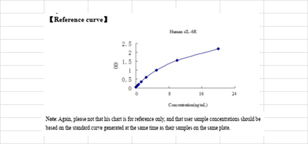 Standard Curve (Sample)
soluble Interleukin 6 Receptor (sIL-6R), ELISA Kit (Cat# AAA12869)
Full Name
Human soluble Interleukin 6 Receptor (sIL-6R) ELISA Kit
Gene Names
IL6R; IL6Q; gp80; CD126; IL6RA; IL6RQ; IL-6RA; IL-6R-1
Reactivity
Human
Pricing


 IP (Immunoprecipitation)
(Immunoprecipitation analysis of 300ug extracts of HeLa cells using 3ug CDK4 antibody . Western blot was performed from the immunoprecipitate using CDK4 antibody at a dilition of 1:1000.)
IP (Immunoprecipitation)
(Immunoprecipitation analysis of 300ug extracts of HeLa cells using 3ug CDK4 antibody . Western blot was performed from the immunoprecipitate using CDK4 antibody at a dilition of 1:1000.)
CDK4, Polyclonal Antibody (Cat# AAA28337)
Full Name
CDK4 Rabbit pAb
Gene Names
CDK4; CMM3; PSK-J3
Reactivity
Human, Mouse, Rat
Applications
Western Blot, Immunohistochemistry, Immunofluorescence, Immunoprecipitation
Purity
Affinity purification
Pricing


 Application Data
(Staining of KG1 lymphocytes with Mouse anti Human CD59:FITC)
Application Data
(Staining of KG1 lymphocytes with Mouse anti Human CD59:FITC)
CD59, Monoclonal Antibody (Cat# AAA11917)
Full Name
MOUSE ANTI HUMAN CD59
Gene Names
CD59; 1F5; EJ16; EJ30; EL32; G344; MIN1; MIN2; MIN3; MIRL; HRF20; MACIF; MEM43; MIC11; MSK21; 16.3A5; HRF-20; MAC-IP; p18-20
Applications
Immunohistochemistry, Fluorescence Microscopy, Flow Cytometry, Immunofluorescence, Immunoprecipitation, Immunohistochemistry, Western Blot
Pricing


 Application Data
(Staining of rat thymus cells with Mouse anti Rat CD90 (THY1.1):Alexa Fluor 488)
Application Data
(Staining of rat thymus cells with Mouse anti Rat CD90 (THY1.1):Alexa Fluor 488)
CD90, Monoclonal Antibody (Cat# AAA11978)
Full Name
MOUSE ANTI RAT CD90
Gene Names
Thy1; CD7
Applications
Immunohistochemistry, Flow Cytometry, Immunofluorescence, Immunoprecipitation, Western Blot
Pricing


 WB (Western Blot)
(MARCH7 monoclonal antibody. Western Blot analysis of 'MARCH7 expression in PC-12.)
WB (Western Blot)
(MARCH7 monoclonal antibody. Western Blot analysis of 'MARCH7 expression in PC-12.)
2019-03-07, Monoclonal Antibody (Cat# AAA25744)
Full Name
MARCH7 (E3 Ubiquitin-protein Ligase MARCH7, Axotrophin, Membrane-associated RING Finger Protein 7, Membrane-associated RING-CH Protein VII, MARCH-VII, RING Finger Protein 177, AXOT, RNF177, DKFZp586F1122) (PE)
Gene Names
MARCH7; AXO; AXOT; RNF177; MARCH-VII
Reactivity
Human, Mouse, Rat
Applications
Immunofluorescence, Western Blot
Purity
Purified by Protein A Affinity Chromatography.
Pricing
 WB (Western Blot)
(Western blot analysis of lysates from HT-29 cell line and mouse spleen tissue lysate(from left to right), using JAK1 Antibody (C-term). AAA28692 was diluted at 1:1000 at each lane. A goat anti-rabbit IgG H&L(HRP) at 1:5000 dilution was used as the secondary antibody. Lysates at 35ug per lane.)
WB (Western Blot)
(Western blot analysis of lysates from HT-29 cell line and mouse spleen tissue lysate(from left to right), using JAK1 Antibody (C-term). AAA28692 was diluted at 1:1000 at each lane. A goat anti-rabbit IgG H&L(HRP) at 1:5000 dilution was used as the secondary antibody. Lysates at 35ug per lane.)
JAK1, Polyclonal Antibody (Cat# AAA28692)
Full Name
JAK1 Antibody (C-term)
Gene Names
JAK1; JTK3; JAK1A; JAK1B
Reactivity
Human, mouse
Applications
Western Blot
Purity
Purified Rabbit Polyclonal Antibody (Pab)
Pricing


 FCM (Flow Cytometry)
(Flow cytometric analysis of MCF-7 cells with GRASP65 antibody at 1/50 dilution (red) compared with an unlabelled control (cells without incubation with primary antibody; black). Alexa Fluor 488-conjugated goat anti-rabbit IgG was used as the secondary antibody.)
FCM (Flow Cytometry)
(Flow cytometric analysis of MCF-7 cells with GRASP65 antibody at 1/50 dilution (red) compared with an unlabelled control (cells without incubation with primary antibody; black). Alexa Fluor 488-conjugated goat anti-rabbit IgG was used as the secondary antibody.)
GRASP65, Monoclonal Antibody (Cat# AAA30439)
Full Name
GRASP65 Antibody
Gene Names
GORASP1; P65; GOLPH5; GRASP65
Reactivity
Human
Applications
Western Blot, Immunocytochemistry, Immunofluorescence, Immunohistochemistry, Flow Cytometry, Immunoprecipitation
Purity
ProA affinity purified
Pricing


 ICC (Immunocytochemistry)
(ICC staining EpCAM in SW1990 cells (green). The nuclear counter stain is DAPI (blue). Cells were fixed in paraformaldehyde, permeabilised with 0.25% Triton X100/PBS.)
ICC (Immunocytochemistry)
(ICC staining EpCAM in SW1990 cells (green). The nuclear counter stain is DAPI (blue). Cells were fixed in paraformaldehyde, permeabilised with 0.25% Triton X100/PBS.)
EP-CAM, Monoclonal Antibody (Cat# AAA29891)
Full Name
EP-CAM Antibody
Gene Names
EPCAM; ESA; KSA; M4S1; MK-1; DIAR5; EGP-2; EGP40; KS1/4; MIC18; TROP1; EGP314; HNPCC8; TACSTD1
Reactivity
Human, Mouse
Applications
Western Blot, Immunocytochemistry, Immunohistochemistry, Flow Cytometry
Purity
ProL affinity purified
Pricing


 FCM (Flow Cytometry)
(Figure 6. Flow Cytometry analysis of PC-3 cells using anti-ATG14L antibody (AAA11658).Overlay histogram showing PC-3 cells stained with AAA11658 (Blue line).The cells were blocked with 10% normal goat serum. And then incubated with rabbit anti-ATG14L Antibody (AAA11658,1ug/1x10^6 cells) for 30 min at 20 degree C. DyLight®488 conjugated goat anti-rabbit IgG (5-10ug/1x10^6 cells) was used as secondary antibody for 30 minutes at 20 degree C. Isotype control antibody (Green line) was rabbit IgG (1ug/1x106) used under the same conditions. Unlabelled sample (Red line) was also used as a control.)
FCM (Flow Cytometry)
(Figure 6. Flow Cytometry analysis of PC-3 cells using anti-ATG14L antibody (AAA11658).Overlay histogram showing PC-3 cells stained with AAA11658 (Blue line).The cells were blocked with 10% normal goat serum. And then incubated with rabbit anti-ATG14L Antibody (AAA11658,1ug/1x10^6 cells) for 30 min at 20 degree C. DyLight®488 conjugated goat anti-rabbit IgG (5-10ug/1x10^6 cells) was used as secondary antibody for 30 minutes at 20 degree C. Isotype control antibody (Green line) was rabbit IgG (1ug/1x106) used under the same conditions. Unlabelled sample (Red line) was also used as a control.)
ATG14L, Polyclonal Antibody (Cat# AAA11658)
Full Name
Anti-ATG14L Antibody
Gene Names
ATG14; ATG14L; BARKOR; KIAA0831
Reactivity
Human, Rat
Applications
Western Blot, Immunohistochemistry
Purity
Immunogen Affinity Purified
Pricing


 Application Data
(At 25 degree C. The primary antibody was diluted at 1/200 and incubated with the sample for 1 hour at 37 degree C. An Alexa Fluor 594 conjugated goat anti-rabbit IgG (H+L) antibody(Red), diluted at 1/600, was used as secondary antibody.)
Application Data
(At 25 degree C. The primary antibody was diluted at 1/200 and incubated with the sample for 1 hour at 37 degree C. An Alexa Fluor 594 conjugated goat anti-rabbit IgG (H+L) antibody(Red), diluted at 1/600, was used as secondary antibody.)
JAK3, Polyclonal Antibody (Cat# AAA31404)
Full Name
Phospho-JAK3 (Tyr904) Antibody
Gene Names
JAK3; JAKL; LJAK; JAK-3; L-JAK; JAK3_HUMAN
Reactivity
Human, Mouse, Rat
Predicted Reactivity: Bovine (100%), Sheep (100%), Dog (100%)
Predicted Reactivity: Bovine (100%), Sheep (100%), Dog (100%)
Applications
Western Blot, Immunohistochemistry, Immunofluorescence, Immunocytochemistry, Peptide ELISA
Purity
The antibody is from purified rabbit serum by affinity purification via sequential chromatography on phospho-peptide and non-phospho-peptide affinity columns.
Pricing


 SDS-PAGE
A Disintegrin And Metalloprotease 9 (ADAM9), Recombinant Protein (Cat# AAA20220)
Full Name
Recombinant A Disintegrin And Metalloprotease 9 (ADAM9)
Gene Names
ADAM9; MCMP; MDC9; CORD9; Mltng
Reactivity
Homo sapiens (Human)
Applications
WB
Purity
> 97%
Pricing
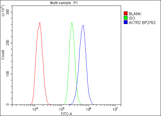


 FCM (Flow Cytometry)
(Figure 6. Flow Cytometry analysis of C6 cells using anti-Arp2/ACTR2 antibody (AAA19284).Overlay histogram showing C6 cells stained with AAA19284 (Blue line). The cells were blocked with 10% normal goat serum. And then incubated with rabbit anti-Arp2/ACTR2 Antibody (AAA19284,1μg/1x106 cells) for 30 min at 20 degree C. DyLight®488 conjugated goat anti-rabbit IgG (5-10μg/1x106 cells) was used as secondary antibody for 30 minutes at 20 degree C. Isotype control antibody (Green line) was rabbit IgG (1μg/1x106) used under the same conditions. Unlabelled sample (Red line) was also used as a control.)
FCM (Flow Cytometry)
(Figure 6. Flow Cytometry analysis of C6 cells using anti-Arp2/ACTR2 antibody (AAA19284).Overlay histogram showing C6 cells stained with AAA19284 (Blue line). The cells were blocked with 10% normal goat serum. And then incubated with rabbit anti-Arp2/ACTR2 Antibody (AAA19284,1μg/1x106 cells) for 30 min at 20 degree C. DyLight®488 conjugated goat anti-rabbit IgG (5-10μg/1x106 cells) was used as secondary antibody for 30 minutes at 20 degree C. Isotype control antibody (Green line) was rabbit IgG (1μg/1x106) used under the same conditions. Unlabelled sample (Red line) was also used as a control.)
Arp2/ACTR2, Polyclonal Antibody (Cat# AAA19284)
Full Name
Anti-Arp2/ACTR2 Antibody
Gene Names
ACTR2; ARP2
Reactivity
Human, Mouse, Monkey, Rat
Applications
WB, IHC-P, ICC, IF, FC/FACS/FCM, EIA
Purity
Immunogen affinity purified.
Pricing


 Application Data
(Published customer image: Leukocyte infiltration in COX-2-M/-M and COX-2+/+ mice. MPO enzymatic activity (panel A) was statistically similar in COX-2-M/-M and COX-2+/+ livers at 6 h and 24 h post-IRI. Ly-6G+ neutrophil (panel B) and granulocyte (panel C) infiltration were also comparable in COX-2-M/-M and COX-2+/+ livers after IRI. Mac-1+ (panel D) and CD68 (panel E) infiltrating macrophages were significantly reduced in COX-2-M/-M livers at 24 h post-reperfusion, but were statistically indistinguishable in COX-2-M/-M and COX-2+/+ livers at 6 h after IRI. No statistical differences in MMP-9 expression (panel F) could be demonstrated in livers of COX-2-M/-M and COX-2+/+ mice post-IRI. Representative immunostaining (panel G) of infiltrating Ly-6G+ (a,b,e,f) and Mac-1+ (c,d,g,h) leukocytes in livers of COX-2+/+ (a,c,e,g) and COX-2-M/-M (b,d,f,h) mice at 6 h (a to d) and 24 h (e to h) post IRI; (n = 5 -6/group; * indicates p)
Application Data
(Published customer image: Leukocyte infiltration in COX-2-M/-M and COX-2+/+ mice. MPO enzymatic activity (panel A) was statistically similar in COX-2-M/-M and COX-2+/+ livers at 6 h and 24 h post-IRI. Ly-6G+ neutrophil (panel B) and granulocyte (panel C) infiltration were also comparable in COX-2-M/-M and COX-2+/+ livers after IRI. Mac-1+ (panel D) and CD68 (panel E) infiltrating macrophages were significantly reduced in COX-2-M/-M livers at 24 h post-reperfusion, but were statistically indistinguishable in COX-2-M/-M and COX-2+/+ livers at 6 h after IRI. No statistical differences in MMP-9 expression (panel F) could be demonstrated in livers of COX-2-M/-M and COX-2+/+ mice post-IRI. Representative immunostaining (panel G) of infiltrating Ly-6G+ (a,b,e,f) and Mac-1+ (c,d,g,h) leukocytes in livers of COX-2+/+ (a,c,e,g) and COX-2-M/-M (b,d,f,h) mice at 6 h (a to d) and 24 h (e to h) post IRI; (n = 5 -6/group; * indicates p)
CD68, Monoclonal Antibody (Cat# AAA12108)
Full Name
RAT ANTI MOUSE CD68:RPE
Gene Names
Cd68; Lamp4; gp110; Scard1
Applications
Flow Cytometry
Pricing
 Standard Curve (Sample)
Platelet Membrane Glycoprotein 2b3a/CD41+CD61, ELISA Kit (Cat# AAA16900)
Full Name
Bovine Platelet Membrane Glycoprotein 2b3a/CD41+CD61 ELISA Kit
Reactivity
Bovine
Pricing


 Application Data
(Staining of NALM6 cells with MOUSE ANTI HUMAN CD10:ALEXA 488)
Application Data
(Staining of NALM6 cells with MOUSE ANTI HUMAN CD10:ALEXA 488)
CD10, Monoclonal Antibody (Cat# AAA26789)
Full Name
CD10 (CALLA, CD 10, Common Acute Lymphocytic Leukemia Antigen, Enkephalinase, gp100, Membrane Metalloendopeptidase, MME, Neprilysin, Neutral Endopeptidase, Pmel17) (MaxLight 490)
Gene Names
MME; NEP; SFE; CD10; CALLA
Reactivity
Human
Applications
Flow Cytometry, Immunohistochemistry, Immunoprecipitation, Western Blot
Purity
Purified by protein G affinity chromatography from tissue culture supernatant.
Pricing


 Application Data
(Staining of human peripheral blood lymphocytes with Mouse anti Human CD20: Pacific Blue)
Application Data
(Staining of human peripheral blood lymphocytes with Mouse anti Human CD20: Pacific Blue)
CD20, Monoclonal Antibody (Cat# AAA11837)
Full Name
MOUSE ANTI HUMAN CD20:APC
Gene Names
MS4A1; B1; S7; Bp35; CD20; CVID5; MS4A2; LEU-16
Applications
Flow Cytometry
Pricing


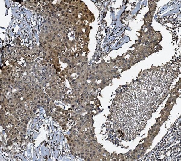
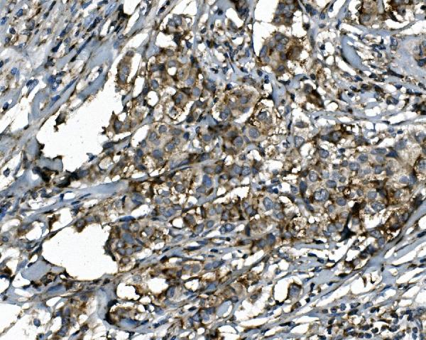
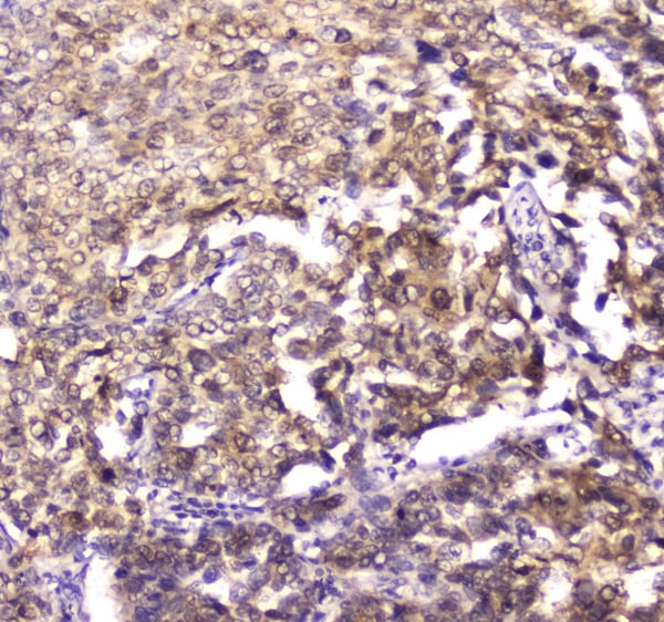
 IHC (Immunohistchemistry)
(Immunohistochemical analysis of paraffin-embedded human-breast-cancer using antibody diluted at 1:50.)
IHC (Immunohistchemistry)
(Immunohistochemical analysis of paraffin-embedded human-breast-cancer using antibody diluted at 1:50.)
CD10, Monoclonal Antibody (Cat# AAA23996)
Full Name
CD10 (7C7) Mouse mAb
Gene Names
MME; NEP; SFE; CD10; CALLA
Reactivity
Human, Rat, Mouse
Applications
IHC-P
Purity
Affinity purified
Pricing


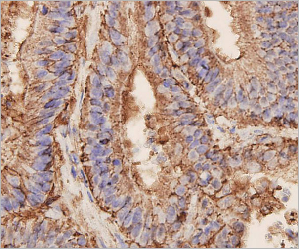
 IHC (Immunohistchemistry)
(Figure 6. IHC analysis of SAE2/UBA2 using anti-SAE2/UBA2 antibody (AAA19171).SAE2/UBA2 was detected in paraffin-embedded section of mouse testis tissue. Heat mediated antigen retrieval was performed in citrate buffer (pH6, epitope retrieval solution) for 20 mins. The tissue section was blocked with 10% goat serum. The tissue section was then incubated with 1ug/ml rabbit anti-SAE2/UBA2 Antibody (AAA19171) overnight at 4 degree C. Biotinylated goat anti-rabbit IgG was used as secondary antibody and incubated for 30 minutes at 37 degree C. The tissue section was developed using Strepavidin-Biotin-Complex (SABC) with DAB as the chromogen.)
IHC (Immunohistchemistry)
(Figure 6. IHC analysis of SAE2/UBA2 using anti-SAE2/UBA2 antibody (AAA19171).SAE2/UBA2 was detected in paraffin-embedded section of mouse testis tissue. Heat mediated antigen retrieval was performed in citrate buffer (pH6, epitope retrieval solution) for 20 mins. The tissue section was blocked with 10% goat serum. The tissue section was then incubated with 1ug/ml rabbit anti-SAE2/UBA2 Antibody (AAA19171) overnight at 4 degree C. Biotinylated goat anti-rabbit IgG was used as secondary antibody and incubated for 30 minutes at 37 degree C. The tissue section was developed using Strepavidin-Biotin-Complex (SABC) with DAB as the chromogen.)
SAE2/UBA2, Polyclonal Antibody (Cat# AAA19171)
Full Name
Anti-SAE2/UBA2 Picoband antibody
Gene Names
UBA2; ARX; SAE2; HRIHFB2115
Reactivity
Human, Mouse, Rat
No cross reactivity with other proteins.
No cross reactivity with other proteins.
Applications
EIA, IHC, WB
Pricing


 FCM (Flow Cytometry)
(Figure 7. Flow Cytometry analysis of Hela cells using anti-Aquaporin 3 antibody (AAA11516).Overlay histogram showing Hela cells stained with AAA11516 (Blue line).The cells were blocked with 10% normal goat serum. And then incubated with rabbit anti-Aquaporin 3 Antibody (AAA11516,1ug/1x10^6 cells) for 30 min at 20 degree C. DyLight®488 conjugated goat anti-rabbit IgG (5-10ug/1x10^6 cells) was used as secondary antibody for 30 minutes at 20 degree C. Isotype control antibody (Green line) was rabbit IgG (1ug/1x106) used under the same conditions. Unlabelled sample (Red line) was also used as a control.)
FCM (Flow Cytometry)
(Figure 7. Flow Cytometry analysis of Hela cells using anti-Aquaporin 3 antibody (AAA11516).Overlay histogram showing Hela cells stained with AAA11516 (Blue line).The cells were blocked with 10% normal goat serum. And then incubated with rabbit anti-Aquaporin 3 Antibody (AAA11516,1ug/1x10^6 cells) for 30 min at 20 degree C. DyLight®488 conjugated goat anti-rabbit IgG (5-10ug/1x10^6 cells) was used as secondary antibody for 30 minutes at 20 degree C. Isotype control antibody (Green line) was rabbit IgG (1ug/1x106) used under the same conditions. Unlabelled sample (Red line) was also used as a control.)
Aquaporin 3, Polyclonal Antibody (Cat# AAA11516)
Full Name
Anti-Aquaporin 3 antibody
Gene Names
AQP3; GIL; AQP-3
Reactivity
Human, Mouse, Rat
Applications
Western Blot, Immunohistochemistry, Immunohistochemistry, Immunocytochemistry
Purity
Immunogen affinity purified.
Pricing


 IF (Immunofluorescence)
(Figure 6. IF analysis of SGK1 using anti- SGK1 antibody (AAA19225).SGK1 was detected in immunocytochemical section of A549 cells. Enzyme antigen retrieval was performed using IHC enzyme antigen retrieval reagent for 15 mins. The cells were blocked with 10% goat serum. And then incubated with 4μg/mL rabbit anti- SGK1 Antibody (AAA19225) overnight at 4 degree C. DyLight®488 Conjugated Goat Anti-Rabbit IgG was used as secondary antibody at 1:100 dilution and incubated for 30 minutes at 37 degree C. The section was counterstained with DAPI. Visualize using a fluorescence microscope and filter sets appropriate for the label used.)
IF (Immunofluorescence)
(Figure 6. IF analysis of SGK1 using anti- SGK1 antibody (AAA19225).SGK1 was detected in immunocytochemical section of A549 cells. Enzyme antigen retrieval was performed using IHC enzyme antigen retrieval reagent for 15 mins. The cells were blocked with 10% goat serum. And then incubated with 4μg/mL rabbit anti- SGK1 Antibody (AAA19225) overnight at 4 degree C. DyLight®488 Conjugated Goat Anti-Rabbit IgG was used as secondary antibody at 1:100 dilution and incubated for 30 minutes at 37 degree C. The section was counterstained with DAPI. Visualize using a fluorescence microscope and filter sets appropriate for the label used.)
SGK1, Polyclonal Antibody (Cat# AAA19225)
Full Name
Anti-SGK1 Antibody
Gene Names
SGK1; SGK
Reactivity
Human
Applications
Western Blot, Immunohistochemistry, Immunocytochemistry, Immunofluorescence, Flow Cytometry
Purity
Immunogen affinity purified.
Pricing


 FCM (Flow Cytometry)
(Figure 8. Flow Cytometry analysis of HEPA1-6 cells using anti-PI-16/PI16 antibody (AAA19330).Overlay histogram showing HEPA1-6 cells stained with AAA19330 (Blue line). The cells were blocked with 10% normal goat serum. And then incubated with rabbit anti-PI-16/PI16 Antibody (AAA19330, 1μg/1x106 cells) for 30 min at 20 degree C. DyLight®488 conjugated goat anti-rabbit IgG (5-10μg/1x106 cells) was used as secondary antibody for 30 minutes at 20 degree C. Isotype control antibody (Green line) was rabbit IgG (1μg/1x106) used under the same conditions. Unlabelled sample (Red line) was also used as a control.)
FCM (Flow Cytometry)
(Figure 8. Flow Cytometry analysis of HEPA1-6 cells using anti-PI-16/PI16 antibody (AAA19330).Overlay histogram showing HEPA1-6 cells stained with AAA19330 (Blue line). The cells were blocked with 10% normal goat serum. And then incubated with rabbit anti-PI-16/PI16 Antibody (AAA19330, 1μg/1x106 cells) for 30 min at 20 degree C. DyLight®488 conjugated goat anti-rabbit IgG (5-10μg/1x106 cells) was used as secondary antibody for 30 minutes at 20 degree C. Isotype control antibody (Green line) was rabbit IgG (1μg/1x106) used under the same conditions. Unlabelled sample (Red line) was also used as a control.)
PI-16/PI16, Polyclonal Antibody (Cat# AAA19330)
Full Name
Anti-PI-16/PI16 Antibody
Gene Names
PI16; PSPBP; CRISP9; MSMBBP
Reactivity
Human, Mouse, Rat, Monkey
Applications
WB, IHC-P, FC/FACS/FCM, EIA
Purity
Immunogen affinity purified.
Pricing


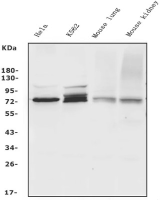
 WB (Western Blot)
(WB Suggested Anti-HCLS1 Antibody Titration: 1.25ug/mlPositive Control: Jurkat cell lysateHCLS1 is supported by BioGPS gene expression data to be expressed in Jurkat)
WB (Western Blot)
(WB Suggested Anti-HCLS1 Antibody Titration: 1.25ug/mlPositive Control: Jurkat cell lysateHCLS1 is supported by BioGPS gene expression data to be expressed in Jurkat)
HCLS1, Polyclonal Antibody (Cat# AAA23399)
Full Name
HCLS1 antibody - N-terminal region
Gene Names
HCLS1; HS1; p75; CTTNL; lckBP1
Reactivity
Cow, Dog, Guinea Pig, Horse, Human, Mouse, Rabbit, Rat
Applications
Immunohistochemistry, Western Blot
Purity
Protein A purified
Pricing


 IF (Immunofluorescence)
(Immunofluorescence analysis of NIH/3T3 cells, untreated(right) and Chloroquine treated(50 ?M, 37? for 20 hours;left) using LC3B antibody at dilution of 1:100. Blue: DAPI for nuclear staining.)
IF (Immunofluorescence)
(Immunofluorescence analysis of NIH/3T3 cells, untreated(right) and Chloroquine treated(50 ?M, 37? for 20 hours;left) using LC3B antibody at dilution of 1:100. Blue: DAPI for nuclear staining.)
LC3B, Monoclonal Antibody (Cat# AAA28305)
Full Name
[KO Validated] LC3B Rabbit mAb
Reactivity
Human, Mouse, Rat
Applications
Western Blot, Immunohistochemistry, Immunofluorescence
Purity
Affinity purification
Pricing
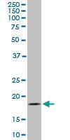


 WB (Western Blot)
(Western Blot analysis of TOMM22 expression in transfected 293T cell line by TOMM22 monoclonal antibody. Lane 1: TOMM22 transfected lysate (15.5kD). Lane 2: Non-transfected lysate.)
WB (Western Blot)
(Western Blot analysis of TOMM22 expression in transfected 293T cell line by TOMM22 monoclonal antibody. Lane 1: TOMM22 transfected lysate (15.5kD). Lane 2: Non-transfected lysate.)
TOMM22, Monoclonal Antibody (Cat# AAA24981)
Full Name
TOMM22 (Translocase of Outer Membrane 22kD Subunit Homolog, Mitochondrial Import Receptor Subunit TOM22 Homolog, TOM22, hTom22, 1C9-2, MST065, MSTP065) (Biotin)
Gene Names
TOMM22; 1C9-2; TOM22; MST065; MSTP065
Reactivity
Human, Mouse, Rat
Applications
EIA, IF, IHC, WB
Purity
Purified by Protein A Affinity Chromatography.
Pricing


 ICC (Immunocytochemistry)
(Immunostaining analysis in HeLa cells. HeLa cells were fixed with 4% paraformaldehyde and permeabilized with 0.1% Triton X-100 in PBS. The cells were immunostained with anti-CDC5L mAb. [Lot No. 2136C1a-1])
ICC (Immunocytochemistry)
(Immunostaining analysis in HeLa cells. HeLa cells were fixed with 4% paraformaldehyde and permeabilized with 0.1% Triton X-100 in PBS. The cells were immunostained with anti-CDC5L mAb. [Lot No. 2136C1a-1])
CDC5L, Monoclonal Antibody (Cat# AAA10605)
Full Name
Mouse monoclonal antibody Anti-Human CDC5L
Gene Names
CDC5L; CDC5; CEF1; PCDC5RP; CDC5-LIKE; dJ319D22.1
Reactivity
Human
Applications
Dot Blot, Immunocytochemistry, Immunoprecipitation, Western Blot, Flow Cytometry
Pricing


 IF (Immunofluorescence)
(ICC/IF analysis of FABP7 in PC3 cells. The cell was stained with AAA11731 (1:100). The secondary antibody (green) was used Alexa Fluor 488. DAPI was stained the cell nucleus (blue).)
IF (Immunofluorescence)
(ICC/IF analysis of FABP7 in PC3 cells. The cell was stained with AAA11731 (1:100). The secondary antibody (green) was used Alexa Fluor 488. DAPI was stained the cell nucleus (blue).)
FABP7, Monoclonal Antibody (Cat# AAA11731)
Full Name
FABP7 antibody
Gene Names
FABP7; MRG; BLBP; FABPB; B-FABP
Reactivity
Human, Mouse
Applications
Western Blot, Immunocytochemistry, Immunofluorescence
Purity
By protein-G affinity chromatography
Pricing


 IF (Immunofluorescence)
(Confocal Immunofluorescent analysis of SK-OV-3 cells using AF488-labeled Isotype Control Monoclonal Antibody (IgG1) (Green).DAPI was used to stain the cell nuclei (blue). (Negative Control))
IF (Immunofluorescence)
(Confocal Immunofluorescent analysis of SK-OV-3 cells using AF488-labeled Isotype Control Monoclonal Antibody (IgG1) (Green).DAPI was used to stain the cell nuclei (blue). (Negative Control))
Ep-CAM /CD326, Monoclonal Antibody (Cat# AAA13838)
Full Name
Ep-CAM /CD326 (Epithelial Marker) Mouse Monoclonal Antibody
Gene Names
EPCAM; ESA; KSA; M4S1; MK-1; DIAR5; EGP-2; EGP40; KS1/4; MIC18; TROP1; EGP314; HNPCC8; TACSTD1
Reactivity
Human.
Does not react with Mouse and Rat
Does not react with Mouse and Rat
Applications
Flow Cytometry, Immunofluorescence, Western Blot, Immunohistochemistry
Pricing


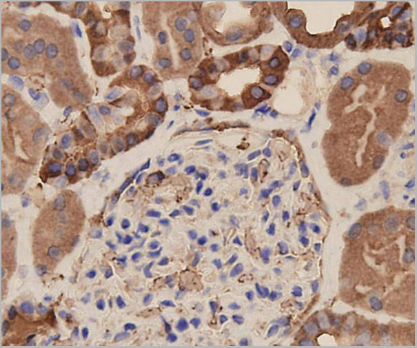
 IHC (Immunohistchemistry)
(Figure 6. IHC analysis of 14-3-3 zeta/delta using anti-14-3-3 zeta/delta antibody (AAA19144).14-3-3 zeta/delta was detected in paraffin-embedded section of rat kidney tissue. Heat mediated antigen retrieval was performed in citrate buffer (pH6, epitope retrieval solution) for 20 mins. The tissue section was blocked with 10% goat serum. The tissue section was then incubated with 1ug/ml rabbit anti-14-3-3 zeta/delta Antibody (AAA19144) overnight at 4 degree C. Biotinylated goat anti-rabbit IgG was used as secondary antibody and incubated for 30 minutes at 37 degree C. The tissue section was developed using Strepavidin-Biotin-Complex (SABC) with DAB as the chromogen.)
IHC (Immunohistchemistry)
(Figure 6. IHC analysis of 14-3-3 zeta/delta using anti-14-3-3 zeta/delta antibody (AAA19144).14-3-3 zeta/delta was detected in paraffin-embedded section of rat kidney tissue. Heat mediated antigen retrieval was performed in citrate buffer (pH6, epitope retrieval solution) for 20 mins. The tissue section was blocked with 10% goat serum. The tissue section was then incubated with 1ug/ml rabbit anti-14-3-3 zeta/delta Antibody (AAA19144) overnight at 4 degree C. Biotinylated goat anti-rabbit IgG was used as secondary antibody and incubated for 30 minutes at 37 degree C. The tissue section was developed using Strepavidin-Biotin-Complex (SABC) with DAB as the chromogen.)
14-3-3 zeta/delta, Polyclonal Antibody (Cat# AAA19144)
Full Name
Anti-14-3-3 zeta/delta Picoband antibody
Gene Names
YWHAZ; HEL4; YWHAD; KCIP-1; HEL-S-3; HEL-S-93; 14-3-3-zeta
Reactivity
Human, Mouse, Rat
No cross reactivity with other proteins.
No cross reactivity with other proteins.
Applications
IHC, WB
Pricing
 FCM (Flow Cytometry)
(Figure 2. Flow Cytometry analysis of A549 cells using anti-WEE1 antibody (AAA19240).Overlay histogram showing A549 cells stained with AAA19240 (Blue line). The cells were blocked with 10% normal goat serum. And then incubated with rabbit anti-WEE1 Antibody (AAA19240, 1μg/1x106 cells) for 30 min at 20 degree C. DyLight®488 conjugated goat anti-rabbit IgG (5-10μg/1x106 cells) was used as secondary antibody for 30 minutes at 20 degree C. Isotype control antibody (Green line) was rabbit IgG (1μg/1x106) used under the same conditions. Unlabelled sample (Red line) was also used as a control.)
FCM (Flow Cytometry)
(Figure 2. Flow Cytometry analysis of A549 cells using anti-WEE1 antibody (AAA19240).Overlay histogram showing A549 cells stained with AAA19240 (Blue line). The cells were blocked with 10% normal goat serum. And then incubated with rabbit anti-WEE1 Antibody (AAA19240, 1μg/1x106 cells) for 30 min at 20 degree C. DyLight®488 conjugated goat anti-rabbit IgG (5-10μg/1x106 cells) was used as secondary antibody for 30 minutes at 20 degree C. Isotype control antibody (Green line) was rabbit IgG (1μg/1x106) used under the same conditions. Unlabelled sample (Red line) was also used as a control.)
WEE1, Polyclonal Antibody (Cat# AAA19240)
Full Name
Anti-WEE1 Antibody
Gene Names
WEE1; WEE1A; WEE1hu
Reactivity
Human, Mouse
Applications
Western Blot, Flow Cytometry, Direct ELISA
Purity
Immunogen affinity purified.
Pricing