Filters
Clonality
Type
Reactivity
Gene Name
Isotype
Host
Application
Clone
3248 results for "Other" - showing 2900-2950
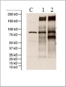
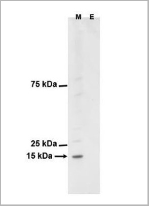
 WB (Western Blot)
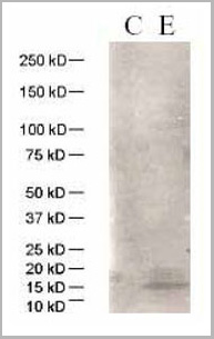
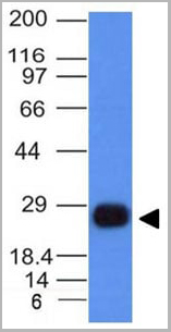
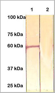
(WB:C: 293 cell extract control E: 293 cell expressing HBeAg antigen (HBV))
WB (Western Blot)
(WB:C: 293 cell extract control E: 293 cell expressing HBeAg antigen (HBV))
HBeAg (HBV), Polyclonal Antibody (Cat# AAA13720)
Full Name
Anti-HBeAg (HBV), rabbit IgG
Applications
Western Blot
Purity
Immunoaffinity chromatography.
Pricing
COVID 19 Nucleocapsid (NP) IgM Humanized Coronavirus, Monoclonal Antibody (Cat# AAA13546)
Full Name
Anti-SARS-CoV-2 Nucleocapsid IgM/COVID-19, Humanized antibody (Positive Control)
Reactivity
Viral
Applications
Lateral Flow
Purity
Purity: >95% by HPLC & SDS-PAGE
Purification: Protein A purified
Purification: Protein A purified
Pricing
Mouse Anti-Monkey IgG, Monoclonal Secondary Antibody (Cat# AAA14895)
Full Name
Mouse Anti-Monkey IgG-HRP
Reactivity
Monkey
Applications
ELISA
Pricing
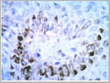
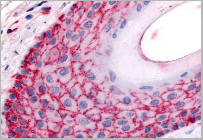
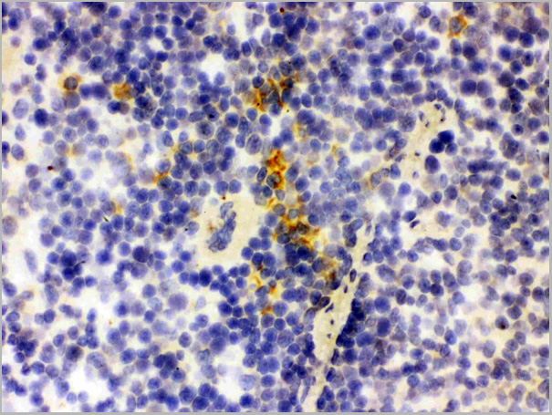
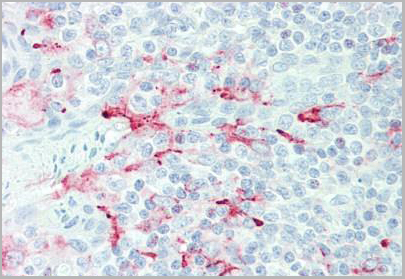
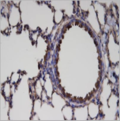
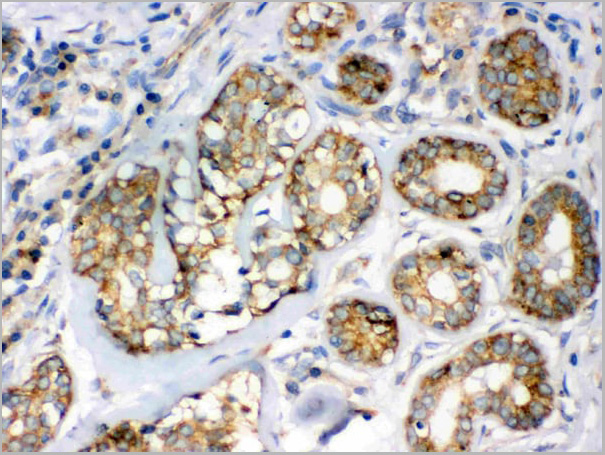
 IHC (Immunohistochemistry)
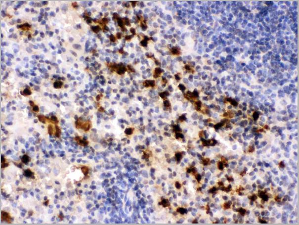
(Anti- S100A9 Picoband antibody, AAA11645, IHC(P)IHC(P): Rat Spleen Tissue)
IHC (Immunohistochemistry)
(Anti- S100A9 Picoband antibody, AAA11645, IHC(P)IHC(P): Rat Spleen Tissue)
S100A9, Polyclonal Antibody (Cat# AAA11645)
Full Name
Anti-S100A9 Antibody
Gene Names
S100a9; p14; Cagb; GAGB; L1Ag; BEE22; MRP14; 60B8Ag; AW546964
Reactivity
Mouse, Rat
Applications
Western Blot, Immunohistochemistry, Immunocytochemistry, Immunofluorescence, Flow Cytometry
Purity
Immunogen Affinity Purified
Pricing
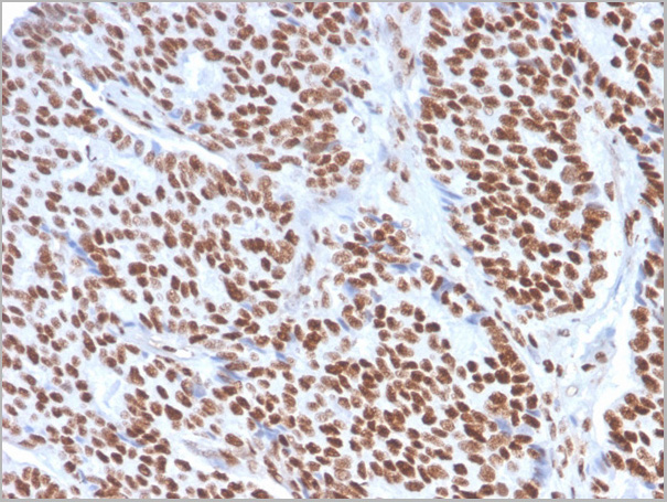
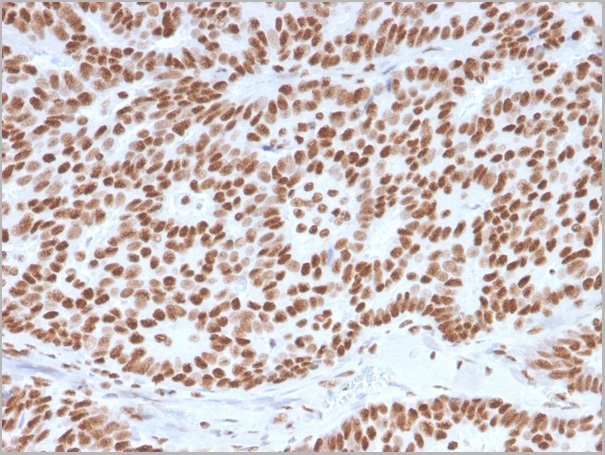
 IHC (Immunohistochemistry)
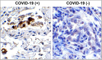
(Immunohistochemistry Validation of SARSCoV-2 (COVID-19) Spike RBD in COVID-19 Patient Lung Immunohistochemical analysis of paraffin-embedded COVID-19 patient lung tissue using anti- SARS-CoV-2 (COVID-19) Spike RBD antibody (AAA11030, 0.5 µg/mL). Tissue was fixed with formaldehyde and blocked with 10% serum for 1 h at RT; antigen retrieval was by heat mediation with a citrate buffer (pH6). Samples were incubated with primary antibody overnight at 4°C. A goat anti-rabbit IgG H&L (HRP) at 1/250 was used as secondary. Counter stained with Hematoxylin. Strong signal of SARS-COV-2 Spike RBD protein was observed in macrophage of COVID-19 patient lung, but not in non-COVID-19 patient lung.)
IHC (Immunohistochemistry)
(Immunohistochemistry Validation of SARSCoV-2 (COVID-19) Spike RBD in COVID-19 Patient Lung Immunohistochemical analysis of paraffin-embedded COVID-19 patient lung tissue using anti- SARS-CoV-2 (COVID-19) Spike RBD antibody (AAA11030, 0.5 µg/mL). Tissue was fixed with formaldehyde and blocked with 10% serum for 1 h at RT; antigen retrieval was by heat mediation with a citrate buffer (pH6). Samples were incubated with primary antibody overnight at 4°C. A goat anti-rabbit IgG H&L (HRP) at 1/250 was used as secondary. Counter stained with Hematoxylin. Strong signal of SARS-COV-2 Spike RBD protein was observed in macrophage of COVID-19 patient lung, but not in non-COVID-19 patient lung.)
COVID 19 Spike RBD Coronavirus, Polyclonal Antibody (Cat# AAA11030)
Full Name
SARS-CoV-2 (COVID-19) Spike RBD Antibody
Gene Names
S; spike glycoprotein
Reactivity
Virus
Applications
Immunofluorescence, Immunohistochemistry, Western Blot
Purity
SARS-CoV-2 (COVID-19) Spike RBD antibody is affinity chromatography purified via peptide column.
Pricing
Endogenous Retrovirus Type K, Envelope Protein, Monoclonal Antibody (Cat# AAA14674)
Full Name
Endogenous Retrovirus Type K, Envelope Protein (HERV K)
Applications
ELISA
Purity
Purified by Protein G affinity chromatography.
Pricing
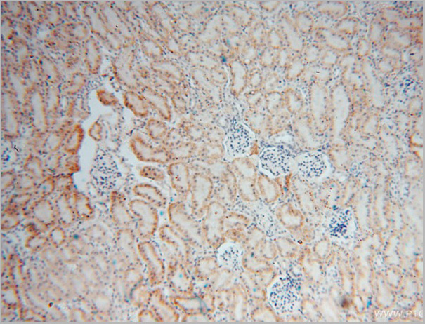
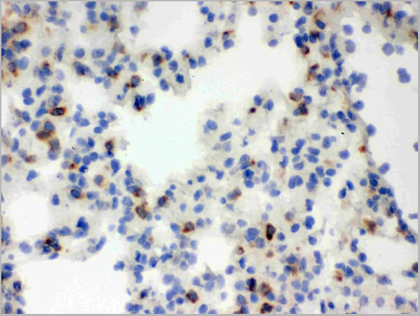
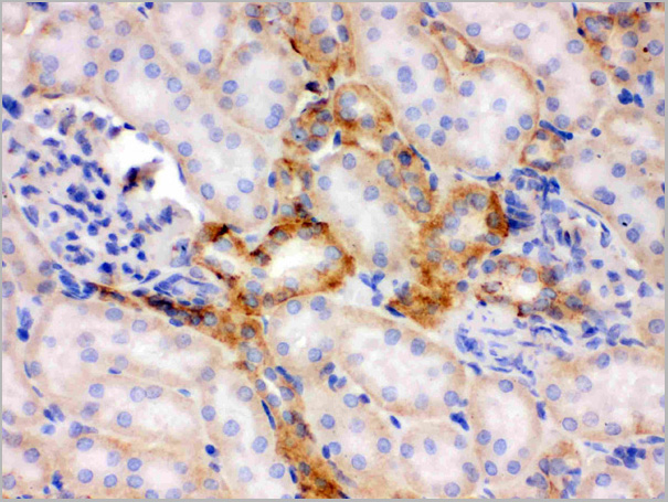
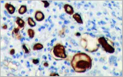 IHC (Immunohistochemistry)
(Immunohistochemistry: Human kidney tissue stained with rabbit Anti-CD133 antibody (Cat# AAA14080) at 1:100 for 10 min @ RT. Staining of formalin-fixed tissue requires boiling tissue sections in 10 mM Citrate Buffer, pH 6.0 for 10 min followed by cooling at RT for 20 min.)
IHC (Immunohistochemistry)
(Immunohistochemistry: Human kidney tissue stained with rabbit Anti-CD133 antibody (Cat# AAA14080) at 1:100 for 10 min @ RT. Staining of formalin-fixed tissue requires boiling tissue sections in 10 mM Citrate Buffer, pH 6.0 for 10 min followed by cooling at RT for 20 min.)
CD133, Antibody (Cat# AAA14080)
Full Name
Rabbit anti CD133 antibody
Reactivity
Human, Mouse, Rat
Applications
Immunohistochemistry, Western Blot, Flow Cytometry, Immunoprecipitation
Purity
The Rabbit IgG is purified by Epitope affinity purification
Pricing
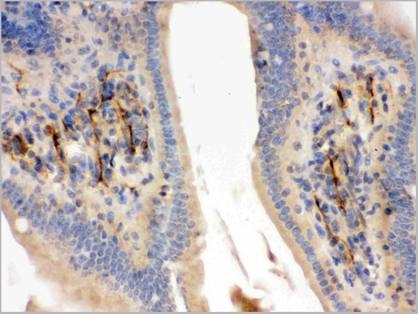
 IHC (Immunohistochemistry)
(IHC staining of distal segment of ligated (7 days) sciatic nerve using AAA14716 at 1:2000 dilution.)
IHC (Immunohistochemistry)
(IHC staining of distal segment of ligated (7 days) sciatic nerve using AAA14716 at 1:2000 dilution.)
Myelin Basic Protein, Degraded, Polyclonal Antibody (Cat# AAA14716)
Full Name
Myelin Basic Protein, Degraded (MBP)
Gene Names
Mbp; Mbps
Applications
Immunohistochemistry
Purity
Serum
Pricing
Borrelia burgdorferi (Lyme) (VlsE), Antigen (Cat# AAA13441)
Full Name
B. burgdorferi VlsE, Recombinant
Applications
Lateral Flow, Western Blot
Purity
95% pure (SDS-PAGE).
Pricing
 SDS-PAGE
Interleukin 31 Receptor A (IL31RA), Recombinant Protein (Cat# AAA20249)
Full Name
Recombinant Interleukin 31 Receptor A (IL31RA)
Gene Names
IL31RA; CRL; GPL; CRL3; GLMR; GLM-R; PLCA2; hGLM-R; IL-31RA; PRO21384
Applications
Western Blot
Purity
> 90%
Pricing
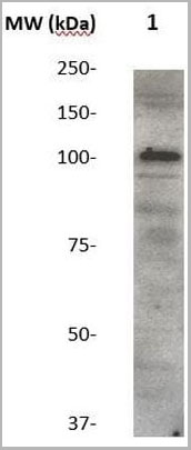
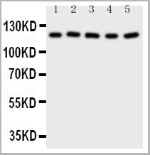 WB (Western Blot)
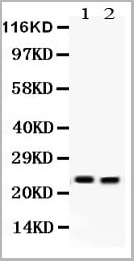
(Anti-Desmoglein 2 antibody, AAA11514, Western blottingAll lanes: Anti Desmoglein 2 (AAA11514) at 0.5ug/mlLane 1: HT1080 Whole Cell Lysate at 40ugLane 2: HELA Whole Cell Lysate at 40ugLane 3: SW620 Whole Cell Lysate at 40ugLane 4: SCG Whole Cell Lysate at 40ugLane 5: COLO320 Whole Cell Lysate at 40ugPredicted bind size: 122KDObserved bind size: 122KD)
WB (Western Blot)
(Anti-Desmoglein 2 antibody, AAA11514, Western blottingAll lanes: Anti Desmoglein 2 (AAA11514) at 0.5ug/mlLane 1: HT1080 Whole Cell Lysate at 40ugLane 2: HELA Whole Cell Lysate at 40ugLane 3: SW620 Whole Cell Lysate at 40ugLane 4: SCG Whole Cell Lysate at 40ugLane 5: COLO320 Whole Cell Lysate at 40ugPredicted bind size: 122KDObserved bind size: 122KD)
Desmoglein 2, Polyclonal Antibody (Cat# AAA11514)
Full Name
Polyclonal Anti-Desmoglein 2, DSG2
Gene Names
DSG2; HDGC; CDHF5; ARVC10; ARVD10; CMD1BB
Reactivity
Human, Mouse, Rat
Applications
Western Blot, Immunohistochemistry
Purity
Immunogen affinity purified
Pricing

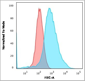
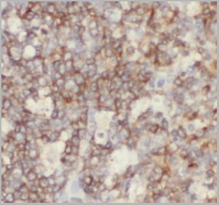
 FCM (Flow Cytometry)
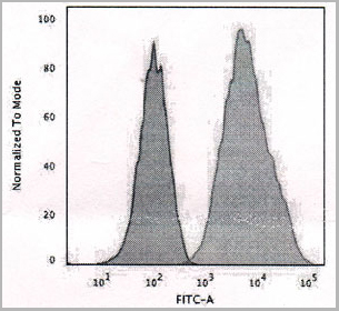
(Flow Cytometric Analysis of human Raji cells using HLA-DR MAb (SPM423) followed by Goat anti-mouse I G-CF488 (Blue); Isotype Control (Red).)
FCM (Flow Cytometry)
(Flow Cytometric Analysis of human Raji cells using HLA-DR MAb (SPM423) followed by Goat anti-mouse I G-CF488 (Blue); Isotype Control (Red).)
HLA-DRB, Monoclonal Antibody (Cat# AAA13826)
Full Name
HLA-DRB (MHC II) Mouse Monoclonal Antibody
Gene Names
HLA-DRB1; SS1; DRB1; DRw10; HLA-DRB; HLA-DR1B
Reactivity
Human, Monkey
Applications
Flow Cytometry, Immunofluorescence, Western Blot, Immunohistochemistry
Pricing
COVID 19 Nucleocapsid (NP) Coronavirus, Recombinant Protein (Cat# AAA13541)
Full Name
Recombinant SARS-Cov-2 Nucleocapsid Protein
Applications
Lateral Flow
Purity
>95% by SDS-PAGE
Pricing
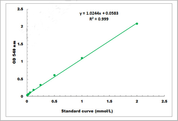
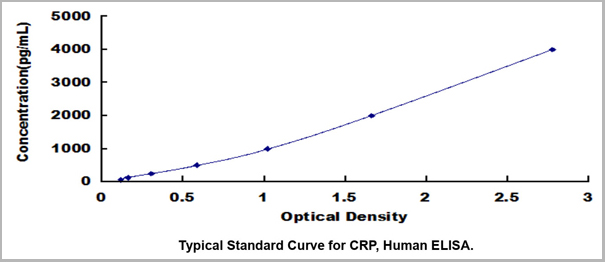 Standard Curve (Sample)
C Reactive Protein (CRP), ELISA Kit (Cat# AAA23109)
Full Name
Human C Reactive Protein (CRP) High Sensitive ELISA Kit
Gene Names
CRP; PTX1
Reactivity
Human
Applications
ELISA
Pricing
Plasmodium falciparum (Malaria) Histidine-Rich Protein 2 (HRP-2), Monoclonal Antibody (Cat# AAA13396)
Full Name
MAb to P. falciparum HRP-2
Applications
Lateral Flow
Purity
Protein A chromatography
Pricing
Morphine, Monoclonal Antibody (Cat# AAA13372)
Full Name
MAb to Morphine
Applications
Lateral Flow
Purity
Purified, Protein G chromatography
Pricing
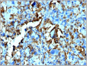
 IHC (Immunohistochemistry)
(High magnification: Paraffin-embedded human skin tissue was prepared using heat-induced epitope retrieval in citrate buffer, pH 6.0. Immunostaining was performed using a 1:100 dilution of AAA14653. Cells lining up under the epidermis consist of numerous mast cells (The dermis has a moderate diffuse inflammatory cell infiltrate).)
IHC (Immunohistochemistry)
(High magnification: Paraffin-embedded human skin tissue was prepared using heat-induced epitope retrieval in citrate buffer, pH 6.0. Immunostaining was performed using a 1:100 dilution of AAA14653. Cells lining up under the epidermis consist of numerous mast cells (The dermis has a moderate diffuse inflammatory cell infiltrate).)
Mast Cell Chymase, Monoclonal Antibody (Cat# AAA14653)
Full Name
Mast Cell Chymase
Gene Names
CMA1; CYH; MCT1; chymase; MGC119890; MGC119891
Reactivity
Human
Applications
Immunohistochemistry
Purity
Purified by Protein G affinity chromatography
Pricing
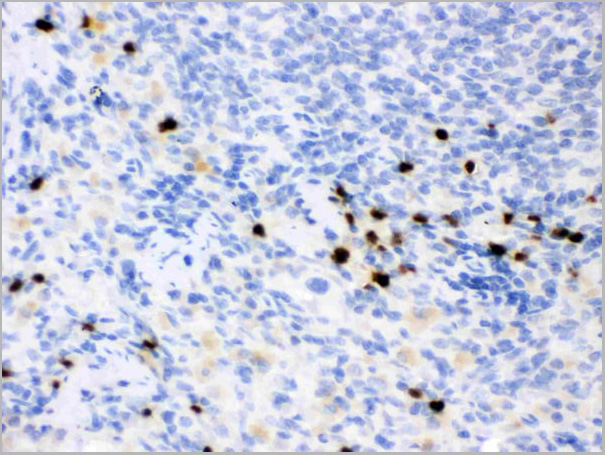
 IHC (Immunohistochemistry)
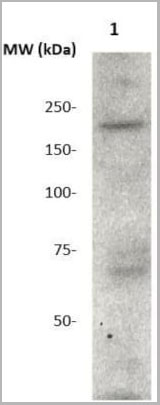
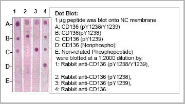
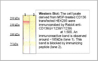
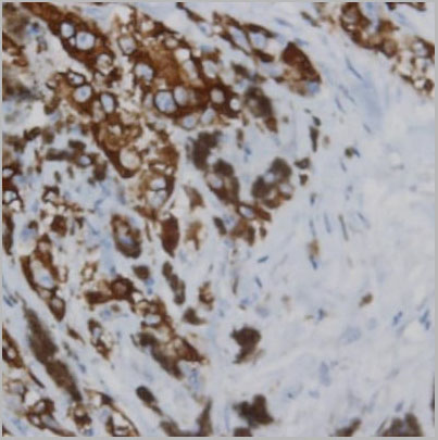
(Immunohistochemistry: Human lymphoma (FFPE) stained with Rabbit anti-CD136 (PY1238/1239) antibody (Cat# AAA14081) at 1:200 for 10 min @ RT. Staining of formalin-fixed tissue requires boiling tissue sections in 10 mM Citrate Buffer, pH 6.0 for 10 min followed by cooling at RT for 20 min.)
IHC (Immunohistochemistry)
(Immunohistochemistry: Human lymphoma (FFPE) stained with Rabbit anti-CD136 (PY1238/1239) antibody (Cat# AAA14081) at 1:200 for 10 min @ RT. Staining of formalin-fixed tissue requires boiling tissue sections in 10 mM Citrate Buffer, pH 6.0 for 10 min followed by cooling at RT for 20 min.)
CD136 (RON) (pY1238/1239), Antibody (Cat# AAA14081)
Full Name
Rabbit anti CD136 Phospho-specific antibody
Reactivity
Human
Applications
Western Blot, Immunohistochemistry, Immunoprecipitation, Flow Cytometry
Purity
The Rabbit IgG is purified by Site-specific Epitope Affinity Purification.
Pricing
 IHC (Immunohistochemistry)
(Anti-Apoptosis inhibitor 5 antibody, AAA11519, IHC(F)IHC(F): Rat Cardiac Muscle Tissue)
IHC (Immunohistochemistry)
(Anti-Apoptosis inhibitor 5 antibody, AAA11519, IHC(F)IHC(F): Rat Cardiac Muscle Tissue)
Apoptosis inhibitor 5, Polyclonal Antibody (Cat# AAA11519)
Full Name
Anti-Apoptosis inhibitor 5 antibody
Gene Names
API5; AAC11; AAC-11
Reactivity
Human, Mouse, Rat
Applications
Western Blot, Immunohistochemistry, Immunohistochemistry, Immunocytochemistry
Purity
Immunogen affinity purified.
Pricing
 IHC (Immunohistochemistry)
(Anti-FAP antibody IHC of human skin, hair follicle. Immunohistochemistry of formalin-fixed, paraffin-embedded tissue after heat-induced antigen retrieval.)
IHC (Immunohistochemistry)
(Anti-FAP antibody IHC of human skin, hair follicle. Immunohistochemistry of formalin-fixed, paraffin-embedded tissue after heat-induced antigen retrieval.)
FAP Alpha, Polyclonal Antibody (Cat# AAA12367)
Full Name
Rabbit Polyclonal to Human FAP Alpha
Gene Names
FAP; FAPA; SIMP; DPPIV
Reactivity
Horse, Human, Monkey
Predicted Reactivity: Bovine, Pig
Predicted Reactivity: Bovine, Pig
Applications
Immunohistochemistry
Purity
Immunoaffinity Purified
Pricing
 Standard Curve (Sample)
Fructosamine, Assay Kit (Cat# AAA27905)
Full Name
Fructosamine Microplate Assay Kit
Reactivity
General
Applications
Functional Assay
Pricing
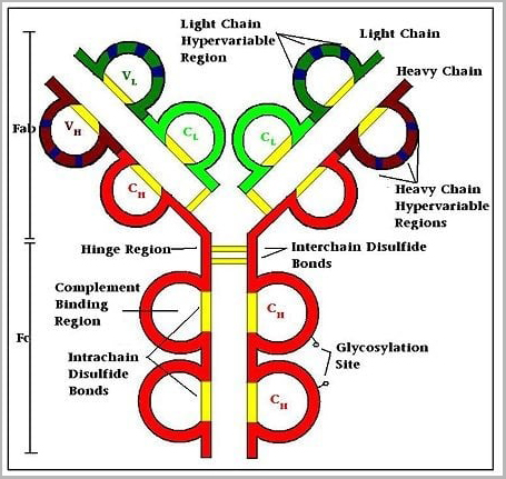 Structure
(Typical Structure of an Antibody)
Structure
(Typical Structure of an Antibody)
Human IgG1 Isotype Control, Monoclonal Isotype Control (Cat# AAA14850)
Full Name
Human IgG1 Isotype Control
Applications
Immunoprecipitation, Flow Cytometry, Immunohistochemistry, Immunocytochemistry
Purity
Purified by fractionation, ion exchange and affinity chromatography; is or >95% (SDS-PAGE).
Pricing
 Application Data
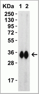
(C: 293 Cell extract control1: 293 cell expressing Spike protein(SARS-CoV BJ02)2: 293 cell expressing Spike protein (Civet SARS-CoV SZ3/2003))
Application Data
(C: 293 Cell extract control1: 293 cell expressing Spike protein(SARS-CoV BJ02)2: 293 cell expressing Spike protein (Civet SARS-CoV SZ3/2003))
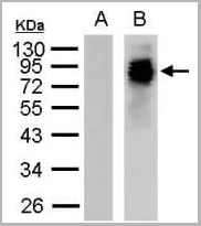
Spike (SARS-CoV), Polyclonal Antibody (Cat# AAA13726)
Full Name
Anti-Spike (SARS-CoV)
Applications
Western Blot
Purity
Immunoaffinity chromatography.
Pricing
 WB (Western Blot)
(Western Blot analysis of 100ng of PEGylated BSA using AAA24071 at 1:30,000 dilution. A: Normal BSA. B: PEGylated BSA.)
WB (Western Blot)
(Western Blot analysis of 100ng of PEGylated BSA using AAA24071 at 1:30,000 dilution. A: Normal BSA. B: PEGylated BSA.)
Polyethylene Glycol, Polyclonal Antibody (Cat# AAA24071)
Full Name
Polyethylene Glycol (PEG)
Applications
Western Blot
Purity
Affinity Purified
Purified by immunoaffinity chromatography
Purified by immunoaffinity chromatography
Pricing
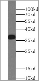 WB (Western Blot)
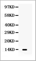
(human heart tissue were subjected to SDS PAGE followed by western blot with AAA27493 (RENALASE antibody) at dilution of 1:2000)
WB (Western Blot)
(human heart tissue were subjected to SDS PAGE followed by western blot with AAA27493 (RENALASE antibody) at dilution of 1:2000)
RENALASE, Monoclonal Antibody (Cat# AAA27493)
Full Name
RENALASE Mouse Monoclonal
Applications
Western Blot, Immunohistochemistry
Purity
Purity: >=95% as determined by SDS-PAGE
Purification: Protein A+G purification
Purification: Protein A+G purification
Pricing
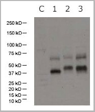
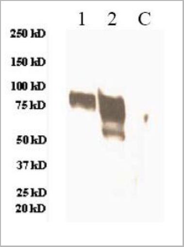 WB (Western Blot)
(C: 293 cell extract control1: 293 cell expressing H3 (A/Wyoming/3/03)(H3N2)2: 293 cell expressing H3 (A/Brisbane/10/07)(H3N2))
WB (Western Blot)
(C: 293 cell extract control1: 293 cell expressing H3 (A/Wyoming/3/03)(H3N2)2: 293 cell expressing H3 (A/Brisbane/10/07)(H3N2))
HAs (A/Wyoming/3/03(H3N2), A/Wisconsin/67/X-161/2005(H3), A/Brisbane/10/2007(H3N2)), Polyclonal Antibody (Cat# AAA13730)
Full Name
Anti-HAs (A/Wyoming/3/03(H3N2), A/Wisconsin/67/X-161/2005(H3), A/Brisbane/10/2007(H3N2)), rabbit IgG
Applications
Western Blot
Purity
Immunoaffinity chromatography
Pricing
Clostridium difficile Glutamate Dehydrogenase (GDH), Recombinant Protein (Cat# AAA13444)
Full Name
Clostridium difficile Glutamate Dehydrogenase (GDH) Recombinant
Applications
Lateral Flow
Purity
> 95% Pure. (Multi-step procedure including affinity chromatography.)
Pricing

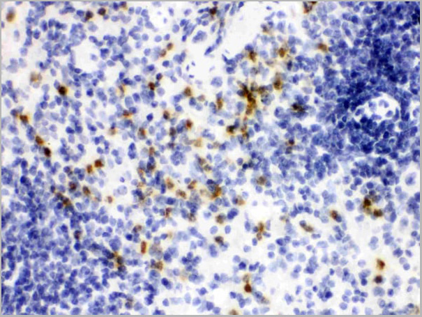
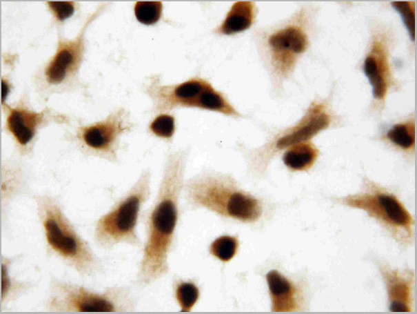
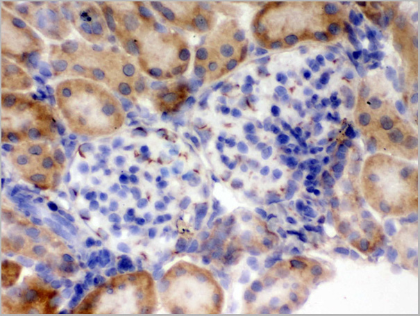
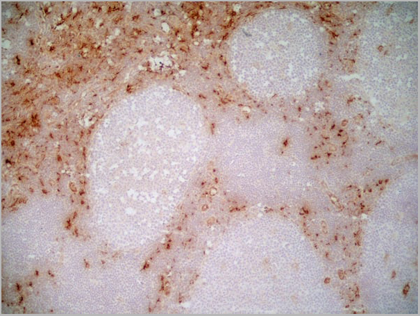
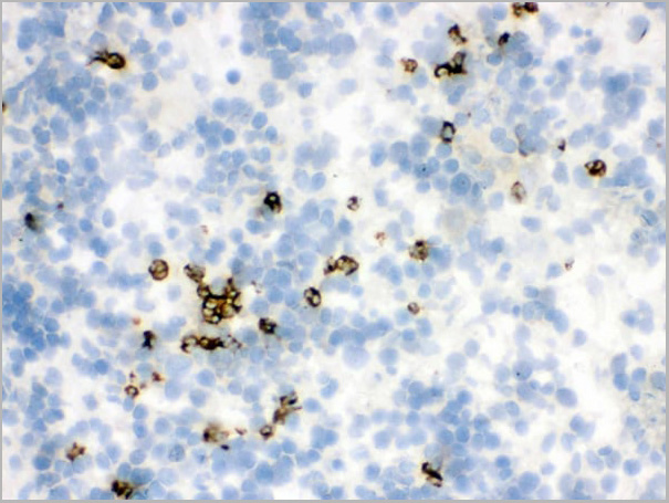 IHC (Immunohistochemistry)
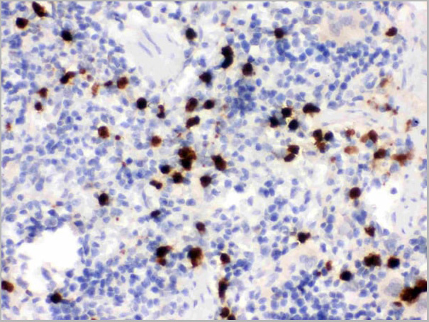
(Figure 7. IHC analysis of Lipocalin 2 using anti-Lipocalin 2 antibody (AAA11663).Lipocalin 2 was detected in frozen section of rat spleen tissue. Heat mediated antigen retrieval was performed in citrate buffer (pH6, epitope retrieval solution) for 20 mins. The tissue section was blocked with 10% goat serum. The tissue section was then incubated with 1ug/ml rabbit anti-Lipocalin 2 Antibody (AAA11663) overnight at 4 degree C. Biotinylated goat anti-rabbit IgG was used as secondary antibody and incubated for 30 minutes at 37 degree C. The tissue section was developed using Strepavidin-Biotin-Complex (SABC) with DAB as the chromogen.)
IHC (Immunohistochemistry)
(Figure 7. IHC analysis of Lipocalin 2 using anti-Lipocalin 2 antibody (AAA11663).Lipocalin 2 was detected in frozen section of rat spleen tissue. Heat mediated antigen retrieval was performed in citrate buffer (pH6, epitope retrieval solution) for 20 mins. The tissue section was blocked with 10% goat serum. The tissue section was then incubated with 1ug/ml rabbit anti-Lipocalin 2 Antibody (AAA11663) overnight at 4 degree C. Biotinylated goat anti-rabbit IgG was used as secondary antibody and incubated for 30 minutes at 37 degree C. The tissue section was developed using Strepavidin-Biotin-Complex (SABC) with DAB as the chromogen.)
Lipocalin 2, Polyclonal Antibody (Cat# AAA11663)
Full Name
Anti-Lipocalin 2 Antibody
Gene Names
Lcn2; Sip24
Reactivity
Human, Mouse, Rat
Applications
Western Blot, Immunohistochemistry, Flow Cytometry
Purity
Immunogen Affinity Purified
Pricing
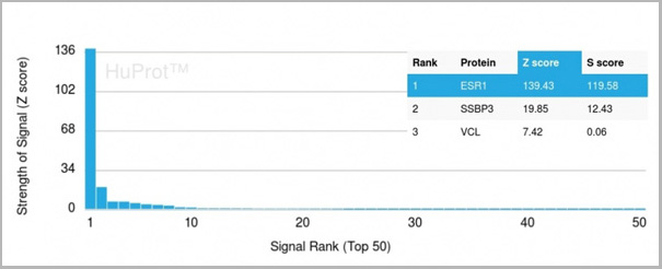 Application Data
(Analysis of Protein Array containing more than 19,000 full-length human proteins using Estrogen Receptor alpha Mouse Monoclonal Antibody (ESR1/3557) Z- and S- Score: The Z-score represents the strength of a signal that a monoclonal antibody (MAb) (in combination with a fluorescently-tagged anti-IgG secondary antibody) produces when binding to a particular protein on the HuProtTM array. Z-scores are described in units of standard deviations (SD’s) above the mean value of all signals generated on that array. If targets on HuProtTM are arranged in descending order of the Z-score, the S-score is the difference (also in units of SD’s) between the Z-score. S-score therefore represents the relative target specificity of a MAb to its intended target. A MAb is considered to specific to its intended target, if the MAb has an S-score of at least 2.5. For example, if a MAb binds to protein X with a Z-score of 43 and to protein Y with a Z-score of 14, then the S-score for the binding of that MAb to protein X is equal to 29.)
Application Data
(Analysis of Protein Array containing more than 19,000 full-length human proteins using Estrogen Receptor alpha Mouse Monoclonal Antibody (ESR1/3557) Z- and S- Score: The Z-score represents the strength of a signal that a monoclonal antibody (MAb) (in combination with a fluorescently-tagged anti-IgG secondary antibody) produces when binding to a particular protein on the HuProtTM array. Z-scores are described in units of standard deviations (SD’s) above the mean value of all signals generated on that array. If targets on HuProtTM are arranged in descending order of the Z-score, the S-score is the difference (also in units of SD’s) between the Z-score. S-score therefore represents the relative target specificity of a MAb to its intended target. A MAb is considered to specific to its intended target, if the MAb has an S-score of at least 2.5. For example, if a MAb binds to protein X with a Z-score of 43 and to protein Y with a Z-score of 14, then the S-score for the binding of that MAb to protein X is equal to 29.)
Estrogen Receptor, alpha, Monoclonal Antibody (Cat# AAA23915)
Full Name
Estrogen Receptor, alpha (Marker of Estrogen Dependence)
Gene Names
ESR1; ER; ESR; Era; ESRA; ESTRR; NR3A1
Reactivity
Human
Applications
Immunofluorescence, Flow Cytometry, Immunohistochemistry
Purity
Purified Ab with BSA and Azide at 200ug/ml OR Purified Ab WITHOUT BSA and Azide at 1.0mg/ml
Pricing
 WB (Western Blot)
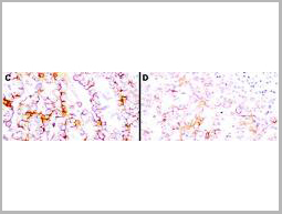
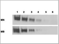
(Anti-Mesothelin Antibodies - Immunohistochemistry. Immunohistochemistry using anti-mesothelin antibodies to detect mesothelin in PEFF human tissue sections treated by antigen retrieval methods. Anti-mesothelin primary antibodies were used to label these sections as follows: C, MAb MB; and D, MAb MN. Reprinted with permission fromClin. Cancer Res. 11(16):5840-6.)
WB (Western Blot)
(Anti-Mesothelin Antibodies - Immunohistochemistry. Immunohistochemistry using anti-mesothelin antibodies to detect mesothelin in PEFF human tissue sections treated by antigen retrieval methods. Anti-mesothelin primary antibodies were used to label these sections as follows: C, MAb MB; and D, MAb MN. Reprinted with permission fromClin. Cancer Res. 11(16):5840-6.)
MSLN / Mesothelin, Monoclonal Antibody (Cat# AAA12374)
Full Name
Mouse Monoclonal [clone MN-1] (IgG) to Human MSLN / Mesothelin
Gene Names
MSLN; MPF; SMRP
Reactivity
Human
Applications
Immunohistochemistry, Western Blot
Purity
Protein A Purified
Pricing
 WB (Western Blot)
(C: 293 cell extract control1: 293 cell expressing H5 (1997) subtype2: 293 cell expressing H5 (2004) subtype3: 293 cell expressing H5 (2005) subtype)
WB (Western Blot)
(C: 293 cell extract control1: 293 cell expressing H5 (1997) subtype2: 293 cell expressing H5 (2004) subtype3: 293 cell expressing H5 (2005) subtype)
HA (H5N1, Human)(A/Vietnam/1203/2004), Antibody (Cat# AAA13715)
Full Name
Anti-HA (H5N1, Human)(A/Vietnam/1203/2004)
Applications
Western Blot
Purity
Immunoaffinity chromatography
Pricing
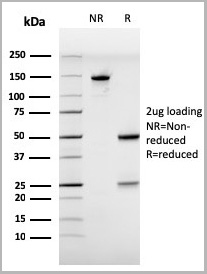
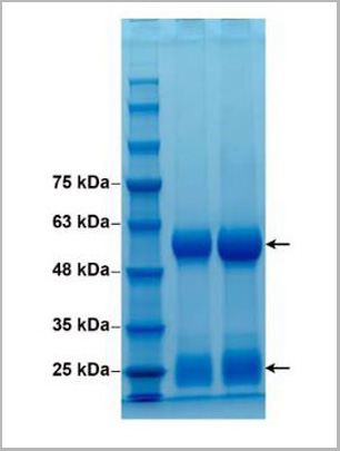 SDS-PAGE
(Figure 2. 10% SDS-PAGE, purified rabbit IgG of anti-SARS-CoV-2 M protein antibody (AAA13787))
SDS-PAGE
(Figure 2. 10% SDS-PAGE, purified rabbit IgG of anti-SARS-CoV-2 M protein antibody (AAA13787))
COVID 19 Membrane Protein Coronavirus, Antibody (Cat# AAA13787)
Full Name
Anti-Membrane Protein (SARS-CoV-2/2019-nCoV) Antibody
Applications
Western Blot
Purity
Protein G chromatography
Pricing
Helicobacter pylori, Native Protein (Cat# AAA14355)
Full Name
Helicobacter pylori protein
Purity
Highly purified
Pricing
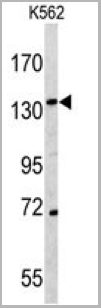
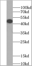 WB (Western Blot)
(HeLa cells were subjected to SDS PAGE followed by western blot with AAA27489 (HLA class I (HLA-A) Antibody) at dilution of 1:4000)
WB (Western Blot)
(HeLa cells were subjected to SDS PAGE followed by western blot with AAA27489 (HLA class I (HLA-A) Antibody) at dilution of 1:4000)
HLA class I (HLA-A), Monoclonal Antibody (Cat# AAA27489)
Full Name
HLA class I (HLA-A) Mouse Monoclonal
Gene Names
HLA-A; HLAA; Aw-33; Aw-74; HLA-A11; HLA-A33; HLA-DQB1; HLA-DRB1
Reactivity
Human
Applications
Western Blot, Flow Cytometry, Immunohistochemistry
Purity
>95% as determined by SDS-PAGE
Protein A+G purification
Protein A+G purification
Pricing
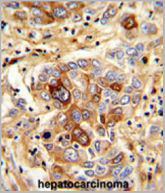 IHC (Immunohistochemistry)
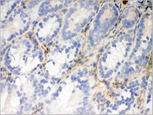
(Immunohistochemistry in formalin-fixed and paraffin-embedded human hepatocarcinoma using AAA24067, which was peroxidase-conjugated to the secondary antibody, followed by DAB staining. This data demonstrates the use of AAA24067 for immunohistochemistry.)
IHC (Immunohistochemistry)
(Immunohistochemistry in formalin-fixed and paraffin-embedded human hepatocarcinoma using AAA24067, which was peroxidase-conjugated to the secondary antibody, followed by DAB staining. This data demonstrates the use of AAA24067 for immunohistochemistry.)
CD163, Polyclonal Antibody (Cat# AAA24067)
Full Name
CD163, NT (CD163, M130, Scavenger receptor cysteine-rich type 1 protein M130, Hemoglobin scavenger receptor, CD163, Soluble CD163)
Gene Names
CD163; M130; MM130
Reactivity
Human
Applications
Western Blot, Immunohistochemistry
Purity
Purified by ammonium sulfate precipitation.
Pricing
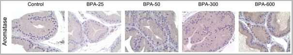
 Application Data
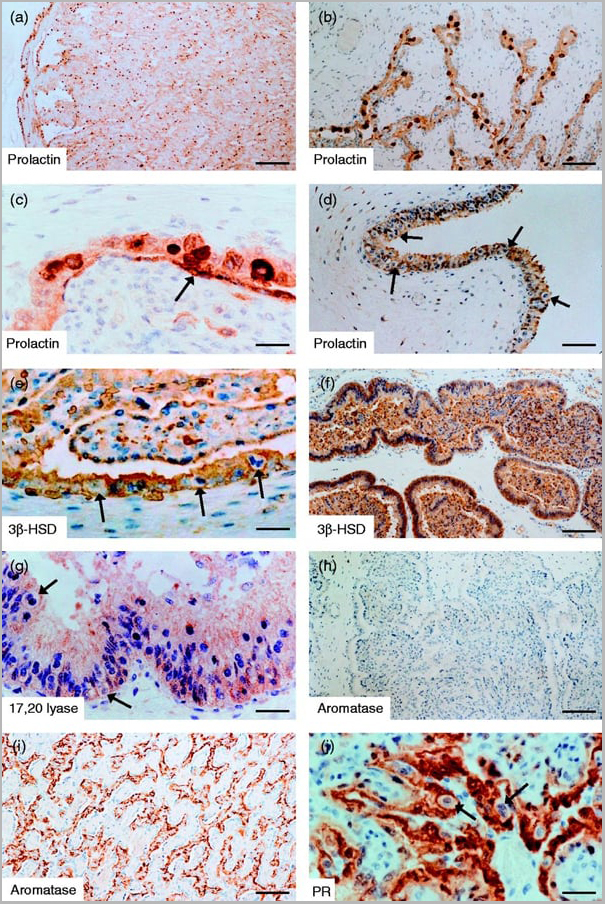
(Published customer image: Mouse anti Human cytochrome p450 aromatase antibody, clone H4 used for the detaction of aromatase in human tissues by Immunohistochemistry on paraffin sectionsImage caption:Morphology and P450 arom immunoreactivity of tumoral region in human testis with seminoma. A-B: Haematoxylin-eosin staining. C-D: Strong P450 arom immunoreactivity in cytoplasm of neoplastic cells (Nc) and unstained lymphocytes (L). Insert: absorption control. Scale bars: A, 20 um; B-C, 12.5 um; D, 5 um.From: Rago V, Romeo F, Aquila S, Montanaro D, And S, Carpino A. Cytochrome P450 aromatase expression in human seminoma. Reprod Biol Endocrinol. 2005 Dec 22;3:72.)
Application Data
(Published customer image: Mouse anti Human cytochrome p450 aromatase antibody, clone H4 used for the detaction of aromatase in human tissues by Immunohistochemistry on paraffin sectionsImage caption:Morphology and P450 arom immunoreactivity of tumoral region in human testis with seminoma. A-B: Haematoxylin-eosin staining. C-D: Strong P450 arom immunoreactivity in cytoplasm of neoplastic cells (Nc) and unstained lymphocytes (L). Insert: absorption control. Scale bars: A, 20 um; B-C, 12.5 um; D, 5 um.From: Rago V, Romeo F, Aquila S, Montanaro D, And S, Carpino A. Cytochrome P450 aromatase expression in human seminoma. Reprod Biol Endocrinol. 2005 Dec 22;3:72.)
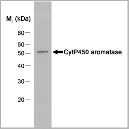
CYTOCHROME P450 AROMATASE, Monoclonal Antibody (Cat# AAA12113)
Full Name
MOUSE ANTI HUMAN CYTOCHROME P450 AROMATASE
Gene Names
CYP19A1; ARO; ARO1; CPV1; CYAR; CYP19; CYPXIX; P-450AROM
Applications
Immunofluorescence, Immunohistochemistry, Western Blot
Pricing


 Application Data
(Published customer image: Mouse anti V5 tag antibody, clone SV5-Pk1 used for the detection of V5 tagged WEEV_nsP3 protein by western blotting and immunofluorescenceImage caption: WEEV nsP3 interaction with host IKKbeta. A) U87MGs were transfected in a 6-well plate with 5 ug of pUC19 and WEEV_nsP3_HA for 24 hours. Cell lysates were resolved using SDS-PAGE and subsequently immunoblotted with V5 antibody and beta-actin served as a loading control. B) U87MGs were transfected with WEEV_nsP3_V5; cells were fixed after 24 hours and stained with antibodies against the endogenous IKKbeta and the V5 tag. Cells were incubated with appropriate secondary Alexa Fluor antibodies and the nuclei stained with DAPI. Co-localization of IKKbeta with WEEV_nsP3_V5 (yellow) was observed as shown by the arrows. B) Panels E -H serve as an example of transfected cells in a given field of view that show co-localization of IKKbeta and WEEV_nsP3_V5 24 hours post transfection. Panels I-L represent magnified images of other cells showing co-localization of IKKbeta and WEEV_nsP3_V5. Panel M is a magnified image of panel L. The co-localization was confirmed by Z-stack analysis. Co-localization was calculated to be approximately in 61% of cells (163 cells were counted of which 44% demonstrated expression of nsP3. Of those cells that expressed nsP3, 61% showed co-localization of both proteins). Images were taken using Nikon Eclipse TE2000-U at 60x magnification and are representative of 2 independent experiments.From: Amaya M, Voss K, Sampey G, Senina S, de la Fuente C, et al. (2014) The Role of IKKbeta in Venezuelan Equine Encephalitis Virus Infection. PLoS ONE 9(2): e86745.)
Application Data
(Published customer image: Mouse anti V5 tag antibody, clone SV5-Pk1 used for the detection of V5 tagged WEEV_nsP3 protein by western blotting and immunofluorescenceImage caption: WEEV nsP3 interaction with host IKKbeta. A) U87MGs were transfected in a 6-well plate with 5 ug of pUC19 and WEEV_nsP3_HA for 24 hours. Cell lysates were resolved using SDS-PAGE and subsequently immunoblotted with V5 antibody and beta-actin served as a loading control. B) U87MGs were transfected with WEEV_nsP3_V5; cells were fixed after 24 hours and stained with antibodies against the endogenous IKKbeta and the V5 tag. Cells were incubated with appropriate secondary Alexa Fluor antibodies and the nuclei stained with DAPI. Co-localization of IKKbeta with WEEV_nsP3_V5 (yellow) was observed as shown by the arrows. B) Panels E -H serve as an example of transfected cells in a given field of view that show co-localization of IKKbeta and WEEV_nsP3_V5 24 hours post transfection. Panels I-L represent magnified images of other cells showing co-localization of IKKbeta and WEEV_nsP3_V5. Panel M is a magnified image of panel L. The co-localization was confirmed by Z-stack analysis. Co-localization was calculated to be approximately in 61% of cells (163 cells were counted of which 44% demonstrated expression of nsP3. Of those cells that expressed nsP3, 61% showed co-localization of both proteins). Images were taken using Nikon Eclipse TE2000-U at 60x magnification and are representative of 2 independent experiments.From: Amaya M, Voss K, Sampey G, Senina S, de la Fuente C, et al. (2014) The Role of IKKbeta in Venezuelan Equine Encephalitis Virus Infection. PLoS ONE 9(2): e86745.)
V5-TAG, Monoclonal Antibody (Cat# AAA12081)
Full Name
MOUSE ANTI V5-TAG:HRP
Applications
Western Blot
Pricing
COVID 19 Spike RBD Coronavirus, Recombinant Protein (Cat# AAA13542)
Full Name
Recombinant SARS-Cov-2 Spike RBD Protein (Detection)
Applications
Lateral Flow
Purity
>95% by SDS-PAGE
Pricing
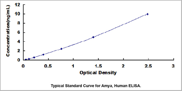 Standard Curve (Sample)
Amylase Alpha (Amya), ELISA Kit (Cat# AAA23113)
Full Name
Human Amylase Alpha (Amya) High Sensitive ELISA Kit
Reactivity
Human
Applications
ELISA
Pricing
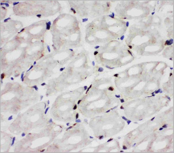
 IHC (Immunohistochemistry)
(Immunohistochemistry: Lung tissue (FFPE) stained with Rabbit anti-ER81 (Cat# AAA14073) at 1:200 for 10 min @ RT. Staining of formalin-fixed tissue requires boiling tissue sections in 10 mM Citrate Buffer, pH 6.0 for 10 min followed by cooling at RT for 20 min.)
IHC (Immunohistochemistry)
(Immunohistochemistry: Lung tissue (FFPE) stained with Rabbit anti-ER81 (Cat# AAA14073) at 1:200 for 10 min @ RT. Staining of formalin-fixed tissue requires boiling tissue sections in 10 mM Citrate Buffer, pH 6.0 for 10 min followed by cooling at RT for 20 min.)
ER81, Polyclonal Antibody (Cat# AAA14073)
Full Name
Rabbit anti ER81 Polyclonal Antibody
Gene Names
ETV1; ER81
Reactivity
Mouse, Human, Rat
Applications
Flow Cytometry, Immunohistochemistry, Western Blot, Immunoprecipitation
Purity
Rabbit IgG is purified by Epitope Affinity Purification
Pricing
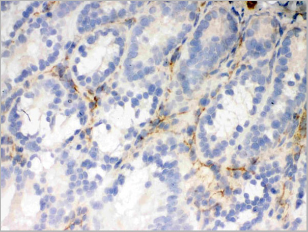
 IHC (Immunohistochemistry)
(Figure 7. IHC analysis of Flotillin 2 using anti-Flotillin 2 antibody (AAA11664).Flotillin 2 was detected in frozen section of rat small intestine tissue. Heat mediated antigen retrieval was performed in citrate buffer (pH6, epitope retrieval solution) for 20 mins. The tissue section was blocked with 10% goat serum. The tissue section was then incubated with 1ug/ml rabbit anti-Flotillin 2 Antibody (AAA11664) overnight at 4 degree C. Biotinylated goat anti-rabbit IgG was used as secondary antibody and incubated for 30 minutes at 37 degree C. The tissue section was developed using Strepavidin-Biotin-Complex (SABC) with DAB as the chromogen.)
IHC (Immunohistochemistry)
(Figure 7. IHC analysis of Flotillin 2 using anti-Flotillin 2 antibody (AAA11664).Flotillin 2 was detected in frozen section of rat small intestine tissue. Heat mediated antigen retrieval was performed in citrate buffer (pH6, epitope retrieval solution) for 20 mins. The tissue section was blocked with 10% goat serum. The tissue section was then incubated with 1ug/ml rabbit anti-Flotillin 2 Antibody (AAA11664) overnight at 4 degree C. Biotinylated goat anti-rabbit IgG was used as secondary antibody and incubated for 30 minutes at 37 degree C. The tissue section was developed using Strepavidin-Biotin-Complex (SABC) with DAB as the chromogen.)
Flotillin 2, Polyclonal Antibody (Cat# AAA11664)
Full Name
Anti-Flotillin 2 Antibody
Gene Names
FLOT2; ESA; ECS1; ESA1; ECS-1; M17S1
Reactivity
Human, Mouse, Rat
Applications
Western Blot, Immunohistochemistry
Purity
Immunogen Affinity Purified
Pricing
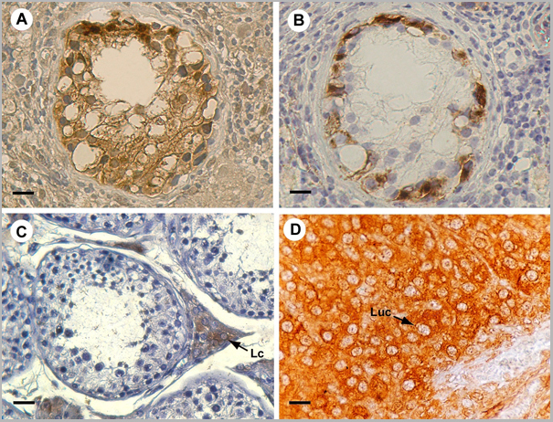
 Application Data
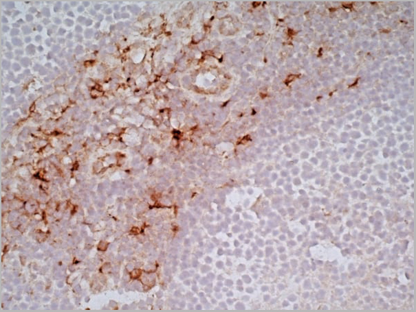
(Immunoperoxidase staining of a human tonsil cryosection with Mouse anti Human CD163 antibody, clone EDHu-1 followed by the Histar detection system . Low power)
Application Data
(Immunoperoxidase staining of a human tonsil cryosection with Mouse anti Human CD163 antibody, clone EDHu-1 followed by the Histar detection system . Low power)
CD163, Monoclonal Antibody (Cat# AAA11943)
Full Name
MOUSE ANTI HUMAN CD163
Gene Names
CD163; M130; MM130
Reactivity
Guinea Pig, Pig, Rhesus Monkey, Sheep
Applications
Immunohistochemistry, Immunohistochemistry, Flow Cytometry, Immunofluorescence, Immunoassay, Western Blot
Pricing
Pseudomonas aeruginosa Exotoxin A, Polyclonal Antibody (Cat# AAA14706)
Full Name
Pseudomonas aeruginosa Exotoxin A
Applications
Direct ELISA, Western Blot
Purity
Serum
Pricing
p. VIVAX Pldh, Recombinant Protein (Cat# AAA13455)
Full Name
p. VIVAX Pldh, Recomb.
Applications
Lateral Flow, Western Blot
Purity
> 95% pure (SDS-PAGE), Immobilized metal affinity chromatography (IMAC) using Ni (II)-NTA.
Pricing
P.falciparum pLDH, Recombinant Protein (Cat# AAA13454)
Full Name
P.falciparum pLDH, Recomb.
Applications
Lateral Flow
Purity
> 95% pure (SDS-PAGE), Immobilized metal affinity chromatography (IMAC) using Ni (II)-NTA.
Pricing
Glucagon Like Peptide-2, Peptide (Cat# AAA10861)
Full Name
Human Glucagon Like Peptide-2
Gene Names
GCG; GLP1; GLP2; GRPP
Purity
Greater than 98.0% as determined by RP-HPLC.
Pricing
Cyfra-21-1, Cytokeratin 19 Fragment Calibrator Grade (Low Cross Reactivity), Native Protein (Cat# AAA13416)
Full Name
Cyfra-21-1 Antigen
Applications
Chemiluminescent Immunoassay
Purity
Ion-Exchange Chromatography
Product is 0.2 um filtered.
Product is 0.2 um filtered.
Pricing
Insulin C-terminal Pentapeptide of B-chain, Monoclonal Antibody (Cat# AAA13370)
Full Name
MAb to Insulin C-terminal
Gene Names
INS; ILPR; IRDN; IDDM2; MODY10
Applications
ELISA
Purity
>90% pure (SDS-PAGE). Protein A chromatography
Pricing
 Molecular Structure
THC mAb1, Monoclonal Antibody (Cat# AAA14169)
Full Name
Monoclonal antibody to tetrahydrocannabinol (THC)
Applications
Lateral Flow
Purity
>95%, Protein A affinity chromatography purified
Pricing
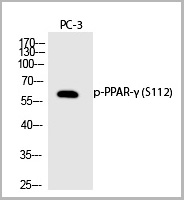
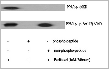 WB (Western Blot)
(Western Blot analysis of various cells using Phospho-PPAR-Gamma (S112) Polyclonal Antibody)
WB (Western Blot)
(Western Blot analysis of various cells using Phospho-PPAR-Gamma (S112) Polyclonal Antibody)
PPAR-gamma, Polyclonal Antibody (Cat# AAA29649)
Full Name
PPAR-gamma (Phospho-Ser112) Polyclonal Antibody
Gene Names
PPARG; GLM1; CIMT1; NR1C3; PPARG1; PPARG2; PPARgamma
Reactivity
Human, Mouse, Rat
Applications
Western Blot
Purity
The antibody was affinity-purified from rabbit antiserum by affinity-chromatography using epitope-specific immunogen.
Pricing