Filters
Clonality
Type
Reactivity
Gene Name
Isotype
Host
Application
Clone
898 results for "HRP Conjugated Secondary Antibody" - showing 200-250


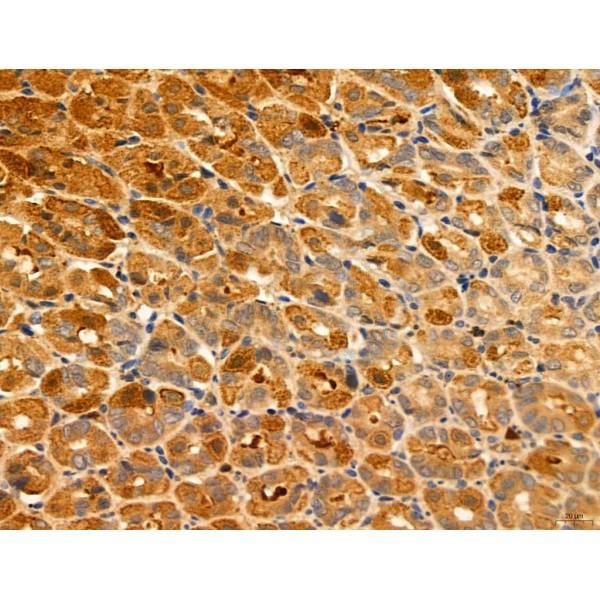
 IHC (Immunohistchemistry)
(At 1/100 staining Mouse brain tissue by IHC-P. The sample was formaldehyde fixed and a heat mediated antigen retrieval step in citrate buffer was performed. The sample was then blocked and incubated with the primary antibody at 4 degree C overnight. An HRP conjugated anti-Rabbit antibody was used as the secondary antibody.)
IHC (Immunohistchemistry)
(At 1/100 staining Mouse brain tissue by IHC-P. The sample was formaldehyde fixed and a heat mediated antigen retrieval step in citrate buffer was performed. The sample was then blocked and incubated with the primary antibody at 4 degree C overnight. An HRP conjugated anti-Rabbit antibody was used as the secondary antibody.)
USF1, Polyclonal Antibody (Cat# AAA31437)
Full Name
Phospho-USF1 (Thr153) Antibody
Gene Names
USF1; UEF; FCHL; MLTF; FCHL1; MLTFI; HYPLIP1; bHLHb11
Reactivity
Human, Mouse, Rat
Predicted Reactivity: Pig (100%), Bovine (100%), Rabbit (100%), Dog (100%)
Predicted Reactivity: Pig (100%), Bovine (100%), Rabbit (100%), Dog (100%)
Applications
Western Blot, Immunohistochemistry, Peptide ELISA
Purity
The antibody is from purified rabbit serum by affinity purification via sequential chromatography on phospho-peptide and non-phospho-peptide affinity columns.
Pricing


 FCM (Flow Cytometry)
(Figure 7. Flow Cytometry analysis of HL-60 cells using anti-Sumo 1/SUMO1 antibody (AAA19224).Overlay histogram showing HL-60 cells stained with AAA19224 (Blue line). The cells were blocked with 10% normal goat serum. And then incubated with rabbit anti-Sumo 1/SUMO1 Antibody (AAA19224, 1μg/1x106 cells) for 30 min at 20 degree C. DyLight®488 conjugated goat anti-rabbit IgG (5-10μg/1x106 cells) was used as secondary antibody for 30 minutes at 20 degree C. Isotype control antibody (Green line) was rabbit IgG (1μg/1x106) used under the same conditions. Unlabelled sample (Red line) was also used as a control.)
FCM (Flow Cytometry)
(Figure 7. Flow Cytometry analysis of HL-60 cells using anti-Sumo 1/SUMO1 antibody (AAA19224).Overlay histogram showing HL-60 cells stained with AAA19224 (Blue line). The cells were blocked with 10% normal goat serum. And then incubated with rabbit anti-Sumo 1/SUMO1 Antibody (AAA19224, 1μg/1x106 cells) for 30 min at 20 degree C. DyLight®488 conjugated goat anti-rabbit IgG (5-10μg/1x106 cells) was used as secondary antibody for 30 minutes at 20 degree C. Isotype control antibody (Green line) was rabbit IgG (1μg/1x106) used under the same conditions. Unlabelled sample (Red line) was also used as a control.)
Sumo 1/SUMO1, Polyclonal Antibody (Cat# AAA19224)
Full Name
Anti-Sumo 1/SUMO1 Antibody
Gene Names
SUMO1; DAP1; GMP1; PIC1; SMT3; UBL1; OFC10; SENP2; SMT3C; SMT3H3
Reactivity
Human, Mouse, Rat
Applications
Immunohistochemistry, Immunocytochemistry, Immunofluorescence, Flow Cytometry
Purity
Immunogen affinity purified.
Pricing


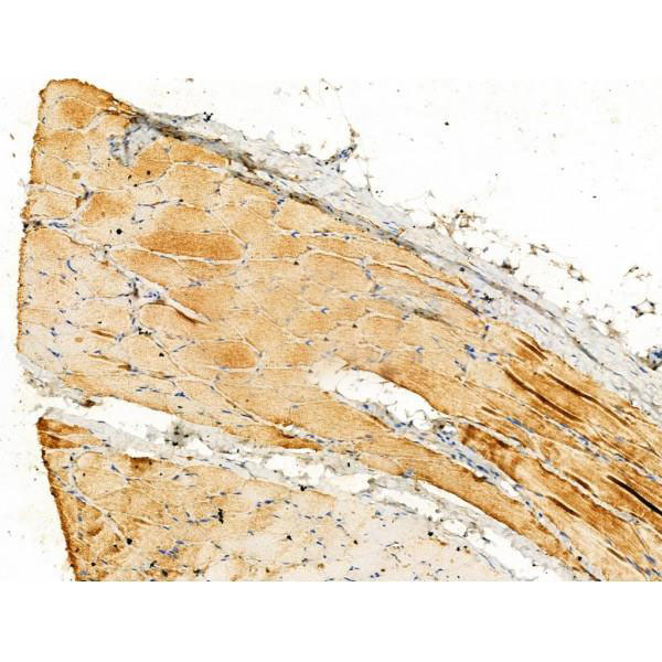
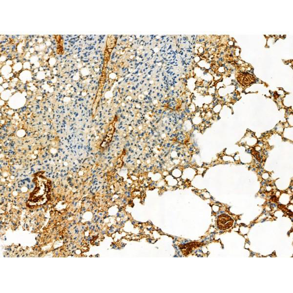
 IHC (Immunohistochemistry)
(At 1/100 staining Mouse lung tissue by IHC-P. The sample was formaldehyde fixed and a heat mediated antigen retrieval step in citrate buffer was performed. The sample was then blocked and incubated with the primary antibody at 4 degree C overnight. An HRP conjugated anti-Rabbit antibody was used as the secondary antibody.)
IHC (Immunohistochemistry)
(At 1/100 staining Mouse lung tissue by IHC-P. The sample was formaldehyde fixed and a heat mediated antigen retrieval step in citrate buffer was performed. The sample was then blocked and incubated with the primary antibody at 4 degree C overnight. An HRP conjugated anti-Rabbit antibody was used as the secondary antibody.)
Histone H2A, Polyclonal Antibody (Cat# AAA31347)
Full Name
Acetyl-Histone H2A (Lys5) Antibody
Gene Names
HIST1H2AB; H2A/m; H2AFM
Reactivity
Human, Mouse, Rat
Applications
Western Blot, Immunohistochemistry, Peptide ELISA
Purity
The antiserum was purified by peptide affinity chromatography using SulfoLink Coupling Resin
Pricing


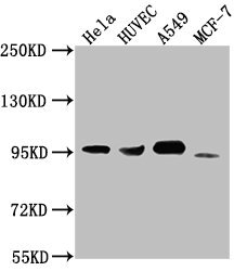
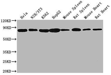
 WB (Western Blot)
(Western blot analysis of extracts of various celllines, using GSK3 alpha/beta Antibody.)
WB (Western Blot)
(Western blot analysis of extracts of various celllines, using GSK3 alpha/beta Antibody.)
GSK3 alpha/beta, Polyclonal Antibody (Cat# AAA31094)
Full Name
GSK3 alpha/beta Antibody
Reactivity
Human, Mouse, Rat
Applications
Western Blot, Immunohistochemistry
Purity
The antiserum was purified by peptide affinity chromatography using SulfoLink Coupling Resin.
Pricing


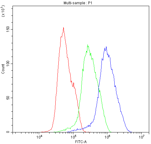
 FCM (Flow Cytometry)
(Overlay Peak curve showing 293F cells transfected with GST stained with AAA28065 (red line) at 1:189. The cells were fixed in 4% formaldehyde and permeated by 0.2% TritonX-100. Then 10% normal goat serum was Incubated to block non-specific protein-protein interactions followed by the antibody (1?g/1*106cells) for 1 h at 4 degree C. The secondary antibody used was FITC-conjugated Goat Anti-Mouse IgG(H+L) at 1/100 dilution for 30min at 4 degree C. Isotype control antibody (green line) was mouse IgG2b (1?g/1*106cells) used under the same conditions. Acquisition of >10,000 events was performed.)
FCM (Flow Cytometry)
(Overlay Peak curve showing 293F cells transfected with GST stained with AAA28065 (red line) at 1:189. The cells were fixed in 4% formaldehyde and permeated by 0.2% TritonX-100. Then 10% normal goat serum was Incubated to block non-specific protein-protein interactions followed by the antibody (1?g/1*106cells) for 1 h at 4 degree C. The secondary antibody used was FITC-conjugated Goat Anti-Mouse IgG(H+L) at 1/100 dilution for 30min at 4 degree C. Isotype control antibody (green line) was mouse IgG2b (1?g/1*106cells) used under the same conditions. Acquisition of >10,000 events was performed.)
GST, Monoclonal Antibody (Cat# AAA28065)
Full Name
GST Monoclonal Antibody
Reactivity
All
Applications
Western Blot, Immunofluorescence, Flow Cytometry, Immunoprecipitation
Purity
>95%, Protein A purified
Pricing


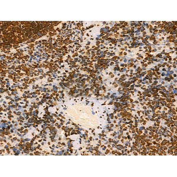
 IHC (Immunohistchemistry)
(At 1/100 staining Mouse heart tissue by IHC-P. The sample was formaldehyde fixed and a heat mediated antigen retrieval step in citrate buffer was performed. The sample was then blocked and incubated with the primary antibody at 4 degree C overnight. An HRP conjugated anti-Rabbit antibody was used as the secondary antibody.)
IHC (Immunohistchemistry)
(At 1/100 staining Mouse heart tissue by IHC-P. The sample was formaldehyde fixed and a heat mediated antigen retrieval step in citrate buffer was performed. The sample was then blocked and incubated with the primary antibody at 4 degree C overnight. An HRP conjugated anti-Rabbit antibody was used as the secondary antibody.)
OTUB1, Polyclonal Antibody (Cat# AAA31284)
Full Name
Phospho-OTUB1 (Ser16) Antibody
Gene Names
OTUB1; OTB1; OTU1; HSPC263
Reactivity
Human, Mouse, Rat
Applications
Immunohistochemistry, Peptide ELISA
Purity
The antibody is from purified rabbit serum by affinity purification via sequential chromatography on phospho-peptide and non-phospho-peptide affinity columns.
Pricing


 FCM (Flow Cytometry)
(Figure 7. Flow Cytometry analysis of A431 cells using anti-BIK antibody (AAA11666).Overlay histogram showing A431 cells stained with AAA11666 (Blue line).The cells were blocked with 10% normal goat serum. And then incubated with rabbit anti-BIK Antibody (AAA11666,1ug/1x10^6 cells) for 30 min at 20 degree C. DyLight®488 conjugated goat anti-rabbit IgG (5-10ug/1x10^6 cells) was used as secondary antibody for 30 minutes at 20 degree C. Isotype control antibody (Green line) was rabbit IgG (1ug/1x106) used under the same conditions. Unlabelled sample (Red line) was also used as a control.)
FCM (Flow Cytometry)
(Figure 7. Flow Cytometry analysis of A431 cells using anti-BIK antibody (AAA11666).Overlay histogram showing A431 cells stained with AAA11666 (Blue line).The cells were blocked with 10% normal goat serum. And then incubated with rabbit anti-BIK Antibody (AAA11666,1ug/1x10^6 cells) for 30 min at 20 degree C. DyLight®488 conjugated goat anti-rabbit IgG (5-10ug/1x10^6 cells) was used as secondary antibody for 30 minutes at 20 degree C. Isotype control antibody (Green line) was rabbit IgG (1ug/1x106) used under the same conditions. Unlabelled sample (Red line) was also used as a control.)
Bik, Polyclonal Antibody (Cat# AAA11666)
Full Name
Anti-Bik Antibody
Gene Names
BIK; BP4; NBK; BIP1
Reactivity
Human, Mouse, Rat
Applications
Western Blot, Immunohistochemistry
Purity
Immunogen Affinity Purified
Pricing


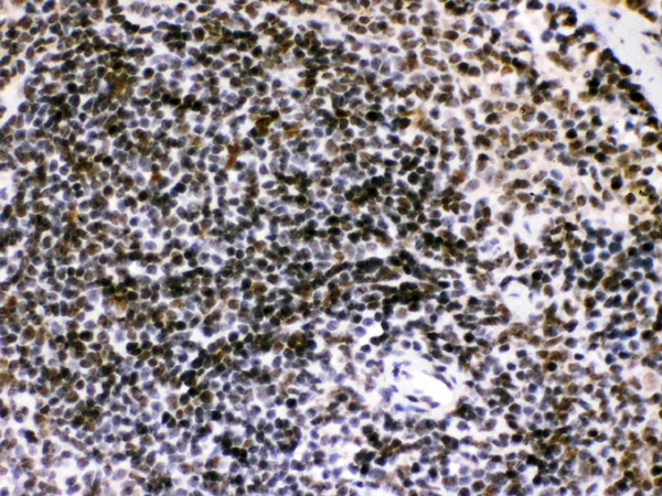
 IHC (Immunohistochemistry)
(At 1/100 staining Mouse heart tissue by IHC-P. The sample was formaldehyde fixed and a heat mediated antigen retrieval step in citrate buffer was performed. The sample was then blocked and incubated with the primary antibody at 4 degree C overnight. An HRP conjugated anti-Rabbit antibody was used as the secondary antibody.)
IHC (Immunohistochemistry)
(At 1/100 staining Mouse heart tissue by IHC-P. The sample was formaldehyde fixed and a heat mediated antigen retrieval step in citrate buffer was performed. The sample was then blocked and incubated with the primary antibody at 4 degree C overnight. An HRP conjugated anti-Rabbit antibody was used as the secondary antibody.)
AR, Polyclonal Antibody (Cat# AAA31420)
Full Name
Phospho-AR (Tyr535) Antibody
Gene Names
AR; KD; AIS; TFM; DHTR; SBMA; HYSP1; NR3C4; SMAX1; HUMARA
Reactivity
Human, Mouse, Rat, Monkey
Predicted Reactivity: Pig (92%), Bovine (85%), Sheep (85%), Rabbit (92%), Dog (92%)
Predicted Reactivity: Pig (92%), Bovine (85%), Sheep (85%), Rabbit (92%), Dog (92%)
Applications
Western Blot, Immunohistochemistry, Peptide ELISA
Purity
The antibody is from purified rabbit serum by affinity purification via sequential chromatography on phospho-peptide and non-phospho-peptide affinity columns.
Pricing


 FCM (Flow Cytometry)
(IDH1 Antibody (Center) flow cytometric analysis of 293 cells (right histogram) compared to a negative control cell (left histogram).FITC-conjugated goat-anti-rabbit secondary antibodies were used for the analysis.)
FCM (Flow Cytometry)
(IDH1 Antibody (Center) flow cytometric analysis of 293 cells (right histogram) compared to a negative control cell (left histogram).FITC-conjugated goat-anti-rabbit secondary antibodies were used for the analysis.)
IDH1, Polyclonal Antibody (Cat# AAA28676)
Full Name
IDH1 Antibody (Center)
Gene Names
IDH1; IDH; IDP; IDCD; IDPC; PICD; HEL-216; HEL-S-26
Reactivity
Human, mouse (Predicted Reactivity: Bovine, Rat)
Applications
Immunohistochemistry, Immunofluorescence, Western Blot, Flow Cytometry
Purity
Purified Rabbit Polyclonal Antibody (Pab)
Pricing


 IHC (Immunohistochemistry)
(Figure 13. IHC analysis of EEF2/Elongation factor 2 using anti-EEF2/Elongation factor 2 antibody (AAA19229).EEF2/Elongation factor 2 was detected in paraffin-embedded section of rat brain tissue. Heat mediated antigen retrieval was performed in EDTA buffer (pH8. 0, epitope retrieval solution). The tissue section was blocked with 10% goat serum. The tissue section was then incubated with 2μg/ml rabbit anti-EEF2/Elongation factor 2 Antibody (AAA19229) overnight at 4 degree C. Biotinylated goat anti-rabbit IgG was used as secondary antibody and incubated for 30 minutes at 37 degree C. The tissue section was developed using Strepavidin-Biotin-Complex (SABC) (Catalog # with DAB as the chromogen.)
IHC (Immunohistochemistry)
(Figure 13. IHC analysis of EEF2/Elongation factor 2 using anti-EEF2/Elongation factor 2 antibody (AAA19229).EEF2/Elongation factor 2 was detected in paraffin-embedded section of rat brain tissue. Heat mediated antigen retrieval was performed in EDTA buffer (pH8. 0, epitope retrieval solution). The tissue section was blocked with 10% goat serum. The tissue section was then incubated with 2μg/ml rabbit anti-EEF2/Elongation factor 2 Antibody (AAA19229) overnight at 4 degree C. Biotinylated goat anti-rabbit IgG was used as secondary antibody and incubated for 30 minutes at 37 degree C. The tissue section was developed using Strepavidin-Biotin-Complex (SABC) (Catalog # with DAB as the chromogen.)
EEF2/Elongation factor 2, Polyclonal Antibody (Cat# AAA19229)
Full Name
Anti-EEF2/Elongation factor 2 Antibody
Gene Names
EEF2; EF2; EF-2; EEF-2
Reactivity
Human, Mouse, Rat, Monkey
Applications
Western Blot, Immunohistochemistry, Immunocytochemistry, Immunofluorescence, Flow Cytometry
Purity
Immunogen affinity purified.
Pricing


 Application Data
(At 25 degree C. The primary antibody was diluted at 1/200 and incubated with the sample for 1 hour at 37 degree C. An Alexa Fluor 594 conjugated goat anti-rabbit IgG (H+L) Ab, diluted at 1/600, was used as the secondary antibody.)
Application Data
(At 25 degree C. The primary antibody was diluted at 1/200 and incubated with the sample for 1 hour at 37 degree C. An Alexa Fluor 594 conjugated goat anti-rabbit IgG (H+L) Ab, diluted at 1/600, was used as the secondary antibody.)
LIMK1, Polyclonal Antibody (Cat# AAA31425)
Full Name
Phospho-LIMK1 (Ser310) Antibody
Gene Names
LIMK1; LIMK; LIMK-1
Reactivity
Human, Mouse, Rat
Predicted Reactivity: Pig (100%), Bovine (100%), Horse (100%), Sheep (100%), Rabbit (100%), Dog (88%), Chicken (100%)
Predicted Reactivity: Pig (100%), Bovine (100%), Horse (100%), Sheep (100%), Rabbit (100%), Dog (88%), Chicken (100%)
Applications
Western Blot, Immunohistochemistry, Immunofluorescence, Immunocytochemistry, Peptide ELISA
Purity
The antibody is from purified rabbit serum by affinity purification via sequential chromatography on phospho-peptide and non-phospho-peptide affinity columns.
Pricing


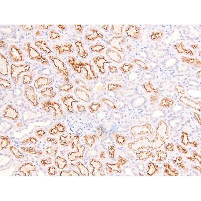 IHC (Immunohistochemistry)
(At 1/200 staining Human kidney tissue sections by IHC-P. The tissue was formaldehyde fixed and a heat mediated antigen retrieval step in citrate buffer was performed. The tissue was then blocked and incubated with the antibody for 1.5 hours at 22 degree C. An HRP conjugated goat anti-rabbit antibody was used as the secondary antibody.)
IHC (Immunohistochemistry)
(At 1/200 staining Human kidney tissue sections by IHC-P. The tissue was formaldehyde fixed and a heat mediated antigen retrieval step in citrate buffer was performed. The tissue was then blocked and incubated with the antibody for 1.5 hours at 22 degree C. An HRP conjugated goat anti-rabbit antibody was used as the secondary antibody.)
RhoA, Polyclonal Antibody (Cat# AAA31383)
Full Name
Phospho-RhoA (Ser188) Antibody
Gene Names
RHOA; ARHA; ARH12; RHO12; RHOH12
Reactivity
Human, Mouse, Rat
Predicted Reactivity: Pig (100%), Bovine (100%), Sheep (100%), Dog (100%)
Predicted Reactivity: Pig (100%), Bovine (100%), Sheep (100%), Dog (100%)
Applications
Western Blot, Immunohistochemistry, Peptide ELISA
Purity
The antibody is from purified rabbit serum by affinity purification via sequential chromatography on phospho-peptide and non-phospho-peptide affinity columns.
Pricing


 FCM (Flow Cytometry)
(Overlay histogram showing Raji cells stained with AAA27027 (red line) at 1:100. The cells were incubated in 1x PBS /10% normal goat serum to block non-specific protein-protein interactions followed by primary antibody for 1 h at 4 degree C. The secondary antibody used was FITC goat anti-mouse IgG(H+L) at 1/200 dilution for 1 h at 4 degree C. Isotype control antibody (green line) was used under the same conditions. Acquisition of >10,000 events was performed.)
FCM (Flow Cytometry)
(Overlay histogram showing Raji cells stained with AAA27027 (red line) at 1:100. The cells were incubated in 1x PBS /10% normal goat serum to block non-specific protein-protein interactions followed by primary antibody for 1 h at 4 degree C. The secondary antibody used was FITC goat anti-mouse IgG(H+L) at 1/200 dilution for 1 h at 4 degree C. Isotype control antibody (green line) was used under the same conditions. Acquisition of >10,000 events was performed.)
CD19, Monoclonal Antibody (Cat# AAA27027)
Full Name
CD19 Monoclonal Antibody
Gene Names
CD19; B4; CVID3
Reactivity
Human
Applications
Western Blot, Immunohistochemistry, Immunofluorescence, Flow Cytometry
Purity
>95%, Protein A Purified
Pricing


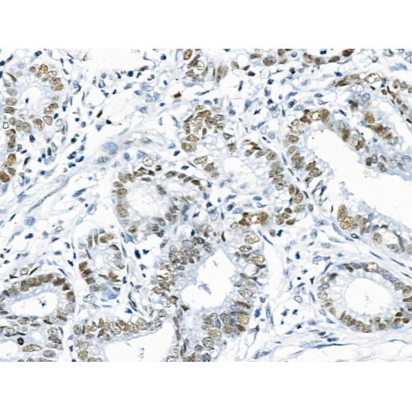
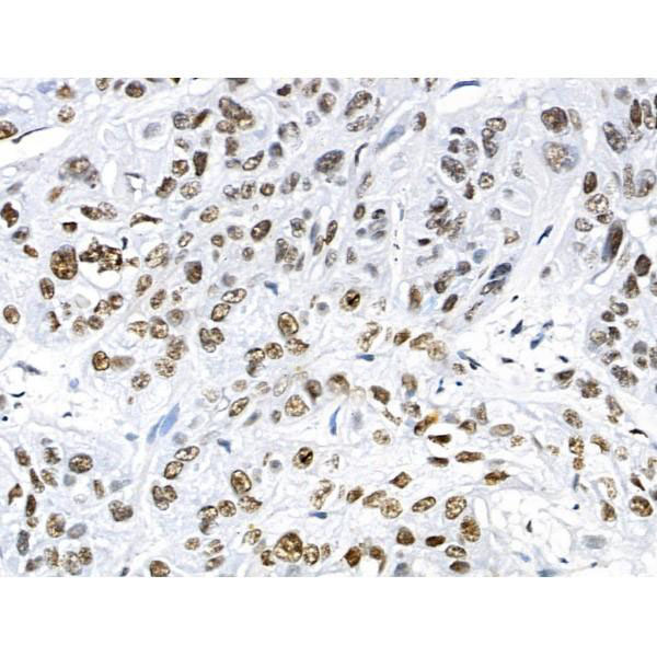
 IHC (Immunohistochemistry)
(At 1/100 staining Human gastric cancer by IHC-P. The sample was formaldehyde fixed and a heat mediated antigen retrieval step in citrate buffer was performed. The sample was then blocked and incubated with the primary antibody at 4 degree C overnight. An HRP conjugated anti-Rabbit antibody was used as the secondary antibody.)
IHC (Immunohistochemistry)
(At 1/100 staining Human gastric cancer by IHC-P. The sample was formaldehyde fixed and a heat mediated antigen retrieval step in citrate buffer was performed. The sample was then blocked and incubated with the primary antibody at 4 degree C overnight. An HRP conjugated anti-Rabbit antibody was used as the secondary antibody.)
Abi 1, Polyclonal Antibody (Cat# AAA31442)
Full Name
Phospho-Abi 1 (Tyr213) Antibody
Gene Names
ABI1; E3B1; ABI-1; ABLBP4; NAP1BP; SSH3BP; SSH3BP1
Reactivity
Human, Mouse, Rat
Predicted Reactivity: Pig (100%), Bovine (100%), Horse (100%), Sheep (100%), Rabbit (100%), Dog (100%), Chicken (100%), Xenopus (100%)
Predicted Reactivity: Pig (100%), Bovine (100%), Horse (100%), Sheep (100%), Rabbit (100%), Dog (100%), Chicken (100%), Xenopus (100%)
Applications
Western Blot, Immunohistochemistry, Peptide ELISA
Purity
The antibody is from purified rabbit serum by affinity purification via sequential chromatography on phospho-peptide and non-phospho-peptide affinity columns.
Pricing


 FCM (Flow Cytometry)
(Figure 12. Flow Cytometry analysis of A549 cells using anti-Transketolase/TKT antibody (AAA19367).Overlay histogram showing A549 cells stained with AAA19367 (Blue line). The cells were blocked with 10% normal goat serum. And then incubated with mouse anti- Transketolase/TKT Antibody (AAA19367, 1μg/1x106 cells) for 30 min at 20 degree C. DyLight®488 conjugated goat anti-mouse IgG (BA1126, 5-10μg/1x106 cells) was used as secondary antibody for 30 minutes at 20 degree C. Isotype control antibody (Green line) was mouse IgG (1μg/1x106) used under the same conditions. Unlabelled sample (Red line) was also used as a control.)
FCM (Flow Cytometry)
(Figure 12. Flow Cytometry analysis of A549 cells using anti-Transketolase/TKT antibody (AAA19367).Overlay histogram showing A549 cells stained with AAA19367 (Blue line). The cells were blocked with 10% normal goat serum. And then incubated with mouse anti- Transketolase/TKT Antibody (AAA19367, 1μg/1x106 cells) for 30 min at 20 degree C. DyLight®488 conjugated goat anti-mouse IgG (BA1126, 5-10μg/1x106 cells) was used as secondary antibody for 30 minutes at 20 degree C. Isotype control antibody (Green line) was mouse IgG (1μg/1x106) used under the same conditions. Unlabelled sample (Red line) was also used as a control.)
Transketolase/TKT, Monoclonal Antibody (Cat# AAA19367)
Full Name
Anti-Transketolase/TKT Antibody (monoclonal, 3E5)
Gene Names
TKT; TK; TKT1; HEL107
Reactivity
Human, Mouse, Rat
Applications
WB, IHC-P, ICC, IF, FC/FACS/FCM
Purity
Immunogen affinity purified.
Pricing


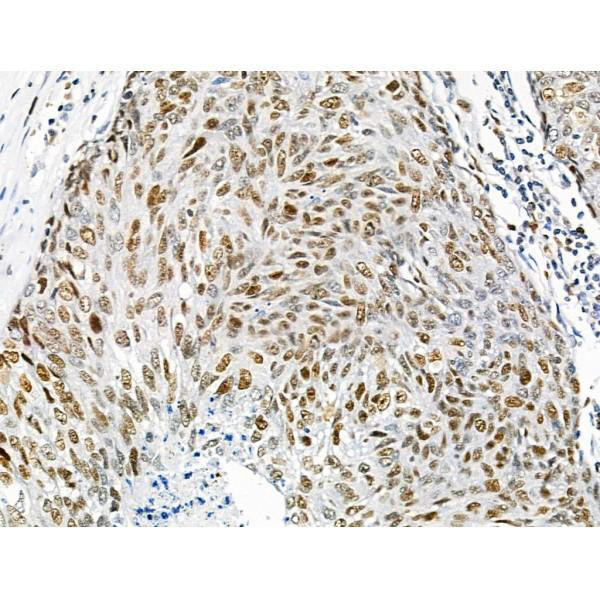
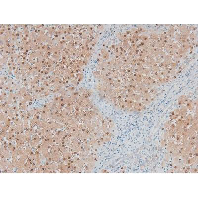
 IHC (Immunohistochemistry)
(At 1/100 staining Human colorectal cancer and adjacent normal tissues by IHC-P. The sample was formaldehyde fixed and a heat mediated antigen retrieval step in citrate buffer was performed. The sample was then blocked and incubated with the primary antibody at 4 degree C overnight. An HRP conjugated anti-Rabbit antibody was used as the secondary antibody.)
IHC (Immunohistochemistry)
(At 1/100 staining Human colorectal cancer and adjacent normal tissues by IHC-P. The sample was formaldehyde fixed and a heat mediated antigen retrieval step in citrate buffer was performed. The sample was then blocked and incubated with the primary antibody at 4 degree C overnight. An HRP conjugated anti-Rabbit antibody was used as the secondary antibody.)
Bamacan, Polyclonal Antibody (Cat# AAA31455)
Full Name
Phospho-Bamacan (Ser1083) Antibody
Gene Names
SMC3; BAM; BMH; HCAP; CDLS3; CSPG6; SMC3L1
Reactivity
Human, Mouse, Rat
Predicted Reactivity: Pig (100%), Horse (100%), Sheep (100%), Rabbit (100%), Dog (100%), Chicken (100%)
Predicted Reactivity: Pig (100%), Horse (100%), Sheep (100%), Rabbit (100%), Dog (100%), Chicken (100%)
Applications
Western Blot, Immunohistochemistry, Peptide ELISA
Purity
The antibody is from purified rabbit serum by affinity purification via sequential chromatography on phospho-peptide and non-phospho-peptide affinity columns.
Pricing


 FCM (Flow Cytometry)
(Overlay histogram showing Raji cells stained with AAA28060 (red line) at 1:500. The cells were incubated in 10% normal goat serum to block non-specific protein-protein interactions followed by the antibody (1ug/1*106cells) for 1 h at 4 degree C. The secondary antibody used was FITC-conjugated Goat Anti-Mouse IgG(H+L) at 1/100 dilution for 30min at 4 degree C. Isotype control antibody (green line) was mouse IgG2b (1ug/1*106cells) used under the same conditions. Acquisition of >10,000 events was performed.)
FCM (Flow Cytometry)
(Overlay histogram showing Raji cells stained with AAA28060 (red line) at 1:500. The cells were incubated in 10% normal goat serum to block non-specific protein-protein interactions followed by the antibody (1ug/1*106cells) for 1 h at 4 degree C. The secondary antibody used was FITC-conjugated Goat Anti-Mouse IgG(H+L) at 1/100 dilution for 30min at 4 degree C. Isotype control antibody (green line) was mouse IgG2b (1ug/1*106cells) used under the same conditions. Acquisition of >10,000 events was performed.)
CD45, Monoclonal Antibody (Cat# AAA28060)
Full Name
CD45 Monoclonal Antibody
Gene Names
PTPRC; LCA; LY5; B220; CD45; L-CA; T200; CD45R; GP180
Reactivity
Human
Applications
Western Blot, Immunohistochemistry, Immunofluorescence, Flow Cytometry
Purity
>95%, Protein A purified
Pricing


 FCM (Flow Cytometry)
(Figure 6. Flow Cytometry analysis of MCF-7 cells using anti-ALK-1/ACVRL1 antibody (AAA19242).Overlay histogram showing MCF-7 cells stained with AAA19242 (Blue line). The cells were blocked with 10% normal goat serum. And then incubated with rabbit anti-ALK-1/ACVRL1 Antibody (AAA19242, 1μg/1x106 cells) for 30 min at 20 degree C. DyLight®488 conjugated goat anti-rabbit IgG (5-10μg/1x106 cells) was used as secondary antibody for 30 minutes at 20 degree C. Isotype control antibody (Green line) was rabbit IgG (1μg/1x106) used under the same conditions. Unlabelled sample (Red line) was also used as a control.)
FCM (Flow Cytometry)
(Figure 6. Flow Cytometry analysis of MCF-7 cells using anti-ALK-1/ACVRL1 antibody (AAA19242).Overlay histogram showing MCF-7 cells stained with AAA19242 (Blue line). The cells were blocked with 10% normal goat serum. And then incubated with rabbit anti-ALK-1/ACVRL1 Antibody (AAA19242, 1μg/1x106 cells) for 30 min at 20 degree C. DyLight®488 conjugated goat anti-rabbit IgG (5-10μg/1x106 cells) was used as secondary antibody for 30 minutes at 20 degree C. Isotype control antibody (Green line) was rabbit IgG (1μg/1x106) used under the same conditions. Unlabelled sample (Red line) was also used as a control.)
ALK-1/ACVRL1, Polyclonal Antibody (Cat# AAA19242)
Full Name
Anti-ALK-1/ACVRL1 Antibody
Gene Names
ACVRL1; HHT; ALK1; HHT2; ORW2; SKR3; ALK-1; TSR-I; ACVRLK1
Reactivity
Human, Mouse, Rat
Applications
Western Blot, Immunohistochemistry, Flow Cytometry, Direct ELISA
Purity
Immunogen affinity purified.
Pricing


 FCM (Flow Cytometry)
(Overlay histogram showing Hela cells stained with AAA27026 (red line) at 1:200. The cells were incubated in 1x PBS /10% normal goat serum to block non-specific protein-protein interactions followed by primary antibody for 1 h at 4 degree C. The secondary antibody used was FITC goat anti-mouse IgG(H+L) at 1/200 dilution for 1 h at 4 degree C. Isotype control antibody (green line) was used under the same conditions. Acquisition of >10,000 events was performed.)
FCM (Flow Cytometry)
(Overlay histogram showing Hela cells stained with AAA27026 (red line) at 1:200. The cells were incubated in 1x PBS /10% normal goat serum to block non-specific protein-protein interactions followed by primary antibody for 1 h at 4 degree C. The secondary antibody used was FITC goat anti-mouse IgG(H+L) at 1/200 dilution for 1 h at 4 degree C. Isotype control antibody (green line) was used under the same conditions. Acquisition of >10,000 events was performed.)
CD44, Monoclonal Antibody (Cat# AAA27026)
Full Name
CD44 Monoclonal Antibody
Gene Names
CD44; IN; LHR; MC56; MDU2; MDU3; MIC4; Pgp1; CDW44; CSPG8; HCELL; HUTCH-I; ECMR-III
Reactivity
Human
Applications
Western Blot, Immunohistochemistry, Immunofluorescence, Flow Cytometry
Purity
>95%, Protein G Purified
Pricing


 FCM (Flow Cytometry)
(Figure 13. Flow Cytometry analysis of SiHa cells using anti-DRP1/DNM1L antibody (AAA19220).Overlay histogram showing SiHa cells stained with AAA19220 (Blue line). The cells were blocked with 10% normal goat serum. And then incubated with rabbit anti-DRP1/DNM1L Antibody (AAA19220, 1μg/1x106 cells) for 30 min at 20 degree C. DyLight®488 conjugated goat anti-rabbit IgG (5-10μg/1x106 cells) was used as secondary antibody for 30 minutes at 20 degree C. Isotype control antibody (Green line) was rabbit IgG (1μg/1x106) used under the same conditions. Unlabelled sample (Red line) was also used as a control.)
FCM (Flow Cytometry)
(Figure 13. Flow Cytometry analysis of SiHa cells using anti-DRP1/DNM1L antibody (AAA19220).Overlay histogram showing SiHa cells stained with AAA19220 (Blue line). The cells were blocked with 10% normal goat serum. And then incubated with rabbit anti-DRP1/DNM1L Antibody (AAA19220, 1μg/1x106 cells) for 30 min at 20 degree C. DyLight®488 conjugated goat anti-rabbit IgG (5-10μg/1x106 cells) was used as secondary antibody for 30 minutes at 20 degree C. Isotype control antibody (Green line) was rabbit IgG (1μg/1x106) used under the same conditions. Unlabelled sample (Red line) was also used as a control.)
DRP1/DNM1L, Polyclonal Antibody (Cat# AAA19220)
Full Name
Anti-DRP1/DNM1L Antibody
Gene Names
DNM1L; DLP1; DRP1; DVLP; EMPF; DYMPLE; HDYNIV
Reactivity
Human, Mouse, Rat
Applications
Western Blot, Immunohistochemistry, Immunocytochemistry, Immunofluorescence, Flow Cytometry, Direct ELISA
Purity
Immunogen affinity purified.
Pricing
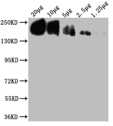
 WB (Western Blot)
(Western BlotPositive WB detected in Recombinantprotein (80ng, 40ng, 20ng, 10ng)All lanes: Human IgG Fc antibody;HRPconjugated at 1:1000Predicted band size: 35 kDaObserved band size: 35 kDa)
WB (Western Blot)
(Western BlotPositive WB detected in Recombinantprotein (80ng, 40ng, 20ng, 10ng)All lanes: Human IgG Fc antibody;HRPconjugated at 1:1000Predicted band size: 35 kDaObserved band size: 35 kDa)
IgG Fc fragment, Polyclonal Secondary Antibody (Cat# AAA15949)
Full Name
Rabbit anti-Human IgG Fc fragment polyclonal Antibody, HRP conjugated
Reactivity
Human
Applications
EIA, WB
Purity
Caprylic Acid Ammonium Sulfate Precipitation purified
Pricing


 FCM (Flow Cytometry)
(Figure 6. Flow Cytometry analysis of MCF-7 cells using anti-Caspase-7/CASP7 antibody (AAA19232).Overlay histogram showing MCF-7 cells stained with AAA19232 (Blue line). The cells were blocked with 10% normal goat serum. And then incubated with rabbit anti-Caspase-7/CASP7 Antibody (AAA19232, 1μg/1x106 cells) for 30 min at 20 degree C. DyLight®488 conjugated goat anti-rabbit IgG (5-10μg/1x106 cells) was used as secondary antibody for 30 minutes at 20 degree C. Isotype control antibody (Green line) was rabbit IgG (1μg/1x106) used under the same conditions. Unlabelled sample (Red line) was also used as a control.)
FCM (Flow Cytometry)
(Figure 6. Flow Cytometry analysis of MCF-7 cells using anti-Caspase-7/CASP7 antibody (AAA19232).Overlay histogram showing MCF-7 cells stained with AAA19232 (Blue line). The cells were blocked with 10% normal goat serum. And then incubated with rabbit anti-Caspase-7/CASP7 Antibody (AAA19232, 1μg/1x106 cells) for 30 min at 20 degree C. DyLight®488 conjugated goat anti-rabbit IgG (5-10μg/1x106 cells) was used as secondary antibody for 30 minutes at 20 degree C. Isotype control antibody (Green line) was rabbit IgG (1μg/1x106) used under the same conditions. Unlabelled sample (Red line) was also used as a control.)
Caspase-7/CASP7, Polyclonal Antibody (Cat# AAA19232)
Full Name
Anti-Caspase-7/CASP7 Antibody
Gene Names
CASP7; MCH3; CMH-1; LICE2; CASP-7; ICE-LAP3
Reactivity
Human, Rat
Applications
Western Blot, Immunohistochemistry, Immunocytochemistry, Immunofluorescence, Flow Cytometry, Direct ELISA
Purity
Immunogen affinity purified.
Pricing


 FCM (Flow Cytometry)
(Figure 6. Flow Cytometry analysis of PC-3 cells using anti-APP antibody (AAA11597).Overlay histogram showing PC-3 cells stained with AAA11597 (Blue line).The cells were blocked with 10% normal goat serum. And then incubated with rabbit anti-APP Antibody (AAA11597,1ug/1x10^6 cells) for 30 min at 20 degree C. DyLight®488 conjugated goat anti-rabbit IgG (5-10ug/1x10^6 cells) was used as secondary antibody for 30 minutes at 20 degree C. Isotype control antibody (Green line) was rabbit IgG (1ug/1x106) used under the same conditions. Unlabelled sample (Red line) was also used as a control.)
FCM (Flow Cytometry)
(Figure 6. Flow Cytometry analysis of PC-3 cells using anti-APP antibody (AAA11597).Overlay histogram showing PC-3 cells stained with AAA11597 (Blue line).The cells were blocked with 10% normal goat serum. And then incubated with rabbit anti-APP Antibody (AAA11597,1ug/1x10^6 cells) for 30 min at 20 degree C. DyLight®488 conjugated goat anti-rabbit IgG (5-10ug/1x10^6 cells) was used as secondary antibody for 30 minutes at 20 degree C. Isotype control antibody (Green line) was rabbit IgG (1ug/1x106) used under the same conditions. Unlabelled sample (Red line) was also used as a control.)
beta Amyloid, Polyclonal Antibody (Cat# AAA11597)
Full Name
Anti-beta Amyloid Antibody
Gene Names
APP; AAA; AD1; PN2; ABPP; APPI; CVAP; ABETA; PN-II; CTFgamma
Reactivity
Human, Mouse, Rat
Applications
Western Blot, Immunohistochemistry
Purity
Immunogen affinity purified.
Pricing


 Application Data
(At 25 degree C. The primary antibody was diluted at 1/200 and incubated with the sample for 1 hour at 37 degree C. An Alexa Fluor 594 conjugated goat anti-rabbit IgG (H+L) Ab, diluted at 1/600, was used as the secondary antibody.)
Application Data
(At 25 degree C. The primary antibody was diluted at 1/200 and incubated with the sample for 1 hour at 37 degree C. An Alexa Fluor 594 conjugated goat anti-rabbit IgG (H+L) Ab, diluted at 1/600, was used as the secondary antibody.)
PKC delta, Polyclonal Antibody (Cat# AAA31409)
Full Name
Phospho-PKC delta (Ser664) Antibody
Gene Names
PRKCD; MAY1; PKCD; nPKC-delta
Reactivity
Human, Mouse, Rat
Predicted Reactivity: Pig (82%), Bovine (91%), Horse (91%), Sheep (91%), Dog (91%), Chicken (91%), Xenopus (82%)
Predicted Reactivity: Pig (82%), Bovine (91%), Horse (91%), Sheep (91%), Dog (91%), Chicken (91%), Xenopus (82%)
Applications
Western Blot, Immunohistochemistry, Immunofluorescence, Immunocytochemistry, Peptide ELISA
Purity
The antibody is from purified rabbit serum by affinity purification via sequential chromatography on phospho-peptide and non-phospho-peptide affinity columns.
Pricing
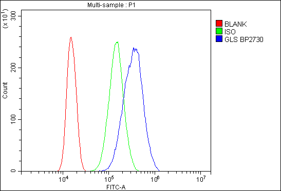


 FCM (Flow Cytometry)
(Figure 6. Flow Cytometry analysis of SiHa cells using anti-Glutaminase/GLS antibody (AAA19239).Overlay histogram showing SiHa cells stained with AAA19239 (Blue line). The cells were blocked with 10% normal goat serum. And then incubated with rabbit anti-Glutaminase/GLS Antibody (AAA19239,1μg/1x106 cells) for 30 min at 20 degree C. DyLight®488 conjugated goat anti-rabbit IgG (5-10μg/1x106 cells) was used as secondary antibody for 30 minutes at 20 degree C. Isotype control antibody (Green line) was rabbit IgG (1μg/1x106) used under the same conditions. Unlabelled sample (Red line) was also used as a control.)
FCM (Flow Cytometry)
(Figure 6. Flow Cytometry analysis of SiHa cells using anti-Glutaminase/GLS antibody (AAA19239).Overlay histogram showing SiHa cells stained with AAA19239 (Blue line). The cells were blocked with 10% normal goat serum. And then incubated with rabbit anti-Glutaminase/GLS Antibody (AAA19239,1μg/1x106 cells) for 30 min at 20 degree C. DyLight®488 conjugated goat anti-rabbit IgG (5-10μg/1x106 cells) was used as secondary antibody for 30 minutes at 20 degree C. Isotype control antibody (Green line) was rabbit IgG (1μg/1x106) used under the same conditions. Unlabelled sample (Red line) was also used as a control.)
Glutaminase/GLS, Polyclonal Antibody (Cat# AAA19239)
Full Name
Anti-Glutaminase/GLS Antibody
Gene Names
GLS; GAC; GAM; KGA; GLS1; AAD20
Reactivity
Human, Mouse, Rat, Monkey
Applications
Western Blot, Immunohistochemistry, Immunocytochemistry, Immunofluorescence, Flow Cytometry, Direct ELISA
Purity
Immunogen affinity purified.
Pricing


 Application Data
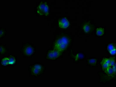
(At 25 degree C. Samples were then incubated with primary Ab(At 37 degree C. An AlexaFluor594 conjugated goat anti-rabbit IgG(H+L) Ab(Red) and an AlexaFluor488 conjugated goat anti-mouse IgG(H+L) Ab(Green) were used as the secondary antibody.The nuclear counter stain is DAPI(blue).)
Application Data
(At 25 degree C. Samples were then incubated with primary Ab(At 37 degree C. An AlexaFluor594 conjugated goat anti-rabbit IgG(H+L) Ab(Red) and an AlexaFluor488 conjugated goat anti-mouse IgG(H+L) Ab(Green) were used as the secondary antibody.The nuclear counter stain is DAPI(blue).)
INCENP, Polyclonal Antibody (Cat# AAA31457)
Full Name
Phospho-INCENP (Thr59) Antibody
Reactivity
Human, Mouse, Rat
Predicted Reactivity: Pig (100%), Zebrafish (100%), Horse (100%), Sheep (89%), Rabbit (100%), Dog (100%), Chicken (100%)
Predicted Reactivity: Pig (100%), Zebrafish (100%), Horse (100%), Sheep (89%), Rabbit (100%), Dog (100%), Chicken (100%)
Applications
Western Blot, Immunohistochemistry, Immunofluorescence, Immunocytochemistry, Peptide ELISA
Purity
The antibody is from purified rabbit serum by affinity purification via sequential chromatography on phospho-peptide and non-phospho-peptide affinity columns.
Pricing


 WB (Western Blot)
(Anti-ANP32A rabbit polyclonal antibody at 1:500 dilution Lane A: ANP32A konckout Hela Whole Cell Lysate Lane B: Hela Whole Cell Lysate Lysates/proteins at 20 ug per lane. Secondary Goat Anti-Rabbit IgG (H+L)/HRP at 1/10000 dilution. Developed using the ECL technique. Performed under reducing conditions. Predicted band size:28 kDa Observed band size:28 kDa)
WB (Western Blot)
(Anti-ANP32A rabbit polyclonal antibody at 1:500 dilution Lane A: ANP32A konckout Hela Whole Cell Lysate Lane B: Hela Whole Cell Lysate Lysates/proteins at 20 ug per lane. Secondary Goat Anti-Rabbit IgG (H+L)/HRP at 1/10000 dilution. Developed using the ECL technique. Performed under reducing conditions. Predicted band size:28 kDa Observed band size:28 kDa)
ANP32A, Polyclonal Antibody (Cat# AAA27748)
Full Name
Anti-ANP32A Antibody, Rabbit Polyclonal
Gene Names
ANP32A; LANP; MAPM; PP32; HPPCn; PHAP1; PHAPI; I1PP2A; C15orf1
Reactivity
Human
Applications
Western Blot, Immunohistochemistry, Immunocytochemistry, Immunofluorescence, Immunoprecipitation
Purity
Protein A & Antigen Affinity
Pricing


 IHC (Immunohistochemistry)
(At 1/100 staining Mouse kidney tissue by IHC-P. The sample was formaldehyde fixed and a heat mediated antigen retrieval step in citrate buffer was performed. The sample was then blocked and incubated with the primary antibody at 4 degree C overnight. An HRP conjugated anti-Rabbit antibody was used as the secondary antibody.)
IHC (Immunohistochemistry)
(At 1/100 staining Mouse kidney tissue by IHC-P. The sample was formaldehyde fixed and a heat mediated antigen retrieval step in citrate buffer was performed. The sample was then blocked and incubated with the primary antibody at 4 degree C overnight. An HRP conjugated anti-Rabbit antibody was used as the secondary antibody.)
LC3A, Polyclonal Antibody (Cat# AAA31334)
Full Name
LC3A Antibody
Gene Names
MAP1LC3A; LC3; LC3A; ATG8E; MAP1ALC3; MAP1BLC3
Reactivity
Human, Mouse, Rat
Predicted Reactivity: Pig (100%), Zebrafish (100%), Bovine (100%), Horse (100%), Sheep (100%), Dog (100%), Chicken (100%), Xenopus (100%)
Predicted Reactivity: Pig (100%), Zebrafish (100%), Bovine (100%), Horse (100%), Sheep (100%), Dog (100%), Chicken (100%), Xenopus (100%)
Applications
Western Blot, Immunohistochemistry, Immunofluorescence, Immunocytochemistry, Peptide ELISA
Purity
The antiserum was purified by peptide affinity chromatography using SulfoLink Coupling Resin
Pricing


 IHC (Immunohistchemistry)
(At 1/200 staining Human kidney tissue sections by IHC-P. The tissue was formaldehyde fixed and a heat mediated antigen retrieval step in citrate buffer was performed. The tissue was then blocked and incubated with the antibody for 1.5 hours at 22 degree C. An HRP conjugated goat anti-rabbit antibody was used as the secondary antibody.)
IHC (Immunohistchemistry)
(At 1/200 staining Human kidney tissue sections by IHC-P. The tissue was formaldehyde fixed and a heat mediated antigen retrieval step in citrate buffer was performed. The tissue was then blocked and incubated with the antibody for 1.5 hours at 22 degree C. An HRP conjugated goat anti-rabbit antibody was used as the secondary antibody.)
BAG3, Polyclonal Antibody (Cat# AAA31421)
Full Name
Phospho-BAG3 (Tyr457) Antibody
Gene Names
BAG3; BIS; BAG-3; CAIR-1
Reactivity
Human, Mouse, Rat
Predicted Reactivity: Pig (100%), Bovine (100%), Horse (100%), Sheep (100%), Rabbit (100%), Dog (100%)
Predicted Reactivity: Pig (100%), Bovine (100%), Horse (100%), Sheep (100%), Rabbit (100%), Dog (100%)
Applications
Western Blot, Immunohistochemistry, Peptide ELISA
Purity
The antibody is from purified rabbit serum by affinity purification via sequential chromatography on phospho-peptide and non-phospho-peptide affinity columns.
Pricing


 WB (Western Blot)
(Western Blot Analysis: Representative lot data. Lysate from HEK-293 cells was resolved by electrophoresis, transferred to PVDF and probed with anti-PI3 Kinase, p110beta (0.05 ug/mL). Proteins were visualized using donkey anti-rabbit secondary antibody conjugated to HRP and chemiluminescence detection. Arrow indicates PI3 Kinase, p110beta (~110kD).)
WB (Western Blot)
(Western Blot Analysis: Representative lot data. Lysate from HEK-293 cells was resolved by electrophoresis, transferred to PVDF and probed with anti-PI3 Kinase, p110beta (0.05 ug/mL). Proteins were visualized using donkey anti-rabbit secondary antibody conjugated to HRP and chemiluminescence detection. Arrow indicates PI3 Kinase, p110beta (~110kD).)
Phosphoinositide 3 Kinase, p110 beta, Polyclonal Antibody (Cat# AAA26898)
Full Name
Phosphoinositide 3 Kinase, p110 beta (DKFZp779K1237, MGC133043, p110beta, Phosphatidylinositol 3 Kinase Catalytic beta Polypeptide, Phosphatidylinositol-4,5-bisphosphate 3-kinase Catalytic Subunit beta Isoform, Phosphoinositide 3 Kinase Catalytic beta Pol
Gene Names
PIK3CA; MCM; CWS5; MCAP; PI3K; CLOVE; MCMTC; p110-alpha
Reactivity
Human
Applications
Immunocytochemistry, Western Blot, Immunoprecipitation, Immunohistochemistry
Purity
Purified by Immunoaffinity chromatography.
Pricing


 FCM (Flow Cytometry)
(Figure 7. Flow Cytometry analysis of U251 cells using anti-Serum Response Factor/SRF antibody (AAA19221).Overlay histogram showing U251 cells stained with AAA19221 (Blue line). The cells were blocked with 10% normal goat serum. And then incubated with rabbit anti-Serum Response Factor/SRF Antibody (AAA19221, 1μg/1x106 cells) for 30 min at 20 degree C. DyLight®488 conjugated goat anti-rabbit IgG (5-10μg/1x106 cells) was used as secondary antibody for 30 minutes at 20 degree C. Isotype control antibody (Green line) was rabbit IgG (1μg/1x106) used under the same conditions. Unlabelled sample (Red line) was also used as a control.)
FCM (Flow Cytometry)
(Figure 7. Flow Cytometry analysis of U251 cells using anti-Serum Response Factor/SRF antibody (AAA19221).Overlay histogram showing U251 cells stained with AAA19221 (Blue line). The cells were blocked with 10% normal goat serum. And then incubated with rabbit anti-Serum Response Factor/SRF Antibody (AAA19221, 1μg/1x106 cells) for 30 min at 20 degree C. DyLight®488 conjugated goat anti-rabbit IgG (5-10μg/1x106 cells) was used as secondary antibody for 30 minutes at 20 degree C. Isotype control antibody (Green line) was rabbit IgG (1μg/1x106) used under the same conditions. Unlabelled sample (Red line) was also used as a control.)
Serum Response Factor/SRF, Polyclonal Antibody (Cat# AAA19221)
Full Name
Anti-Serum Response Factor/SRF Antibody
Gene Names
SRF; MCM1
Reactivity
Human, Mouse, Rat
Applications
Western Blot, Immunohistochemistry, Immunocytochemistry, Immunofluorescence, Flow Cytometry, Direct ELISA
Purity
Immunogen affinity purified.
Pricing


 Application Data
(At 25 degree C. Samples were then incubated with primary Ab(At 37 degree C. An AlexaFluor594 conjugated goat anti-rabbit IgG(H+L) Ab(Red) and an AlexaFluor488 conjugated goat anti-mouse IgG(H+L) Ab(Green) were used as the secondary antibody.The nuclear counter stain is DAPI (blue).)
Application Data
(At 25 degree C. Samples were then incubated with primary Ab(At 37 degree C. An AlexaFluor594 conjugated goat anti-rabbit IgG(H+L) Ab(Red) and an AlexaFluor488 conjugated goat anti-mouse IgG(H+L) Ab(Green) were used as the secondary antibody.The nuclear counter stain is DAPI (blue).)
TOPBP1, Polyclonal Antibody (Cat# AAA31285)
Full Name
Phospho-TOPBP1 (Ser1138) Antibody
Gene Names
TOPBP1; TOP2BP1
Reactivity
Human, Mouse, Rat
Applications
Immunohistochemistry, Immunofluorescence, Immunocytochemistry, Peptide ELISA
Purity
The antibody is from purified rabbit serum by affinity purification via sequential chromatography on phospho-peptide and non-phospho-peptide affinity columns.
Pricing


 Application Data
(At 25 degree C. Samples were then incubated with primary Ab(At 37 degree C. An AlexaFluor594 conjugated goat anti-rabbit IgG(H+L) Ab(Red) and an AlexaFluor488 conjugated goat anti-mouse IgG(H+L) Ab(Green) were used as the secondary antibody.The nuclear counter stain is DAPI(blue).)
Application Data
(At 25 degree C. Samples were then incubated with primary Ab(At 37 degree C. An AlexaFluor594 conjugated goat anti-rabbit IgG(H+L) Ab(Red) and an AlexaFluor488 conjugated goat anti-mouse IgG(H+L) Ab(Green) were used as the secondary antibody.The nuclear counter stain is DAPI(blue).)
MARK2, Polyclonal Antibody (Cat# AAA31392)
Full Name
Phospho-MARK2 (Thr596) Antibody
Gene Names
MARK2; EMK1; EMK-1; PAR-1; Par1b; Par-1b
Reactivity
Human, Mouse, Rat
Predicted Reactivity: Pig (100%), Bovine (100%), Horse (100%), Sheep (100%), Rabbit (100%), Dog (100%), Xenopus (100%)
Predicted Reactivity: Pig (100%), Bovine (100%), Horse (100%), Sheep (100%), Rabbit (100%), Dog (100%), Xenopus (100%)
Applications
Western Blot, Immunohistochemistry, Immunofluorescence, Immunocytochemistry, Peptide ELISA
Purity
The antibody is from purified rabbit serum by affinity purification via sequential chromatography on phospho-peptide and non-phospho-peptide affinity columns.
Pricing

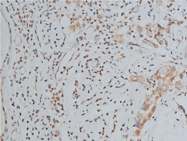


 IHC (Immunohistochemistry)
(AAA30929 at 1/200 staining human colon tissue sections by IHC-P. The tissue was formaldehyde fixed and a heat mediated antigen retrieval step in citrate buffer was performed. The tissue was then blocked and incubated with the antibody for 1.5 hours at 22 degree C. An HRP conjugated goat anti-rabbit antibody was used as the secondary.)
IHC (Immunohistochemistry)
(AAA30929 at 1/200 staining human colon tissue sections by IHC-P. The tissue was formaldehyde fixed and a heat mediated antigen retrieval step in citrate buffer was performed. The tissue was then blocked and incubated with the antibody for 1.5 hours at 22 degree C. An HRP conjugated goat anti-rabbit antibody was used as the secondary.)
Histone H3, Polyclonal Antibody (Cat# AAA30929)
Full Name
Histone H3 Antibody
Gene Names
HIST1H3A; H3/A; H3FA
Reactivity
Human, Mouse, Rat
Applications
Western Blot, Immunohistochemistry, Immunofluorescence, Immunocytochemistry
Purity
The antiserum was purified by peptide affinity chromatography using SulfoLink Coupling Resin.
Pricing


 IHC (Immunohistochemistry)
(At 1/100 staining Rat heart tissue by IHC-P. The sample was formaldehyde fixed and a heat mediated antigen retrieval step in citrate buffer was performed. The sample was then blocked and incubated with the primary antibody at 4 degree C overnight. An HRP conjugated anti-Rabbit antibody was used as the secondary antibody.)
IHC (Immunohistochemistry)
(At 1/100 staining Rat heart tissue by IHC-P. The sample was formaldehyde fixed and a heat mediated antigen retrieval step in citrate buffer was performed. The sample was then blocked and incubated with the primary antibody at 4 degree C overnight. An HRP conjugated anti-Rabbit antibody was used as the secondary antibody.)
ULK1, Polyclonal Antibody (Cat# AAA31359)
Full Name
Phospho-ULK1 (Ser757)[Ser758] Antibody
Gene Names
ULK1; ATG1; ATG1A; UNC51; hATG1; Unc51.1
Reactivity
Human, Mouse, Rat
Predicted Reactivity: Pig (100%), Bovine (100%), Horse (100%), Sheep (100%), Rabbit (100%), Dog (100%), Xenopus (88%)
Predicted Reactivity: Pig (100%), Bovine (100%), Horse (100%), Sheep (100%), Rabbit (100%), Dog (100%), Xenopus (88%)
Applications
Western Blot, Immunohistochemistry, Peptide ELISA
Purity
The antibody is from purified rabbit serum by affinity purification via sequential chromatography on phospho-peptide and non-phospho-peptide affinity columns.
Pricing


 Application Data
(At 25 degree C. Samples were then incubated with primary Ab(At 37 degree C. An AlexaFluor594 conjugated goat anti-rabbit IgG(H+L) Ab(Red) and an AlexaFluor488 conjugated goat anti-mouse IgG(H+L) Ab(Green) were used as the secondary antibody.The nuclear counter stain is DAPI (blue).)
Application Data
(At 25 degree C. Samples were then incubated with primary Ab(At 37 degree C. An AlexaFluor594 conjugated goat anti-rabbit IgG(H+L) Ab(Red) and an AlexaFluor488 conjugated goat anti-mouse IgG(H+L) Ab(Green) were used as the secondary antibody.The nuclear counter stain is DAPI (blue).)
Histone H3, Polyclonal Antibody (Cat# AAA31322)
Full Name
Phospho-Histone H3 (Thr3) Antibody
Gene Names
HIST1H3A; H3/A; H3FA
Reactivity
Human, Mouse, Rat
Applications
Immunohistochemistry, Immunofluorescence, Immunocytochemistry, Peptide ELISA
Purity
The antibody is from purified rabbit serum by affinity purification via sequential chromatography on phospho-peptide and non-phospho-peptide affinity columns.
Pricing


 IP (Immunoprecipitation)
(Immunoprecipitating GAPDH in Hela whole cell lysate Lane 1: Mouse control IgG instead of AAA27043 in Hela whole cell lysate. Lane 2: AAA27043 (5ul) + Hela whole cell lysate (500ug) Lane 3: Hela whole cell lysate (10ug) For western blotting, the blot was detected with AAA27043 at 1:5000, and a HRPconjugated Protein G antibody was used as the secondary antibody at 1:2000)
IP (Immunoprecipitation)
(Immunoprecipitating GAPDH in Hela whole cell lysate Lane 1: Mouse control IgG instead of AAA27043 in Hela whole cell lysate. Lane 2: AAA27043 (5ul) + Hela whole cell lysate (500ug) Lane 3: Hela whole cell lysate (10ug) For western blotting, the blot was detected with AAA27043 at 1:5000, and a HRPconjugated Protein G antibody was used as the secondary antibody at 1:2000)
GAPDH, Monoclonal Antibody (Cat# AAA27043)
Full Name
GAPDH Monoclonal Antibody
Reactivity
Human, Rat, Rabbit, Mouse
Applications
Western Blot, Immunohistochemistry, Immunoprecipitation, Immunofluorescence
Purity
>95%, Protein G purified
Pricing


 Application Data
(At 25 degree C. Samples were then incubated with primary Ab(At 37 degree C. An AlexaFluor594 conjugated goat anti-rabbit IgG(H+L) Ab(Red) and an AlexaFluor488 conjugated goat anti-mouse IgG(H+L) Ab(Green) were used as the secondary antibody.The nuclear counter stain is DAPI (blue).)
Application Data
(At 25 degree C. Samples were then incubated with primary Ab(At 37 degree C. An AlexaFluor594 conjugated goat anti-rabbit IgG(H+L) Ab(Red) and an AlexaFluor488 conjugated goat anti-mouse IgG(H+L) Ab(Green) were used as the secondary antibody.The nuclear counter stain is DAPI (blue).)
PFKFB3, Polyclonal Antibody (Cat# AAA31288)
Full Name
Phospho-PFKFB3 (Ser461) Antibody
Gene Names
PFKFB3; PFK2; IPFK2
Reactivity
Human, Rat
Applications
Western Blot, Immunohistochemistry, Immunofluorescence, Immunocytochemistry, Peptide ELISA
Purity
The antibody is from purified rabbit serum by affinity purification via sequential chromatography on phospho-peptide and non-phospho-peptide affinity columns.
Pricing


 IP (Immunoprecipitation)
(Immunoprecipitating HSPA8 in Hela whole cell lysate Lane 1: Mouse control IgG2b instead dilution for 30min at 4 degree C. Isotype control antibody (green line) was mouse IgG2b used under the same conditions. Acquisition of >10,000 events was performed.)
IP (Immunoprecipitation)
(Immunoprecipitating HSPA8 in Hela whole cell lysate Lane 1: Mouse control IgG2b instead dilution for 30min at 4 degree C. Isotype control antibody (green line) was mouse IgG2b used under the same conditions. Acquisition of >10,000 events was performed.)
HSPA8, Monoclonal Antibody (Cat# AAA27045)
Full Name
HSPA8 Monoclonal Antibody
Gene Names
HSPA8; LAP1; HSC54; HSC70; HSC71; HSP71; HSP73; NIP71; HSPA10
Reactivity
Human, Rat, Mouse
Applications
Western Blot, Immunohistochemistry, Flow Cytometry, Immunoprecipitation
Purity
>95%, Protein A purified
Pricing


 FCM (Flow Cytometry)
(Figure 7. Flow Cytometry analysis of U87 cells using anti-PDE6 beta/PDE6B antibody (AAA19261).Overlay histogram showing U87 cells stained with AAA19261 (Blue line). The cells were blocked with 10% normal goat serum. And then incubated with rabbit anti-PDE6 beta/PDE6B Antibody (AAA19261,1μg/1x106 cells) for 30 min at 20 degree C. DyLight®488 conjugated goat anti-rabbit IgG (5-10μg/1x106 cells) was used as secondary antibody for 30 minutes at 20 degree C. Isotype control antibody (Green line) was rabbit IgG (1μg/1x106) used under the same conditions. Unlabelled sample (Red line) was also used as a control.)
FCM (Flow Cytometry)
(Figure 7. Flow Cytometry analysis of U87 cells using anti-PDE6 beta/PDE6B antibody (AAA19261).Overlay histogram showing U87 cells stained with AAA19261 (Blue line). The cells were blocked with 10% normal goat serum. And then incubated with rabbit anti-PDE6 beta/PDE6B Antibody (AAA19261,1μg/1x106 cells) for 30 min at 20 degree C. DyLight®488 conjugated goat anti-rabbit IgG (5-10μg/1x106 cells) was used as secondary antibody for 30 minutes at 20 degree C. Isotype control antibody (Green line) was rabbit IgG (1μg/1x106) used under the same conditions. Unlabelled sample (Red line) was also used as a control.)
PDE6 beta/PDE6B, Polyclonal Antibody (Cat# AAA19261)
Full Name
Anti-PDE6 beta/PDE6B Antibody
Reactivity
Human, Mouse, Rat
Applications
Western Blot, Immunohistochemistry, Immunocytochemistry, Immunofluorescence, Flow Cytometry, Direct ELISA
Purity
Immunogen affinity purified.
Pricing


 WB (Western Blot)
(Anti-Beta-Tubulin mouse monoclonal antibody at 1:2000 dilution Lane A: HepG2 Whole Cell Lysate Lane B: Daudi Whole Cell Lysate Lane C: MOLT-4 Whole Cell Lysate Lane D: A549 Whole Cell Lysate Lane E: 293T Whole Cell Lysate Lane F: HelaS3 Whole Cell Lysate Lysates/proteins at 30 ug per lane. Secondary Goat Anti-Mouse IgG H&L (Dylight800) at 1/15000 dilution. Developed using the Odyssey technique. Performed under reducing conditions. Predicted band size:50 kDa Observed band size:54 kDa)
WB (Western Blot)
(Anti-Beta-Tubulin mouse monoclonal antibody at 1:2000 dilution Lane A: HepG2 Whole Cell Lysate Lane B: Daudi Whole Cell Lysate Lane C: MOLT-4 Whole Cell Lysate Lane D: A549 Whole Cell Lysate Lane E: 293T Whole Cell Lysate Lane F: HelaS3 Whole Cell Lysate Lysates/proteins at 30 ug per lane. Secondary Goat Anti-Mouse IgG H&L (Dylight800) at 1/15000 dilution. Developed using the Odyssey technique. Performed under reducing conditions. Predicted band size:50 kDa Observed band size:54 kDa)
Beta-Tubulin, Monoclonal Antibody (Cat# AAA27713)
Full Name
Beta-Tubulin Loading Control Antibody, Mouse MAb
Reactivity
Human
Applications
Western Blot, Immunohistochemistry, Immunocytochemistry, Immunofluorescence, Immunoprecipitation
Purity
Protein A
Pricing


 FCM (Flow Cytometry)
(Figure 6. Flow Cytometry analysis of PC-3 cells using anti-ADK antibody (AAA11690).Overlay histogram showing PC-3 cells stained with AAA11690 (Blue line).The cells were blocked with 10% normal goat serum. And then incubated with rabbit anti-ADK Antibody (AAA11690,1ug/1x10^6 cells) for 30 min at 20 degree C. DyLight®488 conjugated goat anti-rabbit IgG (5-10ug/1x10^6 cells) was used as secondary antibody for 30 minutes at 20 degree C. Isotype control antibody (Green line) was rabbit IgG (1ug/1x106) used under the same conditions. Unlabelled sample (Red line) was also used as a control.)
FCM (Flow Cytometry)
(Figure 6. Flow Cytometry analysis of PC-3 cells using anti-ADK antibody (AAA11690).Overlay histogram showing PC-3 cells stained with AAA11690 (Blue line).The cells were blocked with 10% normal goat serum. And then incubated with rabbit anti-ADK Antibody (AAA11690,1ug/1x10^6 cells) for 30 min at 20 degree C. DyLight®488 conjugated goat anti-rabbit IgG (5-10ug/1x10^6 cells) was used as secondary antibody for 30 minutes at 20 degree C. Isotype control antibody (Green line) was rabbit IgG (1ug/1x106) used under the same conditions. Unlabelled sample (Red line) was also used as a control.)
ADK, Polyclonal Antibody (Cat# AAA11690)
Full Name
Anti-ADK Antibody
Gene Names
ADK; AK
Reactivity
Human, Mouse, Rat
Applications
Western Blot, Immunohistochemistry
Purity
Immunogen affinity purified.
Pricing


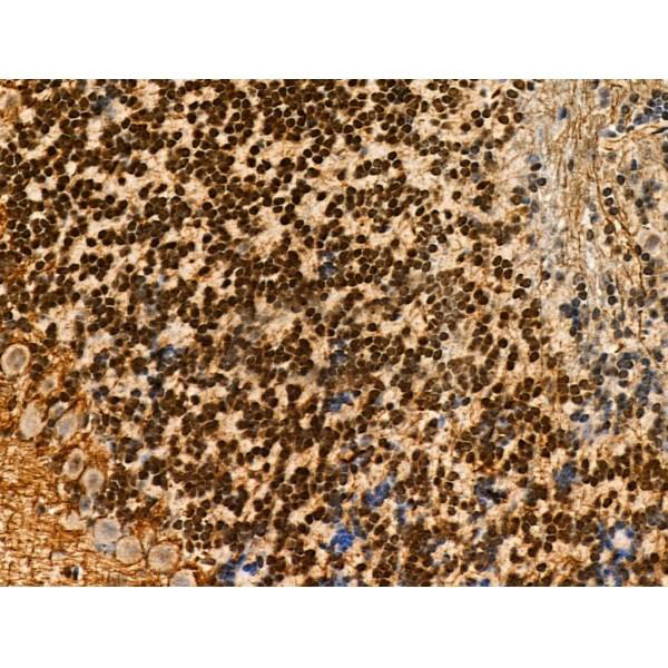
 IHC (Immunohistochemistry)
(At 1/100 staining Human gastric cancer and adjacent normal tissues by IHC-P. The sample was formaldehyde fixed and a heat mediated antigen retrieval step in citrate buffer was performed. The sample was then blocked and incubated with the primary antibody at 4 degree C overnight. An HRP conjugated anti-Rabbit antibody was used as the secondary antibody.)
IHC (Immunohistochemistry)
(At 1/100 staining Human gastric cancer and adjacent normal tissues by IHC-P. The sample was formaldehyde fixed and a heat mediated antigen retrieval step in citrate buffer was performed. The sample was then blocked and incubated with the primary antibody at 4 degree C overnight. An HRP conjugated anti-Rabbit antibody was used as the secondary antibody.)
AIRE, Polyclonal Antibody (Cat# AAA31324)
Full Name
Phospho-AIRE (Ser156) Antibody
Gene Names
AIRE; APS1; APSI; PGA1; AIRE1; APECED
Reactivity
Human, Mouse, Rat
Applications
Immunohistochemistry, Peptide ELISA
Purity
The antibody is from purified rabbit serum by affinity purification via sequential chromatography on phospho-peptide and non-phospho-peptide affinity columns.
Pricing


 IP (Immunoprecipitation)
(MAP2K3 was immunoprecipitated using: Lane A:0.5 mg Jurkat Whole Cell Lysate Lane B:0.5 mg HepG2 Whole Cell Lysate Lane C:0.5 mg HeLa Whole Cell Lysate 2 uL anti-MAP2K3 rabbit polyclonal antibody and 60 ug of Immunomagnetic beads Protein A/G. Primary antibody: Anti-MAP2K3 rabbit polyclonal antibody,at 1:100 dilution Secondary antibody: Clean-Blot IP Detection Reagent (HRP) at 1:1000dilution Developed using the ECL technique. Performed under reducing conditions. Predicted band size: 39 kDa Observed band size :39 kDa)
IP (Immunoprecipitation)
(MAP2K3 was immunoprecipitated using: Lane A:0.5 mg Jurkat Whole Cell Lysate Lane B:0.5 mg HepG2 Whole Cell Lysate Lane C:0.5 mg HeLa Whole Cell Lysate 2 uL anti-MAP2K3 rabbit polyclonal antibody and 60 ug of Immunomagnetic beads Protein A/G. Primary antibody: Anti-MAP2K3 rabbit polyclonal antibody,at 1:100 dilution Secondary antibody: Clean-Blot IP Detection Reagent (HRP) at 1:1000dilution Developed using the ECL technique. Performed under reducing conditions. Predicted band size: 39 kDa Observed band size :39 kDa)
MEK3/MKK3, Polyclonal Antibody (Cat# AAA27720)
Full Name
Anti-MEK3/MKK3 Antibody, Rabbit Polyclonal
Gene Names
MAP2K3; MEK3; MKK3; MAPKK3; PRKMK3; SAPKK2; SAPKK-2
Reactivity
Human, Mouse
Applications
Western Blot, Immunohistochemistry, Immunocytochemistry, Immunofluorescence, Immunoprecipitation
Purity
Protein A & Antigen Affinity
Pricing


 IHC (Immunohistochemistry)
(At 1/200 staining Human kidney tissue sections by IHC-P. The tissue was formaldehyde fixed and a heat mediated antigen retrieval step in citrate buffer was performed. The tissue was then blocked and incubated with the antibody for 1.5 hours at 22 degree C. An HRP conjugated goat anti-rabbit antibody was used as the secondary antibody.)
IHC (Immunohistochemistry)
(At 1/200 staining Human kidney tissue sections by IHC-P. The tissue was formaldehyde fixed and a heat mediated antigen retrieval step in citrate buffer was performed. The tissue was then blocked and incubated with the antibody for 1.5 hours at 22 degree C. An HRP conjugated goat anti-rabbit antibody was used as the secondary antibody.)
CtBP1/2, Polyclonal Antibody (Cat# AAA31423)
Full Name
Phospho-CtBP1/2 (Ser158/Ser164) Antibody
Gene Names
CTBP1; BARS
Reactivity
Human, Mouse, Rat
Predicted Reactivity: Pig (100%), Zebrafish (82%), Bovine (100%), Horse (100%), Dog (100%), Chicken (100%), Xenopus (100%)
Predicted Reactivity: Pig (100%), Zebrafish (82%), Bovine (100%), Horse (100%), Dog (100%), Chicken (100%), Xenopus (100%)
Applications
Western Blot, Immunohistochemistry, Peptide ELISA
Purity
The antibody is from purified rabbit serum by affinity purification via sequential chromatography on phospho-peptide and non-phospho-peptide affinity columns.
Pricing


 FCM (Flow Cytometry)
(Figure 8. Flow Cytometry analysis of THP-1 cells using anti-YTHDF2 antibody (AAA19260).Overlay histogram showing THP-1 cells stained with AAA19260 (Blue line). The cells were blocked with 10% normal goat serum. And then incubated with rabbit anti-YTHDF2 Antibody (AAA19260, 1μg/1x106 cells) for 30 min at 20 degree C. DyLight®488 conjugated goat anti-rabbit IgG (5-10μg/1x106 cells) was used as secondary antibody for 30 minutes at 20 degree C. Isotype control antibody (Green line) was rabbit IgG (1μg/1x106) used under the same conditions. Unlabelled sample (Red line) was also used as a control.)
FCM (Flow Cytometry)
(Figure 8. Flow Cytometry analysis of THP-1 cells using anti-YTHDF2 antibody (AAA19260).Overlay histogram showing THP-1 cells stained with AAA19260 (Blue line). The cells were blocked with 10% normal goat serum. And then incubated with rabbit anti-YTHDF2 Antibody (AAA19260, 1μg/1x106 cells) for 30 min at 20 degree C. DyLight®488 conjugated goat anti-rabbit IgG (5-10μg/1x106 cells) was used as secondary antibody for 30 minutes at 20 degree C. Isotype control antibody (Green line) was rabbit IgG (1μg/1x106) used under the same conditions. Unlabelled sample (Red line) was also used as a control.)
YTHDF2, Polyclonal Antibody (Cat# AAA19260)
Full Name
Anti-YTHDF2 Antibody
Gene Names
YTHDF2; HGRG8; NY-REN-2
Reactivity
Human, Mouse, Rat
Applications
Western Blot, Immunohistochemistry, Immunocytochemistry, Immunofluorescence, Flow Cytometry
Purity
Immunogen affinity purified.
Pricing


 Application Data
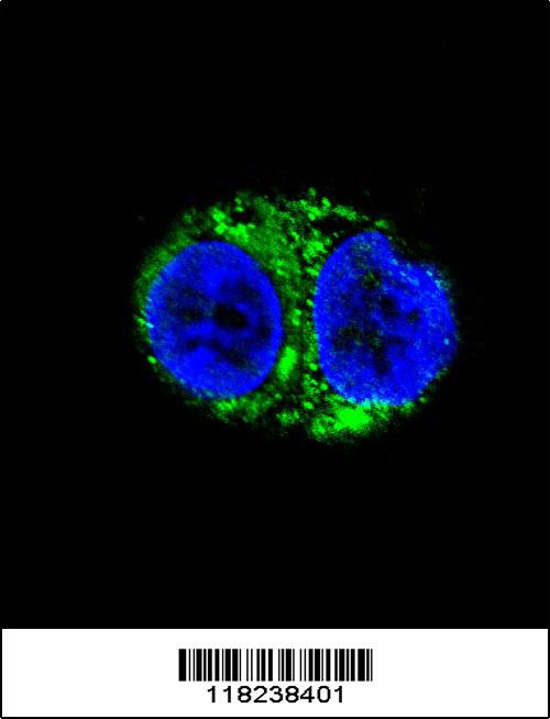
(At 25 degree C. The primary antibody was diluted at 1/200 and incubated with the sample for 1 hour at 37 degree C. An Alexa Fluor 594 conjugated goat anti-rabbit IgG (H+L) Ab, diluted at 1/600, was used as the secondary antibody.)
Application Data
(At 25 degree C. The primary antibody was diluted at 1/200 and incubated with the sample for 1 hour at 37 degree C. An Alexa Fluor 594 conjugated goat anti-rabbit IgG (H+L) Ab, diluted at 1/600, was used as the secondary antibody.)
p27 Kip1, Polyclonal Antibody (Cat# AAA31388)
Full Name
Phospho-p27 Kip1 (Ser178) Antibody
Gene Names
CDKN1B; KIP1; MEN4; CDKN4; MEN1B; P27KIP1
Reactivity
Human, Mouse, Rat
Predicted Reactivity: Bovine (100%), Sheep (100%), Rabbit (100%), Dog (100%)
Predicted Reactivity: Bovine (100%), Sheep (100%), Rabbit (100%), Dog (100%)
Applications
Western Blot, Immunohistochemistry, Immunofluorescence, Immunocytochemistry, Peptide ELISA
Purity
The antibody is from purified rabbit serum by affinity purification via sequential chromatography on phospho-peptide and non-phospho-peptide affinity columns.
Pricing


 IP (Immunoprecipitation)
(Immunoprecipitating GFP in 293F whole cell lysate transfected with GFPLane 1: Mouse control IgG2b instead of AAA28064 in 293F whole cell lysate transfected with GFPLane 2: AAA28064 (4ug) + 293F whole cell lysate transfected with GFP (500ug)Lane 3: 293F whole cell lysate transfected with GFP (5ug)For western blotting, the blot was detected with AAA28064 at 1:2000, and a HRP-conjugated Protein G antibody was used as the secondary antibody at 1:50000)
IP (Immunoprecipitation)
(Immunoprecipitating GFP in 293F whole cell lysate transfected with GFPLane 1: Mouse control IgG2b instead of AAA28064 in 293F whole cell lysate transfected with GFPLane 2: AAA28064 (4ug) + 293F whole cell lysate transfected with GFP (500ug)Lane 3: 293F whole cell lysate transfected with GFP (5ug)For western blotting, the blot was detected with AAA28064 at 1:2000, and a HRP-conjugated Protein G antibody was used as the secondary antibody at 1:50000)
GFP, Monoclonal Antibody (Cat# AAA28064)
Full Name
GFP Monoclonal Antibody
Reactivity
All
Applications
Western Blot, Immunofluorescence, Flow Cytometry, Immunoprecipitation
Purity
>95%,Protein G purified
Pricing


 Application Data
(At 25 degree C. Samples were then incubated with primary Ab(At 37 degree C. An AlexaFluor594 conjugated goat anti-rabbit IgG(H+L) Ab(Red) and an AlexaFluor488 conjugated goat anti-mouse IgG(H+L) Ab(Green) were used as the secondary antibody.The nuclear counter stain is DAPI(blue).)
Application Data
(At 25 degree C. Samples were then incubated with primary Ab(At 37 degree C. An AlexaFluor594 conjugated goat anti-rabbit IgG(H+L) Ab(Red) and an AlexaFluor488 conjugated goat anti-mouse IgG(H+L) Ab(Green) were used as the secondary antibody.The nuclear counter stain is DAPI(blue).)
TLK1, Polyclonal Antibody (Cat# AAA31385)
Full Name
Phospho-TLK1 (Ser741) Antibody
Gene Names
TLK1; PKU-beta
Reactivity
Human, Mouse, Rat
Predicted Reactivity: Pig (100%), Bovine (100%), Horse (100%), Sheep (100%), Rabbit (100%), Dog (100%), Chicken (100%)
Predicted Reactivity: Pig (100%), Bovine (100%), Horse (100%), Sheep (100%), Rabbit (100%), Dog (100%), Chicken (100%)
Applications
Western Blot, Immunohistochemistry, Immunofluorescence, Immunocytochemistry, Peptide ELISA
Purity
The antibody is from purified rabbit serum by affinity purification via sequential chromatography on phospho-peptide and non-phospho-peptide affinity columns.
Pricing


 Application Data
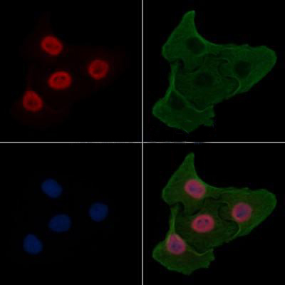
(At 25 degree C. Samples were then incubated with primary Ab(At 37 degree C. An AlexaFluor594 conjugated goat anti-rabbit IgG(H+L) Ab(Red) and an AlexaFluor488 conjugated goat anti-mouse IgG(H+L) Ab(Green) were used as the secondary antibody.The nuclear counter stain is DAPI(blue).)
Application Data
(At 25 degree C. Samples were then incubated with primary Ab(At 37 degree C. An AlexaFluor594 conjugated goat anti-rabbit IgG(H+L) Ab(Red) and an AlexaFluor488 conjugated goat anti-mouse IgG(H+L) Ab(Green) were used as the secondary antibody.The nuclear counter stain is DAPI(blue).)
USP28, Polyclonal Antibody (Cat# AAA31438)
Full Name
Phospho-USP28 (Ser714) Antibody
Reactivity
Human, Mouse, Monkey
Applications
Western Blot, Immunohistochemistry, Immunofluorescence, Immunocytochemistry, Peptide ELISA
Purity
The antibody is from purified rabbit serum by affinity purification via sequential chromatography on phospho-peptide and non-phospho-peptide affinity columns.
Pricing