Filters
Clonality
Type
Reactivity
Gene Name
Isotype
Host
Application
Clone
301 results for " biotin conjugated antibody" - showing 200-250


 FCM (Flow Cytometry)
(Figure 8. Flow Cytometry analysis of U937 cells using anti-SEC14L3/TAP2 antibody (AAA19342).Overlay histogram showing U937 cells stained with AAA19342 (Blue line). The cells were blocked with 10% normal goat serum. And then incubated with rabbit anti-SEC14L3/TAP2 Antibody (AAA19342, 1μg/1x106 cells) for 30 min at 20 degree C. DyLight®488 conjugated goat anti-rabbit IgG (5-10μg/1x106 cells) was used as secondary antibody for 30 minutes at 20 degree C. Isotype control antibody (Green line) was rabbit IgG (1μg/1x106) used under the same conditions. Unlabelled sample (Red line) was also used as a control.)
FCM (Flow Cytometry)
(Figure 8. Flow Cytometry analysis of U937 cells using anti-SEC14L3/TAP2 antibody (AAA19342).Overlay histogram showing U937 cells stained with AAA19342 (Blue line). The cells were blocked with 10% normal goat serum. And then incubated with rabbit anti-SEC14L3/TAP2 Antibody (AAA19342, 1μg/1x106 cells) for 30 min at 20 degree C. DyLight®488 conjugated goat anti-rabbit IgG (5-10μg/1x106 cells) was used as secondary antibody for 30 minutes at 20 degree C. Isotype control antibody (Green line) was rabbit IgG (1μg/1x106) used under the same conditions. Unlabelled sample (Red line) was also used as a control.)
SEC14L3/TAP2, Polyclonal Antibody (Cat# AAA19342)
Full Name
Anti-SEC14L3/TAP2 Antibody
Gene Names
SEC14L3; TAP2
Reactivity
Human, Mouse, Rat
Applications
WB, IHC-P, ICC, IF, FC/FACS/FCM, EIA
Purity
Immunogen affinity purified.
Pricing


 FCM (Flow Cytometry)
(Figure 6. Flow Cytometry analysis of Hela cells using anti-PDE4D antibody (AAA19143).Overlay histogram showing Hela cells stained with AAA19143 (Blue line).The cells were blocked with 10% normal goat serum. And then incubated with rabbit anti-PDE4D Antibody (AAA19143,1ug/1x10^6 cells) for 30 min at 20 degree C. DyLight®488 conjugated goat anti-rabbit IgG (5-10ug/1x10^6 cells) was used as secondary antibody for 30 minutes at 20 degree C. Isotype control antibody (Green line) was rabbit IgG (1ug/1x106) used under the same conditions. Unlabelled sample (Red line) was also used as a control.)
FCM (Flow Cytometry)
(Figure 6. Flow Cytometry analysis of Hela cells using anti-PDE4D antibody (AAA19143).Overlay histogram showing Hela cells stained with AAA19143 (Blue line).The cells were blocked with 10% normal goat serum. And then incubated with rabbit anti-PDE4D Antibody (AAA19143,1ug/1x10^6 cells) for 30 min at 20 degree C. DyLight®488 conjugated goat anti-rabbit IgG (5-10ug/1x10^6 cells) was used as secondary antibody for 30 minutes at 20 degree C. Isotype control antibody (Green line) was rabbit IgG (1ug/1x106) used under the same conditions. Unlabelled sample (Red line) was also used as a control.)
PDE4D, Polyclonal Antibody (Cat# AAA19143)
Full Name
Anti-PDE4D Picoband antibody
Gene Names
PDE4D; DPDE3; PDE43; STRK1; ACRDYS2; HSPDE4D; PDE4DN2
Reactivity
Human, Mouse, Rat
No cross reactivity with other proteins.
No cross reactivity with other proteins.
Applications
EIA, FC/FACS, IHC, ICC, WB
Pricing


 IF (Immunofluorescence)
(Figure 8. IF analysis of eRF1/ETF1 using anti- eRF1/ETF1 antibody (AAA19383).eRF1/ETF1 was detected in immunocytochemical section of A431 cells. Enzyme antigen retrieval was performed using IHC enzyme antigen retrieval reagent for 15 mins. The cells were blocked with 10% goat serum. And then incubated with 5μg/mL mouse anti- eRF1/ETF1 Antibody (AAA19383) overnight at 4 degree C. DyLight®488 Conjugated Goat Anti-Mouse IgG (BA1126) was used as secondary antibody at 1:100 dilution and incubated for 30 minutes at 37 degree C. The section was counterstained with DAPI. Visualize using a fluorescence microscope and filter sets appropriate for the label used.)
IF (Immunofluorescence)
(Figure 8. IF analysis of eRF1/ETF1 using anti- eRF1/ETF1 antibody (AAA19383).eRF1/ETF1 was detected in immunocytochemical section of A431 cells. Enzyme antigen retrieval was performed using IHC enzyme antigen retrieval reagent for 15 mins. The cells were blocked with 10% goat serum. And then incubated with 5μg/mL mouse anti- eRF1/ETF1 Antibody (AAA19383) overnight at 4 degree C. DyLight®488 Conjugated Goat Anti-Mouse IgG (BA1126) was used as secondary antibody at 1:100 dilution and incubated for 30 minutes at 37 degree C. The section was counterstained with DAPI. Visualize using a fluorescence microscope and filter sets appropriate for the label used.)
eRF1/ETF1, Monoclonal Antibody (Cat# AAA19383)
Full Name
Anti-eRF1/ETF1 Antibody (monoclonal, 3E5)
Gene Names
ETF1; ERF; RF1; ERF1; TB3-1; D5S1995; SUP45L1
Reactivity
Human, Mouse, Rat
Applications
WB, IHC-P, ICC, IF, FC/FACS/FCM
Purity
Immunogen affinity purified.
Pricing


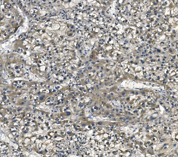
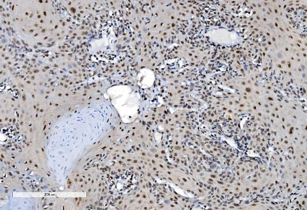
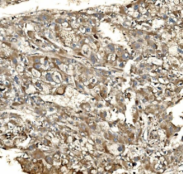
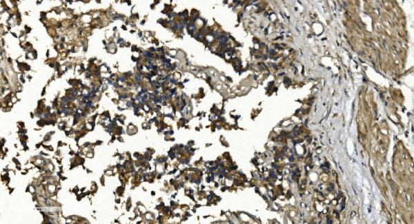
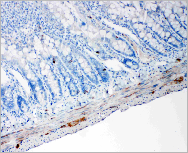
 IHC (Immunohistochemistry)
(Figure 7. IHC analysis of GCHFR using anti-GCHFR antibody (AAA19322).GCHFR was detected in paraffin-embedded section of mouse liver tissue. Heat mediated antigen retrieval was performed in EDTA buffer (pH8. 0, epitope retrieval solution). The tissue section was blocked with 10% goat serum. The tissue section was then incubated with 2μg/ml rabbit anti-GCHFR Antibody (AAA19322) overnight at 4 degree C. Biotinylated goat anti-rabbit IgG was used as secondary antibody and incubated for 30 minutes at 37 degree C. The tissue section was developed using Strepavidin-Biotin-Complex (SABC) (Catalog # with DAB as the chromogen.)
IHC (Immunohistochemistry)
(Figure 7. IHC analysis of GCHFR using anti-GCHFR antibody (AAA19322).GCHFR was detected in paraffin-embedded section of mouse liver tissue. Heat mediated antigen retrieval was performed in EDTA buffer (pH8. 0, epitope retrieval solution). The tissue section was blocked with 10% goat serum. The tissue section was then incubated with 2μg/ml rabbit anti-GCHFR Antibody (AAA19322) overnight at 4 degree C. Biotinylated goat anti-rabbit IgG was used as secondary antibody and incubated for 30 minutes at 37 degree C. The tissue section was developed using Strepavidin-Biotin-Complex (SABC) (Catalog # with DAB as the chromogen.)
GCHFR, Polyclonal Antibody (Cat# AAA19322)
Full Name
Anti-GCHFR Antibody
Gene Names
GCHFR; P35; GFRP; HsT16933
Reactivity
Human, Mouse, Rat
Applications
WB, IHC-P, ICC, IF, FC/FACS/FCM, EIA
Purity
Immunogen affinity purified.
Pricing


 FCM (Flow Cytometry)
(Figure 8. Flow Cytometry analysis of LLC cells using anti-Synaptotagmin 1 antibody (AAA19158).Overlay histogram showing LLC cells stained with AAA19158 (Blue line).The cells were blocked with 10% normal goat serum. And then incubated with rabbit anti-Synaptotagmin 1 Antibody (AAA19158,1ug/1x10^6 cells) for 30 min at 20 degree C. DyLight®488 conjugated goat anti-rabbit IgG (5-10ug/1x10^6 cells) was used as secondary antibody for 30 minutes at 20 degree C. Isotype control antibody (Green line) was rabbit IgG (1ug/1x106) used under the same conditions. Unlabelled sample (Red line) was also used as a control.)
FCM (Flow Cytometry)
(Figure 8. Flow Cytometry analysis of LLC cells using anti-Synaptotagmin 1 antibody (AAA19158).Overlay histogram showing LLC cells stained with AAA19158 (Blue line).The cells were blocked with 10% normal goat serum. And then incubated with rabbit anti-Synaptotagmin 1 Antibody (AAA19158,1ug/1x10^6 cells) for 30 min at 20 degree C. DyLight®488 conjugated goat anti-rabbit IgG (5-10ug/1x10^6 cells) was used as secondary antibody for 30 minutes at 20 degree C. Isotype control antibody (Green line) was rabbit IgG (1ug/1x106) used under the same conditions. Unlabelled sample (Red line) was also used as a control.)
Synaptotagmin 1, Polyclonal Antibody (Cat# AAA19158)
Full Name
Anti-Synaptotagmin 1 Picoband antibody
Gene Names
SYT1; P65; SYT; SVP65
Reactivity
Human, Mouse, Rat
No cross reactivity with other proteins.
No cross reactivity with other proteins.
Applications
IHC, WB
Pricing


 FCM (Flow Cytometry)
(Figure 10. Flow Cytometry analysis of MCF-7 cells using anti-DDX1 antibody (AAA19378).Overlay histogram showing MCF-7 cells stained with AAA19378 (Blue line). The cells were blocked with 10% normal goat serum. And then incubated with mouse anti- DDX1 Antibody (AAA19378, 1μg/1x106 cells) for 30 min at 20 degree C. DyLight®488 conjugated goat anti-mouse IgG (BA1126, 5-10μg/1x106 cells) was used as secondary antibody for 30 minutes at 20 degree C. Isotype control antibody (Green line) was mouse IgG (1μg/1x106) used under the same conditions. Unlabelled sample (Red line) was also used as a control.)
FCM (Flow Cytometry)
(Figure 10. Flow Cytometry analysis of MCF-7 cells using anti-DDX1 antibody (AAA19378).Overlay histogram showing MCF-7 cells stained with AAA19378 (Blue line). The cells were blocked with 10% normal goat serum. And then incubated with mouse anti- DDX1 Antibody (AAA19378, 1μg/1x106 cells) for 30 min at 20 degree C. DyLight®488 conjugated goat anti-mouse IgG (BA1126, 5-10μg/1x106 cells) was used as secondary antibody for 30 minutes at 20 degree C. Isotype control antibody (Green line) was mouse IgG (1μg/1x106) used under the same conditions. Unlabelled sample (Red line) was also used as a control.)
DDX1, Monoclonal Antibody (Cat# AAA19378)
Full Name
Anti-DDX1 Antibody (monoclonal, 3I10)
Gene Names
DDX1; DBP-RB; UKVH5d
Reactivity
Human, Mouse, Rat
Applications
WB, IHC-P, FC/FACS/FCM
Purity
Immunogen affinity purified.
Pricing


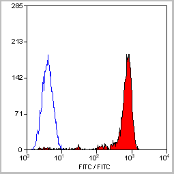
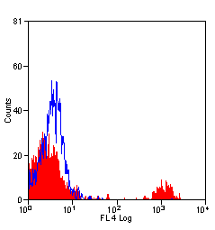
 FCM (Flow Cytometry)
(Figure 11. Flow Cytometry analysis of THP-1 cells using anti-EPRS1/PARS antibody (AAA19268).Overlay histogram showing THP-1 cells stained with AAA19268 (Blue line). The cells were blocked with 10% normal goat serum. And then incubated with rabbit anti-EPRS1/PARS Antibody (AAA19268, 1μg/1x106 cells) for 30 min at 20 degree C. DyLight®488 conjugated goat anti-rabbit IgG (5-10μg/1x106 cells) was used as secondary antibody for 30 minutes at 20 degree C. Isotype control antibody (Green line) was rabbit IgG (1μg/1x106) used under the same conditions. Unlabelled sample (Red line) was also used as a control.)
FCM (Flow Cytometry)
(Figure 11. Flow Cytometry analysis of THP-1 cells using anti-EPRS1/PARS antibody (AAA19268).Overlay histogram showing THP-1 cells stained with AAA19268 (Blue line). The cells were blocked with 10% normal goat serum. And then incubated with rabbit anti-EPRS1/PARS Antibody (AAA19268, 1μg/1x106 cells) for 30 min at 20 degree C. DyLight®488 conjugated goat anti-rabbit IgG (5-10μg/1x106 cells) was used as secondary antibody for 30 minutes at 20 degree C. Isotype control antibody (Green line) was rabbit IgG (1μg/1x106) used under the same conditions. Unlabelled sample (Red line) was also used as a control.)
EPRS1/PARS, Polyclonal Antibody (Cat# AAA19268)
Full Name
Anti-EPRS1/PARS Antibody
Gene Names
EPRS; EARS; PARS; QARS; QPRS; PIG32; GLUPRORS
Reactivity
Human, Mouse, Rat
Applications
WB, IHC-P, ICC, IF, FC/FACS/FCM, EIA
Purity
Immunogen affinity purified.
Pricing


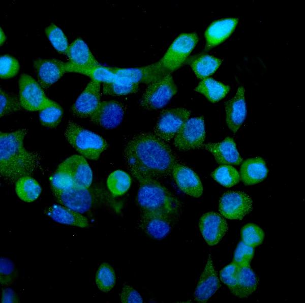
 IF (Immunofluorescence)
(Immunofluorescent analysis of 4% paraformaldehyde-fixed, 0.1% Triton X-100 permeabilized MCF-7 (human breast cancer cell line) cells labeling Pdx1 with at 1:25 dilution, followed by DyLight 488-conjugated IgG goat anti-rabbit secondary antibody at 1:200 dilution (green). Immunofluorescence image showing cytoplasm staining on MCF-7 cell line. Cytoplasmic actin is detected with DyLight 554 Phalloidin (PD18466410) at 1:100 dilution (red). The nuclear counter stain is DAPI (blue).)
IF (Immunofluorescence)
(Immunofluorescent analysis of 4% paraformaldehyde-fixed, 0.1% Triton X-100 permeabilized MCF-7 (human breast cancer cell line) cells labeling Pdx1 with at 1:25 dilution, followed by DyLight 488-conjugated IgG goat anti-rabbit secondary antibody at 1:200 dilution (green). Immunofluorescence image showing cytoplasm staining on MCF-7 cell line. Cytoplasmic actin is detected with DyLight 554 Phalloidin (PD18466410) at 1:100 dilution (red). The nuclear counter stain is DAPI (blue).)
OPN-a/b, Polyclonal Antibody (Cat# AAA26858)
Full Name
OPN-a/b, NT (SPP1, BNSP, OPN, Osteopontin, Bone sialoprotein 1, Nephropontin, Secreted phosphoprotein 1, Urinary stone protein, Uropontin) (Biotin)
Gene Names
SPP1; OPN; BNSP; BSPI; ETA-1
Reactivity
Human
Applications
WB, IHC, IF, EIA
Purity
Purified by Protein A and Peptide Affinity Chromatography.
Pricing


 FCM (Flow Cytometry)
(Figure 8. Flow Cytometry analysis of U20S cells using anti-SV2A antibody (AAA19279).Overlay histogram showing U20S cells stained with AAA19279 (Blue line). The cells were blocked with 10% normal goat serum. And then incubated with rabbit anti-SV2A Antibody (AAA19279, 1μg/1x106 cells) for 30 min at 20 degree C. DyLight®488 conjugated goat anti-rabbit IgG (5-10μg/1x106 cells) was used as secondary antibody for 30 minutes at 20 degree C. Isotype control antibody (Green line) was rabbit IgG (1μg/1x106) used under the same conditions. Unlabelled sample (Red line) was also used as a control.)
FCM (Flow Cytometry)
(Figure 8. Flow Cytometry analysis of U20S cells using anti-SV2A antibody (AAA19279).Overlay histogram showing U20S cells stained with AAA19279 (Blue line). The cells were blocked with 10% normal goat serum. And then incubated with rabbit anti-SV2A Antibody (AAA19279, 1μg/1x106 cells) for 30 min at 20 degree C. DyLight®488 conjugated goat anti-rabbit IgG (5-10μg/1x106 cells) was used as secondary antibody for 30 minutes at 20 degree C. Isotype control antibody (Green line) was rabbit IgG (1μg/1x106) used under the same conditions. Unlabelled sample (Red line) was also used as a control.)
SV2A, Polyclonal Antibody (Cat# AAA19279)
Full Name
Anti-SV2A Antibody
Gene Names
SV2A; SV2
Reactivity
Human, Mouse, Rat
Applications
WB, IHC-P, FC/FACS/FCM
Purity
Immunogen affinity purified.
Pricing


 FCM (Flow Cytometry)
(Figure 8. Flow Cytometry analysis of HepG2 cells using anti-REA/PHB2 antibody (AAA19374).Overlay histogram showing HepG2 cells stained with AAA19374 (Blue line). The cells were blocked with 10% normal goat serum. And then incubated with mouse anti- REA/PHB2 Antibody (AAA19374, 1μg/1x106 cells) for 30 min at 20 degree C. DyLight®488 conjugated goat anti-mouse IgG (BA1126, 5-10μg/1x106 cells) was used as secondary antibody for 30 minutes at 20 degree C. Isotype control antibody (Green line) was mouse IgG (1μg/1x106) used under the same conditions. Unlabelled sample (Red line) was also used as a control.)
FCM (Flow Cytometry)
(Figure 8. Flow Cytometry analysis of HepG2 cells using anti-REA/PHB2 antibody (AAA19374).Overlay histogram showing HepG2 cells stained with AAA19374 (Blue line). The cells were blocked with 10% normal goat serum. And then incubated with mouse anti- REA/PHB2 Antibody (AAA19374, 1μg/1x106 cells) for 30 min at 20 degree C. DyLight®488 conjugated goat anti-mouse IgG (BA1126, 5-10μg/1x106 cells) was used as secondary antibody for 30 minutes at 20 degree C. Isotype control antibody (Green line) was mouse IgG (1μg/1x106) used under the same conditions. Unlabelled sample (Red line) was also used as a control.)
REA/PHB2, Monoclonal Antibody (Cat# AAA19374)
Full Name
Anti-REA/PHB2 Antibody (monoclonal, 2B5)
Gene Names
PHB2; BAP; REA; p22; Bap37; BCAP37; PNAS-141
Reactivity
Human, Mouse, Rat
Applications
WB, IHC-P, FC/FACS/FCM
Purity
Immunogen affinity purified.
Pricing


 FCM (Flow Cytometry)
(Figure 7. Flow Cytometry analysis of HEPG2 cells using anti-ALDH1L1 antibody (AAA19297).Overlay histogram showing HEPG2 cells stained with AAA19297 (Blue line). The cells were blocked with 10% normal goat serum. And then incubated with rabbit anti-ALDH1L1 Antibody (AAA19297 1μg/1x106 cells) for 30 min at 20 degree C. DyLight®488 conjugated goat anti-rabbit IgG (5-10μg/1x106 cells) was used as secondary antibody for 30 minutes at 20 degree C. Isotype control antibody (Green line) was rabbit IgG (1μg/1x106) used under the same conditions. Unlabelled sample (Red line) was also used as a control.)
FCM (Flow Cytometry)
(Figure 7. Flow Cytometry analysis of HEPG2 cells using anti-ALDH1L1 antibody (AAA19297).Overlay histogram showing HEPG2 cells stained with AAA19297 (Blue line). The cells were blocked with 10% normal goat serum. And then incubated with rabbit anti-ALDH1L1 Antibody (AAA19297 1μg/1x106 cells) for 30 min at 20 degree C. DyLight®488 conjugated goat anti-rabbit IgG (5-10μg/1x106 cells) was used as secondary antibody for 30 minutes at 20 degree C. Isotype control antibody (Green line) was rabbit IgG (1μg/1x106) used under the same conditions. Unlabelled sample (Red line) was also used as a control.)
ALDH1L1, Polyclonal Antibody (Cat# AAA19297)
Full Name
Anti-ALDH1L1 Antibody
Gene Names
ALDH1L1; FDH; FTHFD; 10-fTHF; 10-FTHFDH
Reactivity
Human, Mouse, Rat, Monkey
Applications
WB, IHC-P, ICC, IF, FC/FACS/FCM, EIA
Purity
Immunogen affinity purified.
Pricing


 FCM (Flow Cytometry)
(Figure 10. Flow Cytometry analysis of SiHa cells using anti-PRDM14 antibody (AAA19283).Overlay histogram showing SiHa cells stained with AAA19283 (Blue line). The cells were blocked with 10% normal goat serum. And then incubated with rabbit anti-PRDM14 Antibody (AAA19283, 1μg/1x106 cells) for 30 min at 20 degree C. DyLight®488 conjugated goat anti-rabbit IgG (5-10μg/1x106 cells) was used as secondary antibody for 30 minutes at 20 degree C. Isotype control antibody (Green line) was rabbit IgG (1μg/1x106) used under the same conditions. Unlabelled sample (Red line) was also used as a control.)
FCM (Flow Cytometry)
(Figure 10. Flow Cytometry analysis of SiHa cells using anti-PRDM14 antibody (AAA19283).Overlay histogram showing SiHa cells stained with AAA19283 (Blue line). The cells were blocked with 10% normal goat serum. And then incubated with rabbit anti-PRDM14 Antibody (AAA19283, 1μg/1x106 cells) for 30 min at 20 degree C. DyLight®488 conjugated goat anti-rabbit IgG (5-10μg/1x106 cells) was used as secondary antibody for 30 minutes at 20 degree C. Isotype control antibody (Green line) was rabbit IgG (1μg/1x106) used under the same conditions. Unlabelled sample (Red line) was also used as a control.)
PRDM14, Polyclonal Antibody (Cat# AAA19283)
Full Name
Anti-PRDM14 Antibody
Gene Names
PRDM14; PFM11
Reactivity
Human, Mouse, Rat
Applications
WB, IHC-P, ICC, IF, FC/FACS/FCM
Purity
Immunogen affinity purified.
Pricing


 FCM (Flow Cytometry)
(Figure 8. Flow Cytometry analysis of Jurkat cells using anti-Ki67 antibody (AAA19350).Overlay histogram showing Jurkat cells stained with AAA19350 (Blue line). The cells were blocked with 10% normal goat serum. And then incubated with mouse anti-Ki67 Antibody (AAA19350, 1μg/1x106 cells) for 30 min at 20 degree C. DyLight®488 conjugated goat anti-mouse IgG (BA1126, 5-10μg/1x106 cells) was used as secondary antibody for 30 minutes at 20 degree C. Isotype control antibody (Green line) was mouse IgG (1μg/1x106) used under the same conditions. Unlabelled sample (Red line) was also used as a control.)
FCM (Flow Cytometry)
(Figure 8. Flow Cytometry analysis of Jurkat cells using anti-Ki67 antibody (AAA19350).Overlay histogram showing Jurkat cells stained with AAA19350 (Blue line). The cells were blocked with 10% normal goat serum. And then incubated with mouse anti-Ki67 Antibody (AAA19350, 1μg/1x106 cells) for 30 min at 20 degree C. DyLight®488 conjugated goat anti-mouse IgG (BA1126, 5-10μg/1x106 cells) was used as secondary antibody for 30 minutes at 20 degree C. Isotype control antibody (Green line) was mouse IgG (1μg/1x106) used under the same conditions. Unlabelled sample (Red line) was also used as a control.)
Ki67, Monoclonal Antibody (Cat# AAA19350)
Full Name
Anti-Ki67 Antibody (monoclonal, 5E12)
Gene Names
MKI67; KIA
Reactivity
Human
Applications
IHC-P, ICC, IF, FC/FACS/FCM
Purity
Immunogen affinity purified.
Pricing


 FCM (Flow Cytometry)
(Figure 7. Flow Cytometry analysis of A549 cells using anti-DR6/TNFRSF21 antibody (AAA19292).Overlay histogram showing A549 cells stained with AAA19292 (Blue line). The cells were blocked with 10% normal goat serum. And then incubated with rabbit anti-DR6/TNFRSF21 Antibody (AAA19292, 1μg/1x106 cells) for 30 min at 20 degree C. DyLight®488 conjugated goat anti-rabbit IgG (5-10μg/1x106 cells) was used as secondary antibody for 30 minutes at 20 degree C. Isotype control antibody (Green line) was rabbit IgG (1μg/1x106) used under the same conditions. Unlabelled sample (Red line) was also used as a control.)
FCM (Flow Cytometry)
(Figure 7. Flow Cytometry analysis of A549 cells using anti-DR6/TNFRSF21 antibody (AAA19292).Overlay histogram showing A549 cells stained with AAA19292 (Blue line). The cells were blocked with 10% normal goat serum. And then incubated with rabbit anti-DR6/TNFRSF21 Antibody (AAA19292, 1μg/1x106 cells) for 30 min at 20 degree C. DyLight®488 conjugated goat anti-rabbit IgG (5-10μg/1x106 cells) was used as secondary antibody for 30 minutes at 20 degree C. Isotype control antibody (Green line) was rabbit IgG (1μg/1x106) used under the same conditions. Unlabelled sample (Red line) was also used as a control.)
DR6/TNFRSF21, Polyclonal Antibody (Cat# AAA19292)
Full Name
Anti-DR6/TNFRSF21 Antibody
Gene Names
TNFRSF21; DR6; CD358; BM-018
Reactivity
Human, Mouse, Rat
Applications
WB, IHC-P, FC/FACS/FCM
Purity
Immunogen affinity purified.
Pricing


 FCM (Flow Cytometry)
(Figure 9. Flow Cytometry analysis of 293T cells using anti-CDC45L antibody (AAA19360).Overlay histogram showing 293T cells stained with AAA19360 (Blue line). The cells were blocked with 10% normal goat serum. And then incubated with mouse anti- CDC45L Antibody (AAA19360, 1μg/1x106 cells) for 30 min at 20 degree C. DyLight®488 conjugated goat anti-mouse IgG (BA1126, 5-10μg/1x106 cells) was used as secondary antibody for 30 minutes at 20 degree C. Isotype control antibody (Green line) was mouse IgG (1μg/1x106) used under the same conditions. Unlabelled sample (Red line) was also used as a control.)
FCM (Flow Cytometry)
(Figure 9. Flow Cytometry analysis of 293T cells using anti-CDC45L antibody (AAA19360).Overlay histogram showing 293T cells stained with AAA19360 (Blue line). The cells were blocked with 10% normal goat serum. And then incubated with mouse anti- CDC45L Antibody (AAA19360, 1μg/1x106 cells) for 30 min at 20 degree C. DyLight®488 conjugated goat anti-mouse IgG (BA1126, 5-10μg/1x106 cells) was used as secondary antibody for 30 minutes at 20 degree C. Isotype control antibody (Green line) was mouse IgG (1μg/1x106) used under the same conditions. Unlabelled sample (Red line) was also used as a control.)
CDC45L, Monoclonal Antibody (Cat# AAA19360)
Full Name
Anti-CDC45L Antibody (monoclonal, 6H6)
Gene Names
CDC45; CDC45L; CDC45L2; PORC-PI-1
Reactivity
Human, Mouse, Rat
Applications
WB, IHC-P, FC/FACS/FCM
Purity
Immunogen affinity purified.
Pricing


 FCM (Flow Cytometry)
(Figure 10. Flow Cytometry analysis of U87 cells using anti-GNG2 antibody (AAA19312).Overlay histogram showing U87 cells stained with AAA19312 (Blue line). The cells were blocked with 10% normal goat serum. And then incubated with rabbit anti-GNG2 Antibody (AAA19312, 1μg/1x106 cells) for 30 min at 20 degree C. DyLight®488 conjugated goat anti-rabbit IgG (5-10μg/1x106 cells) was used as secondary antibody for 30 minutes at 20 degree C. Isotype control antibody (Green line) was rabbit IgG (1μg/1x106) used under the same conditions. Unlabelled sample (Red line) was also used as a control.)
FCM (Flow Cytometry)
(Figure 10. Flow Cytometry analysis of U87 cells using anti-GNG2 antibody (AAA19312).Overlay histogram showing U87 cells stained with AAA19312 (Blue line). The cells were blocked with 10% normal goat serum. And then incubated with rabbit anti-GNG2 Antibody (AAA19312, 1μg/1x106 cells) for 30 min at 20 degree C. DyLight®488 conjugated goat anti-rabbit IgG (5-10μg/1x106 cells) was used as secondary antibody for 30 minutes at 20 degree C. Isotype control antibody (Green line) was rabbit IgG (1μg/1x106) used under the same conditions. Unlabelled sample (Red line) was also used as a control.)
GNG2, Polyclonal Antibody (Cat# AAA19312)
Full Name
Anti-GNG2 Antibody
Reactivity
Human, Mouse, Rat
Applications
WB, IHC-P, FC/FACS/FCM, EIA
Purity
Immunogen affinity purified.
Pricing


 FCM (Flow Cytometry)
(Figure 13. Flow Cytometry analysis of MCF-7 cells using anti-EHD3 antibody (AAA19295).Overlay histogram showing MCF-7 cells stained with AAA19295 (Blue line). The cells were blocked with 10% normal goat serum. And then incubated with rabbit anti-EHD3 Antibody (AAA19295, 1μg/1x106 cells) for 30 min at 20 degree C. DyLight®488 conjugated goat anti-rabbit IgG (5-10μg/1x106 cells) was used as secondary antibody for 30 minutes at 20 degree C. Isotype control antibody (Green line) was rabbit IgG (1μg/1x106) used under the same conditions. Unlabelled sample (Red line) was also used as a control.)
FCM (Flow Cytometry)
(Figure 13. Flow Cytometry analysis of MCF-7 cells using anti-EHD3 antibody (AAA19295).Overlay histogram showing MCF-7 cells stained with AAA19295 (Blue line). The cells were blocked with 10% normal goat serum. And then incubated with rabbit anti-EHD3 Antibody (AAA19295, 1μg/1x106 cells) for 30 min at 20 degree C. DyLight®488 conjugated goat anti-rabbit IgG (5-10μg/1x106 cells) was used as secondary antibody for 30 minutes at 20 degree C. Isotype control antibody (Green line) was rabbit IgG (1μg/1x106) used under the same conditions. Unlabelled sample (Red line) was also used as a control.)
EHD3, Polyclonal Antibody (Cat# AAA19295)
Full Name
Anti-EHD3 Antibody
Gene Names
EHD2; PAST2
Reactivity
Human, Monkey, Mouse, Rat
Applications
WB, IHC-P, ICC, IF, FC/FACS/FCM
Purity
Immunogen affinity purified.
Pricing


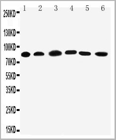
 WB (Western Blot)
(Figure 1. Western blot analysis of CISD2 using anti-CISD2 antibody (AAA19311).Electrophoresis was performed on a 5-20% SDS-PAGE gel at 70V (Stacking gel) / 90V (Resolving gel) for 2-3 hours. The sample well of each lane was loaded with 50ug of sample under reducing conditions.Lane 1: human HEK293 whole cell lysatesLane 2: human HELA whole cell lysatesLane 3: human MCF-7 whole cell lysatesLane 4: monkey kidney tissue lysatesLane 5: human SW620 whole cell lysatesLane 6: human Raji whole cell lysatesLane 7: rat kidney tissue lysatesLane 8: mouse kidney tissue lysates.After Electrophoresis, proteins were transferred to a Nitrocellulose membrane at 150mA for 50-90 minutes. Blocked the membrane with 5% Non-fat Milk/ TBS for 1. 5 hour at RT. The membrane was incubated with rabbit anti-CISD2 antigen affinity purified polyclonal antibody (Catalog # AAA19311) at 0. 5 μg/mL overnight at 4 degree C, then washed with TBS-0. 1%Tween 3 times with 5 minutes each and probed with a goat anti-rabbit IgG-HRP secondary antibody at a dilution of 1:10000 for 1. 5 hour at RT. The signal is developed using an Enhanced Chemiluminescent detection (ECL) kit (Catalog # with Tanon 5200 system. A specific band was detected for CISD2 at approximately 15KD. The expected band size for CISD2 is at 15KD.)
WB (Western Blot)
(Figure 1. Western blot analysis of CISD2 using anti-CISD2 antibody (AAA19311).Electrophoresis was performed on a 5-20% SDS-PAGE gel at 70V (Stacking gel) / 90V (Resolving gel) for 2-3 hours. The sample well of each lane was loaded with 50ug of sample under reducing conditions.Lane 1: human HEK293 whole cell lysatesLane 2: human HELA whole cell lysatesLane 3: human MCF-7 whole cell lysatesLane 4: monkey kidney tissue lysatesLane 5: human SW620 whole cell lysatesLane 6: human Raji whole cell lysatesLane 7: rat kidney tissue lysatesLane 8: mouse kidney tissue lysates.After Electrophoresis, proteins were transferred to a Nitrocellulose membrane at 150mA for 50-90 minutes. Blocked the membrane with 5% Non-fat Milk/ TBS for 1. 5 hour at RT. The membrane was incubated with rabbit anti-CISD2 antigen affinity purified polyclonal antibody (Catalog # AAA19311) at 0. 5 μg/mL overnight at 4 degree C, then washed with TBS-0. 1%Tween 3 times with 5 minutes each and probed with a goat anti-rabbit IgG-HRP secondary antibody at a dilution of 1:10000 for 1. 5 hour at RT. The signal is developed using an Enhanced Chemiluminescent detection (ECL) kit (Catalog # with Tanon 5200 system. A specific band was detected for CISD2 at approximately 15KD. The expected band size for CISD2 is at 15KD.)
CISD2, Polyclonal Antibody (Cat# AAA19311)
Full Name
Anti-CISD2 Antibody
Gene Names
CISD2; ERIS; WFS2; ZCD2; NAF-1; Miner1
Reactivity
Human, Mouse, Rat, Monkey
Applications
WB, IHC-P, ICC, IF, FC/FACS/FCM, EIA
Purity
Immunogen affinity purified.
Pricing


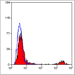
 FCM (Flow Cytometry)
(Figure 7. Flow Cytometry analysis of RH35 cells using anti- EIF4A1 antibody (AAA19380).Overlay histogram showing RH35 cells stained with AAA19380 (Blue line). The cells were blocked with 10% normal goat serum. And then incubated with mouse anti-EIF4A1 Antibody (AAA19380, 1μg/1x106 cells) for 30 min at 20 degree C. DyLight®488 conjugated goat anti-mouse IgG (BA1126, 5-10μg/1x106 cells) was used as secondary antibody for 30 minutes at 20 degree C. Isotype control antibody (Green line) was mouse IgG (1μg/1x106) used under the same conditions. Unlabelled sample (Red line) was also used as a control.)
FCM (Flow Cytometry)
(Figure 7. Flow Cytometry analysis of RH35 cells using anti- EIF4A1 antibody (AAA19380).Overlay histogram showing RH35 cells stained with AAA19380 (Blue line). The cells were blocked with 10% normal goat serum. And then incubated with mouse anti-EIF4A1 Antibody (AAA19380, 1μg/1x106 cells) for 30 min at 20 degree C. DyLight®488 conjugated goat anti-mouse IgG (BA1126, 5-10μg/1x106 cells) was used as secondary antibody for 30 minutes at 20 degree C. Isotype control antibody (Green line) was mouse IgG (1μg/1x106) used under the same conditions. Unlabelled sample (Red line) was also used as a control.)
EIF4A1, Monoclonal Antibody (Cat# AAA19380)
Full Name
Anti-EIF4A1 Antibody (monoclonal, 11B8)
Gene Names
EIF4A1; DDX2A; EIF4A; EIF-4A; eIF4A-I; eIF-4A-I
Reactivity
Human, Mouse, Rat
Applications
WB, IHC-P, ICC, IF, FC/FACS/FCM
Purity
Immunogen affinity purified.
Pricing


 IF (Immunofluorescence)
(Figure 7. IF analysis of METTL1 using anti- METTL1 antibody (AAA19336).METTL1 was detected in immunocytochemical section of A431 cells. Enzyme antigen retrieval was performed using IHC enzyme antigen retrieval reagent for 15 mins. The cells were blocked with 10% goat serum. And then incubated with 5μg/mL rabbit anti-METTL1 Antibody (AAA19336) overnight at 4 degree C. DyLight®488 Conjugated Goat Anti-Rabbit IgG was used as secondary antibody at 1:100 dilution and incubated for 30 minutes at 37 degree C. The section was counterstained with DAPI. Visualize using a fluorescence microscope and filter sets appropriate for the label used.)
IF (Immunofluorescence)
(Figure 7. IF analysis of METTL1 using anti- METTL1 antibody (AAA19336).METTL1 was detected in immunocytochemical section of A431 cells. Enzyme antigen retrieval was performed using IHC enzyme antigen retrieval reagent for 15 mins. The cells were blocked with 10% goat serum. And then incubated with 5μg/mL rabbit anti-METTL1 Antibody (AAA19336) overnight at 4 degree C. DyLight®488 Conjugated Goat Anti-Rabbit IgG was used as secondary antibody at 1:100 dilution and incubated for 30 minutes at 37 degree C. The section was counterstained with DAPI. Visualize using a fluorescence microscope and filter sets appropriate for the label used.)
METTL1, Polyclonal Antibody (Cat# AAA19336)
Full Name
Anti-METTL1 Antibody
Gene Names
METTL1; TRM8; TRMT8; C12orf1; YDL201w
Reactivity
Human, Rat
Applications
WB, IHC-P, ICC, IF, EIA
Purity
Immunogen affinity purified.
Pricing


 FCM (Flow Cytometry)
(Figure 11. Flow Cytometry analysis of U937 cells using anti-Coronin 1a/TACO/CORO1A antibody (AAA19291).Overlay histogram showing U937 cells stained with AAA19291 (Blue line). The cells were blocked with 10% normal goat serum. And then incubated with rabbit anti-Coronin 1a/TACO/CORO1A Antibody (AAA19291, 1μg/1x106 cells) for 30 min at 20 degree C. DyLight®488 conjugated goat anti-rabbit IgG (5-10μg/1x106 cells) was used as secondary antibody for 30 minutes at 20 degree C. Isotype control antibody (Green line) was rabbit IgG (1μg/1x106) used under the same conditions. Unlabelled sample (Red line) was also used as a control.)
FCM (Flow Cytometry)
(Figure 11. Flow Cytometry analysis of U937 cells using anti-Coronin 1a/TACO/CORO1A antibody (AAA19291).Overlay histogram showing U937 cells stained with AAA19291 (Blue line). The cells were blocked with 10% normal goat serum. And then incubated with rabbit anti-Coronin 1a/TACO/CORO1A Antibody (AAA19291, 1μg/1x106 cells) for 30 min at 20 degree C. DyLight®488 conjugated goat anti-rabbit IgG (5-10μg/1x106 cells) was used as secondary antibody for 30 minutes at 20 degree C. Isotype control antibody (Green line) was rabbit IgG (1μg/1x106) used under the same conditions. Unlabelled sample (Red line) was also used as a control.)
Coronin 1a/TACO/CORO1A, Polyclonal Antibody (Cat# AAA19291)
Full Name
Anti-Coronin 1a/TACO/CORO1A Antibody
Gene Names
CORO1A; p57; TACO; CLABP; HCORO1; CLIPINA
Reactivity
Human, Mouse, Rat
Applications
WB, IHC-P, FC/FACS/FCM, EIA
Purity
Immunogen affinity purified.
Pricing


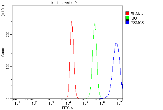 FCM (Flow Cytometry)
(Figure 7. Flow Cytometry analysis of 293T cells using anti-TBP-1/PSMC3 antibody (AAA19314).Overlay histogram showing 293T cells stained with AAA19314 (Blue line). The cells were blocked with 10% normal goat serum. And then incubated with rabbit anti-TBP-1/PSMC3 Antibody (AAA19314, 1μg/1x106 cells) for 30 min at 20 degree C. DyLight®488 conjugated goat anti-rabbit IgG (5-10μg/1x106 cells) was used as secondary antibody for 30 minutes at 20 degree C. Isotype control antibody (Green line) was rabbit IgG (1μg/1x106) used under the same conditions. Unlabelled sample (Red line) was also used as a control.)
FCM (Flow Cytometry)
(Figure 7. Flow Cytometry analysis of 293T cells using anti-TBP-1/PSMC3 antibody (AAA19314).Overlay histogram showing 293T cells stained with AAA19314 (Blue line). The cells were blocked with 10% normal goat serum. And then incubated with rabbit anti-TBP-1/PSMC3 Antibody (AAA19314, 1μg/1x106 cells) for 30 min at 20 degree C. DyLight®488 conjugated goat anti-rabbit IgG (5-10μg/1x106 cells) was used as secondary antibody for 30 minutes at 20 degree C. Isotype control antibody (Green line) was rabbit IgG (1μg/1x106) used under the same conditions. Unlabelled sample (Red line) was also used as a control.)
TBP-1/PSMC3, Polyclonal Antibody (Cat# AAA19314)
Full Name
Anti-TBP-1/PSMC3 Antibody
Gene Names
PSMC3; TBP1
Reactivity
Human, Mouse, Rat
Applications
WB, IHC-P, FC/FACS/FCM, EIA
Purity
Immunogen affinity purified.
Pricing


 FCM (Flow Cytometry)
(Figure 6. Flow Cytometry analysis of A431 cells using anti-TMPRSS3 antibody (AAA19289).Overlay histogram showing A431 cells stained with AAA19289 (Blue line). The cells were blocked with 10% normal goat serum. And then incubated with rabbit anti-TMPRSS3 Antibody (AAA19289, 1μg/1x106 cells) for 30 min at 20 degree C. DyLight®488 conjugated goat anti-rabbit IgG (5-10μg/1x106 cells) was used as secondary antibody for 30 minutes at 20 degree C. Isotype control antibody (Green line) was rabbit IgG (1μg/1x106) used under the same conditions. Unlabelled sample (Red line) was also used as a control.)
FCM (Flow Cytometry)
(Figure 6. Flow Cytometry analysis of A431 cells using anti-TMPRSS3 antibody (AAA19289).Overlay histogram showing A431 cells stained with AAA19289 (Blue line). The cells were blocked with 10% normal goat serum. And then incubated with rabbit anti-TMPRSS3 Antibody (AAA19289, 1μg/1x106 cells) for 30 min at 20 degree C. DyLight®488 conjugated goat anti-rabbit IgG (5-10μg/1x106 cells) was used as secondary antibody for 30 minutes at 20 degree C. Isotype control antibody (Green line) was rabbit IgG (1μg/1x106) used under the same conditions. Unlabelled sample (Red line) was also used as a control.)
TMPRSS3, Polyclonal Antibody (Cat# AAA19289)
Full Name
Anti-TMPRSS3 Antibody
Gene Names
TMPRSS3; DFNB8; DFNB10; ECHOS1; TADG12
Reactivity
Human, Mouse, Rat
Applications
WB, IHC-P, FC/FACS/FCM, EIA
Purity
Immunogen affinity purified.
Pricing


 FCM (Flow Cytometry)
(Figure 6. Flow Cytometry analysis of U937 cells using anti-BDH1 antibody (AAA19328).Overlay histogram showing U937 cells stained with AAA19328 (Blue line). The cells were blocked with 10% normal goat serum. And then incubated with rabbit anti-BDH1 Antibody (AAA19328,1μg/1x106 cells) for 30 min at 20 degree C. DyLight®488 conjugated goat anti-rabbit IgG (5-10μg/1x106 cells) was used as secondary antibody for 30 minutes at 20 degree C. Isotype control antibody (Green line) was rabbit IgG (1μg/1x106) used under the same conditions. Unlabelled sample (Red line) was also used as a control.)
FCM (Flow Cytometry)
(Figure 6. Flow Cytometry analysis of U937 cells using anti-BDH1 antibody (AAA19328).Overlay histogram showing U937 cells stained with AAA19328 (Blue line). The cells were blocked with 10% normal goat serum. And then incubated with rabbit anti-BDH1 Antibody (AAA19328,1μg/1x106 cells) for 30 min at 20 degree C. DyLight®488 conjugated goat anti-rabbit IgG (5-10μg/1x106 cells) was used as secondary antibody for 30 minutes at 20 degree C. Isotype control antibody (Green line) was rabbit IgG (1μg/1x106) used under the same conditions. Unlabelled sample (Red line) was also used as a control.)
BDH1, Polyclonal Antibody (Cat# AAA19328)
Full Name
Anti-BDH1 Antibody
Gene Names
BDH1; BDH; SDR9C1
Reactivity
Human, Rat
Applications
WB, IHC-P, ICC, IF, FC/FACS/FCM, EIA
Purity
Immunogen affinity purified.
Pricing


 FCM (Flow Cytometry)
(Figure 10. Flow Cytometry analysis of A549 cells using anti- U2AF65/U2AF2 antibody (AAA19375).Overlay histogram showing A549 cells stained with AAA19375 (Blue line). The cells were blocked with 10% normal goat serum. And then incubated with mouse anti-U2AF65/U2AF2 Antibody (AAA19375, 1μg/1x106 cells) for 30 min at 20 degree C. DyLight®488 conjugated goat anti-mouse IgG (BA1126, 5-10μg/1x106 cells) was used as secondary antibody for 30 minutes at 20 degree C. Isotype control antibody (Green line) was mouse IgG (1μg/1x106) used under the same conditions. Unlabelled sample (Red line) was also used as a control.)
FCM (Flow Cytometry)
(Figure 10. Flow Cytometry analysis of A549 cells using anti- U2AF65/U2AF2 antibody (AAA19375).Overlay histogram showing A549 cells stained with AAA19375 (Blue line). The cells were blocked with 10% normal goat serum. And then incubated with mouse anti-U2AF65/U2AF2 Antibody (AAA19375, 1μg/1x106 cells) for 30 min at 20 degree C. DyLight®488 conjugated goat anti-mouse IgG (BA1126, 5-10μg/1x106 cells) was used as secondary antibody for 30 minutes at 20 degree C. Isotype control antibody (Green line) was mouse IgG (1μg/1x106) used under the same conditions. Unlabelled sample (Red line) was also used as a control.)
U2AF65/U2AF2, Monoclonal Antibody (Cat# AAA19375)
Full Name
Anti-U2AF65/U2AF2 Antibody (monoclonal, 10F4)
Gene Names
U2AF2; U2AF65
Reactivity
Human, Mouse, Rat
Applications
WB, IHC-P, ICC, IF, FC/FACS/FCM
Purity
Immunogen affinity purified.
Pricing


 FCM (Flow Cytometry)
(Figure 7. Flow Cytometry analysis of HELA cells using anti-DNAJC10 antibody (AAA19320).Overlay histogram showing HELA cells stained with AAA19320 (Blue line). The cells were blocked with 10% normal goat serum. And then incubated with rabbit anti-DNAJC10 Antibody (AAA19320, 1μg/1x106 cells) for 30 min at 20 degree C. DyLight®488 conjugated goat anti-rabbit IgG (5-10μg/1x106 cells) was used as secondary antibody for 30 minutes at 20 degree C. Isotype control antibody (Green line) was rabbit IgG (1μg/1x106) used under the same conditions. Unlabelled sample (Red line) was also used as a control.)
FCM (Flow Cytometry)
(Figure 7. Flow Cytometry analysis of HELA cells using anti-DNAJC10 antibody (AAA19320).Overlay histogram showing HELA cells stained with AAA19320 (Blue line). The cells were blocked with 10% normal goat serum. And then incubated with rabbit anti-DNAJC10 Antibody (AAA19320, 1μg/1x106 cells) for 30 min at 20 degree C. DyLight®488 conjugated goat anti-rabbit IgG (5-10μg/1x106 cells) was used as secondary antibody for 30 minutes at 20 degree C. Isotype control antibody (Green line) was rabbit IgG (1μg/1x106) used under the same conditions. Unlabelled sample (Red line) was also used as a control.)
DNAJC10, Polyclonal Antibody (Cat# AAA19320)
Full Name
Anti-DNAJC10 Antibody
Gene Names
DNAJC10; JPDI; MTHr; ERdj5; PDIA19
Reactivity
Human, Mouse, Rat
Applications
WB, IHC-P, ICC, IF, FC/FACS/FCM, EIA
Purity
Immunogen affinity purified.
Pricing


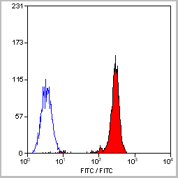
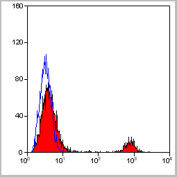
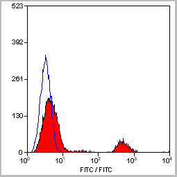
 FCM (Flow Cytometry)
(Figure 6. Flow Cytometry analysis of K562 cells using anti-D2AP antibody (M01756). Overlay histogram showing K562 cells stained with M01756 (Blue line).The cells were blocked with 10% normal goat serum. And then incubated with mouse anti-D2AP Antibody (M01756, 1ug/1x106 cells) for 30 min at 20 degree C. DyLight488 conjugated goat anti-mouse IgG (BA1126, 5-10ug/1x106 cells) was used as secondary antibody for 30 minutes at 20 degree C. Isotype control antibody (Green line) was mouse IgG (1ug/1x106) used under the same conditions. Unlabelled sample (Red line) was also used as a control.)
FCM (Flow Cytometry)
(Figure 6. Flow Cytometry analysis of K562 cells using anti-D2AP antibody (M01756). Overlay histogram showing K562 cells stained with M01756 (Blue line).The cells were blocked with 10% normal goat serum. And then incubated with mouse anti-D2AP Antibody (M01756, 1ug/1x106 cells) for 30 min at 20 degree C. DyLight488 conjugated goat anti-mouse IgG (BA1126, 5-10ug/1x106 cells) was used as secondary antibody for 30 minutes at 20 degree C. Isotype control antibody (Green line) was mouse IgG (1ug/1x106) used under the same conditions. Unlabelled sample (Red line) was also used as a control.)
CD2AP, Monoclonal Antibody (Cat# AAA19185)
Full Name
Anti-CD2AP Antibody (monoclonal, 5F8)
Gene Names
CD2AP; CMS
Reactivity
Human, Mouse, Rat
Applications
WB, IHC, FC/FACS
Purity
Immunogen Affinity Purified
Pricing


 FCM (Flow Cytometry)
(Figure 8. Flow Cytometry analysis of A549 cells using anti-Transketolase/TKT antibody (AAA19368).Overlay histogram showing A549 cells stained with AAA19368 (Blue line). The cells were blocked with 10% normal goat serum. And then incubated with mouse anti- Transketolase/TKT Antibody (AAA19368, 1μg/1x106 cells) for 30 min at 20 degree C. DyLight®488 conjugated goat anti-mouse IgG (BA1126, 5-10μg/1x106 cells) was used as secondary antibody for 30 minutes at 20 degree C. Isotype control antibody (Green line) was mouse IgG (1μg/1x106) used under the same conditions. Unlabelled sample (Red line) was also used as a control.)
FCM (Flow Cytometry)
(Figure 8. Flow Cytometry analysis of A549 cells using anti-Transketolase/TKT antibody (AAA19368).Overlay histogram showing A549 cells stained with AAA19368 (Blue line). The cells were blocked with 10% normal goat serum. And then incubated with mouse anti- Transketolase/TKT Antibody (AAA19368, 1μg/1x106 cells) for 30 min at 20 degree C. DyLight®488 conjugated goat anti-mouse IgG (BA1126, 5-10μg/1x106 cells) was used as secondary antibody for 30 minutes at 20 degree C. Isotype control antibody (Green line) was mouse IgG (1μg/1x106) used under the same conditions. Unlabelled sample (Red line) was also used as a control.)
Transketolase/TKT, Monoclonal Antibody (Cat# AAA19368)
Full Name
Anti-Transketolase/TKT Antibody (monoclonal, 2I3)
Gene Names
TKT; TK; TKT1; HEL107
Reactivity
Human, Mouse, Rat
Applications
WB, IHC-P, ICC, IF, FC/FACS/FCM
Purity
Immunogen affinity purified.
Pricing


 FCM (Flow Cytometry)
(Figure 8. Flow Cytometry analysis of THP-1 cells using anti-INPPL1 antibody (AAA19364).Overlay histogram showing THP-1 cells stained with AAA19364 (Blue line). The cells were blocked with 10% normal goat serum. And then incubated with mouse anti- INPPL1 Antibody (AAA19364, 1μg/1x106 cells) for 30 min at 20 degree C. DyLight®488 conjugated goat anti-mouse IgG (BA1126, 5-10μg/1x106 cells) was used as secondary antibody for 30 minutes at 20 degree C. Isotype control antibody (Green line) was mouse IgG (1μg/1x106) used under the same conditions. Unlabelled sample (Red line) was also used as a control.)
FCM (Flow Cytometry)
(Figure 8. Flow Cytometry analysis of THP-1 cells using anti-INPPL1 antibody (AAA19364).Overlay histogram showing THP-1 cells stained with AAA19364 (Blue line). The cells were blocked with 10% normal goat serum. And then incubated with mouse anti- INPPL1 Antibody (AAA19364, 1μg/1x106 cells) for 30 min at 20 degree C. DyLight®488 conjugated goat anti-mouse IgG (BA1126, 5-10μg/1x106 cells) was used as secondary antibody for 30 minutes at 20 degree C. Isotype control antibody (Green line) was mouse IgG (1μg/1x106) used under the same conditions. Unlabelled sample (Red line) was also used as a control.)
INPPL1, Monoclonal Antibody (Cat# AAA19364)
Full Name
Anti-INPPL1 Antibody (monoclonal, 8C13)
Gene Names
INPPL1; OPSMD; SHIP2
Reactivity
Human, Mouse, Rat
Applications
WB, IHC-P, ICC, IF, FC/FACS/FCM
Purity
Immunogen affinity purified.
Pricing
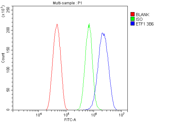


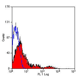
 FCM (Flow Cytometry)
(Figure 11. Flow Cytometry analysis of RH35 cells using anti- eRF1/ETF1 antibody (AAA19382).Overlay histogram showing RH35 cells stained with AAA19382 (Blue line). The cells were blocked with 10% normal goat serum. And then incubated with mouse anti-eRF1/ETF1 Antibody (AAA19382, 1μg/1x106 cells) for 30 min at 20 degree C. DyLight®488 conjugated goat anti-mouse IgG (BA1126, 5-10μg/1x106 cells) was used as secondary antibody for 30 minutes at 20 degree C. Isotype control antibody (Green line) was mouse IgG (1μg/1x106) used under the same conditions. Unlabelled sample (Red line) was also used as a control.)
FCM (Flow Cytometry)
(Figure 11. Flow Cytometry analysis of RH35 cells using anti- eRF1/ETF1 antibody (AAA19382).Overlay histogram showing RH35 cells stained with AAA19382 (Blue line). The cells were blocked with 10% normal goat serum. And then incubated with mouse anti-eRF1/ETF1 Antibody (AAA19382, 1μg/1x106 cells) for 30 min at 20 degree C. DyLight®488 conjugated goat anti-mouse IgG (BA1126, 5-10μg/1x106 cells) was used as secondary antibody for 30 minutes at 20 degree C. Isotype control antibody (Green line) was mouse IgG (1μg/1x106) used under the same conditions. Unlabelled sample (Red line) was also used as a control.)
eRF1/ETF1, Monoclonal Antibody (Cat# AAA19382)
Full Name
Anti-eRF1/ETF1 Antibody (monoclonal, 3B6)
Gene Names
ETF1; ERF; RF1; ERF1; TB3-1; D5S1995; SUP45L1
Reactivity
Human, Mouse, Rat
Applications
WB, IHC-P, ICC, IF, FC/FACS/FCM
Purity
Immunogen affinity purified.
Pricing


 FCM (Flow Cytometry)
(Figure 9. Flow Cytometry analysis of U251 cells using anti-RAB1B antibody (AAA19296).Overlay histogram showing U251 cells stained with AAA19296 (Blue line). The cells were blocked with 10% normal goat serum. And then incubated with rabbit anti-RAB1B Antibody (AAA19296, 1μg/1x106 cells) for 30 min at 20 degree C. DyLight®488 conjugated goat anti-rabbit IgG (5-10μg/1x106 cells) was used as secondary antibody for 30 minutes at 20 degree C. Isotype control antibody (Green line) was rabbit IgG (1μg/1x106) used under the same conditions. Unlabelled sample (Red line) was also used as a control.)
FCM (Flow Cytometry)
(Figure 9. Flow Cytometry analysis of U251 cells using anti-RAB1B antibody (AAA19296).Overlay histogram showing U251 cells stained with AAA19296 (Blue line). The cells were blocked with 10% normal goat serum. And then incubated with rabbit anti-RAB1B Antibody (AAA19296, 1μg/1x106 cells) for 30 min at 20 degree C. DyLight®488 conjugated goat anti-rabbit IgG (5-10μg/1x106 cells) was used as secondary antibody for 30 minutes at 20 degree C. Isotype control antibody (Green line) was rabbit IgG (1μg/1x106) used under the same conditions. Unlabelled sample (Red line) was also used as a control.)
RAB1B, Polyclonal Antibody (Cat# AAA19296)
Full Name
Anti-RAB1B Antibody
Reactivity
Human, Mouse, Rat
Applications
WB, IHC-P, ICC, IF, FC/FACS/FCM
Purity
Immunogen affinity purified.
Pricing


 FCM (Flow Cytometry)
(Figure 7. Flow Cytometry analysis of MCF-7 cells using anti-RGS6 antibody (AAA19308).Overlay histogram showing MCF-7 cells stained with AAA19308 (Blue line). The cells were blocked with 10% normal goat serum. And then incubated with rabbit anti-RGS6 Antibody (AAA19308, 1μg/1x106 cells) for 30 min at 20 degree C. DyLight®488 conjugated goat anti-rabbit IgG (5-10μg/1x106 cells) was used as secondary antibody for 30 minutes at 20 degree C. Isotype control antibody (Green line) was rabbit IgG (1μg/1x106) used under the same conditions. Unlabelled sample (Red line) was also used as a control.)
FCM (Flow Cytometry)
(Figure 7. Flow Cytometry analysis of MCF-7 cells using anti-RGS6 antibody (AAA19308).Overlay histogram showing MCF-7 cells stained with AAA19308 (Blue line). The cells were blocked with 10% normal goat serum. And then incubated with rabbit anti-RGS6 Antibody (AAA19308, 1μg/1x106 cells) for 30 min at 20 degree C. DyLight®488 conjugated goat anti-rabbit IgG (5-10μg/1x106 cells) was used as secondary antibody for 30 minutes at 20 degree C. Isotype control antibody (Green line) was rabbit IgG (1μg/1x106) used under the same conditions. Unlabelled sample (Red line) was also used as a control.)
RGS6, Polyclonal Antibody (Cat# AAA19308)
Full Name
Anti-RGS6 Antibody
Gene Names
RGS6; GAP
Reactivity
Human, Mouse, Rat
Applications
WB, IHC-P, FC/FACS/FCM, EIA
Purity
Immunogen affinity purified.
Pricing


 FCM (Flow Cytometry)
(Figure 6. Flow Cytometry analysis of THP-1 cells using anti-PRMT8 antibody (AAA19321).Overlay histogram showing THP-1 cells stained with AAA19321 (Blue line). The cells were blocked with 10% normal goat serum. And then incubated with rabbit anti-PRMT8 Antibody (AAA19321,1μg/1x106 cells) for 30 min at 20 degree C. DyLight®488 conjugated goat anti-rabbit IgG (5-10μg/1x106 cells) was used as secondary antibody for 30 minutes at 20 degree C. Isotype control antibody (Green line) was rabbit IgG (1μg/1x106) used under the same conditions. Unlabelled sample (Red line) was also used as a control.)
FCM (Flow Cytometry)
(Figure 6. Flow Cytometry analysis of THP-1 cells using anti-PRMT8 antibody (AAA19321).Overlay histogram showing THP-1 cells stained with AAA19321 (Blue line). The cells were blocked with 10% normal goat serum. And then incubated with rabbit anti-PRMT8 Antibody (AAA19321,1μg/1x106 cells) for 30 min at 20 degree C. DyLight®488 conjugated goat anti-rabbit IgG (5-10μg/1x106 cells) was used as secondary antibody for 30 minutes at 20 degree C. Isotype control antibody (Green line) was rabbit IgG (1μg/1x106) used under the same conditions. Unlabelled sample (Red line) was also used as a control.)
PRMT8, Polyclonal Antibody (Cat# AAA19321)
Full Name
Anti-PRMT8 Antibody
Gene Names
PRMT8; HRMT1L3; HRMT1L4
Reactivity
Human, Rat
Applications
WB, IHC-P, FC/FACS/FCM, EIA
Purity
Immunogen affinity purified.
Pricing


 FCM (Flow Cytometry)
(Figure 12. Flow Cytometry analysis of U937 cells using anti-DYNLL1/PIN antibody (AAA19276).Overlay histogram showing U937 cells stained with AAA19276 (Blue line). The cells were blocked with 10% normal goat serum. And then incubated with rabbit anti-DYNLL1/PIN Antibody (AAA19276, 1μg/1x106 cells) for 30 min at 20 degree C. DyLight®488 conjugated goat anti-rabbit IgG (5-10μg/1x106 cells) was used as secondary antibody for 30 minutes at 20 degree C. Isotype control antibody (Green line) was rabbit IgG (1μg/1x106) used under the same conditions. Unlabelled sample (Red line) was also used as a control.)
FCM (Flow Cytometry)
(Figure 12. Flow Cytometry analysis of U937 cells using anti-DYNLL1/PIN antibody (AAA19276).Overlay histogram showing U937 cells stained with AAA19276 (Blue line). The cells were blocked with 10% normal goat serum. And then incubated with rabbit anti-DYNLL1/PIN Antibody (AAA19276, 1μg/1x106 cells) for 30 min at 20 degree C. DyLight®488 conjugated goat anti-rabbit IgG (5-10μg/1x106 cells) was used as secondary antibody for 30 minutes at 20 degree C. Isotype control antibody (Green line) was rabbit IgG (1μg/1x106) used under the same conditions. Unlabelled sample (Red line) was also used as a control.)
DYNLL1/PIN, Polyclonal Antibody (Cat# AAA19276)
Full Name
Anti-DYNLL1/PIN Antibody
Gene Names
DYNLL1; LC8; PIN; DLC1; DLC8; LC8a; DNCL1; hdlc1; DNCLC1
Reactivity
Human, Mouse, Rat
Applications
WB, IHC-P, ICC, IF, FC/FACS/FCM, EIA
Purity
Immunogen affinity purified.
Pricing


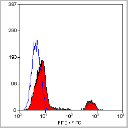
 FCM (Flow Cytometry)
(Figure 7. Flow Cytometry analysis of A549 cells using anti-Hsp90 alpha antibody (AAA19359).Overlay histogram showing A549 cells stained with AAA19359 (Blue line). The cells were blocked with 10% normal goat serum. And then incubated with mouse anti-Hsp90 alpha Antibody (AAA19359, 1μg/1x106 cells) for 30 min at 20 degree C. DyLight®488 conjugated goat anti-mouse IgG (BA1126, 5-10μg/1x106 cells) was used as secondary antibody for 30 minutes at 20 degree C. Isotype control antibody (Green line) was mouse IgG (1μg/1x106) used under the same conditions. Unlabelled sample (Red line) was also used as a control.)
FCM (Flow Cytometry)
(Figure 7. Flow Cytometry analysis of A549 cells using anti-Hsp90 alpha antibody (AAA19359).Overlay histogram showing A549 cells stained with AAA19359 (Blue line). The cells were blocked with 10% normal goat serum. And then incubated with mouse anti-Hsp90 alpha Antibody (AAA19359, 1μg/1x106 cells) for 30 min at 20 degree C. DyLight®488 conjugated goat anti-mouse IgG (BA1126, 5-10μg/1x106 cells) was used as secondary antibody for 30 minutes at 20 degree C. Isotype control antibody (Green line) was mouse IgG (1μg/1x106) used under the same conditions. Unlabelled sample (Red line) was also used as a control.)
Hsp90 alpha, Monoclonal Antibody (Cat# AAA19359)
Full Name
Anti-Hsp90 alpha Antibody (monoclonal, 6B5)
Gene Names
HSP90AA1; EL52; HSPN; LAP2; HSP86; HSPC1; HSPCA; Hsp89; Hsp90; HSP89A; HSP90A; HSP90N; HSPCAL1; HSPCAL4
Reactivity
Human, Mouse, Rat, Monkey
Applications
WB, IHC-P, ICC, IF, FC/FACS/FCM
Purity
Immunogen affinity purified.
Pricing


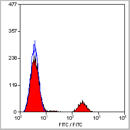
 FCM (Flow Cytometry)
(Figure 7. Flow Cytometry analysis of U20S cells using anti- ADA antibody (AAA19184). Overlay histogram showing U20S cells stained with AAA19184 (Blue line).The cells were blocked with 10% normal goat serum. And then incubated with mouse anti- ADA Antibody (AAA19184, 1ug/1x106 cells) for 30 min at 20 degree C. DyLight488 conjugated goat anti-mouse IgG (BA1126, 5-10ug/1x106 cells) was used as secondary antibody for 30 minutes at 20 degree C. Isotype control antibody (Green line) was mouse IgG (1ug/1x106) used under the same conditions. Unlabelled sample (Red line) was also used as a control.)
FCM (Flow Cytometry)
(Figure 7. Flow Cytometry analysis of U20S cells using anti- ADA antibody (AAA19184). Overlay histogram showing U20S cells stained with AAA19184 (Blue line).The cells were blocked with 10% normal goat serum. And then incubated with mouse anti- ADA Antibody (AAA19184, 1ug/1x106 cells) for 30 min at 20 degree C. DyLight488 conjugated goat anti-mouse IgG (BA1126, 5-10ug/1x106 cells) was used as secondary antibody for 30 minutes at 20 degree C. Isotype control antibody (Green line) was mouse IgG (1ug/1x106) used under the same conditions. Unlabelled sample (Red line) was also used as a control.)
ADA, Monoclonal Antibody (Cat# AAA19184)
Full Name
Anti-ADA Antibody (monoclonal, 6D4)
Reactivity
Human
Applications
WB, IHC, FC/FACS
Purity
Immunogen Affinity Purified
Pricing


 IF (Immunofluorescence)
(Figure 11. IF analysis of Tubulin alpha using anti- Tubulin alpha antibody (AAA19381).Tubulin alpha was detected in immunocytochemical section of CACO-2 cells. Enzyme antigen retrieval was performed using IHC enzyme antigen retrieval reagent for 15 mins. The cells were blocked with 10% goat serum. And then incubated with 5μg/mL mouse anti- Tubulin alpha Antibody (AAA19381) overnight at 4 degree C. DyLight®594 Conjugated Goat Anti-Mouse IgG (BA1141) was used as secondary antibody at 1:100 dilution and incubated for 30 minutes at 37 degree C. The section was counterstained with DAPI. Visualize using a fluorescence microscope and filter sets appropriate for the label used.)
IF (Immunofluorescence)
(Figure 11. IF analysis of Tubulin alpha using anti- Tubulin alpha antibody (AAA19381).Tubulin alpha was detected in immunocytochemical section of CACO-2 cells. Enzyme antigen retrieval was performed using IHC enzyme antigen retrieval reagent for 15 mins. The cells were blocked with 10% goat serum. And then incubated with 5μg/mL mouse anti- Tubulin alpha Antibody (AAA19381) overnight at 4 degree C. DyLight®594 Conjugated Goat Anti-Mouse IgG (BA1141) was used as secondary antibody at 1:100 dilution and incubated for 30 minutes at 37 degree C. The section was counterstained with DAPI. Visualize using a fluorescence microscope and filter sets appropriate for the label used.)
Tubulin alpha, Monoclonal Antibody (Cat# AAA19381)
Full Name
Anti-Tubulin alpha Antibody (monoclonal, 7B12)
Gene Names
TUBA1A; LIS3; TUBA3; B-ALPHA-1
Reactivity
Human, Mouse, Rat
Applications
WB, IHC-P, ICC, IF, FC/FACS/FCM
Purity
Immunogen affinity purified.
Pricing


 FCM (Flow Cytometry)
(Figure 8. Flow Cytometry analysis of K562 cells using anti-PDIA6 antibody (AAA19282).Overlay histogram showing K562 cells stained with AAA19282 (Blue line). The cells were blocked with 10% normal goat serum. And then incubated with rabbit anti-PDIA6 Antibody (AAA19282, 1μg/1x106 cells) for 30 min at 20 degree C. DyLight®488 conjugated goat anti-rabbit IgG (5-10μg/1x106 cells) was used as secondary antibody for 30 minutes at 20 degree C. Isotype control antibody (Green line) was rabbit IgG (1μg/1x106) used under the same conditions. Unlabelled sample (Red line) was also used as a control.)
FCM (Flow Cytometry)
(Figure 8. Flow Cytometry analysis of K562 cells using anti-PDIA6 antibody (AAA19282).Overlay histogram showing K562 cells stained with AAA19282 (Blue line). The cells were blocked with 10% normal goat serum. And then incubated with rabbit anti-PDIA6 Antibody (AAA19282, 1μg/1x106 cells) for 30 min at 20 degree C. DyLight®488 conjugated goat anti-rabbit IgG (5-10μg/1x106 cells) was used as secondary antibody for 30 minutes at 20 degree C. Isotype control antibody (Green line) was rabbit IgG (1μg/1x106) used under the same conditions. Unlabelled sample (Red line) was also used as a control.)
PDIA6, Polyclonal Antibody (Cat# AAA19282)
Full Name
Anti-PDIA6 Antibody
Gene Names
PDIA6; P5; ERP5; TXNDC7
Reactivity
Human, Mouse, Rat
Applications
WB, IHC-P, ICC, IF, FC/FACS/FCM, EIA
Purity
Immunogen affinity purified.
Pricing


 FCM (Flow Cytometry)
(Figure 9. Flow Cytometry analysis of K562 cells using anti-MCM6 antibody (AAA19372).Overlay histogram showing K562 cells stained with AAA19372 (Blue line). The cells were blocked with 10% normal goat serum. And then incubated with mouse anti- MCM6 Antibody (AAA19372, 1μg/1x106 cells) for 30 min at 20 degree C. DyLight®488 conjugated goat anti-mouse IgG (BA1126, 5-10μg/1x106 cells) was used as secondary antibody for 30 minutes at 20 degree C. Isotype control antibody (Green line) was mouse IgG (1μg/1x106) used under the same conditions. Unlabelled sample (Red line) was also used as a control.)
FCM (Flow Cytometry)
(Figure 9. Flow Cytometry analysis of K562 cells using anti-MCM6 antibody (AAA19372).Overlay histogram showing K562 cells stained with AAA19372 (Blue line). The cells were blocked with 10% normal goat serum. And then incubated with mouse anti- MCM6 Antibody (AAA19372, 1μg/1x106 cells) for 30 min at 20 degree C. DyLight®488 conjugated goat anti-mouse IgG (BA1126, 5-10μg/1x106 cells) was used as secondary antibody for 30 minutes at 20 degree C. Isotype control antibody (Green line) was mouse IgG (1μg/1x106) used under the same conditions. Unlabelled sample (Red line) was also used as a control.)
MCM6, Monoclonal Antibody (Cat# AAA19372)
Full Name
Anti-MCM6 Antibody (monoclonal, 10I9)
Gene Names
MCM6; Mis5; P105MCM; MCG40308
Reactivity
Human
Applications
WB, IHC-P, ICC, IF, FC/FACS/FCM
Purity
Immunogen affinity purified.
Pricing
hGH, Monoclonal Antibody (Cat# AAA14628)
Full Name
Biotin conjugated mAb anti-hGH
Reactivity
Human. Others not tested
Applications
ELISA
Purity
Protein G affinity purified
Pricing
ATP1A3, Polyclonal Antibody (Cat# AAA26972)
Full Name
ATP1A3 Antibody, Biotin conjugated
Gene Names
ATP1A3; RDP; AHC2; CAPOS; DYT12; ATP1A1
Reactivity
Human
Applications
ELISA
Purity
>95%, Protein G purified
Pricing
dnaK, Polyclonal Antibody (Cat# AAA26989)
Full Name
dnaK Antibody, Biotin conjugated
Reactivity
Escherichia coli
Applications
ELISA
Purity
>95%, Protein G purified
Pricing
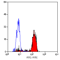

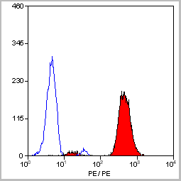
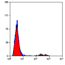
 Application Data
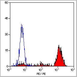
(Staining of human peripheral blood monocytes with Mouse anti Human CD14:RPE)
Application Data
(Staining of human peripheral blood monocytes with Mouse anti Human CD14:RPE)
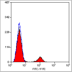
CD14, Monoclonal Antibody (Cat# AAA11884)
Full Name
MOUSE ANTI HUMAN CD14:FITC
Applications
Flow Cytometry
Pricing

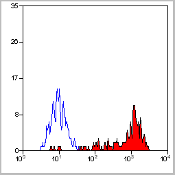
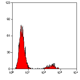
 Application Data
(Staining of human peripheral blood granulocytes with CD18: Alexa Fluor 647)
Application Data
(Staining of human peripheral blood granulocytes with CD18: Alexa Fluor 647)
CD18, Monoclonal Antibody (Cat# AAA12051)
Full Name
RAT ANTI HUMAN CD18:RPE
Gene Names
ITGB2; LAD; CD18; MF17; MFI7; LCAMB; LFA-1; MAC-1
Applications
Flow Cytometry
Pricing


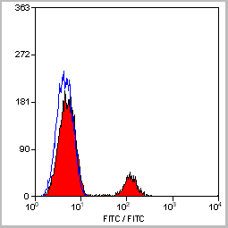
 Application Data
(Staining of human peripheral blood granulocytes with CD18: Alexa Fluor 647 (AAA11986A647))
Application Data
(Staining of human peripheral blood granulocytes with CD18: Alexa Fluor 647 (AAA11986A647))
CD18, Monoclonal Antibody (Cat# AAA11986)
Full Name
RAT ANTI HUMAN CD18
Gene Names
ITGB2; LAD; CD18; MF17; MFI7; LCAMB; LFA-1; MAC-1
Reactivity
Dog, Guinea Pig
Applications
Immunohistochemistry, Flow Cytometry, Immunoprecipitation
Pricing
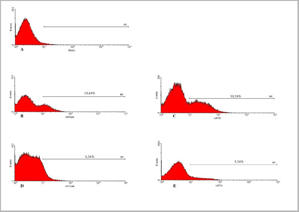
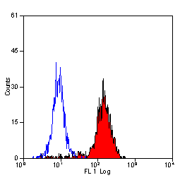
 Application Data
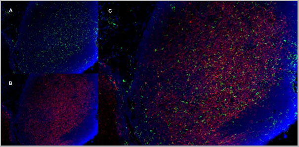
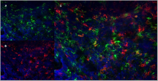
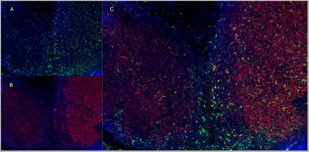
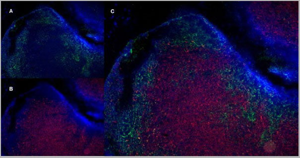
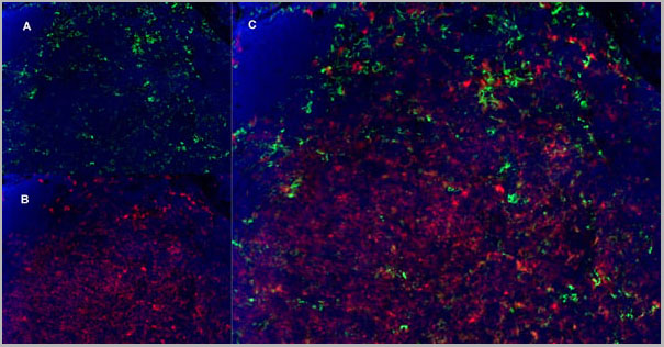
(Immunofluorescence staining of mouse lymph node cryosection with Rat anti Mouse CD11b, clone 5C6 , green in A and Rat anti Mouse CD8, clone YTS105.18 , red in B. C is the merged image with nuclei counterstained blue using DAPI. High power)
Application Data
(Immunofluorescence staining of mouse lymph node cryosection with Rat anti Mouse CD11b, clone 5C6 , green in A and Rat anti Mouse CD8, clone YTS105.18 , red in B. C is the merged image with nuclei counterstained blue using DAPI. High power)
CD8 ALPHA, Monoclonal Antibody (Cat# AAA11918)
Full Name
RAT ANTI MOUSE CD8
Gene Names
Cd8a; Ly-2; Ly-B; Ly-35; Lyt-2; BB154331
Applications
Immunohistochemistry, Flow Cytometry, Immunofluorescence
Pricing


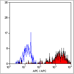
 Application Data
(Staining of human peripheral blood monocytes with Mouse anti Human CD14:RPE)
Application Data
(Staining of human peripheral blood monocytes with Mouse anti Human CD14:RPE)
CD14, Monoclonal Antibody (Cat# AAA12178)
Full Name
MOUSE ANTI HUMAN CD14:APC
Applications
Flow Cytometry
Pricing
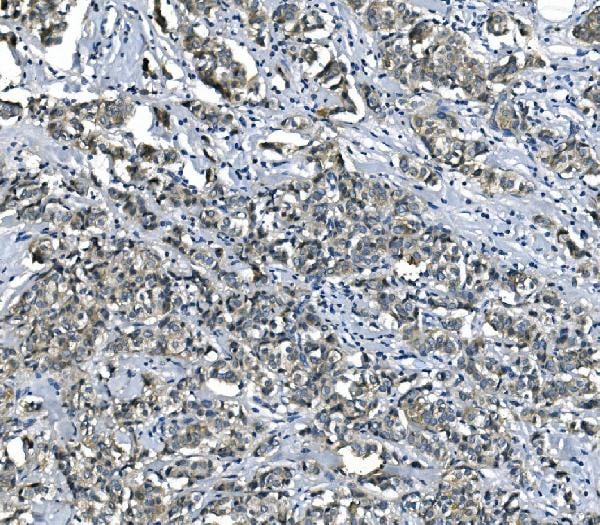
 IF (Immunofluorescence)
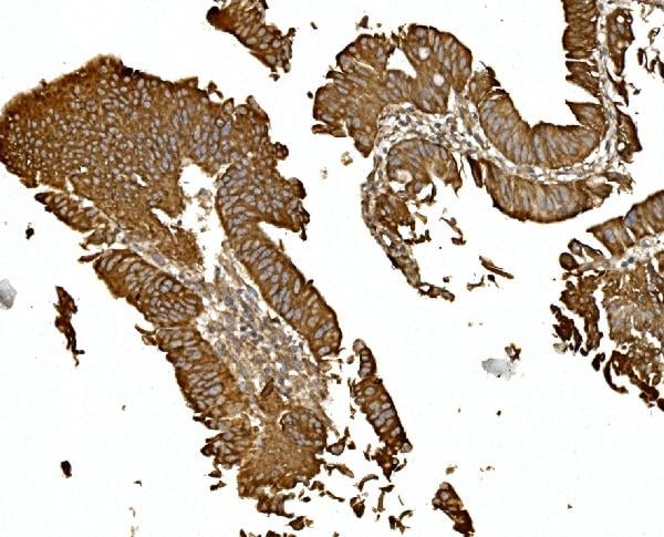
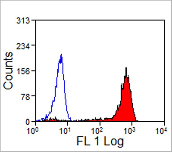
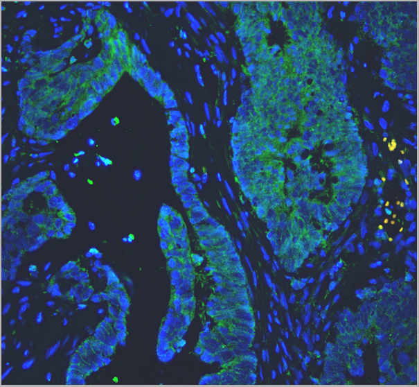
(Figure 3. IF analysis of PC1/3/PCSK1 using anti-PC1/3/PCSK1 antibody PC1/3/PCSK1 was detected in paraffin-embedded section of human colon cancer tissues. Heat mediated antigen retrieval was performed in citrate buffer (pH6, epitope retrieval solution ) for 20 mins. The tissue section was blocked with 10% goat serum. The tissue section was then incubated with 1ug/mL rabbit anti-PC1/3/PCSK1 Antibody overnight at 4 degree C. Biotin conjugated goat anti-rabbit IgG was used as secondary antibody and incubated for 30 minutes at 37 degree C. The tissue section was developed using DyLight 488 Conjugated Avidin. The section was counterstained with DAPI. Visualize using a fluorescence microscope and filter sets appropriate for the label used.)
IF (Immunofluorescence)
(Figure 3. IF analysis of PC1/3/PCSK1 using anti-PC1/3/PCSK1 antibody PC1/3/PCSK1 was detected in paraffin-embedded section of human colon cancer tissues. Heat mediated antigen retrieval was performed in citrate buffer (pH6, epitope retrieval solution ) for 20 mins. The tissue section was blocked with 10% goat serum. The tissue section was then incubated with 1ug/mL rabbit anti-PC1/3/PCSK1 Antibody overnight at 4 degree C. Biotin conjugated goat anti-rabbit IgG was used as secondary antibody and incubated for 30 minutes at 37 degree C. The tissue section was developed using DyLight 488 Conjugated Avidin. The section was counterstained with DAPI. Visualize using a fluorescence microscope and filter sets appropriate for the label used.)
PC1/3, Polyclonal Antibody (Cat# AAA11585)
Full Name
Anti-PC1/3 antibody
Gene Names
PCSK1; PC1; PC3; NEC1; SPC3; BMIQ12
Reactivity
Reacts with: Human, rat; Predicted to work with: Mouse
Applications
Western Blot, Immunohistochemistry, Immunofluorescence
Purity
Immunogen affinity purified.
Pricing
 Application Data
(Staining of human peripheral blood lymphocytes with Mouse anti Human CD19: RPE- Alexa Fluor 647)
Application Data
(Staining of human peripheral blood lymphocytes with Mouse anti Human CD19: RPE- Alexa Fluor 647)
CD19, Monoclonal Antibody (Cat# AAA12034)
Full Name
MOUSE ANTI HUMAN CD19:RPE
Gene Names
CD19; B4; CVID3
Applications
Flow Cytometry
Pricing
 Application Data
(Staining of human peripheral blood lymphocytes with Mouse anti Human CD20: Pacific Blue)
Application Data
(Staining of human peripheral blood lymphocytes with Mouse anti Human CD20: Pacific Blue)
CD20, Monoclonal Antibody (Cat# AAA12032)
Full Name
MOUSE ANTI HUMAN CD20:RPE
Gene Names
MS4A1; B1; S7; Bp35; CD20; CVID5; MS4A2; LEU-16
Applications
Flow Cytometry
Pricing