Filters
Clonality
Type
Reactivity
Gene Name
Isotype
Host
Application
Clone
527 results for " Membrane Proteins" - showing 200-250


 IHC (Immunohistochemistry)
(Figure 7. IHC analysis of L1CAM using anti-L1CAM antibody (AAA19138).L1CAM was detected in paraffin-embedded section of human mammary cancer tissue. Heat mediated antigen retrieval was performed in citrate buffer (pH6, epitope retrieval solution) for 20 mins. The tissue section was blocked with 10% goat serum. The tissue section was then incubated with 1ug/ml rabbit anti-L1CAM Antibody (AAA19138) overnight at 4 degree C. Biotinylated goat anti-rabbit IgG was used as secondary antibody and incubated for 30 minutes at 37 degree C. The tissue section was developed using Strepavidin-Biotin-Complex (SABC) with DAB as the chromogen.)
IHC (Immunohistochemistry)
(Figure 7. IHC analysis of L1CAM using anti-L1CAM antibody (AAA19138).L1CAM was detected in paraffin-embedded section of human mammary cancer tissue. Heat mediated antigen retrieval was performed in citrate buffer (pH6, epitope retrieval solution) for 20 mins. The tissue section was blocked with 10% goat serum. The tissue section was then incubated with 1ug/ml rabbit anti-L1CAM Antibody (AAA19138) overnight at 4 degree C. Biotinylated goat anti-rabbit IgG was used as secondary antibody and incubated for 30 minutes at 37 degree C. The tissue section was developed using Strepavidin-Biotin-Complex (SABC) with DAB as the chromogen.)
L1CAM, Polyclonal Antibody (Cat# AAA19138)
Full Name
Anti-L1CAM antibody
Gene Names
L1CAM; S10; HSAS; MASA; MIC5; SPG1; CAML1; CD171; HSAS1; N-CAML1; NCAM-L1; N-CAM-L1
Reactivity
Human, Mouse, Rat
No cross reactivity with other proteins.
No cross reactivity with other proteins.
Applications
IHC
Pricing


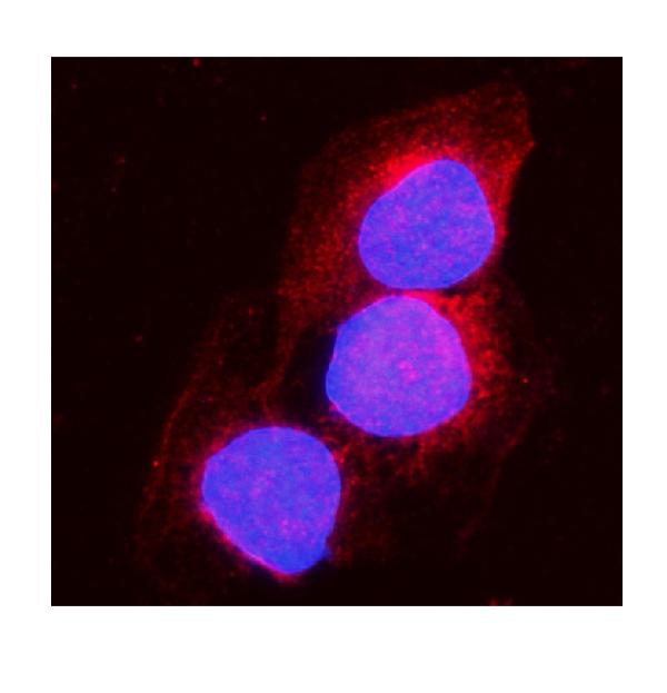
 IF (Immunofluorescence)
(Immunofluorescence analysis of U-2 OS cells using GAPDH Rabbit pAb at dilution of 1:100 (40x lens). Blue: DAPI for nuclear staining.)
IF (Immunofluorescence)
(Immunofluorescence analysis of U-2 OS cells using GAPDH Rabbit pAb at dilution of 1:100 (40x lens). Blue: DAPI for nuclear staining.)
GAPDH, Polyclonal Antibody (Cat# AAA28397)
Full Name
GAPDH Rabbit pAb
Gene Names
GAPDH; G3PD; GAPD
Reactivity
Human, Mouse, Rat
Applications
WB, IHC, IF, IP
Purity
Affinity purification
Pricing


 FCM (Flow Cytometry)
(Figure 6. Flow Cytometry analysis of Raji cells using anti-CD38 antibody (AAA19217).Overlay histogram showing Raji cells stained with AAA19217 (Blue line). The cells were blocked with 10% normal goat serum. And then incubated with rabbit anti-CD38 Antibody (AAA19217, 1μg/1x106 cells) for 30 min at 20 degree C. DyLight®488 conjugated goat anti-rabbit IgG (5-10μg/1x106 cells) was used as secondary antibody for 30 minutes at 20 degree C. Isotype control antibody (Green line) was rabbit IgG (1μg/1x106) used under the same conditions. Unlabelled sample (Red line) was also used as a control.)
FCM (Flow Cytometry)
(Figure 6. Flow Cytometry analysis of Raji cells using anti-CD38 antibody (AAA19217).Overlay histogram showing Raji cells stained with AAA19217 (Blue line). The cells were blocked with 10% normal goat serum. And then incubated with rabbit anti-CD38 Antibody (AAA19217, 1μg/1x106 cells) for 30 min at 20 degree C. DyLight®488 conjugated goat anti-rabbit IgG (5-10μg/1x106 cells) was used as secondary antibody for 30 minutes at 20 degree C. Isotype control antibody (Green line) was rabbit IgG (1μg/1x106) used under the same conditions. Unlabelled sample (Red line) was also used as a control.)
CD38, Polyclonal Antibody (Cat# AAA19217)
Full Name
Anti-CD38 Antibody
Gene Names
CD38; T10; ADPRC 1
Reactivity
Human
Applications
Western Blot, Immunohistochemistry, Immunocytochemistry, Immunofluorescence, Flow Cytometry
Purity
Immunogen affinity purified.
Pricing


 FCM (Flow Cytometry)
(Figure 6. Flow Cytometry analysis of RAW264. 7 cells using anti-TRIM25/EFP antibody (AAA19272).Overlay histogram showing RAW264. 7 cells stained with AAA19272 (Blue line). The cells were blocked with 10% normal goat serum. And then incubated with rabbit anti-TRIM25/EFP Antibody (AAA19272, 1μg/1x106 cells) for 30 min at 20 degree C. DyLight®488 conjugated goat anti-rabbit IgG (5-10μg/1x106 cells) was used as secondary antibody for 30 minutes at 20 degree C. Isotype control antibody (Green line) was rabbit IgG (1μg/1x106) used under the same conditions. Unlabelled sample (Red line) was also used as a control.)
FCM (Flow Cytometry)
(Figure 6. Flow Cytometry analysis of RAW264. 7 cells using anti-TRIM25/EFP antibody (AAA19272).Overlay histogram showing RAW264. 7 cells stained with AAA19272 (Blue line). The cells were blocked with 10% normal goat serum. And then incubated with rabbit anti-TRIM25/EFP Antibody (AAA19272, 1μg/1x106 cells) for 30 min at 20 degree C. DyLight®488 conjugated goat anti-rabbit IgG (5-10μg/1x106 cells) was used as secondary antibody for 30 minutes at 20 degree C. Isotype control antibody (Green line) was rabbit IgG (1μg/1x106) used under the same conditions. Unlabelled sample (Red line) was also used as a control.)
TRIM25/EFP, Polyclonal Antibody (Cat# AAA19272)
Full Name
Anti-TRIM25/EFP Antibody
Gene Names
Trim25; EFP; Zfp147; AA960166; AL022677
Reactivity
Mouse, Rat
Applications
WB, IHC-P, ICC, IF, FC/FACS/FCM, EIA
Purity
Immunogen affinity purified.
Pricing


 Application Data
(Staining of KG1 lymphocytes with Mouse anti Human CD59:FITC)
Application Data
(Staining of KG1 lymphocytes with Mouse anti Human CD59:FITC)
CD59, Monoclonal Antibody (Cat# AAA11857)
Full Name
MOUSE ANTI HUMAN CD59:FITC
Gene Names
CD59; 1F5; EJ16; EJ30; EL32; G344; MIN1; MIN2; MIN3; MIRL; HRF20; MACIF; MEM43; MIC11; MSK21; 16.3A5; HRF-20; MAC-IP; p18-20
Applications
Flow Cytometry
Pricing


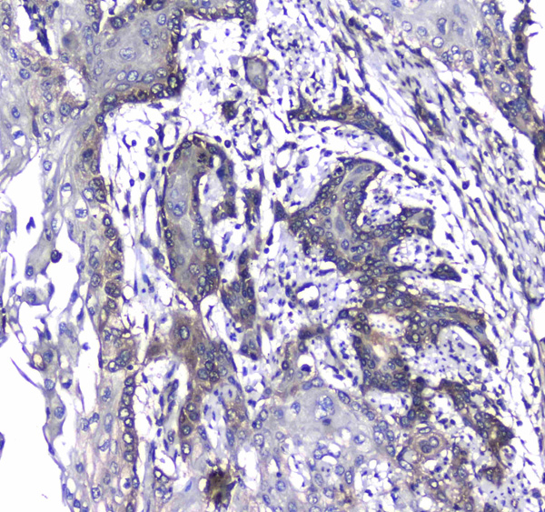
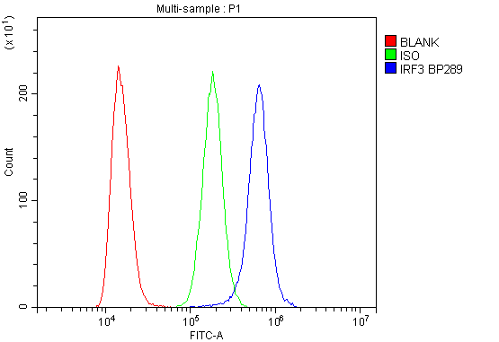 FCM (Flow Cytometry)
(Figure 6. Flow Cytometry analysis of K562 cells using anti-IRF3 antibody (AAA19216).Overlay histogram showing K562 cells stained with AAA19216 (Blue line). The cells were blocked with 10% normal goat serum. And then incubated with rabbit anti-IRF3 Antibody (AAA19216, 1μg/1x106 cells) for 30 min at 20 degree C. DyLight®488 conjugated goat anti-rabbit IgG (5-10μg/1x106 cells) was used as secondary antibody for 30 minutes at 20 degree C. Isotype control antibody (Green line) was rabbit IgG (1μg/1x106) used under the same conditions. Unlabelled sample (Red line) was also used as a control.)
FCM (Flow Cytometry)
(Figure 6. Flow Cytometry analysis of K562 cells using anti-IRF3 antibody (AAA19216).Overlay histogram showing K562 cells stained with AAA19216 (Blue line). The cells were blocked with 10% normal goat serum. And then incubated with rabbit anti-IRF3 Antibody (AAA19216, 1μg/1x106 cells) for 30 min at 20 degree C. DyLight®488 conjugated goat anti-rabbit IgG (5-10μg/1x106 cells) was used as secondary antibody for 30 minutes at 20 degree C. Isotype control antibody (Green line) was rabbit IgG (1μg/1x106) used under the same conditions. Unlabelled sample (Red line) was also used as a control.)
IRF3, Polyclonal Antibody (Cat# AAA19216)
Full Name
Anti-IRF3 Antibody
Reactivity
Human
Applications
Western Blot, Immunohistochemistry, Flow Cytometry, Direct ELISA
Purity
Immunogen affinity purified.
Pricing


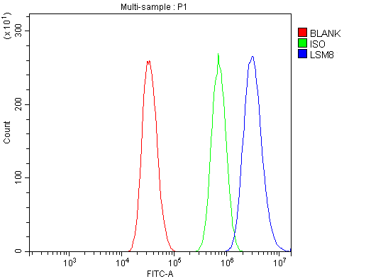 FCM (Flow Cytometry)
(Figure 13. Flow Cytometry analysis of A431 cells using anti-LSM8 antibody (AAA19337).Overlay histogram showing A431 cells stained with AAA19337 (Blue line). The cells were blocked with 10% normal goat serum. And then incubated with rabbit anti-LSM8 Antibody (AAA19337, 1μg/1x106 cells) for 30 min at 20 degree C. DyLight®488 conjugated goat anti-rabbit IgG (5-10μg/1x106 cells) was used as secondary antibody for 30 minutes at 20 degree C. Isotype control antibody (Green line) was rabbit IgG (1μg/1x106) used under the same conditions. Unlabelled sample (Red line) was also used as a control.)
FCM (Flow Cytometry)
(Figure 13. Flow Cytometry analysis of A431 cells using anti-LSM8 antibody (AAA19337).Overlay histogram showing A431 cells stained with AAA19337 (Blue line). The cells were blocked with 10% normal goat serum. And then incubated with rabbit anti-LSM8 Antibody (AAA19337, 1μg/1x106 cells) for 30 min at 20 degree C. DyLight®488 conjugated goat anti-rabbit IgG (5-10μg/1x106 cells) was used as secondary antibody for 30 minutes at 20 degree C. Isotype control antibody (Green line) was rabbit IgG (1μg/1x106) used under the same conditions. Unlabelled sample (Red line) was also used as a control.)
LSM8, Polyclonal Antibody (Cat# AAA19337)
Full Name
Anti-LSM8 Antibody
Reactivity
Human, Mouse, Rat
Applications
WB, IHC-P, ICC, IF, FC/FACS/FCM, EIA
Purity
Immunogen affinity purified.
Pricing


 FCM (Flow Cytometry)
(Figure 7. Flow Cytometry analysis of SiHa cells using anti-Aconitase 1/ACO1 antibody (AAA19267).Overlay histogram showing SiHa cells stained with AAA19267 (Blue line). The cells were blocked with 10% normal goat serum. And then incubated with rabbit anti-Aconitase 1/ACO1 Antibody (AAA19267,1μg/1x106 cells) for 30 min at 20 degree C. DyLight®488 conjugated goat anti-rabbit IgG (5-10μg/1x106 cells) was used as secondary antibody for 30 minutes at 20 degree C. Isotype control antibody (Green line) was rabbit IgG (1μg/1x106) used under the same conditions. Unlabelled sample (Red line) was also used as a control.)
FCM (Flow Cytometry)
(Figure 7. Flow Cytometry analysis of SiHa cells using anti-Aconitase 1/ACO1 antibody (AAA19267).Overlay histogram showing SiHa cells stained with AAA19267 (Blue line). The cells were blocked with 10% normal goat serum. And then incubated with rabbit anti-Aconitase 1/ACO1 Antibody (AAA19267,1μg/1x106 cells) for 30 min at 20 degree C. DyLight®488 conjugated goat anti-rabbit IgG (5-10μg/1x106 cells) was used as secondary antibody for 30 minutes at 20 degree C. Isotype control antibody (Green line) was rabbit IgG (1μg/1x106) used under the same conditions. Unlabelled sample (Red line) was also used as a control.)
Aconitase 1/ACO1, Polyclonal Antibody (Cat# AAA19267)
Full Name
Anti-Aconitase 1/ACO1 Antibody
Gene Names
ACO1; IRP1; ACONS; IREB1; IREBP; IREBP1
Reactivity
Human, Mouse, Monkey, Rat
Applications
WB, IHC-P, ICC, IF, FC/FACS/FCM, EIA
Purity
Immunogen affinity purified.
Pricing


 IF (Immunofluorescence)
(Immunofluorescence analysis of NIH-3T3 cells using IFNAR1 Rabbit pAb at dilution of 1:100 (40x lens). Blue: DAPI for nuclear staining.)
IF (Immunofluorescence)
(Immunofluorescence analysis of NIH-3T3 cells using IFNAR1 Rabbit pAb at dilution of 1:100 (40x lens). Blue: DAPI for nuclear staining.)
IFNAR1, Polyclonal Antibody (Cat# AAA28371)
Full Name
IFNAR1 Rabbit pAb
Gene Names
IFNAR1; AVP; IFRC; IFNAR; IFNBR; IFN-alpha-REC
Reactivity
Human, Mouse, Rat
Applications
WB, IHC, IF
Purity
Affinity purification
Pricing


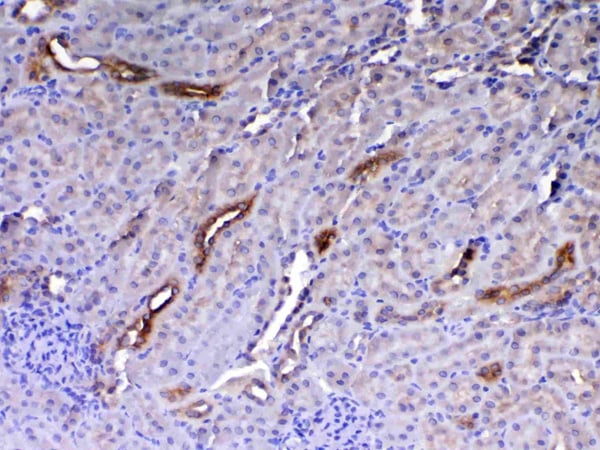
 IHC (Immunohistchemistry)
(Figure 6. IHC analysis of Flt3 / CD135 using anti-Flt3 / CD135 antibody (AAA19130).Flt3 / CD135 was detected in paraffin-embedded section of mouse kidney tissue. Heat mediated antigen retrieval was performed in citrate buffer (pH6, epitope retrieval solution) for 20 mins. The tissue section was blocked with 10% goat serum. The tissue section was then incubated with 1ug/ml rabbit anti-Flt3 / CD135 Antibody (AAA19130) overnight at 4 degree C. Biotinylated goat anti-rabbit IgG was used as secondary antibody and incubated for 30 minutes at 37 degree C. The tissue section was developed using Strepavidin-Biotin-Complex (SABC) with DAB as the chromogen.)
IHC (Immunohistchemistry)
(Figure 6. IHC analysis of Flt3 / CD135 using anti-Flt3 / CD135 antibody (AAA19130).Flt3 / CD135 was detected in paraffin-embedded section of mouse kidney tissue. Heat mediated antigen retrieval was performed in citrate buffer (pH6, epitope retrieval solution) for 20 mins. The tissue section was blocked with 10% goat serum. The tissue section was then incubated with 1ug/ml rabbit anti-Flt3 / CD135 Antibody (AAA19130) overnight at 4 degree C. Biotinylated goat anti-rabbit IgG was used as secondary antibody and incubated for 30 minutes at 37 degree C. The tissue section was developed using Strepavidin-Biotin-Complex (SABC) with DAB as the chromogen.)
Flt3/CD135, Polyclonal Antibody (Cat# AAA19130)
Full Name
Anti-Flt3/CD135 Picoband Antibody
Reactivity
Mouse, Rat
No cross reactivity with other proteins.
No cross reactivity with other proteins.
Applications
EIA, IHC, WB
Purity
Immunogen affinity purified
Pricing


 FCM (Flow Cytometry)
(Figure 6. Flow Cytometry analysis of PC-3 cells using anti-MitoNEET/CISD1 antibody (AAA19293).Overlay histogram showing PC-3 cells stained with AAA19293 (Blue line). The cells were blocked with 10% normal goat serum. And then incubated with rabbit anti-MitoNEET/CISD1 Antibody (AAA19293,1μg/1x106 cells) for 30 min at 20 degree C. DyLight®488 conjugated goat anti-rabbit IgG (5-10μg/1x106 cells) was used as secondary antibody for 30 minutes at 20 degree C. Isotype control antibody (Green line) was rabbit IgG (1μg/1x106) used under the same conditions. Unlabelled sample (Red line) was also used as a control.)
FCM (Flow Cytometry)
(Figure 6. Flow Cytometry analysis of PC-3 cells using anti-MitoNEET/CISD1 antibody (AAA19293).Overlay histogram showing PC-3 cells stained with AAA19293 (Blue line). The cells were blocked with 10% normal goat serum. And then incubated with rabbit anti-MitoNEET/CISD1 Antibody (AAA19293,1μg/1x106 cells) for 30 min at 20 degree C. DyLight®488 conjugated goat anti-rabbit IgG (5-10μg/1x106 cells) was used as secondary antibody for 30 minutes at 20 degree C. Isotype control antibody (Green line) was rabbit IgG (1μg/1x106) used under the same conditions. Unlabelled sample (Red line) was also used as a control.)
MitoNEET/CISD1, Polyclonal Antibody (Cat# AAA19293)
Full Name
Anti-MitoNEET/CISD1 Antibody
Gene Names
CISD1; ZCD1; MDS029; C10orf70; mitoNEET
Reactivity
Human, Mouse, Rat, Monkey
Applications
WB, IHC-P, FC/FACS/FCM, EIA
Purity
Immunogen affinity purified.
Pricing


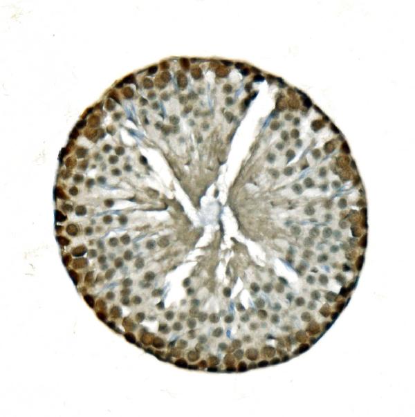
 IHC (Immunohistochemistry)
(Figure 7. IHC analysis of Periostin using anti-Periostin antibody (AAA19149).Periostin was detected in paraffin-embedded section of rat small intestine tissue. Heat mediated antigen retrieval was performed in citrate buffer (pH6, epitope retrieval solution) for 20 mins. The tissue section was blocked with 10% goat serum. The tissue section was then incubated with 1ug/ml rabbit anti-Periostin Antibody (AAA19149) overnight at 4 degree C. Biotinylated goat anti-rabbit IgG was used as secondary antibody and incubated for 30 minutes at 37 degree C. The tissue section was developed using Strepavidin-Biotin-Complex (SABC) with DAB as the chromogen.)
IHC (Immunohistochemistry)
(Figure 7. IHC analysis of Periostin using anti-Periostin antibody (AAA19149).Periostin was detected in paraffin-embedded section of rat small intestine tissue. Heat mediated antigen retrieval was performed in citrate buffer (pH6, epitope retrieval solution) for 20 mins. The tissue section was blocked with 10% goat serum. The tissue section was then incubated with 1ug/ml rabbit anti-Periostin Antibody (AAA19149) overnight at 4 degree C. Biotinylated goat anti-rabbit IgG was used as secondary antibody and incubated for 30 minutes at 37 degree C. The tissue section was developed using Strepavidin-Biotin-Complex (SABC) with DAB as the chromogen.)
Periostin, Polyclonal Antibody (Cat# AAA19149)
Full Name
Anti-Periostin Picoband Antibody
Gene Names
Postn; Plf
Reactivity
Mouse, Rat
No cross reactivity with other proteins.
No cross reactivity with other proteins.
Applications
EIA, IHC, WB
Purity
Immunogen affinity purified
Pricing


 IHC (Immunohistchemistry)
(Figure 6. IHC analysis of HE4 using anti-HE4 antibody (AAA19161).HE4 was detected in paraffin-embedded section of rat small intestine tissue. Heat mediated antigen retrieval was performed in citrate buffer (pH6, epitope retrieval solution) for 20 mins. The tissue section was blocked with 10% goat serum. The tissue section was then incubated with 1ug/ml rabbit anti-HE4 Antibody (AAA19161) overnight at 4 degree C. Biotinylated goat anti-rabbit IgG was used as secondary antibody and incubated for 30 minutes at 37 degree C. The tissue section was developed using Strepavidin-Biotin-Complex (SABC) with DAB as the chromogen.)
IHC (Immunohistchemistry)
(Figure 6. IHC analysis of HE4 using anti-HE4 antibody (AAA19161).HE4 was detected in paraffin-embedded section of rat small intestine tissue. Heat mediated antigen retrieval was performed in citrate buffer (pH6, epitope retrieval solution) for 20 mins. The tissue section was blocked with 10% goat serum. The tissue section was then incubated with 1ug/ml rabbit anti-HE4 Antibody (AAA19161) overnight at 4 degree C. Biotinylated goat anti-rabbit IgG was used as secondary antibody and incubated for 30 minutes at 37 degree C. The tissue section was developed using Strepavidin-Biotin-Complex (SABC) with DAB as the chromogen.)
HE4, Polyclonal Antibody (Cat# AAA19161)
Full Name
Anti-HE4 Picoband Antibody
Gene Names
Wfdc2; re4
Reactivity
Mouse, Rat
No cross reactivity with other proteins.
No cross reactivity with other proteins.
Applications
IHC, WB
Purity
Immunogen affinity purified
Pricing


 IHC (Immunohistchemistry)
(Figure 6. IHC analysis of HP1 gamma using anti-HP1 gamma antibody (AAA19145).HP1 gamma was detected in paraffin-embedded section of rat small intestine tissue. Heat mediated antigen retrieval was performed in citrate buffer (pH6, epitope retrieval solution) for 20 mins. The tissue section was blocked with 10% goat serum. The tissue section was then incubated with 1ug/ml rabbit anti-HP1 gamma Antibody (AAA19145) overnight at 4 degree C. Biotinylated goat anti-rabbit IgG was used as secondary antibody and incubated for 30 minutes at 37 degree C. The tissue section was developed using Strepavidin-Biotin-Complex (SABC) with DAB as the chromogen.)
IHC (Immunohistchemistry)
(Figure 6. IHC analysis of HP1 gamma using anti-HP1 gamma antibody (AAA19145).HP1 gamma was detected in paraffin-embedded section of rat small intestine tissue. Heat mediated antigen retrieval was performed in citrate buffer (pH6, epitope retrieval solution) for 20 mins. The tissue section was blocked with 10% goat serum. The tissue section was then incubated with 1ug/ml rabbit anti-HP1 gamma Antibody (AAA19145) overnight at 4 degree C. Biotinylated goat anti-rabbit IgG was used as secondary antibody and incubated for 30 minutes at 37 degree C. The tissue section was developed using Strepavidin-Biotin-Complex (SABC) with DAB as the chromogen.)
HP1 gamma, Polyclonal Antibody (Cat# AAA19145)
Full Name
Anti-HP1 gamma Picoband antibody
Gene Names
CBX3; HECH; HP1-GAMMA; HP1Hs-gamma
Reactivity
Human, Mouse, Rat
No cross reactivity with other proteins.
No cross reactivity with other proteins.
Applications
EIA, IHC, WB
Pricing


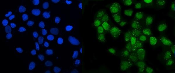
 IF (Immunofluorescence)
(Figure 6. IF analysis of GRSF1 using anti- GRSF1 antibody (AAA19315).GRSF1 was detected in immunocytochemical section of MCF-7 cells. Enzyme antigen retrieval was performed using IHC enzyme antigen retrieval reagent for 15 mins. The cells were blocked with 10% goat serum. And then incubated with 5μg/mL rabbit anti- GRSF1 Antibody (AAA19315) overnight at 4 degree C. DyLight®488 Conjugated Goat Anti-Rabbit IgG was used as secondary antibody at 1:100 dilution and incubated for 30 minutes at 37 degree C. The section was counterstained with DAPI. Visualize using a fluorescence microscope and filter sets appropriate for the label used.)
IF (Immunofluorescence)
(Figure 6. IF analysis of GRSF1 using anti- GRSF1 antibody (AAA19315).GRSF1 was detected in immunocytochemical section of MCF-7 cells. Enzyme antigen retrieval was performed using IHC enzyme antigen retrieval reagent for 15 mins. The cells were blocked with 10% goat serum. And then incubated with 5μg/mL rabbit anti- GRSF1 Antibody (AAA19315) overnight at 4 degree C. DyLight®488 Conjugated Goat Anti-Rabbit IgG was used as secondary antibody at 1:100 dilution and incubated for 30 minutes at 37 degree C. The section was counterstained with DAPI. Visualize using a fluorescence microscope and filter sets appropriate for the label used.)
GRSF1, Polyclonal Antibody (Cat# AAA19315)
Full Name
Anti-GRSF1 Antibody
Reactivity
Human
Applications
WB, IHC-P, ICC, IF, FC/FACS/FCM, EIA
Purity
Immunogen affinity purified.
Pricing


 FCM (Flow Cytometry)
(Figure 9. Flow Cytometry analysis of U251 cells using anti-VPRBP/DCAF1 antibody (AAA19343).Overlay histogram showing U251 cells stained with AAA19343 (Blue line). The cells were blocked with 10% normal goat serum. And then incubated with rabbit anti-VPRBP/DCAF1 Antibody (AAA19343, 1μg/1x106 cells) for 30 min at 20 degree C. DyLight®488 conjugated goat anti-rabbit IgG (5-10μg/1x106 cells) was used as secondary antibody for 30 minutes at 20 degree C. Isotype control antibody (Green line) was rabbit IgG (1μg/1x106) used under the same conditions. Unlabelled sample (Red line) was also used as a control.)
FCM (Flow Cytometry)
(Figure 9. Flow Cytometry analysis of U251 cells using anti-VPRBP/DCAF1 antibody (AAA19343).Overlay histogram showing U251 cells stained with AAA19343 (Blue line). The cells were blocked with 10% normal goat serum. And then incubated with rabbit anti-VPRBP/DCAF1 Antibody (AAA19343, 1μg/1x106 cells) for 30 min at 20 degree C. DyLight®488 conjugated goat anti-rabbit IgG (5-10μg/1x106 cells) was used as secondary antibody for 30 minutes at 20 degree C. Isotype control antibody (Green line) was rabbit IgG (1μg/1x106) used under the same conditions. Unlabelled sample (Red line) was also used as a control.)
VPRBP/DCAF1, Polyclonal Antibody (Cat# AAA19343)
Full Name
Anti-VPRBP/DCAF1 Antibody
Gene Names
DCAF1; RIP; VPRBP
Reactivity
Human, Mouse, Rat
Applications
WB, IHC-P, ICC, IF, FC/FACS/FCM, EIA
Purity
Immunogen affinity purified.
Pricing


 FCM (Flow Cytometry)
(Figure 10. Flow Cytometry analysis of A431 cells using anti-TIMM8A/DDP antibody (AAA19317).Overlay histogram showing A431 cells stained with AAA19317 (Blue line). The cells were blocked with 10% normal goat serum. And then incubated with rabbit anti-TIMM8A/DDP Antibody (AAA19317, 1μg/1x106 cells) for 30 min at 20 degree C. DyLight®488 conjugated goat anti-rabbit IgG (5-10μg/1x106 cells) was used as secondary antibody for 30 minutes at 20 degree C. Isotype control antibody (Green line) was rabbit IgG (1μg/1x106) used under the same conditions. Unlabelled sample (Red line) was also used as a control.)
FCM (Flow Cytometry)
(Figure 10. Flow Cytometry analysis of A431 cells using anti-TIMM8A/DDP antibody (AAA19317).Overlay histogram showing A431 cells stained with AAA19317 (Blue line). The cells were blocked with 10% normal goat serum. And then incubated with rabbit anti-TIMM8A/DDP Antibody (AAA19317, 1μg/1x106 cells) for 30 min at 20 degree C. DyLight®488 conjugated goat anti-rabbit IgG (5-10μg/1x106 cells) was used as secondary antibody for 30 minutes at 20 degree C. Isotype control antibody (Green line) was rabbit IgG (1μg/1x106) used under the same conditions. Unlabelled sample (Red line) was also used as a control.)
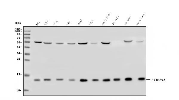
TIMM8A/DDP, Polyclonal Antibody (Cat# AAA19317)
Full Name
Anti-TIMM8A/DDP Antibody
Gene Names
TIMM8A; DDP; MTS; DDP1; DFN1; TIM8
Reactivity
Human, Mouse, Rat, Monkey
Applications
WB, IHC-P, ICC, IF, EIA
Purity
Immunogen affinity purified.
Pricing


 FCM (Flow Cytometry)
(Figure 8. Flow Cytometry analysis of K562 cells using anti-HP1 alpha/CBX5 antibody (AAA19266).Overlay histogram showing K562 cells stained with AAA19266 (Blue line). The cells were blocked with 10% normal goat serum. And then incubated with rabbit anti-HP1 alpha/CBX5 Antibody (AAA19266,1μg/1x106 cells) for 30 min at 20 degree C. DyLight®488 conjugated goat anti-rabbit IgG (5-10μg/1x106 cells) was used as secondary antibody for 30 minutes at 20 degree C. Isotype control antibody (Green line) was rabbit IgG (1μg/1x106) used under the same conditions. Unlabelled sample (Red line) was also used as a control.)
FCM (Flow Cytometry)
(Figure 8. Flow Cytometry analysis of K562 cells using anti-HP1 alpha/CBX5 antibody (AAA19266).Overlay histogram showing K562 cells stained with AAA19266 (Blue line). The cells were blocked with 10% normal goat serum. And then incubated with rabbit anti-HP1 alpha/CBX5 Antibody (AAA19266,1μg/1x106 cells) for 30 min at 20 degree C. DyLight®488 conjugated goat anti-rabbit IgG (5-10μg/1x106 cells) was used as secondary antibody for 30 minutes at 20 degree C. Isotype control antibody (Green line) was rabbit IgG (1μg/1x106) used under the same conditions. Unlabelled sample (Red line) was also used as a control.)
HP1 alpha/CBX5, Polyclonal Antibody (Cat# AAA19266)
Full Name
Anti-HP1 alpha/CBX5 Antibody
Gene Names
CBX5; HP1; HP1A
Reactivity
Human, Mouse, Rat
Applications
WB, IHC-P, ICC, IF, FC/FACS/FCM, EIA
Purity
Immunogen affinity purified.
Pricing


 WB (Western Blot)
(VIM antibody - C-terminal region validated by WB using MCF-7 whole cell lysates at 1.0ug/ml.)
WB (Western Blot)
(VIM antibody - C-terminal region validated by WB using MCF-7 whole cell lysates at 1.0ug/ml.)
VIM, Polyclonal Antibody (Cat# AAA23531)
Full Name
VIM antibody - C-terminal region
Reactivity
Cow, Dog, Guinea Pig, Horse, Human, Mouse, Rabbit, Rat, Sheep, Zebrafish
Applications
IHC, WB
Purity
Affinity Purified
Pricing


 FCM (Flow Cytometry)
(Flow cytometry analysis of GOT1 in HeLa cell line, staining at 2-5ug for 1x10⁶cells (red line). The secondary antibody used goat anti-mouse IgG Alexa fluor 488 conjugate. Isotype control antibody was mouse IgG (black line).)
FCM (Flow Cytometry)
(Flow cytometry analysis of GOT1 in HeLa cell line, staining at 2-5ug for 1x10⁶cells (red line). The secondary antibody used goat anti-mouse IgG Alexa fluor 488 conjugate. Isotype control antibody was mouse IgG (black line).)
GOT1, Monoclonal Antibody (Cat# AAA11733)
Full Name
GOT1 antibody
Gene Names
GOT1; AST1; cCAT; GIG18; cAspAT; ASTQTL1
Reactivity
Human
Applications
Western Blot, Flow Cytometry, Immunocytochemistry, Immunofluorescence
Purity
By protein-A affinity chromatography
Pricing


 IHC (Immunohistchemistry)
(Figure 6. IHC analysis of Cytoglobin using anti- Cytoglobin antibody (AAA11681).Cytoglobin was detected in paraffin-embedded section of human lung cancer tissues. Heat mediated antigen retrieval was performed in citrate buffer (pH6, epitope retrieval solution) for 20 mins. The tissue section was blocked with 10% goat serum. The tissue section was then incubated with 1ug/ml rabbit anti- Cytoglobin Antibody (AAA11681) overnight at 4 degree C. Biotinylated goat anti-rabbit IgG was used as secondary antibody and incubated for 30 minutes at 37 degree C. The tissue section was developed using Strepavidin-Biotin-Complex (SABC) with DAB as the chromogen.)
IHC (Immunohistchemistry)
(Figure 6. IHC analysis of Cytoglobin using anti- Cytoglobin antibody (AAA11681).Cytoglobin was detected in paraffin-embedded section of human lung cancer tissues. Heat mediated antigen retrieval was performed in citrate buffer (pH6, epitope retrieval solution) for 20 mins. The tissue section was blocked with 10% goat serum. The tissue section was then incubated with 1ug/ml rabbit anti- Cytoglobin Antibody (AAA11681) overnight at 4 degree C. Biotinylated goat anti-rabbit IgG was used as secondary antibody and incubated for 30 minutes at 37 degree C. The tissue section was developed using Strepavidin-Biotin-Complex (SABC) with DAB as the chromogen.)
Cytoglobin, Polyclonal Antibody (Cat# AAA11681)
Full Name
Anti-Cytoglobin Antibody
Gene Names
CYGB; HGB; STAP
Reactivity
Human, Mouse, Rat
Applications
Western Blot, Immunohistochemistry
Purity
Immunogen affinity purified.
Pricing


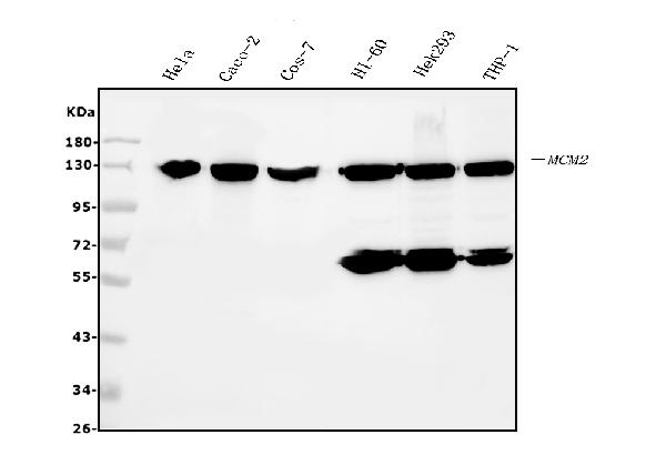
 FCM (Flow Cytometry)
(Figure 6. Flow Cytometry analysis of HL-60 cells using anti- MCM2 antibody (AAA19351).Overlay histogram showing HL-60 cells stained with AAA19351 (Blue line). The cells were blocked with 10% normal goat serum. And then incubated with mouse anti-MCM2 Antibody (AAA19351, 1μg/1x106 cells) for 30 min at 20 degree C. DyLight®488 conjugated goat anti-mouse IgG (BA1126, 5-10μg/1x106 cells) was used as secondary antibody for 30 minutes at 20 degree C. Isotype control antibody (Green line) was mouse IgG (1μg/1x106) used under the same conditions. Unlabelled sample (Red line) was also used as a control.)
FCM (Flow Cytometry)
(Figure 6. Flow Cytometry analysis of HL-60 cells using anti- MCM2 antibody (AAA19351).Overlay histogram showing HL-60 cells stained with AAA19351 (Blue line). The cells were blocked with 10% normal goat serum. And then incubated with mouse anti-MCM2 Antibody (AAA19351, 1μg/1x106 cells) for 30 min at 20 degree C. DyLight®488 conjugated goat anti-mouse IgG (BA1126, 5-10μg/1x106 cells) was used as secondary antibody for 30 minutes at 20 degree C. Isotype control antibody (Green line) was mouse IgG (1μg/1x106) used under the same conditions. Unlabelled sample (Red line) was also used as a control.)
MCM2, Monoclonal Antibody (Cat# AAA19351)
Full Name
Anti-MCM2 Antibody (monoclonal, 11C4)
Gene Names
MCM2; BM28; CCNL1; CDCL1; cdc19; D3S3194; MITOTIN
Reactivity
Human, Monkey
Applications
WB, IHC-P, ICC, IF, FC/FACS/FCM
Purity
Immunogen affinity purified.
Pricing


 IHC (Immunohistchemistry)
(Formalin-fixed, paraffin-embedded human Breast Carcinoma stained with MUC1 / EMA Monoclonal Antibody (MUC1/845).)
IHC (Immunohistchemistry)
(Formalin-fixed, paraffin-embedded human Breast Carcinoma stained with MUC1 / EMA Monoclonal Antibody (MUC1/845).)
MUC1 / EMA /CD227, Monoclonal Antibody (Cat# AAA13841)
Full Name
MUC1 / EMA /CD227 (Epithelial Marker) Mouse Monoclonal Antibody
Gene Names
MUC1; EMA; MCD; PEM; PUM; KL-6; MAM6; MCKD; PEMT; CD227; H23AG; MCKD1; MUC-1; ADMCKD; ADMCKD1; CA 15-3; MUC-1/X; MUC1/ZD; MUC-1/SEC
Reactivity
Human
Applications
Flow Cytometry, Immunofluorescence, Immunohistochemistry
Pricing


 FCM (Flow Cytometry)
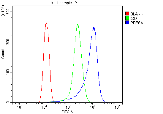
(Figure 6. Flow Cytometry analysis of HELA cells using anti-PDE6 alpha/PDE6A antibody (AAA19318).Overlay histogram showing HELA cells stained with AAA19318 (Blue line). The cells were blocked with 10% normal goat serum. And then incubated with rabbit anti-PDE6 alpha/PDE6A Antibody (AAA19318, 1μg/1x106 cells) for 30 min at 20 degree C. DyLight®488 conjugated goat anti-rabbit IgG (5-10μg/1x106 cells) was used as secondary antibody for 30 minutes at 20 degree C. Isotype control antibody (Green line) was rabbit IgG (1μg/1x106) used under the same conditions. Unlabelled sample (Red line) was also used as a control.)
FCM (Flow Cytometry)
(Figure 6. Flow Cytometry analysis of HELA cells using anti-PDE6 alpha/PDE6A antibody (AAA19318).Overlay histogram showing HELA cells stained with AAA19318 (Blue line). The cells were blocked with 10% normal goat serum. And then incubated with rabbit anti-PDE6 alpha/PDE6A Antibody (AAA19318, 1μg/1x106 cells) for 30 min at 20 degree C. DyLight®488 conjugated goat anti-rabbit IgG (5-10μg/1x106 cells) was used as secondary antibody for 30 minutes at 20 degree C. Isotype control antibody (Green line) was rabbit IgG (1μg/1x106) used under the same conditions. Unlabelled sample (Red line) was also used as a control.)
PDE6 alpha/PDE6A, Polyclonal Antibody (Cat# AAA19318)
Full Name
Anti-PDE6 alpha/PDE6A Antibody
Gene Names
PDE6A; PDEA; RP43; CGPR-A; PDE6A
Reactivity
Human, Mouse, Rat
Applications
WB, IHC-P, ICC, IF, FC/FACS/FCM, EIA
Purity
Immunogen affinity purified.
Pricing


 WB (Western Blot)
(WB Suggested Anti-ATG5 Antibody Titration: 1 ug/mlPositive Control: HepG2 cell lysate)
WB (Western Blot)
(WB Suggested Anti-ATG5 Antibody Titration: 1 ug/mlPositive Control: HepG2 cell lysate)
ATG5, Polyclonal Antibody (Cat# AAA23551)
Full Name
ATG5 antibody - C-terminal region
Gene Names
ATG5; ASP; APG5; APG5L; hAPG5; SCAR25; APG5-LIKE
Reactivity
Cow, Dog, Guinea Pig, Horse, Human, Mouse, Rabbit, Rat, Zebrafish
Applications
IHC, WB
Purity
Affinity Purified
Pricing


 IHC (Immunohistchemistry)
(Figure 6. IHC analysis of GALE using anti-GALE antibody (AAA19135).GALE was detected in paraffin-embedded section of rat kidney tissue. Heat mediated antigen retrieval was performed in citrate buffer (pH6, epitope retrieval solution) for 20 mins. The tissue section was blocked with 10% goat serum. The tissue section was then incubated with 1ug/ml rabbit anti-GALE Antibody (AAA19135) overnight at 4 degree C. Biotinylated goat anti-rabbit IgG was used as secondary antibody and incubated for 30 minutes at 37 degree C. The tissue section was developed using Strepavidin-Biotin-Complex (SABC) with DAB as the chromogen.)
IHC (Immunohistchemistry)
(Figure 6. IHC analysis of GALE using anti-GALE antibody (AAA19135).GALE was detected in paraffin-embedded section of rat kidney tissue. Heat mediated antigen retrieval was performed in citrate buffer (pH6, epitope retrieval solution) for 20 mins. The tissue section was blocked with 10% goat serum. The tissue section was then incubated with 1ug/ml rabbit anti-GALE Antibody (AAA19135) overnight at 4 degree C. Biotinylated goat anti-rabbit IgG was used as secondary antibody and incubated for 30 minutes at 37 degree C. The tissue section was developed using Strepavidin-Biotin-Complex (SABC) with DAB as the chromogen.)
GALE, Polyclonal Antibody (Cat# AAA19135)
Full Name
Anti-GALE Picoband antibody
Gene Names
GALE; SDR1E1
Reactivity
Human, Mouse, Rat
No cross reactivity with other proteins.
No cross reactivity with other proteins.
Applications
EIA, IHC, WB
Pricing


 FCM (Flow Cytometry)
(Figure 9. Flow Cytometry analysis of THP-1 cells using anti-CHCHD10 antibody (AAA19310).Overlay histogram showing THP-1 cells stained with AAA19310 (Blue line). The cells were blocked with 10% normal goat serum. And then incubated with rabbit anti-CHCHD10 Antibody (AAA19310, 1μg/1x106 cells) for 30 min at 20 degree C. DyLight®488 conjugated goat anti-rabbit IgG (5-10μg/1x106 cells) was used as secondary antibody for 30 minutes at 20 degree C. Isotype control antibody (Green line) was rabbit IgG (1μg/1x106) used under the same conditions. Unlabelled sample (Red line) was also used as a control.)
FCM (Flow Cytometry)
(Figure 9. Flow Cytometry analysis of THP-1 cells using anti-CHCHD10 antibody (AAA19310).Overlay histogram showing THP-1 cells stained with AAA19310 (Blue line). The cells were blocked with 10% normal goat serum. And then incubated with rabbit anti-CHCHD10 Antibody (AAA19310, 1μg/1x106 cells) for 30 min at 20 degree C. DyLight®488 conjugated goat anti-rabbit IgG (5-10μg/1x106 cells) was used as secondary antibody for 30 minutes at 20 degree C. Isotype control antibody (Green line) was rabbit IgG (1μg/1x106) used under the same conditions. Unlabelled sample (Red line) was also used as a control.)
CHCHD10, Polyclonal Antibody (Cat# AAA19310)
Full Name
Anti-CHCHD10 Antibody
Gene Names
CHCHD10; FTDALS2; N27C7-4; C22orf16
Reactivity
Human, Mouse, Rat, Monkey
Applications
WB, IHC-P, ICC, IF, FC/FACS/FCM, EIA
Purity
Immunogen affinity purified.
Pricing


 FCM (Flow Cytometry)
(Figure 7. Flow Cytometry analysis of PC-3 cells using anti-PVRL1/NECTIN1 antibody (AAA19303).Overlay histogram showing PC-3 cells stained with AAA19303 (Blue line). The cells were blocked with 10% normal goat serum. And then incubated with rabbit anti-PVRL1/NECTIN1 Antibody (AAA19303, 1μg/1x106 cells) for 30 min at 20 degree C. DyLight®488 conjugated goat anti-rabbit IgG (5-10μg/1x106 cells) was used as secondary antibody for 30 minutes at 20 degree C. Isotype control antibody (Green line) was rabbit IgG (1μg/1x106) used under the same conditions. Unlabelled sample (Red line) was also used as a control.)
FCM (Flow Cytometry)
(Figure 7. Flow Cytometry analysis of PC-3 cells using anti-PVRL1/NECTIN1 antibody (AAA19303).Overlay histogram showing PC-3 cells stained with AAA19303 (Blue line). The cells were blocked with 10% normal goat serum. And then incubated with rabbit anti-PVRL1/NECTIN1 Antibody (AAA19303, 1μg/1x106 cells) for 30 min at 20 degree C. DyLight®488 conjugated goat anti-rabbit IgG (5-10μg/1x106 cells) was used as secondary antibody for 30 minutes at 20 degree C. Isotype control antibody (Green line) was rabbit IgG (1μg/1x106) used under the same conditions. Unlabelled sample (Red line) was also used as a control.)
PVRL1/NECTIN1, Polyclonal Antibody (Cat# AAA19303)
Full Name
Anti-PVRL1/NECTIN1 Antibody
Gene Names
PVRL1; ED4; PRR; HIgR; HVEC; OFC7; PRR1; PVRR; CD111; PVRR1; SK-12; CLPED1; nectin-1
Reactivity
Human, Mouse, Rat
Applications
WB, IHC-P, FC/FACS/FCM, EIA
Purity
Immunogen affinity purified.
Pricing


 FCM (Flow Cytometry)
(Figure 8. Flow Cytometry analysis of C6 cells using anti-Clathrin heavy chain/CLTC antibody (AAA19270).Overlay histogram showing C6 cells stained with AAA19270 (Blue line). The cells were blocked with 10% normal goat serum. And then incubated with rabbit anti-Clathrin heavy chain/CLTC Antibody (AAA19270,1μg/1x106 cells) for 30 min at 20 degree C. DyLight®488 conjugated goat anti-rabbit IgG (5-10μg/1x106 cells) was used as secondary antibody for 30 minutes at 20 degree C. Isotype control antibody (Green line) was rabbit IgG (1μg/1x106) used under the same conditions. Unlabelled sample (Red line) was also used as a control.)
FCM (Flow Cytometry)
(Figure 8. Flow Cytometry analysis of C6 cells using anti-Clathrin heavy chain/CLTC antibody (AAA19270).Overlay histogram showing C6 cells stained with AAA19270 (Blue line). The cells were blocked with 10% normal goat serum. And then incubated with rabbit anti-Clathrin heavy chain/CLTC Antibody (AAA19270,1μg/1x106 cells) for 30 min at 20 degree C. DyLight®488 conjugated goat anti-rabbit IgG (5-10μg/1x106 cells) was used as secondary antibody for 30 minutes at 20 degree C. Isotype control antibody (Green line) was rabbit IgG (1μg/1x106) used under the same conditions. Unlabelled sample (Red line) was also used as a control.)
Clathrin heavy chain/CLTC, Polyclonal Antibody (Cat# AAA19270)
Full Name
Anti-Clathrin heavy chain/CLTC Antibody
Gene Names
CLTC; Hc; CHC; CHC17; CLH-17; CLTCL2
Reactivity
Human, Mouse, Rat
Applications
WB, IHC-P, ICC, IF, FC/FACS/FCM, EIA
Purity
Immunogen affinity purified.
Pricing


 IHC (Immunohistochemistry)
(Figure 7. IHC analysis of IDH2 using anti-IDH2 antibody (AAA11640).IDH2 was detected in frozen section of rat small intestine tissue. Heat mediated antigen retrieval was performed in citrate buffer (pH6, epitope retrieval solution) for 20 mins. The tissue section was blocked with 10% goat serum. The tissue section was then incubated with 1ug/ml rabbit anti-IDH2 Antibody (AAA11640) overnight at 4 degree C. Biotinylated goat anti-rabbit IgG was used as secondary antibody and incubated for 30 minutes at 37 degree C. The tissue section was developed using Strepavidin-Biotin-Complex (SABC) with DAB as the chromogen.)
IHC (Immunohistochemistry)
(Figure 7. IHC analysis of IDH2 using anti-IDH2 antibody (AAA11640).IDH2 was detected in frozen section of rat small intestine tissue. Heat mediated antigen retrieval was performed in citrate buffer (pH6, epitope retrieval solution) for 20 mins. The tissue section was blocked with 10% goat serum. The tissue section was then incubated with 1ug/ml rabbit anti-IDH2 Antibody (AAA11640) overnight at 4 degree C. Biotinylated goat anti-rabbit IgG was used as secondary antibody and incubated for 30 minutes at 37 degree C. The tissue section was developed using Strepavidin-Biotin-Complex (SABC) with DAB as the chromogen.)
IDH2, Polyclonal Antibody (Cat# AAA11640)
Full Name
Anti-IDH2 Antibody
Gene Names
IDH2; IDH; IDP; IDHM; IDPM; ICD-M; D2HGA2; mNADP-IDH
Reactivity
Human, Mouse, Rat
Applications
Western Blot, Immunohistochemistry
Purity
Immunogen Affinity Purified
Pricing


 FCM (Flow Cytometry)
(Figure 7. Flow Cytometry analysis of CACO-2 cells using anti-Glycine decarboxylase/GLDC antibody (AAA19300).Overlay histogram showing CACO-2 cells stained with AAA19300 (Blue line). The cells were blocked with 10% normal goat serum. And then incubated with rabbit anti-Glycine decarboxylase/GLDC Antibody (AAA19300, 1μg/1x106 cells) for 30 min at 20 degree C. DyLight®488 conjugated goat anti-rabbit IgG (5-10μg/1x106 cells) was used as secondary antibody for 30 minutes at 20 degree C. Isotype control antibody (Green line) was rabbit IgG (1μg/1x106) used under the same conditions. Unlabelled sample (Red line) was also used as a control.)
FCM (Flow Cytometry)
(Figure 7. Flow Cytometry analysis of CACO-2 cells using anti-Glycine decarboxylase/GLDC antibody (AAA19300).Overlay histogram showing CACO-2 cells stained with AAA19300 (Blue line). The cells were blocked with 10% normal goat serum. And then incubated with rabbit anti-Glycine decarboxylase/GLDC Antibody (AAA19300, 1μg/1x106 cells) for 30 min at 20 degree C. DyLight®488 conjugated goat anti-rabbit IgG (5-10μg/1x106 cells) was used as secondary antibody for 30 minutes at 20 degree C. Isotype control antibody (Green line) was rabbit IgG (1μg/1x106) used under the same conditions. Unlabelled sample (Red line) was also used as a control.)
Glycine decarboxylase/GLDC, Polyclonal Antibody (Cat# AAA19300)
Full Name
Anti-Glycine decarboxylase/GLDC Antibody
Gene Names
GLDC; GCE; GCSP; HYGN1
Reactivity
Human, Mouse, Rat
Applications
WB, IHC-P, FC/FACS/FCM, EIA
Purity
Immunogen affinity purified.
Pricing


 IHC (Immunohistochemistry)
(Figure 7. IHC analysis of splicing factor 1 using anti-splicing factor 1 antibody (AAA19141).splicing factor 1 was detected in paraffin-embedded section of rat small intestine tissue. Heat mediated antigen retrieval was performed in citrate buffer (pH6, epitope retrieval solution) for 20 mins. The tissue section was blocked with 10% goat serum. The tissue section was then incubated with 1ug/ml rabbit anti-splicing factor 1 Antibody (AAA19141) overnight at 4 degree C. Biotinylated goat anti-rabbit IgG was used as secondary antibody and incubated for 30 minutes at 37 degree C. The tissue section was developed using Strepavidin-Biotin-Complex (SABC) with DAB as the chromogen.)
IHC (Immunohistochemistry)
(Figure 7. IHC analysis of splicing factor 1 using anti-splicing factor 1 antibody (AAA19141).splicing factor 1 was detected in paraffin-embedded section of rat small intestine tissue. Heat mediated antigen retrieval was performed in citrate buffer (pH6, epitope retrieval solution) for 20 mins. The tissue section was blocked with 10% goat serum. The tissue section was then incubated with 1ug/ml rabbit anti-splicing factor 1 Antibody (AAA19141) overnight at 4 degree C. Biotinylated goat anti-rabbit IgG was used as secondary antibody and incubated for 30 minutes at 37 degree C. The tissue section was developed using Strepavidin-Biotin-Complex (SABC) with DAB as the chromogen.)
splicing factor 1, Polyclonal Antibody (Cat# AAA19141)
Full Name
Anti-splicing factor 1 Picoband antibody
Gene Names
SF1; BBP; MBBP; ZFM1; ZNF162; D11S636; ZCCHC25
Reactivity
Human, Mouse, Rat
No cross reactivity with other proteins.
No cross reactivity with other proteins.
Applications
EIA, IHC, WB
Pricing


 FCM (Flow Cytometry)
(Figure 8. Flow Cytometry analysis of U-87 cells using anti-MVP antibody (AAA19137).Overlay histogram showing U-87 cells stained with AAA19137 (Blue line).The cells were blocked with 10% normal goat serum. And then incubated with rabbit anti-MVP Antibody (AAA19137,1ug/1x10^6 cells) for 30 min at 20 degree C. DyLight®488 conjugated goat anti-rabbit IgG (5-10ug/1x10^6 cells) was used as secondary antibody for 30 minutes at 20 degree C. Isotype control antibody (Green line) was rabbit IgG (1ug/1x106) used under the same conditions. Unlabelled sample (Red line) was also used as a control.)
FCM (Flow Cytometry)
(Figure 8. Flow Cytometry analysis of U-87 cells using anti-MVP antibody (AAA19137).Overlay histogram showing U-87 cells stained with AAA19137 (Blue line).The cells were blocked with 10% normal goat serum. And then incubated with rabbit anti-MVP Antibody (AAA19137,1ug/1x10^6 cells) for 30 min at 20 degree C. DyLight®488 conjugated goat anti-rabbit IgG (5-10ug/1x10^6 cells) was used as secondary antibody for 30 minutes at 20 degree C. Isotype control antibody (Green line) was rabbit IgG (1ug/1x106) used under the same conditions. Unlabelled sample (Red line) was also used as a control.)
MVP, Polyclonal Antibody (Cat# AAA19137)
Full Name
Anti-MVP Picoband antibody
Gene Names
MVP; LRP; VAULT1
Reactivity
Human, Mouse, Rat
Applications
EIA, FC/FACS, IHC, ICC, WB
Pricing


 IF (Immunofluorescence)
(Immunofluorescence analysis of U-2 OS cells using GAPDH Mouse mAb at dilution of 1:100 (40x lens). Blue: DAPI for nuclear staining.)
IF (Immunofluorescence)
(Immunofluorescence analysis of U-2 OS cells using GAPDH Mouse mAb at dilution of 1:100 (40x lens). Blue: DAPI for nuclear staining.)
GAPDH, Monoclonal Antibody (Cat# AAA28398)
Full Name
GAPDH Mouse mAb (High Dilution)
Gene Names
GAPDH; G3PD; GAPD
Reactivity
Human, Mouse, Rat
Applications
WB, IHC, IF
Purity
Affinity purification
Pricing


 IHC (Immunohistchemistry)
(Figure 6. IHC analysis of Integrin alpha 5 using anti-Integrin alpha 5 antibody (AAA19156).Integrin alpha 5 was detected in paraffin-embedded section of human placenta tissue. Heat mediated antigen retrieval was performed in citrate buffer (pH6, epitope retrieval solution) for 20 mins. The tissue section was blocked with 10% goat serum. The tissue section was then incubated with 1ug/ml rabbit anti-Integrin alpha 5 Antibody (AAA19156) overnight at 4 degree C. Biotinylated goat anti-rabbit IgG was used as secondary antibody and incubated for 30 minutes at 37 degree C. The tissue section was developed using Strepavidin-Biotin-Complex (SABC) with DAB as the chromogen.)
IHC (Immunohistchemistry)
(Figure 6. IHC analysis of Integrin alpha 5 using anti-Integrin alpha 5 antibody (AAA19156).Integrin alpha 5 was detected in paraffin-embedded section of human placenta tissue. Heat mediated antigen retrieval was performed in citrate buffer (pH6, epitope retrieval solution) for 20 mins. The tissue section was blocked with 10% goat serum. The tissue section was then incubated with 1ug/ml rabbit anti-Integrin alpha 5 Antibody (AAA19156) overnight at 4 degree C. Biotinylated goat anti-rabbit IgG was used as secondary antibody and incubated for 30 minutes at 37 degree C. The tissue section was developed using Strepavidin-Biotin-Complex (SABC) with DAB as the chromogen.)
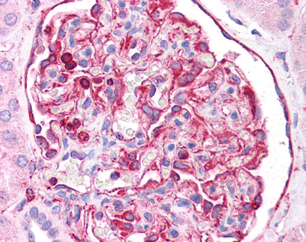
Integrin alpha 5, Polyclonal Antibody (Cat# AAA19156)
Full Name
Anti-Integrin alpha 5 Picoband antibody
Gene Names
ITGA5; FNRA; CD49e; VLA-5; VLA5A
Reactivity
Human, Mouse, Rat
No cross reactivity with other proteins.
No cross reactivity with other proteins.
Applications
EIA, IHC, WB
Pricing


 IHC (Immunohistochemistry)
(Figure 8. IHC analysis of MED18 using anti-MED18 antibody (AAA19179).MED18 was detected in paraffin-embedded section of human mammary cancer tissue. Heat mediated antigen retrieval was performed in citrate buffer (pH6, epitope retrieval solution) for 20 mins. The tissue section was blocked with 10% goat serum. The tissue section was then incubated with 1ug/ml rabbit anti-MED18 Antibody (AAA19179) overnight at 4 degree C. Biotinylated goat anti-rabbit IgG was used as secondary antibody and incubated for 30 minutes at 37 degree C. The tissue section was developed using Strepavidin-Biotin-Complex (SABC) with DAB as the chromogen.)
IHC (Immunohistochemistry)
(Figure 8. IHC analysis of MED18 using anti-MED18 antibody (AAA19179).MED18 was detected in paraffin-embedded section of human mammary cancer tissue. Heat mediated antigen retrieval was performed in citrate buffer (pH6, epitope retrieval solution) for 20 mins. The tissue section was blocked with 10% goat serum. The tissue section was then incubated with 1ug/ml rabbit anti-MED18 Antibody (AAA19179) overnight at 4 degree C. Biotinylated goat anti-rabbit IgG was used as secondary antibody and incubated for 30 minutes at 37 degree C. The tissue section was developed using Strepavidin-Biotin-Complex (SABC) with DAB as the chromogen.)
MED18, Polyclonal Antibody (Cat# AAA19179)
Full Name
Anti-MED18 Picoband Antibody
Gene Names
MED18; SRB5; p28b
Reactivity
Human, Mouse, Rat
No cross reactivity with other proteins.
No cross reactivity with other proteins.
Applications
EIA, IHC, WB
Purity
Immunogen affinity purified
Pricing


 FCM (Flow Cytometry)
(Figure 14. Flow Cytometry analysis of A431 cells using anti-SAMDH1 antibody (AAA19356).Overlay histogram showing A431 cells stained with AAA19356 (Blue line). The cells were blocked with 10% normal goat serum. And then incubated with mouse anti- SAMDH1 Antibody (AAA19356, 1μg/1x106 cells) for 30 min at 20 degree C. DyLight®488 conjugated goat anti-mouse IgG (BA1126, 5-10μg/1x106 cells) was used as secondary antibody for 30 minutes at 20 degree C. Isotype control antibody (Green line) was mouse IgG (1μg/1x106) used under the same conditions. Unlabelled sample (Red line) was also used as a control.)
FCM (Flow Cytometry)
(Figure 14. Flow Cytometry analysis of A431 cells using anti-SAMDH1 antibody (AAA19356).Overlay histogram showing A431 cells stained with AAA19356 (Blue line). The cells were blocked with 10% normal goat serum. And then incubated with mouse anti- SAMDH1 Antibody (AAA19356, 1μg/1x106 cells) for 30 min at 20 degree C. DyLight®488 conjugated goat anti-mouse IgG (BA1126, 5-10μg/1x106 cells) was used as secondary antibody for 30 minutes at 20 degree C. Isotype control antibody (Green line) was mouse IgG (1μg/1x106) used under the same conditions. Unlabelled sample (Red line) was also used as a control.)
SAMHD1, Monoclonal Antibody (Cat# AAA19356)
Full Name
Anti-SAMHD1 Antibody (monoclonal, 10H8)
Gene Names
SAMHD1; DCIP; CHBL2; HDDC1; MOP-5; SBBI88
Reactivity
Human
Applications
WB, IHC-P, ICC, IF, FC/FACS/FCM
Purity
Immunogen affinity purified.
Pricing


 Application Data
(Analysis of Protein Array containing more than 19, 000 full-length human proteins using Mouse Emerin Monoclonal Antibody (EMD/2167) Z- and S- Score: The Z-score represents the strength of a signal that a monoclonal antibody (MAb) (in combination with a fluorescently-tagged anti-IgG secondary antibody) produces when binding to a particular protein on the HuProtTM array. Z-scores are described in units of standard deviations (SD's) above the mean value of all signals generated on that array. If targets on HuProtTM are arranged in descending order of the Z-score, the S-score is the difference (also in units of SD's) between the Z-score. S-score therefore represents the relative target specificity of a MAb to its intended target. A MAb is considered to specific to its intended target, if the MAb has an S-score of at least 2.5. For example, if a MAb binds to protein X with a Z-score of 43 and to protein Y with a Z-score of 14, then the S-score for the binding of that MAb to protein X is equal to 29.)
Application Data
(Analysis of Protein Array containing more than 19, 000 full-length human proteins using Mouse Emerin Monoclonal Antibody (EMD/2167) Z- and S- Score: The Z-score represents the strength of a signal that a monoclonal antibody (MAb) (in combination with a fluorescently-tagged anti-IgG secondary antibody) produces when binding to a particular protein on the HuProtTM array. Z-scores are described in units of standard deviations (SD's) above the mean value of all signals generated on that array. If targets on HuProtTM are arranged in descending order of the Z-score, the S-score is the difference (also in units of SD's) between the Z-score. S-score therefore represents the relative target specificity of a MAb to its intended target. A MAb is considered to specific to its intended target, if the MAb has an S-score of at least 2.5. For example, if a MAb binds to protein X with a Z-score of 43 and to protein Y with a Z-score of 14, then the S-score for the binding of that MAb to protein X is equal to 29.)
Emerin, Monoclonal Antibody (Cat# AAA23898)
Full Name
Emerin (Papillary Thyroid Carcinoma and EDMD Marker)
Gene Names
EMD; STA; EDMD; LEMD5
Reactivity
Human. Others not known.
Applications
Immunohistochemistry
Pricing


 FCM (Flow Cytometry)
(Figure 6. Flow Cytometry analysis of A549 cells using anti-Ch TOG/CKAP5 antibody (AAA19386).Overlay histogram showing A549 cells stained with AAA19386 (Blue line). The cells were blocked with 10% normal goat serum. And then incubated with mouse anti- Ch TOG/CKAP5 Antibody (AAA19386, 1μg/1x106 cells) for 30 min at 20 degree C. DyLight®488 conjugated goat anti-mouse IgG (BA1126, 5-10μg/1x106 cells) was used as secondary antibody for 30 minutes at 20 degree C. Isotype control antibody (Green line) was mouse IgG (1μg/1x106) used under the same conditions. Unlabelled sample (Red line) was also used as a control.)
FCM (Flow Cytometry)
(Figure 6. Flow Cytometry analysis of A549 cells using anti-Ch TOG/CKAP5 antibody (AAA19386).Overlay histogram showing A549 cells stained with AAA19386 (Blue line). The cells were blocked with 10% normal goat serum. And then incubated with mouse anti- Ch TOG/CKAP5 Antibody (AAA19386, 1μg/1x106 cells) for 30 min at 20 degree C. DyLight®488 conjugated goat anti-mouse IgG (BA1126, 5-10μg/1x106 cells) was used as secondary antibody for 30 minutes at 20 degree C. Isotype control antibody (Green line) was mouse IgG (1μg/1x106) used under the same conditions. Unlabelled sample (Red line) was also used as a control.)
ch TOG/CKAP5, Monoclonal Antibody (Cat# AAA19386)
Full Name
Anti-ch TOG/CKAP5 Antibody (monoclonal, 3C13)
Gene Names
CKAP5; TOG; MSPS; TOGp; CHTOG; ch-TOG
Reactivity
Human, Mouse, Rat
Applications
WB, IHC-P, ICC, IF, FC/FACS/FCM
Purity
Immunogen affinity purified.
Pricing


 WB (Western Blot)
(WB Suggested Anti-FOLR1 antibody Titration: 1 ug/mLSample Type: Human liver)
WB (Western Blot)
(WB Suggested Anti-FOLR1 antibody Titration: 1 ug/mLSample Type: Human liver)
FOLR1, Polyclonal Antibody (Cat# AAA23491)
Full Name
FOLR1 antibody - middle region
Gene Names
FOLR1; FBP; FOLR
Reactivity
Cow, Dog, Guinea Pig, Horse, Human, Mouse, Rabbit, Rat
Applications
WB, IHC
Purity
Affinity Purified
Pricing


 IF (Immunofluorescence)
(Immunofluorescence analysis of HeLa cells using TOMM20 antibody at dilution of 1:100 (40x lens). Blue: DAPI for nuclear staining.)
IF (Immunofluorescence)
(Immunofluorescence analysis of HeLa cells using TOMM20 antibody at dilution of 1:100 (40x lens). Blue: DAPI for nuclear staining.)
TOMM20, Polyclonal Antibody (Cat# AAA28123)
Full Name
TOMM20 Polyclonal Antibody
Gene Names
TOMM20; MAS20; MOM19; TOM20
Reactivity
Human, Mouse, Rat
Applications
WB, IHC, IF, IP
Purity
Affinity Purification
Pricing


 FCM (Flow Cytometry)
(Figure 6. Flow Cytometry analysis of SiHa cells using anti- Aldolase/ALDOA antibody (AAA19385).Overlay histogram showing SiHa cells stained with AAA19385 (Blue line). The cells were blocked with 10% normal goat serum. And then incubated with mouse anti-Aldolase/ALDOA Antibody (AAA19385, 1μg/1x106 cells) for 30 min at 20 degree C. DyLight®488 conjugated goat anti-mouse IgG (BA1126, 5-10μg/1x106 cells) was used as secondary antibody for 30 minutes at 20 degree C. Isotype control antibody (Green line) was mouse IgG (1μg/1x106) used under the same conditions. Unlabelled sample (Red line) was also used as a control.)
FCM (Flow Cytometry)
(Figure 6. Flow Cytometry analysis of SiHa cells using anti- Aldolase/ALDOA antibody (AAA19385).Overlay histogram showing SiHa cells stained with AAA19385 (Blue line). The cells were blocked with 10% normal goat serum. And then incubated with mouse anti-Aldolase/ALDOA Antibody (AAA19385, 1μg/1x106 cells) for 30 min at 20 degree C. DyLight®488 conjugated goat anti-mouse IgG (BA1126, 5-10μg/1x106 cells) was used as secondary antibody for 30 minutes at 20 degree C. Isotype control antibody (Green line) was mouse IgG (1μg/1x106) used under the same conditions. Unlabelled sample (Red line) was also used as a control.)
Aldolase/ALDOA, Monoclonal Antibody (Cat# AAA19385)
Full Name
Anti-Aldolase/ALDOA Antibody (monoclonal, 6H8)
Gene Names
ALDOA; ALDA; GSD12
Reactivity
Human
Applications
WB, IHC-P, ICC, IF, FC/FACS/FCM
Purity
Immunogen affinity purified.
Pricing


 FCM (Flow Cytometry)
(Figure 7. Flow Cytometry analysis of U20S cells using anti-VDAC3 antibody (AAA19301).Overlay histogram showing U20S cells stained with AAA19301 (Blue line). The cells were blocked with 10% normal goat serum. And then incubated with rabbit anti-VDAC3 Antibody (AAA19301, 1μg/1x106 cells) for 30 min at 20 degree C. DyLight®488 conjugated goat anti-rabbit IgG (5-10μg/1x106 cells) was used as secondary antibody for 30 minutes at 20 degree C. Isotype control antibody (Green line) was rabbit IgG (1μg/1x106) used under the same conditions. Unlabelled sample (Red line) was also used as a control.)
FCM (Flow Cytometry)
(Figure 7. Flow Cytometry analysis of U20S cells using anti-VDAC3 antibody (AAA19301).Overlay histogram showing U20S cells stained with AAA19301 (Blue line). The cells were blocked with 10% normal goat serum. And then incubated with rabbit anti-VDAC3 Antibody (AAA19301, 1μg/1x106 cells) for 30 min at 20 degree C. DyLight®488 conjugated goat anti-rabbit IgG (5-10μg/1x106 cells) was used as secondary antibody for 30 minutes at 20 degree C. Isotype control antibody (Green line) was rabbit IgG (1μg/1x106) used under the same conditions. Unlabelled sample (Red line) was also used as a control.)
VDAC3, Polyclonal Antibody (Cat# AAA19301)
Full Name
Anti-VDAC3 Antibody
Gene Names
VDAC3; VDAC-3; HD-VDAC3
Reactivity
Human, Mouse, Rat
Applications
WB, IHC-P, FC/FACS/FCM, EIA
Purity
Immunogen affinity purified.
Pricing


 FCM (Flow Cytometry)
(Figure 12. Flow Cytometry analysis of HEPA1-6 cells using anti-LSM5 antibody (AAA19332).Overlay histogram showing HEPA1-6 cells stained with AAA19332 (Blue line). The cells were blocked with 10% normal goat serum. And then incubated with rabbit anti-LSM5 Antibody (AAA19332, 1μg/1x106 cells) for 30 min at 20 degree C. DyLight®488 conjugated goat anti-rabbit IgG (5-10μg/1x106 cells) was used as secondary antibody for 30 minutes at 20 degree C. Isotype control antibody (Green line) was rabbit IgG (1μg/1x106) used under the same conditions. Unlabelled sample (Red line) was also used as a control.)
FCM (Flow Cytometry)
(Figure 12. Flow Cytometry analysis of HEPA1-6 cells using anti-LSM5 antibody (AAA19332).Overlay histogram showing HEPA1-6 cells stained with AAA19332 (Blue line). The cells were blocked with 10% normal goat serum. And then incubated with rabbit anti-LSM5 Antibody (AAA19332, 1μg/1x106 cells) for 30 min at 20 degree C. DyLight®488 conjugated goat anti-rabbit IgG (5-10μg/1x106 cells) was used as secondary antibody for 30 minutes at 20 degree C. Isotype control antibody (Green line) was rabbit IgG (1μg/1x106) used under the same conditions. Unlabelled sample (Red line) was also used as a control.)
LSM5, Polyclonal Antibody (Cat# AAA19332)
Full Name
Anti-LSM5 Antibody
Gene Names
LSM5; YER146W
Reactivity
Human, Mouse, Rat
Applications
WB, IHC-P, ICC, IF, FC/FACS/FCM, EIA
Purity
Immunogen affinity purified.
Pricing


 FCM (Flow Cytometry)
(Figure 8. Flow Cytometry analysis of U20S cells using anti-Dynamin 1 antibody (AAA19160).Overlay histogram showing U20S cells stained with AAA19160 (Blue line).The cells were blocked with 10% normal goat serum. And then incubated with rabbit anti-DNM1 Antibody (AAA19160,1ug/1x10^6 cells) for 30 min at 20 degree C. DyLight®488 conjugated goat anti-rabbit IgG (5-10ug/1x10^6 cells) was used as secondary antibody for 30 minutes at 20 degree C. Isotype control antibody (Green line) was rabbit IgG (1ug/1x106) used under the same conditions. Unlabelled sample (Red line) was also used as a control.)
FCM (Flow Cytometry)
(Figure 8. Flow Cytometry analysis of U20S cells using anti-Dynamin 1 antibody (AAA19160).Overlay histogram showing U20S cells stained with AAA19160 (Blue line).The cells were blocked with 10% normal goat serum. And then incubated with rabbit anti-DNM1 Antibody (AAA19160,1ug/1x10^6 cells) for 30 min at 20 degree C. DyLight®488 conjugated goat anti-rabbit IgG (5-10ug/1x10^6 cells) was used as secondary antibody for 30 minutes at 20 degree C. Isotype control antibody (Green line) was rabbit IgG (1ug/1x106) used under the same conditions. Unlabelled sample (Red line) was also used as a control.)
Dynamin 1, Polyclonal Antibody (Cat# AAA19160)
Full Name
Anti-Dynamin 1 Picoband antibody
Gene Names
DNM1; DNM; EIEE31
Reactivity
Human, Mouse, Rat
No cross reactivity with other proteins.
No cross reactivity with other proteins.
Applications
EIA, FC/FACS, IHC, ICC, WB
Pricing


 FCM (Flow Cytometry)
(Figure 7. Flow Cytometry analysis of Hela cells using anti- ETF/TEAD2 antibody (AAA19325).Overlay histogram showing Hela cells stained withAAA19325 (Blue line). The cells were blocked with 10% normal goat serum. And then incubated with rabbit anti- ETF/TEAD2 Antibody (AAA19325, 1μg/1x106 cells) for 30 min at 20 degree C. DyLight®488 conjugated goat anti-rabbit IgG (5-10μg/1x106 cells) was used as secondary antibody for 30 minutes at 20 degree C. Isotype control antibody (Green line) was rabbit IgG (1μg/1x106) used under the same conditions. Unlabelled sample (Red line) was also used as a control.)
FCM (Flow Cytometry)
(Figure 7. Flow Cytometry analysis of Hela cells using anti- ETF/TEAD2 antibody (AAA19325).Overlay histogram showing Hela cells stained withAAA19325 (Blue line). The cells were blocked with 10% normal goat serum. And then incubated with rabbit anti- ETF/TEAD2 Antibody (AAA19325, 1μg/1x106 cells) for 30 min at 20 degree C. DyLight®488 conjugated goat anti-rabbit IgG (5-10μg/1x106 cells) was used as secondary antibody for 30 minutes at 20 degree C. Isotype control antibody (Green line) was rabbit IgG (1μg/1x106) used under the same conditions. Unlabelled sample (Red line) was also used as a control.)
ETF/TEAD2, Polyclonal Antibody (Cat# AAA19325)
Full Name
Anti-ETF/TEAD2 Antibody
Gene Names
TEAD2; ETF; TEF4; TEF-4; TEAD-2
Reactivity
Human, Mouse, Rat
Applications
WB, IHC-P, FC/FACS/FCM, EIA
Purity
Immunogen affinity purified.
Pricing


 FCM (Flow Cytometry)
(Figure 6. Flow Cytometry analysis of A431 cells using anti-NDUFB5 antibody (AAA19333).Overlay histogram showing A431 cells stained with AAA19333 (Blue line). The cells were blocked with 10% normal goat serum. And then incubated with rabbit anti-NDUFB5 Antibody (AAA19333, 1μg/1x106 cells) for 30 min at 20 degree C. DyLight®488 conjugated goat anti-rabbit IgG (5-10μg/1x106 cells) was used as secondary antibody for 30 minutes at 20 degree C. Isotype control antibody (Green line) was rabbit IgG (1μg/1x106) used under the same conditions. Unlabelled sample (Red line) was also used as a control.)
FCM (Flow Cytometry)
(Figure 6. Flow Cytometry analysis of A431 cells using anti-NDUFB5 antibody (AAA19333).Overlay histogram showing A431 cells stained with AAA19333 (Blue line). The cells were blocked with 10% normal goat serum. And then incubated with rabbit anti-NDUFB5 Antibody (AAA19333, 1μg/1x106 cells) for 30 min at 20 degree C. DyLight®488 conjugated goat anti-rabbit IgG (5-10μg/1x106 cells) was used as secondary antibody for 30 minutes at 20 degree C. Isotype control antibody (Green line) was rabbit IgG (1μg/1x106) used under the same conditions. Unlabelled sample (Red line) was also used as a control.)
NDUFB5, Polyclonal Antibody (Cat# AAA19333)
Full Name
Anti-NDUFB5 Antibody
Gene Names
NDUFB5; SGDH; CISGDH
Reactivity
Human, Mouse, Rat
Applications
WB, IHC-P, ICC, IF, FC/FACS/FCM, EIA
Purity
Immunogen affinity purified.
Pricing


 IHC (Immunohistochemistry)
(Figure 7. IHC analysis of VEGF Receptor 3 using anti-VEGF Receptor 3 antibody (AAA19147).VEGF Receptor 3 was detected in paraffin-embedded section of mouse liver tissue. Heat mediated antigen retrieval was performed in citrate buffer (pH6, epitope retrieval solution) for 20 mins. The tissue section was blocked with 10% goat serum. The tissue section was then incubated with 1ug/ml rabbit anti-VEGF Receptor 3 Antibody (AAA19147) overnight at 4 degree C. Biotinylated goat anti-rabbit IgG was used as secondary antibody and incubated for 30 minutes at 37 degree C. The tissue section was developed using Strepavidin-Biotin-Complex (SABC) with DAB as the chromogen.)
IHC (Immunohistochemistry)
(Figure 7. IHC analysis of VEGF Receptor 3 using anti-VEGF Receptor 3 antibody (AAA19147).VEGF Receptor 3 was detected in paraffin-embedded section of mouse liver tissue. Heat mediated antigen retrieval was performed in citrate buffer (pH6, epitope retrieval solution) for 20 mins. The tissue section was blocked with 10% goat serum. The tissue section was then incubated with 1ug/ml rabbit anti-VEGF Receptor 3 Antibody (AAA19147) overnight at 4 degree C. Biotinylated goat anti-rabbit IgG was used as secondary antibody and incubated for 30 minutes at 37 degree C. The tissue section was developed using Strepavidin-Biotin-Complex (SABC) with DAB as the chromogen.)
VEGF Receptor 3, Polyclonal Antibody (Cat# AAA19147)
Full Name
Anti-VEGF Receptor 3 Picoband antibody
Gene Names
FLT4; PCL; FLT-4; FLT41; LMPH1A; VEGFR3; VEGFR-3
Reactivity
Human, Mouse, Rat
Applications
EIA, IHC, WB, FC
Purity
Immunoggen affinity purified
Pricing


 FCM (Flow Cytometry)
(Figure 11. Flow Cytometry analysis of THP-1 cells using anti-CDC123 antibody (AAA19323).Overlay histogram showing THP-1 cells stained with AAA19323 (Blue line). The cells were blocked with 10% normal goat serum. And then incubated with rabbit anti-CDC123 Antibody (AAA19323, 1μg/1x106 cells) for 30 min at 20 degree C. DyLight®488 conjugated goat anti-rabbit IgG (5-10μg/1x106 cells) was used as secondary antibody for 30 minutes at 20 degree C. Isotype control antibody (Green line) was rabbit IgG (1μg/1x106) used under the same conditions. Unlabelled sample (Red line) was also used as a control.)
FCM (Flow Cytometry)
(Figure 11. Flow Cytometry analysis of THP-1 cells using anti-CDC123 antibody (AAA19323).Overlay histogram showing THP-1 cells stained with AAA19323 (Blue line). The cells were blocked with 10% normal goat serum. And then incubated with rabbit anti-CDC123 Antibody (AAA19323, 1μg/1x106 cells) for 30 min at 20 degree C. DyLight®488 conjugated goat anti-rabbit IgG (5-10μg/1x106 cells) was used as secondary antibody for 30 minutes at 20 degree C. Isotype control antibody (Green line) was rabbit IgG (1μg/1x106) used under the same conditions. Unlabelled sample (Red line) was also used as a control.)
CDC123, Polyclonal Antibody (Cat# AAA19323)
Full Name
Anti-CDC123 Antibody
Gene Names
CDC123; D123; C10orf7
Reactivity
Human
Applications
WB, IHC-P, ICC, IF, FC/FACS/FCM, EIA
Purity
Immunogen affinity purified.
Pricing


 FCM (Flow Cytometry)
(Figure 7. Flow Cytometry analysis of U87 cells using anti-MPI antibody (AAA19349).Overlay histogram showing U87 cells stained with AAA19349 (Blue line). The cells were blocked with 10% normal goat serum. And then incubated with mouse anti- MPI Antibody (AAA19349, 1μg/1x106 cells) for 30 min at 20 degree C. DyLight®488 conjugated goat anti-mouse IgG (BA1126, 5-10μg/1x106 cells) was used as secondary antibody for 30 minutes at 20 degree C. Isotype control antibody (Green line) was mouse IgG (1μg/1x106) used under the same conditions. Unlabelled sample (Red line) was also used as a control.)
FCM (Flow Cytometry)
(Figure 7. Flow Cytometry analysis of U87 cells using anti-MPI antibody (AAA19349).Overlay histogram showing U87 cells stained with AAA19349 (Blue line). The cells were blocked with 10% normal goat serum. And then incubated with mouse anti- MPI Antibody (AAA19349, 1μg/1x106 cells) for 30 min at 20 degree C. DyLight®488 conjugated goat anti-mouse IgG (BA1126, 5-10μg/1x106 cells) was used as secondary antibody for 30 minutes at 20 degree C. Isotype control antibody (Green line) was mouse IgG (1μg/1x106) used under the same conditions. Unlabelled sample (Red line) was also used as a control.)
MPI, Monoclonal Antibody (Cat# AAA19349)
Full Name
Anti-MPI Antibody (monoclonal, 5G5)
Gene Names
MPI; PMI; PMI1; CDG1B
Reactivity
Human, Rat
Applications
WB, IHC-P, ICC, IF, FC/FACS/FCM
Purity
Immunogen affinity purified.
Pricing