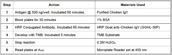
Filters
Clonality
Type
Reactivity
Gene Name
Isotype
Host
Application
Clone
2458 results for "Control Antibody" - showing 2200-2250
 WB (Western Blot)
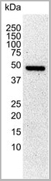
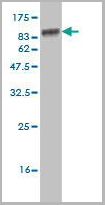
(Positive WB detected in Recombinant proteinAll lanes: BZLF1 antibody at 1:1000SecondaryGoat polyclonal to rabbit IgG at 1/50000 dilutionPredicted band size: 46 kDaObserved band size: 50 kDa)
WB (Western Blot)
(Positive WB detected in Recombinant proteinAll lanes: BZLF1 antibody at 1:1000SecondaryGoat polyclonal to rabbit IgG at 1/50000 dilutionPredicted band size: 46 kDaObserved band size: 50 kDa)
BZLF1, Polyclonal Antibody (Cat# AAA27059)
Full Name
Rabbit anti-Epstein-Barr virus (strain B95-8)(HHV-4)(Human herpesvirus 4) BZLF1 Polyclonal Antibody
Reactivity
Epstein-Barr virus
Applications
Western Blot
Purity
> 95%, Protein G purified
Pricing
S100A9, Monoclonal Antibody (Cat# AAA13837)
Full Name
S100A9 (Macrophage Marker) Mouse Monoclonal Antibody
Gene Names
S100A9; MIF; NIF; P14; CAGB; CFAG; CGLB; L1AG; LIAG; MRP14; 60B8AG; MAC387
Reactivity
Human and Rat. Others not tested.
Applications
Flow Cytometry, Immunofluorescence, Immunohistochemistry
Pricing
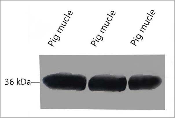
 WB (Western Blot)
(Western blot analysis of ZO-1 using various lysatesLanes 1 - 2: Merged signal (red and green). Green - AAA31075 observed at 195 kDa. Red - loading control, , observed at 55 kDa. Blots were developed with Goat Anti- Rabbit IgG(H+L) FITC–conjugated and Goat Anti-Mouse IgG(H+L) Alexa Fluor 594–conjugated secondary antibodies)
WB (Western Blot)
(Western blot analysis of ZO-1 using various lysatesLanes 1 - 2: Merged signal (red and green). Green - AAA31075 observed at 195 kDa. Red - loading control, , observed at 55 kDa. Blots were developed with Goat Anti- Rabbit IgG(H+L) FITC–conjugated and Goat Anti-Mouse IgG(H+L) Alexa Fluor 594–conjugated secondary antibodies)
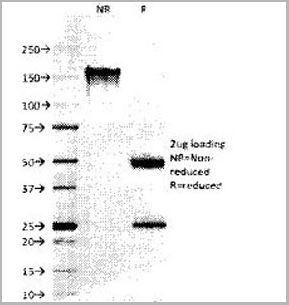
ZO 1, Polyclonal Antibody (Cat# AAA31075)
Full Name
ZO 1 Antibody
Gene Names
TJP1; ZO-1
Reactivity
Human, Mouse, Rat, Pig, Monkey
Applications
Western Blot, Immunohistochemistry, Immunofluorescence, Immunocytochemistry
Purity
The antiserum was purified by peptide affinity chromatography using SulfoLink Coupling Resin
Pricing
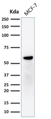 WB (Western Blot)
(Western Blot Analysis of human MCF-7 cell lysate using ER-beta1 Mouse Monoclonal Antibody (ERb455).)
WB (Western Blot)
(Western Blot Analysis of human MCF-7 cell lysate using ER-beta1 Mouse Monoclonal Antibody (ERb455).)
ER-beta1 (Estrogen Receptor beta-1), Monoclonal Antibody (Cat# AAA13814)
Full Name
ER-beta1 (Estrogen Receptor beta-1) Mouse Monoclonal Antibody
Gene Names
ESR2; Erb; ESRB; ESTRB; NR3A2; ER-BETA; ESR-BETA
Reactivity
Human
Applications
Flow Cytometry, Immunofluorescence, Western Blot, Immunohistochemistry
Pricing
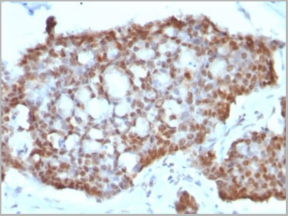
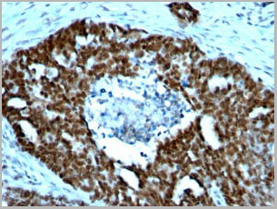
 Application Data
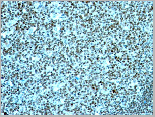
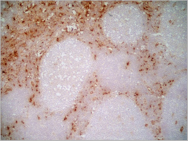
(Immunoperoxidase staining of human tonsil cryosection using Mouse anti Human CD1a antibody followed by HISTAR detection system. Medium power)
Application Data
(Immunoperoxidase staining of human tonsil cryosection using Mouse anti Human CD1a antibody followed by HISTAR detection system. Medium power)
CD1a, Monoclonal Antibody (Cat# AAA12011)
Full Name
MOUSE ANTI HUMAN CD1a
Gene Names
CD1A; R4; T6; CD1; FCB6; HTA1
Reactivity
Cynomolgus monkey, Dog
Applications
Immunohistochemistry, Flow Cytometry
Pricing


 Application Data
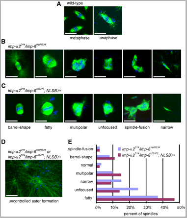
(Published customer image: Spindle abnormalities in embryos derived from imp-a2D14/imp-betaKetRE34 and imp-a2D14/imp-betac02473; NLSB-/+ females. (A -D) Wild-type and mutant embryos stained for a-tubulin (green) and DNA (blue). (A) Mitotic spindles in wild-type embryos at metaphase and anaphase. (B, C) Categories of spindle abnormalities found in embryos derived from (B) imp-a2D14/imp-betaKetRE34 and (C) imp-a2D14/imp-betac02743; NLSB-/+ females. (D) Formation of aster networks found in both genotypes. Scale bar: 10 um. (E) Frequency of spindle defects in embryos from both types of mutant females. Female genotypes are displayed at the upper right corner. At least 200 spindles were scored for both genotypes.From: Specific Cooperation Between Imp-a2 and Imp-beta/Ketel in Spindle Assembly During Drosophila Early Nuclear Divisions Erika Vir¡gh, M¡ty¡s Gorj¡n¡cz, Istv¡n T¶r¶k, Tolga Eichhorn, Sowjanya Kallakuri, Tam¡s Szlanka, Istv¡n Kiss, and Bernard M. Mechler G3 January 2012 2:1-14.)
Application Data
(Published customer image: Spindle abnormalities in embryos derived from imp-a2D14/imp-betaKetRE34 and imp-a2D14/imp-betac02473; NLSB-/+ females. (A -D) Wild-type and mutant embryos stained for a-tubulin (green) and DNA (blue). (A) Mitotic spindles in wild-type embryos at metaphase and anaphase. (B, C) Categories of spindle abnormalities found in embryos derived from (B) imp-a2D14/imp-betaKetRE34 and (C) imp-a2D14/imp-betac02743; NLSB-/+ females. (D) Formation of aster networks found in both genotypes. Scale bar: 10 um. (E) Frequency of spindle defects in embryos from both types of mutant females. Female genotypes are displayed at the upper right corner. At least 200 spindles were scored for both genotypes.From: Specific Cooperation Between Imp-a2 and Imp-beta/Ketel in Spindle Assembly During Drosophila Early Nuclear Divisions Erika Vir¡gh, M¡ty¡s Gorj¡n¡cz, Istv¡n T¶r¶k, Tolga Eichhorn, Sowjanya Kallakuri, Tam¡s Szlanka, Istv¡n Kiss, and Bernard M. Mechler G3 January 2012 2:1-14.)
TUBULIN ALPHA, Monoclonal Antibody (Cat# AAA12232)
Full Name
RAT ANTI TUBULIN ALPHA:HRP
Applications
Immunohistochemistry, Western Blot
Pricing


 Application Data
(Published customer image: Spindle abnormalities in embryos derived from imp-a2D14/imp-betaKetRE34 and imp-a2D14/imp-betac02473; NLSB-/+ females. (A -D) Wild-type and mutant embryos stained for a-tubulin (green) and DNA (blue). (A) Mitotic spindles in wild-type embryos at metaphase and anaphase. (B, C) Categories of spindle abnormalities found in embryos derived from (B) imp-a2D14/imp-betaKetRE34 and (C) imp-a2D14/imp-betac02743; NLSB-/+ females. (D) Formation of aster networks found in both genotypes. Scale bar: 10 um. (E) Frequency of spindle defects in embryos from both types of mutant females. Female genotypes are displayed at the upper right corner. At least 200 spindles were scored for both genotypes.From: Specific Cooperation Between Imp-a2 and Imp-beta/Ketel in Spindle Assembly During Drosophila Early Nuclear Divisions Erika Vir¡gh, M¡ty¡s Gorj¡n¡cz, Istv¡n T¶r¶k, Tolga Eichhorn, Sowjanya Kallakuri, Tam¡s Szlanka, Istv¡n Kiss, and Bernard M. Mechler G3 January 2012 2:1-14.)
Application Data
(Published customer image: Spindle abnormalities in embryos derived from imp-a2D14/imp-betaKetRE34 and imp-a2D14/imp-betac02473; NLSB-/+ females. (A -D) Wild-type and mutant embryos stained for a-tubulin (green) and DNA (blue). (A) Mitotic spindles in wild-type embryos at metaphase and anaphase. (B, C) Categories of spindle abnormalities found in embryos derived from (B) imp-a2D14/imp-betaKetRE34 and (C) imp-a2D14/imp-betac02743; NLSB-/+ females. (D) Formation of aster networks found in both genotypes. Scale bar: 10 um. (E) Frequency of spindle defects in embryos from both types of mutant females. Female genotypes are displayed at the upper right corner. At least 200 spindles were scored for both genotypes.From: Specific Cooperation Between Imp-a2 and Imp-beta/Ketel in Spindle Assembly During Drosophila Early Nuclear Divisions Erika Vir¡gh, M¡ty¡s Gorj¡n¡cz, Istv¡n T¶r¶k, Tolga Eichhorn, Sowjanya Kallakuri, Tam¡s Szlanka, Istv¡n Kiss, and Bernard M. Mechler G3 January 2012 2:1-14.)
TUBULIN ALPHA, Monoclonal Antibody (Cat# AAA12009)
Full Name
RAT ANTI TUBULIN ALPHA
Applications
Immunohistochemistry, Immunofluorescence, Immunoprecipitation, Radioimmunoassay, Western Blot
Pricing


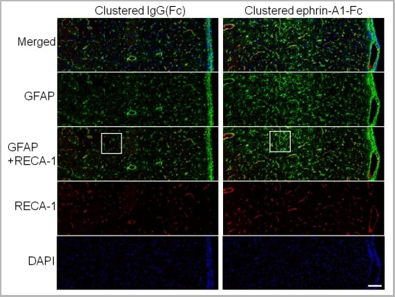
 Application Data
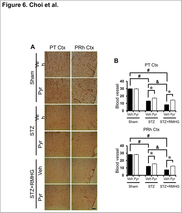
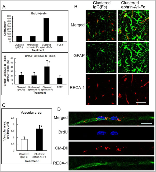
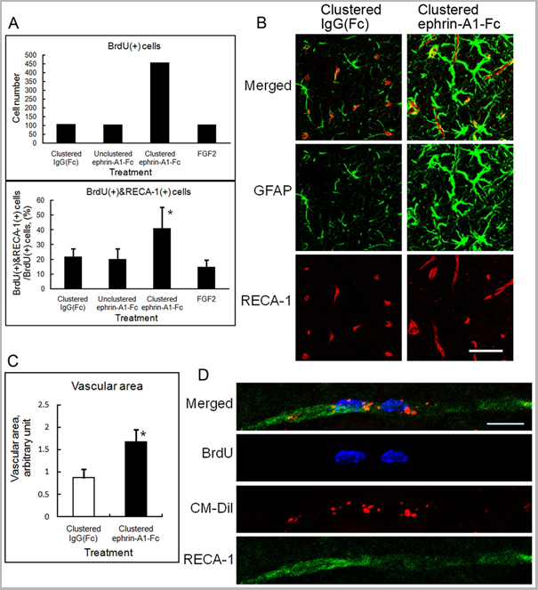
(Published customer image: Effect of clustered ephrin-A1-Fc on vascular formation in the rat striatum. Clustered ephrin-A1-Fc was injected into the lesioned side of the lateral ventricle in the unilaterally lesioned rats. Brains taken 6 weeks after injection were sectioned coronally and stained for GFAP (green) and RECA-1 (red) and with DAPI (nuclei; blue). The rectangular insets are shown in Fig. 8B. Scale bar: 100 um.From: Jing X, Miwa H, Sawada T, Nakanishi I, Kondo T, et al. (2012) Ephrin-A1-Mediated Dopaminergic Neurogenesis and Angiogenesis in a Rat Model of Parkinson's Disease. PLoS ONE 7(2): e32019.)
Application Data
(Published customer image: Effect of clustered ephrin-A1-Fc on vascular formation in the rat striatum. Clustered ephrin-A1-Fc was injected into the lesioned side of the lateral ventricle in the unilaterally lesioned rats. Brains taken 6 weeks after injection were sectioned coronally and stained for GFAP (green) and RECA-1 (red) and with DAPI (nuclei; blue). The rectangular insets are shown in Fig. 8B. Scale bar: 100 um.From: Jing X, Miwa H, Sawada T, Nakanishi I, Kondo T, et al. (2012) Ephrin-A1-Mediated Dopaminergic Neurogenesis and Angiogenesis in a Rat Model of Parkinson's Disease. PLoS ONE 7(2): e32019.)
RECA-1, Monoclonal Antibody (Cat# AAA12018)
Full Name
MOUSE ANTI RAT RECA-1
Applications
Immunohistochemistry, Immunofluorescence
Pricing
 Application Data
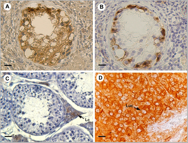
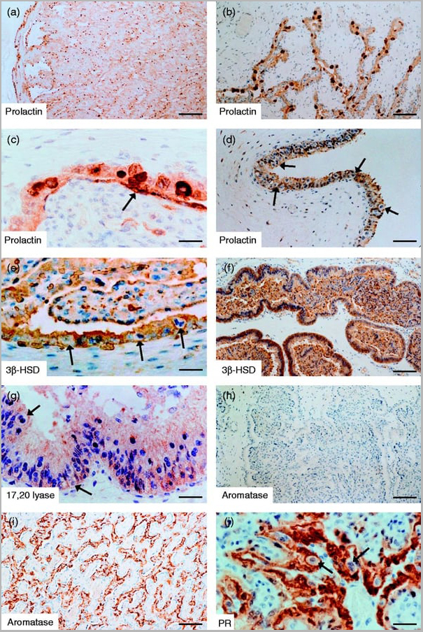
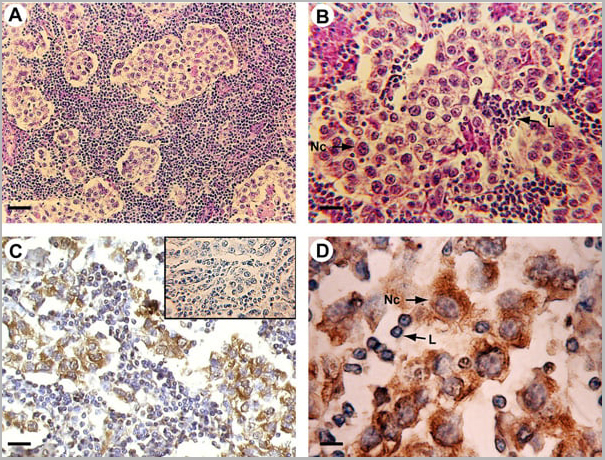
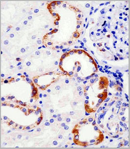
(Published customer image: Mouse anti Human cytochrome p450 aromatase antibody, clone H4 used for the detaction of aromatase in human tissues by Immunohistochemistry on paraffin sectionsImage caption:Morphology and P450 arom immunoreactivity of tumoral region in human testis with seminoma. A-B: Haematoxylin-eosin staining. C-D: Strong P450 arom immunoreactivity in cytoplasm of neoplastic cells (Nc) and unstained lymphocytes (L). Insert: absorption control. Scale bars: A, 20 um; B-C, 12.5 um; D, 5 um.From: Rago V, Romeo F, Aquila S, Montanaro D, And S, Carpino A. Cytochrome P450 aromatase expression in human seminoma. Reprod Biol Endocrinol. 2005 Dec 22;3:72.)
Application Data
(Published customer image: Mouse anti Human cytochrome p450 aromatase antibody, clone H4 used for the detaction of aromatase in human tissues by Immunohistochemistry on paraffin sectionsImage caption:Morphology and P450 arom immunoreactivity of tumoral region in human testis with seminoma. A-B: Haematoxylin-eosin staining. C-D: Strong P450 arom immunoreactivity in cytoplasm of neoplastic cells (Nc) and unstained lymphocytes (L). Insert: absorption control. Scale bars: A, 20 um; B-C, 12.5 um; D, 5 um.From: Rago V, Romeo F, Aquila S, Montanaro D, And S, Carpino A. Cytochrome P450 aromatase expression in human seminoma. Reprod Biol Endocrinol. 2005 Dec 22;3:72.)
CYTOCHROME P450 AROMATASE, Monoclonal Antibody (Cat# AAA12113)
Full Name
MOUSE ANTI HUMAN CYTOCHROME P450 AROMATASE
Gene Names
CYP19A1; ARO; ARO1; CPV1; CYAR; CYP19; CYPXIX; P-450AROM
Applications
Immunofluorescence, Immunohistochemistry, Western Blot
Pricing


 Application Data
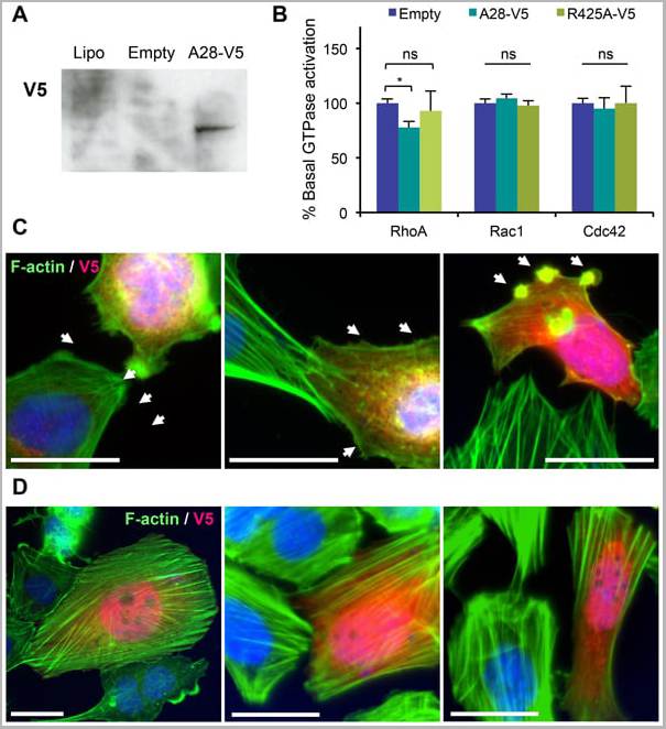
(Published customer image: Mouse anti V5 tag antibody, clone SV5-Pk1 used for the detection of V5 tagged WEEV_nsP3 protein by western blotting and immunofluorescenceImage caption: WEEV nsP3 interaction with host IKKbeta. A) U87MGs were transfected in a 6-well plate with 5 ug of pUC19 and WEEV_nsP3_HA for 24 hours. Cell lysates were resolved using SDS-PAGE and subsequently immunoblotted with V5 antibody and beta-actin served as a loading control. B) U87MGs were transfected with WEEV_nsP3_V5; cells were fixed after 24 hours and stained with antibodies against the endogenous IKKbeta and the V5 tag. Cells were incubated with appropriate secondary Alexa Fluor antibodies and the nuclei stained with DAPI. Co-localization of IKKbeta with WEEV_nsP3_V5 (yellow) was observed as shown by the arrows. B) Panels E -H serve as an example of transfected cells in a given field of view that show co-localization of IKKbeta and WEEV_nsP3_V5 24 hours post transfection. Panels I-L represent magnified images of other cells showing co-localization of IKKbeta and WEEV_nsP3_V5. Panel M is a magnified image of panel L. The co-localization was confirmed by Z-stack analysis. Co-localization was calculated to be approximately in 61% of cells (163 cells were counted of which 44% demonstrated expression of nsP3. Of those cells that expressed nsP3, 61% showed co-localization of both proteins). Images were taken using Nikon Eclipse TE2000-U at 60x magnification and are representative of 2 independent experiments.From: Amaya M, Voss K, Sampey G, Senina S, de la Fuente C, et al. (2014) The Role of IKKbeta in Venezuelan Equine Encephalitis Virus Infection. PLoS ONE 9(2): e86745.)
Application Data
(Published customer image: Mouse anti V5 tag antibody, clone SV5-Pk1 used for the detection of V5 tagged WEEV_nsP3 protein by western blotting and immunofluorescenceImage caption: WEEV nsP3 interaction with host IKKbeta. A) U87MGs were transfected in a 6-well plate with 5 ug of pUC19 and WEEV_nsP3_HA for 24 hours. Cell lysates were resolved using SDS-PAGE and subsequently immunoblotted with V5 antibody and beta-actin served as a loading control. B) U87MGs were transfected with WEEV_nsP3_V5; cells were fixed after 24 hours and stained with antibodies against the endogenous IKKbeta and the V5 tag. Cells were incubated with appropriate secondary Alexa Fluor antibodies and the nuclei stained with DAPI. Co-localization of IKKbeta with WEEV_nsP3_V5 (yellow) was observed as shown by the arrows. B) Panels E -H serve as an example of transfected cells in a given field of view that show co-localization of IKKbeta and WEEV_nsP3_V5 24 hours post transfection. Panels I-L represent magnified images of other cells showing co-localization of IKKbeta and WEEV_nsP3_V5. Panel M is a magnified image of panel L. The co-localization was confirmed by Z-stack analysis. Co-localization was calculated to be approximately in 61% of cells (163 cells were counted of which 44% demonstrated expression of nsP3. Of those cells that expressed nsP3, 61% showed co-localization of both proteins). Images were taken using Nikon Eclipse TE2000-U at 60x magnification and are representative of 2 independent experiments.From: Amaya M, Voss K, Sampey G, Senina S, de la Fuente C, et al. (2014) The Role of IKKbeta in Venezuelan Equine Encephalitis Virus Infection. PLoS ONE 9(2): e86745.)
V5-TAG, Monoclonal Antibody (Cat# AAA12081)
Full Name
MOUSE ANTI V5-TAG:HRP
Applications
Western Blot
Pricing
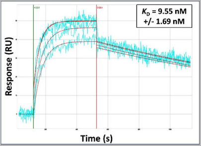 Application Data
(Surface Plasmon Resonance Kinetic Characterization of Polyclonal Antibody Affinity. Purified polyclonal antibodies were immobilized on a Protein A/G coated Carterra LSA sensor chip (PAGH200M) at concentrations of 5, and 50 ug/mL in duplicate. Antibodies on the surface were exposed to interaction with peptides sequentially via microfluidic controlled flow at 333nM peptide concentration for kinetic characterization of the binders for affinity and specificity, followed by curve fitting using the Kinetics software. Kd determinations for both concentrations were averaged and results and standard deviation are shown.)
Application Data
(Surface Plasmon Resonance Kinetic Characterization of Polyclonal Antibody Affinity. Purified polyclonal antibodies were immobilized on a Protein A/G coated Carterra LSA sensor chip (PAGH200M) at concentrations of 5, and 50 ug/mL in duplicate. Antibodies on the surface were exposed to interaction with peptides sequentially via microfluidic controlled flow at 333nM peptide concentration for kinetic characterization of the binders for affinity and specificity, followed by curve fitting using the Kinetics software. Kd determinations for both concentrations were averaged and results and standard deviation are shown.)
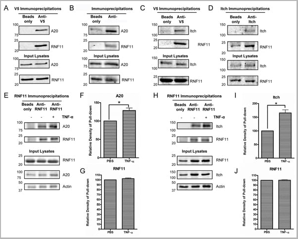
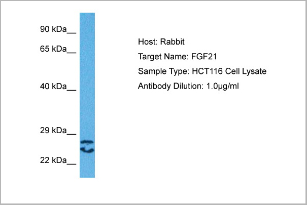
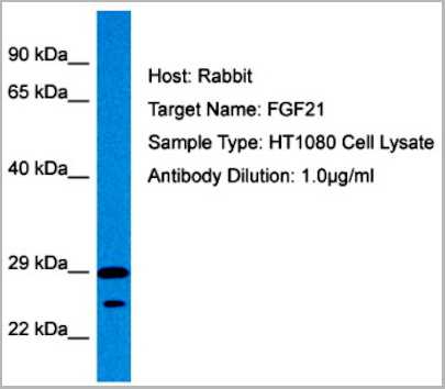
FGF21, Polyclonal Antibody (Cat# AAA23563)
Full Name
FGF21 antibody - N-terminal region
Reactivity
Tested Species Reactivity: Human
Predicted Species Reactivity: Human, Mouse, Rat, Cow, Dog, Guinea Pig, Horse, Rabbit
Predicted Species Reactivity: Human, Mouse, Rat, Cow, Dog, Guinea Pig, Horse, Rabbit
Applications
Immunohistochemistry, Western Blot
Purity
Affinity Purified
Pricing
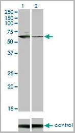
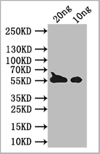
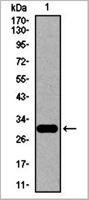
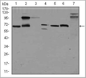
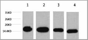
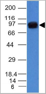
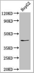
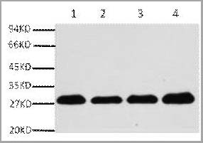
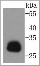
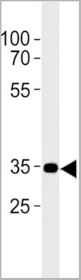
 WB (Western Blot)
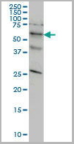
(STK33 monoclonal antibody, Western Blot analysis of STK33 expression in HeLa.)
WB (Western Blot)
(STK33 monoclonal antibody, Western Blot analysis of STK33 expression in HeLa.)
STK33, Monoclonal Antibody (Cat# AAA25856)
Full Name
STK33 (Serine/Threonine Kinase 33) (PE)
Reactivity
Human
Applications
Immunofluorescence, Immunoprecipitation, Western Blot
Purity
Purified by Protein A Affinity Chromatography.
Pricing
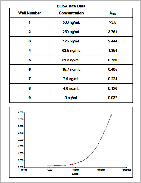 ELISA
(Raw Data)
ELISA
(Raw Data)
Purified IgY, Isotype Control (Cat# AAA14460)
Full Name
Purified Chicken IgY
Applications
Direct ELISA
Pricing
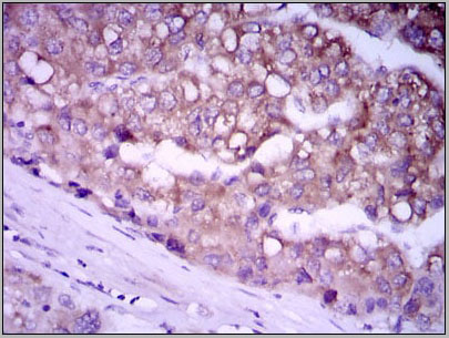
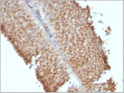
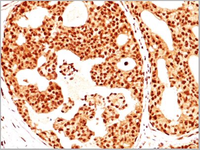
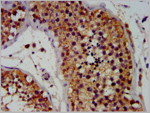
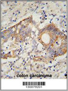
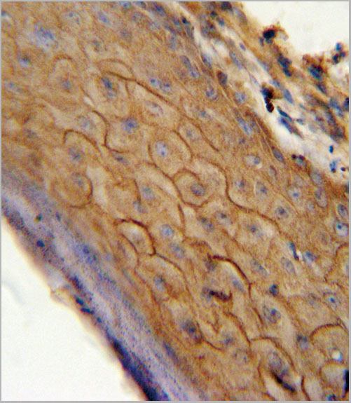
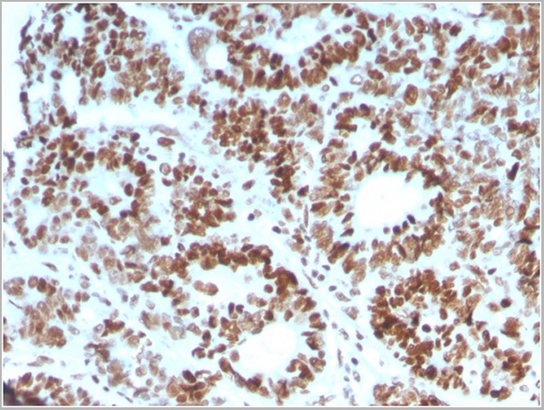
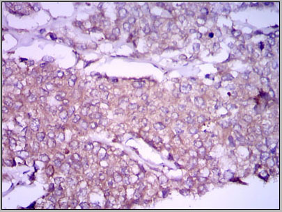 IHC (Immunohistchemistry)
(Immunohistochemical analysis of paraffin-embedded breast cancer tissues using GPC3 mouse mAb with DAB staining.)
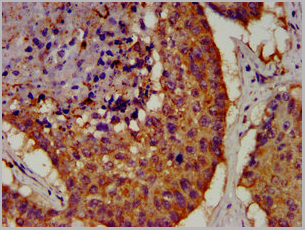
IHC (Immunohistchemistry)
(Immunohistochemical analysis of paraffin-embedded breast cancer tissues using GPC3 mouse mAb with DAB staining.)
GPC3, Monoclonal Antibody (Cat# AAA14118)
Full Name
Anti-GPC3 Mouse mAb
Gene Names
GPC3; SGB; DGSX; MXR7; SDYS; SGBS; OCI-5; SGBS1; GTR2-2
Reactivity
Human, Mouse
Applications
Immunohistochemistry, Immunocytochemistry, Immunofluorescence, Flow Cytometry
Purity
Affinity Purified
Pricing
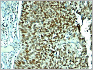
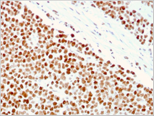
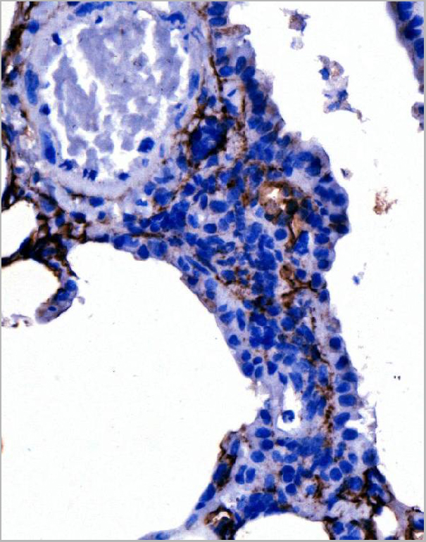
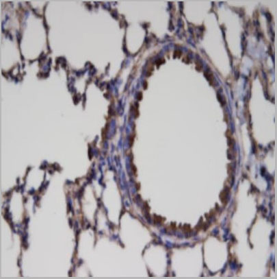 IHC (Immunohistochemistry)
(Immunohistochemistry: Lung tissue (FFPE) stained with Rabbit anti-ER81 (Cat# AAA14073) at 1:200 for 10 min @ RT. Staining of formalin-fixed tissue requires boiling tissue sections in 10 mM Citrate Buffer, pH 6.0 for 10 min followed by cooling at RT for 20 min.)
IHC (Immunohistochemistry)
(Immunohistochemistry: Lung tissue (FFPE) stained with Rabbit anti-ER81 (Cat# AAA14073) at 1:200 for 10 min @ RT. Staining of formalin-fixed tissue requires boiling tissue sections in 10 mM Citrate Buffer, pH 6.0 for 10 min followed by cooling at RT for 20 min.)
ER81, Polyclonal Antibody (Cat# AAA14073)
Full Name
Rabbit anti ER81 Polyclonal Antibody
Gene Names
ETV1; ER81
Reactivity
Mouse, Human, Rat
Applications
Flow Cytometry, Immunohistochemistry, Western Blot, Immunoprecipitation
Purity
Rabbit IgG is purified by Epitope Affinity Purification
Pricing
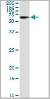
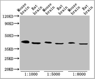
 WB (Western Blot)
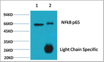
(Fig.8 Western blot analysis of Hela, GAPDH Monoclonal Antibody (2B5) was diluted at 1:10000 (25°C, 3h).)
WB (Western Blot)
(Fig.8 Western blot analysis of Hela, GAPDH Monoclonal Antibody (2B5) was diluted at 1:10000 (25°C, 3h).)
GAPDH, Monoclonal Antibody (Cat# AAA31539)
Full Name
Anti-GAPDH Mouse Monoclonal Antibody (2B5)
Gene Names
GAPDH; G3PD; GAPD
Reactivity
Human, Mouse, Rat, Monkey, Dog, Chicken, Rabbit, Pig, Sheep, Yeast, Hamster
Applications
Western Blot, Immunohistochemistry, Immunofluorescence
Purity
The antibody was affinity-purified from mouse ascites by affinity-chromatography using specific immunogen
Pricing
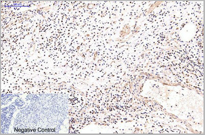
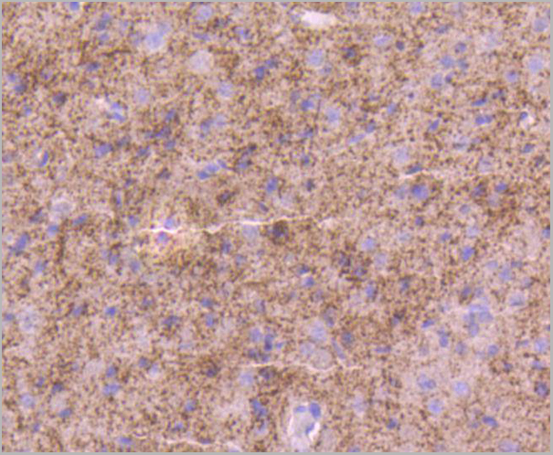
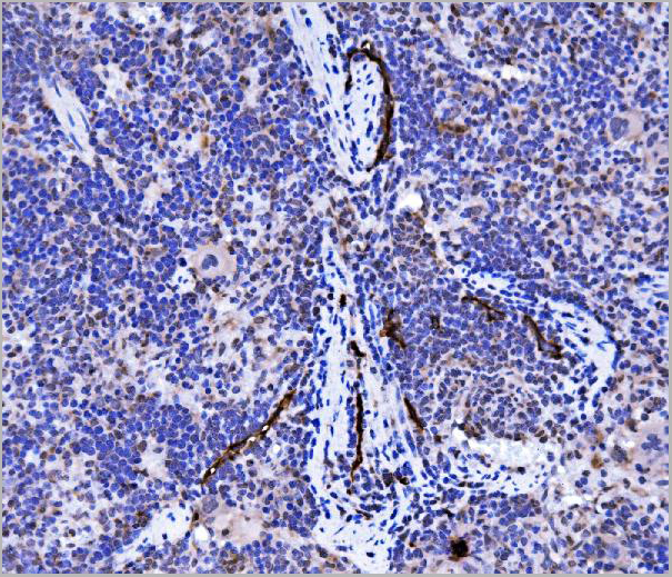
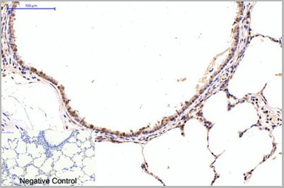 IHC (Immunohistchemistry)
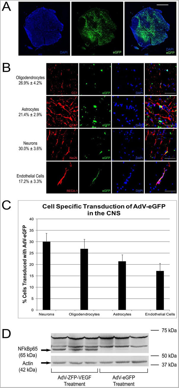
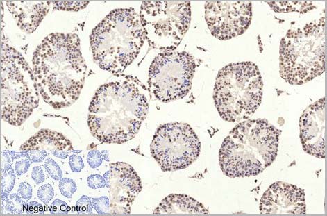
(Immunohistochemical analysis of paraffin-embedded Rat-lung tissue. 1,NFkB p65 Monoclonal Antibody(5G6) was diluted at 1:200(4C,overnight). 2, Sodium citrate pH 6.0 was used for antibody retrieval(>98C,20min). 3,Secondary antibody was diluted at 1:200(room tempeRature, 30min). Negative control was used by secondary antibody only.)
IHC (Immunohistchemistry)
(Immunohistochemical analysis of paraffin-embedded Rat-lung tissue. 1,NFkB p65 Monoclonal Antibody(5G6) was diluted at 1:200(4C,overnight). 2, Sodium citrate pH 6.0 was used for antibody retrieval(>98C,20min). 3,Secondary antibody was diluted at 1:200(room tempeRature, 30min). Negative control was used by secondary antibody only.)
NFkappaB p65, Monoclonal Antibody (Cat# AAA29642)
Full Name
NFkappaB p65 Mouse Monoclonal Antibody
Gene Names
RELA; p65; NFKB3
Reactivity
Human, Rat, Mouse
Applications
Western Blot, Immunohistochemistry, Immunofluorescence
Purity
Affinity purification using immunogen.
Pricing
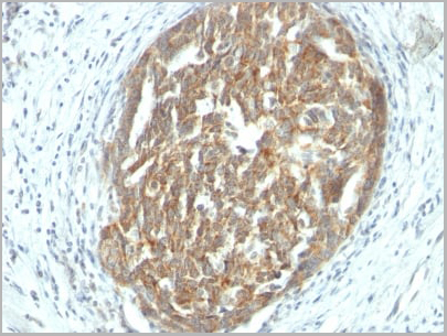 IHC (Immunohistochemistry)
(Formalin-fixed, paraffin-embedded human Ovarian Carcinoma stained with VEGF Monoclonal Antibody (SPM225).)
IHC (Immunohistochemistry)
(Formalin-fixed, paraffin-embedded human Ovarian Carcinoma stained with VEGF Monoclonal Antibody (SPM225).)
VEGF (Vascular Endothelial Growth Factor), Monoclonal Antibody (Cat# AAA13827)
Full Name
VEGF (Vascular Endothelial Growth Factor) Mouse Monoclonal Antibody
Gene Names
VEGFA; VPF; VEGF; MVCD1
Reactivity
Human, Mouse, Rat, Rabbit, Dog. Others not known.
Applications
Immunofluorescence, Immunohistochemistry
Pricing
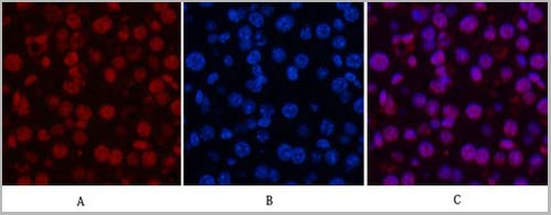
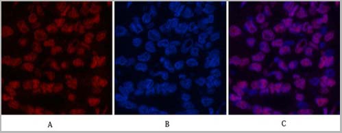
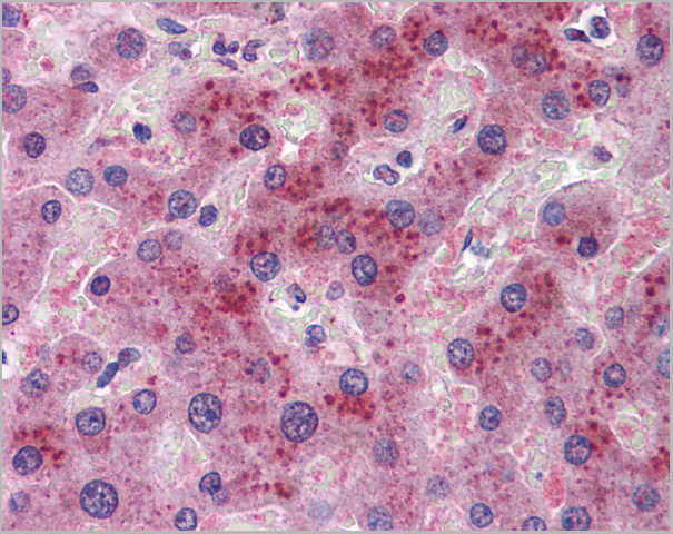
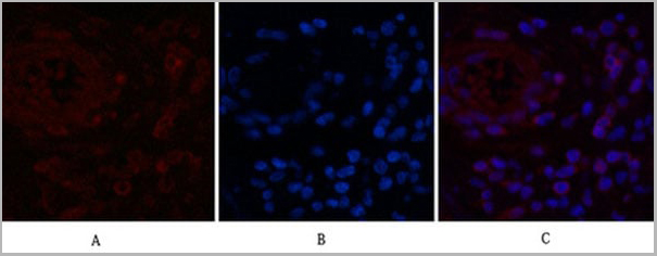
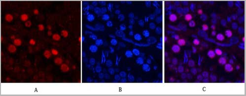
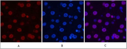 IF (Immunofluorescence)
(Fig.7. Immunofluorescence analysis of rat liver tissue. 1, Histone H3 Monoclonal Antibody (2D10) (red) was diluted at 1:200 (4°C, overnight). 2, Cy3 Labeled secondary antibody was diluted at 1:300 (room temperature, 50min). 3, Picture B: DAPI (blue) 10min. Picture A: Target. Picture B: DAPI. Picture C: merge of A+B.)
IF (Immunofluorescence)
(Fig.7. Immunofluorescence analysis of rat liver tissue. 1, Histone H3 Monoclonal Antibody (2D10) (red) was diluted at 1:200 (4°C, overnight). 2, Cy3 Labeled secondary antibody was diluted at 1:300 (room temperature, 50min). 3, Picture B: DAPI (blue) 10min. Picture A: Target. Picture B: DAPI. Picture C: merge of A+B.)
Histone H3, Monoclonal Antibody (Cat# AAA31543)
Full Name
Anti-Histone H3 Mouse Monoclonal Antibody (2D10)
Gene Names
HIST1H3A; H3/A; H3FA
Reactivity
Human, Mouse, Rat, Yeast
Applications
Western Blot, Immunohistochemistry, Immunofluorescence, Immunoprecipitation
Pricing
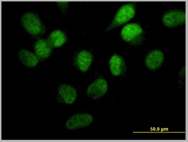
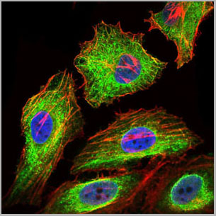
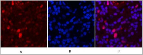
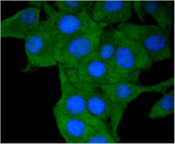
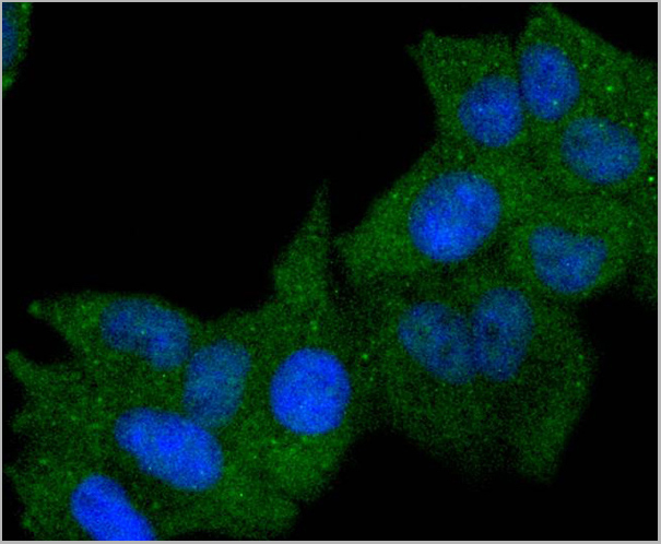
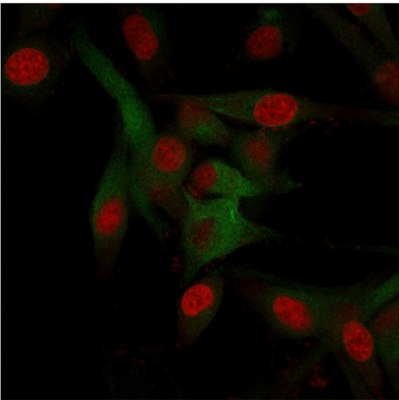
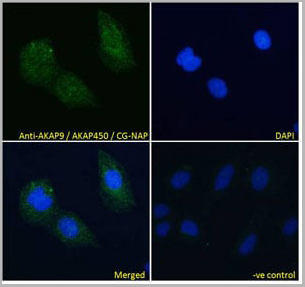 IF (Immunofluorescence)
(AAA13648 Immunofluorescence analysis of paraformaldehyde fixed U2OS cells, permeabilized with 0.15% Triton. Primary incubation 1hr (10ug/ml) followed by Alexa Fluor 488 secondary antibody (4ug/ml), showing cytoplasmic staining. The nuclear stain is DAPI (blue). Negative control: Unimmunized goat IgG (10ug/ml) followed by Alexa Fluor 488 secondary antibody (4ug/ml).)
IF (Immunofluorescence)
(AAA13648 Immunofluorescence analysis of paraformaldehyde fixed U2OS cells, permeabilized with 0.15% Triton. Primary incubation 1hr (10ug/ml) followed by Alexa Fluor 488 secondary antibody (4ug/ml), showing cytoplasmic staining. The nuclear stain is DAPI (blue). Negative control: Unimmunized goat IgG (10ug/ml) followed by Alexa Fluor 488 secondary antibody (4ug/ml).)
AKAP9/AKAP450/CG-NAP, Polyclonal Antibody (Cat# AAA13648)
Full Name
Goat anti-AKAP9/AKAP450/CG-NAP Antibody
Gene Names
AKAP9; LQT11; PRKA9; AKAP-9; CG-NAP; YOTIAO; AKAP350; AKAP450; PPP1R45; HYPERION; MU-RMS-40.16A
Reactivity
Tested: Human; Expected from sequence similarity: Human, Mouse
Applications
Peptide ELISA, Western Blot, Immunofluorescence
Purity
Purified from goat serum by ammonium sulphate precipitation followed by antigen affinity chromatography using the immunizing peptide.
Pricing
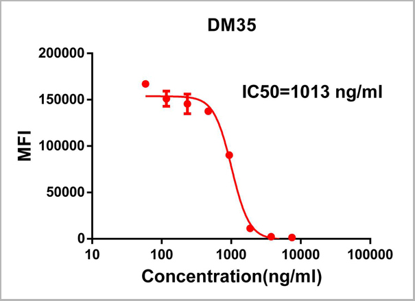 FCM (Flow Cytometry)
(Figure 1. Elisa plate pre-coated by 2 ug/ml (100ul/well) SARS-CoV-2 RBD protein can bind Rabbit Anti-SARS-CoV-2 RBD monoclonal antibody ( clone:DM35) in a linear range of 0.19-200 ng/ml.)
FCM (Flow Cytometry)
(Figure 1. Elisa plate pre-coated by 2 ug/ml (100ul/well) SARS-CoV-2 RBD protein can bind Rabbit Anti-SARS-CoV-2 RBD monoclonal antibody ( clone:DM35) in a linear range of 0.19-200 ng/ml.)
COVID 19 Spike RBD (DM35) Coronavirus, Monoclonal Antibody (Cat# AAA11707)
Full Name
Anti-SARS-CoV-2 RBD antibody (DM35), Rabbit mAb
Reactivity
SARS-CoV-2
Applications
Flow Cytometry
Purity
Purified from cell culture supernatant by affinity chromatography
Pricing
 FCM (Flow Cytometry)
(Flow Cytometric Analysis of paraformaldehyde-fixed Jurkat cells using CD31 Mouse Monoclonal Antibody (JC/70A) followed by goat anti- Mouse- IgG-CF488 (Blue); Isotype Control (Red).)
FCM (Flow Cytometry)
(Flow Cytometric Analysis of paraformaldehyde-fixed Jurkat cells using CD31 Mouse Monoclonal Antibody (JC/70A) followed by goat anti- Mouse- IgG-CF488 (Blue); Isotype Control (Red).)
CD31 / PECAM-1, Monoclonal Antibody (Cat# AAA13818)
Full Name
CD31 / PECAM-1 (Endothelial Cell Marker) Mouse Monoclonal Antibody
Gene Names
PECAM1; CD31; PECA1; GPIIA'; PECAM-1; endoCAM; CD31/EndoCAM
Reactivity
Human, Cynomolgus Monkey, Rabbit.
Does not react with Rat and Pig
Does not react with Rat and Pig
Applications
Flow Cytometry, Immunofluorescence, Western Blot, Immunohistochemistry
Pricing

 IF (Immunofluorescence)
Nucleolin, Monoclonal Antibody (Cat# AAA13823)
Full Name
Nucleolin (Marker of Human Cells) Mouse Monoclonal Antibody
Gene Names
NCL; C23
Reactivity
Human.
Does not react with Mouse, Rat and Cow
Does not react with Mouse, Rat and Cow
Applications
Flow Cytometry, Immunofluorescence, Western Blot, Immunohistochemistry
Pricing
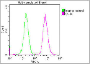
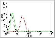
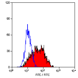
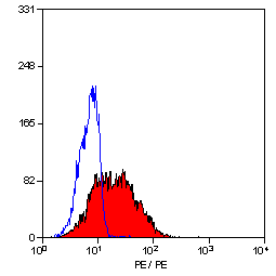
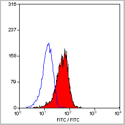
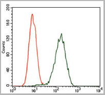
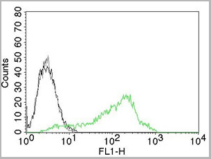
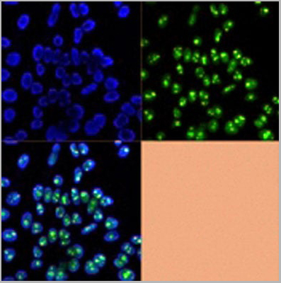
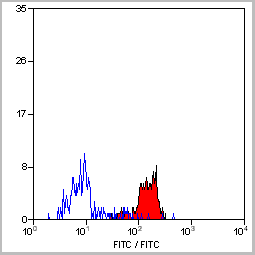
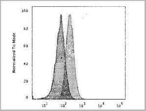
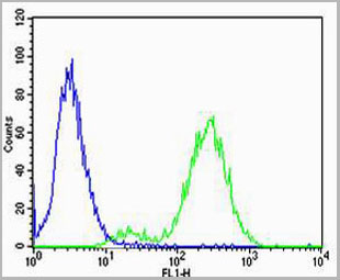
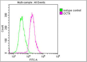 FCM (Flow Cytometry)
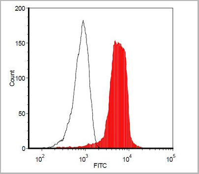
(Overlay histogram showing Ntera-2 cells stained with CSB-MA018403A0m (red line) at 1:100. The cells were incubated in 1x PBS /10% normal goat serum to block non-specific protein-protein interactions followed by primary antibody for 1 h at 4 degree C. The secondary antibody used was FITC goat anti-mouse IgG(H+L) at 1/200 dilution for 1 h at 4 degree C. Isotype control antibody (green line) was used under the same conditions. Acquisition of >10,000 events was performed.)
FCM (Flow Cytometry)
(Overlay histogram showing Ntera-2 cells stained with CSB-MA018403A0m (red line) at 1:100. The cells were incubated in 1x PBS /10% normal goat serum to block non-specific protein-protein interactions followed by primary antibody for 1 h at 4 degree C. The secondary antibody used was FITC goat anti-mouse IgG(H+L) at 1/200 dilution for 1 h at 4 degree C. Isotype control antibody (green line) was used under the same conditions. Acquisition of >10,000 events was performed.)
OCT4, Monoclonal Antibody (Cat# AAA27015)
Full Name
OCT4 Monoclonal Antibody
Gene Names
POU5F1; OCT3; OCT4; OTF3; OTF4; OTF-3; Oct-3; Oct-4
Reactivity
Human, Mouse, Rat
Applications
Western Blot, Immunohistochemistry, Immunofluorescence, Flow Cytometry
Purity
>95%
Protein G Purified
Protein G Purified
Pricing
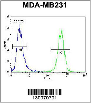
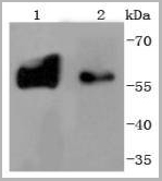
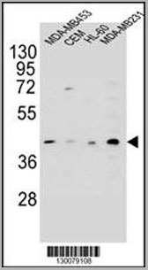 WB (Western Blot)
(HHLA2 Antibody (N-term) western blot analysis in MDA-MB453,CEM,HL-60,MDA-MB231 cell line lysates (35ug/lane).This demonstrates the HHLA2 antibody detected the HHLA2 protein (arrow).)
WB (Western Blot)
(HHLA2 Antibody (N-term) western blot analysis in MDA-MB453,CEM,HL-60,MDA-MB231 cell line lysates (35ug/lane).This demonstrates the HHLA2 antibody detected the HHLA2 protein (arrow).)
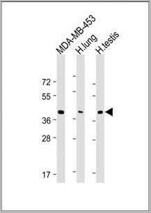
HHLA2, Polyclonal Antibody (Cat# AAA28722)
Full Name
HHLA2 Antibody (N-term)
Gene Names
HHLA2; B7H7; B7-H7
Reactivity
Human
Applications
Western Blot, Immunohistochemistry, Flow Cytometry
Purity
Peptide Affinity Purified Rabbit Polyclonal Antibody (Pab)
Pricing
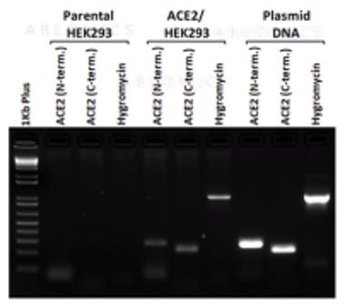
 Application Data
(Fig-5: Cell Surface staining of human ACE2 in the ACE2/HEK293 stable cell line. ATTO 488-conjugated anti-hACE2 antibody (clone AC18F; was used at 1ug/ 1x10^6 cells (Red). ATTO 488-conjugated mouse IgG1 was used as isotype control at 1ug/ 1x10^6 cells (Green).)
Application Data
(Fig-5: Cell Surface staining of human ACE2 in the ACE2/HEK293 stable cell line. ATTO 488-conjugated anti-hACE2 antibody (clone AC18F; was used at 1ug/ 1x10^6 cells (Red). ATTO 488-conjugated mouse IgG1 was used as isotype control at 1ug/ 1x10^6 cells (Green).)
ACE2, Cell Line (Cat# AAA14888)
Full Name
ACE2/HEK293 Stable Cell Line
Gene Names
ACE2; ACEH
Applications
Functional Assay
Pricing


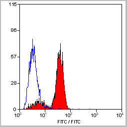
 Application Data
(Staining of mouse peritoneal macrophages with Rat anti Mouse Beta-glucan Receptor: FITC)
Application Data
(Staining of mouse peritoneal macrophages with Rat anti Mouse Beta-glucan Receptor: FITC)
DECTIN-1, Monoclonal Antibody (Cat# AAA12137)
Full Name
RAT ANTI MOUSE DECTIN-1
Gene Names
Clec7a; BGR; beta-GR; Clecsf12
Applications
Immunohistochemistry, Flow Cytometry, Immunoprecipitation
Pricing
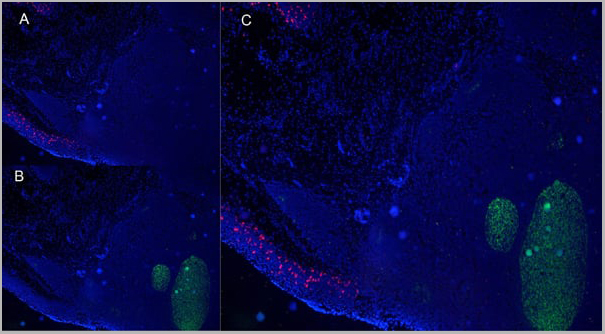
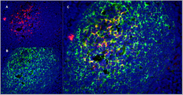
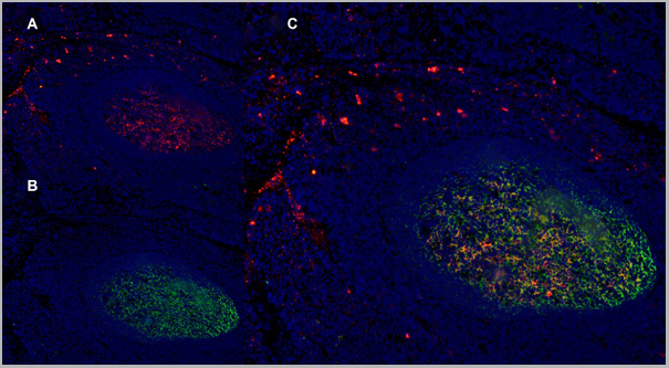
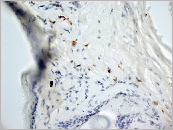
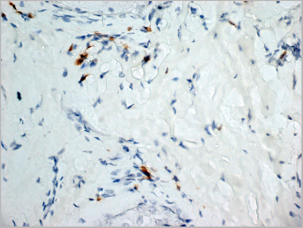
 Application Data
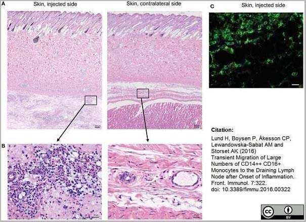
(Mouse anti Human CD14 antibody, clone Tük4 used to identify bovine monocytes in the skin and subcutaneous tissue of adjuvant injected calves by immunofluorescence.Image caption:Cellular recruitment to skin and subcutaneous tissues. (A) HE stained sections of skin with subcutaneous tissue from the side injected with adjuvant and the contralateral side, at 24 h post-injection. Scale bars: 200 ?m. (B) Enlargement of outlined areas in A, as indicated. Scale bars: 20 ?m. (C) Immunofluorescent labeling of subcutaneous tissue on the injected side with antibody against CD14 (green). Scale bar: 20 ?m.From: Lund H, Boysen P, Åkesson CP, Lewandowska-Sabat AM and Storset AK (2016)Transient Migration of Large Numbers of CD14++ CD16+ Monocytes to the Draining Lymph Node after Onset of Inflammation.Front. Immunol. 7:322.This is from an open access article distributed under the terms of the Creative Commons Attribution License.)
Application Data
(Mouse anti Human CD14 antibody, clone Tük4 used to identify bovine monocytes in the skin and subcutaneous tissue of adjuvant injected calves by immunofluorescence.Image caption:Cellular recruitment to skin and subcutaneous tissues. (A) HE stained sections of skin with subcutaneous tissue from the side injected with adjuvant and the contralateral side, at 24 h post-injection. Scale bars: 200 ?m. (B) Enlargement of outlined areas in A, as indicated. Scale bars: 20 ?m. (C) Immunofluorescent labeling of subcutaneous tissue on the injected side with antibody against CD14 (green). Scale bar: 20 ?m.From: Lund H, Boysen P, Åkesson CP, Lewandowska-Sabat AM and Storset AK (2016)Transient Migration of Large Numbers of CD14++ CD16+ Monocytes to the Draining Lymph Node after Onset of Inflammation.Front. Immunol. 7:322.This is from an open access article distributed under the terms of the Creative Commons Attribution License.)
CD14, Monoclonal Antibody (Cat# AAA12266)
Full Name
Mouse Anti Human CD14: Amethyst Orange
Reactivity
Human
Applications
Flow Cytometry
Purity
Purified IgG prepared by affinity chromatography on Protein A from tissue culture supernatant.
Pricing


 Application Data
(Staining of mouse spleen with Hamster anti Mouse CD81: Alexa Fluor 488)
Application Data
(Staining of mouse spleen with Hamster anti Mouse CD81: Alexa Fluor 488)
CD81, Monoclonal Antibody (Cat# AAA11869)
Full Name
HAMSTER ANTI MOUSE CD81:FITC
Gene Names
Cd81; Tapa1; Tapa-1; Tspan28
Applications
Flow Cytometry
Pricing


 Application Data
(PE conjugatedMouse anti Human CD169 antibody, clone 7-239 used to block CD169 function on myeloid cells.Image caption:Siglec-1 mediates HIV-1 uptake into a storage compartment and enhances HIV-1 trans-infection specially in IFN?-treated monocytes and DCs. A. Uptake of HIV-1NL4–3 by different myeloid cells exposed to IFN?. Cells were cultured with HIV-1 to measure p24Gag by ELISA. Mean values and SEM from four experiments include cells from 12 donors. B. Fold change in HIV-1NL4–3 uptake of cells treated with bafilomycin A1 compared to untreated cells. Mean values and SEM include cells from three donors. C. Relative uptake of HIV-1NL4–3 by IFN?-treated myeloid cells pre-incubated with the indicated mAbs. Values are normalized to the level of HIV-1 uptake by mock-treated cells (set at 100%). Mean values and SEM from two experiments include cells from six donors. D. Confocal microscopy analysis of different IFN?-treated myeloid cells pulsed with HIV-1Cherry and stained for Siglec-1 (Alexa 488), HLA-DR (Alexa 647) and DAPI. (Top) Representative viral pattern for each kind of myeloid cell analyzed, showing maximum fluorescence intensity of four channels. (Bottom) Percentage of myeloid cells with distinct viral patterns: random distribution, polarized accumulation, and sac-like compartment formation, as illustrated in the left drawing. Mean values of 50 cells from two different donors are shown. E. HIV-1 transmission from IFN?-treated myeloid cells to a luciferase reporter CD4+ cell line. HIV-1 infection was determined by induced luciferase activity in relative light units (RLUs). Mean values and SEM from four experiments include cells from 12 donors. F. Relative HIV-1 transmission from IFN?-treated myeloid cells pre-incubated with the indicated mAbs. Values are normalized to the level of HIV-1 trans-infected by mock-treated cells. Mean values and SEM from two experiments include cells from six donors. Statistical differences were assessed with a paired t test in A and E, and with a one sample t-test in B, C and F.From: Pino M, Erkizia I, Benet S, Erikson E, Fernández-Figueras MT, Guerrero D, Dalmau J, Ouchi D, Rausell A, Ciuffi A, Keppler OT, Telenti A, Kräusslich HG, Martinez-Picado J, Izquierdo-Useros N.HIV-1 immune activation induces Siglec-1 expression and enhances viral trans-infection in blood and tissue myeloid cells.Retrovirology. 2015 May 7;12:37.This image is from an open access article distributed under the terms of the Creative Commons Attribution License.)
Application Data
(PE conjugatedMouse anti Human CD169 antibody, clone 7-239 used to block CD169 function on myeloid cells.Image caption:Siglec-1 mediates HIV-1 uptake into a storage compartment and enhances HIV-1 trans-infection specially in IFN?-treated monocytes and DCs. A. Uptake of HIV-1NL4–3 by different myeloid cells exposed to IFN?. Cells were cultured with HIV-1 to measure p24Gag by ELISA. Mean values and SEM from four experiments include cells from 12 donors. B. Fold change in HIV-1NL4–3 uptake of cells treated with bafilomycin A1 compared to untreated cells. Mean values and SEM include cells from three donors. C. Relative uptake of HIV-1NL4–3 by IFN?-treated myeloid cells pre-incubated with the indicated mAbs. Values are normalized to the level of HIV-1 uptake by mock-treated cells (set at 100%). Mean values and SEM from two experiments include cells from six donors. D. Confocal microscopy analysis of different IFN?-treated myeloid cells pulsed with HIV-1Cherry and stained for Siglec-1 (Alexa 488), HLA-DR (Alexa 647) and DAPI. (Top) Representative viral pattern for each kind of myeloid cell analyzed, showing maximum fluorescence intensity of four channels. (Bottom) Percentage of myeloid cells with distinct viral patterns: random distribution, polarized accumulation, and sac-like compartment formation, as illustrated in the left drawing. Mean values of 50 cells from two different donors are shown. E. HIV-1 transmission from IFN?-treated myeloid cells to a luciferase reporter CD4+ cell line. HIV-1 infection was determined by induced luciferase activity in relative light units (RLUs). Mean values and SEM from four experiments include cells from 12 donors. F. Relative HIV-1 transmission from IFN?-treated myeloid cells pre-incubated with the indicated mAbs. Values are normalized to the level of HIV-1 trans-infected by mock-treated cells. Mean values and SEM from two experiments include cells from six donors. Statistical differences were assessed with a paired t test in A and E, and with a one sample t-test in B, C and F.From: Pino M, Erkizia I, Benet S, Erikson E, Fernández-Figueras MT, Guerrero D, Dalmau J, Ouchi D, Rausell A, Ciuffi A, Keppler OT, Telenti A, Kräusslich HG, Martinez-Picado J, Izquierdo-Useros N.HIV-1 immune activation induces Siglec-1 expression and enhances viral trans-infection in blood and tissue myeloid cells.Retrovirology. 2015 May 7;12:37.This image is from an open access article distributed under the terms of the Creative Commons Attribution License.)
CD169, Monoclonal Antibody (Cat# AAA12265)
Full Name
Mouse Anti Human CD169: RPE
Gene Names
SIGLEC1; SN; CD169; SIGLEC-1
Reactivity
Human
Applications
Flow Cytometry
Purity
>95% by SDS PAGE
Purified IgG prepared by affinity chromatography on Protein A from tissue culture supernatant.
Purified IgG prepared by affinity chromatography on Protein A from tissue culture supernatant.
Pricing


 Application Data
(Staining of mouse spleen with Hamster anti Mouse CD81: Alexa Fluor 488)
Application Data
(Staining of mouse spleen with Hamster anti Mouse CD81: Alexa Fluor 488)
CD81, Monoclonal Antibody (Cat# AAA12033)
Full Name
HAMSTER ANTI MOUSE CD81:RPE
Gene Names
Cd81; Tapa1; Tapa-1; Tspan28
Applications
Flow Cytometry
Pricing


 Application Data
(Published customer image: Effect of clustered ephrin-A1-Fc on vascular formation in the rat striatum. Clustered ephrin-A1-Fc was injected into the lesioned side of the lateral ventricle in the unilaterally lesioned rats. Brains taken 6 weeks after injection were sectioned coronally and stained for GFAP (green) and RECA-1 (red) and with DAPI (nuclei; blue). The rectangular insets are shown in Fig. 8B. Scale bar: 100 um.From: Jing X, Miwa H, Sawada T, Nakanishi I, Kondo T, et al. (2012) Ephrin-A1-Mediated Dopaminergic Neurogenesis and Angiogenesis in a Rat Model of Parkinson's Disease. PLoS ONE 7(2): e32019.)
Application Data
(Published customer image: Effect of clustered ephrin-A1-Fc on vascular formation in the rat striatum. Clustered ephrin-A1-Fc was injected into the lesioned side of the lateral ventricle in the unilaterally lesioned rats. Brains taken 6 weeks after injection were sectioned coronally and stained for GFAP (green) and RECA-1 (red) and with DAPI (nuclei; blue). The rectangular insets are shown in Fig. 8B. Scale bar: 100 um.From: Jing X, Miwa H, Sawada T, Nakanishi I, Kondo T, et al. (2012) Ephrin-A1-Mediated Dopaminergic Neurogenesis and Angiogenesis in a Rat Model of Parkinson's Disease. PLoS ONE 7(2): e32019.)
RECA-1, Monoclonal Antibody (Cat# AAA12019)
Full Name
MOUSE ANTI RAT RECA-1
Applications
Immunohistochemistry, Immunofluorescence
Pricing
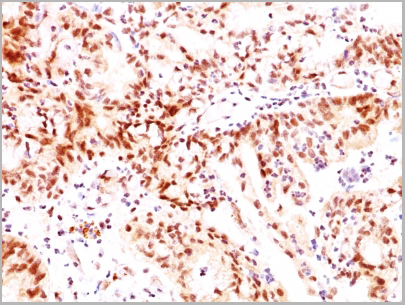
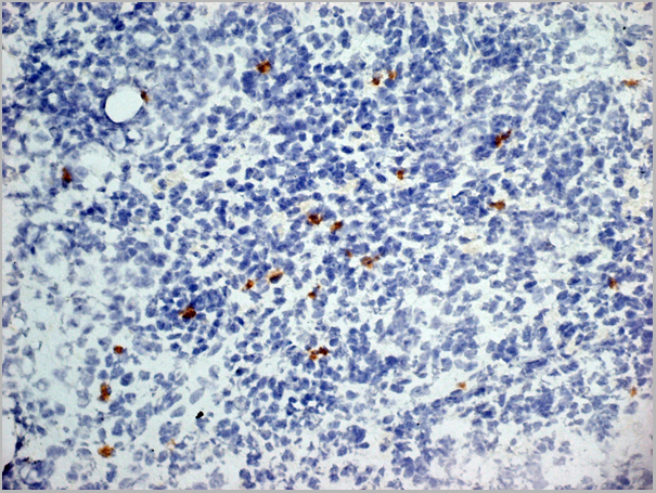
 Application Data
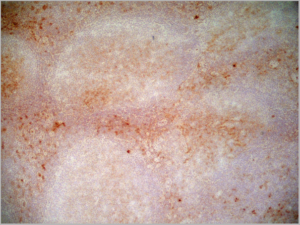
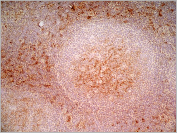
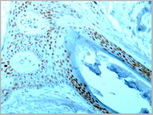
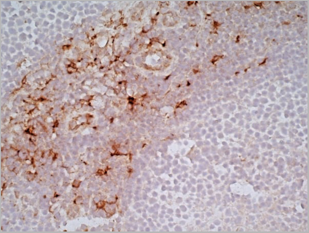
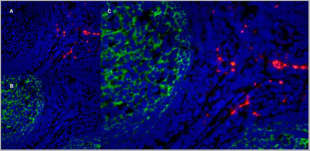
(Immunoperoxidase staining of a human tonsil cryosection with Mouse anti Human CD163 antibody, clone EDHu-1 followed by the Histar detection system . Low power)
Application Data
(Immunoperoxidase staining of a human tonsil cryosection with Mouse anti Human CD163 antibody, clone EDHu-1 followed by the Histar detection system . Low power)
CD163, Monoclonal Antibody (Cat# AAA11943)
Full Name
MOUSE ANTI HUMAN CD163
Gene Names
CD163; M130; MM130
Reactivity
Guinea Pig, Pig, Rhesus Monkey, Sheep
Applications
Immunohistochemistry, Immunohistochemistry, Flow Cytometry, Immunofluorescence, Immunoassay, Western Blot
Pricing
 FCM (Flow Cytometry)
(PE conjugated polystyrene beads were stained with (filled histogram) or rabbit IgG, FITC isotype control (open histogram).)
FCM (Flow Cytometry)
(PE conjugated polystyrene beads were stained with (filled histogram) or rabbit IgG, FITC isotype control (open histogram).)
R-PE, Antibody (Cat# AAA14282)
Full Name
Purified Rabbit Anti-R-PE, Polyclonal Antibody
Applications
Flow Cytometry, Immunohistochemistry
Pricing
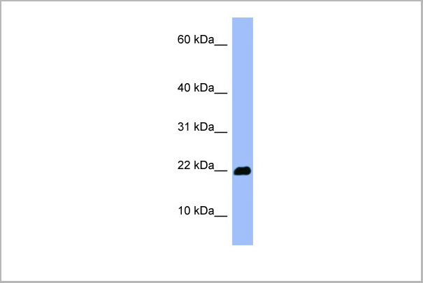
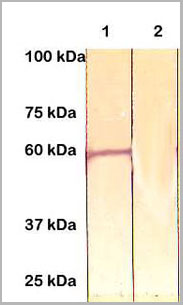
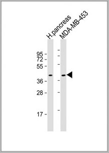
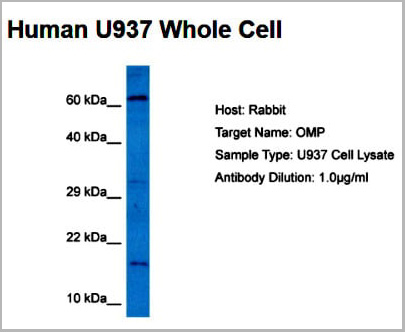 WB (Western Blot)
(Host: RabbitTarget Name: OMPSample Tissue: Human U937 Whole CellAntibody Dilution: 5ug/ml)
WB (Western Blot)
(Host: RabbitTarget Name: OMPSample Tissue: Human U937 Whole CellAntibody Dilution: 5ug/ml)
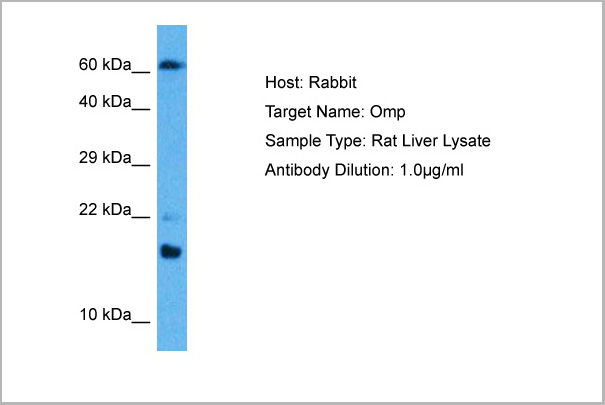
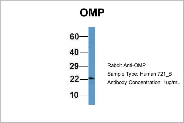
OMP, Polyclonal Antibody (Cat# AAA23571)
Full Name
OMP antibody - middle region
Reactivity
Tested Reactivity: Human, Rat
Predicted Species Reactivity: Goat, Human, Mouse, Rat
Predicted Species Reactivity: Goat, Human, Mouse, Rat
Applications
Western Blot
Purity
Affinity Purified
Pricing
 IHC (Immunohistochemistry)
Actin, Smooth Muscle, Antibody (Cat# AAA14079)
Full Name
Rabbit anti SMA antibody (Smooth Muscle Actin)
Reactivity
Human, Mouse, Rat
Applications
Western Blot, Immunohistochemistry, Immunoprecipitation
Purity
The Rabbit IgG is purified by Epitope affinity purification
Pricing
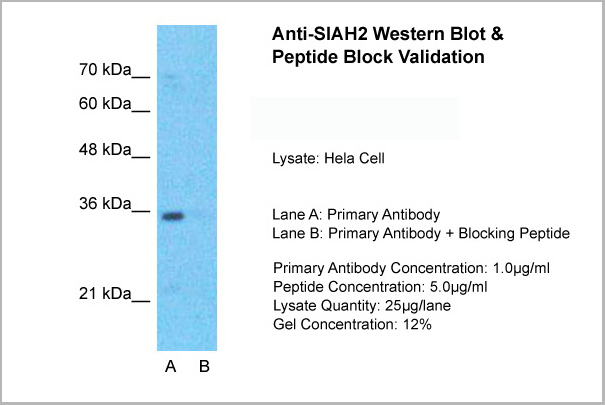
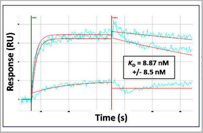 Application Data
(Surface Plasmon Resonance Kinetic Characterization of Polyclonal Antibody Affinity. Purified polyclonal antibodies were immobilized on a Protein A/G coated Carterra LSA sensor chip at concentrations of 0.5, 5, and 50 ug/mL. Antibodies on the surface were exposed to interaction with peptides sequentially via microfluidic controlled flow at 333nM peptide concentration for kinetic characterization of the binders for affinity and specificity, followed by curve fitting using the Kinetics software. Kd determinations for either two or three concentrations were averaged and results and standard deviation are shown.)
Application Data
(Surface Plasmon Resonance Kinetic Characterization of Polyclonal Antibody Affinity. Purified polyclonal antibodies were immobilized on a Protein A/G coated Carterra LSA sensor chip at concentrations of 0.5, 5, and 50 ug/mL. Antibodies on the surface were exposed to interaction with peptides sequentially via microfluidic controlled flow at 333nM peptide concentration for kinetic characterization of the binders for affinity and specificity, followed by curve fitting using the Kinetics software. Kd determinations for either two or three concentrations were averaged and results and standard deviation are shown.)
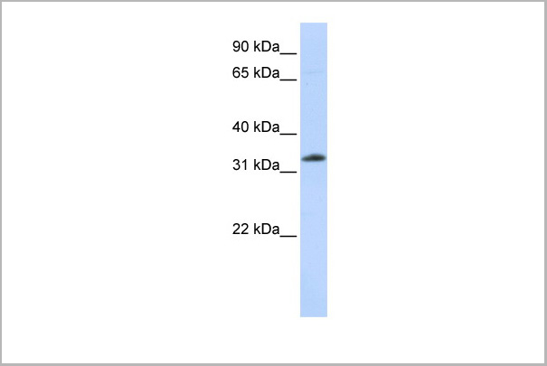
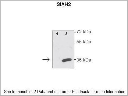
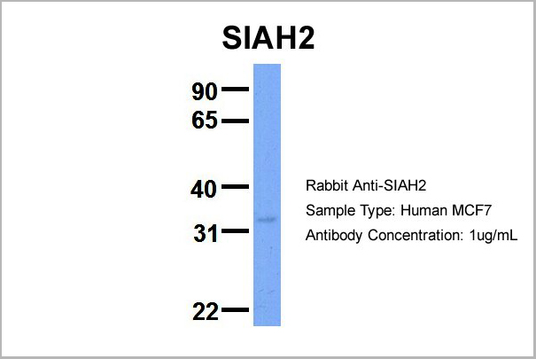
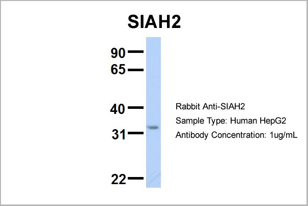
SIAH2, Polyclonal Antibody (Cat# AAA23577)
Full Name
SIAH2 antibody - N-terminal region
Gene Names
SIAH2; hSiah2
Reactivity
Tested Species Reactivity: Human
Predicted Species Reactivity: Human, Mouse, Rat, Cow, Dog, Pig
Predicted Species Reactivity: Human, Mouse, Rat, Cow, Dog, Pig
Applications
Western Blot
Purity
Affinity Purified
Pricing
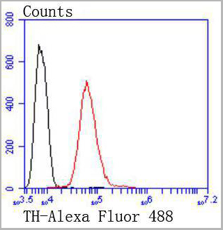 FCM (Flow Cytometry)
(Flow cytometric analysis of SH-SY-5Y cells with Tyrosine Hydroxylase antibody at 1/50 dilution (red) compared with an unlabelled control (cells without incubation with primary antibody; black). Alexa Fluor 488-conjugated goat anti rabbit IgG was use as the secondary antibody)
FCM (Flow Cytometry)
(Flow cytometric analysis of SH-SY-5Y cells with Tyrosine Hydroxylase antibody at 1/50 dilution (red) compared with an unlabelled control (cells without incubation with primary antibody; black). Alexa Fluor 488-conjugated goat anti rabbit IgG was use as the secondary antibody)
Tyrosine Hydroxylase, Monoclonal Antibody (Cat# AAA30209)
Full Name
Tyrosine Hydroxylase Antibody
Gene Names
TH; TYH; DYT14; DYT5b
Reactivity
Human, Mouse, Rat
Applications
Western Blot, Immunocytochemistry, Immunohistochemistry, Flow Cytometry
Purity
ProA affinity purified
Pricing
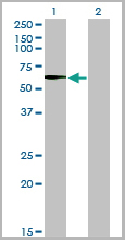
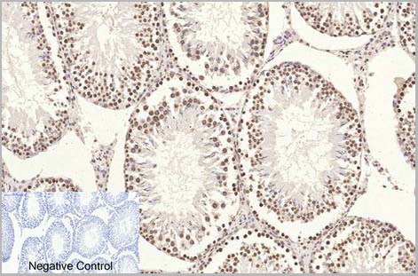
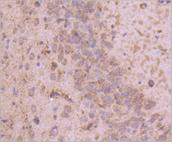
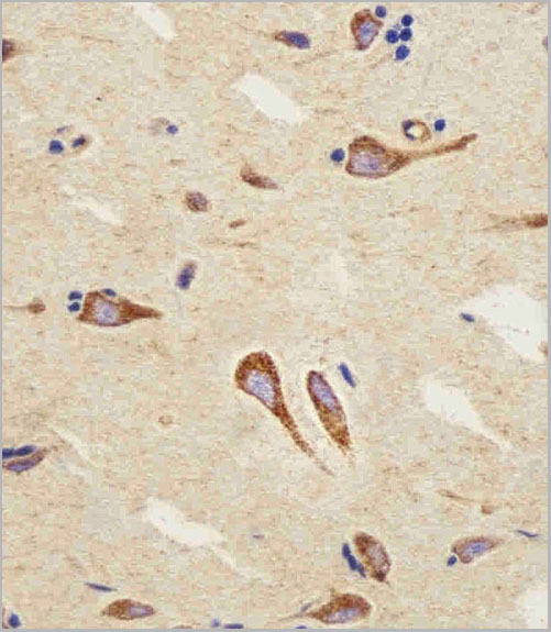
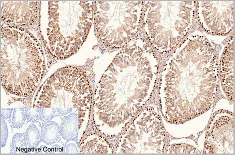 IHC (Immunohistchemistry)
(Fig.6. Immunohistochemical analysis of paraffin-embedded rat testis tissue. 1, PCNA Monoclonal Antibod y(1D7) was diluted at 1:200 (4°C, overnight). 2, Sodium citrate pH 6.0 was used for antibody retrieval(>98°C, 20min). 3, secondary antibody was diluted at 1:200 (room temperature, 30min). Negative control was used by secondary antibody only.)
IHC (Immunohistchemistry)
(Fig.6. Immunohistochemical analysis of paraffin-embedded rat testis tissue. 1, PCNA Monoclonal Antibod y(1D7) was diluted at 1:200 (4°C, overnight). 2, Sodium citrate pH 6.0 was used for antibody retrieval(>98°C, 20min). 3, secondary antibody was diluted at 1:200 (room temperature, 30min). Negative control was used by secondary antibody only.)
PCNA, Monoclonal Antibody (Cat# AAA31541)
Full Name
Anti-PCNA Mouse Monoclonal Antibody (1D7)
Reactivity
Human, Mouse, Rat
Applications
Western Blot, Immunohistochemistry, Immunofluorescence
Purity
The antibody was affinity-purified from mouse ascites by affinity-chromatography using specific immunogen
Pricing
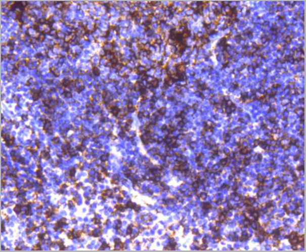
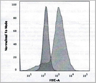
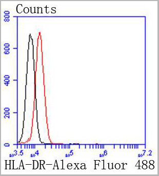 FCM (Flow Cytometry)
(Flow cytometric analysis of Jurkat cells with HLA-DR antibody at 1/50 dilution (red) compared with an unlabelled control (cells without incubation with primary antibody; black). Alexa Fluor 488-conjugated goat anti rabbit IgG was used as the secondary antibody.)
FCM (Flow Cytometry)
(Flow cytometric analysis of Jurkat cells with HLA-DR antibody at 1/50 dilution (red) compared with an unlabelled control (cells without incubation with primary antibody; black). Alexa Fluor 488-conjugated goat anti rabbit IgG was used as the secondary antibody.)
HLA-DR, Monoclonal Antibody (Cat# AAA30136)
Full Name
HLA-DR Antibody
Gene Names
HLA-DRA; MLRW; HLA-DRA1
Reactivity
Human, Mouse, Rat
Applications
Western Blot, Immunocytochemistry, Immunohistochemistry, Flow Cytometry
Purity
ProA affinity purified
Pricing
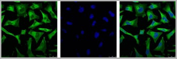
 ICC (Immunocytochemistry)
(Immunofluorescence staining of U87MG cells using CD68 Mouse Monoclonal Antibody (C68/684) followed by goat anti-Mouse IgG conjugated to (green). Nuclei are stained with Red dot)
ICC (Immunocytochemistry)
(Immunofluorescence staining of U87MG cells using CD68 Mouse Monoclonal Antibody (C68/684) followed by goat anti-Mouse IgG conjugated to (green). Nuclei are stained with Red dot)
CD68, Antibody (Cat# AAA13807)
Full Name
CD68 (Macrophage Marker) Mouse Monoclonal Antibody
Gene Names
CD68; GP110; LAMP4; SCARD1
Reactivity
Human, Monkey, Mouse, and Rat. Others not known.
Applications
Western Blot, Immunofluorescence, Flow Cytometry, Immunohistochemistry
Pricing
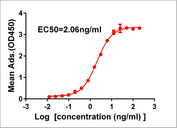
 ELISA
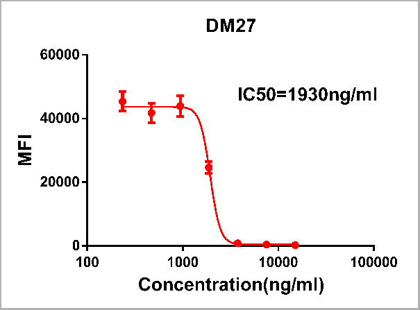
(Figure 1. Competition flow cytometry assay demonstrating Rabbit anti-RBD monoclonal antibody ( clone: DM27) blockade of SARS-CoV-2 (COVID-19) S protein RBD (1ug/ml, [getskuurl sku=PME100497 binding to Expi 293 cell line transfected with human ACE2. IC50=1930ng/ml. The Y-axis represents the geometric mean fluorescence intensity (MFI) while the X-axis represents the concentration of IgG used.)
ELISA
(Figure 1. Competition flow cytometry assay demonstrating Rabbit anti-RBD monoclonal antibody ( clone: DM27) blockade of SARS-CoV-2 (COVID-19) S protein RBD (1ug/ml, [getskuurl sku=PME100497 binding to Expi 293 cell line transfected with human ACE2. IC50=1930ng/ml. The Y-axis represents the geometric mean fluorescence intensity (MFI) while the X-axis represents the concentration of IgG used.)
COVID 19 Spike RBD (DM27) Coronavirus, Monoclonal Antibody (Cat# AAA11706)
Full Name
Anti-SARS-CoV-2 RBD antibody (DM27), Rabbit mAb
Reactivity
SARS-CoV-2
Applications
Flow Cytometry
Purity
Purified from cell culture supernatant by affinity chromatography
Pricing
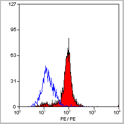
 FCM (Flow Cytometry)
(Flow Cytometric Analysis of Human Raji Cells. using HLA-Mouse Monoclonal Antibody followed by Goat anti-mouse IgG (Blue); Isotype control (Red).)
FCM (Flow Cytometry)
(Flow Cytometric Analysis of Human Raji Cells. using HLA-Mouse Monoclonal Antibody followed by Goat anti-mouse IgG (Blue); Isotype control (Red).)
HLA-A, Monoclonal Antibody (Cat# AAA13821)
Full Name
HLA-A (MHC I) Mouse Monoclonal Antibody
Gene Names
HLA-B; AS; HLAB; Bw-47; Bw-50; SPDA1; B-4901; B-5001; HLA-Cw; HLA-B15; HLA-B39; HLA-B49; HLA-B50; HLA-B55; HLA-B59; HLA-B61; HEL-S-83; HLA-DRB1; HLA-B*45ZJ; HLA-B-3506; HLA-B-3905; HLA-B-5502; HLA-B-5602
Reactivity
Human
Applications
Flow Cytometry, Immunofluorescence
Pricing
 FCM (Flow Cytometry)
(CB2 Antibody (C-term) flow cytometric analysis of Jurkat cells (right histogram) compared to a negative control cell (left histogram).FITC-conjugated goat-anti-rabbit secondary antibodies were used for the analysis.)
FCM (Flow Cytometry)
(CB2 Antibody (C-term) flow cytometric analysis of Jurkat cells (right histogram) compared to a negative control cell (left histogram).FITC-conjugated goat-anti-rabbit secondary antibodies were used for the analysis.)
CB2, Polyclonal Antibody (Cat# AAA28672)
Full Name
CB2 Antibody (C-term)
Gene Names
CNR2; CB2; CX5; CB-2
Reactivity
Human
Applications
Immunohistochemistry, Flow Cytometry, Western Blot
Purity
This antibody is purified through a protein A column, followed by peptide affinity purification.
Pricing
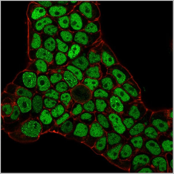

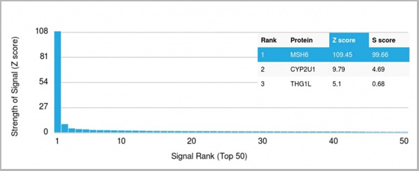 Application Data
(Analysis of Protein Array containing >19,000 full-length human proteins using MSH6 Mouse Monoclonal Antibody (MSH6/3086) Z- and S- Score: The Z-score represents the strength of a signal that a monoclonal antibody (MAb) (in combination with a fluorescently-tagged anti-IgG secondary antibody) produces when binding to a particular protein on the HuProtTM array. Z-scores are described in units of standard deviations (SD’s) above the mean value of all signals generated on that array. If targets on HuProtTM are arranged in descending order of the Z-score, the S-score is the difference (also in units of SD’s) between the Z-score. S-score therefore represents the relative target specificity of a MAb to its intended target. A MAb is considered to specific to its intended target, if the MAb has an S-score of at least 2.5. For example, if a MAb binds to protein X with a Z-score of 43 and to protein Y with a Z-score of 14, then the S-score for the binding of that MAb to protein X is equal to 29.)
Application Data
(Analysis of Protein Array containing >19,000 full-length human proteins using MSH6 Mouse Monoclonal Antibody (MSH6/3086) Z- and S- Score: The Z-score represents the strength of a signal that a monoclonal antibody (MAb) (in combination with a fluorescently-tagged anti-IgG secondary antibody) produces when binding to a particular protein on the HuProtTM array. Z-scores are described in units of standard deviations (SD’s) above the mean value of all signals generated on that array. If targets on HuProtTM are arranged in descending order of the Z-score, the S-score is the difference (also in units of SD’s) between the Z-score. S-score therefore represents the relative target specificity of a MAb to its intended target. A MAb is considered to specific to its intended target, if the MAb has an S-score of at least 2.5. For example, if a MAb binds to protein X with a Z-score of 43 and to protein Y with a Z-score of 14, then the S-score for the binding of that MAb to protein X is equal to 29.)
MSH6, Monoclonal Antibody (Cat# AAA23917)
Full Name
MSH6 (DNA Mismatch Repair Protein)
Gene Names
MSH6; GTBP; HSAP; p160; GTMBP; HNPCC5
Reactivity
Human
Applications
Flow Cytometry, Immunofluorescence, Western Blot, Immunohistochemistry
Purity
Purified Ab with BSA and Azide at 200ug/ml OR Purified Ab WITHOUT BSA and Azide at 1.0mg/ml
Pricing
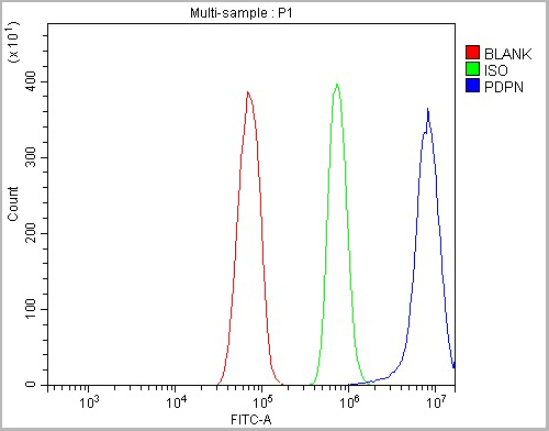 FCM (Flow Cytometry)
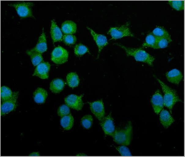
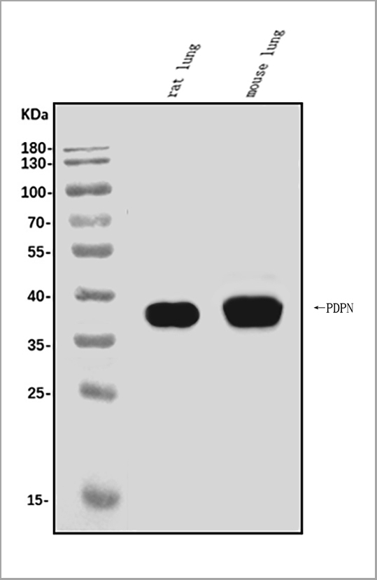
(Figure 5. Flow Cytometry analysis of RH35 cells using anti-Podoplanin/gp36/Pdpn antibody (AAA19233).Overlay histogram showing RH35 cells stained with AAA19233 (Blue line). The cells were blocked with 10% normal goat serum. And then incubated with rabbit anti-Podoplanin/gp36/Pdpn Antibody (AAA19233, 1μg/1x106 cells) for 30 min at 20 degree C. DyLight®488 conjugated goat anti-rabbit IgG (5-10μg/1x106 cells) was used as secondary antibody for 30 minutes at 20 degree C. Isotype control antibody (Green line) was rabbit IgG (1μg/1x106) used under the same conditions. Unlabelled sample (Red line) was also used as a control.)
FCM (Flow Cytometry)
(Figure 5. Flow Cytometry analysis of RH35 cells using anti-Podoplanin/gp36/Pdpn antibody (AAA19233).Overlay histogram showing RH35 cells stained with AAA19233 (Blue line). The cells were blocked with 10% normal goat serum. And then incubated with rabbit anti-Podoplanin/gp36/Pdpn Antibody (AAA19233, 1μg/1x106 cells) for 30 min at 20 degree C. DyLight®488 conjugated goat anti-rabbit IgG (5-10μg/1x106 cells) was used as secondary antibody for 30 minutes at 20 degree C. Isotype control antibody (Green line) was rabbit IgG (1μg/1x106) used under the same conditions. Unlabelled sample (Red line) was also used as a control.)
Podoplanin/gp36/Pdpn, Polyclonal Antibody (Cat# AAA19233)
Full Name
Anti-Podoplanin/gp36/Pdpn Antibody
Gene Names
Pdpn; T1a; Gp38; OTS-8; T1alpha; RANDAM-2; T1-alpha
Reactivity
Mouse, Rat
Applications
Western Blot, Immunohistochemistry, Immunocytochemistry, Immunofluorescence, Flow Cytometry, Direct ELISA
Purity
Immunogen affinity purified.
Pricing


 Application Data
(Staining of mouse peritoneal macrophages with Rat anti Mouse Beta-glucan Receptor: FITC)
Application Data
(Staining of mouse peritoneal macrophages with Rat anti Mouse Beta-glucan Receptor: FITC)
DECTIN-1, Monoclonal Antibody (Cat# AAA12128)
Full Name
RAT ANTI MOUSE DECTIN-1:Biotin
Gene Names
Clec7a; BGR; beta-GR; Clecsf12
Applications
Flow Cytometry
Pricing
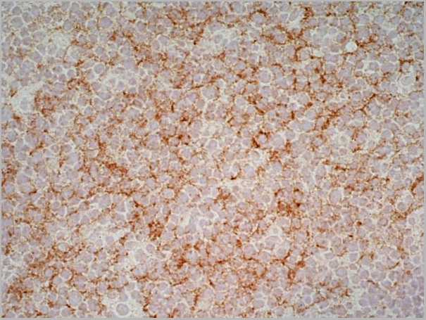
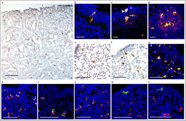
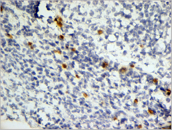
 Application Data
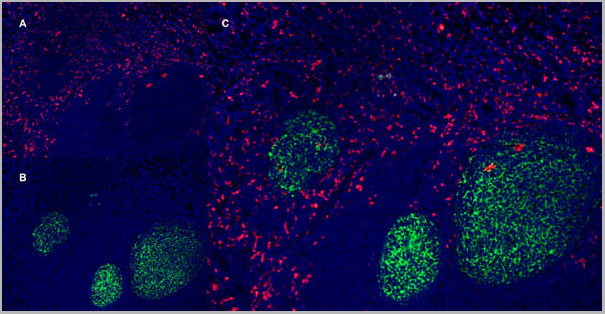
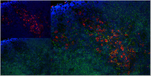
(Immunofluorescence staining of rat lymph node cryosection with Mouse anti Rat CD163 antibody , red an A and Mouse anti Rat CD8 MCA48), green in B. C is the merged image with nuclei counter-stained blue using DAPI. High power)
Application Data
(Immunofluorescence staining of rat lymph node cryosection with Mouse anti Rat CD163 antibody , red an A and Mouse anti Rat CD8 MCA48), green in B. C is the merged image with nuclei counter-stained blue using DAPI. High power)
CD8 ALPHA, Monoclonal Antibody (Cat# AAA11981)
Full Name
MOUSE ANTI RAT CD8 ALPHA
Applications
Immunohistochemistry, Flow Cytometry, Immunofluorescence, Immunoprecipitation, Immunohistochemistry, Western Blot
Pricing
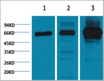
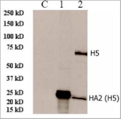 WB (Western Blot)
(lane C, 293 cell extract controlLane 1, 293 cell expressing HA2 (H5N1)Lane 2, 293 cell expressing HA (H5N1))
WB (Western Blot)
(lane C, 293 cell extract controlLane 1, 293 cell expressing HA2 (H5N1)Lane 2, 293 cell expressing HA (H5N1))
HA2 (A/Vietnam/1203/2004)(H5N1), Polyclonal Antibody (Cat# AAA13718)
Full Name
Anti-HA2 (A/Vietnam/1203/2004)(H5N1), rabbit IgG
Applications
Western Blot
Purity
Immunoaffinity chromatography
Pricing
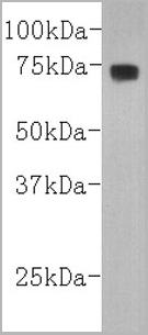 WB (Western Blot)
(Western blot analysis on human serum using anti- Complement C3 mouse monoclonal antibody.)
WB (Western Blot)
(Western blot analysis on human serum using anti- Complement C3 mouse monoclonal antibody.)
Complement C3, Antibody (Cat# AAA17956)
Full Name
Complement C3 (betachain) Antibody
Gene Names
C3; ASP; C3a; C3b; AHUS5; ARMD9; CPAMD1; HEL-S-62p
Reactivity
Human
Applications
Western Blot
Pricing