Filters
Clonality
Type
Reactivity
Gene Name
Isotype
Host
Application
Clone
2050 results for " ELISA K" - showing 1950-2000
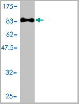
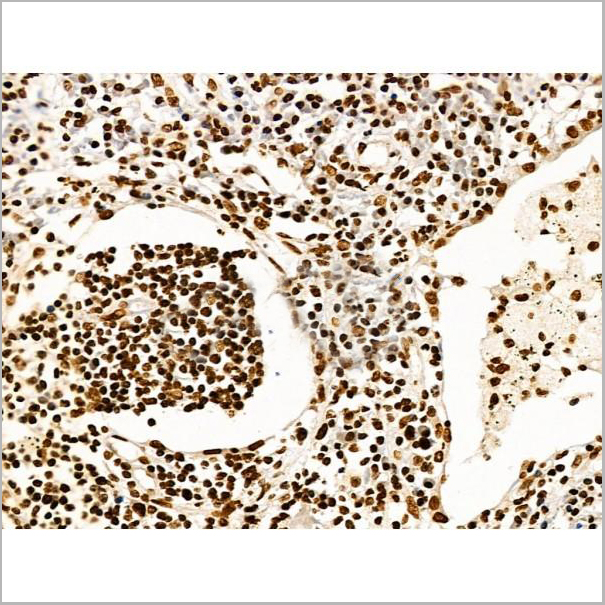
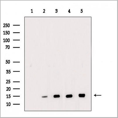
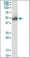
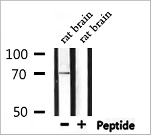
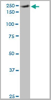
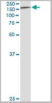
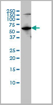
 Application Data
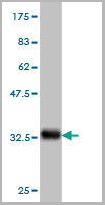
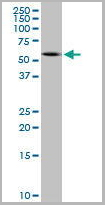
gp120 (ADA) (HIV-1/Clade B), Recombinant Protein (Cat# AAA13750)
Full Name
gp120 (ADA) (HIV-1/Clade B) (aa 34-518), monomer only
Applications
Western Blot
Purity
>= 95% (SDS-PAGE)
Pricing
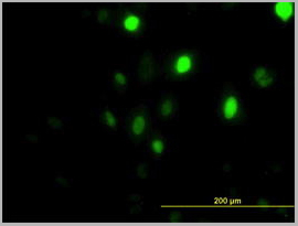
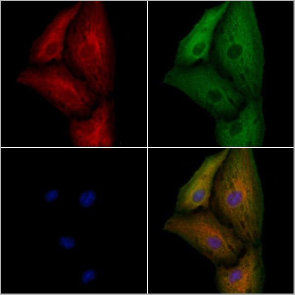
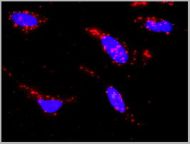
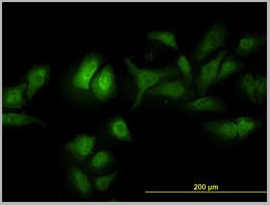
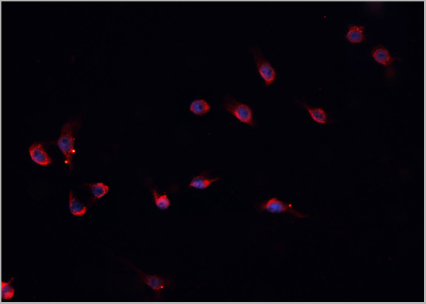
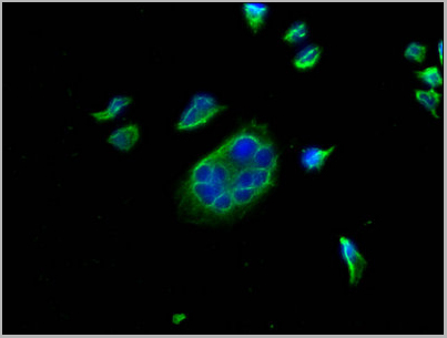
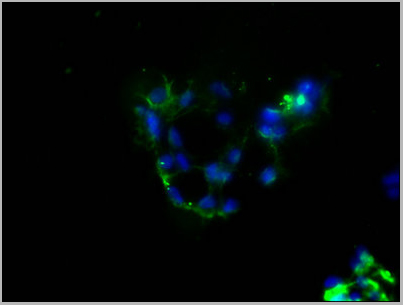
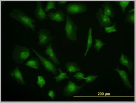
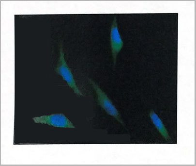
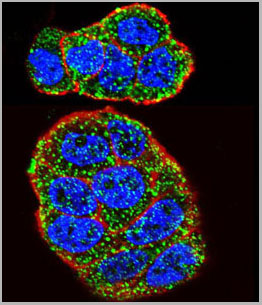
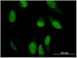
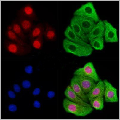 IF (Immunofluorescence)
(AAA31190 staining Hela cells by IF/ICC. The samples were fixed with PFA and permeabilized in 0.1% Triton X-100,then blocked in 10% serum for 45 minutes at 25°C. Samples were then incubated with primary Ab(AAA31190 1:200) and mouse anti-beta tubulin Ab(T0023 1:200) for 1 hour at 37°C. An AlexaFluor594 conjugated goat anti-rabbit IgG(H+L) Ab(Red) and an AlexaFluor488 conjugated goat anti-mouse IgG(H+L) Ab(Green) were used as the secondary antibody.The nuclear counter stain is DAPI(blue).)
IF (Immunofluorescence)
(AAA31190 staining Hela cells by IF/ICC. The samples were fixed with PFA and permeabilized in 0.1% Triton X-100,then blocked in 10% serum for 45 minutes at 25°C. Samples were then incubated with primary Ab(AAA31190 1:200) and mouse anti-beta tubulin Ab(T0023 1:200) for 1 hour at 37°C. An AlexaFluor594 conjugated goat anti-rabbit IgG(H+L) Ab(Red) and an AlexaFluor488 conjugated goat anti-mouse IgG(H+L) Ab(Green) were used as the secondary antibody.The nuclear counter stain is DAPI(blue).)
KDM4B/JMJD2B, Polyclonal Antibody (Cat# AAA31190)
Full Name
KDM4B/JMJD2B Antibody
Gene Names
KDM4B; JMJD2B; TDRD14B
Reactivity
Human, Mouse, Rat
Predicted Reactivity: Pig(100%), Bovine(100%), Sheep(100%), Dog(88%)
Predicted Reactivity: Pig(100%), Bovine(100%), Sheep(100%), Dog(88%)
Applications
Western Blot, Immunofluorescence, Immunocytochemistry
Purity
The antiserum was purified by peptide affinity chromatography using SulfoLink Coupling Resin (Thermo Fisher Scientific).
Pricing


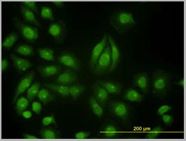
 IF (Immunofluorescence)
(Figure 8 Immunofluorescence Validation of JMJD3 in K562 Cells Immunofluorescent analysis of 4% paraformaldehyde fixed K562 cells labeling JMJD3 with AAA10925 at 20 ug/mL, followed by goat anti-rabbit IgG secondary antibody at 1/500 dilution (red).)
IF (Immunofluorescence)
(Figure 8 Immunofluorescence Validation of JMJD3 in K562 Cells Immunofluorescent analysis of 4% paraformaldehyde fixed K562 cells labeling JMJD3 with AAA10925 at 20 ug/mL, followed by goat anti-rabbit IgG secondary antibody at 1/500 dilution (red).)
JMJD3, Antibody (Cat# AAA10925)
Full Name
JMJD3 Antibody
Gene Names
KDM6B; JMJD3
Reactivity
Human, Mouse, Rat
Applications
Immunocytochemistry, Immunofluorescence, Western Blot
Purity
JMJD3 Antibody is affinity chromatography purified via peptide column.
Pricing
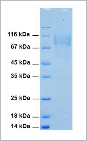
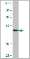
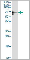
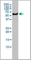
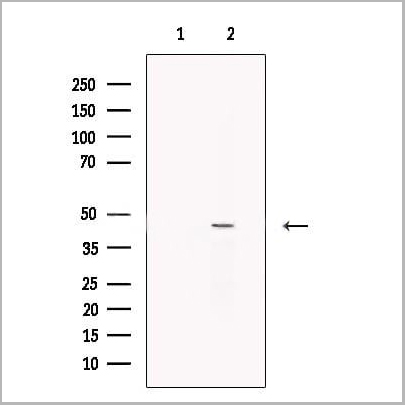
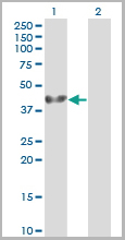
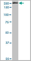
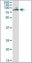
 Application Data
(SDS-PAGE: Purified HIV-1 gp120 (JRFL) (Clade B) protein)
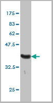
Application Data
(SDS-PAGE: Purified HIV-1 gp120 (JRFL) (Clade B) protein)
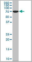
gp120 (JRFL) (HIV-1/Clade B), Recombinant Protein (Cat# AAA13752)
Full Name
gp120 (JRFL) (HIV-1/Clade B) (aa 34-518)
Applications
Western Blot
Purity
≥ 95% purity (SDS-PAGE)
Pricing
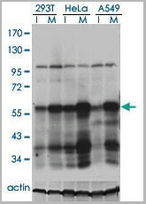
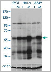
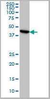
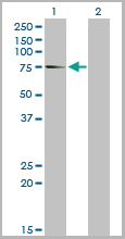
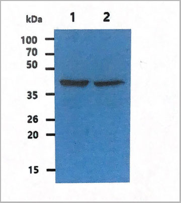
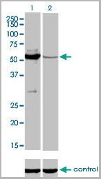
 Application Data
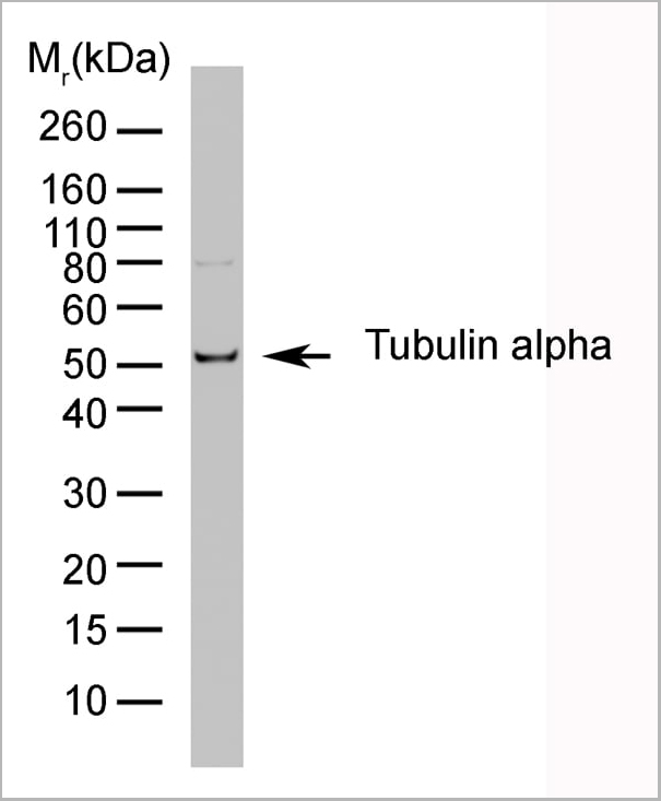
(Western blot analysis of CD107b on J774 cell lysate probed with Rat anti Mouse CD107b at 1/500, 1/1000, 1/2000. Rat anti tubulin alpha is included as a loading control. Detection is with Goat anti Rat IgG:Dylight®)
Application Data
(Western blot analysis of CD107b on J774 cell lysate probed with Rat anti Mouse CD107b at 1/500, 1/1000, 1/2000. Rat anti tubulin alpha is included as a loading control. Detection is with Goat anti Rat IgG:Dylight®)
TUBULIN ALPHA, Monoclonal Antibody (Cat# AAA12010)
Full Name
RAT ANTI TUBULIN ALPHA
Applications
Immunohistochemistry, Immunofluorescence, Radioimmunoassay, Western Blot
Pricing
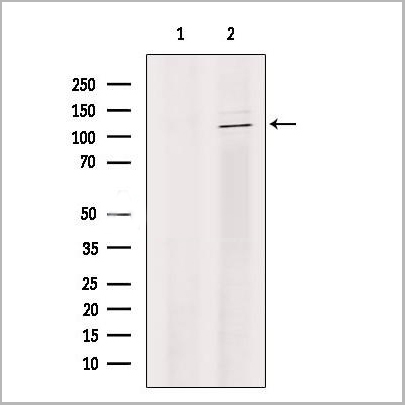
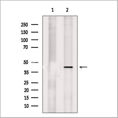
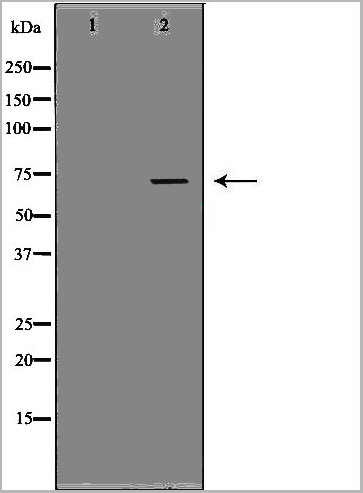
 WB (Western Blot)
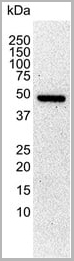
(K-562 cells were subjected to SDS PAGE followed by western blot with AAA27491 (phospho (403/404) -TDP43 Antibody) at dilution of 1:3000)
WB (Western Blot)
(K-562 cells were subjected to SDS PAGE followed by western blot with AAA27491 (phospho (403/404) -TDP43 Antibody) at dilution of 1:3000)
phospho(403/404)-TDP43, Monoclonal Antibody (Cat# AAA27491)
Full Name
phospho(403/404)-TDP43 Mouse Monoclonal
Reactivity
Human, Mouse
Applications
Western Blot
Purity
Purity: >95% as determined by SDS-PAGE
Purification: Protein A+G purification
Purification: Protein A+G purification
Pricing
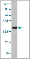
 Application Data
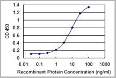
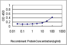
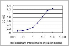
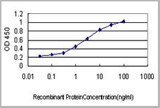
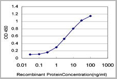
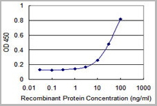
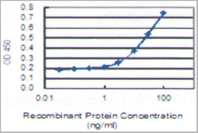
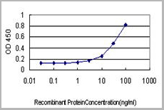
(Detection limit for recombinant GST tagged ROBO3 is 0.1ng/ml using AAA24095 as a capture antibody.)
Application Data
(Detection limit for recombinant GST tagged ROBO3 is 0.1ng/ml using AAA24095 as a capture antibody.)
ROBO3, Monoclonal Antibody (Cat# AAA24095)
Full Name
ROBO3 (Roundabout Homolog 3, Roundabout-like Protein 3, HGPS, HGPPS, RBIG1, RIG1)
Gene Names
ROBO3; HGPS; RIG1; HGPPS; RBIG1
Reactivity
Human
Applications
Western Blot
Purity
Affinity Purified
Purified by Protein A affinity chromatography.
Purified by Protein A affinity chromatography.
Pricing
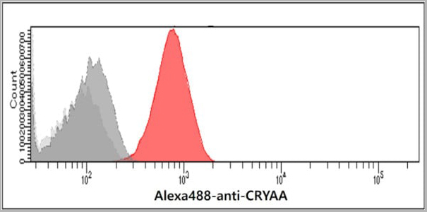 FCM (Flow Cytometry)
(Flow cytometry analysis of Crystallin alpha A in Balb/3T3 cells. The cell was stained with AAA11723 at 2-5ug for 1x10^6cells (red). A Goat anti mouse IgG (Alexa fluor 488) was used as the secondary antibody. Mouse monoclonal IgG was used as the isotype control (dark gray), cells without incubation with primary and secondary antibody was used as the negative control (light gray).)
FCM (Flow Cytometry)
(Flow cytometry analysis of Crystallin alpha A in Balb/3T3 cells. The cell was stained with AAA11723 at 2-5ug for 1x10^6cells (red). A Goat anti mouse IgG (Alexa fluor 488) was used as the secondary antibody. Mouse monoclonal IgG was used as the isotype control (dark gray), cells without incubation with primary and secondary antibody was used as the negative control (light gray).)
A crystallin A, Monoclonal Antibody (Cat# AAA11723)
Full Name
A crystallin A antibody
Gene Names
CRYAA; CRYA1; HSPB4; CTRCT9
Reactivity
Human
Applications
Western Blot, Flow Cytometry
Purity
By protein-G affinity chromatography
Pricing
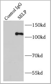
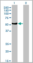
 WB (Western Blot)
(Jurkat cells were subjected to SDS PAGE followed by western blot with AAA27492 (SELP Antibody) at dilution of 1:500)
WB (Western Blot)
(Jurkat cells were subjected to SDS PAGE followed by western blot with AAA27492 (SELP Antibody) at dilution of 1:500)
P-selectin, Monoclonal Antibody (Cat# AAA27492)
Full Name
P-selectin Mouse Monoclonal
Gene Names
SELP; CD62; GRMP; PSEL; CD62P; GMP140; LECAM3; PADGEM
Reactivity
Human, Rat
Applications
Immunohistochemistry, Western Blot, Immunoprecipitation
Purity
>95% as determined by SDS-PAGE
Protein A+G purification
Protein A+G purification
Pricing
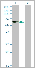
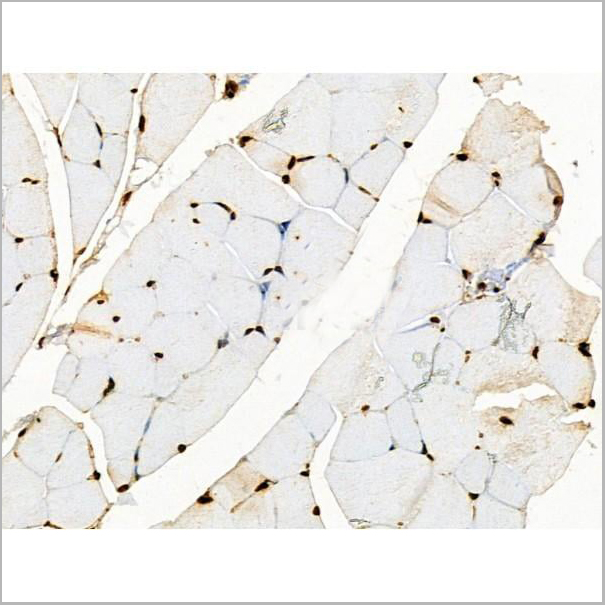
ENO3, Monoclonal Antibody (Cat# AAA24202)
Full Name
ENO3 (Enolase 3, 2-phospho-D-glycerate Hydro-lyase, beta-Enolase, Muscle-specific Enolase, MSE, Skeletal Muscle Enolase) (AP)
Gene Names
ENO3; MSE; GSD13
Reactivity
Human
Applications
EIA, IHC, WB
Purity
Purified by Protein A Affinity Chromatography.
Pricing
 Application Data
(Detection limit for recombinant GST tagged PLK1 is ~1ng/ml as a capture antibody.)
Application Data
(Detection limit for recombinant GST tagged PLK1 is ~1ng/ml as a capture antibody.)
PLK1, Monoclonal Antibody (Cat# AAA24321)
Full Name
PLK1 (STPK13, Polo-like Kinase 1, Serine/Threonine-protein Kinase 13, Serine/Threonine-protein Kinase PLK1, PLK, PLK-1) (AP)
Gene Names
PLK1; PLK; STPK13
Reactivity
Human
Applications
Immunoprecipitation, Western Blot
Purity
Purified by Protein A Affinity Chromatography.
Pricing
Endogenous Retrovirus Type K, Envelope Protein, Monoclonal Antibody (Cat# AAA14674)
Full Name
Endogenous Retrovirus Type K, Envelope Protein (HERV K)
Applications
ELISA
Purity
Purified by Protein G affinity chromatography.
Pricing
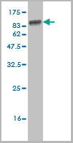
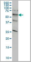
ERO1L, Monoclonal Antibody (Cat# AAA24499)
Full Name
ERO1L (ERO1-like Protein alpha, ERO1-L, ERO1-L-alpha, Endoplasmic Oxidoreductin-1-like Protein, Oxidoreductin-1-L-alpha, UNQ434/PRO865) APC
Gene Names
ERO1A; ERO1L; ERO1-L; ERO1LA; Ero1alpha; ERO1-alpha; ERO1-L-alpha
Reactivity
Human, Rat
Applications
EIA, IHC, WB
Purity
Purified by Protein A Affinity Chromatography.
Pricing
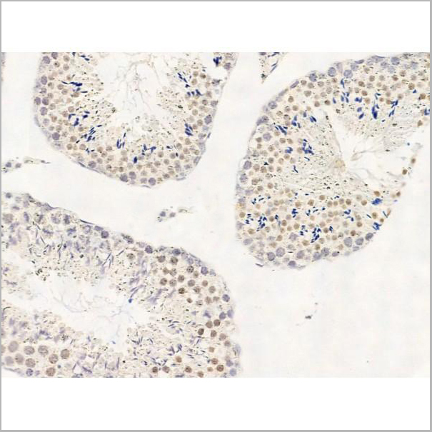
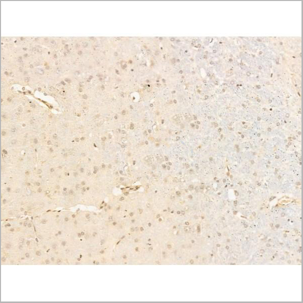
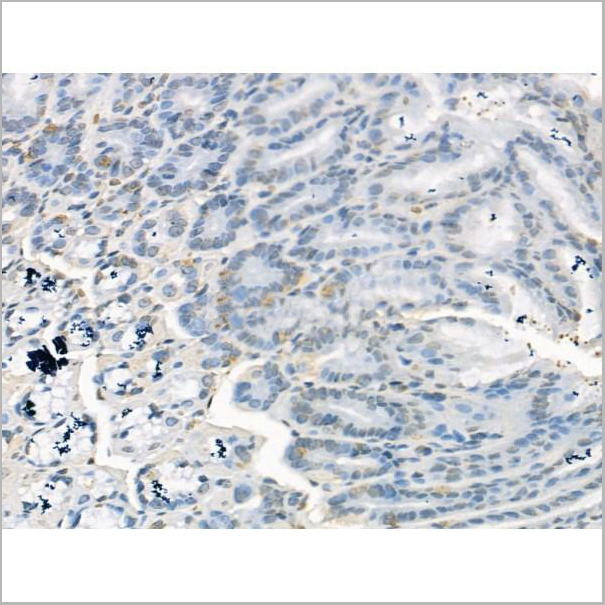
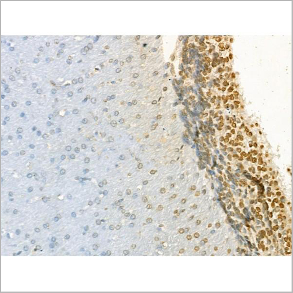
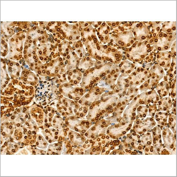
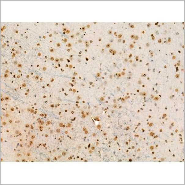
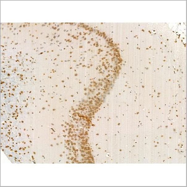
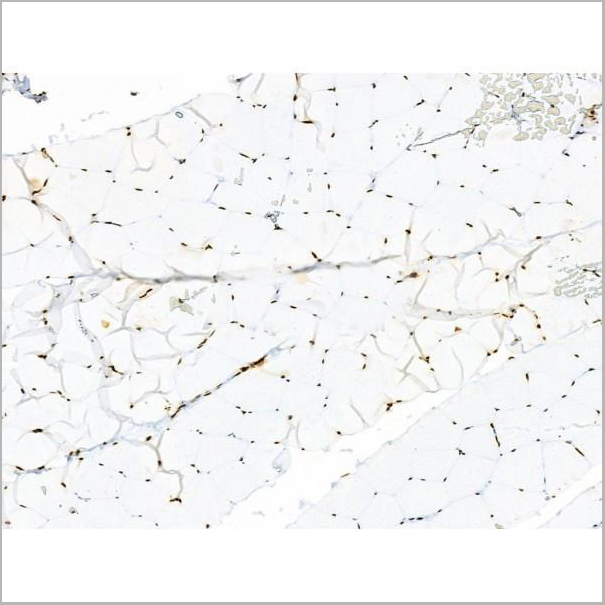
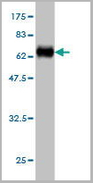
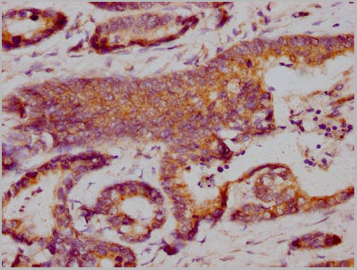
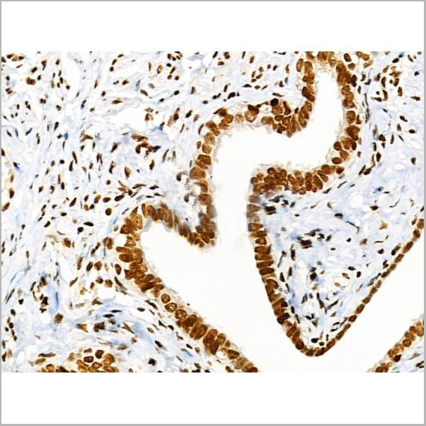
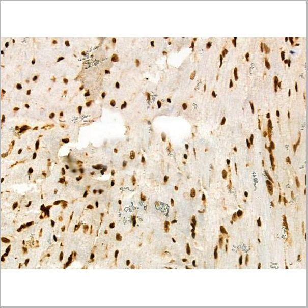
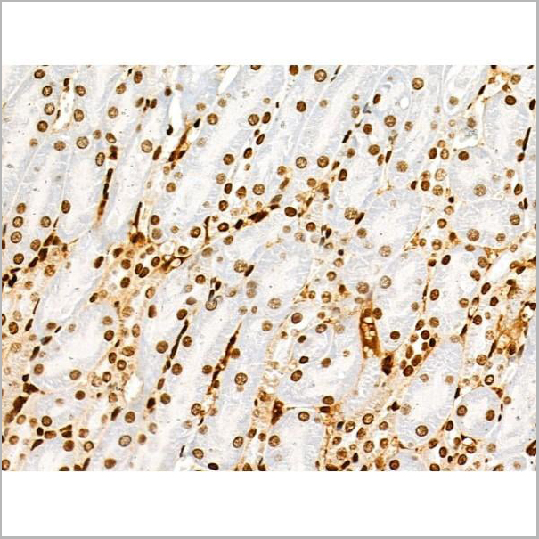
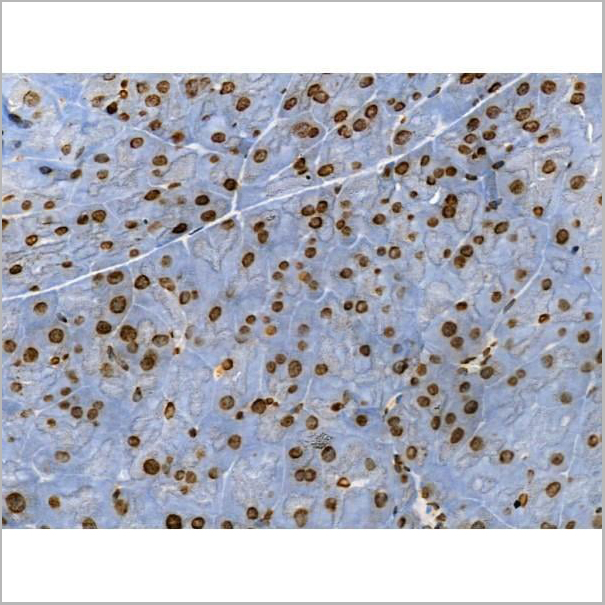
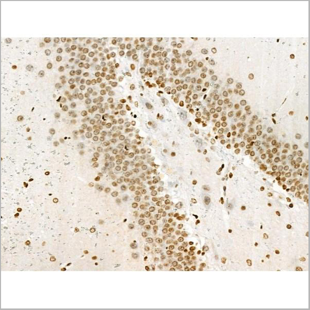
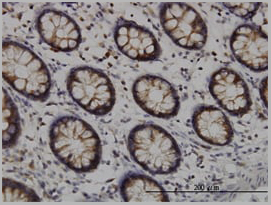
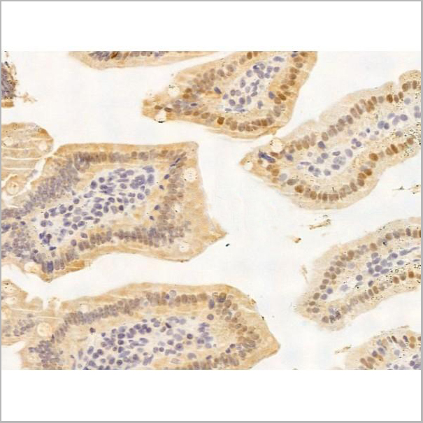 IHC (Immunohistchemistry-Paraffin)
(AAA31252 at 1/100 staining Mouse colorectal tissue by IHC-P. The sample was formaldehyde fixed and a heat mediated antigen retrieval step in citrate buffer was performed. The sample was then blocked and incubated with the primary antibody at 4°C overnight. An HRP conjugated anti-Rabbit antibody was used as the secondary antibody.)
IHC (Immunohistchemistry-Paraffin)
(AAA31252 at 1/100 staining Mouse colorectal tissue by IHC-P. The sample was formaldehyde fixed and a heat mediated antigen retrieval step in citrate buffer was performed. The sample was then blocked and incubated with the primary antibody at 4°C overnight. An HRP conjugated anti-Rabbit antibody was used as the secondary antibody.)
KDM5C/Jarid1C/SMCX, Polyclonal Antibody (Cat# AAA31252)
Full Name
KDM5C/Jarid1C/SMCX Antibody
Gene Names
KDM5C; MRXJ; SMCX; MRX13; MRXSJ; XE169; MRXSCJ; JARID1C; DXS1272E
Reactivity
Human, Mouse, Rat
Predicted Reactivity: Pig(100%), Bovine(100%), Horse(100%), Sheep(100%), Rabbit(100%), Dog(100%)
Predicted Reactivity: Pig(100%), Bovine(100%), Horse(100%), Sheep(100%), Rabbit(100%), Dog(100%)
Applications
ELISA
Purity
The antiserum was purified by peptide affinity chromatography using SulfoLink Coupling Resin (Thermo Fisher Scientific).
Pricing
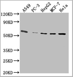
 IF (Immunofluorescence)
(AAA31225 staining A549 cells by IF/ICC. The samples were fixed with PFA and permeabilized in 0.1% Triton X-100,then blocked in 10% serum for 45 minutes at 25°C. Samples were then incubated with primary Ab(AAA31225) and mouse anti-beta tubulin Ab(T0023) for 1 hour at 37°C. An AlexaFluor594 conjugated goat anti-rabbit IgG(H+L) Ab(Red) and an AlexaFluor488 conjugated goat anti-mouse IgG(H+L) Ab(Green) were used as the secondary antibody.The nuclear counter stain is DAPI (blue).)
IF (Immunofluorescence)
(AAA31225 staining A549 cells by IF/ICC. The samples were fixed with PFA and permeabilized in 0.1% Triton X-100,then blocked in 10% serum for 45 minutes at 25°C. Samples were then incubated with primary Ab(AAA31225) and mouse anti-beta tubulin Ab(T0023) for 1 hour at 37°C. An AlexaFluor594 conjugated goat anti-rabbit IgG(H+L) Ab(Red) and an AlexaFluor488 conjugated goat anti-mouse IgG(H+L) Ab(Green) were used as the secondary antibody.The nuclear counter stain is DAPI (blue).)
KCNK9, Polyclonal Antibody (Cat# AAA31225)
Full Name
KCNK9 Antibody
Gene Names
KCNK9; KT3.2; TASK3; K2p9.1; TASK-3
Reactivity
Human, Mouse, Rat
Predicted Reactivity: Pig(100%), Bovine(100%), Horse(100%), Rabbit(100%), Chicken(83%), Xenopus(83%)
Predicted Reactivity: Pig(100%), Bovine(100%), Horse(100%), Rabbit(100%), Chicken(83%), Xenopus(83%)
Applications
ELISA
Purity
The antiserum was purified by peptide affinity chromatography using SulfoLink Coupling Resin (Thermo Fisher Scientific).
Pricing
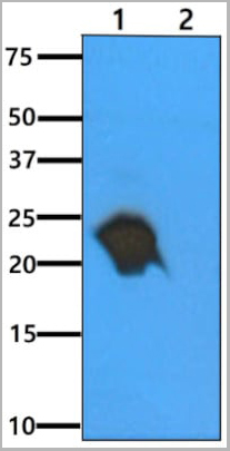
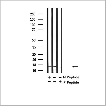
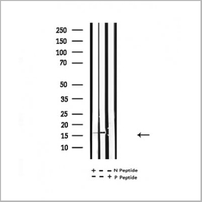
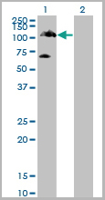
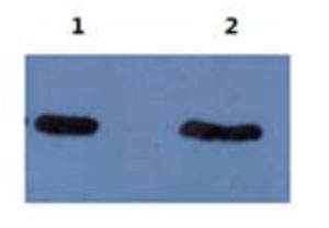 WB (Western Blot)
(The following image is the result of using mAb 5B8 ascites at 1:5000 dilution to detect Abeta40 and Abeta42 on Western blot. Lane 1: Abeta42, Lane 2: Abeta40.)
WB (Western Blot)
(The following image is the result of using mAb 5B8 ascites at 1:5000 dilution to detect Abeta40 and Abeta42 on Western blot. Lane 1: Abeta42, Lane 2: Abeta40.)
amyloid beta peptide N-terminal, Monoclonal Antibody (Cat# AAA14629)
Full Name
mAb anti-human amyloid beta peptide N-terminus
Reactivity
Human
Applications
Western Blot
Purity
Protein G affinity purified
Pricing
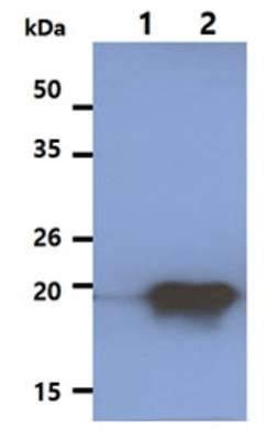
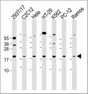
 WB (Western Blot)
(HSPA1L monoclonal antibody Western Blot analysis of HSPA1L expression in PC-12.)
WB (Western Blot)
(HSPA1L monoclonal antibody Western Blot analysis of HSPA1L expression in PC-12.)
HSPA1L, Monoclonal Antibody (Cat# AAA24833)
Full Name
HSPA1L (Heat Shock 70kD Protein 1-like, HSP70-Hom, Heat shock 70kD protein 1L, Heat Shock 70kD Protein 1-Hom) (Biotin)
Gene Names
HSPA1L; HSP70T; hum70t; HSP70-1L; HSP70-HOM
Reactivity
Human, Rat
Applications
Immunohistochemistry, Western Blot
Purity
Purified by Protein A Affinity Chromatography.
Pricing


 Application Data
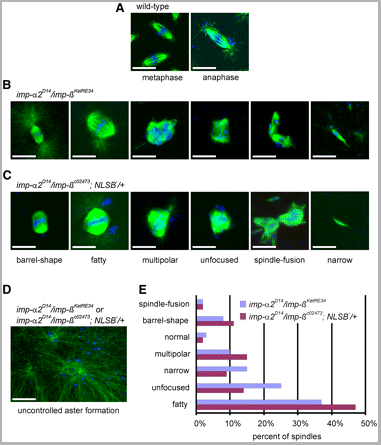
(Published customer image: Spindle abnormalities in embryos derived from imp-a2D14/imp-betaKetRE34 and imp-a2D14/imp-betac02473; NLSB-/+ females. (A -D) Wild-type and mutant embryos stained for a-tubulin (green) and DNA (blue). (A) Mitotic spindles in wild-type embryos at metaphase and anaphase. (B, C) Categories of spindle abnormalities found in embryos derived from (B) imp-a2D14/imp-betaKetRE34 and (C) imp-a2D14/imp-betac02743; NLSB-/+ females. (D) Formation of aster networks found in both genotypes. Scale bar: 10 um. (E) Frequency of spindle defects in embryos from both types of mutant females. Female genotypes are displayed at the upper right corner. At least 200 spindles were scored for both genotypes.From: Specific Cooperation Between Imp-a2 and Imp-beta/Ketel in Spindle Assembly During Drosophila Early Nuclear Divisions Erika Vir¡gh, M¡ty¡s Gorj¡n¡cz, Istv¡n T¶r¶k, Tolga Eichhorn, Sowjanya Kallakuri, Tam¡s Szlanka, Istv¡n Kiss, and Bernard M. Mechler G3 January 2012 2:1-14.)
Application Data
(Published customer image: Spindle abnormalities in embryos derived from imp-a2D14/imp-betaKetRE34 and imp-a2D14/imp-betac02473; NLSB-/+ females. (A -D) Wild-type and mutant embryos stained for a-tubulin (green) and DNA (blue). (A) Mitotic spindles in wild-type embryos at metaphase and anaphase. (B, C) Categories of spindle abnormalities found in embryos derived from (B) imp-a2D14/imp-betaKetRE34 and (C) imp-a2D14/imp-betac02743; NLSB-/+ females. (D) Formation of aster networks found in both genotypes. Scale bar: 10 um. (E) Frequency of spindle defects in embryos from both types of mutant females. Female genotypes are displayed at the upper right corner. At least 200 spindles were scored for both genotypes.From: Specific Cooperation Between Imp-a2 and Imp-beta/Ketel in Spindle Assembly During Drosophila Early Nuclear Divisions Erika Vir¡gh, M¡ty¡s Gorj¡n¡cz, Istv¡n T¶r¶k, Tolga Eichhorn, Sowjanya Kallakuri, Tam¡s Szlanka, Istv¡n Kiss, and Bernard M. Mechler G3 January 2012 2:1-14.)
TUBULIN ALPHA, Monoclonal Antibody (Cat# AAA12232)
Full Name
RAT ANTI TUBULIN ALPHA:HRP
Applications
Immunohistochemistry, Western Blot
Pricing


 Application Data
(Published customer image: Spindle abnormalities in embryos derived from imp-a2D14/imp-betaKetRE34 and imp-a2D14/imp-betac02473; NLSB-/+ females. (A -D) Wild-type and mutant embryos stained for a-tubulin (green) and DNA (blue). (A) Mitotic spindles in wild-type embryos at metaphase and anaphase. (B, C) Categories of spindle abnormalities found in embryos derived from (B) imp-a2D14/imp-betaKetRE34 and (C) imp-a2D14/imp-betac02743; NLSB-/+ females. (D) Formation of aster networks found in both genotypes. Scale bar: 10 um. (E) Frequency of spindle defects in embryos from both types of mutant females. Female genotypes are displayed at the upper right corner. At least 200 spindles were scored for both genotypes.From: Specific Cooperation Between Imp-a2 and Imp-beta/Ketel in Spindle Assembly During Drosophila Early Nuclear Divisions Erika Vir¡gh, M¡ty¡s Gorj¡n¡cz, Istv¡n T¶r¶k, Tolga Eichhorn, Sowjanya Kallakuri, Tam¡s Szlanka, Istv¡n Kiss, and Bernard M. Mechler G3 January 2012 2:1-14.)
Application Data
(Published customer image: Spindle abnormalities in embryos derived from imp-a2D14/imp-betaKetRE34 and imp-a2D14/imp-betac02473; NLSB-/+ females. (A -D) Wild-type and mutant embryos stained for a-tubulin (green) and DNA (blue). (A) Mitotic spindles in wild-type embryos at metaphase and anaphase. (B, C) Categories of spindle abnormalities found in embryos derived from (B) imp-a2D14/imp-betaKetRE34 and (C) imp-a2D14/imp-betac02743; NLSB-/+ females. (D) Formation of aster networks found in both genotypes. Scale bar: 10 um. (E) Frequency of spindle defects in embryos from both types of mutant females. Female genotypes are displayed at the upper right corner. At least 200 spindles were scored for both genotypes.From: Specific Cooperation Between Imp-a2 and Imp-beta/Ketel in Spindle Assembly During Drosophila Early Nuclear Divisions Erika Vir¡gh, M¡ty¡s Gorj¡n¡cz, Istv¡n T¶r¶k, Tolga Eichhorn, Sowjanya Kallakuri, Tam¡s Szlanka, Istv¡n Kiss, and Bernard M. Mechler G3 January 2012 2:1-14.)
TUBULIN ALPHA, Monoclonal Antibody (Cat# AAA12009)
Full Name
RAT ANTI TUBULIN ALPHA
Applications
Immunohistochemistry, Immunofluorescence, Immunoprecipitation, Radioimmunoassay, Western Blot
Pricing


 Application Data
(Published customer image: Mouse anti V5 tag antibody, clone SV5-Pk1 used for the detection of V5 tagged WEEV_nsP3 protein by western blotting and immunofluorescenceImage caption: WEEV nsP3 interaction with host IKKbeta. A) U87MGs were transfected in a 6-well plate with 5 ug of pUC19 and WEEV_nsP3_HA for 24 hours. Cell lysates were resolved using SDS-PAGE and subsequently immunoblotted with V5 antibody and beta-actin served as a loading control. B) U87MGs were transfected with WEEV_nsP3_V5; cells were fixed after 24 hours and stained with antibodies against the endogenous IKKbeta and the V5 tag. Cells were incubated with appropriate secondary Alexa Fluor antibodies and the nuclei stained with DAPI. Co-localization of IKKbeta with WEEV_nsP3_V5 (yellow) was observed as shown by the arrows. B) Panels E -H serve as an example of transfected cells in a given field of view that show co-localization of IKKbeta and WEEV_nsP3_V5 24 hours post transfection. Panels I-L represent magnified images of other cells showing co-localization of IKKbeta and WEEV_nsP3_V5. Panel M is a magnified image of panel L. The co-localization was confirmed by Z-stack analysis. Co-localization was calculated to be approximately in 61% of cells (163 cells were counted of which 44% demonstrated expression of nsP3. Of those cells that expressed nsP3, 61% showed co-localization of both proteins). Images were taken using Nikon Eclipse TE2000-U at 60x magnification and are representative of 2 independent experiments.From: Amaya M, Voss K, Sampey G, Senina S, de la Fuente C, et al. (2014) The Role of IKKbeta in Venezuelan Equine Encephalitis Virus Infection. PLoS ONE 9(2): e86745.)
Application Data
(Published customer image: Mouse anti V5 tag antibody, clone SV5-Pk1 used for the detection of V5 tagged WEEV_nsP3 protein by western blotting and immunofluorescenceImage caption: WEEV nsP3 interaction with host IKKbeta. A) U87MGs were transfected in a 6-well plate with 5 ug of pUC19 and WEEV_nsP3_HA for 24 hours. Cell lysates were resolved using SDS-PAGE and subsequently immunoblotted with V5 antibody and beta-actin served as a loading control. B) U87MGs were transfected with WEEV_nsP3_V5; cells were fixed after 24 hours and stained with antibodies against the endogenous IKKbeta and the V5 tag. Cells were incubated with appropriate secondary Alexa Fluor antibodies and the nuclei stained with DAPI. Co-localization of IKKbeta with WEEV_nsP3_V5 (yellow) was observed as shown by the arrows. B) Panels E -H serve as an example of transfected cells in a given field of view that show co-localization of IKKbeta and WEEV_nsP3_V5 24 hours post transfection. Panels I-L represent magnified images of other cells showing co-localization of IKKbeta and WEEV_nsP3_V5. Panel M is a magnified image of panel L. The co-localization was confirmed by Z-stack analysis. Co-localization was calculated to be approximately in 61% of cells (163 cells were counted of which 44% demonstrated expression of nsP3. Of those cells that expressed nsP3, 61% showed co-localization of both proteins). Images were taken using Nikon Eclipse TE2000-U at 60x magnification and are representative of 2 independent experiments.From: Amaya M, Voss K, Sampey G, Senina S, de la Fuente C, et al. (2014) The Role of IKKbeta in Venezuelan Equine Encephalitis Virus Infection. PLoS ONE 9(2): e86745.)
V5-TAG, Monoclonal Antibody (Cat# AAA12081)
Full Name
MOUSE ANTI V5-TAG:HRP
Applications
Western Blot
Pricing
 WB (Western Blot)
(STK33 monoclonal antibody, Western Blot analysis of STK33 expression in HeLa.)
WB (Western Blot)
(STK33 monoclonal antibody, Western Blot analysis of STK33 expression in HeLa.)
STK33, Monoclonal Antibody (Cat# AAA25856)
Full Name
STK33 (Serine/Threonine Kinase 33) (PE)
Reactivity
Human
Applications
Immunofluorescence, Immunoprecipitation, Western Blot
Purity
Purified by Protein A Affinity Chromatography.
Pricing
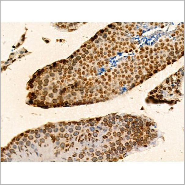
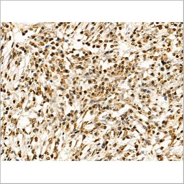
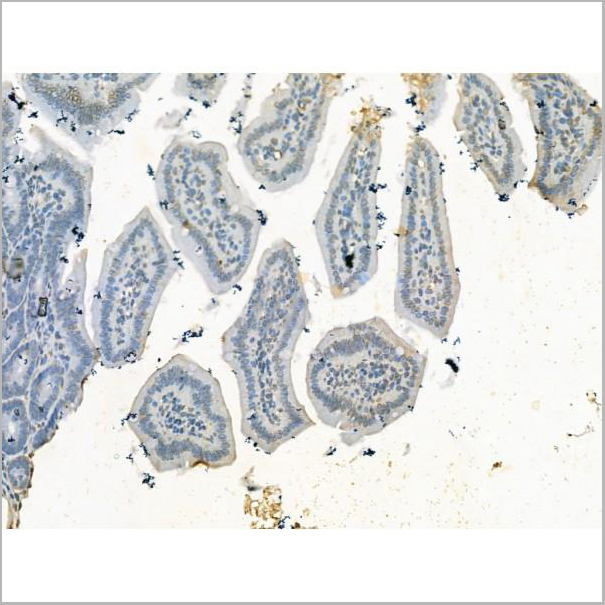
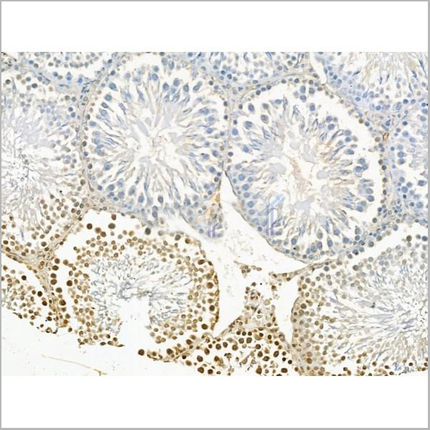
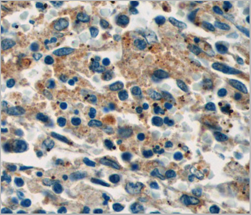
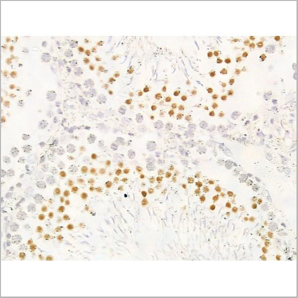
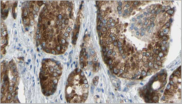
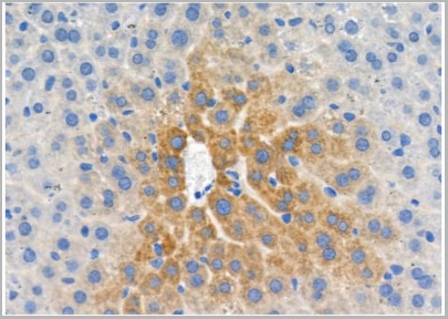
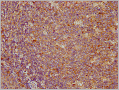
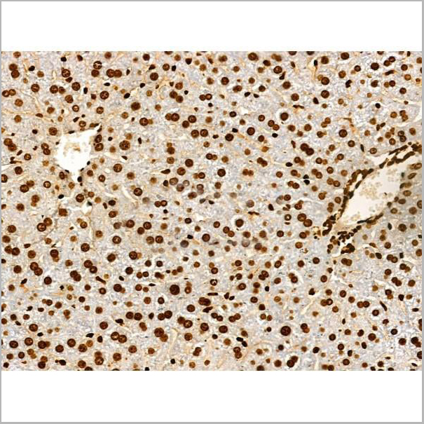
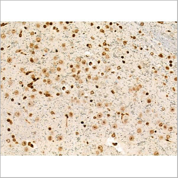
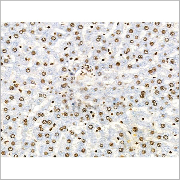
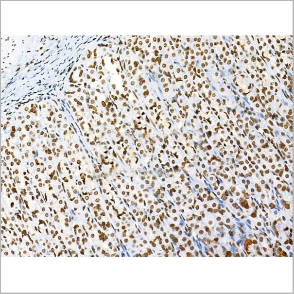
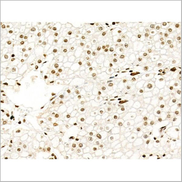
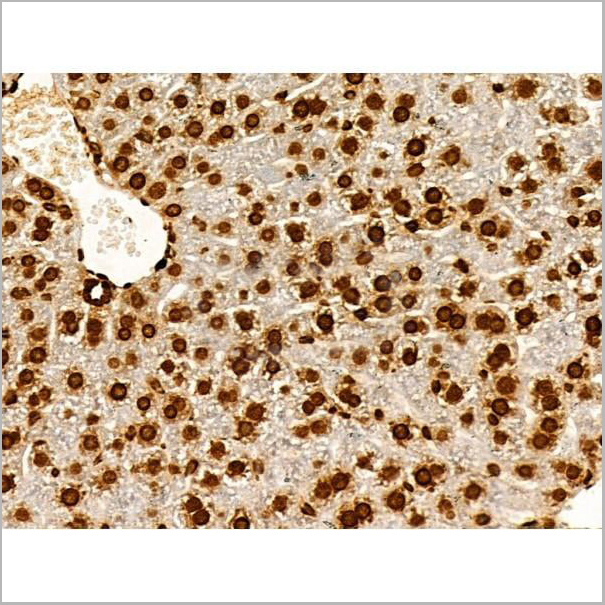 IHC (Immunohistochemistry)
(At 1/100 staining Mouse liver tissue by IHC-P. The sample was formaldehyde fixed and a heat mediated antigen retrieval step in citrate buffer was performed. The sample was then blocked and incubated with the primary antibody at 4 degree C overnight. An HRP conjugated anti-Rabbit antibody was used as the secondary antibody.)
IHC (Immunohistochemistry)
(At 1/100 staining Mouse liver tissue by IHC-P. The sample was formaldehyde fixed and a heat mediated antigen retrieval step in citrate buffer was performed. The sample was then blocked and incubated with the primary antibody at 4 degree C overnight. An HRP conjugated anti-Rabbit antibody was used as the secondary antibody.)
Histone H2B, Polyclonal Antibody (Cat# AAA31352)
Full Name
Acetyl-Histone H2B (Lys20) Antibody
Reactivity
Human, Mouse, Rat
Applications
Western Blot, Immunohistochemistry, Immunofluorescence, Immunocytochemistry, Peptide ELISA
Purity
The antiserum was purified by peptide affinity chromatography using SulfoLink Coupling Resin
Pricing


 Application Data
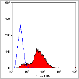
(Staining of mouse spleen with Hamster anti Mouse CD81: Alexa Fluor 488)
Application Data
(Staining of mouse spleen with Hamster anti Mouse CD81: Alexa Fluor 488)
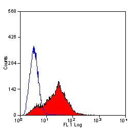
CD81, Monoclonal Antibody (Cat# AAA11869)
Full Name
HAMSTER ANTI MOUSE CD81:FITC
Gene Names
Cd81; Tapa1; Tapa-1; Tspan28
Applications
Flow Cytometry
Pricing


 Application Data
(Staining of mouse spleen with Hamster anti Mouse CD81: Alexa Fluor 488)
Application Data
(Staining of mouse spleen with Hamster anti Mouse CD81: Alexa Fluor 488)
CD81, Monoclonal Antibody (Cat# AAA12033)
Full Name
HAMSTER ANTI MOUSE CD81:RPE
Gene Names
Cd81; Tapa1; Tapa-1; Tspan28
Applications
Flow Cytometry
Pricing
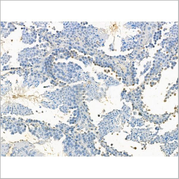
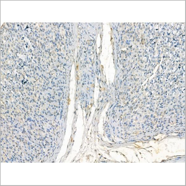
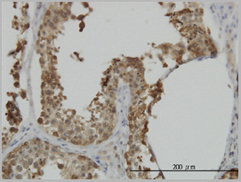
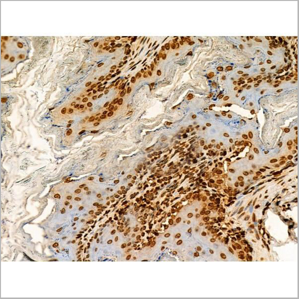
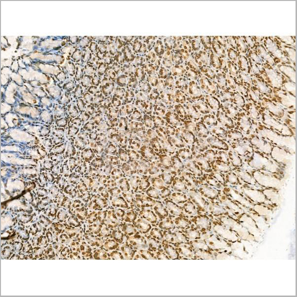
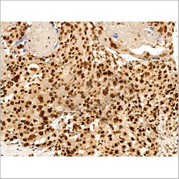
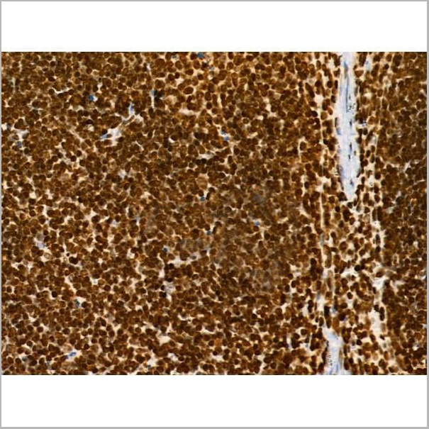
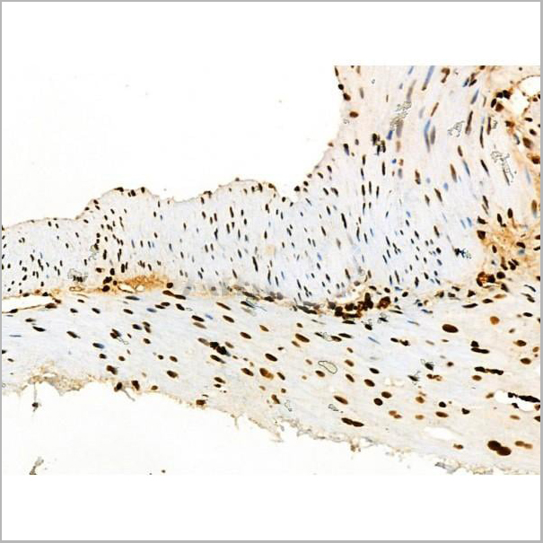
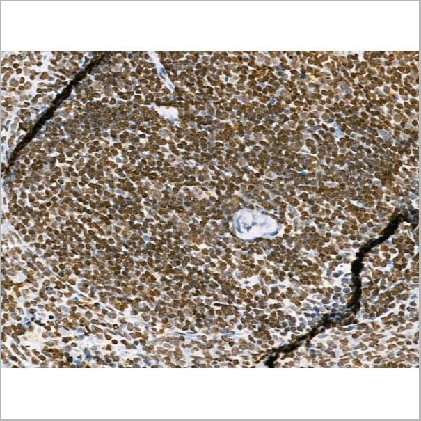
 IHC (Immunohistochemistry)
(AAA31102 at 1/100 staining Human prostate tissue by IHC-P. The sample was formaldehyde fixed and a heat mediated antigen retrieval step in citrate buffer was performed. The sample was then blocked and incubated with the antibody for 1.5 hours at 22 degree C. An HRP conjugated goat anti-rabbit antibody was used as the secondary.)
IHC (Immunohistochemistry)
(AAA31102 at 1/100 staining Human prostate tissue by IHC-P. The sample was formaldehyde fixed and a heat mediated antigen retrieval step in citrate buffer was performed. The sample was then blocked and incubated with the antibody for 1.5 hours at 22 degree C. An HRP conjugated goat anti-rabbit antibody was used as the secondary.)
ATP12A, Polyclonal Antibody (Cat# AAA31102)
Full Name
ATP12A Antibody
Gene Names
ATP12A; HK; ATP1AL1
Reactivity
Human, Mouse
Applications
Western Blot, Immunohistochemistry
Purity
The antiserum was purified by peptide affinity chromatography using SulfoLink™ Coupling Resin (Thermo Fisher Scientific).
Pricing
 Application Data
(Detection limit for recombinant GST tagged ADRM1 is ~0.1ng/ml as a capture antibody.)
Application Data
(Detection limit for recombinant GST tagged ADRM1 is ~0.1ng/ml as a capture antibody.)
ADRM1, Monoclonal Antibody (Cat# AAA25601)
Full Name
ADRM1 (GP110, Proteasomal Ubiquitin Receptor ADRM1, 110kD Cell Membrane Glycoprotein, Adhesion-regulating Molecule 1, Proteasome Regulatory Particle Non-ATPase 13, Rpn13 Homolog, MGC29536) (PE)
Gene Names
ADRM1; ARM1; ARM-1; GP110
Reactivity
Human
Applications
Immunofluorescence, Western Blot
Purity
Purified by Protein A Affinity Chromatography.
Pricing
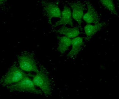
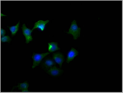
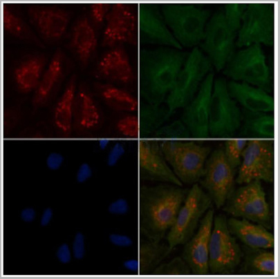 IF (Immunofluorescence)
(AAA31124 staining A549 cells by IF/ICC. The samples were fixed with PFA and permeabilized in 0.1% Triton X-100,then blocked in 10% serum for 45 minutes at 25°C. Samples were then incubated with primary Ab(AAA31124) and mouse anti-beta tubulin Ab for 1 hour at 37°C. An AlexaFluor594 conjugated goat anti-rabbit IgG(H+L) Ab(Red) and an AlexaFluor488 conjugated goat anti-mouse IgG(H+L) Ab(Green) were used as the secondary Ab. The nuclear counter stain is DAPI (blue).)
IF (Immunofluorescence)
(AAA31124 staining A549 cells by IF/ICC. The samples were fixed with PFA and permeabilized in 0.1% Triton X-100,then blocked in 10% serum for 45 minutes at 25°C. Samples were then incubated with primary Ab(AAA31124) and mouse anti-beta tubulin Ab for 1 hour at 37°C. An AlexaFluor594 conjugated goat anti-rabbit IgG(H+L) Ab(Red) and an AlexaFluor488 conjugated goat anti-mouse IgG(H+L) Ab(Green) were used as the secondary Ab. The nuclear counter stain is DAPI (blue).)
PROS1, Polyclonal Antibody (Cat# AAA31124)
Full Name
PROS1 Antibody
Gene Names
PROS1; PSA; PROS; PS21; PS22; PS23; PS24; PS25; THPH5; THPH6
Reactivity
Human, Mouse, Rat
Applications
Western Blot, Immunohistochemistry, Immunofluorescence, Immunocytochemistry
Purity
The antiserum was purified by peptide affinity chromatography using SulfoLink™ Coupling Resin (Thermo Fisher Scientific).
Pricing
FUSIP1, Monoclonal Antibody (Cat# AAA24513)
Full Name
FUSIP1 (SRSF10, FUSIP1, FUSIP2, SFRS13A, TASR, Serine/Arginine-rich Splicing Factor 10, 40kD SR-repressor Protein, FUS-interacting Serine-arginine-rich Protein 1, Splicing Factor SRp38, Splicing Factor, Arginine/Serine-rich 13A, TLS-associated Protein wit
Gene Names
SRSF10; NSSR; TASR; SRp38; TASR1; TASR2; FUSIP1; FUSIP2; SFRS13; SRrp40; SFRS13A; PPP1R149
Reactivity
Human
Applications
EIA, IF, IHC, WB
Purity
Purified by Protein A Affinity Chromatography.
Pricing
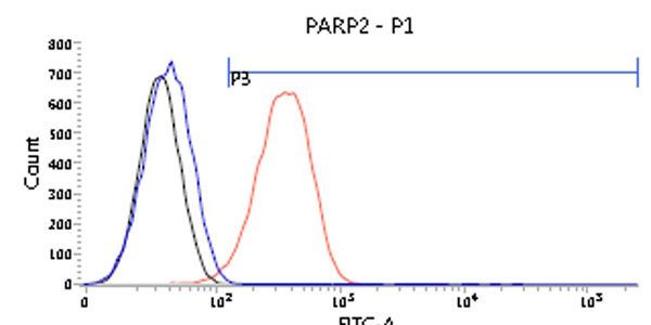 FCM (Flow Cytometry)
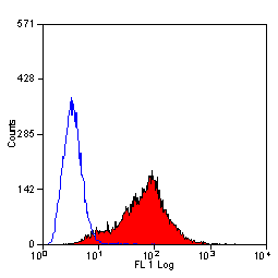
(Flow cytometry analysis of PARP2 in U87MG cells. The cell was stained with AAA11732 at 2-5ug for 1x10^6cells (red). A Goat anti mouse IgG (Alexa fluor 488) was used as the secondary antibody. Mouse monoclonal IgG was used as the isotype control (blue), cells without incubation with primary and secondary antibody was used as the negative control (black))
FCM (Flow Cytometry)
(Flow cytometry analysis of PARP2 in U87MG cells. The cell was stained with AAA11732 at 2-5ug for 1x10^6cells (red). A Goat anti mouse IgG (Alexa fluor 488) was used as the secondary antibody. Mouse monoclonal IgG was used as the isotype control (blue), cells without incubation with primary and secondary antibody was used as the negative control (black))
PARP2, Monoclonal Antibody (Cat# AAA11732)
Full Name
PARP2 antibody
Gene Names
PARP2; ARTD2; ADPRT2; PARP-2; ADPRTL2; ADPRTL3; pADPRT-2
Reactivity
Human
Applications
Western Blot, Flow Cytometry, Immunocytochemistry, Immunofluorescence
Purity
By protein-A affinity chromatography
Pricing
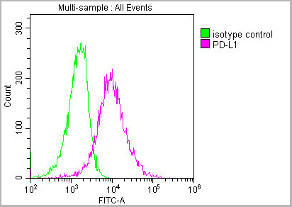
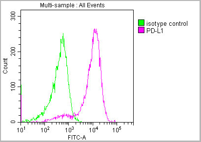
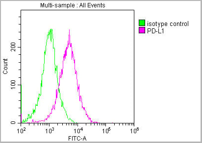 FCM (Flow Cytometry)
(Overlay histogram showing Hela cells stained with AAA27016 (red line) at 1:150. The cells were incubated in 1x PBS /10% normal goat serum to block non-specific protein-protein interactions followed by primary antibody for 1 h at 4 degree C. The secondary antibody used was FITC goat anti-mouse IgG(H+L) at 1/200 dilution for 1 h at 4 degree C. Isotype control antibody (green line) was used under the same conditions. Acquisition of >10,000 events was performed.)
FCM (Flow Cytometry)
(Overlay histogram showing Hela cells stained with AAA27016 (red line) at 1:150. The cells were incubated in 1x PBS /10% normal goat serum to block non-specific protein-protein interactions followed by primary antibody for 1 h at 4 degree C. The secondary antibody used was FITC goat anti-mouse IgG(H+L) at 1/200 dilution for 1 h at 4 degree C. Isotype control antibody (green line) was used under the same conditions. Acquisition of >10,000 events was performed.)
PD-L1, Monoclonal Antibody (Cat# AAA27016)
Full Name
PD-L1 Monoclonal Antibody
Gene Names
CD274; B7-H; B7H1; PDL1; PD-L1; PDCD1L1; PDCD1LG1
Reactivity
Human
Applications
Western Blot, Immunohistochemistry, Immunofluorescence, Flow Cytometry
Purity
>95%, Protein G purified
Pricing
 WB (Western Blot)
(WB: A431 cell lysate was probed with anti-Catenin, beta, active (ABC). Arrow indicates beta-Catenin (92kD))
WB (Western Blot)
(WB: A431 cell lysate was probed with anti-Catenin, beta, active (ABC). Arrow indicates beta-Catenin (92kD))
Catenin, beta, active, Monoclonal Antibody (Cat# AAA14699)
Full Name
Catenin, beta, active (ABC) (BSA, Azide & Glycerol Free)
Reactivity
Human, Mouse, Rat
Applications
Western Blot, Immunohistochemistry, Immunocytochemistry
Purity
Purified by Protein G affinity chromatography.
Pricing


 Application Data
(Published customer image: Mouse anti V5 tag antibody, clone SV5-Pk1 used for the detection of V5 tagged WEEV_nsP3 protein by western blotting and immunofluorescenceImage caption: WEEV nsP3 interaction with host IKKbeta. A) U87MGs were transfected in a 6-well plate with 5 ug of pUC19 and WEEV_nsP3_HA for 24 hours. Cell lysates were resolved using SDS-PAGE and subsequently immunoblotted with V5 antibody and beta-actin served as a loading control. B) U87MGs were transfected with WEEV_nsP3_V5; cells were fixed after 24 hours and stained with antibodies against the endogenous IKKbeta and the V5 tag. Cells were incubated with appropriate secondary Alexa Fluor antibodies and the nuclei stained with DAPI. Co-localization of IKKbeta with WEEV_nsP3_V5 (yellow) was observed as shown by the arrows. B) Panels E -H serve as an example of transfected cells in a given field of view that show co-localization of IKKbeta and WEEV_nsP3_V5 24 hours post transfection. Panels I-L represent magnified images of other cells showing co-localization of IKKbeta and WEEV_nsP3_V5. Panel M is a magnified image of panel L. The co-localization was confirmed by Z-stack analysis. Co-localization was calculated to be approximately in 61% of cells (163 cells were counted of which 44% demonstrated expression of nsP3. Of those cells that expressed nsP3, 61% showed co-localization of both proteins). Images were taken using Nikon Eclipse TE2000-U at 60x magnification and are representative of 2 independent experiments.From: Amaya M, Voss K, Sampey G, Senina S, de la Fuente C, et al. (2014) The Role of IKKbeta in Venezuelan Equine Encephalitis Virus Infection. PLoS ONE 9(2): e86745.)
Application Data
(Published customer image: Mouse anti V5 tag antibody, clone SV5-Pk1 used for the detection of V5 tagged WEEV_nsP3 protein by western blotting and immunofluorescenceImage caption: WEEV nsP3 interaction with host IKKbeta. A) U87MGs were transfected in a 6-well plate with 5 ug of pUC19 and WEEV_nsP3_HA for 24 hours. Cell lysates were resolved using SDS-PAGE and subsequently immunoblotted with V5 antibody and beta-actin served as a loading control. B) U87MGs were transfected with WEEV_nsP3_V5; cells were fixed after 24 hours and stained with antibodies against the endogenous IKKbeta and the V5 tag. Cells were incubated with appropriate secondary Alexa Fluor antibodies and the nuclei stained with DAPI. Co-localization of IKKbeta with WEEV_nsP3_V5 (yellow) was observed as shown by the arrows. B) Panels E -H serve as an example of transfected cells in a given field of view that show co-localization of IKKbeta and WEEV_nsP3_V5 24 hours post transfection. Panels I-L represent magnified images of other cells showing co-localization of IKKbeta and WEEV_nsP3_V5. Panel M is a magnified image of panel L. The co-localization was confirmed by Z-stack analysis. Co-localization was calculated to be approximately in 61% of cells (163 cells were counted of which 44% demonstrated expression of nsP3. Of those cells that expressed nsP3, 61% showed co-localization of both proteins). Images were taken using Nikon Eclipse TE2000-U at 60x magnification and are representative of 2 independent experiments.From: Amaya M, Voss K, Sampey G, Senina S, de la Fuente C, et al. (2014) The Role of IKKbeta in Venezuelan Equine Encephalitis Virus Infection. PLoS ONE 9(2): e86745.)
V5-TAG, Monoclonal Antibody (Cat# AAA11930)
Full Name
MOUSE ANTI V5-TAG
Applications
Immunohistochemistry, Flow Cytometry, Immunofluorescence, Immunoprecipitation, Western Blot, Radioimmunoassay
Pricing


 Application Data
(Staining of mouse spleen with Hamster anti Mouse CD81: Alexa Fluor 488)
Application Data
(Staining of mouse spleen with Hamster anti Mouse CD81: Alexa Fluor 488)
CD81, Monoclonal Antibody (Cat# AAA11942)
Full Name
HAMSTER ANTI MOUSE CD81
Gene Names
Cd81; Tapa1; Tapa-1; Tspan28
Reactivity
Rat
Applications
Immunohistochemistry, Flow Cytometry, Immunoprecipitation, Western Blot
Pricing


 Application Data
(Published customer image: Mouse anti V5 tag antibody, clone SV5-Pk1 used for the detection of V5 tagged WEEV_nsP3 protein by western blotting and immunofluorescenceImage caption: WEEV nsP3 interaction with host IKKbeta. A) U87MGs were transfected in a 6-well plate with 5 ug of pUC19 and WEEV_nsP3_HA for 24 hours. Cell lysates were resolved using SDS-PAGE and subsequently immunoblotted with V5 antibody and beta-actin served as a loading control. B) U87MGs were transfected with WEEV_nsP3_V5; cells were fixed after 24 hours and stained with antibodies against the endogenous IKKbeta and the V5 tag. Cells were incubated with appropriate secondary Alexa Fluor antibodies and the nuclei stained with DAPI. Co-localization of IKKbeta with WEEV_nsP3_V5 (yellow) was observed as shown by the arrows. B) Panels E -H serve as an example of transfected cells in a given field of view that show co-localization of IKKbeta and WEEV_nsP3_V5 24 hours post transfection. Panels I-L represent magnified images of other cells showing co-localization of IKKbeta and WEEV_nsP3_V5. Panel M is a magnified image of panel L. The co-localization was confirmed by Z-stack analysis. Co-localization was calculated to be approximately in 61% of cells (163 cells were counted of which 44% demonstrated expression of nsP3. Of those cells that expressed nsP3, 61% showed co-localization of both proteins). Images were taken using Nikon Eclipse TE2000-U at 60x magnification and are representative of 2 independent experiments.From: Amaya M, Voss K, Sampey G, Senina S, de la Fuente C, et al. (2014) The Role of IKKbeta in Venezuelan Equine Encephalitis Virus Infection. PLoS ONE 9(2): e86745.)
Application Data
(Published customer image: Mouse anti V5 tag antibody, clone SV5-Pk1 used for the detection of V5 tagged WEEV_nsP3 protein by western blotting and immunofluorescenceImage caption: WEEV nsP3 interaction with host IKKbeta. A) U87MGs were transfected in a 6-well plate with 5 ug of pUC19 and WEEV_nsP3_HA for 24 hours. Cell lysates were resolved using SDS-PAGE and subsequently immunoblotted with V5 antibody and beta-actin served as a loading control. B) U87MGs were transfected with WEEV_nsP3_V5; cells were fixed after 24 hours and stained with antibodies against the endogenous IKKbeta and the V5 tag. Cells were incubated with appropriate secondary Alexa Fluor antibodies and the nuclei stained with DAPI. Co-localization of IKKbeta with WEEV_nsP3_V5 (yellow) was observed as shown by the arrows. B) Panels E -H serve as an example of transfected cells in a given field of view that show co-localization of IKKbeta and WEEV_nsP3_V5 24 hours post transfection. Panels I-L represent magnified images of other cells showing co-localization of IKKbeta and WEEV_nsP3_V5. Panel M is a magnified image of panel L. The co-localization was confirmed by Z-stack analysis. Co-localization was calculated to be approximately in 61% of cells (163 cells were counted of which 44% demonstrated expression of nsP3. Of those cells that expressed nsP3, 61% showed co-localization of both proteins). Images were taken using Nikon Eclipse TE2000-U at 60x magnification and are representative of 2 independent experiments.From: Amaya M, Voss K, Sampey G, Senina S, de la Fuente C, et al. (2014) The Role of IKKbeta in Venezuelan Equine Encephalitis Virus Infection. PLoS ONE 9(2): e86745.)
V5-TAG, Monoclonal Antibody (Cat# AAA12211)
Full Name
MOUSE ANTI V5-TAG
Applications
Immunohistochemistry, Flow Cytometry, Immunofluorescence, Immunoprecipitation, Western Blot, Radioimmunoassay
Pricing
HERV-K Envelop Antibody (Anti-HERVKE), ELISA Kit (Cat# AAA10579)
Full Name
Qualitative Human HERV-K Envelop Antibody (Anti-HERVKE) ELISA Kit
Reactivity
Human
Pricing
Staphylococcal Enterotoxin B, Monoclonal Antibody (Cat# AAA14625)
Full Name
mAb anti-Staphylococcal Enterotoxin B [SEB]
Reactivity
Staphylococcal enterotoxin B
Applications
ELISA
Purity
Protein G affinity purified
Pricing
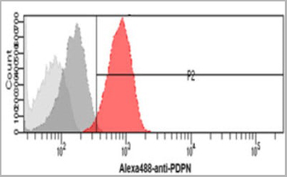 FCM (Flow Cytometry)
(Flow cytometry analysis of Podoplanin in HeLa cells. The cell was stained with (AAA11728) at 2-5ug for 1x10^6cells (red). A Goat anti mouse IgG (Alexa fluor 488) was used as the secondary antibody. Mouse monoclonal IgG was used as the isotype control (dark gray), cells without incubation with primary and secondary antibody was used as the negative control (light gray).)
FCM (Flow Cytometry)
(Flow cytometry analysis of Podoplanin in HeLa cells. The cell was stained with (AAA11728) at 2-5ug for 1x10^6cells (red). A Goat anti mouse IgG (Alexa fluor 488) was used as the secondary antibody. Mouse monoclonal IgG was used as the isotype control (dark gray), cells without incubation with primary and secondary antibody was used as the negative control (light gray).)
PDPN, Monoclonal Antibody (Cat# AAA11728)
Full Name
PDPN antibody
Gene Names
PDPN; T1A; GP36; GP40; Gp38; OTS8; T1A2; TI1A; T1A-2; AGGRUS; HT1A-1; PA2.26
Reactivity
Human
Applications
Flow Cytometry, Immunocytochemistry, Immunofluorescence
Purity
By protein-G affinity chromatography
Pricing
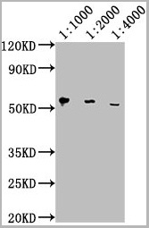
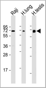 WB (Western Blot)
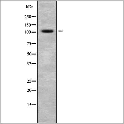
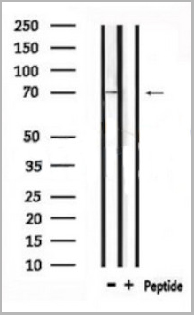
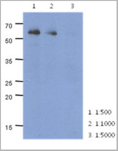
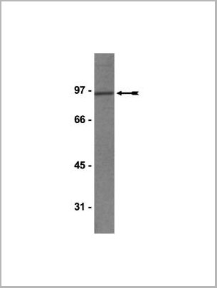
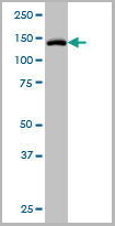
(All lanes : Anti-ERVK-7 Antibody (N-Term) at 1:1000-1:2000 dilutionLane 1: Raji whole cell lysateLane 2: human lung lysateLane 3: human testis lysateLysates/proteins at 20 ug per lane.SecondaryGoat Anti-Rabbit IgG, (H+L), Peroxidase conjugated at 1/10000 dilution.Predicted band size : 67 kDaBlocking/Dilution buffer: 5% NFDM/TBST.)
WB (Western Blot)
(All lanes : Anti-ERVK-7 Antibody (N-Term) at 1:1000-1:2000 dilutionLane 1: Raji whole cell lysateLane 2: human lung lysateLane 3: human testis lysateLysates/proteins at 20 ug per lane.SecondaryGoat Anti-Rabbit IgG, (H+L), Peroxidase conjugated at 1/10000 dilution.Predicted band size : 67 kDaBlocking/Dilution buffer: 5% NFDM/TBST.)
ERVK-7, Polyclonal Antibody (Cat# AAA28797)
Full Name
ERVK-7 Antibody (N-Term)
Reactivity
Human
Applications
Western Blot, Immunohistochemistry
Purity
This antibody is purified through a protein A column, followed by peptide affinity purification.
Pricing
E Coli, Polyclonal Antibody (Cat# AAA14318)
Full Name
E Coli antibody
Applications
ELISA
Purity
> 95% pure
Pricing
ERO1L, Monoclonal Antibody (Cat# AAA24203)
Full Name
ERO1L (ERO1-like Protein alpha, ERO1-L, ERO1-L-alpha, Endoplasmic Oxidoreductin-1-like Protein, Oxidoreductin-1-L-alpha, UNQ434/PRO865) (AP)
Gene Names
ERO1A; ERO1L; ERO1-L; ERO1LA; Ero1alpha; ERO1-alpha; ERO1-L-alpha
Reactivity
Human, Rat
Applications
EIA, IHC, WB
Purity
Purified by Protein A Affinity Chromatography.
Pricing


 Application Data
(Published customer image: Mouse anti V5 tag antibody, clone SV5-Pk1 used for the detection of V5 tagged WEEV_nsP3 protein by western blotting and immunofluorescenceImage caption: WEEV nsP3 interaction with host IKKbeta. A) U87MGs were transfected in a 6-well plate with 5 ug of pUC19 and WEEV_nsP3_HA for 24 hours. Cell lysates were resolved using SDS-PAGE and subsequently immunoblotted with V5 antibody and beta-actin served as a loading control. B) U87MGs were transfected with WEEV_nsP3_V5; cells were fixed after 24 hours and stained with antibodies against the endogenous IKKbeta and the V5 tag. Cells were incubated with appropriate secondary Alexa Fluor antibodies and the nuclei stained with DAPI. Co-localization of IKKbeta with WEEV_nsP3_V5 (yellow) was observed as shown by the arrows. B) Panels E -H serve as an example of transfected cells in a given field of view that show co-localization of IKKbeta and WEEV_nsP3_V5 24 hours post transfection. Panels I-L represent magnified images of other cells showing co-localization of IKKbeta and WEEV_nsP3_V5. Panel M is a magnified image of panel L. The co-localization was confirmed by Z-stack analysis. Co-localization was calculated to be approximately in 61% of cells (163 cells were counted of which 44% demonstrated expression of nsP3. Of those cells that expressed nsP3, 61% showed co-localization of both proteins). Images were taken using Nikon Eclipse TE2000-U at 60x magnification and are representative of 2 independent experiments.From: Amaya M, Voss K, Sampey G, Senina S, de la Fuente C, et al. (2014) The Role of IKKbeta in Venezuelan Equine Encephalitis Virus Infection. PLoS ONE 9(2): e86745.)
Application Data
(Published customer image: Mouse anti V5 tag antibody, clone SV5-Pk1 used for the detection of V5 tagged WEEV_nsP3 protein by western blotting and immunofluorescenceImage caption: WEEV nsP3 interaction with host IKKbeta. A) U87MGs were transfected in a 6-well plate with 5 ug of pUC19 and WEEV_nsP3_HA for 24 hours. Cell lysates were resolved using SDS-PAGE and subsequently immunoblotted with V5 antibody and beta-actin served as a loading control. B) U87MGs were transfected with WEEV_nsP3_V5; cells were fixed after 24 hours and stained with antibodies against the endogenous IKKbeta and the V5 tag. Cells were incubated with appropriate secondary Alexa Fluor antibodies and the nuclei stained with DAPI. Co-localization of IKKbeta with WEEV_nsP3_V5 (yellow) was observed as shown by the arrows. B) Panels E -H serve as an example of transfected cells in a given field of view that show co-localization of IKKbeta and WEEV_nsP3_V5 24 hours post transfection. Panels I-L represent magnified images of other cells showing co-localization of IKKbeta and WEEV_nsP3_V5. Panel M is a magnified image of panel L. The co-localization was confirmed by Z-stack analysis. Co-localization was calculated to be approximately in 61% of cells (163 cells were counted of which 44% demonstrated expression of nsP3. Of those cells that expressed nsP3, 61% showed co-localization of both proteins). Images were taken using Nikon Eclipse TE2000-U at 60x magnification and are representative of 2 independent experiments.From: Amaya M, Voss K, Sampey G, Senina S, de la Fuente C, et al. (2014) The Role of IKKbeta in Venezuelan Equine Encephalitis Virus Infection. PLoS ONE 9(2): e86745.)
V5-TAG, Monoclonal Antibody (Cat# AAA11850)
Full Name
MOUSE ANTI V5-TAG:Biotin
Applications
Immunohistochemistry, Western Blot
Pricing
 WB (Western Blot)
(EIF4G1 monoclonal antibody. Western Blot analysis of EIF4G1 expression in NIH/3T3.)
WB (Western Blot)
(EIF4G1 monoclonal antibody. Western Blot analysis of EIF4G1 expression in NIH/3T3.)
EIF4G1, Monoclonal Antibody (Cat# AAA25088)
Full Name
EIF4G1 (Eukaryotic Translation Initiation Factor 4 gamma 1, eIF-4-gamma 1, eIF-4G 1, eIF-4G1, p220, EIF4G1, EIF4F, EIF4G, EIF4GI) (FITC)
Gene Names
EIF4G1; P220; EIF4F; EIF4G; EIF4GI; PARK18; EIF-4G1
Reactivity
Human, Mouse, Rat
Applications
Immunofluorescence, Western Blot
Purity
Purified by Protein A Affinity Chromatography.
Pricing
 IF (Immunofluorescence)
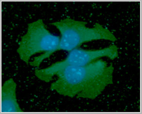
(Immunofluorescence (IFC) analysis of PC3 cells using AAA14697 (1:100). The secondary antibody (green) was used Alexa Fluor 488. DAPI was stained the cell nucleus (blue).)
IF (Immunofluorescence)
(Immunofluorescence (IFC) analysis of PC3 cells using AAA14697 (1:100). The secondary antibody (green) was used Alexa Fluor 488. DAPI was stained the cell nucleus (blue).)
Ribonuclease inhibitor 1, Monoclonal Antibody (Cat# AAA14697)
Full Name
Ribonuclease inhibitor 1 (RNH1)
Gene Names
RNH1; RAI; RNH; MGC4569; MGC18200; MGC54054
Applications
Immunofluorescence, Western Blot
Purity
Affinity Purified
Purified by Protein G affinity chromatography.
Purified by Protein G affinity chromatography.
Pricing
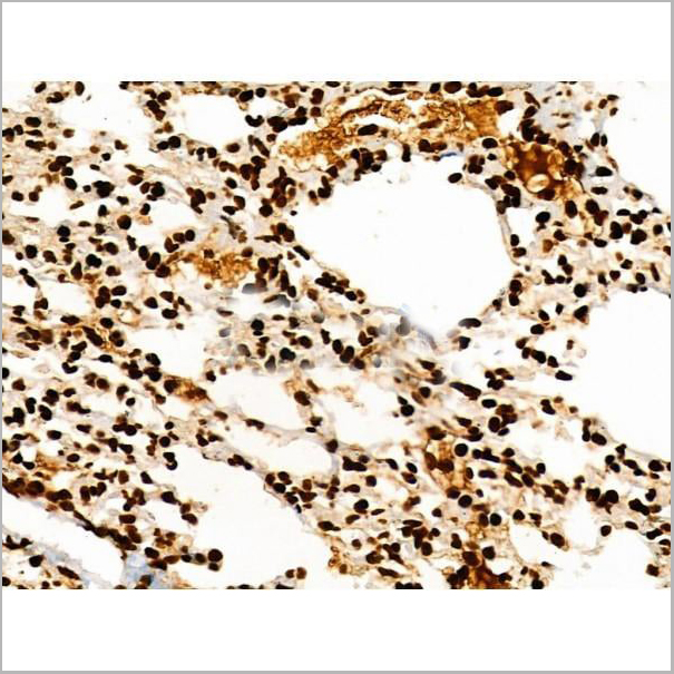
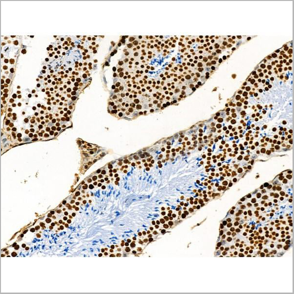

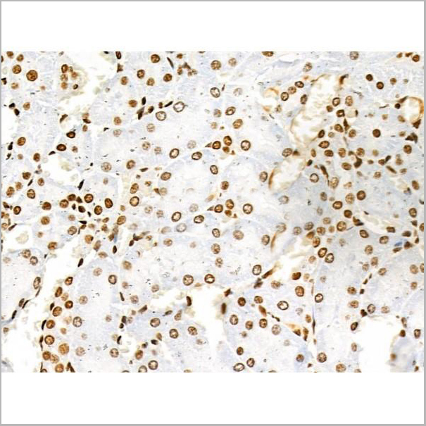
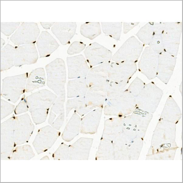
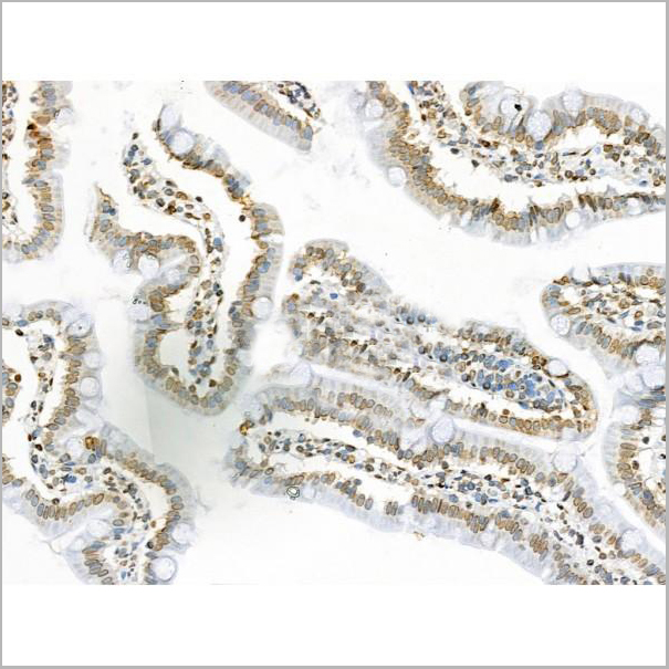
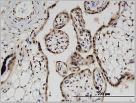
 IHC (Immunohistochemistry)
(At 1/100 staining Human kidney cancer by IHC-P. The sample was formaldehyde fixed and a heat mediated antigen retrieval step in citrate buffer was performed. The sample was then blocked and incubated with the primary antibody at 4 degree C overnight. An HRP conjugated anti-Rabbit antibody was used as the secondary antibody.)
IHC (Immunohistochemistry)
(At 1/100 staining Human kidney cancer by IHC-P. The sample was formaldehyde fixed and a heat mediated antigen retrieval step in citrate buffer was performed. The sample was then blocked and incubated with the primary antibody at 4 degree C overnight. An HRP conjugated anti-Rabbit antibody was used as the secondary antibody.)
Histone H3, Polyclonal Antibody (Cat# AAA31353)
Full Name
Acetyl-Histone H3 (Lys122) Antibody
Gene Names
HIST1H3A; H3/A; H3FA
Reactivity
Human, Mouse, Rat
Predicted Reactivity: Bovine (100%)
Predicted Reactivity: Bovine (100%)
Applications
Western Blot, Immunohistochemistry, Peptide ELISA
Purity
The antiserum was purified by peptide affinity chromatography using SulfoLink Coupling Resin
Pricing
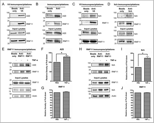
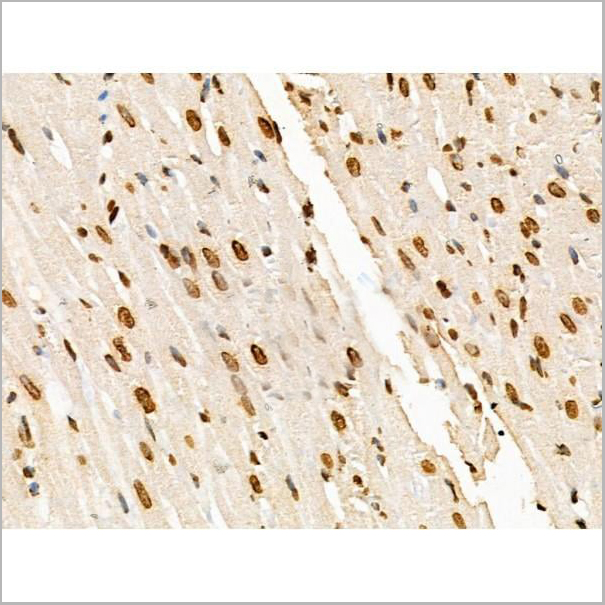
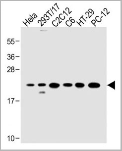
2019-03-07, Monoclonal Antibody (Cat# AAA24266)
Full Name
MARCH7 (E3 Ubiquitin-protein Ligase MARCH7, Axotrophin, Membrane-associated RING Finger Protein 7, Membrane-associated RING-CH Protein VII, MARCH-VII, RING Finger Protein 177, AXOT, RNF177, DKFZp586F1122) (AP)
Gene Names
MARCH7; AXO; AXOT; RNF177; MARCH-VII
Reactivity
Human, Mouse, Rat
Applications
EIA, WB
Purity
Purified by Protein A Affinity Chromatography.
Pricing
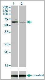
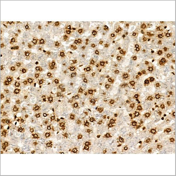
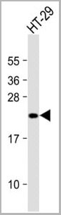
DUSP6, Monoclonal Antibody (Cat# AAA24194)
Full Name
DUSP6 (Dual Specificity Protein Phosphatase 6, Dual Specificity Protein Phosphatase PYST1, Mitogen-activated Protein Kinase Phosphatase 3, MAP Kinase Phosphatase 3, MKP3, MKP-3, PYST1) (AP)
Gene Names
DUSP6; HH19; MKP3; PYST1
Reactivity
Human
Applications
EIA, IHC, WB
Purity
Purified by Protein A Affinity Chromatography.
Pricing
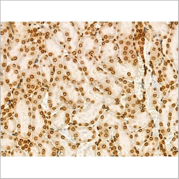
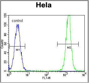 FCM (Flow Cytometry)
(KRAS Antibody (C-term)(AAA28748) flow cytometric analysis of Hela cells (right histogram) compared to a negative control cell (left histogram).FITC-conjugated goat-anti-rabbit secondary antibodies were used for the analysis.)
FCM (Flow Cytometry)
(KRAS Antibody (C-term)(AAA28748) flow cytometric analysis of Hela cells (right histogram) compared to a negative control cell (left histogram).FITC-conjugated goat-anti-rabbit secondary antibodies were used for the analysis.)
KRAS, Polyclonal Antibody (Cat# AAA28748)
Full Name
KRAS Antibody (C-term)
Gene Names
KRAS; NS; NS3; CFC2; KRAS1; KRAS2; RASK2; KI-RAS; C-K-RAS; K-RAS2A; K-RAS2B; K-RAS4A; K-RAS4B
Reactivity
Human, Mouse, Rat (Predicted: Rat)
Applications
Western Blot, Immunofluorescence, Flow Cytometry
Purity
This antibody is purified through a protein A column, followed by peptide affinity purification.
Pricing
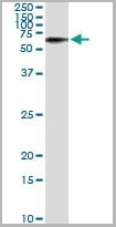
 IF (Immunofluorescence)
(Immunofluorescence of on HeLa cell using AAA24920 (10ug/ml).)
IF (Immunofluorescence)
(Immunofluorescence of on HeLa cell using AAA24920 (10ug/ml).)
PRPH, Monoclonal Antibody (Cat# AAA24920)
Full Name
PRPH (Peripherin, Neurofilament 4, NEF4, PRPH1) (Biotin)
Gene Names
PRPH; NEF4; PRPH1
Reactivity
Human
Applications
Immunofluorescence, Immunohistochemistry, Western Blot
Purity
Purified by Protein A Affinity Chromatography.
Pricing
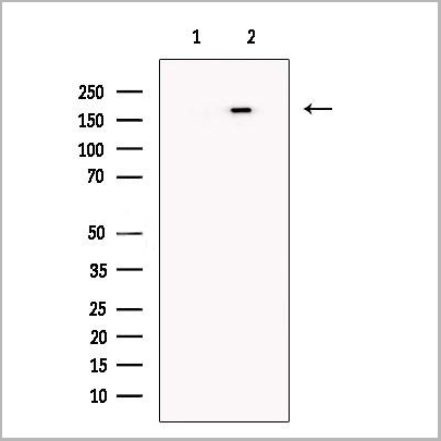
 IP (Immunoprecipitation)
(Immunoprecipitation of TYK2 transfected lysate using TYK2 monoclonal antibody and Protein A Magnetic Bead and immunoblotted with TYK2 rabbit polyclonal antibody.)
IP (Immunoprecipitation)
(Immunoprecipitation of TYK2 transfected lysate using TYK2 monoclonal antibody and Protein A Magnetic Bead and immunoblotted with TYK2 rabbit polyclonal antibody.)
TYK2, Monoclonal Antibody (Cat# AAA25877)
Full Name
TYK2 (Non-receptor Tyrosine-protein Kinase TYK2, JTK1) (PE)
Gene Names
TYK2; JTK1; IMD35
Reactivity
Human
Applications
Immunofluorescence, Immunohistochemistry, Immunoprecipitation, Western Blot
Purity
Purified by Protein A Affinity Chromatography.
Pricing
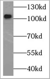
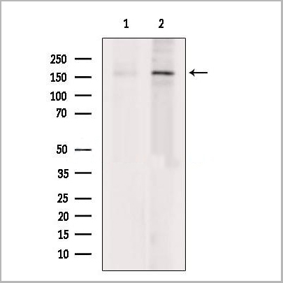
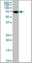
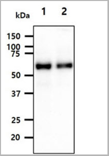
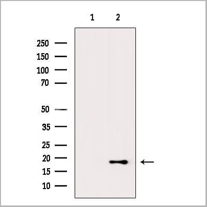
 WB (Western Blot)
(Western blot analysis of MUTYH over-expressed 293 cell line, cotransfected with MUTYH Validated Chimera RNAi (Lane 2) or non-transfected control (Lane 1). Blot probed with MUTYH monoclonal antibody GAPDH (36.1kD) used as specificity and loading control.)
WB (Western Blot)
(Western blot analysis of MUTYH over-expressed 293 cell line, cotransfected with MUTYH Validated Chimera RNAi (Lane 2) or non-transfected control (Lane 1). Blot probed with MUTYH monoclonal antibody GAPDH (36.1kD) used as specificity and loading control.)
MUTYH, Monoclonal Antibody (Cat# AAA24577)
Full Name
MUTYH (A/G-specific Adenine DNA Glycosylase, MutY Homolog, hMYH, MYH) APC
Gene Names
MUTYH; MYH
Reactivity
Human
Applications
Immunoprecipitation, Western Blot
Purity
Purified by Protein A Affinity Chromatography.
Pricing