Filters
Clonality
Type
Reactivity
Gene Name
Isotype
Host
Application
Clone
228 results for "Rat IgG Isotype Control" - showing 150-200


 FCM (Flow Cytometry)
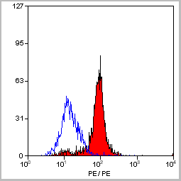
(Figure 8. Flow Cytometry analysis of U20S cells using anti-SV2A antibody (AAA19279).Overlay histogram showing U20S cells stained with AAA19279 (Blue line). The cells were blocked with 10% normal goat serum. And then incubated with rabbit anti-SV2A Antibody (AAA19279, 1μg/1x106 cells) for 30 min at 20 degree C. DyLight®488 conjugated goat anti-rabbit IgG (5-10μg/1x106 cells) was used as secondary antibody for 30 minutes at 20 degree C. Isotype control antibody (Green line) was rabbit IgG (1μg/1x106) used under the same conditions. Unlabelled sample (Red line) was also used as a control.)
FCM (Flow Cytometry)
(Figure 8. Flow Cytometry analysis of U20S cells using anti-SV2A antibody (AAA19279).Overlay histogram showing U20S cells stained with AAA19279 (Blue line). The cells were blocked with 10% normal goat serum. And then incubated with rabbit anti-SV2A Antibody (AAA19279, 1μg/1x106 cells) for 30 min at 20 degree C. DyLight®488 conjugated goat anti-rabbit IgG (5-10μg/1x106 cells) was used as secondary antibody for 30 minutes at 20 degree C. Isotype control antibody (Green line) was rabbit IgG (1μg/1x106) used under the same conditions. Unlabelled sample (Red line) was also used as a control.)
SV2A, Polyclonal Antibody (Cat# AAA19279)
Full Name
Anti-SV2A Antibody
Gene Names
SV2A; SV2
Reactivity
Human, Mouse, Rat
Applications
WB, IHC-P, FC/FACS/FCM
Purity
Immunogen affinity purified.
Pricing


 FCM (Flow Cytometry)
(Figure 8. Flow Cytometry analysis of HepG2 cells using anti-REA/PHB2 antibody (AAA19374).Overlay histogram showing HepG2 cells stained with AAA19374 (Blue line). The cells were blocked with 10% normal goat serum. And then incubated with mouse anti- REA/PHB2 Antibody (AAA19374, 1μg/1x106 cells) for 30 min at 20 degree C. DyLight®488 conjugated goat anti-mouse IgG (BA1126, 5-10μg/1x106 cells) was used as secondary antibody for 30 minutes at 20 degree C. Isotype control antibody (Green line) was mouse IgG (1μg/1x106) used under the same conditions. Unlabelled sample (Red line) was also used as a control.)
FCM (Flow Cytometry)
(Figure 8. Flow Cytometry analysis of HepG2 cells using anti-REA/PHB2 antibody (AAA19374).Overlay histogram showing HepG2 cells stained with AAA19374 (Blue line). The cells were blocked with 10% normal goat serum. And then incubated with mouse anti- REA/PHB2 Antibody (AAA19374, 1μg/1x106 cells) for 30 min at 20 degree C. DyLight®488 conjugated goat anti-mouse IgG (BA1126, 5-10μg/1x106 cells) was used as secondary antibody for 30 minutes at 20 degree C. Isotype control antibody (Green line) was mouse IgG (1μg/1x106) used under the same conditions. Unlabelled sample (Red line) was also used as a control.)
REA/PHB2, Monoclonal Antibody (Cat# AAA19374)
Full Name
Anti-REA/PHB2 Antibody (monoclonal, 2B5)
Gene Names
PHB2; BAP; REA; p22; Bap37; BCAP37; PNAS-141
Reactivity
Human, Mouse, Rat
Applications
WB, IHC-P, FC/FACS/FCM
Purity
Immunogen affinity purified.
Pricing


 FCM (Flow Cytometry)
(Figure 7. Flow Cytometry analysis of HEPG2 cells using anti-ALDH1L1 antibody (AAA19297).Overlay histogram showing HEPG2 cells stained with AAA19297 (Blue line). The cells were blocked with 10% normal goat serum. And then incubated with rabbit anti-ALDH1L1 Antibody (AAA19297 1μg/1x106 cells) for 30 min at 20 degree C. DyLight®488 conjugated goat anti-rabbit IgG (5-10μg/1x106 cells) was used as secondary antibody for 30 minutes at 20 degree C. Isotype control antibody (Green line) was rabbit IgG (1μg/1x106) used under the same conditions. Unlabelled sample (Red line) was also used as a control.)
FCM (Flow Cytometry)
(Figure 7. Flow Cytometry analysis of HEPG2 cells using anti-ALDH1L1 antibody (AAA19297).Overlay histogram showing HEPG2 cells stained with AAA19297 (Blue line). The cells were blocked with 10% normal goat serum. And then incubated with rabbit anti-ALDH1L1 Antibody (AAA19297 1μg/1x106 cells) for 30 min at 20 degree C. DyLight®488 conjugated goat anti-rabbit IgG (5-10μg/1x106 cells) was used as secondary antibody for 30 minutes at 20 degree C. Isotype control antibody (Green line) was rabbit IgG (1μg/1x106) used under the same conditions. Unlabelled sample (Red line) was also used as a control.)
ALDH1L1, Polyclonal Antibody (Cat# AAA19297)
Full Name
Anti-ALDH1L1 Antibody
Gene Names
ALDH1L1; FDH; FTHFD; 10-fTHF; 10-FTHFDH
Reactivity
Human, Mouse, Rat, Monkey
Applications
WB, IHC-P, ICC, IF, FC/FACS/FCM, EIA
Purity
Immunogen affinity purified.
Pricing


 FCM (Flow Cytometry)
(Figure 10. Flow Cytometry analysis of SiHa cells using anti-PRDM14 antibody (AAA19283).Overlay histogram showing SiHa cells stained with AAA19283 (Blue line). The cells were blocked with 10% normal goat serum. And then incubated with rabbit anti-PRDM14 Antibody (AAA19283, 1μg/1x106 cells) for 30 min at 20 degree C. DyLight®488 conjugated goat anti-rabbit IgG (5-10μg/1x106 cells) was used as secondary antibody for 30 minutes at 20 degree C. Isotype control antibody (Green line) was rabbit IgG (1μg/1x106) used under the same conditions. Unlabelled sample (Red line) was also used as a control.)
FCM (Flow Cytometry)
(Figure 10. Flow Cytometry analysis of SiHa cells using anti-PRDM14 antibody (AAA19283).Overlay histogram showing SiHa cells stained with AAA19283 (Blue line). The cells were blocked with 10% normal goat serum. And then incubated with rabbit anti-PRDM14 Antibody (AAA19283, 1μg/1x106 cells) for 30 min at 20 degree C. DyLight®488 conjugated goat anti-rabbit IgG (5-10μg/1x106 cells) was used as secondary antibody for 30 minutes at 20 degree C. Isotype control antibody (Green line) was rabbit IgG (1μg/1x106) used under the same conditions. Unlabelled sample (Red line) was also used as a control.)
PRDM14, Polyclonal Antibody (Cat# AAA19283)
Full Name
Anti-PRDM14 Antibody
Gene Names
PRDM14; PFM11
Reactivity
Human, Mouse, Rat
Applications
WB, IHC-P, ICC, IF, FC/FACS/FCM
Purity
Immunogen affinity purified.
Pricing


 FCM (Flow Cytometry)
(Figure 7. Flow Cytometry analysis of A549 cells using anti-DR6/TNFRSF21 antibody (AAA19292).Overlay histogram showing A549 cells stained with AAA19292 (Blue line). The cells were blocked with 10% normal goat serum. And then incubated with rabbit anti-DR6/TNFRSF21 Antibody (AAA19292, 1μg/1x106 cells) for 30 min at 20 degree C. DyLight®488 conjugated goat anti-rabbit IgG (5-10μg/1x106 cells) was used as secondary antibody for 30 minutes at 20 degree C. Isotype control antibody (Green line) was rabbit IgG (1μg/1x106) used under the same conditions. Unlabelled sample (Red line) was also used as a control.)
FCM (Flow Cytometry)
(Figure 7. Flow Cytometry analysis of A549 cells using anti-DR6/TNFRSF21 antibody (AAA19292).Overlay histogram showing A549 cells stained with AAA19292 (Blue line). The cells were blocked with 10% normal goat serum. And then incubated with rabbit anti-DR6/TNFRSF21 Antibody (AAA19292, 1μg/1x106 cells) for 30 min at 20 degree C. DyLight®488 conjugated goat anti-rabbit IgG (5-10μg/1x106 cells) was used as secondary antibody for 30 minutes at 20 degree C. Isotype control antibody (Green line) was rabbit IgG (1μg/1x106) used under the same conditions. Unlabelled sample (Red line) was also used as a control.)
DR6/TNFRSF21, Polyclonal Antibody (Cat# AAA19292)
Full Name
Anti-DR6/TNFRSF21 Antibody
Gene Names
TNFRSF21; DR6; CD358; BM-018
Reactivity
Human, Mouse, Rat
Applications
WB, IHC-P, FC/FACS/FCM
Purity
Immunogen affinity purified.
Pricing


 FCM (Flow Cytometry)
(Figure 10. Flow Cytometry analysis of U87 cells using anti-GNG2 antibody (AAA19312).Overlay histogram showing U87 cells stained with AAA19312 (Blue line). The cells were blocked with 10% normal goat serum. And then incubated with rabbit anti-GNG2 Antibody (AAA19312, 1μg/1x106 cells) for 30 min at 20 degree C. DyLight®488 conjugated goat anti-rabbit IgG (5-10μg/1x106 cells) was used as secondary antibody for 30 minutes at 20 degree C. Isotype control antibody (Green line) was rabbit IgG (1μg/1x106) used under the same conditions. Unlabelled sample (Red line) was also used as a control.)
FCM (Flow Cytometry)
(Figure 10. Flow Cytometry analysis of U87 cells using anti-GNG2 antibody (AAA19312).Overlay histogram showing U87 cells stained with AAA19312 (Blue line). The cells were blocked with 10% normal goat serum. And then incubated with rabbit anti-GNG2 Antibody (AAA19312, 1μg/1x106 cells) for 30 min at 20 degree C. DyLight®488 conjugated goat anti-rabbit IgG (5-10μg/1x106 cells) was used as secondary antibody for 30 minutes at 20 degree C. Isotype control antibody (Green line) was rabbit IgG (1μg/1x106) used under the same conditions. Unlabelled sample (Red line) was also used as a control.)
GNG2, Polyclonal Antibody (Cat# AAA19312)
Full Name
Anti-GNG2 Antibody
Reactivity
Human, Mouse, Rat
Applications
WB, IHC-P, FC/FACS/FCM, EIA
Purity
Immunogen affinity purified.
Pricing


 FCM (Flow Cytometry)
(Figure 9. Flow Cytometry analysis of 293T cells using anti-CDC45L antibody (AAA19360).Overlay histogram showing 293T cells stained with AAA19360 (Blue line). The cells were blocked with 10% normal goat serum. And then incubated with mouse anti- CDC45L Antibody (AAA19360, 1μg/1x106 cells) for 30 min at 20 degree C. DyLight®488 conjugated goat anti-mouse IgG (BA1126, 5-10μg/1x106 cells) was used as secondary antibody for 30 minutes at 20 degree C. Isotype control antibody (Green line) was mouse IgG (1μg/1x106) used under the same conditions. Unlabelled sample (Red line) was also used as a control.)
FCM (Flow Cytometry)
(Figure 9. Flow Cytometry analysis of 293T cells using anti-CDC45L antibody (AAA19360).Overlay histogram showing 293T cells stained with AAA19360 (Blue line). The cells were blocked with 10% normal goat serum. And then incubated with mouse anti- CDC45L Antibody (AAA19360, 1μg/1x106 cells) for 30 min at 20 degree C. DyLight®488 conjugated goat anti-mouse IgG (BA1126, 5-10μg/1x106 cells) was used as secondary antibody for 30 minutes at 20 degree C. Isotype control antibody (Green line) was mouse IgG (1μg/1x106) used under the same conditions. Unlabelled sample (Red line) was also used as a control.)
CDC45L, Monoclonal Antibody (Cat# AAA19360)
Full Name
Anti-CDC45L Antibody (monoclonal, 6H6)
Gene Names
CDC45; CDC45L; CDC45L2; PORC-PI-1
Reactivity
Human, Mouse, Rat
Applications
WB, IHC-P, FC/FACS/FCM
Purity
Immunogen affinity purified.
Pricing


 FCM (Flow Cytometry)
(Figure 7. Flow Cytometry analysis of RH35 cells using anti- EIF4A1 antibody (AAA19380).Overlay histogram showing RH35 cells stained with AAA19380 (Blue line). The cells were blocked with 10% normal goat serum. And then incubated with mouse anti-EIF4A1 Antibody (AAA19380, 1μg/1x106 cells) for 30 min at 20 degree C. DyLight®488 conjugated goat anti-mouse IgG (BA1126, 5-10μg/1x106 cells) was used as secondary antibody for 30 minutes at 20 degree C. Isotype control antibody (Green line) was mouse IgG (1μg/1x106) used under the same conditions. Unlabelled sample (Red line) was also used as a control.)
FCM (Flow Cytometry)
(Figure 7. Flow Cytometry analysis of RH35 cells using anti- EIF4A1 antibody (AAA19380).Overlay histogram showing RH35 cells stained with AAA19380 (Blue line). The cells were blocked with 10% normal goat serum. And then incubated with mouse anti-EIF4A1 Antibody (AAA19380, 1μg/1x106 cells) for 30 min at 20 degree C. DyLight®488 conjugated goat anti-mouse IgG (BA1126, 5-10μg/1x106 cells) was used as secondary antibody for 30 minutes at 20 degree C. Isotype control antibody (Green line) was mouse IgG (1μg/1x106) used under the same conditions. Unlabelled sample (Red line) was also used as a control.)
EIF4A1, Monoclonal Antibody (Cat# AAA19380)
Full Name
Anti-EIF4A1 Antibody (monoclonal, 11B8)
Gene Names
EIF4A1; DDX2A; EIF4A; EIF-4A; eIF4A-I; eIF-4A-I
Reactivity
Human, Mouse, Rat
Applications
WB, IHC-P, ICC, IF, FC/FACS/FCM
Purity
Immunogen affinity purified.
Pricing


 FCM (Flow Cytometry)
(Figure 13. Flow Cytometry analysis of MCF-7 cells using anti-EHD3 antibody (AAA19295).Overlay histogram showing MCF-7 cells stained with AAA19295 (Blue line). The cells were blocked with 10% normal goat serum. And then incubated with rabbit anti-EHD3 Antibody (AAA19295, 1μg/1x106 cells) for 30 min at 20 degree C. DyLight®488 conjugated goat anti-rabbit IgG (5-10μg/1x106 cells) was used as secondary antibody for 30 minutes at 20 degree C. Isotype control antibody (Green line) was rabbit IgG (1μg/1x106) used under the same conditions. Unlabelled sample (Red line) was also used as a control.)
FCM (Flow Cytometry)
(Figure 13. Flow Cytometry analysis of MCF-7 cells using anti-EHD3 antibody (AAA19295).Overlay histogram showing MCF-7 cells stained with AAA19295 (Blue line). The cells were blocked with 10% normal goat serum. And then incubated with rabbit anti-EHD3 Antibody (AAA19295, 1μg/1x106 cells) for 30 min at 20 degree C. DyLight®488 conjugated goat anti-rabbit IgG (5-10μg/1x106 cells) was used as secondary antibody for 30 minutes at 20 degree C. Isotype control antibody (Green line) was rabbit IgG (1μg/1x106) used under the same conditions. Unlabelled sample (Red line) was also used as a control.)
EHD3, Polyclonal Antibody (Cat# AAA19295)
Full Name
Anti-EHD3 Antibody
Gene Names
EHD2; PAST2
Reactivity
Human, Monkey, Mouse, Rat
Applications
WB, IHC-P, ICC, IF, FC/FACS/FCM
Purity
Immunogen affinity purified.
Pricing


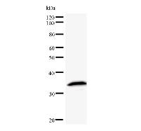
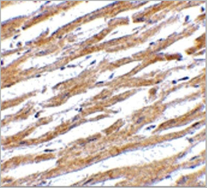
 WB (Western Blot)
(Figure 1. Western blot analysis of CISD2 using anti-CISD2 antibody (AAA19311).Electrophoresis was performed on a 5-20% SDS-PAGE gel at 70V (Stacking gel) / 90V (Resolving gel) for 2-3 hours. The sample well of each lane was loaded with 50ug of sample under reducing conditions.Lane 1: human HEK293 whole cell lysatesLane 2: human HELA whole cell lysatesLane 3: human MCF-7 whole cell lysatesLane 4: monkey kidney tissue lysatesLane 5: human SW620 whole cell lysatesLane 6: human Raji whole cell lysatesLane 7: rat kidney tissue lysatesLane 8: mouse kidney tissue lysates.After Electrophoresis, proteins were transferred to a Nitrocellulose membrane at 150mA for 50-90 minutes. Blocked the membrane with 5% Non-fat Milk/ TBS for 1. 5 hour at RT. The membrane was incubated with rabbit anti-CISD2 antigen affinity purified polyclonal antibody (Catalog # AAA19311) at 0. 5 μg/mL overnight at 4 degree C, then washed with TBS-0. 1%Tween 3 times with 5 minutes each and probed with a goat anti-rabbit IgG-HRP secondary antibody at a dilution of 1:10000 for 1. 5 hour at RT. The signal is developed using an Enhanced Chemiluminescent detection (ECL) kit (Catalog # with Tanon 5200 system. A specific band was detected for CISD2 at approximately 15KD. The expected band size for CISD2 is at 15KD.)
WB (Western Blot)
(Figure 1. Western blot analysis of CISD2 using anti-CISD2 antibody (AAA19311).Electrophoresis was performed on a 5-20% SDS-PAGE gel at 70V (Stacking gel) / 90V (Resolving gel) for 2-3 hours. The sample well of each lane was loaded with 50ug of sample under reducing conditions.Lane 1: human HEK293 whole cell lysatesLane 2: human HELA whole cell lysatesLane 3: human MCF-7 whole cell lysatesLane 4: monkey kidney tissue lysatesLane 5: human SW620 whole cell lysatesLane 6: human Raji whole cell lysatesLane 7: rat kidney tissue lysatesLane 8: mouse kidney tissue lysates.After Electrophoresis, proteins were transferred to a Nitrocellulose membrane at 150mA for 50-90 minutes. Blocked the membrane with 5% Non-fat Milk/ TBS for 1. 5 hour at RT. The membrane was incubated with rabbit anti-CISD2 antigen affinity purified polyclonal antibody (Catalog # AAA19311) at 0. 5 μg/mL overnight at 4 degree C, then washed with TBS-0. 1%Tween 3 times with 5 minutes each and probed with a goat anti-rabbit IgG-HRP secondary antibody at a dilution of 1:10000 for 1. 5 hour at RT. The signal is developed using an Enhanced Chemiluminescent detection (ECL) kit (Catalog # with Tanon 5200 system. A specific band was detected for CISD2 at approximately 15KD. The expected band size for CISD2 is at 15KD.)
CISD2, Polyclonal Antibody (Cat# AAA19311)
Full Name
Anti-CISD2 Antibody
Gene Names
CISD2; ERIS; WFS2; ZCD2; NAF-1; Miner1
Reactivity
Human, Mouse, Rat, Monkey
Applications
WB, IHC-P, ICC, IF, FC/FACS/FCM, EIA
Purity
Immunogen affinity purified.
Pricing


 FCM (Flow Cytometry)
(Figure 11. Flow Cytometry analysis of U937 cells using anti-Coronin 1a/TACO/CORO1A antibody (AAA19291).Overlay histogram showing U937 cells stained with AAA19291 (Blue line). The cells were blocked with 10% normal goat serum. And then incubated with rabbit anti-Coronin 1a/TACO/CORO1A Antibody (AAA19291, 1μg/1x106 cells) for 30 min at 20 degree C. DyLight®488 conjugated goat anti-rabbit IgG (5-10μg/1x106 cells) was used as secondary antibody for 30 minutes at 20 degree C. Isotype control antibody (Green line) was rabbit IgG (1μg/1x106) used under the same conditions. Unlabelled sample (Red line) was also used as a control.)
FCM (Flow Cytometry)
(Figure 11. Flow Cytometry analysis of U937 cells using anti-Coronin 1a/TACO/CORO1A antibody (AAA19291).Overlay histogram showing U937 cells stained with AAA19291 (Blue line). The cells were blocked with 10% normal goat serum. And then incubated with rabbit anti-Coronin 1a/TACO/CORO1A Antibody (AAA19291, 1μg/1x106 cells) for 30 min at 20 degree C. DyLight®488 conjugated goat anti-rabbit IgG (5-10μg/1x106 cells) was used as secondary antibody for 30 minutes at 20 degree C. Isotype control antibody (Green line) was rabbit IgG (1μg/1x106) used under the same conditions. Unlabelled sample (Red line) was also used as a control.)
Coronin 1a/TACO/CORO1A, Polyclonal Antibody (Cat# AAA19291)
Full Name
Anti-Coronin 1a/TACO/CORO1A Antibody
Gene Names
CORO1A; p57; TACO; CLABP; HCORO1; CLIPINA
Reactivity
Human, Mouse, Rat
Applications
WB, IHC-P, FC/FACS/FCM, EIA
Purity
Immunogen affinity purified.
Pricing


 FCM (Flow Cytometry)
(Figure 6. Flow Cytometry analysis of A431 cells using anti-TMPRSS3 antibody (AAA19289).Overlay histogram showing A431 cells stained with AAA19289 (Blue line). The cells were blocked with 10% normal goat serum. And then incubated with rabbit anti-TMPRSS3 Antibody (AAA19289, 1μg/1x106 cells) for 30 min at 20 degree C. DyLight®488 conjugated goat anti-rabbit IgG (5-10μg/1x106 cells) was used as secondary antibody for 30 minutes at 20 degree C. Isotype control antibody (Green line) was rabbit IgG (1μg/1x106) used under the same conditions. Unlabelled sample (Red line) was also used as a control.)
FCM (Flow Cytometry)
(Figure 6. Flow Cytometry analysis of A431 cells using anti-TMPRSS3 antibody (AAA19289).Overlay histogram showing A431 cells stained with AAA19289 (Blue line). The cells were blocked with 10% normal goat serum. And then incubated with rabbit anti-TMPRSS3 Antibody (AAA19289, 1μg/1x106 cells) for 30 min at 20 degree C. DyLight®488 conjugated goat anti-rabbit IgG (5-10μg/1x106 cells) was used as secondary antibody for 30 minutes at 20 degree C. Isotype control antibody (Green line) was rabbit IgG (1μg/1x106) used under the same conditions. Unlabelled sample (Red line) was also used as a control.)
TMPRSS3, Polyclonal Antibody (Cat# AAA19289)
Full Name
Anti-TMPRSS3 Antibody
Gene Names
TMPRSS3; DFNB8; DFNB10; ECHOS1; TADG12
Reactivity
Human, Mouse, Rat
Applications
WB, IHC-P, FC/FACS/FCM, EIA
Purity
Immunogen affinity purified.
Pricing


 FCM (Flow Cytometry)
(Figure 7. Flow Cytometry analysis of 293T cells using anti-TBP-1/PSMC3 antibody (AAA19314).Overlay histogram showing 293T cells stained with AAA19314 (Blue line). The cells were blocked with 10% normal goat serum. And then incubated with rabbit anti-TBP-1/PSMC3 Antibody (AAA19314, 1μg/1x106 cells) for 30 min at 20 degree C. DyLight®488 conjugated goat anti-rabbit IgG (5-10μg/1x106 cells) was used as secondary antibody for 30 minutes at 20 degree C. Isotype control antibody (Green line) was rabbit IgG (1μg/1x106) used under the same conditions. Unlabelled sample (Red line) was also used as a control.)
FCM (Flow Cytometry)
(Figure 7. Flow Cytometry analysis of 293T cells using anti-TBP-1/PSMC3 antibody (AAA19314).Overlay histogram showing 293T cells stained with AAA19314 (Blue line). The cells were blocked with 10% normal goat serum. And then incubated with rabbit anti-TBP-1/PSMC3 Antibody (AAA19314, 1μg/1x106 cells) for 30 min at 20 degree C. DyLight®488 conjugated goat anti-rabbit IgG (5-10μg/1x106 cells) was used as secondary antibody for 30 minutes at 20 degree C. Isotype control antibody (Green line) was rabbit IgG (1μg/1x106) used under the same conditions. Unlabelled sample (Red line) was also used as a control.)
TBP-1/PSMC3, Polyclonal Antibody (Cat# AAA19314)
Full Name
Anti-TBP-1/PSMC3 Antibody
Gene Names
PSMC3; TBP1
Reactivity
Human, Mouse, Rat
Applications
WB, IHC-P, FC/FACS/FCM, EIA
Purity
Immunogen affinity purified.
Pricing


 FCM (Flow Cytometry)
(Figure 6. Flow Cytometry analysis of U937 cells using anti-BDH1 antibody (AAA19328).Overlay histogram showing U937 cells stained with AAA19328 (Blue line). The cells were blocked with 10% normal goat serum. And then incubated with rabbit anti-BDH1 Antibody (AAA19328,1μg/1x106 cells) for 30 min at 20 degree C. DyLight®488 conjugated goat anti-rabbit IgG (5-10μg/1x106 cells) was used as secondary antibody for 30 minutes at 20 degree C. Isotype control antibody (Green line) was rabbit IgG (1μg/1x106) used under the same conditions. Unlabelled sample (Red line) was also used as a control.)
FCM (Flow Cytometry)
(Figure 6. Flow Cytometry analysis of U937 cells using anti-BDH1 antibody (AAA19328).Overlay histogram showing U937 cells stained with AAA19328 (Blue line). The cells were blocked with 10% normal goat serum. And then incubated with rabbit anti-BDH1 Antibody (AAA19328,1μg/1x106 cells) for 30 min at 20 degree C. DyLight®488 conjugated goat anti-rabbit IgG (5-10μg/1x106 cells) was used as secondary antibody for 30 minutes at 20 degree C. Isotype control antibody (Green line) was rabbit IgG (1μg/1x106) used under the same conditions. Unlabelled sample (Red line) was also used as a control.)
BDH1, Polyclonal Antibody (Cat# AAA19328)
Full Name
Anti-BDH1 Antibody
Gene Names
BDH1; BDH; SDR9C1
Reactivity
Human, Rat
Applications
WB, IHC-P, ICC, IF, FC/FACS/FCM, EIA
Purity
Immunogen affinity purified.
Pricing


 IF (Immunofluorescence)
(Immunofluorescence Analysis of PFA fixed U87 cells labeling VCL-Monospecific Mouse Monoclonal Antibody (VCL/3617) followed by Goat anti-mouse IgG-CF488 (Green).)
IF (Immunofluorescence)
(Immunofluorescence Analysis of PFA fixed U87 cells labeling VCL-Monospecific Mouse Monoclonal Antibody (VCL/3617) followed by Goat anti-mouse IgG-CF488 (Green).)
Vinculin, Monoclonal Antibody (Cat# AAA23953)
Full Name
Vinculin (Marker of Age-related Macular Degeneration)
Gene Names
VCL; MV; MVCL; CMD1W; CMH15; HEL114
Reactivity
Human, Mouse, Rat, Cow, Pig, Rabbit, Frog, Fish, Bird
Applications
FC/FACS, IF, IHC
Purity
Purified Ab with BSA and Azide at 200ug/ml or Purified Ab with BSA and Azide at 200ug/ml or Purified Ab WITHOUT BSA and Azide at 1.0mg/ml
Pricing


 FCM (Flow Cytometry)
(Figure 10. Flow Cytometry analysis of A549 cells using anti- U2AF65/U2AF2 antibody (AAA19375).Overlay histogram showing A549 cells stained with AAA19375 (Blue line). The cells were blocked with 10% normal goat serum. And then incubated with mouse anti-U2AF65/U2AF2 Antibody (AAA19375, 1μg/1x106 cells) for 30 min at 20 degree C. DyLight®488 conjugated goat anti-mouse IgG (BA1126, 5-10μg/1x106 cells) was used as secondary antibody for 30 minutes at 20 degree C. Isotype control antibody (Green line) was mouse IgG (1μg/1x106) used under the same conditions. Unlabelled sample (Red line) was also used as a control.)
FCM (Flow Cytometry)
(Figure 10. Flow Cytometry analysis of A549 cells using anti- U2AF65/U2AF2 antibody (AAA19375).Overlay histogram showing A549 cells stained with AAA19375 (Blue line). The cells were blocked with 10% normal goat serum. And then incubated with mouse anti-U2AF65/U2AF2 Antibody (AAA19375, 1μg/1x106 cells) for 30 min at 20 degree C. DyLight®488 conjugated goat anti-mouse IgG (BA1126, 5-10μg/1x106 cells) was used as secondary antibody for 30 minutes at 20 degree C. Isotype control antibody (Green line) was mouse IgG (1μg/1x106) used under the same conditions. Unlabelled sample (Red line) was also used as a control.)
U2AF65/U2AF2, Monoclonal Antibody (Cat# AAA19375)
Full Name
Anti-U2AF65/U2AF2 Antibody (monoclonal, 10F4)
Gene Names
U2AF2; U2AF65
Reactivity
Human, Mouse, Rat
Applications
WB, IHC-P, ICC, IF, FC/FACS/FCM
Purity
Immunogen affinity purified.
Pricing


 FCM (Flow Cytometry)
(Figure 7. Flow Cytometry analysis of HELA cells using anti-DNAJC10 antibody (AAA19320).Overlay histogram showing HELA cells stained with AAA19320 (Blue line). The cells were blocked with 10% normal goat serum. And then incubated with rabbit anti-DNAJC10 Antibody (AAA19320, 1μg/1x106 cells) for 30 min at 20 degree C. DyLight®488 conjugated goat anti-rabbit IgG (5-10μg/1x106 cells) was used as secondary antibody for 30 minutes at 20 degree C. Isotype control antibody (Green line) was rabbit IgG (1μg/1x106) used under the same conditions. Unlabelled sample (Red line) was also used as a control.)
FCM (Flow Cytometry)
(Figure 7. Flow Cytometry analysis of HELA cells using anti-DNAJC10 antibody (AAA19320).Overlay histogram showing HELA cells stained with AAA19320 (Blue line). The cells were blocked with 10% normal goat serum. And then incubated with rabbit anti-DNAJC10 Antibody (AAA19320, 1μg/1x106 cells) for 30 min at 20 degree C. DyLight®488 conjugated goat anti-rabbit IgG (5-10μg/1x106 cells) was used as secondary antibody for 30 minutes at 20 degree C. Isotype control antibody (Green line) was rabbit IgG (1μg/1x106) used under the same conditions. Unlabelled sample (Red line) was also used as a control.)
DNAJC10, Polyclonal Antibody (Cat# AAA19320)
Full Name
Anti-DNAJC10 Antibody
Gene Names
DNAJC10; JPDI; MTHr; ERdj5; PDIA19
Reactivity
Human, Mouse, Rat
Applications
WB, IHC-P, ICC, IF, FC/FACS/FCM, EIA
Purity
Immunogen affinity purified.
Pricing


 FCM (Flow Cytometry)
(Figure 8. Flow Cytometry analysis of A549 cells using anti-Transketolase/TKT antibody (AAA19368).Overlay histogram showing A549 cells stained with AAA19368 (Blue line). The cells were blocked with 10% normal goat serum. And then incubated with mouse anti- Transketolase/TKT Antibody (AAA19368, 1μg/1x106 cells) for 30 min at 20 degree C. DyLight®488 conjugated goat anti-mouse IgG (BA1126, 5-10μg/1x106 cells) was used as secondary antibody for 30 minutes at 20 degree C. Isotype control antibody (Green line) was mouse IgG (1μg/1x106) used under the same conditions. Unlabelled sample (Red line) was also used as a control.)
FCM (Flow Cytometry)
(Figure 8. Flow Cytometry analysis of A549 cells using anti-Transketolase/TKT antibody (AAA19368).Overlay histogram showing A549 cells stained with AAA19368 (Blue line). The cells were blocked with 10% normal goat serum. And then incubated with mouse anti- Transketolase/TKT Antibody (AAA19368, 1μg/1x106 cells) for 30 min at 20 degree C. DyLight®488 conjugated goat anti-mouse IgG (BA1126, 5-10μg/1x106 cells) was used as secondary antibody for 30 minutes at 20 degree C. Isotype control antibody (Green line) was mouse IgG (1μg/1x106) used under the same conditions. Unlabelled sample (Red line) was also used as a control.)
Transketolase/TKT, Monoclonal Antibody (Cat# AAA19368)
Full Name
Anti-Transketolase/TKT Antibody (monoclonal, 2I3)
Gene Names
TKT; TK; TKT1; HEL107
Reactivity
Human, Mouse, Rat
Applications
WB, IHC-P, ICC, IF, FC/FACS/FCM
Purity
Immunogen affinity purified.
Pricing


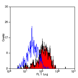
 FCM (Flow Cytometry)
(Figure 6. Flow Cytometry analysis of K562 cells using anti-D2AP antibody (M01756). Overlay histogram showing K562 cells stained with M01756 (Blue line).The cells were blocked with 10% normal goat serum. And then incubated with mouse anti-D2AP Antibody (M01756, 1ug/1x106 cells) for 30 min at 20 degree C. DyLight488 conjugated goat anti-mouse IgG (BA1126, 5-10ug/1x106 cells) was used as secondary antibody for 30 minutes at 20 degree C. Isotype control antibody (Green line) was mouse IgG (1ug/1x106) used under the same conditions. Unlabelled sample (Red line) was also used as a control.)
FCM (Flow Cytometry)
(Figure 6. Flow Cytometry analysis of K562 cells using anti-D2AP antibody (M01756). Overlay histogram showing K562 cells stained with M01756 (Blue line).The cells were blocked with 10% normal goat serum. And then incubated with mouse anti-D2AP Antibody (M01756, 1ug/1x106 cells) for 30 min at 20 degree C. DyLight488 conjugated goat anti-mouse IgG (BA1126, 5-10ug/1x106 cells) was used as secondary antibody for 30 minutes at 20 degree C. Isotype control antibody (Green line) was mouse IgG (1ug/1x106) used under the same conditions. Unlabelled sample (Red line) was also used as a control.)
CD2AP, Monoclonal Antibody (Cat# AAA19185)
Full Name
Anti-CD2AP Antibody (monoclonal, 5F8)
Gene Names
CD2AP; CMS
Reactivity
Human, Mouse, Rat
Applications
WB, IHC, FC/FACS
Purity
Immunogen Affinity Purified
Pricing


 FCM (Flow Cytometry)
(Figure 11. Flow Cytometry analysis of RH35 cells using anti- eRF1/ETF1 antibody (AAA19382).Overlay histogram showing RH35 cells stained with AAA19382 (Blue line). The cells were blocked with 10% normal goat serum. And then incubated with mouse anti-eRF1/ETF1 Antibody (AAA19382, 1μg/1x106 cells) for 30 min at 20 degree C. DyLight®488 conjugated goat anti-mouse IgG (BA1126, 5-10μg/1x106 cells) was used as secondary antibody for 30 minutes at 20 degree C. Isotype control antibody (Green line) was mouse IgG (1μg/1x106) used under the same conditions. Unlabelled sample (Red line) was also used as a control.)
FCM (Flow Cytometry)
(Figure 11. Flow Cytometry analysis of RH35 cells using anti- eRF1/ETF1 antibody (AAA19382).Overlay histogram showing RH35 cells stained with AAA19382 (Blue line). The cells were blocked with 10% normal goat serum. And then incubated with mouse anti-eRF1/ETF1 Antibody (AAA19382, 1μg/1x106 cells) for 30 min at 20 degree C. DyLight®488 conjugated goat anti-mouse IgG (BA1126, 5-10μg/1x106 cells) was used as secondary antibody for 30 minutes at 20 degree C. Isotype control antibody (Green line) was mouse IgG (1μg/1x106) used under the same conditions. Unlabelled sample (Red line) was also used as a control.)
eRF1/ETF1, Monoclonal Antibody (Cat# AAA19382)
Full Name
Anti-eRF1/ETF1 Antibody (monoclonal, 3B6)
Gene Names
ETF1; ERF; RF1; ERF1; TB3-1; D5S1995; SUP45L1
Reactivity
Human, Mouse, Rat
Applications
WB, IHC-P, ICC, IF, FC/FACS/FCM
Purity
Immunogen affinity purified.
Pricing


 FCM (Flow Cytometry)
(Figure 8. Flow Cytometry analysis of THP-1 cells using anti-INPPL1 antibody (AAA19364).Overlay histogram showing THP-1 cells stained with AAA19364 (Blue line). The cells were blocked with 10% normal goat serum. And then incubated with mouse anti- INPPL1 Antibody (AAA19364, 1μg/1x106 cells) for 30 min at 20 degree C. DyLight®488 conjugated goat anti-mouse IgG (BA1126, 5-10μg/1x106 cells) was used as secondary antibody for 30 minutes at 20 degree C. Isotype control antibody (Green line) was mouse IgG (1μg/1x106) used under the same conditions. Unlabelled sample (Red line) was also used as a control.)
FCM (Flow Cytometry)
(Figure 8. Flow Cytometry analysis of THP-1 cells using anti-INPPL1 antibody (AAA19364).Overlay histogram showing THP-1 cells stained with AAA19364 (Blue line). The cells were blocked with 10% normal goat serum. And then incubated with mouse anti- INPPL1 Antibody (AAA19364, 1μg/1x106 cells) for 30 min at 20 degree C. DyLight®488 conjugated goat anti-mouse IgG (BA1126, 5-10μg/1x106 cells) was used as secondary antibody for 30 minutes at 20 degree C. Isotype control antibody (Green line) was mouse IgG (1μg/1x106) used under the same conditions. Unlabelled sample (Red line) was also used as a control.)
INPPL1, Monoclonal Antibody (Cat# AAA19364)
Full Name
Anti-INPPL1 Antibody (monoclonal, 8C13)
Gene Names
INPPL1; OPSMD; SHIP2
Reactivity
Human, Mouse, Rat
Applications
WB, IHC-P, ICC, IF, FC/FACS/FCM
Purity
Immunogen affinity purified.
Pricing


 FCM (Flow Cytometry)
(Figure 9. Flow Cytometry analysis of U251 cells using anti-RAB1B antibody (AAA19296).Overlay histogram showing U251 cells stained with AAA19296 (Blue line). The cells were blocked with 10% normal goat serum. And then incubated with rabbit anti-RAB1B Antibody (AAA19296, 1μg/1x106 cells) for 30 min at 20 degree C. DyLight®488 conjugated goat anti-rabbit IgG (5-10μg/1x106 cells) was used as secondary antibody for 30 minutes at 20 degree C. Isotype control antibody (Green line) was rabbit IgG (1μg/1x106) used under the same conditions. Unlabelled sample (Red line) was also used as a control.)
FCM (Flow Cytometry)
(Figure 9. Flow Cytometry analysis of U251 cells using anti-RAB1B antibody (AAA19296).Overlay histogram showing U251 cells stained with AAA19296 (Blue line). The cells were blocked with 10% normal goat serum. And then incubated with rabbit anti-RAB1B Antibody (AAA19296, 1μg/1x106 cells) for 30 min at 20 degree C. DyLight®488 conjugated goat anti-rabbit IgG (5-10μg/1x106 cells) was used as secondary antibody for 30 minutes at 20 degree C. Isotype control antibody (Green line) was rabbit IgG (1μg/1x106) used under the same conditions. Unlabelled sample (Red line) was also used as a control.)
RAB1B, Polyclonal Antibody (Cat# AAA19296)
Full Name
Anti-RAB1B Antibody
Reactivity
Human, Mouse, Rat
Applications
WB, IHC-P, ICC, IF, FC/FACS/FCM
Purity
Immunogen affinity purified.
Pricing


 WB (Western Blot)
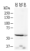
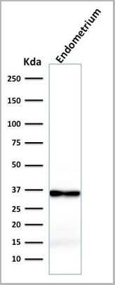
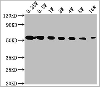
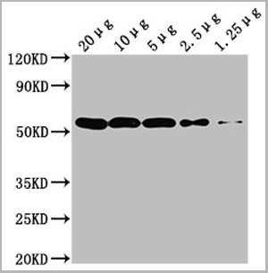
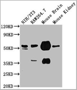
(Western blot analysis of lysates from 293, Hela, mouse NIH/3T3, rat PC-12 cell line and rat brain tissue lysate(from left to right), using RPS6 Antibody (N-term). AAA28643 was diluted at 1:2000 at each lane. A goat anti-mouse IgG H&L(HRP) at 1:3000 dilution was used as the secondary antibody. Lysates at 35ug per lane.)
WB (Western Blot)
(Western blot analysis of lysates from 293, Hela, mouse NIH/3T3, rat PC-12 cell line and rat brain tissue lysate(from left to right), using RPS6 Antibody (N-term). AAA28643 was diluted at 1:2000 at each lane. A goat anti-mouse IgG H&L(HRP) at 1:3000 dilution was used as the secondary antibody. Lysates at 35ug per lane.)
RPS6, Monoclonal Antibody (Cat# AAA28643)
Full Name
RPS6 Antibody (N-term)
Gene Names
RPS6; S6
Reactivity
Human, rat (Predicted Reactivity: Monkey, Mouse)
Applications
EIA, IHC, WB, FC/FACS, IF
Purity
Purified Mouse Monoclonal Antibody (Mab)
Pricing


 FCM (Flow Cytometry)
(Figure 7. Flow Cytometry analysis of MCF-7 cells using anti-RGS6 antibody (AAA19308).Overlay histogram showing MCF-7 cells stained with AAA19308 (Blue line). The cells were blocked with 10% normal goat serum. And then incubated with rabbit anti-RGS6 Antibody (AAA19308, 1μg/1x106 cells) for 30 min at 20 degree C. DyLight®488 conjugated goat anti-rabbit IgG (5-10μg/1x106 cells) was used as secondary antibody for 30 minutes at 20 degree C. Isotype control antibody (Green line) was rabbit IgG (1μg/1x106) used under the same conditions. Unlabelled sample (Red line) was also used as a control.)
FCM (Flow Cytometry)
(Figure 7. Flow Cytometry analysis of MCF-7 cells using anti-RGS6 antibody (AAA19308).Overlay histogram showing MCF-7 cells stained with AAA19308 (Blue line). The cells were blocked with 10% normal goat serum. And then incubated with rabbit anti-RGS6 Antibody (AAA19308, 1μg/1x106 cells) for 30 min at 20 degree C. DyLight®488 conjugated goat anti-rabbit IgG (5-10μg/1x106 cells) was used as secondary antibody for 30 minutes at 20 degree C. Isotype control antibody (Green line) was rabbit IgG (1μg/1x106) used under the same conditions. Unlabelled sample (Red line) was also used as a control.)
RGS6, Polyclonal Antibody (Cat# AAA19308)
Full Name
Anti-RGS6 Antibody
Gene Names
RGS6; GAP
Reactivity
Human, Mouse, Rat
Applications
WB, IHC-P, FC/FACS/FCM, EIA
Purity
Immunogen affinity purified.
Pricing


 FCM (Flow Cytometry)
(Figure 6. Flow Cytometry analysis of THP-1 cells using anti-PRMT8 antibody (AAA19321).Overlay histogram showing THP-1 cells stained with AAA19321 (Blue line). The cells were blocked with 10% normal goat serum. And then incubated with rabbit anti-PRMT8 Antibody (AAA19321,1μg/1x106 cells) for 30 min at 20 degree C. DyLight®488 conjugated goat anti-rabbit IgG (5-10μg/1x106 cells) was used as secondary antibody for 30 minutes at 20 degree C. Isotype control antibody (Green line) was rabbit IgG (1μg/1x106) used under the same conditions. Unlabelled sample (Red line) was also used as a control.)
FCM (Flow Cytometry)
(Figure 6. Flow Cytometry analysis of THP-1 cells using anti-PRMT8 antibody (AAA19321).Overlay histogram showing THP-1 cells stained with AAA19321 (Blue line). The cells were blocked with 10% normal goat serum. And then incubated with rabbit anti-PRMT8 Antibody (AAA19321,1μg/1x106 cells) for 30 min at 20 degree C. DyLight®488 conjugated goat anti-rabbit IgG (5-10μg/1x106 cells) was used as secondary antibody for 30 minutes at 20 degree C. Isotype control antibody (Green line) was rabbit IgG (1μg/1x106) used under the same conditions. Unlabelled sample (Red line) was also used as a control.)
PRMT8, Polyclonal Antibody (Cat# AAA19321)
Full Name
Anti-PRMT8 Antibody
Gene Names
PRMT8; HRMT1L3; HRMT1L4
Reactivity
Human, Rat
Applications
WB, IHC-P, FC/FACS/FCM, EIA
Purity
Immunogen affinity purified.
Pricing


 FCM (Flow Cytometry)
(Figure 12. Flow Cytometry analysis of U937 cells using anti-DYNLL1/PIN antibody (AAA19276).Overlay histogram showing U937 cells stained with AAA19276 (Blue line). The cells were blocked with 10% normal goat serum. And then incubated with rabbit anti-DYNLL1/PIN Antibody (AAA19276, 1μg/1x106 cells) for 30 min at 20 degree C. DyLight®488 conjugated goat anti-rabbit IgG (5-10μg/1x106 cells) was used as secondary antibody for 30 minutes at 20 degree C. Isotype control antibody (Green line) was rabbit IgG (1μg/1x106) used under the same conditions. Unlabelled sample (Red line) was also used as a control.)
FCM (Flow Cytometry)
(Figure 12. Flow Cytometry analysis of U937 cells using anti-DYNLL1/PIN antibody (AAA19276).Overlay histogram showing U937 cells stained with AAA19276 (Blue line). The cells were blocked with 10% normal goat serum. And then incubated with rabbit anti-DYNLL1/PIN Antibody (AAA19276, 1μg/1x106 cells) for 30 min at 20 degree C. DyLight®488 conjugated goat anti-rabbit IgG (5-10μg/1x106 cells) was used as secondary antibody for 30 minutes at 20 degree C. Isotype control antibody (Green line) was rabbit IgG (1μg/1x106) used under the same conditions. Unlabelled sample (Red line) was also used as a control.)
DYNLL1/PIN, Polyclonal Antibody (Cat# AAA19276)
Full Name
Anti-DYNLL1/PIN Antibody
Gene Names
DYNLL1; LC8; PIN; DLC1; DLC8; LC8a; DNCL1; hdlc1; DNCLC1
Reactivity
Human, Mouse, Rat
Applications
WB, IHC-P, ICC, IF, FC/FACS/FCM, EIA
Purity
Immunogen affinity purified.
Pricing


 FCM (Flow Cytometry)
(Figure 7. Flow Cytometry analysis of A549 cells using anti-Hsp90 alpha antibody (AAA19359).Overlay histogram showing A549 cells stained with AAA19359 (Blue line). The cells were blocked with 10% normal goat serum. And then incubated with mouse anti-Hsp90 alpha Antibody (AAA19359, 1μg/1x106 cells) for 30 min at 20 degree C. DyLight®488 conjugated goat anti-mouse IgG (BA1126, 5-10μg/1x106 cells) was used as secondary antibody for 30 minutes at 20 degree C. Isotype control antibody (Green line) was mouse IgG (1μg/1x106) used under the same conditions. Unlabelled sample (Red line) was also used as a control.)
FCM (Flow Cytometry)
(Figure 7. Flow Cytometry analysis of A549 cells using anti-Hsp90 alpha antibody (AAA19359).Overlay histogram showing A549 cells stained with AAA19359 (Blue line). The cells were blocked with 10% normal goat serum. And then incubated with mouse anti-Hsp90 alpha Antibody (AAA19359, 1μg/1x106 cells) for 30 min at 20 degree C. DyLight®488 conjugated goat anti-mouse IgG (BA1126, 5-10μg/1x106 cells) was used as secondary antibody for 30 minutes at 20 degree C. Isotype control antibody (Green line) was mouse IgG (1μg/1x106) used under the same conditions. Unlabelled sample (Red line) was also used as a control.)
Hsp90 alpha, Monoclonal Antibody (Cat# AAA19359)
Full Name
Anti-Hsp90 alpha Antibody (monoclonal, 6B5)
Gene Names
HSP90AA1; EL52; HSPN; LAP2; HSP86; HSPC1; HSPCA; Hsp89; Hsp90; HSP89A; HSP90A; HSP90N; HSPCAL1; HSPCAL4
Reactivity
Human, Mouse, Rat, Monkey
Applications
WB, IHC-P, ICC, IF, FC/FACS/FCM
Purity
Immunogen affinity purified.
Pricing


 FCM (Flow Cytometry)
(Figure 7. Flow Cytometry analysis of U20S cells using anti- ADA antibody (AAA19184). Overlay histogram showing U20S cells stained with AAA19184 (Blue line).The cells were blocked with 10% normal goat serum. And then incubated with mouse anti- ADA Antibody (AAA19184, 1ug/1x106 cells) for 30 min at 20 degree C. DyLight488 conjugated goat anti-mouse IgG (BA1126, 5-10ug/1x106 cells) was used as secondary antibody for 30 minutes at 20 degree C. Isotype control antibody (Green line) was mouse IgG (1ug/1x106) used under the same conditions. Unlabelled sample (Red line) was also used as a control.)
FCM (Flow Cytometry)
(Figure 7. Flow Cytometry analysis of U20S cells using anti- ADA antibody (AAA19184). Overlay histogram showing U20S cells stained with AAA19184 (Blue line).The cells were blocked with 10% normal goat serum. And then incubated with mouse anti- ADA Antibody (AAA19184, 1ug/1x106 cells) for 30 min at 20 degree C. DyLight488 conjugated goat anti-mouse IgG (BA1126, 5-10ug/1x106 cells) was used as secondary antibody for 30 minutes at 20 degree C. Isotype control antibody (Green line) was mouse IgG (1ug/1x106) used under the same conditions. Unlabelled sample (Red line) was also used as a control.)
ADA, Monoclonal Antibody (Cat# AAA19184)
Full Name
Anti-ADA Antibody (monoclonal, 6D4)
Reactivity
Human
Applications
WB, IHC, FC/FACS
Purity
Immunogen Affinity Purified
Pricing


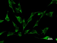
 IF (Immunofluorescence)
(Figure 11. IF analysis of Tubulin alpha using anti- Tubulin alpha antibody (AAA19381).Tubulin alpha was detected in immunocytochemical section of CACO-2 cells. Enzyme antigen retrieval was performed using IHC enzyme antigen retrieval reagent for 15 mins. The cells were blocked with 10% goat serum. And then incubated with 5μg/mL mouse anti- Tubulin alpha Antibody (AAA19381) overnight at 4 degree C. DyLight®594 Conjugated Goat Anti-Mouse IgG (BA1141) was used as secondary antibody at 1:100 dilution and incubated for 30 minutes at 37 degree C. The section was counterstained with DAPI. Visualize using a fluorescence microscope and filter sets appropriate for the label used.)
IF (Immunofluorescence)
(Figure 11. IF analysis of Tubulin alpha using anti- Tubulin alpha antibody (AAA19381).Tubulin alpha was detected in immunocytochemical section of CACO-2 cells. Enzyme antigen retrieval was performed using IHC enzyme antigen retrieval reagent for 15 mins. The cells were blocked with 10% goat serum. And then incubated with 5μg/mL mouse anti- Tubulin alpha Antibody (AAA19381) overnight at 4 degree C. DyLight®594 Conjugated Goat Anti-Mouse IgG (BA1141) was used as secondary antibody at 1:100 dilution and incubated for 30 minutes at 37 degree C. The section was counterstained with DAPI. Visualize using a fluorescence microscope and filter sets appropriate for the label used.)
Tubulin alpha, Monoclonal Antibody (Cat# AAA19381)
Full Name
Anti-Tubulin alpha Antibody (monoclonal, 7B12)
Gene Names
TUBA1A; LIS3; TUBA3; B-ALPHA-1
Reactivity
Human, Mouse, Rat
Applications
WB, IHC-P, ICC, IF, FC/FACS/FCM
Purity
Immunogen affinity purified.
Pricing


 FCM (Flow Cytometry)
(Figure 8. Flow Cytometry analysis of K562 cells using anti-PDIA6 antibody (AAA19282).Overlay histogram showing K562 cells stained with AAA19282 (Blue line). The cells were blocked with 10% normal goat serum. And then incubated with rabbit anti-PDIA6 Antibody (AAA19282, 1μg/1x106 cells) for 30 min at 20 degree C. DyLight®488 conjugated goat anti-rabbit IgG (5-10μg/1x106 cells) was used as secondary antibody for 30 minutes at 20 degree C. Isotype control antibody (Green line) was rabbit IgG (1μg/1x106) used under the same conditions. Unlabelled sample (Red line) was also used as a control.)
FCM (Flow Cytometry)
(Figure 8. Flow Cytometry analysis of K562 cells using anti-PDIA6 antibody (AAA19282).Overlay histogram showing K562 cells stained with AAA19282 (Blue line). The cells were blocked with 10% normal goat serum. And then incubated with rabbit anti-PDIA6 Antibody (AAA19282, 1μg/1x106 cells) for 30 min at 20 degree C. DyLight®488 conjugated goat anti-rabbit IgG (5-10μg/1x106 cells) was used as secondary antibody for 30 minutes at 20 degree C. Isotype control antibody (Green line) was rabbit IgG (1μg/1x106) used under the same conditions. Unlabelled sample (Red line) was also used as a control.)
PDIA6, Polyclonal Antibody (Cat# AAA19282)
Full Name
Anti-PDIA6 Antibody
Gene Names
PDIA6; P5; ERP5; TXNDC7
Reactivity
Human, Mouse, Rat
Applications
WB, IHC-P, ICC, IF, FC/FACS/FCM, EIA
Purity
Immunogen affinity purified.
Pricing

 ICC (Immunocytochemistry)
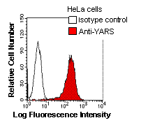
(Immunostaining analysis in HeLa cells. HeLa cells were fixed with 4% paraformaldehyde and permeabilized with 0.1% Triton X-100 in PBS. The cells were immunostained with anti-YARS mAb. [Lot No. YAR5H08-1])
ICC (Immunocytochemistry)
(Immunostaining analysis in HeLa cells. HeLa cells were fixed with 4% paraformaldehyde and permeabilized with 0.1% Triton X-100 in PBS. The cells were immunostained with anti-YARS mAb. [Lot No. YAR5H08-1])
YARS, Monoclonal Antibody (Cat# AAA10598)
Full Name
Mouse monoclonal antibody Anti-Human YARS
Gene Names
YARS; YRS; YTS; TYRRS; CMTDIC
Reactivity
Human
Applications
Immunocytochemistry, Immunoprecipitation, Western Blot, Flow Cytometry
Pricing
 IF (Immunofluorescence)
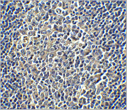
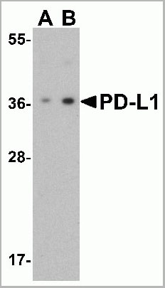
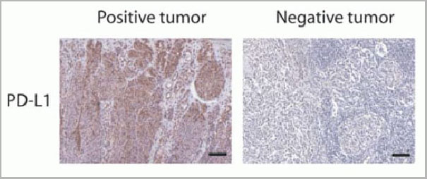
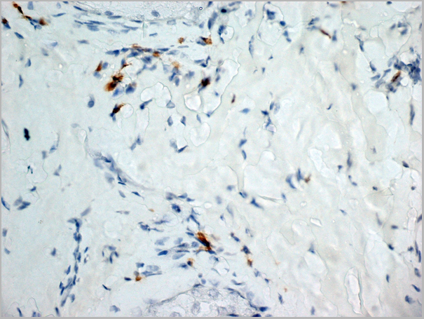
(Figure 12 Immunohistochemistry Validation of PD-L1in Human Tumors (Gadiot et al., 2011)Immunohistochemical analysis of patient tumors labeling PD-L1 with anti-PD-L1 antibodies (AAA10941). Several anti-PD-L1 antibodies were tested for staining, “Only 1 antibody gave no background staining and was competitively blocked by the addition of PD-L1Fc protein (AAA10941)”.)
IF (Immunofluorescence)
(Figure 12 Immunohistochemistry Validation of PD-L1in Human Tumors (Gadiot et al., 2011)Immunohistochemical analysis of patient tumors labeling PD-L1 with anti-PD-L1 antibodies (AAA10941). Several anti-PD-L1 antibodies were tested for staining, “Only 1 antibody gave no background staining and was competitively blocked by the addition of PD-L1Fc protein (AAA10941)”.)
PDL-1, Polyclonal Antibody (Cat# AAA10941)
Full Name
PDL-1 Antibody
Gene Names
CD274; B7-H; B7H1; PDL1; PD-L1; PDCD1L1; PDCD1LG1
Applications
Western Blot, Immunohistochemistry, Immunofluorescence, Flow Cytometry
Purity
PD-L1 Antibody is affinity chromatography purified via peptide column.
Pricing
 Application Data
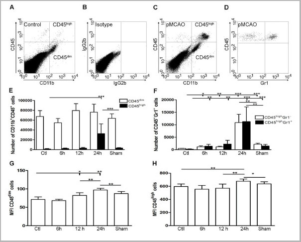
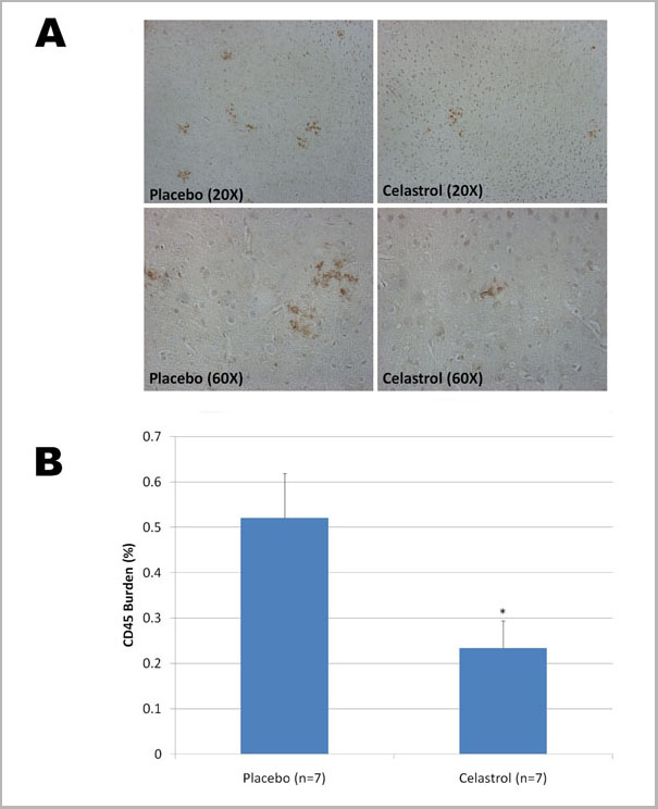
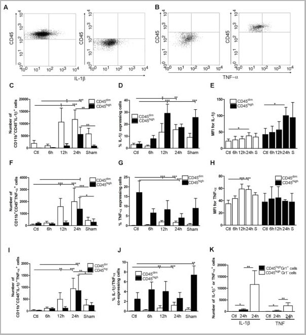
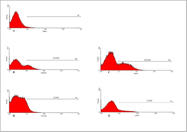
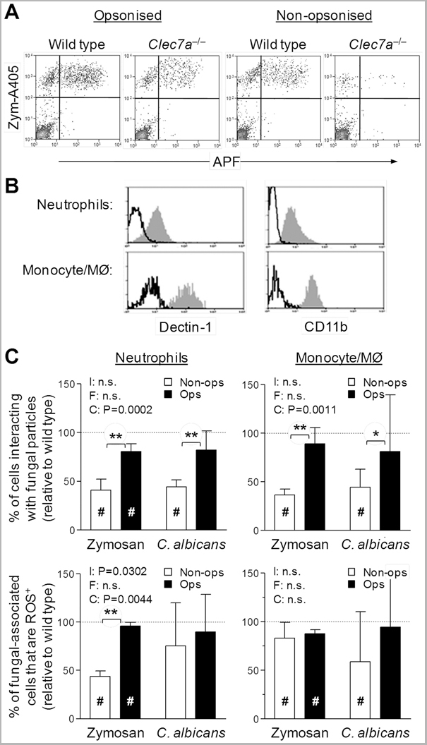
(Published customer image: Cytokine expression in segregated populations of cells following stroke. (A, B) Dot plots showing CD11b+CD45high macrophages/granulocytes (upper right quadrants) and CD11b+CD45dim microglia (bottom right quadrants) expressing IL-1beta (A) or TNF-a (B). (C-J) Bar graphs showing numbers and proportions of IL-1beta (C, D), TNF-a (F, G) and IL-1beta/TNF-a co-expressing (I, J) CD11b+CD45dim microglia and CD11b+CD45high macrophages/granulocytes in unmanipulated control mice (n = 10), in mice 6 (n = 7), 12 (n = 7), or 24 hours after pMCAO (n = 10), and in sham-operated mice 24 hours after pMCAO (n = 7). (E, H) Comparison of the MFI values for IL-1beta (E) and TNF-a (H) in viable CD11b+CD45dim microglia and CD11b+CD45high macrophages/granulocytes in unmanipulated mice, in mice 6, 12, or 24 hours after pMCAO, and in sham-operated mice 24 hours after pMCAO. Macrophages/granulocytes express significantly more IL-1beta than do microglial in unmanipulated mice, in mice 6, 12, or 24 hours after pMCAO, and in sham-operated mice 24 hours after pMCAO (E), whereas microglial cells express significantly higher levels of TNF-a than do macrophages/granulocytes at 12 h and 24 hours, and in sham-operated mice 24 hours after pMCAO (H). (K) CD11b+CD45highGr1- macrophages and not CD11b+CD45highGr1+ granulocytes are the main producers of IL-1beta and TNF-a 24 hours after pMCAO. *P < 0.05, **P < 0.01, and ***P < 0.001.From: http://www.jneuroinflammation.com/content/5/1/46.)
Application Data
(Published customer image: Cytokine expression in segregated populations of cells following stroke. (A, B) Dot plots showing CD11b+CD45high macrophages/granulocytes (upper right quadrants) and CD11b+CD45dim microglia (bottom right quadrants) expressing IL-1beta (A) or TNF-a (B). (C-J) Bar graphs showing numbers and proportions of IL-1beta (C, D), TNF-a (F, G) and IL-1beta/TNF-a co-expressing (I, J) CD11b+CD45dim microglia and CD11b+CD45high macrophages/granulocytes in unmanipulated control mice (n = 10), in mice 6 (n = 7), 12 (n = 7), or 24 hours after pMCAO (n = 10), and in sham-operated mice 24 hours after pMCAO (n = 7). (E, H) Comparison of the MFI values for IL-1beta (E) and TNF-a (H) in viable CD11b+CD45dim microglia and CD11b+CD45high macrophages/granulocytes in unmanipulated mice, in mice 6, 12, or 24 hours after pMCAO, and in sham-operated mice 24 hours after pMCAO. Macrophages/granulocytes express significantly more IL-1beta than do microglial in unmanipulated mice, in mice 6, 12, or 24 hours after pMCAO, and in sham-operated mice 24 hours after pMCAO (E), whereas microglial cells express significantly higher levels of TNF-a than do macrophages/granulocytes at 12 h and 24 hours, and in sham-operated mice 24 hours after pMCAO (H). (K) CD11b+CD45highGr1- macrophages and not CD11b+CD45highGr1+ granulocytes are the main producers of IL-1beta and TNF-a 24 hours after pMCAO. *P < 0.05, **P < 0.01, and ***P < 0.001.From: http://www.jneuroinflammation.com/content/5/1/46.)
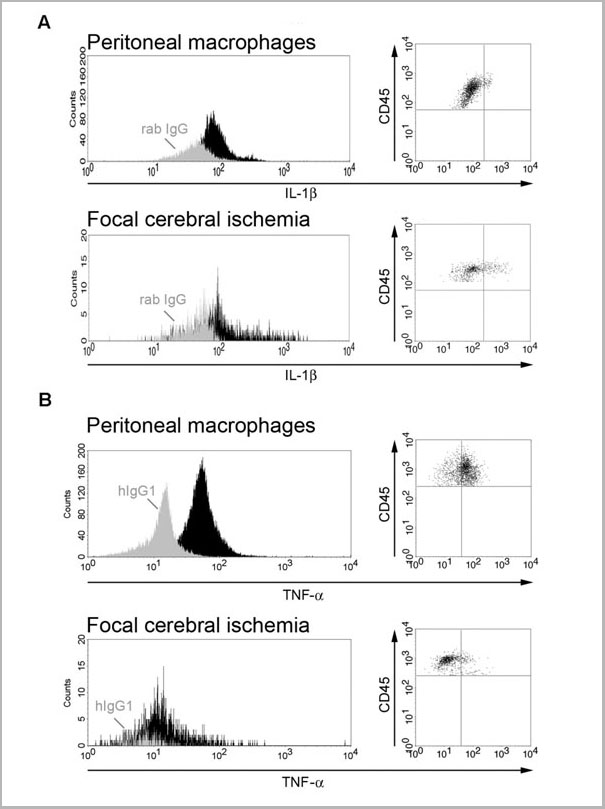
CD45, Monoclonal Antibody (Cat# AAA11896)
Full Name
RAT ANTI MOUSE CD45
Gene Names
Ptprc; loc; B220; Cd45; L-CA; Ly-5; T200; CD45R; Lyt-4
Applications
Immunohistochemistry, Flow Cytometry, Immunofluorescence, Immunoprecipitation
Pricing
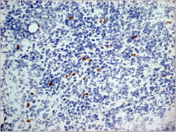
 Application Data
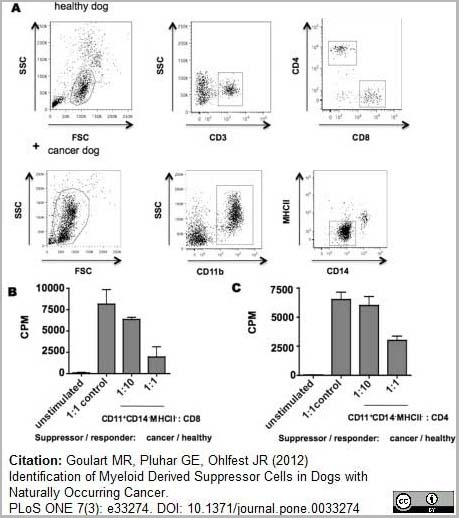
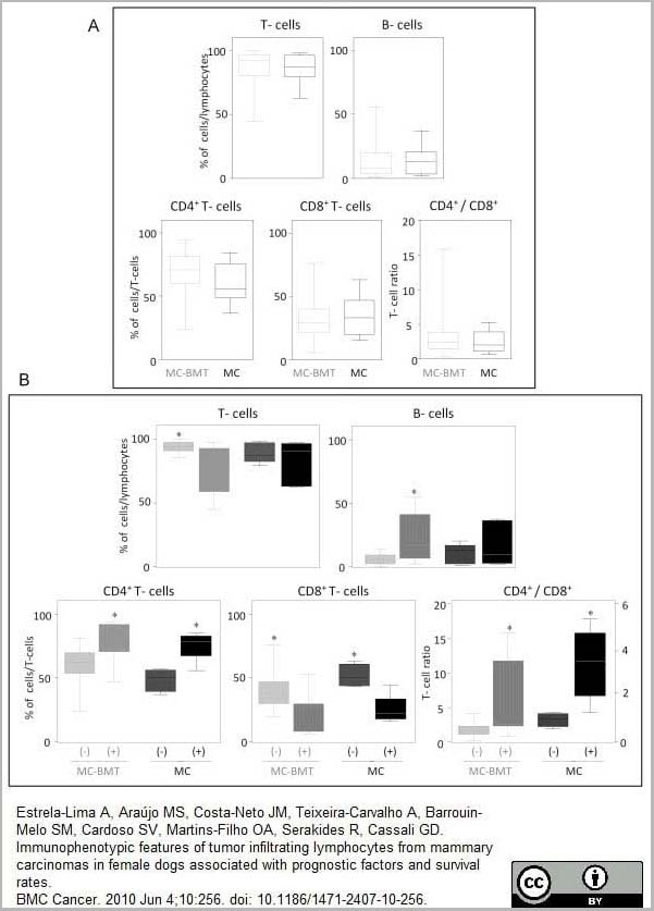
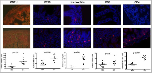
(Published customer image:RPE conjugated Mouse anti Canine CD4 antibody, clone YKIC302.9used for the assessment of CD4 levels on canine cells by flow cytometry.Image caption:Immunophenotypic profile of tumor infiltrating lymphocyte in canine mammary carcinomas. Analysis of tumor infiltrating T-cells, B-lymphocytes and T-cell subsets from MC-BMT or MC (A), further subcategorized according to the absence (-) or presence (+) of lymph node metastasis (-) (B). Lymphocyte populations and subsets were identified by flow cytometric immunostaining as described in Material and Methods. Data were expressed as percentage of positive cells within gated lymphocytes and CD4+/CD8+ T-cell ratio. Significant differences at p < 0.05 are highlighted by asterisk.From: From: Estrela-Lima et al.BMC Cancer 2010 10:256.)
Application Data
(Published customer image:RPE conjugated Mouse anti Canine CD4 antibody, clone YKIC302.9used for the assessment of CD4 levels on canine cells by flow cytometry.Image caption:Immunophenotypic profile of tumor infiltrating lymphocyte in canine mammary carcinomas. Analysis of tumor infiltrating T-cells, B-lymphocytes and T-cell subsets from MC-BMT or MC (A), further subcategorized according to the absence (-) or presence (+) of lymph node metastasis (-) (B). Lymphocyte populations and subsets were identified by flow cytometric immunostaining as described in Material and Methods. Data were expressed as percentage of positive cells within gated lymphocytes and CD4+/CD8+ T-cell ratio. Significant differences at p < 0.05 are highlighted by asterisk.From: From: Estrela-Lima et al.BMC Cancer 2010 10:256.)
CD4, Monoclonal Antibody (Cat# AAA12279)
Full Name
RAT ANTI DOG CD4: RPE-Cy7
Applications
Flow Cytometry
Purity
Purified IgG prepared by affinity chromatography on Protein G from tissue culture supernatant
Pricing


 Application Data
(Staining of human peripheral blood granulocytes with CD18: Alexa Fluor 647)
Application Data
(Staining of human peripheral blood granulocytes with CD18: Alexa Fluor 647)
CD18, Monoclonal Antibody (Cat# AAA12051)
Full Name
RAT ANTI HUMAN CD18:RPE
Gene Names
ITGB2; LAD; CD18; MF17; MFI7; LCAMB; LFA-1; MAC-1
Applications
Flow Cytometry
Pricing


 Application Data
(Staining of human peripheral blood granulocytes with CD18: Alexa Fluor 647 (AAA11986A647))
Application Data
(Staining of human peripheral blood granulocytes with CD18: Alexa Fluor 647 (AAA11986A647))
CD18, Monoclonal Antibody (Cat# AAA11986)
Full Name
RAT ANTI HUMAN CD18
Gene Names
ITGB2; LAD; CD18; MF17; MFI7; LCAMB; LFA-1; MAC-1
Reactivity
Dog, Guinea Pig
Applications
Immunohistochemistry, Flow Cytometry, Immunoprecipitation
Pricing
 Application Data
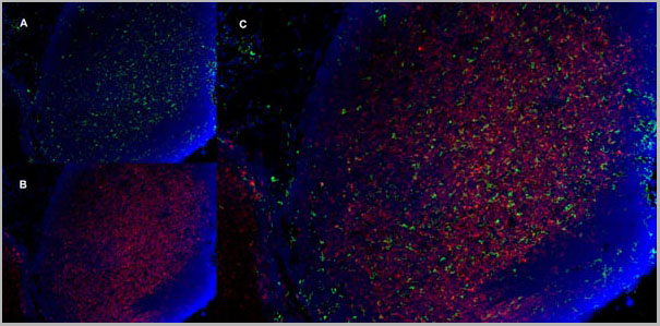
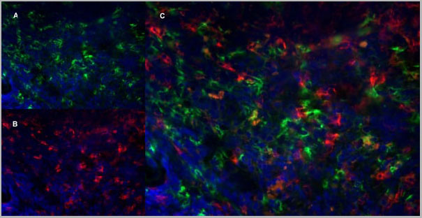
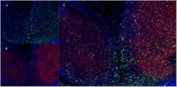
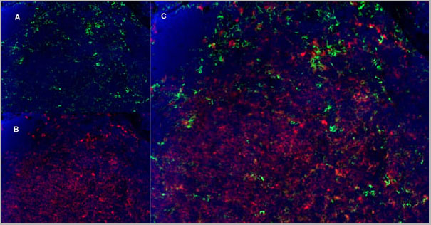
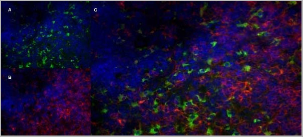
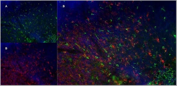
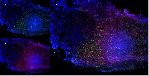
(Immunofluorescence staining of mouse lymph node cryosection with Rat anti Mouse CD11b, clone 5C6 , green in A and Rat anti Mouse CD8, clone YTS105.18 , red in B. C is the merged image with nuclei counterstained blue using DAPI. High power)
Application Data
(Immunofluorescence staining of mouse lymph node cryosection with Rat anti Mouse CD11b, clone 5C6 , green in A and Rat anti Mouse CD8, clone YTS105.18 , red in B. C is the merged image with nuclei counterstained blue using DAPI. High power)
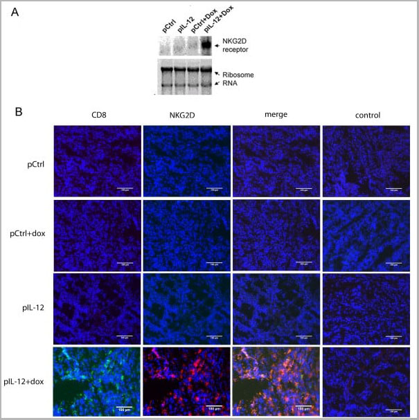
CD8 ALPHA, Monoclonal Antibody (Cat# AAA11918)
Full Name
RAT ANTI MOUSE CD8
Gene Names
Cd8a; Ly-2; Ly-B; Ly-35; Lyt-2; BB154331
Applications
Immunohistochemistry, Flow Cytometry, Immunofluorescence
Pricing
 Application Data
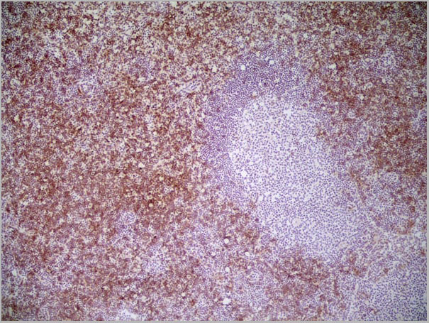
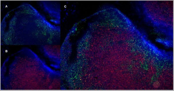
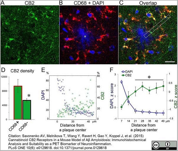
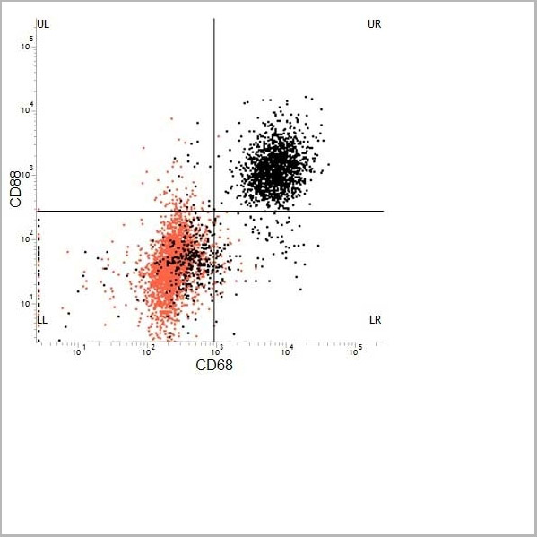
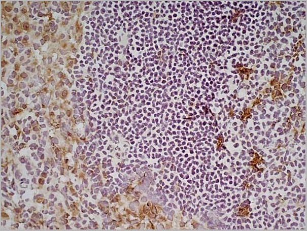
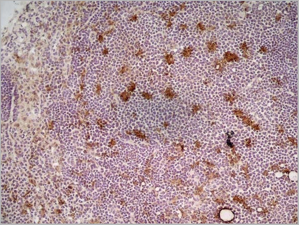
(Published customer image:Rat anti Mouse CD68 antibody, clone FA-11 used for the identification of microglia in mouse brain by immunofluorescence.Image caption:Comparison of CB2 immunoreactivity in neurons, activated microglia and astrocytes. ()
Application Data
(Published customer image:Rat anti Mouse CD68 antibody, clone FA-11 used for the identification of microglia in mouse brain by immunofluorescence.Image caption:Comparison of CB2 immunoreactivity in neurons, activated microglia and astrocytes. ()
CD68, Monoclonal Antibody (Cat# AAA12289)
Full Name
Rat anti Mouse CD68:Amethyst Orange
Gene Names
Cd68; Lamp4; gp110; Scard1
Reactivity
Mouse
Applications
Flow Cytometry
Purity
Purified IgG prepared by affinity chromatography on Protein G from tissue culture supernatant
Pricing


 Application Data
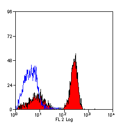
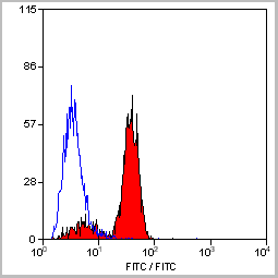
(Staining of mouse peritoneal macrophages with Rat anti Mouse Beta-glucan Receptor: FITC)
Application Data
(Staining of mouse peritoneal macrophages with Rat anti Mouse Beta-glucan Receptor: FITC)
DECTIN-1, Monoclonal Antibody (Cat# AAA12129)
Full Name
RAT ANTI MOUSE DECTIN-1:Low Endotoxin
Gene Names
Clec7a; BGR; beta-GR; Clecsf12
Applications
Immunohistochemistry, Flow Cytometry, Functional Assay, Immunoprecipitation
Pricing


 Application Data
(Immunofluorescence staining of mouse lymph node cryosection with Rat anti Mouse CD11b, clone 5C6 , green in A and Rat anti Mouse CD8, clone YTS105.18 , red in B. C is the merged image with nuclei counterstained blue using DAPI. High power)
Application Data
(Immunofluorescence staining of mouse lymph node cryosection with Rat anti Mouse CD11b, clone 5C6 , green in A and Rat anti Mouse CD8, clone YTS105.18 , red in B. C is the merged image with nuclei counterstained blue using DAPI. High power)
CD8 ALPHA, Monoclonal Antibody (Cat# AAA12069)
Full Name
RAT ANTI MOUSE CD8
Gene Names
Cd8a; Ly-2; Ly-B; Ly-35; Lyt-2; BB154331
Reactivity
Mouse
Applications
Immunohistochemistry, Flow Cytometry, Immunofluorescence
Pricing


 Application Data
(Staining of mouse peritoneal macrophages with Rat anti Mouse Beta-glucan Receptor: FITC)
Application Data
(Staining of mouse peritoneal macrophages with Rat anti Mouse Beta-glucan Receptor: FITC)
DECTIN-1, Monoclonal Antibody (Cat# AAA12130)
Full Name
RAT ANTI MOUSE DECTIN-1:FITC
Gene Names
Clec7a; BGR; beta-GR; Clecsf12
Applications
Flow Cytometry
Pricing


 Application Data
(Staining of mouse peritoneal macrophages with Rat anti Mouse Beta-glucan Receptor: FITC)
Application Data
(Staining of mouse peritoneal macrophages with Rat anti Mouse Beta-glucan Receptor: FITC)
DECTIN-1, Monoclonal Antibody (Cat# AAA12136)
Full Name
RAT ANTI MOUSE DECTIN-1:RPE
Gene Names
Clec7a; BGR; beta-GR; Clecsf12
Applications
Flow Cytometry
Pricing
 FCM (Flow Cytometry)
(Flow Cytometric Analysis of PFA-fixed K562 cells using Calponin-1 Recombinant Mouse Monoclonal Antibody (rCNN1/832) followed by Goat anti-Mouse IgG-CF488 (Blue); Isotype Control (Red).)
FCM (Flow Cytometry)
(Flow Cytometric Analysis of PFA-fixed K562 cells using Calponin-1 Recombinant Mouse Monoclonal Antibody (rCNN1/832) followed by Goat anti-Mouse IgG-CF488 (Blue); Isotype Control (Red).)
Calponin-1, Monoclonal Antibody (Cat# AAA13848)
Full Name
Calponin-1 (Smooth Muscle Marker)
Gene Names
CNN1; SMCC; Sm-Calp; HEL-S-14
Reactivity
Human, Rat
Applications
Flow Cytometry, Immunofluorescence, Immunohistochemistry
Pricing


 Application Data
(Staining of human peripheral blood granulocytes with CD18: Alexa Fluor 647)
Application Data
(Staining of human peripheral blood granulocytes with CD18: Alexa Fluor 647)
CD18, Monoclonal Antibody (Cat# AAA11881)
Full Name
RAT ANTI HUMAN CD18:FITC
Gene Names
ITGB2; LAD; CD18; MF17; MFI7; LCAMB; LFA-1; MAC-1
Applications
Flow Cytometry
Pricing


 Application Data
(Staining of human peripheral blood granulocytes with CD18: Alexa Fluor 647)
Application Data
(Staining of human peripheral blood granulocytes with CD18: Alexa Fluor 647)
CD18, Monoclonal Antibody (Cat# AAA11987)
Full Name
RAT ANTI HUMAN CD18
Gene Names
ITGB2; LAD; CD18; MF17; MFI7; LCAMB; LFA-1; MAC-1
Applications
Immunohistochemistry, Flow Cytometry, Immunoprecipitation
Pricing
 FCM (Flow Cytometry)
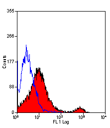
(Flow Cytometric Analysis of U937 cells using CD15 Rabbit Recombinant Monoclonal Antibody (FUT4/1478R) followed by goat anti-rabbit IgG-CF488 (Blue); Isotype Control (Red).)
FCM (Flow Cytometry)
(Flow Cytometric Analysis of U937 cells using CD15 Rabbit Recombinant Monoclonal Antibody (FUT4/1478R) followed by goat anti-rabbit IgG-CF488 (Blue); Isotype Control (Red).)
CD15/FUT4, Monoclonal Antibody (Cat# AAA13849)
Full Name
CD15/FUT4 (Reed-Sternberg Cell Marker)
Gene Names
FUT4; LeX; CD15; ELFT; FCT3A; FUTIV; SSEA-1; FUC-TIV
Reactivity
Human, Mouse, Rat. Others not known.
Applications
Flow Cytometry, Immunofluorescence, Immunohistochemistry
Pricing


 Application Data
(Staining of mouse peritoneal macrophages with Rat anti Mouse Beta-glucan Receptor: FITC)
Application Data
(Staining of mouse peritoneal macrophages with Rat anti Mouse Beta-glucan Receptor: FITC)
DECTIN-1, Monoclonal Antibody (Cat# AAA12131)
Full Name
RAT ANTI MOUSE DECTIN-1:FITC
Gene Names
Clec7a; BGR; beta-GR; Clecsf12
Applications
Flow Cytometry
Pricing
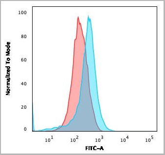
 FCM (Flow Cytometry)
(WB - MCSF Receptor (CSF1R) Antibody (C-term) AAA28757 detail IHC-P - MCSF Receptor (CSF1R) Antibody (C-term) AAA28757 detail IHC-P - MCSF Receptor (CSF1R) Antibody (C-term) AAA28757 detail FC - MCSF Receptor (CSF1R) Antibody (C-term) AAA28757 detail Overlay histogram showing HepG2 cells stained with AAA28757(green line). The cells were fixed with 2% paraformaldehyde (10 min) and then permeabilized with 90% methanol for 10 min. The cells were then icubated in 2% bovine serum albumin to block non-specific protein-protein interactions followed by the antibody (AAA28757, 1:25 dilution) for 60 min at 37ºC. The secondary antibody used was Goat-Anti-Rabbit IgG, DyLight® 488 Conjugated Highly Cross-Adsorbed(1583138) at 1/200 dilution for 40 min at 37ºC. Isotype control antibody (blue line) was rabbit IgG1 (1ug/1x10^6 cells) used under the same conditions. Acquisition of >10, 000 events was performed.)
FCM (Flow Cytometry)
(WB - MCSF Receptor (CSF1R) Antibody (C-term) AAA28757 detail IHC-P - MCSF Receptor (CSF1R) Antibody (C-term) AAA28757 detail IHC-P - MCSF Receptor (CSF1R) Antibody (C-term) AAA28757 detail FC - MCSF Receptor (CSF1R) Antibody (C-term) AAA28757 detail Overlay histogram showing HepG2 cells stained with AAA28757(green line). The cells were fixed with 2% paraformaldehyde (10 min) and then permeabilized with 90% methanol for 10 min. The cells were then icubated in 2% bovine serum albumin to block non-specific protein-protein interactions followed by the antibody (AAA28757, 1:25 dilution) for 60 min at 37ºC. The secondary antibody used was Goat-Anti-Rabbit IgG, DyLight® 488 Conjugated Highly Cross-Adsorbed(1583138) at 1/200 dilution for 40 min at 37ºC. Isotype control antibody (blue line) was rabbit IgG1 (1ug/1x10^6 cells) used under the same conditions. Acquisition of >10, 000 events was performed.)
MCSF Receptor (CSF1R), Polyclonal Antibody (Cat# AAA28757)
Full Name
MCSF Receptor (CSF1R) Antibody (C-term)
Gene Names
CSF1R; FMS; CSFR; FIM2; HDLS; C-FMS; CD115; CSF-1R; M-CSF-R
Reactivity
Human
Applications
Western Blot, Flow Cytometry, Immunohistochemistry
Purity
Purified Rabbit Polyclonal Antibody (Pab)
Pricing
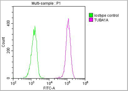
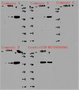
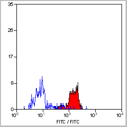
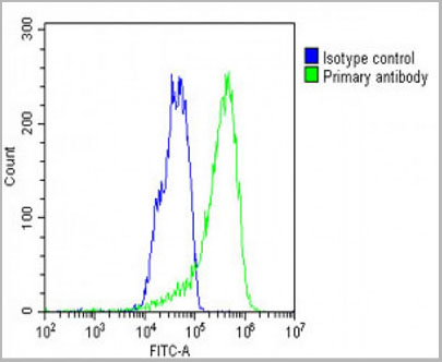
 FCM (Flow Cytometry)
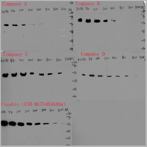
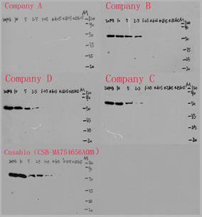
(Overlay histogram showing Hela cells stainedwith Company A (5ug), Company B (5ug), Company C (5ug), Company D (5ug), AAA27031 (5ug) (red line) at 1:150. The cells were incubated in 1x PBS /10% normal goat serum to block non-specific protein-protein interactions followed by primary antibody for 1 hat 4°C. The secondary antibody used was FITC goat anti-mouse IgG(H+L) at 1/200 dilution for 1 h at 4°C. Isotype control antibody (green line) was used under the same conditions. Acquisition of >10,000 events was performed.)
FCM (Flow Cytometry)
(Overlay histogram showing Hela cells stainedwith Company A (5ug), Company B (5ug), Company C (5ug), Company D (5ug), AAA27031 (5ug) (red line) at 1:150. The cells were incubated in 1x PBS /10% normal goat serum to block non-specific protein-protein interactions followed by primary antibody for 1 hat 4°C. The secondary antibody used was FITC goat anti-mouse IgG(H+L) at 1/200 dilution for 1 h at 4°C. Isotype control antibody (green line) was used under the same conditions. Acquisition of >10,000 events was performed.)
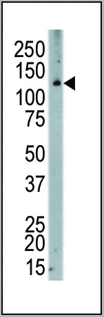
TUBA1A, Monoclonal Antibody (Cat# AAA27031)
Full Name
TUBA1A
Gene Names
Tuba1a; Tuba1; Tuba-1
Reactivity
Human, Rabbit, Rat, Mouse
Applications
Western Blot, Immunohistochemistry, Immunofluorescence, Flow Cytometry, Immunoprecipitation
Purity
>95%, Protein A purified
Pricing


 Application Data
(Staining of mouse peritoneal macrophages with Rat anti Mouse Beta-glucan Receptor: FITC)
Application Data
(Staining of mouse peritoneal macrophages with Rat anti Mouse Beta-glucan Receptor: FITC)
DECTIN-1, Monoclonal Antibody (Cat# AAA12133)
Full Name
RAT ANTI MOUSE DECTIN-1:FITC
Gene Names
Clec7a; BGR; beta-GR; Clecsf12
Applications
Flow Cytometry
Pricing