Filters
Clonality
Type
Reactivity
Gene Name
Isotype
Host
Application
Clone
1769 results for " Anti Human Monoclonal Antibody" - showing 1600-1650
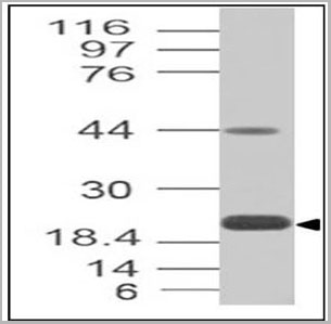
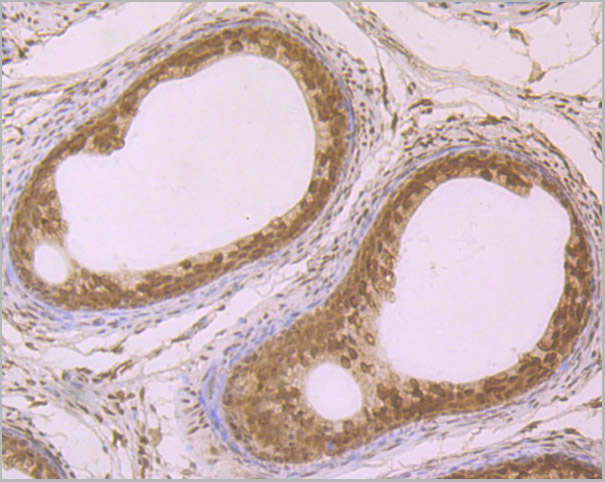
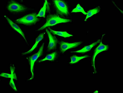
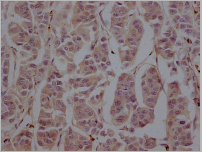
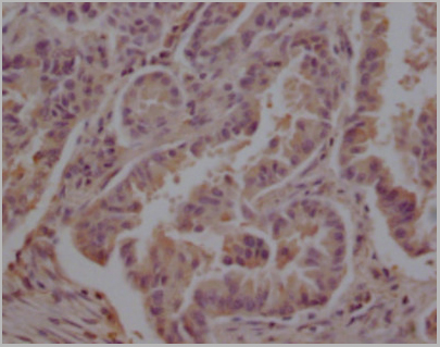
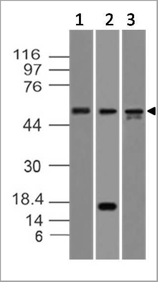
 WB (Western Blot)
(Western blot analysis of PD-L1. Anti-PD-L1 antibody (Clone: ABM4E54) was tested at 2 ug/ml on h Spleen lysate.)
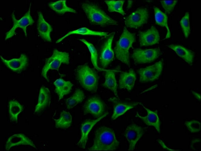
WB (Western Blot)
(Western blot analysis of PD-L1. Anti-PD-L1 antibody (Clone: ABM4E54) was tested at 2 ug/ml on h Spleen lysate.)
PD-L1, Monoclonal Antibody (Cat# AAA14873)
Full Name
Monoclonal Antibody to PD-L1 (Clone: ABM4E54)
Gene Names
CD274; B7-H; B7H1; PDL1; PD-L1; PDCD1L1; PDCD1LG1
Reactivity
Human
Applications
Immunohistochemistry, Flow Cytometry, Western Blot
Purity
Purified
Protein G Chromatography
Protein G Chromatography
Pricing
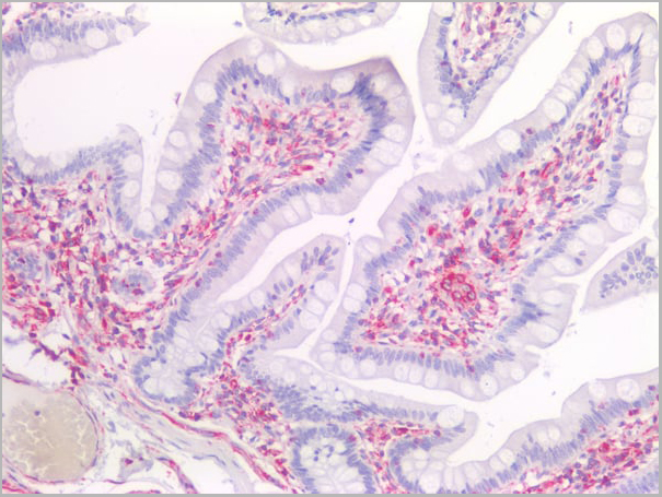
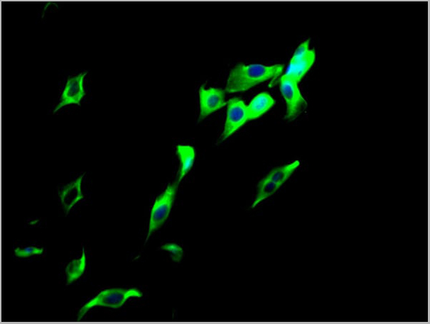
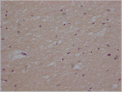
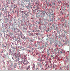
 IS (Immunostaining)
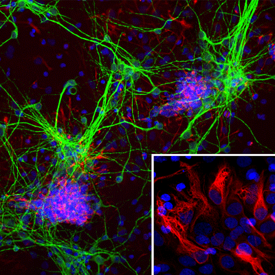
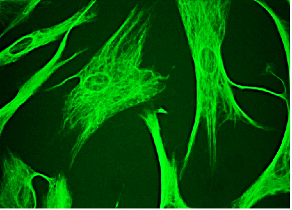
(Immunostaining of cultured E20 rat cortical neurons and glia stained with anti-nestin antibody (AAA14229, red, 1:500) and anti-MAP2 antibody (, green, 1:500). The blue is Hoechst staining for nuclear DNA. The nestin antibody labels developing astrocytes and neuronal stem cells in a clearly filamentous fashion, while the MAP2 antibody stains dendrites and perikarya of mature neurons.)
IS (Immunostaining)
(Immunostaining of cultured E20 rat cortical neurons and glia stained with anti-nestin antibody (AAA14229, red, 1:500) and anti-MAP2 antibody (, green, 1:500). The blue is Hoechst staining for nuclear DNA. The nestin antibody labels developing astrocytes and neuronal stem cells in a clearly filamentous fashion, while the MAP2 antibody stains dendrites and perikarya of mature neurons.)
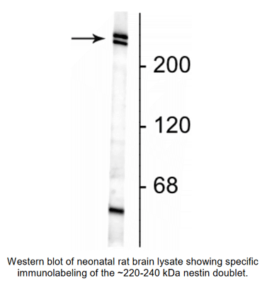
Nestin, Monoclonal Antibody (Cat# AAA14229)
Full Name
Anti-Nestin
Gene Names
NES; Nbla00170
Applications
Western Blot, Immunofluorescence
Purity
Protein G purified culture supernatant
Pricing
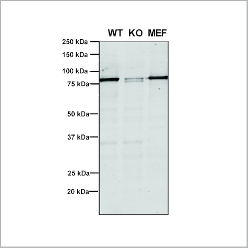
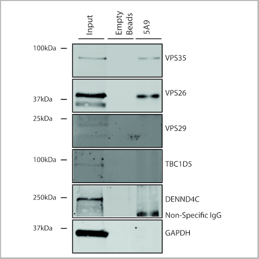
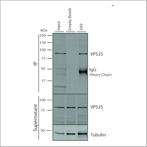
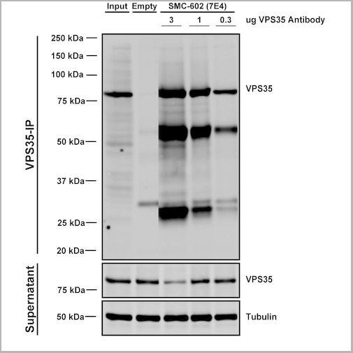
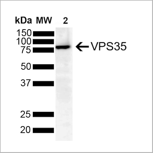
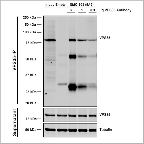 IP (Immunoprecipitation)
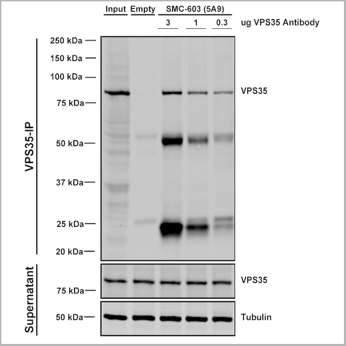
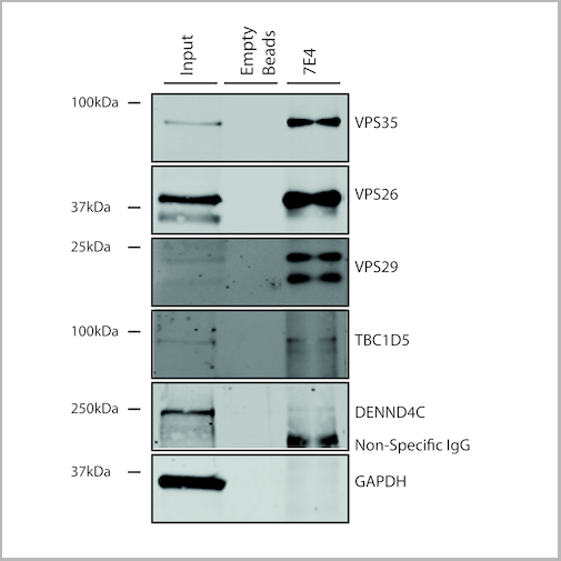
(Immunoprecipitation analysis using Mouse Anti-VPS35 Monoclonal Antibody, Clone 5A9. Tissue: embryonic fibroblast. Species: Mouse. Primary Antibody: Mouse Anti-VPS35 Monoclonal Antibody. Three amounts of (3, 1 and 0.3 ug) were non-covalently coupled to 10uL of A/G sepharose beads for 1 hour at 4 degree C and next incubated with 250ug of MEF lysate for 2 hours at 4 degree C.)
IP (Immunoprecipitation)
(Immunoprecipitation analysis using Mouse Anti-VPS35 Monoclonal Antibody, Clone 5A9. Tissue: embryonic fibroblast. Species: Mouse. Primary Antibody: Mouse Anti-VPS35 Monoclonal Antibody. Three amounts of (3, 1 and 0.3 ug) were non-covalently coupled to 10uL of A/G sepharose beads for 1 hour at 4 degree C and next incubated with 250ug of MEF lysate for 2 hours at 4 degree C.)
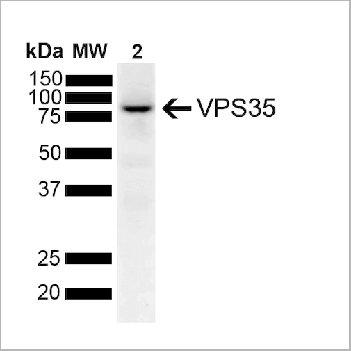
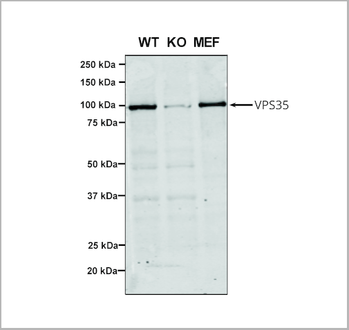
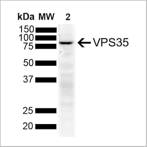
VPS35, Monoclonal Antibody (Cat# AAA27680)
Full Name
VPS35 Antibody, Clone 5A9: PerCP
Gene Names
VPS35; MEM3; PARK17
Reactivity
Human, Mouse, Rat
Applications
Western Blot, Immunocytochemistry, Immunofluorescence, Immunoprecipitation
Purity
Protein G Purified
Pricing
 Application Data
(Staining of human peripheral blood monocytes with Mouse anti Human CD14:RPE)
Application Data
(Staining of human peripheral blood monocytes with Mouse anti Human CD14:RPE)
CD14, Monoclonal Antibody (Cat# AAA11998)
Full Name
MOUSE ANTI HUMAN CD14
Applications
Immunohistochemistry, Flow Cytometry, Immunofluorescence, Immunoprecipitation
Pricing
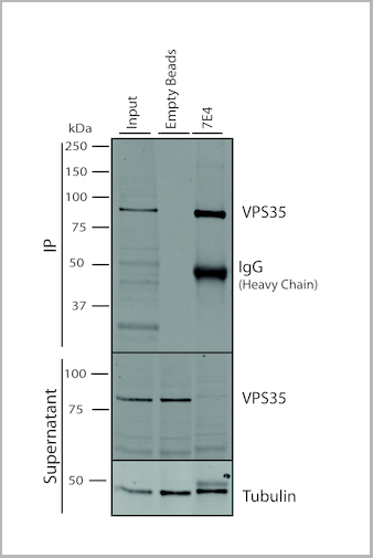
 IP (Immunoprecipitation)
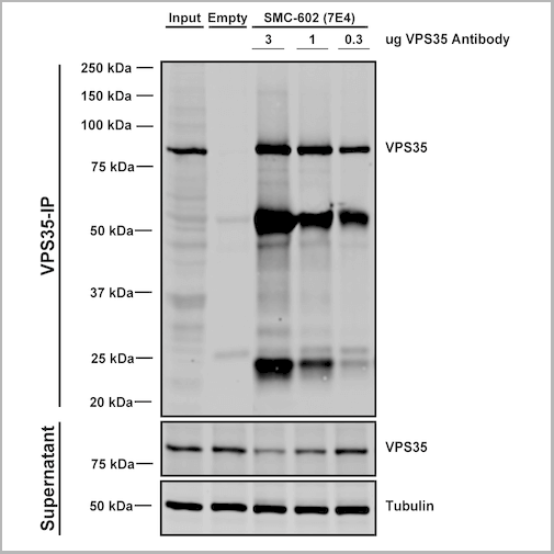
(Immunoprecipitation analysis using Mouse Anti-VPS35 Monoclonal Antibody, Clone 7E4. Tissue: embryonic fibroblast. Species: Mouse. Primary Antibody: Mouse Anti-VPS35 Monoclonal Antibody. Three amounts (3, 1 and 0.3 ug) were non-covalently coupled to 10uL of A/G sepharose beads for 1 hour at 4 degree C and next incubated with 250ug of MEF lysate for 2 hours at 4 degree C.)
IP (Immunoprecipitation)
(Immunoprecipitation analysis using Mouse Anti-VPS35 Monoclonal Antibody, Clone 7E4. Tissue: embryonic fibroblast. Species: Mouse. Primary Antibody: Mouse Anti-VPS35 Monoclonal Antibody. Three amounts (3, 1 and 0.3 ug) were non-covalently coupled to 10uL of A/G sepharose beads for 1 hour at 4 degree C and next incubated with 250ug of MEF lysate for 2 hours at 4 degree C.)
VPS35, Monoclonal Antibody (Cat# AAA27671)
Full Name
VPS35 Antibody, Clone 7E4: PerCP
Gene Names
VPS35; MEM3; PARK17
Reactivity
Human, Mouse, Rat
Applications
Western Blot, Immunocytochemistry, Immunofluorescence, Immunoprecipitation
Purity
Protein G Purified
Pricing
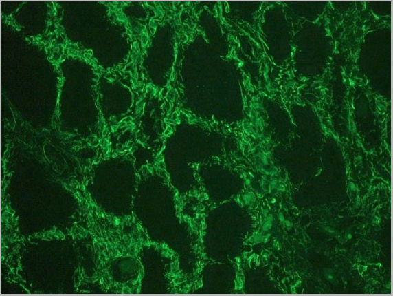
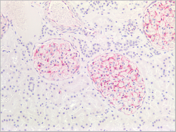
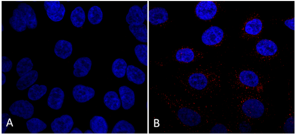
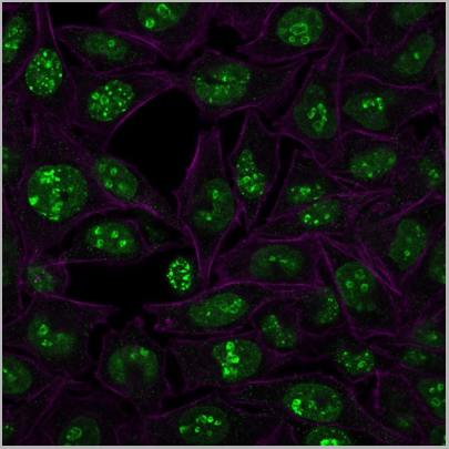
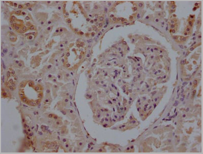
 IF (Immunofluorescence)
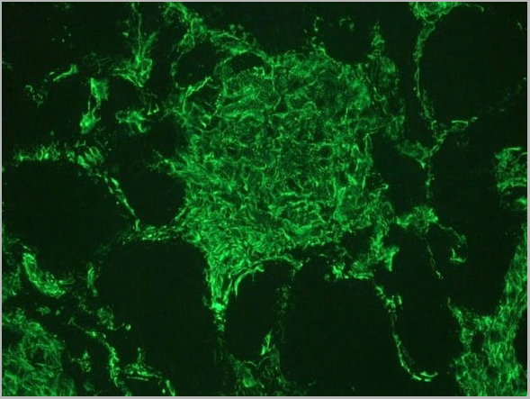
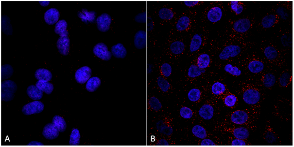
(Figure 2. Indirect immunofluorescence staining of human kidney tissue section with (diluted 1:1000), showing the specific pattern of vimentin in the mesenchymal cell types, such as fibroblasts in the connective tissue, podocytes, and endothelial cells in blood vessels. As expected, no reactivity is seen in the epithelial cell compartment.)
IF (Immunofluorescence)
(Figure 2. Indirect immunofluorescence staining of human kidney tissue section with (diluted 1:1000), showing the specific pattern of vimentin in the mesenchymal cell types, such as fibroblasts in the connective tissue, podocytes, and endothelial cells in blood vessels. As expected, no reactivity is seen in the epithelial cell compartment.)
vimentin, Monoclonal Recombinant Antibody (Cat# AAA14575)
Full Name
Recombinant rabbit anti vimentin
Reactivity
Human, mouse
Applications
Immunocytochemistry, Immunohistochemistry, Immunoblot
Purity
The purity of the antibody is >98% as determined by SDS-Polyacrylamide gel electrophoresis.
Pricing
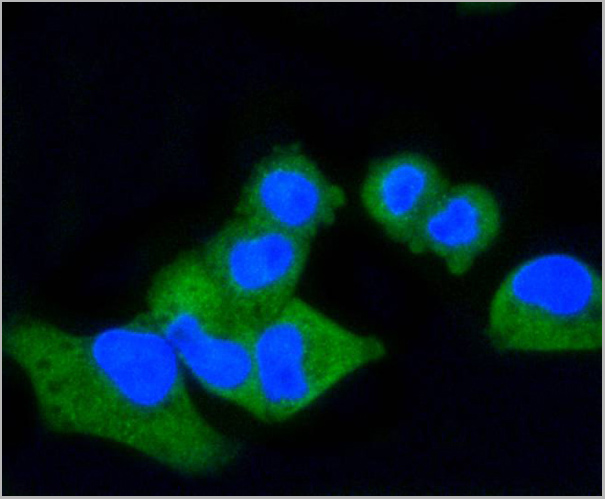
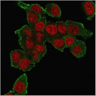 IF (Immunofluorescence)
(Immunofluorescence staining of PFA-fixed HePG2 cells using Tumor Necrosis Factor (TNF alpha) Mouse Monoclonal Antibody (TNF706) followed by goat anti-mouse IgG-CF488 (green). Nuclei stained with RedDot.)
IF (Immunofluorescence)
(Immunofluorescence staining of PFA-fixed HePG2 cells using Tumor Necrosis Factor (TNF alpha) Mouse Monoclonal Antibody (TNF706) followed by goat anti-mouse IgG-CF488 (green). Nuclei stained with RedDot.)
TNF-alpha (Tumor Necrosis Factor alpha), Monoclonal Antibody (Cat# AAA13806)
Full Name
TNF-alpha (Tumor Necrosis Factor alpha) Mouse Monoclonal Antibody
Gene Names
TNF; DIF; TNFA; TNFSF2; TNLG1F; TNF-alpha
Reactivity
Human, Mouse, Rat, Rabbit, Cat, Dog, Zebrafish
Applications
Flow Cytometry, Immunofluorescence, Immunohistochemistry
Pricing
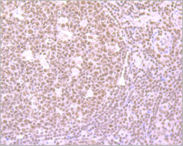
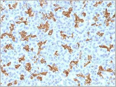
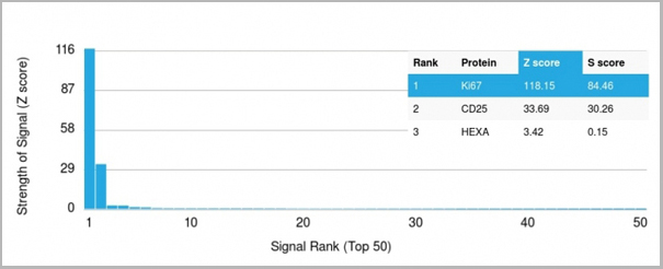 Application Data
(Analysis of Protein Array containing more than 19,000 full-length human proteins using Ki67 Mouse Monoclonal Antibody (MKI67/2466). Z- and S- Score: The Z-score represents the strength of a signal that a monoclonal antibody (MAb) (in combination with a fluorescently-tagged anti-IgG secondary antibody) produces when binding to a particular protein on the HuProtTM array. Z-scores are described in units of standard deviations (SD's) above the mean value of all signals generated on that array. If targets on HuProtTM are arranged in descending order of the Z-score, the S-score is the difference (also in units of SD's) between the Z-score. S-score therefore represents the relative target specificity of a MAb to its intended target. A MAb is considered to specific to its intended target, if the MAb has an S-score of at least 2.5. For example, if a MAb binds to protein X with a Z-score of 43 and to protein Y with a Z-score of 14, then the S-score for the binding of that MAb to protein X is equal to 29.)
Application Data
(Analysis of Protein Array containing more than 19,000 full-length human proteins using Ki67 Mouse Monoclonal Antibody (MKI67/2466). Z- and S- Score: The Z-score represents the strength of a signal that a monoclonal antibody (MAb) (in combination with a fluorescently-tagged anti-IgG secondary antibody) produces when binding to a particular protein on the HuProtTM array. Z-scores are described in units of standard deviations (SD's) above the mean value of all signals generated on that array. If targets on HuProtTM are arranged in descending order of the Z-score, the S-score is the difference (also in units of SD's) between the Z-score. S-score therefore represents the relative target specificity of a MAb to its intended target. A MAb is considered to specific to its intended target, if the MAb has an S-score of at least 2.5. For example, if a MAb binds to protein X with a Z-score of 43 and to protein Y with a Z-score of 14, then the S-score for the binding of that MAb to protein X is equal to 29.)
Ki-67, Monoclonal Antibody (Cat# AAA23909)
Full Name
Ki-67 (Proliferating Cell Marker)
Gene Names
MKI67; KIA; MIB-; MIB-1; PPP1R105
Reactivity
Human. Others not known.
Applications
Flow Cytometry, Immunofluorescence, Immunohistochemistry
Purity
Purified Ab with BSA and Azide at 200ug/ml OR Purified Ab WITHOUT BSA and Azide at 1.0mg/ml
Pricing


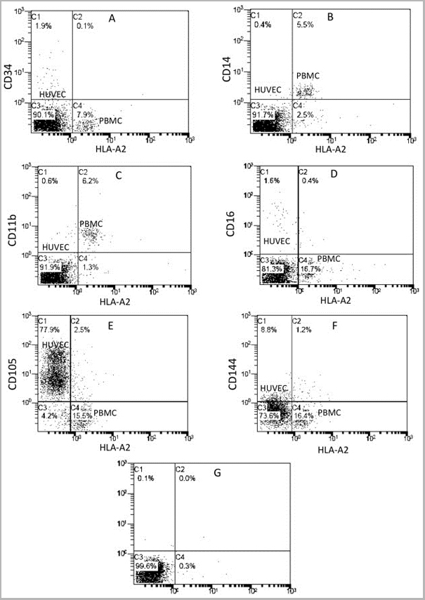
 Application Data
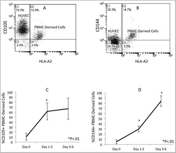
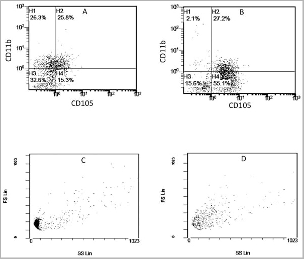
(Published customer image: Mouse anti Human CD105 antibody, clone SN6 used for flow cytometry.Image caption:Phenotype change from HLA-A2+/CD11b+/CD105- to HLA-A2+/CD11b-/CD105+ on endothelium-adherent blood monocyte-derived cells with increase in size and granularity during co-culture. HLA-A2+ PBMCs (1x10⁶ cells/well) were incubated for 2 h (Day 0) with HLA-A2- HUVECs, after which the non-adherent cells were removed by washing. The cell layers were analysed by three-colour flow cytometry staining for HLA-A2, CD11b and CD105 on (A) Day 1 and (B) Day 2. These plots were gated for HLA-A2+ cells. Forward scatter/side scatter dot plots gated for HLA-A2+ cells on Day 0 (C) and Day 2 (D) was shown. These are representative of 2 individual experiments.From: Tso C, Rye K-A, Barter P (2012) Phenotypic and Functional Changes in Blood Monocytes Following Adherence to Endothelium. PLoS ONE 7(5): e37091.)
Application Data
(Published customer image: Mouse anti Human CD105 antibody, clone SN6 used for flow cytometry.Image caption:Phenotype change from HLA-A2+/CD11b+/CD105- to HLA-A2+/CD11b-/CD105+ on endothelium-adherent blood monocyte-derived cells with increase in size and granularity during co-culture. HLA-A2+ PBMCs (1x10⁶ cells/well) were incubated for 2 h (Day 0) with HLA-A2- HUVECs, after which the non-adherent cells were removed by washing. The cell layers were analysed by three-colour flow cytometry staining for HLA-A2, CD11b and CD105 on (A) Day 1 and (B) Day 2. These plots were gated for HLA-A2+ cells. Forward scatter/side scatter dot plots gated for HLA-A2+ cells on Day 0 (C) and Day 2 (D) was shown. These are representative of 2 individual experiments.From: Tso C, Rye K-A, Barter P (2012) Phenotypic and Functional Changes in Blood Monocytes Following Adherence to Endothelium. PLoS ONE 7(5): e37091.)
CD105, Monoclonal Antibody (Cat# AAA12085)
Full Name
MOUSE ANTI HUMAN CD105
Gene Names
ENG; END; HHT1; ORW1
Applications
Immunohistochemistry, Flow Cytometry, Immunoprecipitation, Western Blot
Pricing


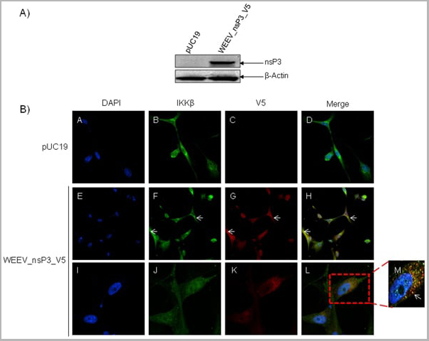 Application Data
(Published customer image: Mouse anti V5 tag antibody, clone SV5-Pk1 used for the detection of V5 tagged WEEV_nsP3 protein by western blotting and immunofluorescenceImage caption: WEEV nsP3 interaction with host IKKbeta. A) U87MGs were transfected in a 6-well plate with 5 ug of pUC19 and WEEV_nsP3_HA for 24 hours. Cell lysates were resolved using SDS-PAGE and subsequently immunoblotted with V5 antibody and beta-actin served as a loading control. B) U87MGs were transfected with WEEV_nsP3_V5; cells were fixed after 24 hours and stained with antibodies against the endogenous IKKbeta and the V5 tag. Cells were incubated with appropriate secondary Alexa Fluor antibodies and the nuclei stained with DAPI. Co-localization of IKKbeta with WEEV_nsP3_V5 (yellow) was observed as shown by the arrows. B) Panels E -H serve as an example of transfected cells in a given field of view that show co-localization of IKKbeta and WEEV_nsP3_V5 24 hours post transfection. Panels I-L represent magnified images of other cells showing co-localization of IKKbeta and WEEV_nsP3_V5. Panel M is a magnified image of panel L. The co-localization was confirmed by Z-stack analysis. Co-localization was calculated to be approximately in 61% of cells (163 cells were counted of which 44% demonstrated expression of nsP3. Of those cells that expressed nsP3, 61% showed co-localization of both proteins). Images were taken using Nikon Eclipse TE2000-U at 60x magnification and are representative of 2 independent experiments.From: Amaya M, Voss K, Sampey G, Senina S, de la Fuente C, et al. (2014) The Role of IKKbeta in Venezuelan Equine Encephalitis Virus Infection. PLoS ONE 9(2): e86745.)
Application Data
(Published customer image: Mouse anti V5 tag antibody, clone SV5-Pk1 used for the detection of V5 tagged WEEV_nsP3 protein by western blotting and immunofluorescenceImage caption: WEEV nsP3 interaction with host IKKbeta. A) U87MGs were transfected in a 6-well plate with 5 ug of pUC19 and WEEV_nsP3_HA for 24 hours. Cell lysates were resolved using SDS-PAGE and subsequently immunoblotted with V5 antibody and beta-actin served as a loading control. B) U87MGs were transfected with WEEV_nsP3_V5; cells were fixed after 24 hours and stained with antibodies against the endogenous IKKbeta and the V5 tag. Cells were incubated with appropriate secondary Alexa Fluor antibodies and the nuclei stained with DAPI. Co-localization of IKKbeta with WEEV_nsP3_V5 (yellow) was observed as shown by the arrows. B) Panels E -H serve as an example of transfected cells in a given field of view that show co-localization of IKKbeta and WEEV_nsP3_V5 24 hours post transfection. Panels I-L represent magnified images of other cells showing co-localization of IKKbeta and WEEV_nsP3_V5. Panel M is a magnified image of panel L. The co-localization was confirmed by Z-stack analysis. Co-localization was calculated to be approximately in 61% of cells (163 cells were counted of which 44% demonstrated expression of nsP3. Of those cells that expressed nsP3, 61% showed co-localization of both proteins). Images were taken using Nikon Eclipse TE2000-U at 60x magnification and are representative of 2 independent experiments.From: Amaya M, Voss K, Sampey G, Senina S, de la Fuente C, et al. (2014) The Role of IKKbeta in Venezuelan Equine Encephalitis Virus Infection. PLoS ONE 9(2): e86745.)
V5-TAG, Monoclonal Antibody (Cat# AAA11930)
Full Name
MOUSE ANTI V5-TAG
Applications
Immunohistochemistry, Flow Cytometry, Immunofluorescence, Immunoprecipitation, Western Blot, Radioimmunoassay
Pricing


 Application Data
(Published customer image: Mouse anti V5 tag antibody, clone SV5-Pk1 used for the detection of V5 tagged WEEV_nsP3 protein by western blotting and immunofluorescenceImage caption: WEEV nsP3 interaction with host IKKbeta. A) U87MGs were transfected in a 6-well plate with 5 ug of pUC19 and WEEV_nsP3_HA for 24 hours. Cell lysates were resolved using SDS-PAGE and subsequently immunoblotted with V5 antibody and beta-actin served as a loading control. B) U87MGs were transfected with WEEV_nsP3_V5; cells were fixed after 24 hours and stained with antibodies against the endogenous IKKbeta and the V5 tag. Cells were incubated with appropriate secondary Alexa Fluor antibodies and the nuclei stained with DAPI. Co-localization of IKKbeta with WEEV_nsP3_V5 (yellow) was observed as shown by the arrows. B) Panels E -H serve as an example of transfected cells in a given field of view that show co-localization of IKKbeta and WEEV_nsP3_V5 24 hours post transfection. Panels I-L represent magnified images of other cells showing co-localization of IKKbeta and WEEV_nsP3_V5. Panel M is a magnified image of panel L. The co-localization was confirmed by Z-stack analysis. Co-localization was calculated to be approximately in 61% of cells (163 cells were counted of which 44% demonstrated expression of nsP3. Of those cells that expressed nsP3, 61% showed co-localization of both proteins). Images were taken using Nikon Eclipse TE2000-U at 60x magnification and are representative of 2 independent experiments.From: Amaya M, Voss K, Sampey G, Senina S, de la Fuente C, et al. (2014) The Role of IKKbeta in Venezuelan Equine Encephalitis Virus Infection. PLoS ONE 9(2): e86745.)
Application Data
(Published customer image: Mouse anti V5 tag antibody, clone SV5-Pk1 used for the detection of V5 tagged WEEV_nsP3 protein by western blotting and immunofluorescenceImage caption: WEEV nsP3 interaction with host IKKbeta. A) U87MGs were transfected in a 6-well plate with 5 ug of pUC19 and WEEV_nsP3_HA for 24 hours. Cell lysates were resolved using SDS-PAGE and subsequently immunoblotted with V5 antibody and beta-actin served as a loading control. B) U87MGs were transfected with WEEV_nsP3_V5; cells were fixed after 24 hours and stained with antibodies against the endogenous IKKbeta and the V5 tag. Cells were incubated with appropriate secondary Alexa Fluor antibodies and the nuclei stained with DAPI. Co-localization of IKKbeta with WEEV_nsP3_V5 (yellow) was observed as shown by the arrows. B) Panels E -H serve as an example of transfected cells in a given field of view that show co-localization of IKKbeta and WEEV_nsP3_V5 24 hours post transfection. Panels I-L represent magnified images of other cells showing co-localization of IKKbeta and WEEV_nsP3_V5. Panel M is a magnified image of panel L. The co-localization was confirmed by Z-stack analysis. Co-localization was calculated to be approximately in 61% of cells (163 cells were counted of which 44% demonstrated expression of nsP3. Of those cells that expressed nsP3, 61% showed co-localization of both proteins). Images were taken using Nikon Eclipse TE2000-U at 60x magnification and are representative of 2 independent experiments.From: Amaya M, Voss K, Sampey G, Senina S, de la Fuente C, et al. (2014) The Role of IKKbeta in Venezuelan Equine Encephalitis Virus Infection. PLoS ONE 9(2): e86745.)
V5-TAG, Monoclonal Antibody (Cat# AAA11864)
Full Name
MOUSE ANTI V5-TAG:FITC
Applications
Immunofluorescence
Pricing
T-Cell Receptor gamma delta, Monoclonal Antibody (Cat# AAA14565)
Full Name
Mouse anti T-Cell Receptor gamma delta
Reactivity
Human
Applications
Flow Cytometry, Immunohistochemistry, Immunoprecipitation
Pricing
COVID 19 Nucleocapsid (NP) Humanized Coronavirus, Monoclonal Antibody (Cat# AAA13535)
Full Name
Anti-SARS-CoV-2 Nucleocapsid / COVID-19, Humanized antibody
Reactivity
Human
Applications
Lateral Flow
Purity
Purity: >95% by HPLC & SDS-PAGE
Purification: Protein A purified
Purification: Protein A purified
Pricing
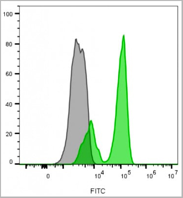
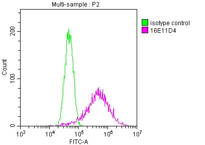
 FCM (Flow Cytometry)
(Figure-1: Cell surface flowcytometry analysis of CLEC1on human PBMC (Monocytes) using 0.5 ug/10^6 Cells of Anti-CLEC1 antibody. Green represent isotype control and red represent Anti human CLEC1 antibody. Goat anti mouse PE conjugated was used as the secondary antibody.)
FCM (Flow Cytometry)
(Figure-1: Cell surface flowcytometry analysis of CLEC1on human PBMC (Monocytes) using 0.5 ug/10^6 Cells of Anti-CLEC1 antibody. Green represent isotype control and red represent Anti human CLEC1 antibody. Goat anti mouse PE conjugated was used as the secondary antibody.)
CLEC1, Monoclonal Antibody (Cat# AAA14882)
Full Name
Monoclonal Antibody to CLEC1 (Clone: ABM2H82)
Gene Names
CLEC1A; CLEC1; CLEC-1
Reactivity
Human
Applications
Flow Cytometry
Purity
Protein G Chromatography Purified
Pricing
 Application Data
HLA Class 1 Antigen A33,B8, Monoclonal Antibody (Cat# AAA14684)
Full Name
Mouse anti-Human HLA Class 1 Antigen A33,B8
Reactivity
Human
Applications
Cell Typing
Purity
Ascites
Pricing
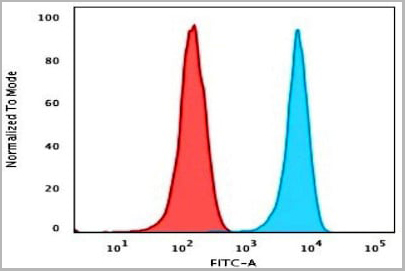
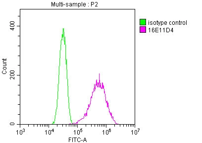
 FCM (Flow Cytometry)
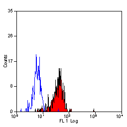
(Intracellular flow cytometric analysis of RIG-I in K562 Cell line using 0.5 ug/10^6 cells of Anti-RIGI antibody (ABM40B5). Green represent isotype control and red represent Anti-RIG I antibody (AAA14865). Goat anti-mouse PE conjugate was used as secondary.)
FCM (Flow Cytometry)
(Intracellular flow cytometric analysis of RIG-I in K562 Cell line using 0.5 ug/10^6 cells of Anti-RIGI antibody (ABM40B5). Green represent isotype control and red represent Anti-RIG I antibody (AAA14865). Goat anti-mouse PE conjugate was used as secondary.)
RIG-I, Monoclonal Antibody (Cat# AAA14865)
Full Name
RIG-I Monoclonal Antibody
Gene Names
DDX58; RIGI; RIG-I; RLR-1; SGMRT2
Reactivity
Human
Applications
Immunohistochemistry, Flow Cytometry, Western Blot
Purity
Protein G Chromatography
Pricing
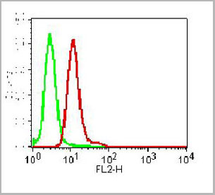
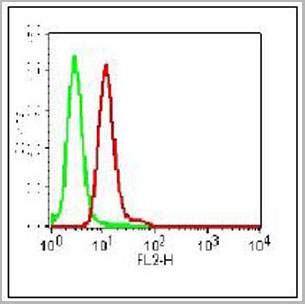
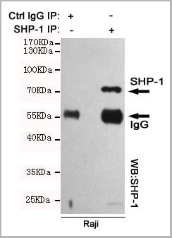 IP (Immunoprecipitation)
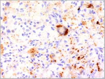
(Immunoprecipitation analysis of Raji cell lysates using SHP-1 mouse mAb.)
IP (Immunoprecipitation)
(Immunoprecipitation analysis of Raji cell lysates using SHP-1 mouse mAb.)
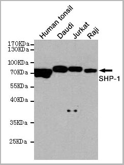
SHP-1, Monoclonal Antibody (Cat# AAA14102)
Full Name
Anti-SHP-1 Mouse mAb
Gene Names
PTPN6; HCP; HCPH; SHP1; SHP-1; HPTP1C; PTP-1C; SHP-1L; SH-PTP1
Reactivity
Human
Applications
Western Blot, Immunoprecipitation
Purity
Affinity purified
Pricing
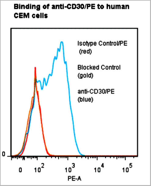 Application Data
CD30, Monoclonal Antibody (Cat# AAA14859)
Full Name
anti-human CD30-R-PE
Reactivity
Human
Applications
Flow Cytometry
Pricing
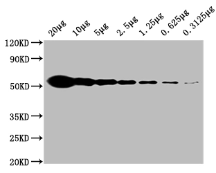
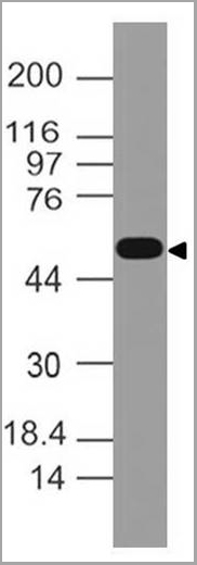
 WB (Western Blot)
(Western blot analysis of C3 expression in HepG2 cell lysate (AAA11693). Electrophoresis was performed on a 5-20% SDS-PAGE gel at 70V (stacking gel) / 90V (resolving gel) for 2-3 hours. The sample well of each lane was loaded with 50ug of sample under reducing conditions. After Electrophoresis, proteins were transferred to a Nitrocellulose membrane at 150mA for 50-90 minutes. Blocked the membrane with 5% Non-fat Milk/TBS for 1.5 hour at RT. The membrane was incubated with rabbit anti-C3 monoclonal antibody (AAA11693) overnight at 4°C, then washed with TBS-0.1%Tween 3 times with 5 minutes each and probed with goat anti-rabbit IgG-HRP secondary antibody at a dilution of 1:10000 for 1.5 hour at RT. The signal is developed using an Enhanced Chemiluminescent detection (ECL) kit (please inquire) with Tanon 5200 system. A specific band was detected for C3.)
WB (Western Blot)
(Western blot analysis of C3 expression in HepG2 cell lysate (AAA11693). Electrophoresis was performed on a 5-20% SDS-PAGE gel at 70V (stacking gel) / 90V (resolving gel) for 2-3 hours. The sample well of each lane was loaded with 50ug of sample under reducing conditions. After Electrophoresis, proteins were transferred to a Nitrocellulose membrane at 150mA for 50-90 minutes. Blocked the membrane with 5% Non-fat Milk/TBS for 1.5 hour at RT. The membrane was incubated with rabbit anti-C3 monoclonal antibody (AAA11693) overnight at 4°C, then washed with TBS-0.1%Tween 3 times with 5 minutes each and probed with goat anti-rabbit IgG-HRP secondary antibody at a dilution of 1:10000 for 1.5 hour at RT. The signal is developed using an Enhanced Chemiluminescent detection (ECL) kit (please inquire) with Tanon 5200 system. A specific band was detected for C3.)
C3/Complement C3, Monoclonal Antibody (Cat# AAA11693)
Full Name
Anti-C3/Complement C3 Rabbit Monoclonal Antibody
Gene Names
C3; ASP; C3a; C3b; AHUS5; ARMD9; CPAMD1; HEL-S-62p
Reactivity
Human
Applications
Flow Cytometry, Immunofluorescence, Immunocytochemistry, Western Blot
Purity
Affinity-chromatography
Pricing
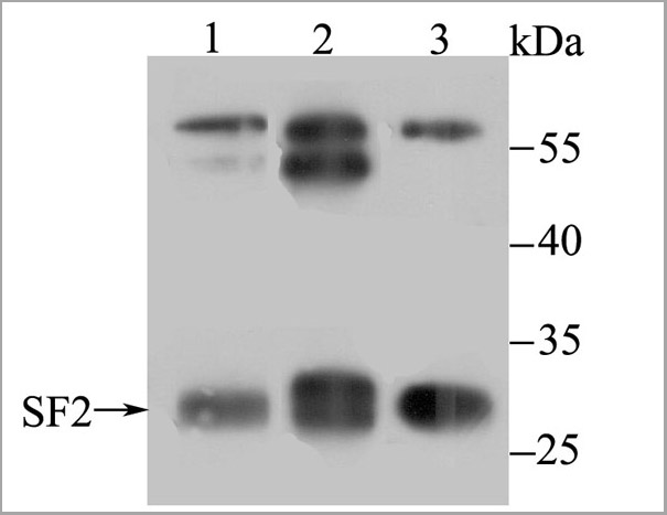
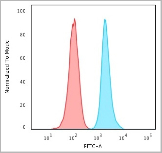
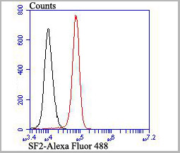 FCM (Flow Cytometry)
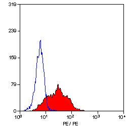
(Flow cytometric analysis of K562 cells with SF2 antibody at 1/100 dilution (red) compared with an unlabelled control (cells without incubation with primary antibody; black). Alexa Fluor 488-conjugated goat anti rabbit IgG was used as the secondary antibody.)
FCM (Flow Cytometry)
(Flow cytometric analysis of K562 cells with SF2 antibody at 1/100 dilution (red) compared with an unlabelled control (cells without incubation with primary antibody; black). Alexa Fluor 488-conjugated goat anti rabbit IgG was used as the secondary antibody.)
SF2, Monoclonal Antibody (Cat# AAA30465)
Full Name
SF2 Antibody
Gene Names
SRSF1; ASF; SF2; SFRS1; SF2p33; SRp30a
Reactivity
Human, Mouse, Rat
Applications
Western Blot, Immunocytochemistry, Immunofluorescence, Immunohistochemistry, Flow Cytometry
Purity
ProA affinity purified
Pricing
 IF (Immunofluorescence)
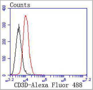
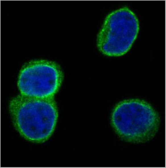
(Immunofluorescence Analysis of methanol-fixed human tonsil cryosection stained with CF488A CD3e clone RIV9 (green), mounted in EverBrite’ Mounting Medium with DAPI (nuclei, blue).)
IF (Immunofluorescence)
(Immunofluorescence Analysis of methanol-fixed human tonsil cryosection stained with CF488A CD3e clone RIV9 (green), mounted in EverBrite’ Mounting Medium with DAPI (nuclei, blue).)
CD3, Monoclonal Antibody (Cat# AAA13839)
Full Name
CD3 (T-Cell Marker) Mouse Monoclonal Antibody
Gene Names
CD3E; T3E; TCRE; IMD18
Reactivity
Human, Mouse, Rat
Applications
Functional Assay, Flow Cytometry, Immunofluorescence
Pricing

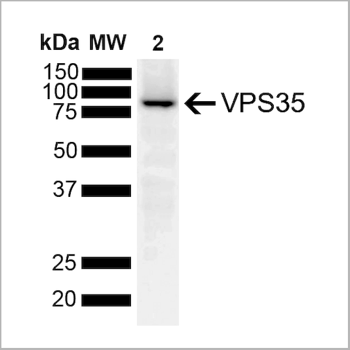

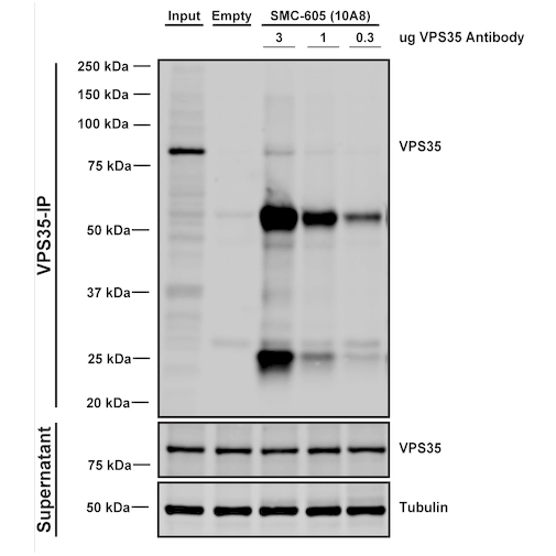
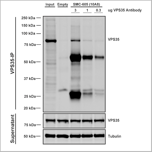 IP (Immunoprecipitation)
(Immunoprecipitation analysis using Mouse Anti-VPS35 Monoclonal Antibody, Clone 10A8. Tissue: embryonic fibroblast. Species: Mouse. Primary Antibody: Mouse Anti-VPS35 Monoclonal Antibody. Three amounts of (3, 1 and 0.3 ug) were non-covalently coupled to 10uL of A/G sepharose beads for 1 hour at 4 degree C and next incubated with 250ug of MEF lysate for 2 hours at 4 degree C.)
IP (Immunoprecipitation)
(Immunoprecipitation analysis using Mouse Anti-VPS35 Monoclonal Antibody, Clone 10A8. Tissue: embryonic fibroblast. Species: Mouse. Primary Antibody: Mouse Anti-VPS35 Monoclonal Antibody. Three amounts of (3, 1 and 0.3 ug) were non-covalently coupled to 10uL of A/G sepharose beads for 1 hour at 4 degree C and next incubated with 250ug of MEF lysate for 2 hours at 4 degree C.)
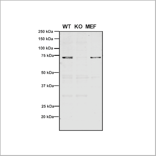
VPS35, Monoclonal Antibody (Cat# AAA27697)
Full Name
VPS35 Antibody, Clone 10A8: HRP
Gene Names
VPS35; MEM3; PARK17
Reactivity
Human, Mouse, Rat
Applications
Western Blot, Immunocytochemistry, Immunofluorescence, Immunoprecipitation
Purity
Protein G Purified
Pricing


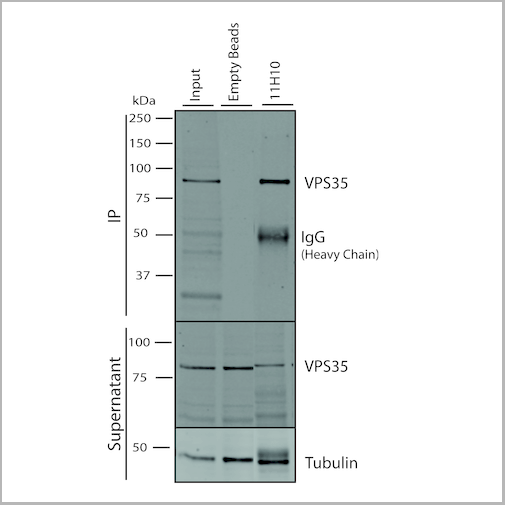
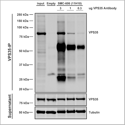 IP (Immunoprecipitation)
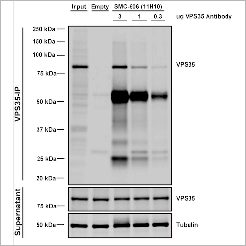
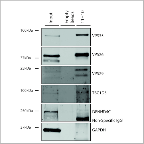
(Immunoprecipitation analysis using Mouse Anti-VPS35 Monoclonal Antibody, Clone 11H10. Tissue: embryonic fibroblast. Species: Mouse. Primary Antibody: Mouse Anti-VPS35 Monoclonal Antibody. Three amounts of (3, 1 and 0.3 ug) were non-covalently coupled to 10uL of A/G sepharose beads for 1 hour at 4 degree C and next incubated with 250ug of MEF lysate for 2 hours at 4 degree C.)
IP (Immunoprecipitation)
(Immunoprecipitation analysis using Mouse Anti-VPS35 Monoclonal Antibody, Clone 11H10. Tissue: embryonic fibroblast. Species: Mouse. Primary Antibody: Mouse Anti-VPS35 Monoclonal Antibody. Three amounts of (3, 1 and 0.3 ug) were non-covalently coupled to 10uL of A/G sepharose beads for 1 hour at 4 degree C and next incubated with 250ug of MEF lysate for 2 hours at 4 degree C.)
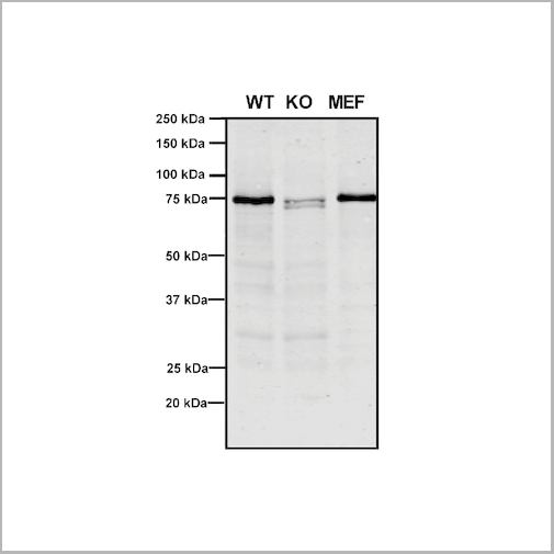
VPS35, Monoclonal Antibody (Cat# AAA27702)
Full Name
VPS35 Antibody, Clone 11H10: ATTO 488
Gene Names
VPS35; MEM3; PARK17
Reactivity
Human, Mouse, Rat
Applications
Western Blot, Immunoprecipitation
Purity
Protein G Purified
Pricing


 IP (Immunoprecipitation)
(Immunoprecipitation analysis using Mouse Anti-VPS35 Monoclonal Antibody, Clone 11H10. Tissue: embryonic fibroblast. Species: Mouse. Primary Antibody: Mouse Anti-VPS35 Monoclonal Antibody. Three amounts of (3, 1 and 0.3 ug) were non-covalently coupled to 10uL of A/G sepharose beads for 1 hour at 4 degree C and next incubated with 250ug of MEF lysate for 2 hours at 4 degree C.)
IP (Immunoprecipitation)
(Immunoprecipitation analysis using Mouse Anti-VPS35 Monoclonal Antibody, Clone 11H10. Tissue: embryonic fibroblast. Species: Mouse. Primary Antibody: Mouse Anti-VPS35 Monoclonal Antibody. Three amounts of (3, 1 and 0.3 ug) were non-covalently coupled to 10uL of A/G sepharose beads for 1 hour at 4 degree C and next incubated with 250ug of MEF lysate for 2 hours at 4 degree C.)
VPS35, Monoclonal Antibody (Cat# AAA27700)
Full Name
VPS35 Antibody, Clone 11H10
Gene Names
VPS35; MEM3; PARK17
Reactivity
Human, Mouse, Rat
Applications
Western Blot, Immunoprecipitation
Purity
Protein G Purified
Pricing

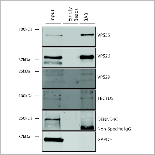

 IP (Immunoprecipitation)
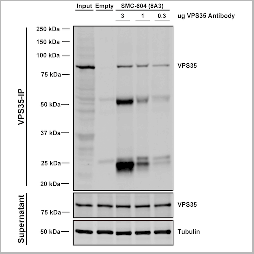
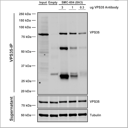
(Immunoprecipitation analysis using Mouse Anti-VPS35 Monoclonal Antibody, Clone 8A3. Tissue: embryonic fibroblast. Species: Mouse. Primary Antibody: Mouse Anti-VPS35 Monoclonal Antibody. Three amounts of (3, 1 and 0.3 ug) were non-covalently coupled to 10uL of A/G sepharose beads for 1 hour at 4 degree C and next incubated with 250ug of MEF lysate for 2 hours at 4 degree C.)
IP (Immunoprecipitation)
(Immunoprecipitation analysis using Mouse Anti-VPS35 Monoclonal Antibody, Clone 8A3. Tissue: embryonic fibroblast. Species: Mouse. Primary Antibody: Mouse Anti-VPS35 Monoclonal Antibody. Three amounts of (3, 1 and 0.3 ug) were non-covalently coupled to 10uL of A/G sepharose beads for 1 hour at 4 degree C and next incubated with 250ug of MEF lysate for 2 hours at 4 degree C.)
VPS35, Monoclonal Antibody (Cat# AAA27687)
Full Name
VPS35 Antibody, Clone 8A3: FITC
Gene Names
VPS35; MEM3; PARK17
Reactivity
Human, Mouse, Rat
Applications
Western Blot, Immunocytochemistry, Immunofluorescence, Immunoprecipitation
Purity
Protein G Purified
Pricing


 IP (Immunoprecipitation)
(Immunoprecipitation analysis using Mouse Anti-VPS35 Monoclonal Antibody, Clone 8A3. Tissue: embryonic fibroblast. Species: Mouse. Primary Antibody: Mouse Anti-VPS35 Monoclonal Antibody. Three amounts of (3, 1 and 0.3 ug) were non-covalently coupled to 10uL of A/G sepharose beads for 1 hour at 4 degree C and next incubated with 250ug of MEF lysate for 2 hours at 4 degree C.)
IP (Immunoprecipitation)
(Immunoprecipitation analysis using Mouse Anti-VPS35 Monoclonal Antibody, Clone 8A3. Tissue: embryonic fibroblast. Species: Mouse. Primary Antibody: Mouse Anti-VPS35 Monoclonal Antibody. Three amounts of (3, 1 and 0.3 ug) were non-covalently coupled to 10uL of A/G sepharose beads for 1 hour at 4 degree C and next incubated with 250ug of MEF lysate for 2 hours at 4 degree C.)
VPS35, Monoclonal Antibody (Cat# AAA27683)
Full Name
VPS35 Antibody, Clone 8A3: ATTO 390
Gene Names
VPS35; MEM3; PARK17
Reactivity
Human, Mouse, Rat
Applications
Western Blot, Immunocytochemistry, Immunofluorescence, Immunoprecipitation
Purity
Protein G Purified
Pricing
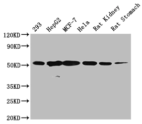
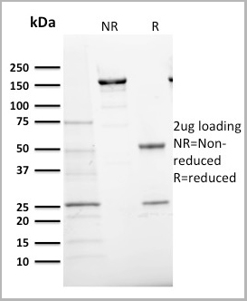
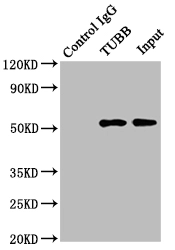 IP (Immunoprecipitation)
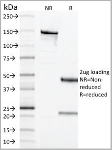
(Immunoprecipitating TUBB in Hela whole cell lysateLane 1: Mouse control IgG instead in Hela whole cell lysate.Lane 2: (2ug) + Hela whole cell lysate (500ug)Lane 3: Hela whole cell lysate (5ug)For western blotting, the blot was detected at 1:2000, and a HRP-conjugated Protein G antibody was used as the secondary antibody at 1:5000)
IP (Immunoprecipitation)
(Immunoprecipitating TUBB in Hela whole cell lysateLane 1: Mouse control IgG instead in Hela whole cell lysate.Lane 2: (2ug) + Hela whole cell lysate (500ug)Lane 3: Hela whole cell lysate (5ug)For western blotting, the blot was detected at 1:2000, and a HRP-conjugated Protein G antibody was used as the secondary antibody at 1:5000)
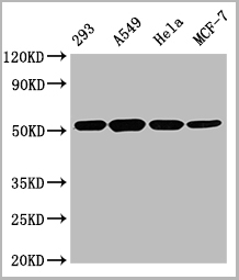
TUBB, Monoclonal Antibody (Cat# AAA28058)
Full Name
TUBB Monoclonal Antibody
Reactivity
Human, Rat, Rabbit, Mouse
Applications
Western Blot, Immunohistochemistry, Immunofluorescence, Flow Cytometry, Immunoprecipitation
Purity
>95%, Protein A purified
Pricing


 Application Data
(Published customer image: Mouse anti Human CD105 antibody, clone SN6 used for flow cytometry.Image caption:Phenotype change from HLA-A2+/CD11b+/CD105- to HLA-A2+/CD11b-/CD105+ on endothelium-adherent blood monocyte-derived cells with increase in size and granularity during co-culture. HLA-A2+ PBMCs (1x10⁶ cells/well) were incubated for 2 h (Day 0) with HLA-A2- HUVECs, after which the non-adherent cells were removed by washing. The cell layers were analysed by three-colour flow cytometry staining for HLA-A2, CD11b and CD105 on (A) Day 1 and (B) Day 2. These plots were gated for HLA-A2+ cells. Forward scatter/side scatter dot plots gated for HLA-A2+ cells on Day 0 (C) and Day 2 (D) was shown. These are representative of 2 individual experiments.From: Tso C, Rye K-A, Barter P (2012) Phenotypic and Functional Changes in Blood Monocytes Following Adherence to Endothelium. PLoS ONE 7(5): e37091.)
Application Data
(Published customer image: Mouse anti Human CD105 antibody, clone SN6 used for flow cytometry.Image caption:Phenotype change from HLA-A2+/CD11b+/CD105- to HLA-A2+/CD11b-/CD105+ on endothelium-adherent blood monocyte-derived cells with increase in size and granularity during co-culture. HLA-A2+ PBMCs (1x10⁶ cells/well) were incubated for 2 h (Day 0) with HLA-A2- HUVECs, after which the non-adherent cells were removed by washing. The cell layers were analysed by three-colour flow cytometry staining for HLA-A2, CD11b and CD105 on (A) Day 1 and (B) Day 2. These plots were gated for HLA-A2+ cells. Forward scatter/side scatter dot plots gated for HLA-A2+ cells on Day 0 (C) and Day 2 (D) was shown. These are representative of 2 individual experiments.From: Tso C, Rye K-A, Barter P (2012) Phenotypic and Functional Changes in Blood Monocytes Following Adherence to Endothelium. PLoS ONE 7(5): e37091.)
CD105, Monoclonal Antibody (Cat# AAA12083)
Full Name
MOUSE ANTI HUMAN CD105:FITC
Gene Names
ENG; END; HHT1; ORW1
Applications
Flow Cytometry
Pricing


 Application Data
(Published customer image: Mouse anti Human CD105 antibody, clone SN6 used for flow cytometry.Image caption:Phenotype change from HLA-A2+/CD11b+/CD105- to HLA-A2+/CD11b-/CD105+ on endothelium-adherent blood monocyte-derived cells with increase in size and granularity during co-culture. HLA-A2+ PBMCs (1x10⁶ cells/well) were incubated for 2 h (Day 0) with HLA-A2- HUVECs, after which the non-adherent cells were removed by washing. The cell layers were analysed by three-colour flow cytometry staining for HLA-A2, CD11b and CD105 on (A) Day 1 and (B) Day 2. These plots were gated for HLA-A2+ cells. Forward scatter/side scatter dot plots gated for HLA-A2+ cells on Day 0 (C) and Day 2 (D) was shown. These are representative of 2 individual experiments.From: Tso C, Rye K-A, Barter P (2012) Phenotypic and Functional Changes in Blood Monocytes Following Adherence to Endothelium. PLoS ONE 7(5): e37091.)
Application Data
(Published customer image: Mouse anti Human CD105 antibody, clone SN6 used for flow cytometry.Image caption:Phenotype change from HLA-A2+/CD11b+/CD105- to HLA-A2+/CD11b-/CD105+ on endothelium-adherent blood monocyte-derived cells with increase in size and granularity during co-culture. HLA-A2+ PBMCs (1x10⁶ cells/well) were incubated for 2 h (Day 0) with HLA-A2- HUVECs, after which the non-adherent cells were removed by washing. The cell layers were analysed by three-colour flow cytometry staining for HLA-A2, CD11b and CD105 on (A) Day 1 and (B) Day 2. These plots were gated for HLA-A2+ cells. Forward scatter/side scatter dot plots gated for HLA-A2+ cells on Day 0 (C) and Day 2 (D) was shown. These are representative of 2 individual experiments.From: Tso C, Rye K-A, Barter P (2012) Phenotypic and Functional Changes in Blood Monocytes Following Adherence to Endothelium. PLoS ONE 7(5): e37091.)
CD105, Monoclonal Antibody (Cat# AAA11865)
Full Name
MOUSE ANTI HUMAN CD105:FITC
Gene Names
ENG; END; HHT1; ORW1
Applications
Flow Cytometry
Pricing


 Application Data
(Published customer image: Mouse anti Human CD105 antibody, clone SN6 used for flow cytometry.Image caption:Phenotype change from HLA-A2+/CD11b+/CD105- to HLA-A2+/CD11b-/CD105+ on endothelium-adherent blood monocyte-derived cells with increase in size and granularity during co-culture. HLA-A2+ PBMCs (1x10⁶ cells/well) were incubated for 2 h (Day 0) with HLA-A2- HUVECs, after which the non-adherent cells were removed by washing. The cell layers were analysed by three-colour flow cytometry staining for HLA-A2, CD11b and CD105 on (A) Day 1 and (B) Day 2. These plots were gated for HLA-A2+ cells. Forward scatter/side scatter dot plots gated for HLA-A2+ cells on Day 0 (C) and Day 2 (D) was shown. These are representative of 2 individual experiments.From: Tso C, Rye K-A, Barter P (2012) Phenotypic and Functional Changes in Blood Monocytes Following Adherence to Endothelium. PLoS ONE 7(5): e37091.)
Application Data
(Published customer image: Mouse anti Human CD105 antibody, clone SN6 used for flow cytometry.Image caption:Phenotype change from HLA-A2+/CD11b+/CD105- to HLA-A2+/CD11b-/CD105+ on endothelium-adherent blood monocyte-derived cells with increase in size and granularity during co-culture. HLA-A2+ PBMCs (1x10⁶ cells/well) were incubated for 2 h (Day 0) with HLA-A2- HUVECs, after which the non-adherent cells were removed by washing. The cell layers were analysed by three-colour flow cytometry staining for HLA-A2, CD11b and CD105 on (A) Day 1 and (B) Day 2. These plots were gated for HLA-A2+ cells. Forward scatter/side scatter dot plots gated for HLA-A2+ cells on Day 0 (C) and Day 2 (D) was shown. These are representative of 2 individual experiments.From: Tso C, Rye K-A, Barter P (2012) Phenotypic and Functional Changes in Blood Monocytes Following Adherence to Endothelium. PLoS ONE 7(5): e37091.)
CD105, Monoclonal Antibody (Cat# AAA12029)
Full Name
MOUSE ANTI HUMAN CD105:RPE
Gene Names
ENG; END; HHT1; ORW1
Applications
Flow Cytometry
Pricing
 Application Data
(Staining of mouse spleen with Hamster anti Mouse CD81: Alexa Fluor 488)
Application Data
(Staining of mouse spleen with Hamster anti Mouse CD81: Alexa Fluor 488)
CD81, Monoclonal Antibody (Cat# AAA11942)
Full Name
HAMSTER ANTI MOUSE CD81
Gene Names
Cd81; Tapa1; Tapa-1; Tspan28
Reactivity
Rat
Applications
Immunohistochemistry, Flow Cytometry, Immunoprecipitation, Western Blot
Pricing


 Application Data
(Published customer image: Mouse anti V5 tag antibody, clone SV5-Pk1 used for the detection of V5 tagged WEEV_nsP3 protein by western blotting and immunofluorescenceImage caption: WEEV nsP3 interaction with host IKKbeta. A) U87MGs were transfected in a 6-well plate with 5 ug of pUC19 and WEEV_nsP3_HA for 24 hours. Cell lysates were resolved using SDS-PAGE and subsequently immunoblotted with V5 antibody and beta-actin served as a loading control. B) U87MGs were transfected with WEEV_nsP3_V5; cells were fixed after 24 hours and stained with antibodies against the endogenous IKKbeta and the V5 tag. Cells were incubated with appropriate secondary Alexa Fluor antibodies and the nuclei stained with DAPI. Co-localization of IKKbeta with WEEV_nsP3_V5 (yellow) was observed as shown by the arrows. B) Panels E -H serve as an example of transfected cells in a given field of view that show co-localization of IKKbeta and WEEV_nsP3_V5 24 hours post transfection. Panels I-L represent magnified images of other cells showing co-localization of IKKbeta and WEEV_nsP3_V5. Panel M is a magnified image of panel L. The co-localization was confirmed by Z-stack analysis. Co-localization was calculated to be approximately in 61% of cells (163 cells were counted of which 44% demonstrated expression of nsP3. Of those cells that expressed nsP3, 61% showed co-localization of both proteins). Images were taken using Nikon Eclipse TE2000-U at 60x magnification and are representative of 2 independent experiments.From: Amaya M, Voss K, Sampey G, Senina S, de la Fuente C, et al. (2014) The Role of IKKbeta in Venezuelan Equine Encephalitis Virus Infection. PLoS ONE 9(2): e86745.)
Application Data
(Published customer image: Mouse anti V5 tag antibody, clone SV5-Pk1 used for the detection of V5 tagged WEEV_nsP3 protein by western blotting and immunofluorescenceImage caption: WEEV nsP3 interaction with host IKKbeta. A) U87MGs were transfected in a 6-well plate with 5 ug of pUC19 and WEEV_nsP3_HA for 24 hours. Cell lysates were resolved using SDS-PAGE and subsequently immunoblotted with V5 antibody and beta-actin served as a loading control. B) U87MGs were transfected with WEEV_nsP3_V5; cells were fixed after 24 hours and stained with antibodies against the endogenous IKKbeta and the V5 tag. Cells were incubated with appropriate secondary Alexa Fluor antibodies and the nuclei stained with DAPI. Co-localization of IKKbeta with WEEV_nsP3_V5 (yellow) was observed as shown by the arrows. B) Panels E -H serve as an example of transfected cells in a given field of view that show co-localization of IKKbeta and WEEV_nsP3_V5 24 hours post transfection. Panels I-L represent magnified images of other cells showing co-localization of IKKbeta and WEEV_nsP3_V5. Panel M is a magnified image of panel L. The co-localization was confirmed by Z-stack analysis. Co-localization was calculated to be approximately in 61% of cells (163 cells were counted of which 44% demonstrated expression of nsP3. Of those cells that expressed nsP3, 61% showed co-localization of both proteins). Images were taken using Nikon Eclipse TE2000-U at 60x magnification and are representative of 2 independent experiments.From: Amaya M, Voss K, Sampey G, Senina S, de la Fuente C, et al. (2014) The Role of IKKbeta in Venezuelan Equine Encephalitis Virus Infection. PLoS ONE 9(2): e86745.)
V5-TAG, Monoclonal Antibody (Cat# AAA12211)
Full Name
MOUSE ANTI V5-TAG
Applications
Immunohistochemistry, Flow Cytometry, Immunofluorescence, Immunoprecipitation, Western Blot, Radioimmunoassay
Pricing
 Standard Curve (Sample)
Mouse Anti-Human IgG (Fc) (gamma chain specific), Monoclonal Secondary Antibody (Cat# AAA14898)
Full Name
Mouse Anti-Human IgG (Fc)-APC
Reactivity
Human
Applications
Flow Cytometry, Western Blot
Pricing
Creatine Kinase MM (CK-MM), Antibody (Cat# AAA13411)
Full Name
Goat anti CK-MM
Gene Names
CKM; CKMM; M-CK
Applications
Lateral Flow
Purity
Immunoaffinity column chromatography
Pricing
CA 125, Monoclonal Antibody (Cat# AAA14303)
Full Name
CA 125 antibody
Applications
Immunoblot, Immunoassay
Pricing
COVID 19 Spike S1 Protein Humanized Coronavirus, Monoclonal Antibody (Cat# AAA13537)
Full Name
Anti-SARS-CoV-2 S1 / COVID-19, Humanized antibody (Detection Ab)
Reactivity
Viral
Applications
Lateral Flow
Purity
Purity: >95% by HPLC & SDS-PAGE
Purification: Protein A purified
Purification: Protein A purified
Pricing
AFP, Monoclonal Antibody (Cat# AAA14291)
Full Name
AFP antibody
Gene Names
AFP; AFPD; FETA; HPAFP
Reactivity
AFP 100%
Applications
ELISA
Pricing
COVID 19 Spike RBD IgG Humanized Coronavirus, Monoclonal Antibody (Cat# AAA13540)
Full Name
Anti-SARS-CoV-2 Spike RBD IgG, Humanized antibody (Positive Control)
Reactivity
Human
Applications
Lateral Flow
Purity
Purity: >95% by HPLC & SDS-PAGE
Purification: Protein A purified
Purification: Protein A purified
Pricing
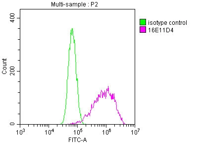
 FCM (Flow Cytometry)
(Intracellular flow cytometric analysis of RIG-I in K562 Cell line using 0.5 ug/10^6 cells of Anti-RIGI antibody (ABM4H29). Green represent isotype control and red represent Anti-RIG I antibody (AAA14869). Goat anti-mouse PE conjugate was used as secondary.)
FCM (Flow Cytometry)
(Intracellular flow cytometric analysis of RIG-I in K562 Cell line using 0.5 ug/10^6 cells of Anti-RIGI antibody (ABM4H29). Green represent isotype control and red represent Anti-RIG I antibody (AAA14869). Goat anti-mouse PE conjugate was used as secondary.)
RIG I /DDx58, Monoclonal Antibody (Cat# AAA14869)
Full Name
Monoclonal Antibody to RIG-I (Clone: ABM4H29)
Gene Names
DDX58; RIGI; RIG-I; RLR-1; SGMRT2
Reactivity
Human
Applications
Immunohistochemistry, Flow Cytometry, Western Blot
Purity
Protein G chromatography
Pricing
HLA Class 1 Antigen A30,A31, Monoclonal Antibody (Cat# AAA14656)
Full Name
Mouse Anti-Human HLA Class 1 Antigen A30,A31 (R0.87)
Gene Names
HLA-A; HLAA
Reactivity
Human
Applications
Cell Typing
Purity
Ascites
Pricing
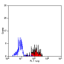
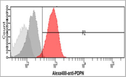 FCM (Flow Cytometry)
(Flow cytometry analysis of Podoplanin in HeLa cells. The cell was stained with (AAA11728) at 2-5ug for 1x10^6cells (red). A Goat anti mouse IgG (Alexa fluor 488) was used as the secondary antibody. Mouse monoclonal IgG was used as the isotype control (dark gray), cells without incubation with primary and secondary antibody was used as the negative control (light gray).)
FCM (Flow Cytometry)
(Flow cytometry analysis of Podoplanin in HeLa cells. The cell was stained with (AAA11728) at 2-5ug for 1x10^6cells (red). A Goat anti mouse IgG (Alexa fluor 488) was used as the secondary antibody. Mouse monoclonal IgG was used as the isotype control (dark gray), cells without incubation with primary and secondary antibody was used as the negative control (light gray).)
PDPN, Monoclonal Antibody (Cat# AAA11728)
Full Name
PDPN antibody
Gene Names
PDPN; T1A; GP36; GP40; Gp38; OTS8; T1A2; TI1A; T1A-2; AGGRUS; HT1A-1; PA2.26
Reactivity
Human
Applications
Flow Cytometry, Immunocytochemistry, Immunofluorescence
Purity
By protein-G affinity chromatography
Pricing
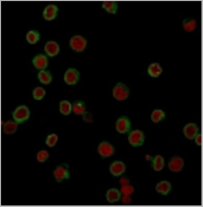
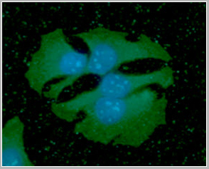
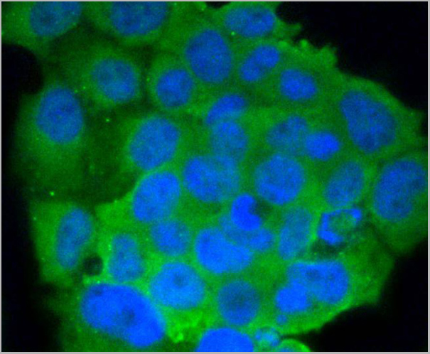
 IF (Immunofluorescence)
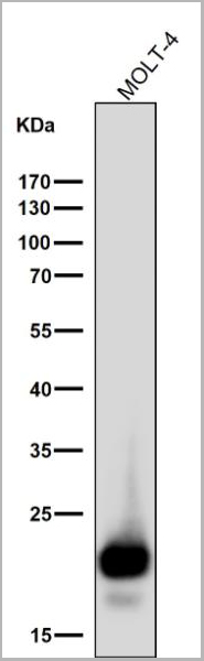
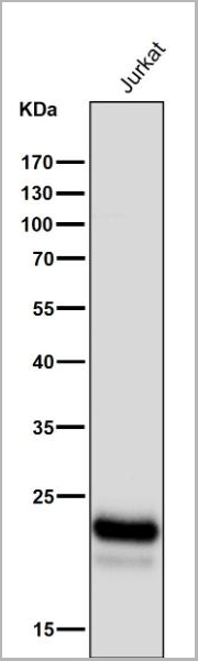
(Immunofluorescent analysis of Jurkat cells, using CD3D Antibody.)
IF (Immunofluorescence)
(Immunofluorescent analysis of Jurkat cells, using CD3D Antibody.)
CD3D, Monoclonal Antibody (Cat# AAA30236)
Full Name
CD3D Antibody
Gene Names
CD3D; T3D; IMD19; CD3-DELTA
Reactivity
Human
Applications
Western Blot, Immunocytochemistry, Immunohistochemistry, Immunoprecipitation, Flow Cytometry
Purity
Affinity - chromatography.
Pricing
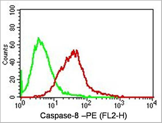
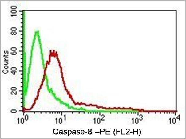
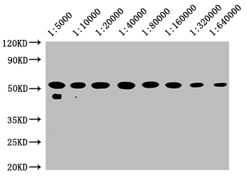
 WB (Western Blot)
(Figure-6: Western blot analysis of Caspase-8. Anti-Caspase-8 antibody (Clone: ABM14C1) was used at 4 ug/ml on EL-4 lysate.)
WB (Western Blot)
(Figure-6: Western blot analysis of Caspase-8. Anti-Caspase-8 antibody (Clone: ABM14C1) was used at 4 ug/ml on EL-4 lysate.)
Caspase-8, Monoclonal Antibody (Cat# AAA14877)
Full Name
Monoclonal Antibody to Caspase-8 (Clone: ABM14C1)
Gene Names
CASP8; CAP4; MACH; MCH5; FLICE; ALPS2B; Casp-8
Reactivity
Human
Applications
Immunohistochemistry, Western Blot, Flow Cytometry
Purity
Purified
Protein G Chromatography
Protein G Chromatography
Pricing
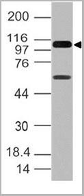
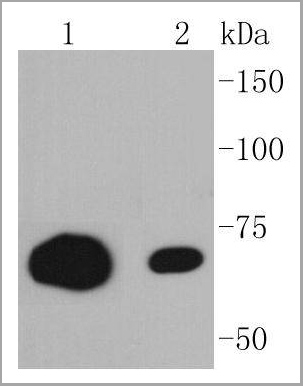
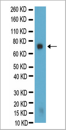 WB (Western Blot)
(Western Blot analysis for galactocerebroside in PC12 lysate. PC12 lysate was resolved by electrophoresis, transferred to PVDF membrane and probed with AAA14685 at 1:500 dilution. Proteins were visualized using a goat anti-mouse secondary antibody conjugated to HRP and a chemiluminescence detection system. Arrow indicates protein Galactocerebroside (~75kD).)
WB (Western Blot)
(Western Blot analysis for galactocerebroside in PC12 lysate. PC12 lysate was resolved by electrophoresis, transferred to PVDF membrane and probed with AAA14685 at 1:500 dilution. Proteins were visualized using a goat anti-mouse secondary antibody conjugated to HRP and a chemiluminescence detection system. Arrow indicates protein Galactocerebroside (~75kD).)
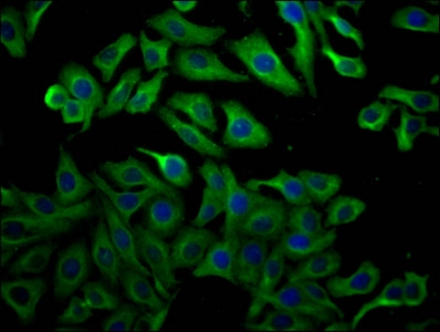
Galactocerebroside, Monoclonal Antibody (Cat# AAA14685)
Full Name
Mouse anti-Bovine Galactocerebroside
Applications
Immunohistochemistry, Immunocytochemistry
Purity
Purified by Protein A affinity chromatography.
Pricing

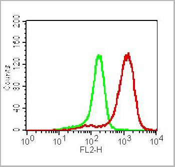
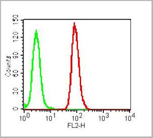
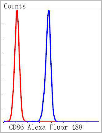 FCM (Flow Cytometry)
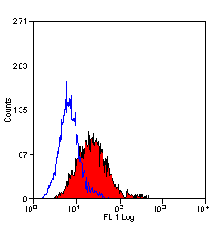
(Flow cytometric analysis of K562 cells with CD86 antibody at 1/50 dilution (blue) compared with an unlabelled control (cells without incubation with primary antibody; red). Alexa Fluor 488-conjugated goat anti rabbit IgG was used as the secondary antibody.)
FCM (Flow Cytometry)
(Flow cytometric analysis of K562 cells with CD86 antibody at 1/50 dilution (blue) compared with an unlabelled control (cells without incubation with primary antibody; red). Alexa Fluor 488-conjugated goat anti rabbit IgG was used as the secondary antibody.)
CD86, Monoclonal Antibody (Cat# AAA30048)
Full Name
CD86 Antibody
Gene Names
CD86; B70; B7-2; B7.2; LAB72; CD28LG2
Reactivity
Human, Rat
Applications
Western Blot, Immunocytochemistry, Immunofluorescence, Immunohistochemistry, Immunoprecipitation, Flow Cytometry
Purity
ProA affinity purified
Pricing


 IP (Immunoprecipitation)
(Immunoprecipitation analysis using Mouse Anti-VPS35 Monoclonal Antibody, Clone 11H10. Tissue: embryonic fibroblast. Species: Mouse. Primary Antibody: Mouse Anti-VPS35 Monoclonal Antibody. Three amounts of (3, 1 and 0.3 ug) were non-covalently coupled to 10uL of A/G sepharose beads for 1 hour at 4 degree C and next incubated with 250ug of MEF lysate for 2 hours at 4 degree C.)
IP (Immunoprecipitation)
(Immunoprecipitation analysis using Mouse Anti-VPS35 Monoclonal Antibody, Clone 11H10. Tissue: embryonic fibroblast. Species: Mouse. Primary Antibody: Mouse Anti-VPS35 Monoclonal Antibody. Three amounts of (3, 1 and 0.3 ug) were non-covalently coupled to 10uL of A/G sepharose beads for 1 hour at 4 degree C and next incubated with 250ug of MEF lysate for 2 hours at 4 degree C.)
VPS35, Monoclonal Antibody (Cat# AAA27706)
Full Name
VPS35 Antibody, Clone 11H10: HRP
Gene Names
VPS35; MEM3; PARK17
Reactivity
Human, Mouse, Rat
Applications
Western Blot, Immunoprecipitation
Purity
Protein G Purified
Pricing


 IP (Immunoprecipitation)
(Immunoprecipitation analysis using Mouse Anti-VPS35 Monoclonal Antibody, Clone 11H10. Tissue: embryonic fibroblast. Species: Mouse. Primary Antibody: Mouse Anti-VPS35 Monoclonal Antibody. Three amounts of (3, 1 and 0.3 ug) were non-covalently coupled to 10uL of A/G sepharose beads for 1 hour at 4 degree C and next incubated with 250ug of MEF lysate for 2 hours at 4 degree C.)
IP (Immunoprecipitation)
(Immunoprecipitation analysis using Mouse Anti-VPS35 Monoclonal Antibody, Clone 11H10. Tissue: embryonic fibroblast. Species: Mouse. Primary Antibody: Mouse Anti-VPS35 Monoclonal Antibody. Three amounts of (3, 1 and 0.3 ug) were non-covalently coupled to 10uL of A/G sepharose beads for 1 hour at 4 degree C and next incubated with 250ug of MEF lysate for 2 hours at 4 degree C.)
VPS35, Monoclonal Antibody (Cat# AAA27708)
Full Name
VPS35 Antibody, Clone 11H10: RPE
Gene Names
VPS35; MEM3; PARK17
Reactivity
Human, Mouse, Rat
Applications
Western Blot, Immunoprecipitation
Purity
Protein G Purified
Pricing


 IP (Immunoprecipitation)
(Immunoprecipitation analysis using Mouse Anti-VPS35 Monoclonal Antibody, Clone 10A8. Tissue: embryonic fibroblast. Species: Mouse. Primary Antibody: Mouse Anti-VPS35 Monoclonal Antibody. Three amounts of (3, 1 and 0.3 ug) were non-covalently coupled to 10uL of A/G sepharose beads for 1 hour at 4 degree C and next incubated with 250ug of MEF lysate for 2 hours at 4 degree C.)
IP (Immunoprecipitation)
(Immunoprecipitation analysis using Mouse Anti-VPS35 Monoclonal Antibody, Clone 10A8. Tissue: embryonic fibroblast. Species: Mouse. Primary Antibody: Mouse Anti-VPS35 Monoclonal Antibody. Three amounts of (3, 1 and 0.3 ug) were non-covalently coupled to 10uL of A/G sepharose beads for 1 hour at 4 degree C and next incubated with 250ug of MEF lysate for 2 hours at 4 degree C.)
VPS35, Monoclonal Antibody (Cat# AAA27693)
Full Name
VPS35 Antibody, Clone 10A8: ATTO 488
Gene Names
VPS35; MEM3; PARK17
Reactivity
Human, Mouse, Rat
Applications
Western Blot, Immunocytochemistry, Immunofluorescence, Immunoprecipitation
Purity
Protein G Purified
Pricing
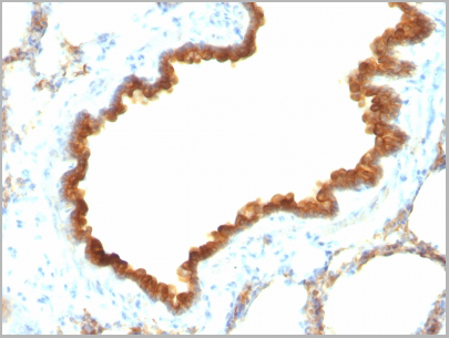
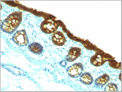
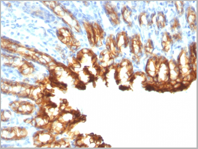
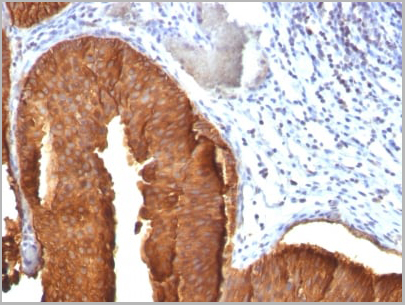
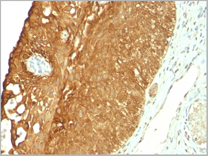
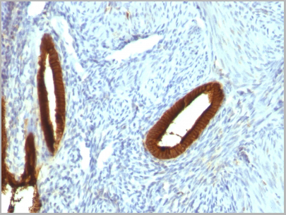
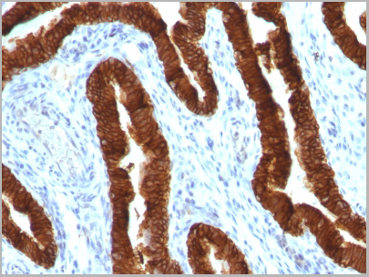
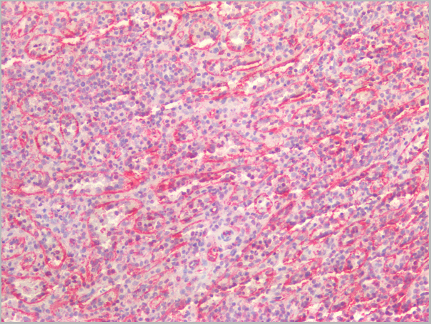
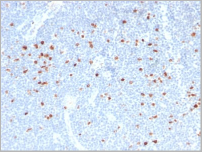
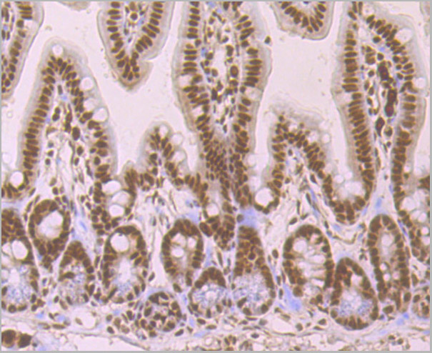
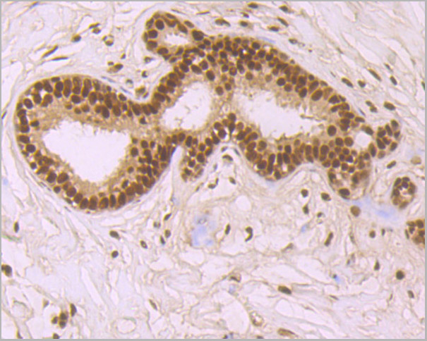
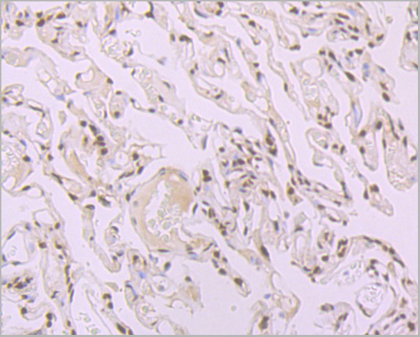
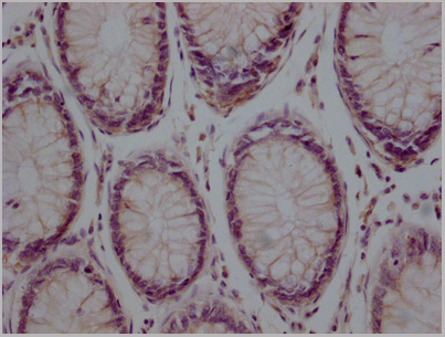
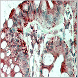
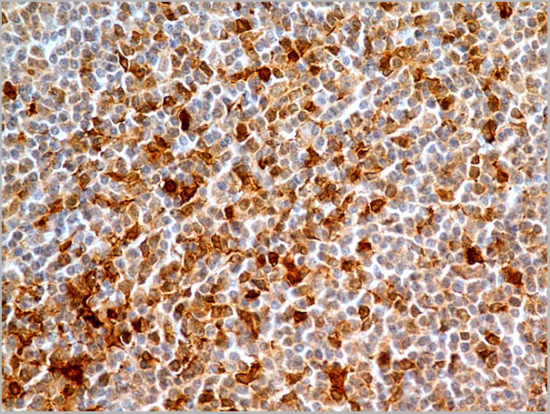
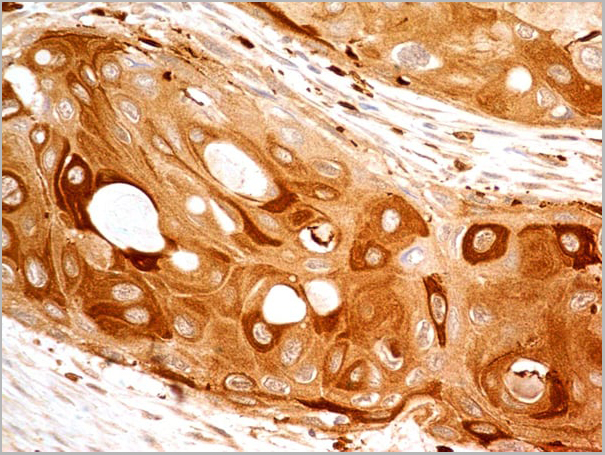
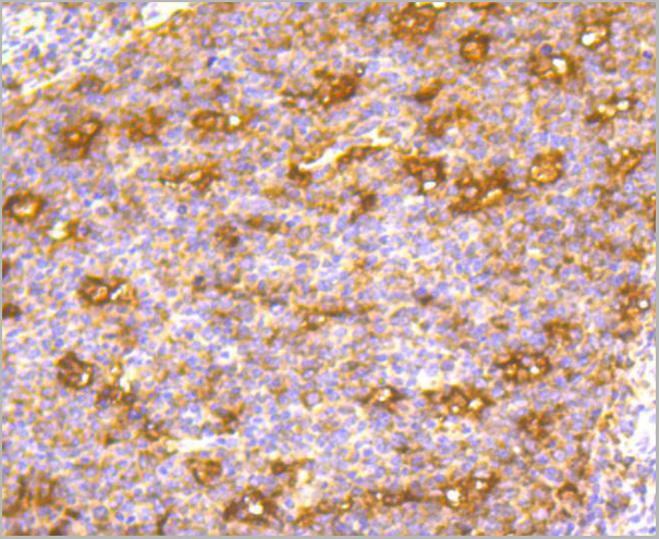
 IHC (Immunohistochemistry)
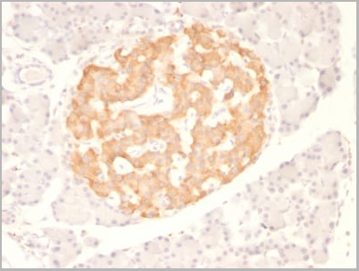
(Formalin-fixed, paraffin-embedded human Pancreas stained with Cytokeratin 19 Monoclonal Antibody (KRT19/800))
IHC (Immunohistochemistry)
(Formalin-fixed, paraffin-embedded human Pancreas stained with Cytokeratin 19 Monoclonal Antibody (KRT19/800))
Cytokeratin 19 (KRT19), Monoclonal Antibody (Cat# AAA13824)
Full Name
Cytokeratin 19 (KRT19) (Pancreatic Stem Cell Marker) Mouse Monoclonal Antibody
Gene Names
KRT19; K19; CK19; K1CS
Reactivity
Human, Mouse, Rat
Applications
Flow Cytometry, Immunofluorescence, Immunohistochemistry
Pricing
 Application Data
(Figure 1. Competition flow cytometry assay demonstrating Rabbit anti-RBD monoclonal antibody ( clone: DM26) blockade of SARS-CoV-2 (COVID-19) S protein RBD (1ug/ml, [getskuurl sku=PME100497 binding to Expi 293 cell line transfected with human ACE2. IC50=1717ng/ml. The Y-axis represents the geometric mean fluorescence intensity (MFI) while the X-axis represents the concentration of IgG used.)
Application Data
(Figure 1. Competition flow cytometry assay demonstrating Rabbit anti-RBD monoclonal antibody ( clone: DM26) blockade of SARS-CoV-2 (COVID-19) S protein RBD (1ug/ml, [getskuurl sku=PME100497 binding to Expi 293 cell line transfected with human ACE2. IC50=1717ng/ml. The Y-axis represents the geometric mean fluorescence intensity (MFI) while the X-axis represents the concentration of IgG used.)
COVID 19 Spike RBD (DM26) Coronavirus, Monoclonal Antibody (Cat# AAA11705)
Full Name
Anti-SARS-CoV-2 RBD antibody (DM26), Rabbit mAb
Reactivity
SARS-CoV-2
Applications
Flow Cytometry
Purity
Purified from cell culture supernatant by affinity chromatography
Pricing