Filters
Clonality
Type
Reactivity
Gene Name
Isotype
Host
Application
Clone
330 results for "Secondary Antibodies" - showing 100-150


 IF (Immunofluorescence)
(Figure 7 Immunofluorescence Validation of TMPRSS2 in Rat BrainImmunofluorescent analysis of 4% paraformaldehyde-fixed rat brain labeling TMPRSS2 at 20ug/mL, followed by goat anti-rabbit IgG secondary antibody at 1/500 dilution (green) and DAPI staining (blue).)
IF (Immunofluorescence)
(Figure 7 Immunofluorescence Validation of TMPRSS2 in Rat BrainImmunofluorescent analysis of 4% paraformaldehyde-fixed rat brain labeling TMPRSS2 at 20ug/mL, followed by goat anti-rabbit IgG secondary antibody at 1/500 dilution (green) and DAPI staining (blue).)
TMPRSS2, Polyclonal Antibody (Cat# AAA11038)
Full Name
TMPRSS2 (IN) Antibody
Gene Names
TMPRSS2; PP9284; PRSS10
Reactivity
Human, Mouse, Rat
Predicted species reactivity based on immunogen sequence: Horse (100%); Rabbit (100%); Monkey (100%); Sheep (100%); Gorilla (100%); Cat (100%).
Predicted species reactivity based on immunogen sequence: Horse (100%); Rabbit (100%); Monkey (100%); Sheep (100%); Gorilla (100%); Cat (100%).
Applications
Immunofluorescence, Western Blot
Purity
TMPRSS2 Antibody is affinity chromatography purified via peptide column.
Pricing


 IHC (Immunohistchemistry)
(Immunohistochemistry of paraffin-embedded mouse stomach using SULT2A1 Antibody at dilution of 1:200 (40x lens).)
IHC (Immunohistchemistry)
(Immunohistochemistry of paraffin-embedded mouse stomach using SULT2A1 Antibody at dilution of 1:200 (40x lens).)
SULT2A1, Polyclonal Antibody (Cat# AAA28271)
Full Name
SULT2A1 Polyclonal Antibody
Gene Names
SULT2A1; HST; ST2; STD; hSTa; DHEAS; ST2A1; ST2A3; DHEA-ST
Reactivity
Human, Mouse, Rat
Applications
Western Blot, Immunohistochemistry
Purity
Affinity Purification
Pricing


 Application Data
(C:FGFR2/isolectinB4 (C) and FGFR1/isolectinB4 (D) staining of apparent mesenchymal cells and the subpopulation of endothelial cells. Virtually all other dispersed apparent mesenchymal cells express FGFR1 and FGFR2 (merged image in E). F: FGFR2 (F) and FGFR1 (G) staining in clustered cells of epithelial origin (inferred by morphology here) demonstrating that epithelial cells express both FGFR1 and FGFR2 (merged image with DAPI staining in H).)
Application Data
(C:FGFR2/isolectinB4 (C) and FGFR1/isolectinB4 (D) staining of apparent mesenchymal cells and the subpopulation of endothelial cells. Virtually all other dispersed apparent mesenchymal cells express FGFR1 and FGFR2 (merged image in E). F: FGFR2 (F) and FGFR1 (G) staining in clustered cells of epithelial origin (inferred by morphology here) demonstrating that epithelial cells express both FGFR1 and FGFR2 (merged image with DAPI staining in H).)
FGFR2, Polyclonal Antibody (Cat# AAA26855)
Full Name
FGFR2, NT (FGFR2, BEK, KGFR, KSAM, Fibroblast growth factor receptor 2, K-sam, Keratinocyte growth factor receptor, CD332) (Azide free) (HRP)
Gene Names
FGFR2; BEK; JWS; BBDS; CEK3; CFD1; ECT1; KGFR; TK14; TK25; BFR-1; CD332; K-SAM
Reactivity
Human, Monkey, Mouse, Rat
Applications
IHC, EIA, WB
Purity
Purified by Protein G Affinity Chromatography.
Pricing


 IHC (Immunohistchemistry)
(Formalin-fixed and paraffin-embedded human testis tissue reacted with PARK2 (Parkin) antibody (C-term) , which was peroxidase-conjugated to the secondary antibody, followed by DAB staining. This data demonstrates the use of this antibody for immunohistochemistry; clinical relevance has not been evaluated.)
IHC (Immunohistchemistry)
(Formalin-fixed and paraffin-embedded human testis tissue reacted with PARK2 (Parkin) antibody (C-term) , which was peroxidase-conjugated to the secondary antibody, followed by DAB staining. This data demonstrates the use of this antibody for immunohistochemistry; clinical relevance has not been evaluated.)
Parkin, Polyclonal Antibody (Cat# AAA28668)
Full Name
Parkin Antibody (C-term)
Gene Names
PARK2; PDJ; PRKN; AR-JP; LPRS2
Reactivity
Human, mouse
Applications
WB, EIA, IF, FC/FACS, IHC
Purity
Purified Rabbit Polyclonal Antibody (Pab)
Pricing


 IHC (Immunohistchemistry)
(Formalin-fixed and paraffin-embedded human cancer tissue reacted with the primary antibody, which was peroxidase-conjugated to the secondary antibody, followed by DAB staining. This data demonstrates the use of this antibody for immunohistochemistry; clinical relevance has not been evaluated. BC = breast carcinoma; HC = hepatocarcinoma.)
IHC (Immunohistchemistry)
(Formalin-fixed and paraffin-embedded human cancer tissue reacted with the primary antibody, which was peroxidase-conjugated to the secondary antibody, followed by DAB staining. This data demonstrates the use of this antibody for immunohistochemistry; clinical relevance has not been evaluated. BC = breast carcinoma; HC = hepatocarcinoma.)
PACSIN2, Polyclonal Antibody (Cat# AAA28758)
Full Name
PACSIN2 Antibody (C-term)
Gene Names
PACSIN2; SDPII
Reactivity
Human, mouse
Applications
Immunohistochemistry, Western Blot, Immunofluorescence
Purity
Purified Rabbit Polyclonal Antibody (Pab)
Pricing


 IF (Immunofluorescence)
(Immunofluorescent analysis of 4% paraformaldehyde-fixed, 0.1% Triton X-100 permeabilized MCF-7 (human breast cancer cell line) cells labeling Pdx1 with at 1:25 dilution, followed by DyLight 488-conjugated IgG goat anti-rabbit secondary antibody at 1:200 dilution (green). Immunofluorescence image showing cytoplasm staining on MCF-7 cell line. Cytoplasmic actin is detected with DyLight 554 Phalloidin (PD18466410) at 1:100 dilution (red). The nuclear counter stain is DAPI (blue).)
IF (Immunofluorescence)
(Immunofluorescent analysis of 4% paraformaldehyde-fixed, 0.1% Triton X-100 permeabilized MCF-7 (human breast cancer cell line) cells labeling Pdx1 with at 1:25 dilution, followed by DyLight 488-conjugated IgG goat anti-rabbit secondary antibody at 1:200 dilution (green). Immunofluorescence image showing cytoplasm staining on MCF-7 cell line. Cytoplasmic actin is detected with DyLight 554 Phalloidin (PD18466410) at 1:100 dilution (red). The nuclear counter stain is DAPI (blue).)
OPN-a/b, Polyclonal Antibody (Cat# AAA26865)
Full Name
OPN-a/b, NT (SPP1, BNSP, OPN, Osteopontin, Bone sialoprotein 1, Nephropontin, Secreted phosphoprotein 1, Urinary stone protein, Uropontin) (PE)
Gene Names
SPP1; OPN; BNSP; BSPI; ETA-1
Reactivity
Human
Applications
WB, IHC, IF
Purity
Purified by Protein A and Peptide Affinity Chromatography.
Pricing


 FCM (Flow Cytometry)
(AGXT Antibody (Center) flow cytometric analysis of HepG2 cells (right histogram) compared to a negative control cell (left histogram).FITC-conjugated goat-anti-rabbit secondary antibodies were used for the analysis.)
FCM (Flow Cytometry)
(AGXT Antibody (Center) flow cytometric analysis of HepG2 cells (right histogram) compared to a negative control cell (left histogram).FITC-conjugated goat-anti-rabbit secondary antibodies were used for the analysis.)
AGXT, Polyclonal Antibody (Cat# AAA28732)
Full Name
AGXT Antibody (Center)
Gene Names
AGXT; AGT; PH1; SPT; AGT1; SPAT; TLH6; AGXT1
Reactivity
Human
Applications
Western Blot, Immunohistochemistry, Flow Cytometry
Purity
Peptide Affinity Purified Rabbit Polyclonal Antibody (Pab)
Pricing


 WB (Western Blot)
(Western blot analysis of anti-NEK2 Antibody (Center) in HL60 cell line lysates (35ug/lane). NEK2(arrow) was detected using the purified Pab.)
WB (Western Blot)
(Western blot analysis of anti-NEK2 Antibody (Center) in HL60 cell line lysates (35ug/lane). NEK2(arrow) was detected using the purified Pab.)
NEK2, Polyclonal Antibody (Cat# AAA28694)
Full Name
NEK2 Antibody (Center)
Gene Names
NEK2; NLK1; RP67; NEK2A; HsPK21; PPP1R111
Reactivity
Human
Applications
Western Blot, Immunofluorescence, Immunohistochemistry
Purity
Purified Rabbit Polyclonal Antibody (Pab)
Pricing


 Application Data
(Staining of mouse peritoneal macrophages with RAT ANTI MOUSE F4/80 ANTIGEN:FITC)
Application Data
(Staining of mouse peritoneal macrophages with RAT ANTI MOUSE F4/80 ANTIGEN:FITC)
EMR1, Monoclonal Antibody (Cat# AAA26814)
Full Name
EMR1 (EGF-like Module Containing Mucin-like Hormone Receptor-like 1, EMR1 Hormone Receptor, Cell Surface Glycoprotein EMR1, Cell Surface Glycoprotein F4/80, DD7A5-7, EGF-TM7, F4/80, Gpf480, Lymphocyte Antigen 71, Ly71, TM7LN3) (MaxLight 550)
Reactivity
Mouse
Applications
FC/FACS, IHC, IP, RIA, WB
Purity
Purified by protein G affinity chromatography from tissue culture supernatant.
Pricing


 WB (Western Blot)
(Western Blot analysis of human muscle lysate (35ug protein in RIPAbuffer) using AAA24036 (0.01ug/ml). Primary incubation was 1 hour. Detected by chemiluminescence.)
WB (Western Blot)
(Western Blot analysis of human muscle lysate (35ug protein in RIPAbuffer) using AAA24036 (0.01ug/ml). Primary incubation was 1 hour. Detected by chemiluminescence.)
COX4I1, Polyclonal Antibody (Cat# AAA24036)
Full Name
COX4I1, CT (Cytochrome C Oxidase Subunit 4 Isoform 1, Mitochondrial, Cytochrome C Oxidase Polypeptide IV, Cytochrome C Oxidase Subunit IV Isoform 1, COX IV-1, COX IV-1)
Gene Names
COX4I1; COX4; COXIV; COX4-1
Reactivity
Human
Applications
EL/EIA, WB, IHC
Purity
Affinity Purified
Purified by immunoaffinity chromatography.
Purified by immunoaffinity chromatography.
Pricing

 WB (Western Blot)
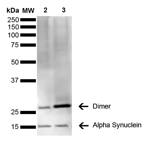
(Western Blot analysis of Mouse, Rat Brain showing detection of 14 kDa Alpha Synuclein protein using Mouse Anti-Alpha Synuclein Monoclonal Antibody, Clone 3C11. Lane 1: Molecular Weight Ladder (MW). Lane 2: Mouse brain cell lysate. Lane 3: Rat brain cell lysate. Load: 15 ug. Block: 5% Skim Milk in 1X TBST. Primary Antibody: Mouse Anti-Alpha Synuclein Monoclonal Antibody at 1:1000 for 2 hours at RT. Secondary Antibody: Goat Anti-Mouse HRP:IgG at 1:3000 for 1 hour at RT. Color Development: ECL solution (Super Signal West Pico) for 5 min in RT. Predicted/Observed Size: 14 kDa. Other Band(s): ~30 kDa (dimer).)
WB (Western Blot)
(Western Blot analysis of Mouse, Rat Brain showing detection of 14 kDa Alpha Synuclein protein using Mouse Anti-Alpha Synuclein Monoclonal Antibody, Clone 3C11. Lane 1: Molecular Weight Ladder (MW). Lane 2: Mouse brain cell lysate. Lane 3: Rat brain cell lysate. Load: 15 ug. Block: 5% Skim Milk in 1X TBST. Primary Antibody: Mouse Anti-Alpha Synuclein Monoclonal Antibody at 1:1000 for 2 hours at RT. Secondary Antibody: Goat Anti-Mouse HRP:IgG at 1:3000 for 1 hour at RT. Color Development: ECL solution (Super Signal West Pico) for 5 min in RT. Predicted/Observed Size: 14 kDa. Other Band(s): ~30 kDa (dimer).)
Alpha Synuclein, Monoclonal Antibody (Cat# AAA17810)
Full Name
Alpha Synuclein Antibody, Clone 3C11
Gene Names
SNCA; PD1; NACP; PARK1; PARK4
Reactivity
Human; Mouse; Rat
Applications
WB, DB, ICC, IF, EIA
Purity
Protein G Purified
Pricing


 IP (Immunoprecipitation)
(Immunoprecipitation analysis of 150ug extracts of MCF7 cells using 3ug HIST3H3 antibody. Western blot was performed from the immunoprecipitate using HIST3H3 antibody at a dilition of 1:500.)
IP (Immunoprecipitation)
(Immunoprecipitation analysis of 150ug extracts of MCF7 cells using 3ug HIST3H3 antibody. Western blot was performed from the immunoprecipitate using HIST3H3 antibody at a dilition of 1:500.)
HIST3H3, Polyclonal Antibody (Cat# AAA28284)
Full Name
HIST3H3 Polyclonal Antibody
Gene Names
HIST3H3; H3t; H3.4; H3/g; H3FT
Reactivity
Human, Mouse, Rat, Other (Wide Range)
Applications
Western Blot, Immunohistochemistry, Immunoprecipitation
Purity
Affinity Purification
Pricing


 IF (Immunofluorescence)
(Immunofluorescent analysis of 4% paraformaldehyde-fixed, 0.1% Triton X-100 permeabilized MCF-7 (human breast cancer cell line) cells labeling Pdx1 with at 1:25 dilution, followed by DyLight 488-conjugated IgG goat anti-rabbit secondary antibody at 1:200 dilution (green). Immunofluorescence image showing cytoplasm staining on MCF-7 cell line. Cytoplasmic actin is detected with DyLight 554 Phalloidin (PD18466410) at 1:100 dilution (red). The nuclear counter stain is DAPI (blue).)
IF (Immunofluorescence)
(Immunofluorescent analysis of 4% paraformaldehyde-fixed, 0.1% Triton X-100 permeabilized MCF-7 (human breast cancer cell line) cells labeling Pdx1 with at 1:25 dilution, followed by DyLight 488-conjugated IgG goat anti-rabbit secondary antibody at 1:200 dilution (green). Immunofluorescence image showing cytoplasm staining on MCF-7 cell line. Cytoplasmic actin is detected with DyLight 554 Phalloidin (PD18466410) at 1:100 dilution (red). The nuclear counter stain is DAPI (blue).)
OPN-a/b, Polyclonal Antibody (Cat# AAA26857)
Full Name
OPN-a/b, NT (SPP1, BNSP, OPN, Osteopontin, Bone sialoprotein 1, Nephropontin, Secreted phosphoprotein 1, Urinary stone protein, Uropontin) (APC)
Gene Names
SPP1; OPN; BNSP; BSPI; ETA-1
Reactivity
Human
Applications
WB, IHC, IF
Purity
Purified by Protein A and Peptide Affinity Chromatography.
Pricing


 FCM (Flow Cytometry)
(SYP Antibody (C-term) flow cytometric analysis of Neuro-2a cells (right histogram) compared to a negative control cell (left histogram).FITC-conjugated goat-anti-rabbit secondary antibodies were used for the analysis.)
FCM (Flow Cytometry)
(SYP Antibody (C-term) flow cytometric analysis of Neuro-2a cells (right histogram) compared to a negative control cell (left histogram).FITC-conjugated goat-anti-rabbit secondary antibodies were used for the analysis.)
SYP, Polyclonal Antibody (Cat# AAA28723)
Full Name
SYP Antibody (C-term)
Gene Names
SYP; MRX96; MRXSYP
Reactivity
Human, mouse (Predicted Reactivity: Rat)
Applications
Flow Cytometry, Immunofluorescence, Immunohistochemistry, Western Blot
Purity
Peptide Affinity Purified Rabbit Polyclonal Antibody (Pab)
Pricing


 IF (Immunofluorescence)
(Immunofluorescent analysis of 4% paraformaldehyde-fixed, 0.1% Triton X-100 permeabilized MCF-7 (human breast cancer cell line) cells labeling Pdx1 with at 1:25 dilution, followed by DyLight 488-conjugated IgG goat anti-rabbit secondary antibody at 1:200 dilution (green). Immunofluorescence image showing cytoplasm staining on MCF-7 cell line. Cytoplasmic actin is detected with DyLight 554 Phalloidin (PD18466410) at 1:100 dilution (red). The nuclear counter stain is DAPI (blue).)
IF (Immunofluorescence)
(Immunofluorescent analysis of 4% paraformaldehyde-fixed, 0.1% Triton X-100 permeabilized MCF-7 (human breast cancer cell line) cells labeling Pdx1 with at 1:25 dilution, followed by DyLight 488-conjugated IgG goat anti-rabbit secondary antibody at 1:200 dilution (green). Immunofluorescence image showing cytoplasm staining on MCF-7 cell line. Cytoplasmic actin is detected with DyLight 554 Phalloidin (PD18466410) at 1:100 dilution (red). The nuclear counter stain is DAPI (blue).)
OPN-a/b, Polyclonal Antibody (Cat# AAA26861)
Full Name
OPN-a/b, NT (SPP1, BNSP, OPN, Osteopontin, Bone sialoprotein 1, Nephropontin, Secreted phosphoprotein 1, Urinary stone protein, Uropontin) (MaxLight 490)
Gene Names
SPP1; OPN; BNSP; BSPI; ETA-1
Reactivity
Human
Applications
WB, IHC, IF
Purity
Purified by Protein A and Peptide Affinity Chromatography.
Pricing


 Application Data
(C:FGFR2/isolectinB4 (C) and FGFR1/isolectinB4 (D) staining of apparent mesenchymal cells and the subpopulation of endothelial cells. Virtually all other dispersed apparent mesenchymal cells express FGFR1 and FGFR2 (merged image in E). F: FGFR2 (F) and FGFR1 (G) staining in clustered cells of epithelial origin (inferred by morphology here) demonstrating that epithelial cells express both FGFR1 and FGFR2 (merged image with DAPI staining in H).)
Application Data
(C:FGFR2/isolectinB4 (C) and FGFR1/isolectinB4 (D) staining of apparent mesenchymal cells and the subpopulation of endothelial cells. Virtually all other dispersed apparent mesenchymal cells express FGFR1 and FGFR2 (merged image in E). F: FGFR2 (F) and FGFR1 (G) staining in clustered cells of epithelial origin (inferred by morphology here) demonstrating that epithelial cells express both FGFR1 and FGFR2 (merged image with DAPI staining in H).)
FGFR2, Polyclonal Antibody (Cat# AAA14790)
Full Name
FGFR2, NT (FGFR2, BEK, KGFR, KSAM, Fibroblast growth factor receptor 2, K-sam, Keratinocyte growth factor receptor, CD332)
Gene Names
FGFR2; BEK; JWS; BBDS; CEK3; CFD1; ECT1; KGFR; TK14; TK25; BFR-1; CD332; K-SAM
Reactivity
Human, Monkey, Mouse, Rat
Applications
EL/EIA, WB, IHC, FC/FACS, IF
Purity
Affinity Purified
Purified by Protein A affinity chromatography.
Purified by Protein A affinity chromatography.
Pricing


 WB (Western Blot)
(The anti-SUMO2/3 C-term Pab is used in Western blot to detect SUMO2/3 in HeLa cell lysate.)
WB (Western Blot)
(The anti-SUMO2/3 C-term Pab is used in Western blot to detect SUMO2/3 in HeLa cell lysate.)
SUMO2/3, Polyclonal Antibody (Cat# AAA28663)
Full Name
SUMO2/3 Antibody (C-term)
Gene Names
SUMO3; SMT3A; Smt3B; SMT3H1; SUMO-3
Reactivity
Human, mouse (Predicted Reactivity: Xenopus, Zebrafish, Bovine, Chicken, Hamster, Monkey, Pig, Rat)
Applications
IF, EIA, WB, IHC
Purity
Purified Rabbit Polyclonal Antibody (Pab)
Pricing


 IF (Immunofluorescence)
(Immunofluorescent analysis of 4% paraformaldehyde-fixed, 0.1% Triton X-100 permeabilized MCF-7 (human breast cancer cell line) cells labeling Pdx1 with at 1:25 dilution, followed by DyLight 488-conjugated IgG goat anti-rabbit secondary antibody at 1:200 dilution (green). Immunofluorescence image showing cytoplasm staining on MCF-7 cell line. Cytoplasmic actin is detected with DyLight 554 Phalloidin (PD18466410) at 1:100 dilution (red). The nuclear counter stain is DAPI (blue).)
IF (Immunofluorescence)
(Immunofluorescent analysis of 4% paraformaldehyde-fixed, 0.1% Triton X-100 permeabilized MCF-7 (human breast cancer cell line) cells labeling Pdx1 with at 1:25 dilution, followed by DyLight 488-conjugated IgG goat anti-rabbit secondary antibody at 1:200 dilution (green). Immunofluorescence image showing cytoplasm staining on MCF-7 cell line. Cytoplasmic actin is detected with DyLight 554 Phalloidin (PD18466410) at 1:100 dilution (red). The nuclear counter stain is DAPI (blue).)
OPN-a/b, Polyclonal Antibody (Cat# AAA26863)
Full Name
OPN-a/b, NT (SPP1, BNSP, OPN, Osteopontin, Bone sialoprotein 1, Nephropontin, Secreted phosphoprotein 1, Urinary stone protein, Uropontin) (MaxLight 650)
Gene Names
SPP1; OPN; BNSP; BSPI; ETA-1
Reactivity
Human
Applications
WB, IHC, IF
Purity
Purified by Protein A and Peptide Affinity Chromatography.
Pricing


 IF (Immunofluorescence)
(Immunofluorescent analysis of 4% paraformaldehyde-fixed, 0.1% Triton X-100 permeabilized MCF-7 (human breast cancer cell line) cells labeling Pdx1 with at 1:25 dilution, followed by DyLight 488-conjugated IgG goat anti-rabbit secondary antibody at 1:200 dilution (green). Immunofluorescence image showing cytoplasm staining on MCF-7 cell line. Cytoplasmic actin is detected with DyLight 554 Phalloidin (PD18466410) at 1:100 dilution (red). The nuclear counter stain is DAPI (blue).)
IF (Immunofluorescence)
(Immunofluorescent analysis of 4% paraformaldehyde-fixed, 0.1% Triton X-100 permeabilized MCF-7 (human breast cancer cell line) cells labeling Pdx1 with at 1:25 dilution, followed by DyLight 488-conjugated IgG goat anti-rabbit secondary antibody at 1:200 dilution (green). Immunofluorescence image showing cytoplasm staining on MCF-7 cell line. Cytoplasmic actin is detected with DyLight 554 Phalloidin (PD18466410) at 1:100 dilution (red). The nuclear counter stain is DAPI (blue).)
OPN-a/b, Polyclonal Antibody (Cat# AAA26864)
Full Name
OPN-a/b, NT (SPP1, BNSP, OPN, Osteopontin, Bone sialoprotein 1, Nephropontin, Secreted phosphoprotein 1, Urinary stone protein, Uropontin) (MaxLight 750)
Gene Names
SPP1; OPN; BNSP; BSPI; ETA-1
Reactivity
Human
Applications
WB, IHC, IF
Purity
Purified by Protein A and Peptide Affinity Chromatography.
Pricing


 Application Data
(Staining of mouse peritoneal macrophages with RAT ANTI MOUSE F4/80 ANTIGEN:FITC)
Application Data
(Staining of mouse peritoneal macrophages with RAT ANTI MOUSE F4/80 ANTIGEN:FITC)
EMR1, Monoclonal Antibody (Cat# AAA26842)
Full Name
EMR1 (EGF-like Module Containing Mucin-like Hormone Receptor-like 1, EMR1 Hormone Receptor, Cell Surface Glycoprotein EMR1, Cell Surface Glycoprotein F4/80, DD7A5-7, EGF-TM7, F4/80, Gpf480, Lymphocyte Antigen 71, Ly71, TM7LN3) (MaxLight 750)
Reactivity
Mouse
Applications
FC/FACS, IHC, IP, RIA, WB
Purity
Purified by protein G affinity chromatography from tissue culture supernatant.
Pricing


 IF (Immunofluorescence)
(Immunofluorescent analysis of 4% paraformaldehyde-fixed, 0.1% Triton X-100 permeabilized MCF-7 (human breast cancer cell line) cells labeling Pdx1 with at 1:25 dilution, followed by DyLight 488-conjugated IgG goat anti-rabbit secondary antibody at 1:200 dilution (green). Immunofluorescence image showing cytoplasm staining on MCF-7 cell line. Cytoplasmic actin is detected with DyLight 554 Phalloidin (PD18466410) at 1:100 dilution (red). The nuclear counter stain is DAPI (blue).)
IF (Immunofluorescence)
(Immunofluorescent analysis of 4% paraformaldehyde-fixed, 0.1% Triton X-100 permeabilized MCF-7 (human breast cancer cell line) cells labeling Pdx1 with at 1:25 dilution, followed by DyLight 488-conjugated IgG goat anti-rabbit secondary antibody at 1:200 dilution (green). Immunofluorescence image showing cytoplasm staining on MCF-7 cell line. Cytoplasmic actin is detected with DyLight 554 Phalloidin (PD18466410) at 1:100 dilution (red). The nuclear counter stain is DAPI (blue).)
OPN-a/b, Polyclonal Antibody (Cat# AAA26862)
Full Name
OPN-a/b, NT (SPP1, BNSP, OPN, Osteopontin, Bone sialoprotein 1, Nephropontin, Secreted phosphoprotein 1, Urinary stone protein, Uropontin) (MaxLight 550)
Gene Names
SPP1; OPN; BNSP; BSPI; ETA-1
Reactivity
Human
Applications
WB, IHC, IF
Purity
Purified by Protein A and Peptide Affinity Chromatography.
Pricing


 Application Data
(C:FGFR2/isolectinB4 (C) and FGFR1/isolectinB4 (D) staining of apparent mesenchymal cells and the subpopulation of endothelial cells. Virtually all other dispersed apparent mesenchymal cells express FGFR1 and FGFR2 (merged image in E). F: FGFR2 (F) and FGFR1 (G) staining in clustered cells of epithelial origin (inferred by morphology here) demonstrating that epithelial cells express both FGFR1 and FGFR2 (merged image with DAPI staining in H).)
Application Data
(C:FGFR2/isolectinB4 (C) and FGFR1/isolectinB4 (D) staining of apparent mesenchymal cells and the subpopulation of endothelial cells. Virtually all other dispersed apparent mesenchymal cells express FGFR1 and FGFR2 (merged image in E). F: FGFR2 (F) and FGFR1 (G) staining in clustered cells of epithelial origin (inferred by morphology here) demonstrating that epithelial cells express both FGFR1 and FGFR2 (merged image with DAPI staining in H).)
FGFR2, Polyclonal Antibody (Cat# AAA26854)
Full Name
FGFR2, NT (FGFR2, BEK, KGFR, KSAM, Fibroblast growth factor receptor 2, K-sam, Keratinocyte growth factor receptor, CD332) (FITC)
Gene Names
FGFR2; BEK; JWS; BBDS; CEK3; CFD1; ECT1; KGFR; TK14; TK25; BFR-1; CD332; K-SAM
Reactivity
Human, Monkey, Mouse, Rat
Applications
WB, IHC, IF, FC/FACS
Purity
Purified by Protein G Affinity Chromatography.
Pricing


 WB (Western Blot)
(Formalin-fixed and paraffin-embedded human cancer tissue reacted with the primary antibody, which was peroxidase-conjugated to the secondary antibody, followed by AEC staining. This data demonstrates the use of this antibody for immunohistochemistry; clinical relevance has not been evaluated. BC = breast carcinoma; HC = hepatocarcinoma.)
WB (Western Blot)
(Formalin-fixed and paraffin-embedded human cancer tissue reacted with the primary antibody, which was peroxidase-conjugated to the secondary antibody, followed by AEC staining. This data demonstrates the use of this antibody for immunohistochemistry; clinical relevance has not been evaluated. BC = breast carcinoma; HC = hepatocarcinoma.)
SIRT3, Polyclonal Antibody (Cat# AAA28669)
Full Name
SIRT3 Antibody (C-term)
Gene Names
SIRT3; SIR2L3
Reactivity
Human, mouse
Applications
WB, EIA, IHC
Purity
Purified Rabbit Polyclonal Antibody (Pab)
Pricing


 FCM (Flow Cytometry)
(GTF2I Antibody (C-term) flow cytometric analysis of k562 cells (bottom histogram) compared to a negative control cell (top histogram).FITC-conjugated goat-anti-rabbit secondary antibodies were used for the analysis.)
FCM (Flow Cytometry)
(GTF2I Antibody (C-term) flow cytometric analysis of k562 cells (bottom histogram) compared to a negative control cell (top histogram).FITC-conjugated goat-anti-rabbit secondary antibodies were used for the analysis.)
GTF2I, Polyclonal Antibody (Cat# AAA28707)
Full Name
GTF2I Antibody (C-term)
Gene Names
GTF2I; WBS; DIWS; SPIN; IB291; BAP135; BTKAP1; TFII-I; WBSCR6; GTFII-I
Reactivity
Human (Predicted Reactivity: Rat)
Applications
Immunofluorescence, Immunohistochemistry, Flow Cytometry, Western Blot
Purity
Peptide Affinity Purified Rabbit Polyclonal Antibody (Pab)
Pricing


 WB (Western Blot)
(Western blot analysis of lysate from HepG2 cell line using PPT1 Antibody. AAA28640 was diluted at 1:1000 at each lane. A goat anti-mouse IgG H&L(HRP) at 1:3000 dilution was used as the secondary antibody. Lysate at 35ug per lane.)
WB (Western Blot)
(Western blot analysis of lysate from HepG2 cell line using PPT1 Antibody. AAA28640 was diluted at 1:1000 at each lane. A goat anti-mouse IgG H&L(HRP) at 1:3000 dilution was used as the secondary antibody. Lysate at 35ug per lane.)
PPT1, Monoclonal Antibody (Cat# AAA28640)
Full Name
PPT1 Antibody
Gene Names
PPT1; PPT; CLN1; INCL
Reactivity
Human
Applications
WB, EIA, FC/FACS, IHC
Purity
Purified Mouse Monoclonal Antibody (Mab)
Pricing


 Application Data
(C:FGFR2/isolectinB4 (C) and FGFR1/isolectinB4 (D) staining of apparent mesenchymal cells and the subpopulation of endothelial cells. Virtually all other dispersed apparent mesenchymal cells express FGFR1 and FGFR2 (merged image in E). F: FGFR2 (F) and FGFR1 (G) staining in clustered cells of epithelial origin (inferred by morphology here) demonstrating that epithelial cells express both FGFR1 and FGFR2 (merged image with DAPI staining in H).)
Application Data
(C:FGFR2/isolectinB4 (C) and FGFR1/isolectinB4 (D) staining of apparent mesenchymal cells and the subpopulation of endothelial cells. Virtually all other dispersed apparent mesenchymal cells express FGFR1 and FGFR2 (merged image in E). F: FGFR2 (F) and FGFR1 (G) staining in clustered cells of epithelial origin (inferred by morphology here) demonstrating that epithelial cells express both FGFR1 and FGFR2 (merged image with DAPI staining in H).)
FGFR2, Polyclonal Antibody (Cat# AAA26851)
Full Name
FGFR2, NT (FGFR2, BEK, KGFR, KSAM, Fibroblast growth factor receptor 2, K-sam, Keratinocyte growth factor receptor, CD332) (AP)
Gene Names
FGFR2; BEK; JWS; BBDS; CEK3; CFD1; ECT1; KGFR; TK14; TK25; BFR-1; CD332; K-SAM
Reactivity
Human, Monkey, Mouse, Rat
Applications
IF, EIA, IHC, WB
Purity
Purified by Protein G Affinity Chromatography.
Pricing


 IF (Immunofluorescence)
(Immunofluorescent analysis of 4% paraformaldehyde-fixed, 0.1% Triton X-100 permeabilized MCF-7 (human breast cancer cell line) cells labeling Pdx1 with at 1:25 dilution, followed by DyLight 488-conjugated IgG goat anti-rabbit secondary antibody at 1:200 dilution (green). Immunofluorescence image showing cytoplasm staining on MCF-7 cell line. Cytoplasmic actin is detected with DyLight 554 Phalloidin (PD18466410) at 1:100 dilution (red). The nuclear counter stain is DAPI (blue).)
IF (Immunofluorescence)
(Immunofluorescent analysis of 4% paraformaldehyde-fixed, 0.1% Triton X-100 permeabilized MCF-7 (human breast cancer cell line) cells labeling Pdx1 with at 1:25 dilution, followed by DyLight 488-conjugated IgG goat anti-rabbit secondary antibody at 1:200 dilution (green). Immunofluorescence image showing cytoplasm staining on MCF-7 cell line. Cytoplasmic actin is detected with DyLight 554 Phalloidin (PD18466410) at 1:100 dilution (red). The nuclear counter stain is DAPI (blue).)
OPN-a/b, Polyclonal Antibody (Cat# AAA26859)
Full Name
OPN-a/b, NT (SPP1, BNSP, OPN, Osteopontin, Bone sialoprotein 1, Nephropontin, Secreted phosphoprotein 1, Urinary stone protein, Uropontin) (FITC)
Gene Names
SPP1; OPN; BNSP; BSPI; ETA-1
Reactivity
Human
Applications
WB, IHC, IF
Purity
Purified by Protein A and Peptide Affinity Chromatography.
Pricing


 FCM (Flow Cytometry)
(PROX-1-S514 Antibody flow cytometric analysis of 293 cells (right histogram) compared to a negative control cell (left histogram).FITC-conjugated goat-anti-rabbit secondary antibodies were used for the analysis.)
FCM (Flow Cytometry)
(PROX-1-S514 Antibody flow cytometric analysis of 293 cells (right histogram) compared to a negative control cell (left histogram).FITC-conjugated goat-anti-rabbit secondary antibodies were used for the analysis.)
PROX-1-S514, Polyclonal Antibody (Cat# AAA28650)
Full Name
PROX-1-S514 Antibody
Reactivity
Human, mouse
Applications
WB, EIA, IHC, FC/FACS
Purity
Peptide Affinity Purified Rabbit Polyclonal Antibody (Pab)
Pricing


 IF (Immunofluorescence)
(Immunofluorescent analysis of 4% paraformaldehyde-fixed, 0.1% Triton X-100 permeabilized MCF-7 (human breast cancer cell line) cells labeling Pdx1 with at 1:25 dilution, followed by DyLight 488-conjugated IgG goat anti-rabbit secondary antibody at 1:200 dilution (green). Immunofluorescence image showing cytoplasm staining on MCF-7 cell line. Cytoplasmic actin is detected with DyLight 554 Phalloidin (PD18466410) at 1:100 dilution (red). The nuclear counter stain is DAPI (blue).)
IF (Immunofluorescence)
(Immunofluorescent analysis of 4% paraformaldehyde-fixed, 0.1% Triton X-100 permeabilized MCF-7 (human breast cancer cell line) cells labeling Pdx1 with at 1:25 dilution, followed by DyLight 488-conjugated IgG goat anti-rabbit secondary antibody at 1:200 dilution (green). Immunofluorescence image showing cytoplasm staining on MCF-7 cell line. Cytoplasmic actin is detected with DyLight 554 Phalloidin (PD18466410) at 1:100 dilution (red). The nuclear counter stain is DAPI (blue).)
OPN-a/b, Polyclonal Antibody (Cat# AAA26860)
Full Name
OPN-a/b, NT (SPP1, BNSP, OPN, Osteopontin, Bone sialoprotein 1, Nephropontin, Secreted phosphoprotein 1, Urinary stone protein, Uropontin) (MaxLight 405)
Gene Names
SPP1; OPN; BNSP; BSPI; ETA-1
Reactivity
Human
Applications
WB, IHC, IF
Purity
Purified by Protein A and Peptide Affinity Chromatography.
Pricing


 WB (Western Blot)
(EGFR Antibody (C-term) western blot analysis in Hela cell line lysates (35ug/lane).This demonstrates the EGFR antibody detected the EGFR protein (arrow).)
WB (Western Blot)
(EGFR Antibody (C-term) western blot analysis in Hela cell line lysates (35ug/lane).This demonstrates the EGFR antibody detected the EGFR protein (arrow).)
EGFR, Monoclonal Antibody (Cat# AAA28637)
Full Name
EGFR Antibody (C-term)
Gene Names
EGFR; ERBB; HER1; mENA; ERBB1; PIG61; NISBD2
Reactivity
Human
Applications
WB, EIA, IHC
Pricing


 WB (Western Blot)
(Formalin-fixed and paraffin-embedded human cancer tissue reacted with the primary antibody, which was peroxidase-conjugated to the secondary antibody, followed by DAB staining. This data demonstrates the use of this antibody for immunohistochemistry; clinical relevance has not been evaluated. BC = breast carcinoma; HC = hepatocarcinoma.)
WB (Western Blot)
(Formalin-fixed and paraffin-embedded human cancer tissue reacted with the primary antibody, which was peroxidase-conjugated to the secondary antibody, followed by DAB staining. This data demonstrates the use of this antibody for immunohistochemistry; clinical relevance has not been evaluated. BC = breast carcinoma; HC = hepatocarcinoma.)
SPAK, Polyclonal Antibody (Cat# AAA28754)
Full Name
SPAK Antibody (Center)
Gene Names
STK39; DCHT; PASK; SPAK
Reactivity
Human, mouse (Predicted Reactivity: Rat)
Applications
Western Blot, Immunohistochemistry
Purity
Purified Rabbit Polyclonal Antibody (Pab)
Pricing


 IF (Immunofluorescence)
(Immunofluorescent analysis of 4% paraformaldehyde-fixed, 0.1% Triton X-100 permeabilized U-2 OS (human osteosarcoma cell line) cells labeling DLL3 with (1:25), followed by Dylight 488- conjugated goat anti- rabbit IgG secondary antibody at (1:200) (green). Immunofluorescence image showing nucleus and weak cytoplasm staining on U-2 OScell line. Cytoplasmic actin is detected with Dylight 554 Phalloidin (1:100) (red).)
IF (Immunofluorescence)
(Immunofluorescent analysis of 4% paraformaldehyde-fixed, 0.1% Triton X-100 permeabilized U-2 OS (human osteosarcoma cell line) cells labeling DLL3 with (1:25), followed by Dylight 488- conjugated goat anti- rabbit IgG secondary antibody at (1:200) (green). Immunofluorescence image showing nucleus and weak cytoplasm staining on U-2 OScell line. Cytoplasmic actin is detected with Dylight 554 Phalloidin (1:100) (red).)
DLL3, Polyclonal Antibody (Cat# AAA26850)
Full Name
DLL3, CT (DLL3, Delta-like protein 3, Drosophila Delta homolog 3) (APC)
Gene Names
DLL3; SCDO1
Reactivity
Human
Applications
IF, IHC, WB
Purity
Purified by Protein A Affinity Chromatography.
Pricing


 FCM (Flow Cytometry)
(ID1 Antibody (Center) flow cytometric analysis of U251 cells (right histogram) compared to a negative control cell (left histogram).FITC-conjugated goat-anti-rabbit secondary antibodies were used for the analysis.)
FCM (Flow Cytometry)
(ID1 Antibody (Center) flow cytometric analysis of U251 cells (right histogram) compared to a negative control cell (left histogram).FITC-conjugated goat-anti-rabbit secondary antibodies were used for the analysis.)
ID1, Polyclonal Antibody (Cat# AAA28652)
Full Name
ID1 Antibody (Center)
Gene Names
ID1; ID; bHLHb24
Reactivity
Human, mouse (Predicted Reactivity: Rat)
Applications
WB, EIA, IHC, IF, FC/FACS
Purity
Peptide Affinity Purified Rabbit Polyclonal Antibody (Pab)
Pricing


 Application Data
(Staining of mouse peritoneal macrophages with RAT ANTI MOUSE F4/80 ANTIGEN:FITC)
Application Data
(Staining of mouse peritoneal macrophages with RAT ANTI MOUSE F4/80 ANTIGEN:FITC)
EMR1, Monoclonal Antibody (Cat# AAA26828)
Full Name
EMR1 (EGF-like Module Containing Mucin-like Hormone Receptor-like 1, EMR1 Hormone Receptor, Cell Surface Glycoprotein EMR1, Cell Surface Glycoprotein F4/80, DD7A5-7, EGF-TM7, F4/80, Gpf480, Lymphocyte Antigen 71, Ly71, TM7LN3) (MaxLight 650)
Reactivity
Mouse
Applications
FC/FACS, IHC, IP, RIA, WB
Purity
Purified by protein G affinity chromatography from tissue culture supernatant.
Pricing


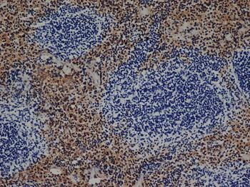
 IHC (Immunohistchemistry)
(LALBA Antibody (Center) (AAA28660)immunohistochemistry analysis in formalin fixed and paraffin embedded human breast carcinoma followed by peroxidase conjugation of the secondary antibody and DAB staining.This data demonstrates the use of LALBA Antibody (Center) for immunohistochemistry. Clinical relevance has not been evaluated.)
IHC (Immunohistchemistry)
(LALBA Antibody (Center) (AAA28660)immunohistochemistry analysis in formalin fixed and paraffin embedded human breast carcinoma followed by peroxidase conjugation of the secondary antibody and DAB staining.This data demonstrates the use of LALBA Antibody (Center) for immunohistochemistry. Clinical relevance has not been evaluated.)
LALBA, Polyclonal Antibody (Cat# AAA28660)
Full Name
LALBA Antibody (Center)
Reactivity
Human
Applications
WB, EIA, IHC
Purity
Peptide Affinity Purified Rabbit Polyclonal Antibody (Pab)
Pricing


 IF (Immunofluorescence)
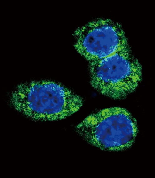
(Immunofluorescent analysis of 4% paraformaldehyde-fixed, 0.1% Triton X-100 permeabilized MCF-7 (human breast cancer cell line) cells labeling Pdx1 with AAA14796 at 1:25 dilution, followed by Dylight® 488-conjugated IgG goat anti-rabbit secondary antibody at 1:200 dilution (green). Immunofluorescence image showing cytoplasm staining on MCF-7 cell line. Cytoplasmic actin is detected with Dylight® 554 Phalloidin (PD18466410) at 1:100 dilution (red). The nuclear counter stain is DAPI (blue).)
IF (Immunofluorescence)
(Immunofluorescent analysis of 4% paraformaldehyde-fixed, 0.1% Triton X-100 permeabilized MCF-7 (human breast cancer cell line) cells labeling Pdx1 with AAA14796 at 1:25 dilution, followed by Dylight® 488-conjugated IgG goat anti-rabbit secondary antibody at 1:200 dilution (green). Immunofluorescence image showing cytoplasm staining on MCF-7 cell line. Cytoplasmic actin is detected with Dylight® 554 Phalloidin (PD18466410) at 1:100 dilution (red). The nuclear counter stain is DAPI (blue).)
OPN-a/b, Polyclonal Antibody (Cat# AAA14796)
Full Name
OPN-a/b, NT (SPP1, BNSP, OPN, Osteopontin, Bone sialoprotein 1, Nephropontin, Secreted phosphoprotein 1, Urinary stone protein, Uropontin)
Reactivity
Human
Applications
EL/EIA, WB, IHC, IF
Purity
Affinity Purified
Purified by Protein A affinity chromatography.
Purified by Protein A affinity chromatography.
Pricing


 Application Data
(C:FGFR2/isolectinB4 (C) and FGFR1/isolectinB4 (D) staining of apparent mesenchymal cells and the subpopulation of endothelial cells. Virtually all other dispersed apparent mesenchymal cells express FGFR1 and FGFR2 (merged image in E). F: FGFR2 (F) and FGFR1 (G) staining in clustered cells of epithelial origin (inferred by morphology here) demonstrating that epithelial cells express both FGFR1 and FGFR2 (merged image with DAPI staining in H).)
Application Data
(C:FGFR2/isolectinB4 (C) and FGFR1/isolectinB4 (D) staining of apparent mesenchymal cells and the subpopulation of endothelial cells. Virtually all other dispersed apparent mesenchymal cells express FGFR1 and FGFR2 (merged image in E). F: FGFR2 (F) and FGFR1 (G) staining in clustered cells of epithelial origin (inferred by morphology here) demonstrating that epithelial cells express both FGFR1 and FGFR2 (merged image with DAPI staining in H).)
FGFR2, Polyclonal Antibody (Cat# AAA26856)
Full Name
FGFR2, NT (FGFR2, BEK, KGFR, KSAM, Fibroblast growth factor receptor 2, K-sam, Keratinocyte growth factor receptor, CD332) (PE)
Gene Names
FGFR2; BEK; JWS; BBDS; CEK3; CFD1; ECT1; KGFR; TK14; TK25; BFR-1; CD332; K-SAM
Reactivity
Human, Monkey, Mouse, Rat
Applications
WB, IHC, IF, FC/FACS
Purity
Purified by Protein G Affinity Chromatography.
Pricing


 WB (Western Blot)
(Western blot analysis of lysates from MDA-MB-468, SW620, T47D cell line, mouse spleen, mouse testis tissue (from left to right), using EZH2 Antibody. AAA28665 was diluted at 1:1000 at each lane. A goat anti-rabbit IgG H&L(HRP) at 1:10000 dilution was used as the secondary antibody. Lysates at 20ug per lane.)
WB (Western Blot)
(Western blot analysis of lysates from MDA-MB-468, SW620, T47D cell line, mouse spleen, mouse testis tissue (from left to right), using EZH2 Antibody. AAA28665 was diluted at 1:1000 at each lane. A goat anti-rabbit IgG H&L(HRP) at 1:10000 dilution was used as the secondary antibody. Lysates at 20ug per lane.)
EZH2, Polyclonal Antibody (Cat# AAA28665)
Full Name
EZH2 Antibody
Gene Names
EZH2; WVS; ENX1; EZH1; KMT6; WVS2; ENX-1; EZH2b; KMT6A
Reactivity
Human, mouse
Applications
WB, EIA, IHC, IF, FC/FACS
Purity
Peptide Affinity Purified Rabbit Polyclonal Antibody (Pab)
Pricing


 FCM (Flow Cytometry)
(TYSY Antibody (C-term) flow cytometric analysis of Hela cells (right histogram) compared to a negative control cell (left histogram).FITC-conjugated goat-anti-rabbit secondary antibodies were used for the analysis.)
FCM (Flow Cytometry)
(TYSY Antibody (C-term) flow cytometric analysis of Hela cells (right histogram) compared to a negative control cell (left histogram).FITC-conjugated goat-anti-rabbit secondary antibodies were used for the analysis.)
TYSY, Polyclonal Antibody (Cat# AAA28647)
Full Name
TYSY Antibody (C-term)
Gene Names
TYMS; TS; TMS; HST422
Reactivity
Human
Applications
WB, EIA, IHC, IF, FC/FACS
Purity
Peptide Affinity Purified Rabbit Polyclonal Antibody (Pab)
Pricing

 FCM (Flow Cytometry)
(TMPRSS2 Antibody (Center)(AAA28646) flow cytometric analysis of HepG2 cells (right histogram) compared to a negative control cell (left histogram).FITC-conjugated goat-anti-rabbit secondary antibodies were used for the analysis.)
FCM (Flow Cytometry)
(TMPRSS2 Antibody (Center)(AAA28646) flow cytometric analysis of HepG2 cells (right histogram) compared to a negative control cell (left histogram).FITC-conjugated goat-anti-rabbit secondary antibodies were used for the analysis.)
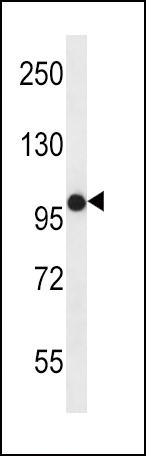
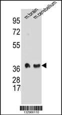
TMPRSS2, Polyclonal Antibody (Cat# AAA28646)
Full Name
TMPRSS2 Antibody (Center)
Gene Names
TMPRSS2; PP9284; PRSS10
Reactivity
Reactivity: Human, Mouse
Predicted: Mouse
Predicted: Mouse
Applications
WB, EIA, FC/FACS
Purity
Purified Rabbit Polyclonal Antibody (Pab)
Pricing


 IF (Immunofluorescence)
(Immunofluorescent analysis of 4% paraformaldehyde-fixed, 0.1% Triton X-100 permeabilized MCF-7 (human breast cancer cell line) cells labeling Pdx1 with at 1:25 dilution, followed by DyLight 488-conjugated IgG goat anti-rabbit secondary antibody at 1:200 dilution (green). Immunofluorescence image showing cytoplasm staining on MCF-7 cell line. Cytoplasmic actin is detected with DyLight 554 Phalloidin (PD18466410) at 1:100 dilution (red). The nuclear counter stain is DAPI (blue).)
IF (Immunofluorescence)
(Immunofluorescent analysis of 4% paraformaldehyde-fixed, 0.1% Triton X-100 permeabilized MCF-7 (human breast cancer cell line) cells labeling Pdx1 with at 1:25 dilution, followed by DyLight 488-conjugated IgG goat anti-rabbit secondary antibody at 1:200 dilution (green). Immunofluorescence image showing cytoplasm staining on MCF-7 cell line. Cytoplasmic actin is detected with DyLight 554 Phalloidin (PD18466410) at 1:100 dilution (red). The nuclear counter stain is DAPI (blue).)
OPN-a/b, Polyclonal Antibody (Cat# AAA26858)
Full Name
OPN-a/b, NT (SPP1, BNSP, OPN, Osteopontin, Bone sialoprotein 1, Nephropontin, Secreted phosphoprotein 1, Urinary stone protein, Uropontin) (Biotin)
Gene Names
SPP1; OPN; BNSP; BSPI; ETA-1
Reactivity
Human
Applications
WB, IHC, IF, EIA
Purity
Purified by Protein A and Peptide Affinity Chromatography.
Pricing


 Application Data
(Staining of mouse peritoneal macrophages with RAT ANTI MOUSE F4/80 ANTIGEN:FITC)
Application Data
(Staining of mouse peritoneal macrophages with RAT ANTI MOUSE F4/80 ANTIGEN:FITC)
EMR1, Monoclonal Antibody (Cat# AAA26845)
Full Name
EMR1 (EGF-like Module Containing Mucin-like Hormone Receptor-like 1, EMR1 Hormone Receptor, Cell Surface Glycoprotein EMR1, Cell Surface Glycoprotein F4/80, DD7A5-7, EGF-TM7, F4/80, Gpf480, Lymphocyte Antigen 71, Ly71, TM7LN3) (APC)
Reactivity
Mouse
Applications
IHC, IP, RIA, WB
Purity
Purified by protein G affinity chromatography from tissue culture supernatant.
Pricing


 WB (Western Blot)
(FG Pancreatic Carcinoma Cell Lines stably expressing vector along (FG-V) the b3 integrin subunit (FG-b3) or a b3 truncation mutant (FG-759x). Src Mab (AAA28639) was diluted 1:500 in 1% BSA/TBST and incubated Overnight at 4 degree C. After washing 3x 5 min. with TBST the blots were incubated with 1:5000 Goat anti-mouse or Goat anti-rabbit secondary antibody for 1 hr at Room temperature. The blots were again washed 3x 5 min. with TBST and developed using ECL reagent.Data and protocol kindly provided by Dr. Weis of Cheresh Lab, UCSD.)
WB (Western Blot)
(FG Pancreatic Carcinoma Cell Lines stably expressing vector along (FG-V) the b3 integrin subunit (FG-b3) or a b3 truncation mutant (FG-759x). Src Mab (AAA28639) was diluted 1:500 in 1% BSA/TBST and incubated Overnight at 4 degree C. After washing 3x 5 min. with TBST the blots were incubated with 1:5000 Goat anti-mouse or Goat anti-rabbit secondary antibody for 1 hr at Room temperature. The blots were again washed 3x 5 min. with TBST and developed using ECL reagent.Data and protocol kindly provided by Dr. Weis of Cheresh Lab, UCSD.)
SRC, Monoclonal Antibody (Cat# AAA28639)
Full Name
SRC Antibody
Gene Names
SRC; ASV; SRC1; c-SRC; p60-Src
Reactivity
Human, mouse
Applications
WB, EIA, IF
Purity
This antibody is purified through a protein G column, followed by dialysis against PBS.
Pricing
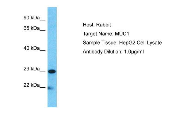


 WB (Western Blot)
(Host: RabbitTarget Name: MUC1Sample Type: HepG2Lane A: Primary AntibodyLane B: Primary Antibody + Blocking PeptidePrimary Antibody Concentration: 1ug/mlPeptide Concentration: 5ug/mlLysate Quantity: 25ug/lane/laneGel Concentration: 0.12)
WB (Western Blot)
(Host: RabbitTarget Name: MUC1Sample Type: HepG2Lane A: Primary AntibodyLane B: Primary Antibody + Blocking PeptidePrimary Antibody Concentration: 1ug/mlPeptide Concentration: 5ug/mlLysate Quantity: 25ug/lane/laneGel Concentration: 0.12)
MUC1, Polyclonal Antibody (Cat# AAA23492)
Full Name
MUC1 antibody - C-terminal region
Gene Names
MUC1; EMA; MCD; PEM; PUM; KL-6; MAM6; MCKD; PEMT; CD227; H23AG; MCKD1; MUC-1; ADMCKD; ADMCKD1; CA 15-3; MUC-1/X; MUC1/ZD; MUC-1/SEC
Reactivity
Cow, Dog, Guinea Pig, Horse, Human, Mouse, Rabbit, Rat, Pig
Applications
IHC, WB
Purity
Affinity Purified
Pricing


 DB (Dot Blot)
(Formalin-fixed and paraffin-embedded human cancer tissue reacted with the primary antibody, which was peroxidase-conjugated to the secondary antibody, followed by AEC staining. This data demonstrates the use of this antibody for immunohistochemistry; clinical relevance has not been evaluated. BC = breast carcinoma; HC = hepatocarcinoma.)
DB (Dot Blot)
(Formalin-fixed and paraffin-embedded human cancer tissue reacted with the primary antibody, which was peroxidase-conjugated to the secondary antibody, followed by AEC staining. This data demonstrates the use of this antibody for immunohistochemistry; clinical relevance has not been evaluated. BC = breast carcinoma; HC = hepatocarcinoma.)
Phospho-HIST1H3B3 (S10), Polyclonal Antibody (Cat# AAA28670)
Full Name
Phospho-HIST1H3B3 (S10) Antibody
Gene Names
HIST1H3A; H3/A; H3FA
Reactivity
Human
Applications
DB, EIA, IHC, WB
Purity
Peptide Affinity Purified Rabbit Polyclonal Antibody (Pab)
Pricing


 IHC (Immunohistchemistry)
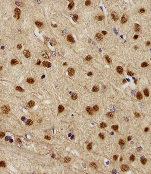
(Figure 6. IHC analysis of AMD1 using anti-AMD1 antibody (AAA19177).AMD1 was detected in paraffin-embedded section of human mammary cancer tissue. Heat mediated antigen retrieval was performed in citrate buffer (pH6, epitope retrieval solution) for 20 mins. The tissue section was blocked with 10% goat serum. The tissue section was then incubated with 1ug/ml rabbit anti-AMD1 Antibody (AAA19177) overnight at 4 degree C. Biotinylated goat anti-rabbit IgG was used as secondary antibody and incubated for 30 minutes at 37 degree C. The tissue section was developed using Strepavidin-Biotin-Complex (SABC) with DAB as the chromogen.)
IHC (Immunohistchemistry)
(Figure 6. IHC analysis of AMD1 using anti-AMD1 antibody (AAA19177).AMD1 was detected in paraffin-embedded section of human mammary cancer tissue. Heat mediated antigen retrieval was performed in citrate buffer (pH6, epitope retrieval solution) for 20 mins. The tissue section was blocked with 10% goat serum. The tissue section was then incubated with 1ug/ml rabbit anti-AMD1 Antibody (AAA19177) overnight at 4 degree C. Biotinylated goat anti-rabbit IgG was used as secondary antibody and incubated for 30 minutes at 37 degree C. The tissue section was developed using Strepavidin-Biotin-Complex (SABC) with DAB as the chromogen.)
AMD1/Adometdc, Polyclonal Antibody (Cat# AAA19177)
Full Name
Anti-AMD1/Adometdc Antibody
Gene Names
AMD1; AMD; SAMDC; ADOMETDC
Reactivity
Human, Mouse, Rat
No cross reactivity with other proteins.
No cross reactivity with other proteins.
Applications
IHC, WB
Purity
Immunogen affinity purified
Pricing


 WB (Western Blot)
(Western blot analysis of lysates from 293, Hela, mouse NIH/3T3, rat PC-12 cell line and rat brain tissue lysate(from left to right), using RPS6 Antibody (N-term). AAA28643 was diluted at 1:2000 at each lane. A goat anti-mouse IgG H&L(HRP) at 1:3000 dilution was used as the secondary antibody. Lysates at 35ug per lane.)
WB (Western Blot)
(Western blot analysis of lysates from 293, Hela, mouse NIH/3T3, rat PC-12 cell line and rat brain tissue lysate(from left to right), using RPS6 Antibody (N-term). AAA28643 was diluted at 1:2000 at each lane. A goat anti-mouse IgG H&L(HRP) at 1:3000 dilution was used as the secondary antibody. Lysates at 35ug per lane.)
RPS6, Monoclonal Antibody (Cat# AAA28643)
Full Name
RPS6 Antibody (N-term)
Gene Names
RPS6; S6
Reactivity
Human, rat (Predicted Reactivity: Monkey, Mouse)
Applications
EIA, IHC, WB, FC/FACS, IF
Purity
Purified Mouse Monoclonal Antibody (Mab)
Pricing


 WB (Western Blot)
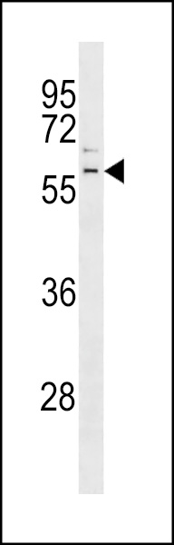
(SLC16A11 Antibody (N-term) western blot analysis in mouse brain,stomach tissue lysates (35ug/lane).This demonstrates the SLC16A11 antibody detected the SLC16A11 protein (arrow).)
WB (Western Blot)
(SLC16A11 Antibody (N-term) western blot analysis in mouse brain,stomach tissue lysates (35ug/lane).This demonstrates the SLC16A11 antibody detected the SLC16A11 protein (arrow).)
SLC16A11, Polyclonal Antibody (Cat# AAA28655)
Full Name
SLC16A11 Antibody (N-term)
Gene Names
SLC16A11; MCT11
Reactivity
Human, mouse
Applications
WB, EIA, FC/FACS, IHC
Purity
Peptide Affinity Purified Rabbit Polyclonal Antibody (Pab)
Pricing


 Application Data
(C:FGFR2/isolectinB4 (C) and FGFR1/isolectinB4 (D) staining of apparent mesenchymal cells and the subpopulation of endothelial cells. Virtually all other dispersed apparent mesenchymal cells express FGFR1 and FGFR2 (merged image in E). F: FGFR2 (F) and FGFR1 (G) staining in clustered cells of epithelial origin (inferred by morphology here) demonstrating that epithelial cells express both FGFR1 and FGFR2 (merged image with DAPI staining in H).)
Application Data
(C:FGFR2/isolectinB4 (C) and FGFR1/isolectinB4 (D) staining of apparent mesenchymal cells and the subpopulation of endothelial cells. Virtually all other dispersed apparent mesenchymal cells express FGFR1 and FGFR2 (merged image in E). F: FGFR2 (F) and FGFR1 (G) staining in clustered cells of epithelial origin (inferred by morphology here) demonstrating that epithelial cells express both FGFR1 and FGFR2 (merged image with DAPI staining in H).)
FGFR2, Polyclonal Antibody (Cat# AAA26852)
Full Name
FGFR2, NT (FGFR2, BEK, KGFR, KSAM, Fibroblast growth factor receptor 2, K-sam, Keratinocyte growth factor receptor, CD332) (APC)
Gene Names
FGFR2; BEK; JWS; BBDS; CEK3; CFD1; ECT1; KGFR; TK14; TK25; BFR-1; CD332; K-SAM
Reactivity
Human, Monkey, Mouse, Rat
Applications
FC/FACS, IF, IHC, WB
Purity
Purified by Protein G Affinity Chromatography.
Pricing


 IHC (Immunohistchemistry)
(PINK1 Monoclonal Antibody immunohistochemistry analysis in formalin fixed and paraffin embedded human kidney tissue followed by peroxidase conjugation of the secondary antibody and DAB staining. This data demonstrates the use of the PINK1 Monoclonal Antibody for immunohistochemistry. Clinical relevance has not been evaluated.)
IHC (Immunohistchemistry)
(PINK1 Monoclonal Antibody immunohistochemistry analysis in formalin fixed and paraffin embedded human kidney tissue followed by peroxidase conjugation of the secondary antibody and DAB staining. This data demonstrates the use of the PINK1 Monoclonal Antibody for immunohistochemistry. Clinical relevance has not been evaluated.)
PINK1, Monoclonal Antibody (Cat# AAA28638)
Full Name
PINK1 Antibody
Gene Names
PINK1; BRPK; PARK6
Reactivity
Human
Applications
EIA, IHC, IF, WB
Purity
Purified Mouse Monoclonal Antibody (Mab)
Pricing