Filters
Clonality
Type
Reactivity
Gene Name
Isotype
Host
Application
Clone
409 results for "Rabbit ELISA Kit" - showing 100-150


 Application Data
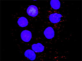
(Proximity Ligation Analysis (PLA) of protein-protein interactions between FGA and F2. HeLa cells were stained with FGA rabbit purified polyclonal 1:1200 and F2 mouse monoclonal antibody 1:50. Signals were detected by 30 Detection Kit 613 (red), and nuclei were counterstained with DAPI (blue). Each red dot represents the detection of protein-protein interaction complex.)
Application Data
(Proximity Ligation Analysis (PLA) of protein-protein interactions between FGA and F2. HeLa cells were stained with FGA rabbit purified polyclonal 1:1200 and F2 mouse monoclonal antibody 1:50. Signals were detected by 30 Detection Kit 613 (red), and nuclei were counterstained with DAPI (blue). Each red dot represents the detection of protein-protein interaction complex.)
F2, Monoclonal Antibody (Cat# AAA25388)
Full Name
F2 (Coagulation Factor II, Prothrombin) (HRP)
Gene Names
F2; PT; THPH1; RPRGL2
Reactivity
Human
Applications
Immunoprecipitation, Western Blot
Purity
Purified by Protein A Affinity Chromatography.
Pricing


 Application Data
(Proximity Ligation Analysis (PLA) of protein-protein interactions between BAD and MAPK8 Mahlavu cells were stained with BAD rabbit purified polyclonal 1:1200 and anti-MAPK8 mouse monoclonal antibody 1:50. Signals were detected 30 Detection Kit 613 (red), and nuclei were counterstained with DAPI (blue). Each red dot represents the detection of protein-protein interaction complex.)
Application Data
(Proximity Ligation Analysis (PLA) of protein-protein interactions between BAD and MAPK8 Mahlavu cells were stained with BAD rabbit purified polyclonal 1:1200 and anti-MAPK8 mouse monoclonal antibody 1:50. Signals were detected 30 Detection Kit 613 (red), and nuclei were counterstained with DAPI (blue). Each red dot represents the detection of protein-protein interaction complex.)
MAPK8, Monoclonal Antibody (Cat# AAA25447)
Full Name
MAPK8 (JNK1, PRKM8, SAPK1, SAPK1C, Mitogen-activated Protein Kinase 8, MAP Kinase 8, MAPK 8, JNK-46, Stress-activated Protein Kinase 1c, Stress-activated Protein Kinase JNK1, c-Jun N-terminal Kinase 1) (HRP)
Gene Names
MAPK8; JNK; JNK1; PRKM8; SAPK1; JNK-46; JNK1A2; SAPK1c; JNK21B1/2
Reactivity
Human
Applications
Western Blot
Purity
Purified by Protein A Affinity Chromatography.
Pricing


 WB (Western Blot)
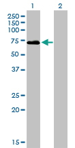
(DAAM1 monoclonal antibody Western Blot analysis of DAAM1 expression in A-431.)
WB (Western Blot)
(DAAM1 monoclonal antibody Western Blot analysis of DAAM1 expression in A-431.)
DAAM1, Monoclonal Antibody (Cat# AAA25661)
Full Name
DAAM1 (Disheveled-associated Activator Of Morphogenesis 1, KIAA0666) (PE)
Reactivity
Human, Rat
Applications
Western Blot
Purity
Purified by Protein A Affinity Chromatography.
Pricing


 IHC (Immunohistchemistry)
(Figure 6. IHC analysis of ADA using anti-ADA antibody (AAA19140).ADA was detected in paraffin-embedded section of rat spleen tissue. Heat mediated antigen retrieval was performed in citrate buffer (pH6, epitope retrieval solution) for 20 mins. The tissue section was blocked with 10% goat serum. The tissue section was then incubated with 1ug/ml rabbit anti-ADA Antibody (AAA19140) overnight at 4 degree C. Biotinylated goat anti-rabbit IgG was used as secondary antibody and incubated for 30 minutes at 37 degree C. The tissue section was developed using Strepavidin-Biotin-Complex (SABC) with DAB as the chromogen.)
IHC (Immunohistchemistry)
(Figure 6. IHC analysis of ADA using anti-ADA antibody (AAA19140).ADA was detected in paraffin-embedded section of rat spleen tissue. Heat mediated antigen retrieval was performed in citrate buffer (pH6, epitope retrieval solution) for 20 mins. The tissue section was blocked with 10% goat serum. The tissue section was then incubated with 1ug/ml rabbit anti-ADA Antibody (AAA19140) overnight at 4 degree C. Biotinylated goat anti-rabbit IgG was used as secondary antibody and incubated for 30 minutes at 37 degree C. The tissue section was developed using Strepavidin-Biotin-Complex (SABC) with DAB as the chromogen.)
ADA/Adenosine Deaminase, Polyclonal Antibody (Cat# AAA19140)
Full Name
Anti-ADA/Adenosine Deaminase Picoband antibody
Reactivity
Mouse, Rat
No cross reactivity with other proteins.
No cross reactivity with other proteins.
Applications
EIA, IHC, WB
Pricing


 FCM (Flow Cytometry)
(Figure 8. Flow Cytometry analysis of HepG2 cells using anti-MERTK antibody (AAA19219).Overlay histogram showing HepG2 cells stained with AAA19219 (Blue line). The cells were blocked with 10% normal goat serum. And then incubated with rabbit anti-MERTK Antibody (AAA19219, 1ug/1x106 cells) for 30 min at 20 degree C. DyLight488 conjugated goat anti-rabbit IgG (5-10ug/1x106 cells) was used as secondary antibody for 30 minutes at 20 degree C. Isotype control antibody (Green line) was rabbit IgG (1ug/1x106) used under the same conditions. Unlabelled sample (Red line) was also used as a control.)
FCM (Flow Cytometry)
(Figure 8. Flow Cytometry analysis of HepG2 cells using anti-MERTK antibody (AAA19219).Overlay histogram showing HepG2 cells stained with AAA19219 (Blue line). The cells were blocked with 10% normal goat serum. And then incubated with rabbit anti-MERTK Antibody (AAA19219, 1ug/1x106 cells) for 30 min at 20 degree C. DyLight488 conjugated goat anti-rabbit IgG (5-10ug/1x106 cells) was used as secondary antibody for 30 minutes at 20 degree C. Isotype control antibody (Green line) was rabbit IgG (1ug/1x106) used under the same conditions. Unlabelled sample (Red line) was also used as a control.)
MERTK, Polyclonal Antibody (Cat# AAA19219)
Full Name
Anti-MERTK Antibody
Gene Names
MERTK; MER; RP38; c-mer
Reactivity
Human, Mouse, Rat
Applications
Western Blot, Immunohistochemistry, Flow Cytometry, Direct ELISA
Purity
Immunogen affinity purified.
Pricing


 FCM (Flow Cytometry)
(Figure 6. Flow Cytometry analysis of U20S cells using anti-PPID antibody (AAA19159).Overlay histogram showing U20S cells stained with AAA19159 (Blue line).The cells were blocked with 10% normal goat serum. And then incubated with rabbit anti-PPID Antibody (AAA19159,1ug/1x10^6 cells) for 30 min at 20 degree C. DyLight®488 conjugated goat anti-rabbit IgG (5-10ug/1x10^6 cells) was used as secondary antibody for 30 minutes at 20 degree C. Isotype control antibody (Green line) was rabbit IgG (1ug/1x106) used under the same conditions. Unlabelled sample (Red line) was also used as a control.)
FCM (Flow Cytometry)
(Figure 6. Flow Cytometry analysis of U20S cells using anti-PPID antibody (AAA19159).Overlay histogram showing U20S cells stained with AAA19159 (Blue line).The cells were blocked with 10% normal goat serum. And then incubated with rabbit anti-PPID Antibody (AAA19159,1ug/1x10^6 cells) for 30 min at 20 degree C. DyLight®488 conjugated goat anti-rabbit IgG (5-10ug/1x10^6 cells) was used as secondary antibody for 30 minutes at 20 degree C. Isotype control antibody (Green line) was rabbit IgG (1ug/1x106) used under the same conditions. Unlabelled sample (Red line) was also used as a control.)
PPID/Cyclophilin 40, Polyclonal Antibody (Cat# AAA19159)
Full Name
Anti-PPID/Cyclophilin 40 Picoband antibody
Gene Names
PPID; CYPD; CYP-40
Reactivity
Human, Mouse, Rat
No cross reactivity with other proteins.
No cross reactivity with other proteins.
Applications
EIA, FC/FACS, IHC, ICC, WB
Pricing


 Application Data
(Proximity Ligation Analysis (PLA) of protein-protein interactions between CASP3 and HCLS1. HeLa cells were stained with anti-CASP3 rabbit purified polyclonal 1:1200 and anti-HCLS1 mouse monoclonal antibody 1:50. Signals were detected by Duolink® 30 Detection Kit 613 (red), and nuclei were counterstained with DAPI (blue). Each red dot represents the detection of protein-protein interaction complex.)
Application Data
(Proximity Ligation Analysis (PLA) of protein-protein interactions between CASP3 and HCLS1. HeLa cells were stained with anti-CASP3 rabbit purified polyclonal 1:1200 and anti-HCLS1 mouse monoclonal antibody 1:50. Signals were detected by Duolink® 30 Detection Kit 613 (red), and nuclei were counterstained with DAPI (blue). Each red dot represents the detection of protein-protein interaction complex.)
HCLS1, Monoclonal Antibody (Cat# AAA25122)
Full Name
HCLS1 (Hematopoietic Lineage Cell-specific Protein, Hematopoietic Cell-specific LYN Substrate 1, LckBP1, p75, HS1) (FITC)
Gene Names
HCLS1; HS1; p75; CTTNL; lckBP1
Reactivity
Human
Applications
Immunohistochemistry, Western Blot
Purity
Purified by Protein A Affinity Chromatography.
Pricing


 WB (Western Blot)
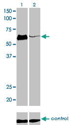
(SMAD3 monoclonal antibody. Western Blot analysis of SMAD3 expression in human colon.)
WB (Western Blot)
(SMAD3 monoclonal antibody. Western Blot analysis of SMAD3 expression in human colon.)
SMAD3, Monoclonal Antibody (Cat# AAA24658)
Full Name
SMAD3 (Mothers Against Decapentaplegic Homolog 3, SMAD 3, Mothers Against DPP Homolog 3, MAD Homolog 3, Mad3, hMAD-3, SMAD Family Member 3, JV15-2, hSMAD3, MADH3, DKFZp586N0721, DKFZp686J10186, HSPC193, HsT17436, MGC60396) APC
Gene Names
SMAD3; LDS3; LDS1C; MADH3; JV15-2; HSPC193; HsT17436
Reactivity
Human, Rat
Applications
Immunohistochemistry, Western Blot
Purity
Purified by Protein A Affinity Chromatography.
Pricing


 FCM (Flow Cytometry)
(Figure 7. Flow Cytometry analysis of HEPA1-6 cells using anti-Rad9b antibody (AAA19340).Overlay histogram showing HEPA1-6 cells stained with AAA19340 (Blue line). The cells were blocked with 10% normal goat serum. And then incubated with rabbit anti-Rad9b Antibody (AAA19340, 1μg/1x106 cells) for 30 min at 20 degree C. DyLight®488 conjugated goat anti-rabbit IgG (5-10μg/1x106 cells) was used as secondary antibody for 30 minutes at 20 degree C. Isotype control antibody (Green line) was rabbit IgG (1μg/1x106) used under the same conditions. Unlabelled sample (Red line) was also used as a control.)
FCM (Flow Cytometry)
(Figure 7. Flow Cytometry analysis of HEPA1-6 cells using anti-Rad9b antibody (AAA19340).Overlay histogram showing HEPA1-6 cells stained with AAA19340 (Blue line). The cells were blocked with 10% normal goat serum. And then incubated with rabbit anti-Rad9b Antibody (AAA19340, 1μg/1x106 cells) for 30 min at 20 degree C. DyLight®488 conjugated goat anti-rabbit IgG (5-10μg/1x106 cells) was used as secondary antibody for 30 minutes at 20 degree C. Isotype control antibody (Green line) was rabbit IgG (1μg/1x106) used under the same conditions. Unlabelled sample (Red line) was also used as a control.)
Rad9b, Polyclonal Antibody (Cat# AAA19340)
Full Name
Anti-Rad9b Antibody
Gene Names
Rad9b; BC021784; A630082N15Rik
Reactivity
Mouse, Rat
Applications
WB, IHC-P, FC/FACS/FCM, EIA
Purity
Immunogen affinity purified.
Pricing


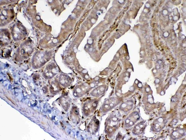
 IHC (Immunohistochemistry)
(Figure 7. IHC analysis of GALNS using anti-GALNS antibody (AAA19164).GALNS was detected in paraffin-embedded section of rat small intestine tissue. Heat mediated antigen retrieval was performed in citrate buffer (pH6, epitope retrieval solution) for 20 mins. The tissue section was blocked with 10% goat serum. The tissue section was then incubated with 1ug/ml rabbit anti-GALNS Antibody (AAA19164) overnight at 4 degree C. Biotinylated goat anti-rabbit IgG was used as secondary antibody and incubated for 30 minutes at 37 degree C. The tissue section was developed using Strepavidin-Biotin-Complex (SABC) with DAB as the chromogen.)
IHC (Immunohistochemistry)
(Figure 7. IHC analysis of GALNS using anti-GALNS antibody (AAA19164).GALNS was detected in paraffin-embedded section of rat small intestine tissue. Heat mediated antigen retrieval was performed in citrate buffer (pH6, epitope retrieval solution) for 20 mins. The tissue section was blocked with 10% goat serum. The tissue section was then incubated with 1ug/ml rabbit anti-GALNS Antibody (AAA19164) overnight at 4 degree C. Biotinylated goat anti-rabbit IgG was used as secondary antibody and incubated for 30 minutes at 37 degree C. The tissue section was developed using Strepavidin-Biotin-Complex (SABC) with DAB as the chromogen.)
GALNS, Antibody (Cat# AAA19164)
Full Name
Anti-GALNS Picoband antibody
Gene Names
GALNS; GAS; MPS4A; GalN6S; GALNAC6S
Reactivity
Reacts with: Human, Mouse, Rat
Applications
WB, EIA
Purity
Immunogen affinity purified
Pricing


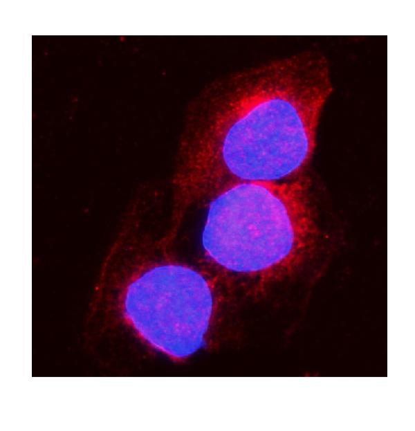
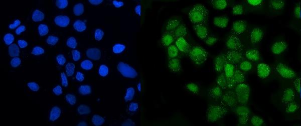
 IF (Immunofluorescence)
(Figure 6. IF analysis of UNG using anti-UNG antibody (AAA19245).UNG was detected in immunocytochemical section of MCF-7 cells. Enzyme antigen retrieval was performed using IHC enzyme antigen retrieval reagent for 15 mins. The cells were blocked with 10% goat serum. And then incubated with 5μg/mL rabbit anti- UNG Antibody (AAA19245) overnight at 4 degree C. DyLight®594 Conjugated Goat Anti-Rabbit IgG (BA1142) was used as secondary antibody at 1:100 dilution and incubated for 30 minutes at 37 degree C. The section was counterstained with DAPI. Visualize using a fluorescence microscope and filter sets appropriate for the label used.)
IF (Immunofluorescence)
(Figure 6. IF analysis of UNG using anti-UNG antibody (AAA19245).UNG was detected in immunocytochemical section of MCF-7 cells. Enzyme antigen retrieval was performed using IHC enzyme antigen retrieval reagent for 15 mins. The cells were blocked with 10% goat serum. And then incubated with 5μg/mL rabbit anti- UNG Antibody (AAA19245) overnight at 4 degree C. DyLight®594 Conjugated Goat Anti-Rabbit IgG (BA1142) was used as secondary antibody at 1:100 dilution and incubated for 30 minutes at 37 degree C. The section was counterstained with DAPI. Visualize using a fluorescence microscope and filter sets appropriate for the label used.)
UNG, Polyclonal Antibody (Cat# AAA19245)
Full Name
Anti-UNG Antibody
Gene Names
UNG; DGU; UDG; UNG1; UNG2; HIGM4; HIGM5; UNG15
Reactivity
Human, Mouse, Rat, Monkey
Applications
Western Blot, Immunohistochemistry, Immunocytochemistry, Immunofluorescence, Flow Cytometry, Direct ELISA
Purity
Immunogen affinity purified.
Pricing
 Standard Curve (Sample)
Cartilage Oligomeric Matrix Protein (COMP), Antibody Pair Kit (Cat# AAA21129)
Full Name
Rat Cartilage Oligomeric Matrix Protein (COMP) Antibody Pair Kit (with Standard)
Gene Names
COMP; MED; EDM1; EPD1; TSP5; PSACH; THBS5
Reactivity
Rat
Applications
EIA, CLIA
Pricing


 WB (Western Blot)
(HSPA1L monoclonal antibody Western Blot analysis of HSPA1L expression in PC-12.)
WB (Western Blot)
(HSPA1L monoclonal antibody Western Blot analysis of HSPA1L expression in PC-12.)
HSPA1L, Monoclonal Antibody (Cat# AAA25424)
Full Name
HSPA1L (Heat Shock 70kD Protein 1-like, HSP70-Hom, Heat shock 70kD protein 1L, Heat Shock 70kD Protein 1-Hom) (HRP)
Gene Names
HSPA1L; HSP70T; hum70t; HSP70-1L; HSP70-HOM
Reactivity
Human, Rat
Applications
Immunohistochemistry, Western Blot
Purity
Purified by Protein A Affinity Chromatography.
Pricing


 FCM (Flow Cytometry)
(Figure 8. Flow Cytometry analysis of U87 cells using anti-Transketolase/TKT antibody (AAA19254).Overlay histogram showing U87 cells stained with AAA19254 (Blue line). The cells were blocked with 10% normal goat serum. And then incubated with rabbit anti-Transketolase/TKT Antibody (AAA19254,1μg/1x106 cells) for 30 min at 20 degree C. DyLight®488 conjugated goat anti-rabbit IgG (5-10μg/1x106 cells) was used as secondary antibody for 30 minutes at 20 degree C. Isotype control antibody (Green line) was rabbit IgG (1μg/1x106) used under the same conditions. Unlabelled sample (Red line) was also used as a control.)
FCM (Flow Cytometry)
(Figure 8. Flow Cytometry analysis of U87 cells using anti-Transketolase/TKT antibody (AAA19254).Overlay histogram showing U87 cells stained with AAA19254 (Blue line). The cells were blocked with 10% normal goat serum. And then incubated with rabbit anti-Transketolase/TKT Antibody (AAA19254,1μg/1x106 cells) for 30 min at 20 degree C. DyLight®488 conjugated goat anti-rabbit IgG (5-10μg/1x106 cells) was used as secondary antibody for 30 minutes at 20 degree C. Isotype control antibody (Green line) was rabbit IgG (1μg/1x106) used under the same conditions. Unlabelled sample (Red line) was also used as a control.)
Transketolase/TKT, Polyclonal Antibody (Cat# AAA19254)
Full Name
Anti-Transketolase/TKT Antibody
Gene Names
TKT; TK; TKT1; HEL107
Reactivity
Human, Mouse, Rat
Applications
Western Blot, Immunohistochemistry, Immunocytochemistry, Immunofluorescence, Flow Cytometry, Direct ELISA
Purity
Immunogen affinity purified.
Pricing


 Application Data
(Proximity Ligation Analysis (PLA) of protein-protein interactions between PIK3R1 and SHC1 Huh7 cells were stained with PIK3R1 rabbit purified polyclonal 1:1200 and anti-SHC1 mouse monoclonal antibody 1:50. Signals were detected 30 Detection Kit 613 (red), and nuclei were counterstained with DAPI (blue). Each red dot represents the detection of protein-protein interaction complex.)
Application Data
(Proximity Ligation Analysis (PLA) of protein-protein interactions between PIK3R1 and SHC1 Huh7 cells were stained with PIK3R1 rabbit purified polyclonal 1:1200 and anti-SHC1 mouse monoclonal antibody 1:50. Signals were detected 30 Detection Kit 613 (red), and nuclei were counterstained with DAPI (blue). Each red dot represents the detection of protein-protein interaction complex.)
SHC, Monoclonal Antibody (Cat# AAA24950)
Full Name
SHC (SHC-transforming Protein 1, SH2 Domain Protein C1, SHC1, SHC-transforming Protein 3, SHC-transforming Protein A, SHCA, Src Homology 2 Domain-containing-transforming Protein C1, FLJ26504) (Biotin)
Gene Names
SHC1; SHC; SHCA
Reactivity
Human
Applications
Immunofluorescence, Immunohistochemistry, Western Blot
Purity
Purified by Protein A Affinity Chromatography.
Pricing


 WB (Western Blot)
(BUB1B monoclonal antibody Western Blot analysis of BUB1B expression in Hela NE.)
WB (Western Blot)
(BUB1B monoclonal antibody Western Blot analysis of BUB1B expression in Hela NE.)
BUBR1, Monoclonal Antibody (Cat# AAA24149)
Full Name
BUBR1 (Mitotic Checkpoint Serine/Threonine-protein Kinase BUB1 beta, MAD3/BUB1-related Protein Kinase, hBUBR1, Mitotic Checkpoint Kinase MAD3L, Protein SSK1, BUB1B, MAD3L, SSK1) (AP)
Gene Names
BUB1B; MVA1; SSK1; BUBR1; Bub1A; MAD3L; hBUBR1; BUB1beta
Reactivity
Human
Applications
Immunohistochemistry, Western Blot
Purity
Purified by Protein A Affinity Chromatography.
Pricing


 IHC (Immunohistchemistry)
(Figure 6. IHC analysis of Integrin beta 4/ITGB4 using anti-Integrin beta 4/ITGB4 antibody (AAA19269).Integrin beta 4/ITGB4 was detected in paraffin-embedded section of rat brain tissue. Heat mediated antigen retrieval was performed in EDTA buffer (pH8. 0, epitope retrieval solution). The tissue section was blocked with 10% goat serum. The tissue section was then incubated with 1μg/ml rabbit anti-Integrin beta 4/ITGB4 Antibody (AAA19269) overnight at 4 degree C. Biotinylated goat anti-rabbit IgG was used as secondary antibody and incubated for 30 minutes at 37 degree C. The tissue section was developed using Strepavidin-Biotin-Complex (SABC) (Catalog # with DAB as the chromogen.)
IHC (Immunohistchemistry)
(Figure 6. IHC analysis of Integrin beta 4/ITGB4 using anti-Integrin beta 4/ITGB4 antibody (AAA19269).Integrin beta 4/ITGB4 was detected in paraffin-embedded section of rat brain tissue. Heat mediated antigen retrieval was performed in EDTA buffer (pH8. 0, epitope retrieval solution). The tissue section was blocked with 10% goat serum. The tissue section was then incubated with 1μg/ml rabbit anti-Integrin beta 4/ITGB4 Antibody (AAA19269) overnight at 4 degree C. Biotinylated goat anti-rabbit IgG was used as secondary antibody and incubated for 30 minutes at 37 degree C. The tissue section was developed using Strepavidin-Biotin-Complex (SABC) (Catalog # with DAB as the chromogen.)
mGluR1/GRM1, Polyclonal Antibody (Cat# AAA19269)
Full Name
Anti-mGluR1/GRM1 Antibody
Gene Names
GRM1; MGLU1; GPRC1A; MGLUR1; SCAR13; PPP1R85
Reactivity
Human, Mouse, Rat
Applications
WB, IHC-P, FC/FACS/FCM, EIA
Purity
Immunogen affinity purified.
Pricing


 FCM (Flow Cytometry)
(Figure 8. Flow Cytometry analysis of HL-60 cells using anti-REA/PHB2 antibody (AAA19273).Overlay histogram showing HL-60 cells stained with AAA19273 (Blue line). The cells were blocked with 10% normal goat serum. And then incubated with rabbit anti-REA/PHB2 Antibody (AAA19273,1μg/1x106 cells) for 30 min at 20 degree C. DyLight®488 conjugated goat anti-rabbit IgG (5-10μg/1x106 cells) was used as secondary antibody for 30 minutes at 20 degree C. Isotype control antibody (Green line) was rabbit IgG (1μg/1x106) used under the same conditions. Unlabelled sample (Red line) was also used as a control.)
FCM (Flow Cytometry)
(Figure 8. Flow Cytometry analysis of HL-60 cells using anti-REA/PHB2 antibody (AAA19273).Overlay histogram showing HL-60 cells stained with AAA19273 (Blue line). The cells were blocked with 10% normal goat serum. And then incubated with rabbit anti-REA/PHB2 Antibody (AAA19273,1μg/1x106 cells) for 30 min at 20 degree C. DyLight®488 conjugated goat anti-rabbit IgG (5-10μg/1x106 cells) was used as secondary antibody for 30 minutes at 20 degree C. Isotype control antibody (Green line) was rabbit IgG (1μg/1x106) used under the same conditions. Unlabelled sample (Red line) was also used as a control.)
REA/PHB2, Polyclonal Antibody (Cat# AAA19273)
Full Name
Anti-REA/PHB2 Antibody
Gene Names
PHB2; BAP; REA; p22; Bap37; BCAP37; PNAS-141
Reactivity
Human, Mouse, Rat
Applications
WB, IHC-P, ICC, IF, FC/FACS/FCM, EIA
Purity
Immunogen affinity purified.
Pricing


 FCM (Flow Cytometry)
(Figure 9. Flow Cytometry analysis of MCF-7 cells using anti-Claudin 3/CLDN3 antibody (AAA19294).Overlay histogram showing MCF-7 cells stained with AAA19294 (Blue line). The cells were blocked with 10% normal goat serum. And then incubated with rabbit anti-Claudin 3/CLDN3 Antibody (AAA19294, 1μg/1x106 cells) for 30 min at 20 degree C. DyLight®488 conjugated goat anti-rabbit IgG (5-10μg/1x106 cells) was used as secondary antibody for 30 minutes at 20 degree C. Isotype control antibody (Green line) was rabbit IgG (1μg/1x106) used under the same conditions. Unlabelled sample (Red line) was also used as a control.)
FCM (Flow Cytometry)
(Figure 9. Flow Cytometry analysis of MCF-7 cells using anti-Claudin 3/CLDN3 antibody (AAA19294).Overlay histogram showing MCF-7 cells stained with AAA19294 (Blue line). The cells were blocked with 10% normal goat serum. And then incubated with rabbit anti-Claudin 3/CLDN3 Antibody (AAA19294, 1μg/1x106 cells) for 30 min at 20 degree C. DyLight®488 conjugated goat anti-rabbit IgG (5-10μg/1x106 cells) was used as secondary antibody for 30 minutes at 20 degree C. Isotype control antibody (Green line) was rabbit IgG (1μg/1x106) used under the same conditions. Unlabelled sample (Red line) was also used as a control.)
Claudin 3/CLDN3, Polyclonal Antibody (Cat# AAA19294)
Full Name
Anti-Claudin 3/CLDN3 Antibody
Gene Names
CLDN3; RVP1; HRVP1; C7orf1; CPE-R2; CPETR2
Reactivity
Human
Applications
WB, IHC-P, ICC, IF, FC/FACS/FCM, EIA
Purity
Immunogen affinity purified.
Pricing


 FCM (Flow Cytometry)
(Figure 9. Flow Cytometry analysis of HL-60 cells using anti-NDUFB10 antibody (AAA19339).Overlay histogram showing HL-60 cells stained with AAA19339 (Blue line). The cells were blocked with 10% normal goat serum. And then incubated with rabbit anti-NDUFB10 Antibody (AAA19339, 1μg/1x106 cells) for 30 min at 20 degree C. DyLight®488 conjugated goat anti-rabbit IgG (5-10μg/1x106 cells) was used as secondary antibody for 30 minutes at 20 degree C. Isotype control antibody (Green line) was rabbit IgG (1μg/1x106) used under the same conditions. Unlabelled sample (Red line) was also used as a control.)
FCM (Flow Cytometry)
(Figure 9. Flow Cytometry analysis of HL-60 cells using anti-NDUFB10 antibody (AAA19339).Overlay histogram showing HL-60 cells stained with AAA19339 (Blue line). The cells were blocked with 10% normal goat serum. And then incubated with rabbit anti-NDUFB10 Antibody (AAA19339, 1μg/1x106 cells) for 30 min at 20 degree C. DyLight®488 conjugated goat anti-rabbit IgG (5-10μg/1x106 cells) was used as secondary antibody for 30 minutes at 20 degree C. Isotype control antibody (Green line) was rabbit IgG (1μg/1x106) used under the same conditions. Unlabelled sample (Red line) was also used as a control.)
NDUFB10, Polyclonal Antibody (Cat# AAA19339)
Full Name
Anti-NDUFB10 Antibody
Gene Names
NDUFB10; PDSW
Reactivity
Human, Mouse, Rat
Applications
WB, IHC-P, ICC, IF, FC/FACS/FCM, EIA
Purity
Immunogen affinity purified.
Pricing
 Standard Curve (Sample)
Vascular Endothelial Growth Factor C (VEGF-C), ELISA Kit (Cat# AAA22438)
Full Name
Rabbit Vascular Endothelial Growth Factor C (VEGF-C) ELISA Kit
Gene Names
VEGFC; VRP; Flt4-L; LMPH1D
Reactivity
Rabbit
Pricing


 WB (Western Blot)
(F3 monoclonal antibody, Western Blot analysis of F3 expression in A-431.)
WB (Western Blot)
(F3 monoclonal antibody, Western Blot analysis of F3 expression in A-431.)
CD142, Monoclonal Antibody (Cat# AAA24160)
Full Name
CD142 (CD142 Antigen, Coagulation Factor III, F3, Tissue Factor, TF, TFA, Thromboplastin) (AP)
Gene Names
F3; TF; TFA; CD142
Reactivity
Human
Applications
Immunoprecipitation, Western Blot
Purity
Purified by Protein A Affinity Chromatography.
Pricing


 Application Data
(Proximity Ligation Analysis (PLA) of protein-protein interactions between CASP3 and HCLS1. HeLa cells were stained with anti-CASP3 rabbit purified polyclonal 1:1200 and anti-HCLS1 mouse monoclonal antibody 1:50. Signals were detected by Duolink® 30 Detection Kit 613 (red), and nuclei were counterstained with DAPI (blue). Each red dot represents the detection of protein-protein interaction complex.)
Application Data
(Proximity Ligation Analysis (PLA) of protein-protein interactions between CASP3 and HCLS1. HeLa cells were stained with anti-CASP3 rabbit purified polyclonal 1:1200 and anti-HCLS1 mouse monoclonal antibody 1:50. Signals were detected by Duolink® 30 Detection Kit 613 (red), and nuclei were counterstained with DAPI (blue). Each red dot represents the detection of protein-protein interaction complex.)
HCLS1, Monoclonal Antibody (Cat# AAA25711)
Full Name
HCLS1 (Hematopoietic Lineage Cell-specific Protein, Hematopoietic Cell-specific LYN Substrate 1, LckBP1, p75, HS1) (PE)
Gene Names
HCLS1; HS1; p75; CTTNL; lckBP1
Reactivity
Human
Applications
Immunohistochemistry, Western Blot
Purity
Purified by Protein A Affinity Chromatography.
Pricing


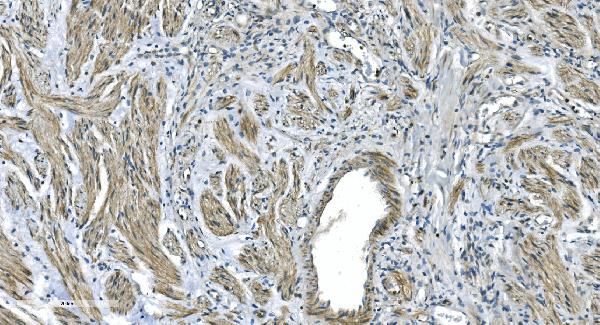


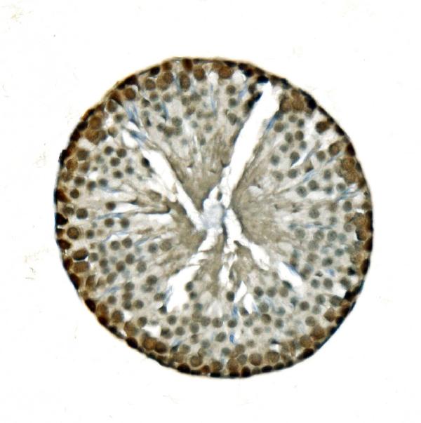
 IHC (Immunohistchemistry)
(Figure 5. IHC analysis of GNG4 using anti-GNG4 antibody (AAA19341).GNG4 was detected in paraffin-embedded section of human placenta tissue. Heat mediated antigen retrieval was performed in EDTA buffer (pH8. 0, epitope retrieval solution). The tissue section was blocked with 10% goat serum. The tissue section was then incubated with 2μg/ml rabbit anti-GNG4 Antibody (AAA19341) overnight at 4 degree C. Biotinylated goat anti-rabbit IgG was used as secondary antibody and incubated for 30 minutes at 37 degree C. The tissue section was developed using Strepavidin-Biotin-Complex (SABC) (Catalog # with DAB as the chromogen.)
IHC (Immunohistchemistry)
(Figure 5. IHC analysis of GNG4 using anti-GNG4 antibody (AAA19341).GNG4 was detected in paraffin-embedded section of human placenta tissue. Heat mediated antigen retrieval was performed in EDTA buffer (pH8. 0, epitope retrieval solution). The tissue section was blocked with 10% goat serum. The tissue section was then incubated with 2μg/ml rabbit anti-GNG4 Antibody (AAA19341) overnight at 4 degree C. Biotinylated goat anti-rabbit IgG was used as secondary antibody and incubated for 30 minutes at 37 degree C. The tissue section was developed using Strepavidin-Biotin-Complex (SABC) (Catalog # with DAB as the chromogen.)
GNG4, Polyclonal Antibody (Cat# AAA19341)
Full Name
Anti-GNG4 Antibody
Reactivity
Human, Mouse, Rat
Applications
WB, IHC-P, FC/FACS/FCM, EIA
Purity
Immunogen affinity purified.
Pricing


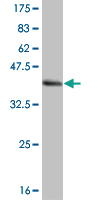
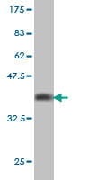
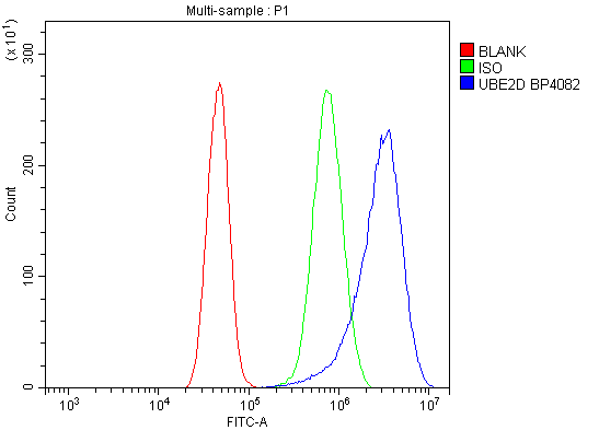 FCM (Flow Cytometry)
(Figure 7. Flow Cytometry analysis of A431 cells using anti-UBE2D1/2/3/4 antibody (AAA19298).Overlay histogram showing A431 cells stained with AAA19298 (Blue line). The cells were blocked with 10% normal goat serum. And then incubated with rabbit anti-UBE2D1/2/3/4 Antibody (AAA19298, 1μg/1x106 cells) for 30 min at 20 degree C. DyLight®488 conjugated goat anti-rabbit IgG (5-10μg/1x106 cells) was used as secondary antibody for 30 minutes at 20 degree C. Isotype control antibody (Green line) was rabbit IgG (1μg/1x106) used under the same conditions. Unlabelled sample (Red line) was also used as a control.)
FCM (Flow Cytometry)
(Figure 7. Flow Cytometry analysis of A431 cells using anti-UBE2D1/2/3/4 antibody (AAA19298).Overlay histogram showing A431 cells stained with AAA19298 (Blue line). The cells were blocked with 10% normal goat serum. And then incubated with rabbit anti-UBE2D1/2/3/4 Antibody (AAA19298, 1μg/1x106 cells) for 30 min at 20 degree C. DyLight®488 conjugated goat anti-rabbit IgG (5-10μg/1x106 cells) was used as secondary antibody for 30 minutes at 20 degree C. Isotype control antibody (Green line) was rabbit IgG (1μg/1x106) used under the same conditions. Unlabelled sample (Red line) was also used as a control.)
UBE2D1/2/3/4, Polyclonal Antibody (Cat# AAA19298)
Full Name
Anti-UBE2D1/2/3/4 Antibody
Gene Names
UBE2D1; SFT; UBCH5; UBC4/5; UBCH5A; E2(17)KB1
Reactivity
Human, Mouse, Rat
Applications
WB, IHC-P, ICC, IF, FC/FACS/FCM, EIA
Purity
Immunogen affinity purified.
Pricing
 Standard Curve (Sample)
Calcitonin (CT), ELISA Kit (Cat# AAA22399)
Full Name
Rabbit Calcitonin (CT) ELISA Kit
Gene Names
CALCA; CT; KC; CGRP; CALC1; CGRP1; CGRP-I
Reactivity
Rabbit
Pricing
 Standard Curve (Sample)
Receptor Activator Of Nuclear Factor Kappa B Ligand (RANkL), ELISA Kit (Cat# AAA22441)
Full Name
Rabbit Receptor Activator Of Nuclear Factor Kappa B Ligand (RANkL) ELISA Kit
Gene Names
TNFSF11; ODF; OPGL; sOdf; CD254; OPTB2; RANKL; TRANCE; hRANKL2
Reactivity
Rabbit
Pricing


 IHC (Immunohistchemistry)
(Figure 6. IHC analysis of DYNLT1 using anti-DYNLT1 antibody (AAA19175).DYNLT1 was detected in paraffin-embedded section of rat lung tissue. Heat mediated antigen retrieval was performed in citrate buffer (pH6, epitope retrieval solution) for 20 mins. The tissue section was blocked with 10% goat serum. The tissue section was then incubated with 1ug/ml rabbit anti-DYNLT1 Antibody (AAA19175) overnight at 4 degree C. Biotinylated goat anti-rabbit IgG was used as secondary antibody and incubated for 30 minutes at 37 degree C. The tissue section was developed using Strepavidin-Biotin-Complex (SABC) with DAB as the chromogen.)
IHC (Immunohistchemistry)
(Figure 6. IHC analysis of DYNLT1 using anti-DYNLT1 antibody (AAA19175).DYNLT1 was detected in paraffin-embedded section of rat lung tissue. Heat mediated antigen retrieval was performed in citrate buffer (pH6, epitope retrieval solution) for 20 mins. The tissue section was blocked with 10% goat serum. The tissue section was then incubated with 1ug/ml rabbit anti-DYNLT1 Antibody (AAA19175) overnight at 4 degree C. Biotinylated goat anti-rabbit IgG was used as secondary antibody and incubated for 30 minutes at 37 degree C. The tissue section was developed using Strepavidin-Biotin-Complex (SABC) with DAB as the chromogen.)
DYNLT1, Polyclonal Antibody (Cat# AAA19175)
Full Name
Anti-DYNLT1 Picoband antibody
Gene Names
DYNLT1; CW-1; TCTEL1; tctex-1
Reactivity
Human, Mouse, Rat
No cross reactivity with other proteins.
No cross reactivity with other proteins.
Applications
EIA, IHC, WB
Pricing
 Standard Curve (Sample)
Prostate specific antigen, ELISA Kit (Cat# AAA15993)
Full Name
Rabbit Prostate specific antigen ELISA Kit
Gene Names
KLK3; APS; PSA; hK3; KLK2A1
Reactivity
Rabbit
Pricing


 WB (Western Blot)
(BUB1B monoclonal antibody Western Blot analysis of BUB1B expression in Hela NE.)
WB (Western Blot)
(BUB1B monoclonal antibody Western Blot analysis of BUB1B expression in Hela NE.)
BUBR1, Monoclonal Antibody (Cat# AAA25332)
Full Name
BUBR1 (Mitotic Checkpoint Serine/Threonine-protein Kinase BUB1 beta, MAD3/BUB1-related Protein Kinase, hBUBR1, Mitotic Checkpoint Kinase MAD3L, Protein SSK1, BUB1B, MAD3L, SSK1) (HRP)
Gene Names
BUB1B; MVA1; SSK1; BUBR1; Bub1A; MAD3L; hBUBR1; BUB1beta
Reactivity
Human
Applications
EIA, IHC, WB
Purity
Purified by Protein A Affinity Chromatography.
Pricing


 FCM (Flow Cytometry)
(Figure 7. Flow Cytometry analysis of A549 cells using anti-AGO3 antibody (AAA19290).Overlay histogram showing A549 cells stained with AAA19290 (Blue line). The cells were blocked with 10% normal goat serum. And then incubated with rabbit anti-AGO3 Antibody (AAA19290, 1μg/1x106 cells) for 30 min at 20 degree C. DyLight®488 conjugated goat anti-rabbit IgG (5-10μg/1x106 cells) was used as secondary antibody for 30 minutes at 20 degree C. Isotype control antibody (Green line) was rabbit IgG (1μg/1x106) used under the same conditions. Unlabelled sample (Red line) was also used as a control.)
FCM (Flow Cytometry)
(Figure 7. Flow Cytometry analysis of A549 cells using anti-AGO3 antibody (AAA19290).Overlay histogram showing A549 cells stained with AAA19290 (Blue line). The cells were blocked with 10% normal goat serum. And then incubated with rabbit anti-AGO3 Antibody (AAA19290, 1μg/1x106 cells) for 30 min at 20 degree C. DyLight®488 conjugated goat anti-rabbit IgG (5-10μg/1x106 cells) was used as secondary antibody for 30 minutes at 20 degree C. Isotype control antibody (Green line) was rabbit IgG (1μg/1x106) used under the same conditions. Unlabelled sample (Red line) was also used as a control.)
AGO3, Polyclonal Antibody (Cat# AAA19290)
Full Name
Anti-AGO3 Antibody
Gene Names
AGO3; EIF2C3
Reactivity
Human
Applications
WB, IHC-P, ICC, IF, FC/FACS/FCM, EIA
Purity
Immunogen affinity purified.
Pricing


 FCM (Flow Cytometry)
(Figure 10. Flow Cytometry analysis of K562 cells using anti-HNRNPH3 antibody (AAA19338).Overlay histogram showing K562 cells stained with AAA19338 (Blue line). The cells were blocked with 10% normal goat serum. And then incubated with rabbit anti-HNRNPH3 Antibody (AAA19338, 1μg/1x106 cells) for 30 min at 20 degree C. DyLight®488 conjugated goat anti-rabbit IgG (5-10μg/1x106 cells) was used as secondary antibody for 30 minutes at 20 degree C. Isotype control antibody (Green line) was rabbit IgG (1μg/1x106) used under the same conditions. Unlabelled sample (Red line) was also used as a control.)
FCM (Flow Cytometry)
(Figure 10. Flow Cytometry analysis of K562 cells using anti-HNRNPH3 antibody (AAA19338).Overlay histogram showing K562 cells stained with AAA19338 (Blue line). The cells were blocked with 10% normal goat serum. And then incubated with rabbit anti-HNRNPH3 Antibody (AAA19338, 1μg/1x106 cells) for 30 min at 20 degree C. DyLight®488 conjugated goat anti-rabbit IgG (5-10μg/1x106 cells) was used as secondary antibody for 30 minutes at 20 degree C. Isotype control antibody (Green line) was rabbit IgG (1μg/1x106) used under the same conditions. Unlabelled sample (Red line) was also used as a control.)
HNRNPH3, Polyclonal Antibody (Cat# AAA19338)
Full Name
Anti-HNRNPH3 Antibody
Gene Names
HNRNPH3; 2H9; HNRPH3
Reactivity
Human, Mouse, Rat
Applications
WB, IHC-P, ICC, IF, FC/FACS/FCM, EIA
Purity
Immunogen affinity purified.
Pricing
 Standard Curve (Sample)
Creatine Kinase BB Isoenzymes, ELISA Kit (Cat# AAA22681)
Full Name
Rabbit Creatine Kinase BB Isoenzymes (CK-BB) ELISA Kit
Reactivity
Rabbit
Pricing
 Standard Curve (Sample)
Triiodothyronine, ELISA Kit (Cat# AAA16524)
Full Name
Rabbit Triiodothyronine ELISA Kit
Reactivity
Rabbit
Pricing


 Application Data
(Proximity Ligation Analysis (PLA) of protein-protein interactions between MAPK3 and RPS6KA2 HeLa cells were stained with anti-MAPK3 rabbit purified polyclonal 1:1200 and anti-RPS6KA2 mouse monoclonal antibody 1:50. Signals were detected by 30 Detection Kit 613 (red), and nuclei were counterstained with DAPI (blue). Each red dot represents the detection of protein-protein interaction complex.)
Application Data
(Proximity Ligation Analysis (PLA) of protein-protein interactions between MAPK3 and RPS6KA2 HeLa cells were stained with anti-MAPK3 rabbit purified polyclonal 1:1200 and anti-RPS6KA2 mouse monoclonal antibody 1:50. Signals were detected by 30 Detection Kit 613 (red), and nuclei were counterstained with DAPI (blue). Each red dot represents the detection of protein-protein interaction complex.)
RSK3, Monoclonal Antibody (Cat# AAA25235)
Full Name
RSK3, CT (Ribosomal S6 Kinase 3, RSK-3, pp90RSK3, 90kD Ribosomal Protein S6 Kinase 2, p90-RSK 2, p90RSK2, MAP Kinase-activated Protein Kinase 1c, MAPK-activated Protein Kinase 1c, MAPKAP Kinase 1c, MAPKAPK1C, MAPKAPK-1c, Ribosomal Protein S6 Kinase alpha-
Gene Names
RPS6KA2; RSK; HU-2; RSK3; p90RSK2; p90-RSK3; pp90RSK3; MAPKAPK1C; S6K-alpha; S6K-alpha2
Reactivity
Human
Applications
Immunofluorescence, Immunohistochemistry, Western Blot
Purity
Purified by Protein A Affinity Chromatography.
Pricing
 Standard Curve (Sample)
Bone Morphogenetic Protein 2 (BMP-2), ELISA Kit (Cat# AAA22416)
Full Name
Rabbit Bone Morphogenetic Protein 2 (BMP-2) ELISA Kit
Gene Names
BMP2; BDA2; BMP2A
Reactivity
Rabbit
Pricing
 Standard Curve (Sample)
Activated protein C, ELISA Kit (Cat# AAA16309)
Full Name
Rabbit Activated protein C ELISA Kit
Gene Names
APC; GS; DP2; DP3; BTPS2; DP2.5; PPP1R46
Reactivity
Rabbit
Pricing


 WB (Western Blot)
(SMAD4 monoclonal antibody. Western Blot analysis of SMAD4 expression in IMR-32.)
WB (Western Blot)
(SMAD4 monoclonal antibody. Western Blot analysis of SMAD4 expression in IMR-32.)
SMAD4, Monoclonal Antibody (Cat# AAA24955)
Full Name
SMAD4 (Mothers Against Decapentaplegic Homolog 4, SMAD 4, Mothers Against DPP Homolog 4, Deletion Target In Pancreatic Carcinoma 4, MAD Homolog 4, SMAD Family Member 4, hSMAD4, DPC4, MADH4) (Biotin)
Gene Names
SMAD4; JIP; DPC4; MADH4; MYHRS
Reactivity
Human
Applications
Immunofluorescence, Western Blot
Purity
Purified by Protein A Affinity Chromatography.
Pricing


 FCM (Flow Cytometry)
(Figure 6. Flow Cytometry analysis of RAW264. 7 cells using anti-TRIM25/EFP antibody (AAA19272).Overlay histogram showing RAW264. 7 cells stained with AAA19272 (Blue line). The cells were blocked with 10% normal goat serum. And then incubated with rabbit anti-TRIM25/EFP Antibody (AAA19272, 1μg/1x106 cells) for 30 min at 20 degree C. DyLight®488 conjugated goat anti-rabbit IgG (5-10μg/1x106 cells) was used as secondary antibody for 30 minutes at 20 degree C. Isotype control antibody (Green line) was rabbit IgG (1μg/1x106) used under the same conditions. Unlabelled sample (Red line) was also used as a control.)
FCM (Flow Cytometry)
(Figure 6. Flow Cytometry analysis of RAW264. 7 cells using anti-TRIM25/EFP antibody (AAA19272).Overlay histogram showing RAW264. 7 cells stained with AAA19272 (Blue line). The cells were blocked with 10% normal goat serum. And then incubated with rabbit anti-TRIM25/EFP Antibody (AAA19272, 1μg/1x106 cells) for 30 min at 20 degree C. DyLight®488 conjugated goat anti-rabbit IgG (5-10μg/1x106 cells) was used as secondary antibody for 30 minutes at 20 degree C. Isotype control antibody (Green line) was rabbit IgG (1μg/1x106) used under the same conditions. Unlabelled sample (Red line) was also used as a control.)
TRIM25/EFP, Polyclonal Antibody (Cat# AAA19272)
Full Name
Anti-TRIM25/EFP Antibody
Gene Names
Trim25; EFP; Zfp147; AA960166; AL022677
Reactivity
Mouse, Rat
Applications
WB, IHC-P, ICC, IF, FC/FACS/FCM, EIA
Purity
Immunogen affinity purified.
Pricing
 Standard Curve (Sample)
Creatine Kinase MB Isoenzyme (CKMB), ELISA Kit (Cat# AAA22475)
Full Name
Rabbit Creatine Kinase MB Isoenzyme (CKMB) ELISA Kit
Gene Names
CKM; CKMM; M-CK
Reactivity
Rabbit
Pricing


 WB (Western Blot)
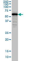
(Western Blot analysis of B2M expression in transfected 293T cell line by B2M monoclonal antibody. Lane 1: B2M transfected lysate (13.7kD). Lane 2: Non-transfected lysate.)
WB (Western Blot)
(Western Blot analysis of B2M expression in transfected 293T cell line by B2M monoclonal antibody. Lane 1: B2M transfected lysate (13.7kD). Lane 2: Non-transfected lysate.)
b2-Microglobulin, Monoclonal Antibody (Cat# AAA14785)
Full Name
b2-Microglobulin (B2M, Beta-2-microglobulin, CDABP0092, HDCMA22P)
Gene Names
HLA-G; MHC-G
Reactivity
Human
Applications
EL/EIA, WB, IHC, IF
Purity
Affinity Purified
Purified by Protein A affinity chromatography.
Purified by Protein A affinity chromatography.
Pricing


 FCM (Flow Cytometry)
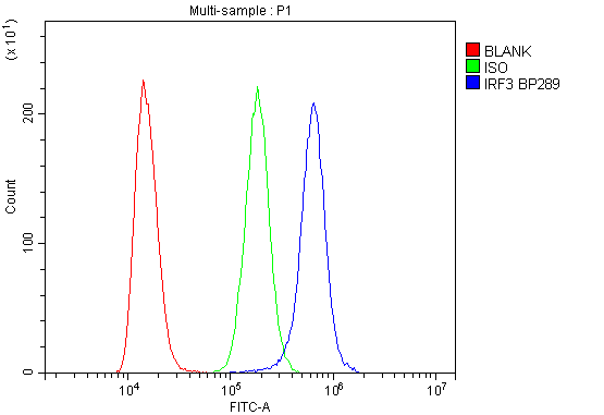
(Figure 6. Flow Cytometry analysis of K562 cells using anti-IRF3 antibody (AAA19216).Overlay histogram showing K562 cells stained with AAA19216 (Blue line). The cells were blocked with 10% normal goat serum. And then incubated with rabbit anti-IRF3 Antibody (AAA19216, 1μg/1x106 cells) for 30 min at 20 degree C. DyLight®488 conjugated goat anti-rabbit IgG (5-10μg/1x106 cells) was used as secondary antibody for 30 minutes at 20 degree C. Isotype control antibody (Green line) was rabbit IgG (1μg/1x106) used under the same conditions. Unlabelled sample (Red line) was also used as a control.)
FCM (Flow Cytometry)
(Figure 6. Flow Cytometry analysis of K562 cells using anti-IRF3 antibody (AAA19216).Overlay histogram showing K562 cells stained with AAA19216 (Blue line). The cells were blocked with 10% normal goat serum. And then incubated with rabbit anti-IRF3 Antibody (AAA19216, 1μg/1x106 cells) for 30 min at 20 degree C. DyLight®488 conjugated goat anti-rabbit IgG (5-10μg/1x106 cells) was used as secondary antibody for 30 minutes at 20 degree C. Isotype control antibody (Green line) was rabbit IgG (1μg/1x106) used under the same conditions. Unlabelled sample (Red line) was also used as a control.)
IRF3, Polyclonal Antibody (Cat# AAA19216)
Full Name
Anti-IRF3 Antibody
Reactivity
Human
Applications
Western Blot, Immunohistochemistry, Flow Cytometry, Direct ELISA
Purity
Immunogen affinity purified.
Pricing


 FCM (Flow Cytometry)
(Figure 7. Flow Cytometry analysis of SiHa cells using anti-Aconitase 1/ACO1 antibody (AAA19267).Overlay histogram showing SiHa cells stained with AAA19267 (Blue line). The cells were blocked with 10% normal goat serum. And then incubated with rabbit anti-Aconitase 1/ACO1 Antibody (AAA19267,1μg/1x106 cells) for 30 min at 20 degree C. DyLight®488 conjugated goat anti-rabbit IgG (5-10μg/1x106 cells) was used as secondary antibody for 30 minutes at 20 degree C. Isotype control antibody (Green line) was rabbit IgG (1μg/1x106) used under the same conditions. Unlabelled sample (Red line) was also used as a control.)
FCM (Flow Cytometry)
(Figure 7. Flow Cytometry analysis of SiHa cells using anti-Aconitase 1/ACO1 antibody (AAA19267).Overlay histogram showing SiHa cells stained with AAA19267 (Blue line). The cells were blocked with 10% normal goat serum. And then incubated with rabbit anti-Aconitase 1/ACO1 Antibody (AAA19267,1μg/1x106 cells) for 30 min at 20 degree C. DyLight®488 conjugated goat anti-rabbit IgG (5-10μg/1x106 cells) was used as secondary antibody for 30 minutes at 20 degree C. Isotype control antibody (Green line) was rabbit IgG (1μg/1x106) used under the same conditions. Unlabelled sample (Red line) was also used as a control.)
Aconitase 1/ACO1, Polyclonal Antibody (Cat# AAA19267)
Full Name
Anti-Aconitase 1/ACO1 Antibody
Gene Names
ACO1; IRP1; ACONS; IREB1; IREBP; IREBP1
Reactivity
Human, Mouse, Monkey, Rat
Applications
WB, IHC-P, ICC, IF, FC/FACS/FCM, EIA
Purity
Immunogen affinity purified.
Pricing


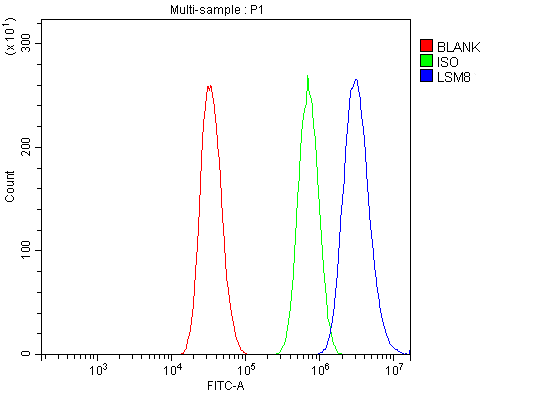 FCM (Flow Cytometry)
(Figure 13. Flow Cytometry analysis of A431 cells using anti-LSM8 antibody (AAA19337).Overlay histogram showing A431 cells stained with AAA19337 (Blue line). The cells were blocked with 10% normal goat serum. And then incubated with rabbit anti-LSM8 Antibody (AAA19337, 1μg/1x106 cells) for 30 min at 20 degree C. DyLight®488 conjugated goat anti-rabbit IgG (5-10μg/1x106 cells) was used as secondary antibody for 30 minutes at 20 degree C. Isotype control antibody (Green line) was rabbit IgG (1μg/1x106) used under the same conditions. Unlabelled sample (Red line) was also used as a control.)
FCM (Flow Cytometry)
(Figure 13. Flow Cytometry analysis of A431 cells using anti-LSM8 antibody (AAA19337).Overlay histogram showing A431 cells stained with AAA19337 (Blue line). The cells were blocked with 10% normal goat serum. And then incubated with rabbit anti-LSM8 Antibody (AAA19337, 1μg/1x106 cells) for 30 min at 20 degree C. DyLight®488 conjugated goat anti-rabbit IgG (5-10μg/1x106 cells) was used as secondary antibody for 30 minutes at 20 degree C. Isotype control antibody (Green line) was rabbit IgG (1μg/1x106) used under the same conditions. Unlabelled sample (Red line) was also used as a control.)
LSM8, Polyclonal Antibody (Cat# AAA19337)
Full Name
Anti-LSM8 Antibody
Reactivity
Human, Mouse, Rat
Applications
WB, IHC-P, ICC, IF, FC/FACS/FCM, EIA
Purity
Immunogen affinity purified.
Pricing
 Standard Curve (Sample)
thrombin-antithrombin complex (TAT), ELISA Kit (Cat# AAA18285)
Full Name
Rabbit thrombin-antithrombin complex, TAT ELISA Kit
Reactivity
Rabbit
Pricing
 Standard Curve (Sample)
Prekallikrein (PK), ELISA Kit (Cat# AAA22359)
Full Name
Rabbit Prekallikrein (PK) ELISA Kit
Gene Names
KLKB1; PPK; KLK3
Reactivity
Rabbit
Pricing


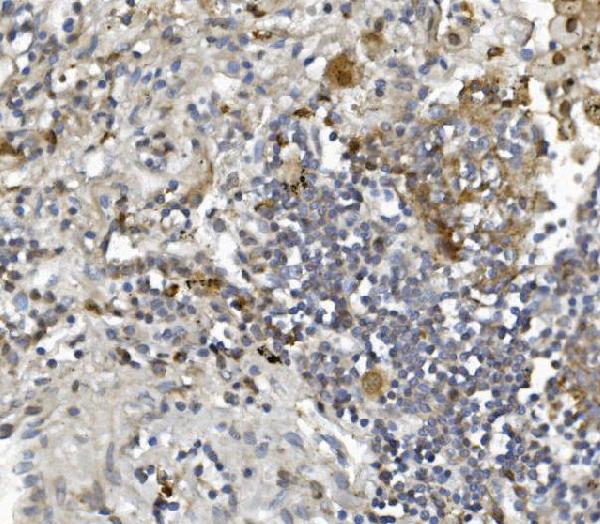
 IHC (Immunohistchemistry)
(Figure 6. IHC analysis of Flt3 / CD135 using anti-Flt3 / CD135 antibody (AAA19130).Flt3 / CD135 was detected in paraffin-embedded section of mouse kidney tissue. Heat mediated antigen retrieval was performed in citrate buffer (pH6, epitope retrieval solution) for 20 mins. The tissue section was blocked with 10% goat serum. The tissue section was then incubated with 1ug/ml rabbit anti-Flt3 / CD135 Antibody (AAA19130) overnight at 4 degree C. Biotinylated goat anti-rabbit IgG was used as secondary antibody and incubated for 30 minutes at 37 degree C. The tissue section was developed using Strepavidin-Biotin-Complex (SABC) with DAB as the chromogen.)
IHC (Immunohistchemistry)
(Figure 6. IHC analysis of Flt3 / CD135 using anti-Flt3 / CD135 antibody (AAA19130).Flt3 / CD135 was detected in paraffin-embedded section of mouse kidney tissue. Heat mediated antigen retrieval was performed in citrate buffer (pH6, epitope retrieval solution) for 20 mins. The tissue section was blocked with 10% goat serum. The tissue section was then incubated with 1ug/ml rabbit anti-Flt3 / CD135 Antibody (AAA19130) overnight at 4 degree C. Biotinylated goat anti-rabbit IgG was used as secondary antibody and incubated for 30 minutes at 37 degree C. The tissue section was developed using Strepavidin-Biotin-Complex (SABC) with DAB as the chromogen.)
Flt3/CD135, Polyclonal Antibody (Cat# AAA19130)
Full Name
Anti-Flt3/CD135 Picoband Antibody
Reactivity
Mouse, Rat
No cross reactivity with other proteins.
No cross reactivity with other proteins.
Applications
EIA, IHC, WB
Purity
Immunogen affinity purified
Pricing


 FCM (Flow Cytometry)
(Figure 6. Flow Cytometry analysis of PC-3 cells using anti-MitoNEET/CISD1 antibody (AAA19293).Overlay histogram showing PC-3 cells stained with AAA19293 (Blue line). The cells were blocked with 10% normal goat serum. And then incubated with rabbit anti-MitoNEET/CISD1 Antibody (AAA19293,1μg/1x106 cells) for 30 min at 20 degree C. DyLight®488 conjugated goat anti-rabbit IgG (5-10μg/1x106 cells) was used as secondary antibody for 30 minutes at 20 degree C. Isotype control antibody (Green line) was rabbit IgG (1μg/1x106) used under the same conditions. Unlabelled sample (Red line) was also used as a control.)
FCM (Flow Cytometry)
(Figure 6. Flow Cytometry analysis of PC-3 cells using anti-MitoNEET/CISD1 antibody (AAA19293).Overlay histogram showing PC-3 cells stained with AAA19293 (Blue line). The cells were blocked with 10% normal goat serum. And then incubated with rabbit anti-MitoNEET/CISD1 Antibody (AAA19293,1μg/1x106 cells) for 30 min at 20 degree C. DyLight®488 conjugated goat anti-rabbit IgG (5-10μg/1x106 cells) was used as secondary antibody for 30 minutes at 20 degree C. Isotype control antibody (Green line) was rabbit IgG (1μg/1x106) used under the same conditions. Unlabelled sample (Red line) was also used as a control.)
MitoNEET/CISD1, Polyclonal Antibody (Cat# AAA19293)
Full Name
Anti-MitoNEET/CISD1 Antibody
Gene Names
CISD1; ZCD1; MDS029; C10orf70; mitoNEET
Reactivity
Human, Mouse, Rat, Monkey
Applications
WB, IHC-P, FC/FACS/FCM, EIA
Purity
Immunogen affinity purified.
Pricing


 WB (Western Blot)
(CDK6 monoclonal antibody, Western Blot analysis of CDK6 expression in Jurkat.)
WB (Western Blot)
(CDK6 monoclonal antibody, Western Blot analysis of CDK6 expression in Jurkat.)
CDK6, Monoclonal Antibody (Cat# AAA24166)
Full Name
CDK6 (Cell Division Protein Kinase 6, Serine/Threonine-protein Kinase PLSTIRE, Crk2, MGC59692) (AP)
Gene Names
CDK6; MCPH12; PLSTIRE
Reactivity
Human, Rat
Applications
Immunohistochemistry, Western Blot
Purity
Purified by Protein A Affinity Chromatography.
Pricing


 Application Data
(Proximity Ligation Analysis (PLA) of protein-protein interactions between MAPK3 and RPS6KA2 HeLa cells were stained with anti-MAPK3 rabbit purified polyclonal 1:1200 and anti-RPS6KA2 mouse monoclonal antibody 1:50. Signals were detected by 30 Detection Kit 613 (red), and nuclei were counterstained with DAPI (blue). Each red dot represents the detection of protein-protein interaction complex.)
Application Data
(Proximity Ligation Analysis (PLA) of protein-protein interactions between MAPK3 and RPS6KA2 HeLa cells were stained with anti-MAPK3 rabbit purified polyclonal 1:1200 and anti-RPS6KA2 mouse monoclonal antibody 1:50. Signals were detected by 30 Detection Kit 613 (red), and nuclei were counterstained with DAPI (blue). Each red dot represents the detection of protein-protein interaction complex.)
RSK3, Monoclonal Antibody (Cat# AAA24939)
Full Name
RSK3, CT (Ribosomal S6 Kinase 3, RSK-3, pp90RSK3, 90kD Ribosomal Protein S6 Kinase 2, p90-RSK 2, p90RSK2, MAP Kinase-activated Protein Kinase 1c, MAPK-activated Protein Kinase 1c, MAPKAP Kinase 1c, MAPKAPK1C, MAPKAPK-1c, Ribosomal Protein S6 Kinase alpha-
Gene Names
RPS6KA2; RSK; HU-2; RSK3; p90RSK2; p90-RSK3; pp90RSK3; MAPKAPK1C; S6K-alpha; S6K-alpha2
Reactivity
Human
Applications
Immunofluorescence, Immunohistochemistry, Western Blot
Purity
Purified by Protein A Affinity Chromatography.
Pricing