Filters
Clonality
Type
Reactivity
Gene Name
Isotype
Host
Application
Clone
150 results for " cell lysate or tissue lysate" - showing 100-150


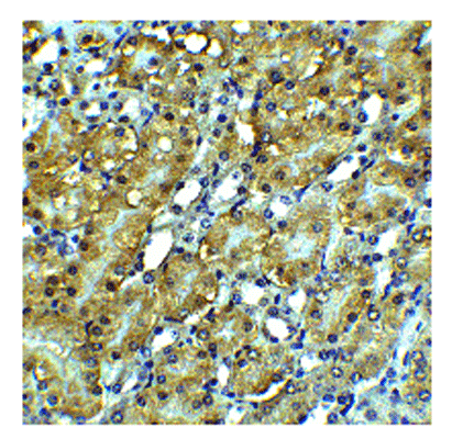
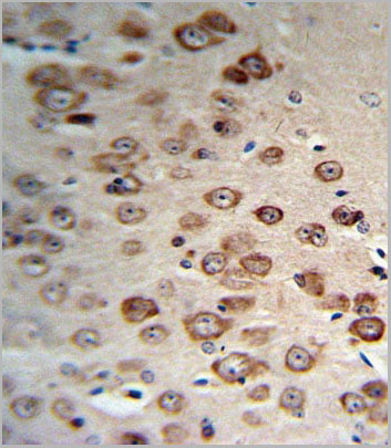
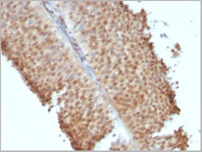
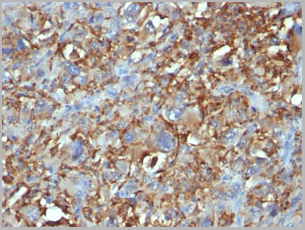
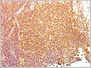
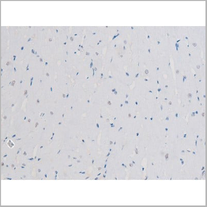
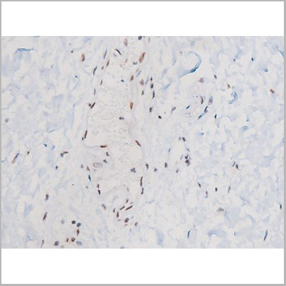
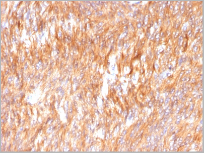
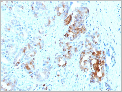
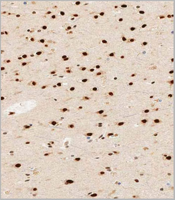
 IHC (Immunohistchemistry)
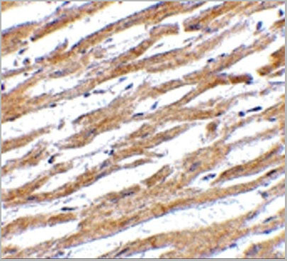
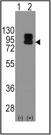
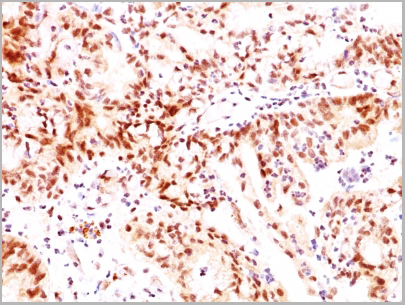
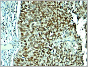
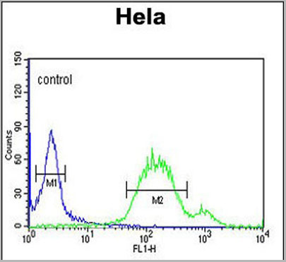
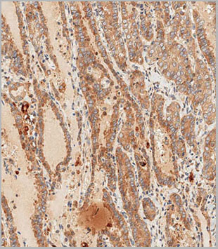
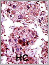
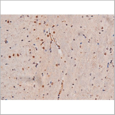
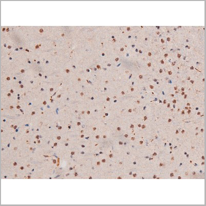
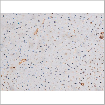
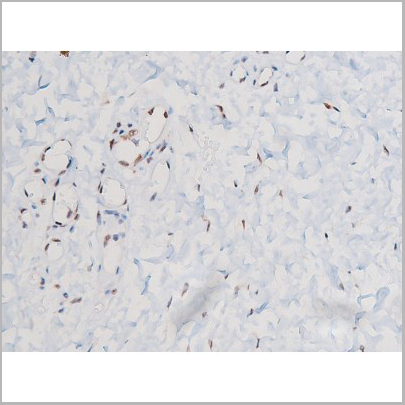
(PINK1 Monoclonal Antibody immunohistochemistry analysis in formalin fixed and paraffin embedded human kidney tissue followed by peroxidase conjugation of the secondary antibody and DAB staining. This data demonstrates the use of the PINK1 Monoclonal Antibody for immunohistochemistry. Clinical relevance has not been evaluated.)
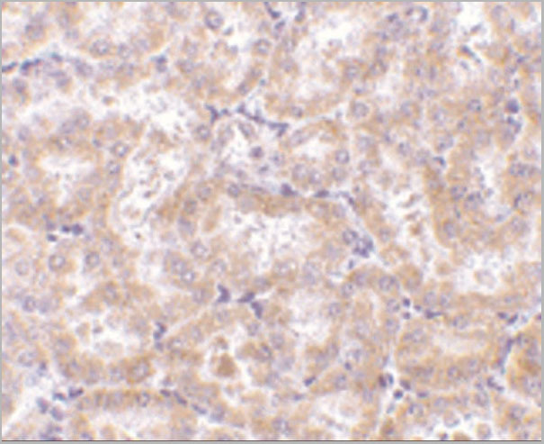
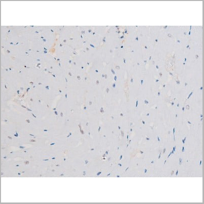
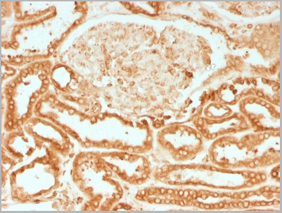
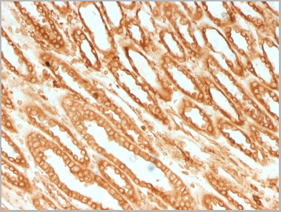
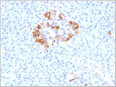
IHC (Immunohistchemistry)
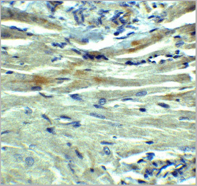
(PINK1 Monoclonal Antibody immunohistochemistry analysis in formalin fixed and paraffin embedded human kidney tissue followed by peroxidase conjugation of the secondary antibody and DAB staining. This data demonstrates the use of the PINK1 Monoclonal Antibody for immunohistochemistry. Clinical relevance has not been evaluated.)
PINK1, Monoclonal Antibody (Cat# AAA28638)
Full Name
PINK1 Antibody
Gene Names
PINK1; BRPK; PARK6
Reactivity
Human
Applications
EIA, IHC, IF, WB
Purity
Purified Mouse Monoclonal Antibody (Mab)
Pricing


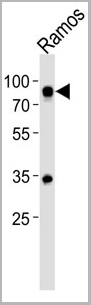
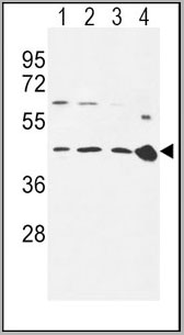
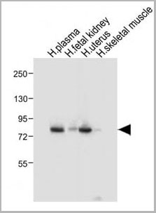
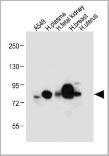
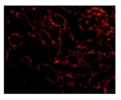
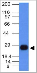
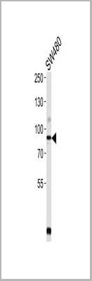
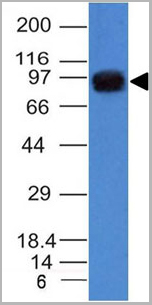
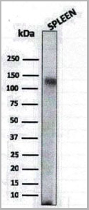
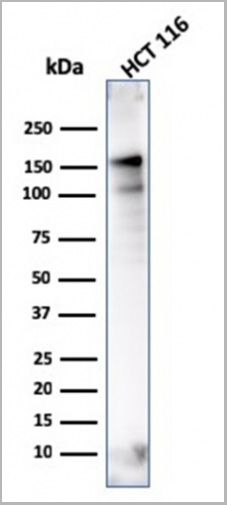
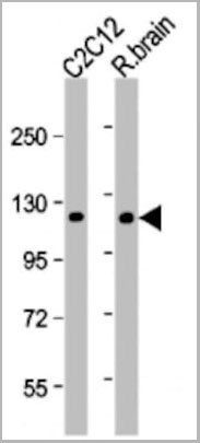
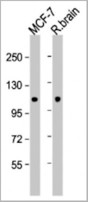
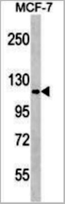
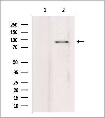
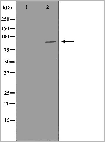
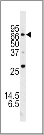
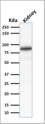
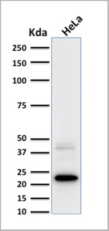
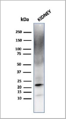
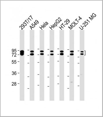
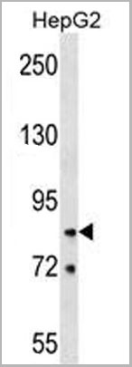
 WB (Western Blot)
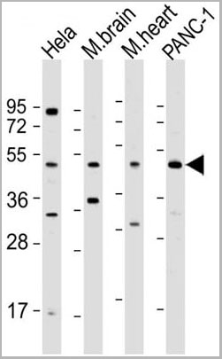
(Western Blot Analysis of human liver tissue lysate using Prohibitin Mouse Monoclonal Antibody (PHB/3225).)
WB (Western Blot)
(Western Blot Analysis of human liver tissue lysate using Prohibitin Mouse Monoclonal Antibody (PHB/3225).)
Prohibitin, Monoclonal Antibody (Cat# AAA23933)
Full Name
Prohibitin (Mitochondrial Marker)
Gene Names
PHB; PHB1; HEL-215; HEL-S-54e
Reactivity
Human
Applications
WB, IF, IHC
Purity
Purified Ab with BSA and Azide at 200ug/ml OR Purified Ab WITHOUT BSA and Azide at 1.0mg/ml
Pricing


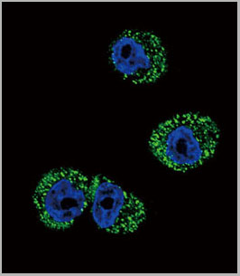
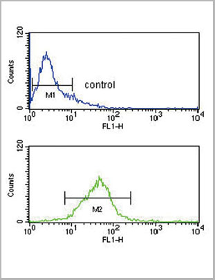
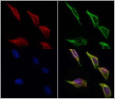
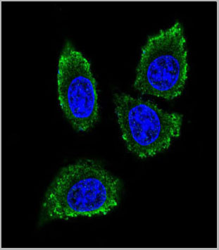
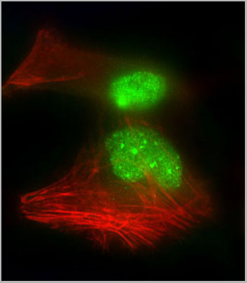
 IF (Immunofluorescence)
MSH2, Monoclonal Antibody (Cat# AAA23925)
Full Name
MSH2 (DNA Mismatch Repair Marker)
Gene Names
MSH2; FCC1; COCA1; HNPCC; LCFS2; hMSH2; HNPCC1
Reactivity
Human
Applications
FC, WB, IHC
Purity
Purified
Pricing


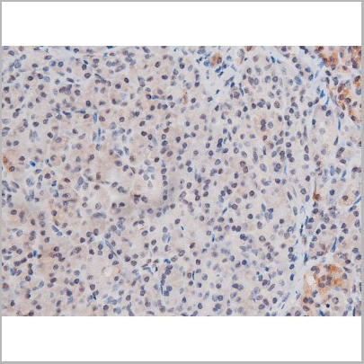
 Application Data
(Analysis of Protein Array containing more than 19,000 full-length human proteins using Cytokeratin 15 Mouse Monoclonal Antibody (KRT15/2957). Z- and S- Score: The Z-score represents the strength of a signal that a monoclonal antibody (MAb) (in combination with a fluorescently-tagged anti-IgG secondary antibody) produces when binding to a particular protein on the HuProtTM array. Z-scores are described in units of standard deviations (SD's) above the mean value of all signals generated on that array. If targets on HuProtTM are arranged in descending order of the Z-score, the S-score is the difference (also in units of SD's) between the Z-score. S-score therefore represents the relative target specificity of a MAb to its intended target. A MAb is considered to specific to its intended target, if the MAb has an S-score of at least 2.5. For example, if a MAb binds to protein X with a Z-score of 43 and to protein Y with a Z-score of 14, then the S-score for the binding of that MAb to protein X is equal to 29.)
Application Data
(Analysis of Protein Array containing more than 19,000 full-length human proteins using Cytokeratin 15 Mouse Monoclonal Antibody (KRT15/2957). Z- and S- Score: The Z-score represents the strength of a signal that a monoclonal antibody (MAb) (in combination with a fluorescently-tagged anti-IgG secondary antibody) produces when binding to a particular protein on the HuProtTM array. Z-scores are described in units of standard deviations (SD's) above the mean value of all signals generated on that array. If targets on HuProtTM are arranged in descending order of the Z-score, the S-score is the difference (also in units of SD's) between the Z-score. S-score therefore represents the relative target specificity of a MAb to its intended target. A MAb is considered to specific to its intended target, if the MAb has an S-score of at least 2.5. For example, if a MAb binds to protein X with a Z-score of 43 and to protein Y with a Z-score of 14, then the S-score for the binding of that MAb to protein X is equal to 29.)
Cytokeratin 15, Monoclonal Antibody (Cat# AAA23923)
Full Name
Cytokeratin 15 (Esophageal Squamous Cell Carcinoma Marker)
Gene Names
KRT15; K15; CK15; K1CO
Reactivity
Human
Applications
FC/FACS, IF, WB, IHC
Purity
Purified Ab with BSA and Azide at 200ug/ml OR Purified Ab WITHOUT BSA and Azide at 1.0mg/ml
Pricing


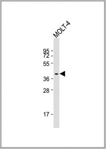
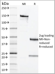
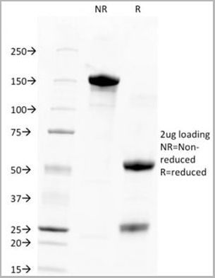
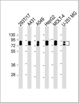
 SDS-PAGE
(SDS-PAGE Analysis Purified Cytokeratin-7 Mouse Monoclonal Antibody (KRT7/2200). Confirmation of Integrity and Purity of Antibody.)
SDS-PAGE
(SDS-PAGE Analysis Purified Cytokeratin-7 Mouse Monoclonal Antibody (KRT7/2200). Confirmation of Integrity and Purity of Antibody.)
Cytokeratin 7, Monoclonal Antibody (Cat# AAA23922)
Full Name
Cytokeratin 7 (Glandular and Transitional Epithelial Marker)
Gene Names
KRT7; K7; CK7; SCL; K2C7
Reactivity
Human
Applications
WB, IHC
Purity
Purified Ab with BSA and Azide at 200ug/ml OR Purified Ab WITHOUT BSA and Azide at 1.0mg/ml
Pricing


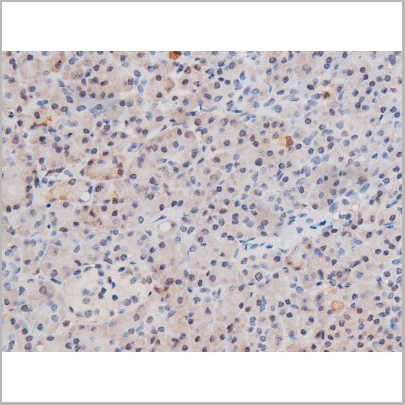
 Application Data
(Analysis of Protein Array containing more than 19,000 full-length human proteins using CAIX-Monospecific Mouse Monoclonal Antibody (CA9/3406). Z- and S- Score: The Z-score represents the strength of a signal that a monoclonal antibody (MAb) (in combination with a fluorescently-tagged anti-IgG secondary antibody) produces when binding to a particular protein on the HuProtTM array. Z-scores are described in units of standard deviations (SD's) above the mean value of all signals generated on that array. If targets on HuProtTM are arranged in descending order of the Z-score, the S-score is the difference (also in units of SD's) between the Z-score. S-score therefore represents the relative target specificity of a MAb to its intended target. A MAb is considered to specific to its intended target, if the MAb has an S-score of at least 2.5. For example, if a MAb binds to protein X with a Z-score of 43 and to protein Y with a Z-score of 14, then the S-score for the binding of that MAb to protein X is equal to 29.)
Application Data
(Analysis of Protein Array containing more than 19,000 full-length human proteins using CAIX-Monospecific Mouse Monoclonal Antibody (CA9/3406). Z- and S- Score: The Z-score represents the strength of a signal that a monoclonal antibody (MAb) (in combination with a fluorescently-tagged anti-IgG secondary antibody) produces when binding to a particular protein on the HuProtTM array. Z-scores are described in units of standard deviations (SD's) above the mean value of all signals generated on that array. If targets on HuProtTM are arranged in descending order of the Z-score, the S-score is the difference (also in units of SD's) between the Z-score. S-score therefore represents the relative target specificity of a MAb to its intended target. A MAb is considered to specific to its intended target, if the MAb has an S-score of at least 2.5. For example, if a MAb binds to protein X with a Z-score of 43 and to protein Y with a Z-score of 14, then the S-score for the binding of that MAb to protein X is equal to 29.)
Renal Cell Carcinoma, Monoclonal Antibody (Cat# AAA23952)
Full Name
Renal Cell Carcinoma (Carbonic Anhydrase IX)
Gene Names
CA9; MN; CAIX
Reactivity
Human
Applications
FC/FACS, IHC, WB
Purity
Purified Ab with BSA and Azide at 200ug/ml or Purified Ab with BSA and Azide at 200ug/ml or Purified Ab WITHOUT BSA and Azide at 1.0mg/ml
Pricing
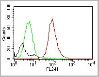
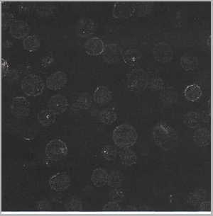
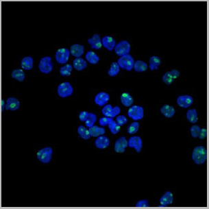
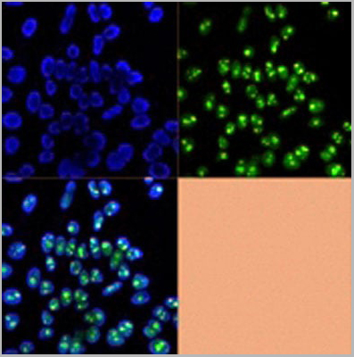
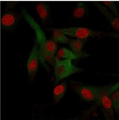
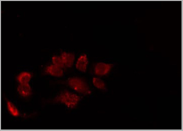
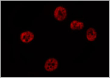
 IF (Immunofluorescence)
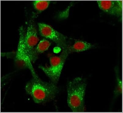
(Immunofluorescence Analysis of PFA-fixed U87MG cells stained using CD63 Mouse Monoclonal Antibody (MX-49.129.5) followed by goat anti-mouse IgG-CF488 (green). CF640R phalloidin (red).)
IF (Immunofluorescence)
(Immunofluorescence Analysis of PFA-fixed U87MG cells stained using CD63 Mouse Monoclonal Antibody (MX-49.129.5) followed by goat anti-mouse IgG-CF488 (green). CF640R phalloidin (red).)
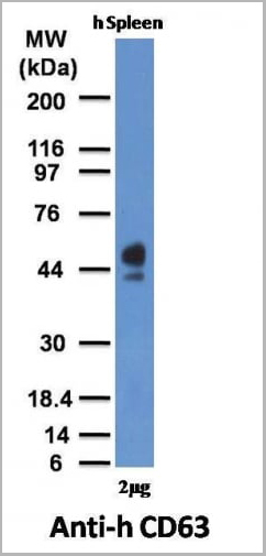
CD63, Monoclonal Antibody (Cat# AAA13803)
Full Name
CD63 (Late Endosomes Marker) Mouse Monoclonal Antibody
Gene Names
CD63; MLA1; ME491; LAMP-3; OMA81H; TSPAN30
Reactivity
Human, Mouse
Applications
Flow Cytometry, Immunofluorescence, Western Blot, Immunohistochemistry
Pricing
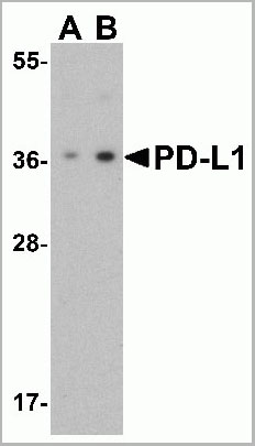
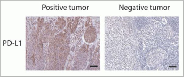 IF (Immunofluorescence)
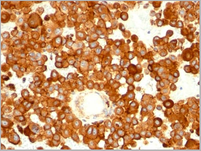
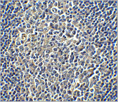
(Figure 12 Immunohistochemistry Validation of PD-L1in Human Tumors (Gadiot et al., 2011)Immunohistochemical analysis of patient tumors labeling PD-L1 with anti-PD-L1 antibodies (AAA10941). Several anti-PD-L1 antibodies were tested for staining, “Only 1 antibody gave no background staining and was competitively blocked by the addition of PD-L1Fc protein (AAA10941)”.)
IF (Immunofluorescence)
(Figure 12 Immunohistochemistry Validation of PD-L1in Human Tumors (Gadiot et al., 2011)Immunohistochemical analysis of patient tumors labeling PD-L1 with anti-PD-L1 antibodies (AAA10941). Several anti-PD-L1 antibodies were tested for staining, “Only 1 antibody gave no background staining and was competitively blocked by the addition of PD-L1Fc protein (AAA10941)”.)
PDL-1, Polyclonal Antibody (Cat# AAA10941)
Full Name
PDL-1 Antibody
Gene Names
CD274; B7-H; B7H1; PDL1; PD-L1; PDCD1L1; PDCD1LG1
Applications
Western Blot, Immunohistochemistry, Immunofluorescence, Flow Cytometry
Purity
PD-L1 Antibody is affinity chromatography purified via peptide column.
Pricing
CAMKIV, Monoclonal Antibody (Cat# AAA24153)
Full Name
CAMKIV (Calcium/Calmodulin-dependent Protein Kinase Type IV, CAM Kinase-GR, CaMK IV, CAMK4) (AP)
Gene Names
CAMK4; caMK; CaMKIV; CaMK IV; CaMK-GR
Reactivity
Human
Applications
Immunohistochemistry, Western Blot
Purity
Purified by Protein A Affinity Chromatography.
Pricing
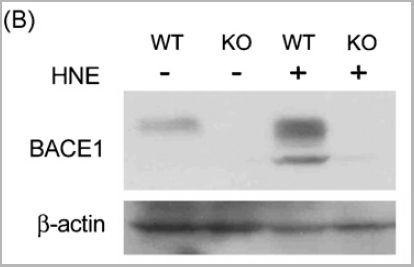
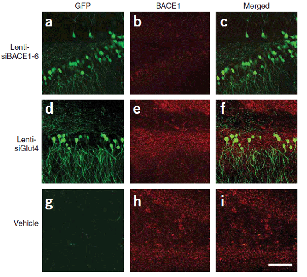

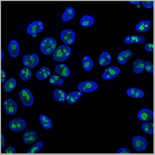
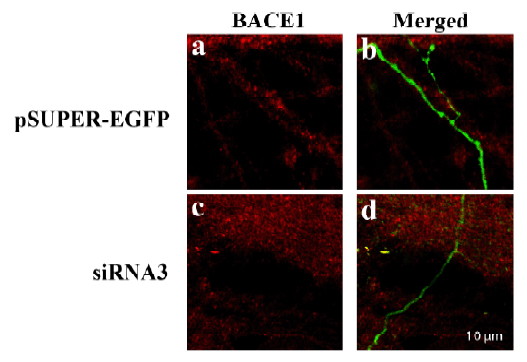 IF (Immunofluorescence)
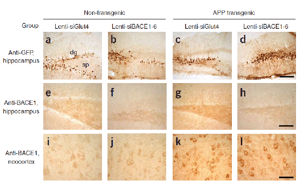
(Figure 10 KD Validation of BACE in DRG (Hyun, 2007)Decreased BACE1 expression in DRG following siRNA3 transfection. DRG neurons were transfected with 1 ug siRNA3 plasmid and incubated for 48 hours in 37°˚C. DRG neurons were stained for BACE1 us¬ing the Anti-BACE antibody (a,b) Neurons transfected with the control plas¬mid pSUPER-EGFP (green) did not display any changes in BACE1 expression (red). (c,d) DRG neurons transfected with siR¬NA3 displayed reduced BACE1 expression in the axon.)
IF (Immunofluorescence)
(Figure 10 KD Validation of BACE in DRG (Hyun, 2007)Decreased BACE1 expression in DRG following siRNA3 transfection. DRG neurons were transfected with 1 ug siRNA3 plasmid and incubated for 48 hours in 37°˚C. DRG neurons were stained for BACE1 us¬ing the Anti-BACE antibody (a,b) Neurons transfected with the control plas¬mid pSUPER-EGFP (green) did not display any changes in BACE1 expression (red). (c,d) DRG neurons transfected with siR¬NA3 displayed reduced BACE1 expression in the axon.)
BACE, Polyclonal Antibody (Cat# AAA10918)
Full Name
BACE Antibody
Gene Names
BACE1; ASP2; BACE; HSPC104
Reactivity
Human, Mouse
Applications
Immunocytochemistry, Immunofluorescence, Immunohistochemistry, Western Blot
Purity
BACE Antibody is affinity chromatography purified via peptide column.
Pricing
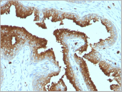
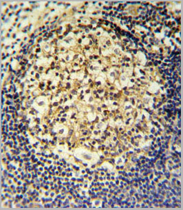
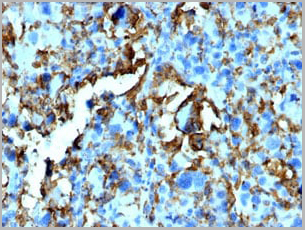
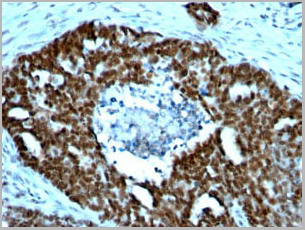
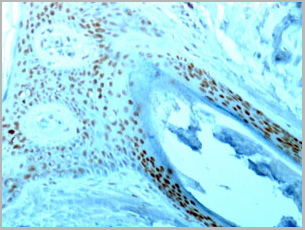
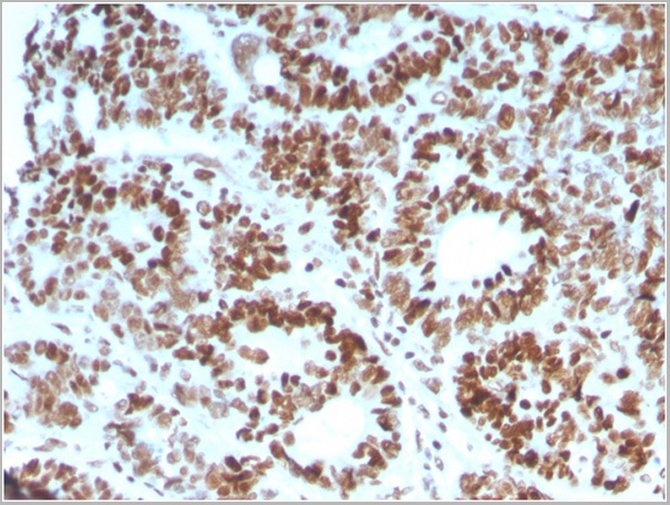
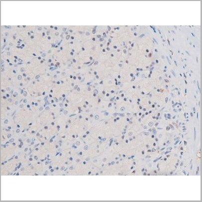
 IHC (Immunohistochemistry)
(Anti-GDF15 antibody IHC of human placenta. Immunohistochemistry of formalin-fixed, paraffin-embedded tissue after heat-induced antigen retrieval. Antibody dilution 10 ug/ml.)
IHC (Immunohistochemistry)
(Anti-GDF15 antibody IHC of human placenta. Immunohistochemistry of formalin-fixed, paraffin-embedded tissue after heat-induced antigen retrieval. Antibody dilution 10 ug/ml.)
GDF15, Monoclonal Antibody (Cat# AAA12357)
Full Name
Rat Monoclonal [clone 6D12.H10.E4] (IgG) to Human GDF15
Gene Names
GDF15; PDF; MIC1; PLAB; MIC-1; NAG-1; PTGFB; GDF-15
Reactivity
Chimpanzee, Macaque
Applications
Immunohistochemistry, Immunohistochemistry, Western Blot
Purity
Protein G Purified chromatography
Pricing

 Application Data
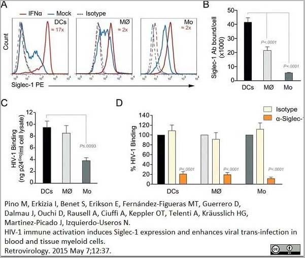
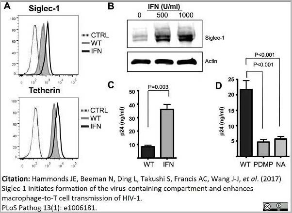
(PE conjugatedMouse anti Human CD169 antibody, clone 7-239 used to block CD169 function on myeloid cells.Image caption:Siglec-1 mediates HIV-1 uptake into a storage compartment and enhances HIV-1 trans-infection specially in IFN?-treated monocytes and DCs. A. Uptake of HIV-1NL4–3 by different myeloid cells exposed to IFN?. Cells were cultured with HIV-1 to measure p24Gag by ELISA. Mean values and SEM from four experiments include cells from 12 donors. B. Fold change in HIV-1NL4–3 uptake of cells treated with bafilomycin A1 compared to untreated cells. Mean values and SEM include cells from three donors. C. Relative uptake of HIV-1NL4–3 by IFN?-treated myeloid cells pre-incubated with the indicated mAbs. Values are normalized to the level of HIV-1 uptake by mock-treated cells (set at 100%). Mean values and SEM from two experiments include cells from six donors. D. Confocal microscopy analysis of different IFN?-treated myeloid cells pulsed with HIV-1Cherry and stained for Siglec-1 (Alexa 488), HLA-DR (Alexa 647) and DAPI. (Top) Representative viral pattern for each kind of myeloid cell analyzed, showing maximum fluorescence intensity of four channels. (Bottom) Percentage of myeloid cells with distinct viral patterns: random distribution, polarized accumulation, and sac-like compartment formation, as illustrated in the left drawing. Mean values of 50 cells from two different donors are shown. E. HIV-1 transmission from IFN?-treated myeloid cells to a luciferase reporter CD4+ cell line. HIV-1 infection was determined by induced luciferase activity in relative light units (RLUs). Mean values and SEM from four experiments include cells from 12 donors. F. Relative HIV-1 transmission from IFN?-treated myeloid cells pre-incubated with the indicated mAbs. Values are normalized to the level of HIV-1 trans-infected by mock-treated cells. Mean values and SEM from two experiments include cells from six donors. Statistical differences were assessed with a paired t test in A and E, and with a one sample t-test in B, C and F.From: Pino M, Erkizia I, Benet S, Erikson E, Fernández-Figueras MT, Guerrero D, Dalmau J, Ouchi D, Rausell A, Ciuffi A, Keppler OT, Telenti A, Kräusslich HG, Martinez-Picado J, Izquierdo-Useros N.HIV-1 immune activation induces Siglec-1 expression and enhances viral trans-infection in blood and tissue myeloid cells.Retrovirology. 2015 May 7;12:37.This image is from an open access article distributed under the terms of the Creative Commons Attribution License.)
Application Data
(PE conjugatedMouse anti Human CD169 antibody, clone 7-239 used to block CD169 function on myeloid cells.Image caption:Siglec-1 mediates HIV-1 uptake into a storage compartment and enhances HIV-1 trans-infection specially in IFN?-treated monocytes and DCs. A. Uptake of HIV-1NL4–3 by different myeloid cells exposed to IFN?. Cells were cultured with HIV-1 to measure p24Gag by ELISA. Mean values and SEM from four experiments include cells from 12 donors. B. Fold change in HIV-1NL4–3 uptake of cells treated with bafilomycin A1 compared to untreated cells. Mean values and SEM include cells from three donors. C. Relative uptake of HIV-1NL4–3 by IFN?-treated myeloid cells pre-incubated with the indicated mAbs. Values are normalized to the level of HIV-1 uptake by mock-treated cells (set at 100%). Mean values and SEM from two experiments include cells from six donors. D. Confocal microscopy analysis of different IFN?-treated myeloid cells pulsed with HIV-1Cherry and stained for Siglec-1 (Alexa 488), HLA-DR (Alexa 647) and DAPI. (Top) Representative viral pattern for each kind of myeloid cell analyzed, showing maximum fluorescence intensity of four channels. (Bottom) Percentage of myeloid cells with distinct viral patterns: random distribution, polarized accumulation, and sac-like compartment formation, as illustrated in the left drawing. Mean values of 50 cells from two different donors are shown. E. HIV-1 transmission from IFN?-treated myeloid cells to a luciferase reporter CD4+ cell line. HIV-1 infection was determined by induced luciferase activity in relative light units (RLUs). Mean values and SEM from four experiments include cells from 12 donors. F. Relative HIV-1 transmission from IFN?-treated myeloid cells pre-incubated with the indicated mAbs. Values are normalized to the level of HIV-1 trans-infected by mock-treated cells. Mean values and SEM from two experiments include cells from six donors. Statistical differences were assessed with a paired t test in A and E, and with a one sample t-test in B, C and F.From: Pino M, Erkizia I, Benet S, Erikson E, Fernández-Figueras MT, Guerrero D, Dalmau J, Ouchi D, Rausell A, Ciuffi A, Keppler OT, Telenti A, Kräusslich HG, Martinez-Picado J, Izquierdo-Useros N.HIV-1 immune activation induces Siglec-1 expression and enhances viral trans-infection in blood and tissue myeloid cells.Retrovirology. 2015 May 7;12:37.This image is from an open access article distributed under the terms of the Creative Commons Attribution License.)
CD169, Monoclonal Antibody (Cat# AAA12264)
Full Name
Mouse Anti Human CD169
Gene Names
SIGLEC1; SN; CD169; SIGLEC-1
Reactivity
Human
Applications
Immunohistochemistry, Flow Cytometry, Functional Assay, Immunoprecipitation, Western Blot
Purity
>95% by SDS PAGE
Purified IgG prepared by affinity chromatography on Protein A from tissue culture supernatant.
Purified IgG prepared by affinity chromatography on Protein A from tissue culture supernatant.
Pricing

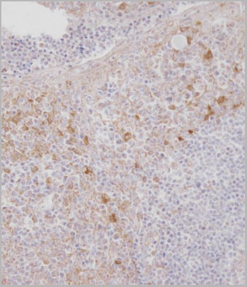
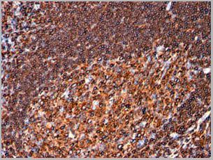
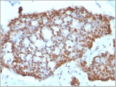
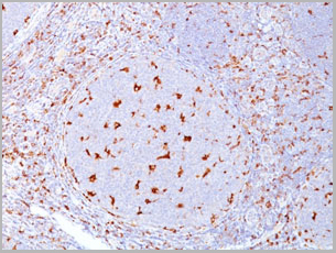
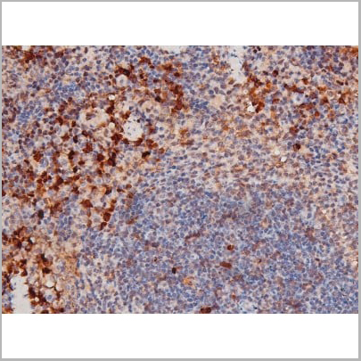
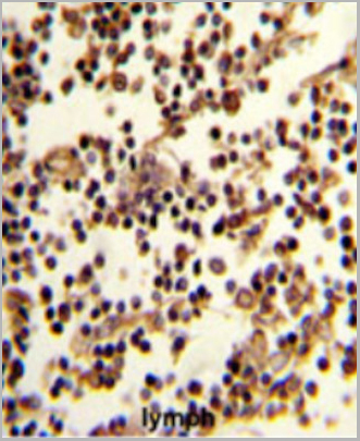 IHC (Immunohistchemistry)
(Formalin-fixed and paraffin-embedded human lymph reacted with CD19 Antibody (N-term), which was peroxidase-conjugated to the secondary antibody, followed by DAB staining. This data demonstrates the use of this antibody for immunohistochemistry; clinical relevance has not been evaluated.)
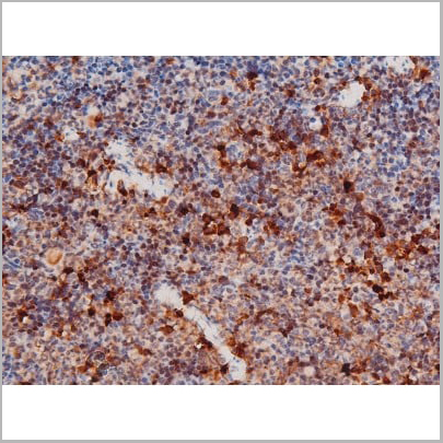
IHC (Immunohistchemistry)
(Formalin-fixed and paraffin-embedded human lymph reacted with CD19 Antibody (N-term), which was peroxidase-conjugated to the secondary antibody, followed by DAB staining. This data demonstrates the use of this antibody for immunohistochemistry; clinical relevance has not been evaluated.)
CD19, Polyclonal Antibody (Cat# AAA28685)
Full Name
CD19 Antibody (N-term)
Gene Names
CD19; B4; CVID3
Reactivity
Human
Applications
Western Blot, Immunofluorescence, Flow Cytometry, Immunohistochemistry
Purity
This antibody is purified through a protein A column, followed by peptide affinity purification.
Pricing
 Application Data
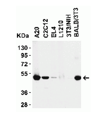
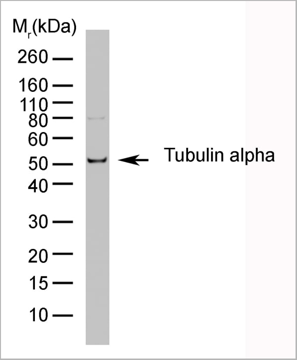
(Western blot analysis of CD107b on J774 cell lysate probed with Rat anti Mouse CD107b at 1/500, 1/1000, 1/2000. Rat anti tubulin alpha is included as a loading control. Detection is with Goat anti Rat IgG:Dylight®)
Application Data
(Western blot analysis of CD107b on J774 cell lysate probed with Rat anti Mouse CD107b at 1/500, 1/1000, 1/2000. Rat anti tubulin alpha is included as a loading control. Detection is with Goat anti Rat IgG:Dylight®)
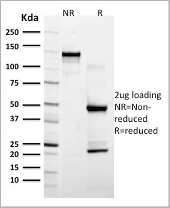
TUBULIN ALPHA, Monoclonal Antibody (Cat# AAA12010)
Full Name
RAT ANTI TUBULIN ALPHA
Applications
Immunohistochemistry, Immunofluorescence, Radioimmunoassay, Western Blot
Pricing
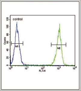
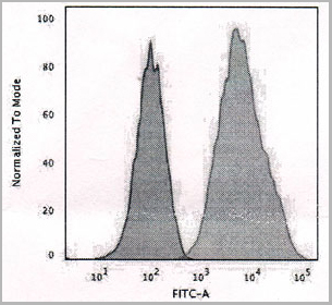
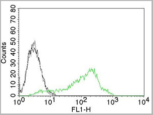
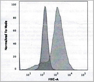
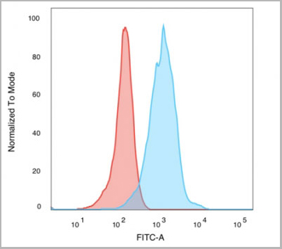
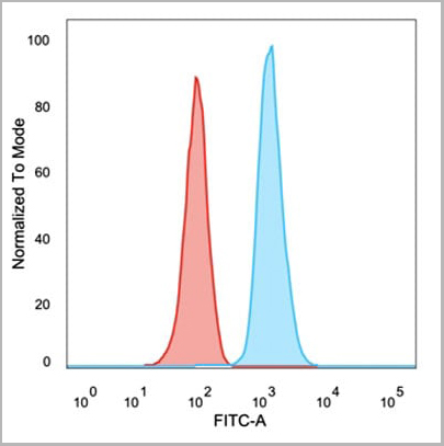
 FCM (Flow Cytometry)
(CCR7 Antibody (N-term) (AAA28701) flow cytometric analysis of 293 cells (right histogram) compared to a negative controlcell (left histogram). FITC-conjugated goat-anti-rabbit secondary antibodies were used for the analysis.)
FCM (Flow Cytometry)
(CCR7 Antibody (N-term) (AAA28701) flow cytometric analysis of 293 cells (right histogram) compared to a negative controlcell (left histogram). FITC-conjugated goat-anti-rabbit secondary antibodies were used for the analysis.)
CCR7, Polyclonal Antibody (Cat# AAA28701)
Full Name
CCR7 Antibody (N-term)
Gene Names
CCR7; BLR2; EBI1; CCR-7; CD197; CDw197; CMKBR7; CC-CKR-7
Reactivity
Human, mouse
Applications
Western Blot, Immunohistochemistry, Immunofluorescence, Flow Cytometry
Purity
Peptide Affinity Purified Rabbit Polyclonal Antibody (Pab)
Pricing

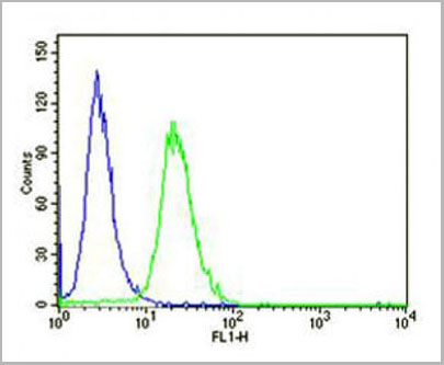
 FCM (Flow Cytometry)
(LTF Antibody flow cytometric analysis of MDA-MB231 cells (right histogram) compared to a negative control cell (lefthistogram).FITC-conjugated goat-anti-rabbit secondary antibodies were used for theanalysis.)
FCM (Flow Cytometry)
(LTF Antibody flow cytometric analysis of MDA-MB231 cells (right histogram) compared to a negative control cell (lefthistogram).FITC-conjugated goat-anti-rabbit secondary antibodies were used for theanalysis.)
LTF, Polyclonal Antibody (Cat# AAA28680)
Full Name
LTF Antibody
Gene Names
LTF; LF; HLF2; GIG12; HEL110
Reactivity
Human, mouse
Applications
Western Blot, Flow Cytometry
Purity
Peptide Affinity Purified Rabbit Polyclonal Antibody (Pab)
Pricing
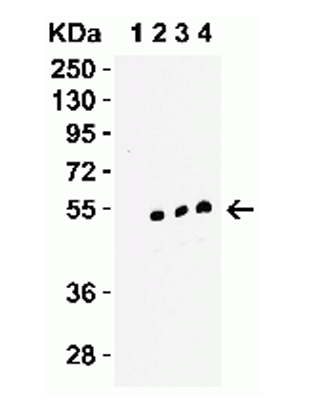
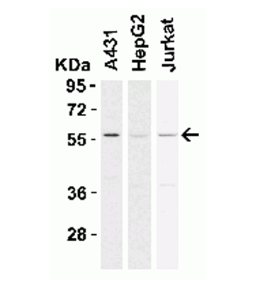
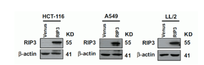 Application Data
(The cancer cell lines were stably expressing DYKDDDDK-tagged RIP3 and RIP expression was detected by anti-RIP3 antibodies (AAA10922) in RIP3-overexpressed cells.)
Application Data
(The cancer cell lines were stably expressing DYKDDDDK-tagged RIP3 and RIP expression was detected by anti-RIP3 antibodies (AAA10922) in RIP3-overexpressed cells.)
RIP3, Polyclonal Antibody (Cat# AAA10922)
Full Name
RIP3 Antibody
Gene Names
Ripk3; Rip3; AW107945; 2610528K09Rik
Reactivity
Human, Mouse, Rat
Applications
Immunofluorescence, Immunohistochemistry, Immunoprecipitation, Western Blot
Purity
RIP3 Antibody is affinity chromatography purified via peptide column.
Pricing
 SDS-PAGE
(SDS-PAGE Analysis Purified EGFR Mouse Monoclonal Antibody (GFR/2596).Confirmation of Integrity and Purity of Antibody)
SDS-PAGE
(SDS-PAGE Analysis Purified EGFR Mouse Monoclonal Antibody (GFR/2596).Confirmation of Integrity and Purity of Antibody)
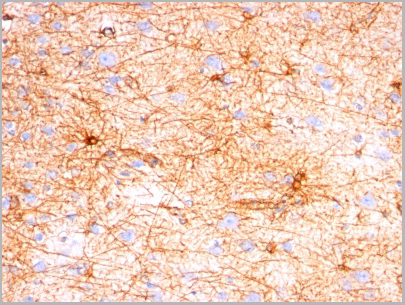
GFAP, Monoclonal Antibody (Cat# AAA13808)
Full Name
GFAP (Astrocyte & Neural Stem Cell Marker) Mouse Monoclonal Antibody
Gene Names
GFAP; ALXDRD
Reactivity
Human, Mouse, Rat, Cow, Pig, Rabbit, Chicken
Applications
Flow Cytometry, Immunofluorescence, Western Blot, Immunohistochemistry
Purity
200ug/ml of Ab purified from Bioreactor Concentrate by Protein A/G.
Pricing
 FCM (Flow Cytometry)
(WB - MCSF Receptor (CSF1R) Antibody (C-term) AAA28757 detail IHC-P - MCSF Receptor (CSF1R) Antibody (C-term) AAA28757 detail IHC-P - MCSF Receptor (CSF1R) Antibody (C-term) AAA28757 detail FC - MCSF Receptor (CSF1R) Antibody (C-term) AAA28757 detail Overlay histogram showing HepG2 cells stained with AAA28757(green line). The cells were fixed with 2% paraformaldehyde (10 min) and then permeabilized with 90% methanol for 10 min. The cells were then icubated in 2% bovine serum albumin to block non-specific protein-protein interactions followed by the antibody (AAA28757, 1:25 dilution) for 60 min at 37ºC. The secondary antibody used was Goat-Anti-Rabbit IgG, DyLight® 488 Conjugated Highly Cross-Adsorbed(1583138) at 1/200 dilution for 40 min at 37ºC. Isotype control antibody (blue line) was rabbit IgG1 (1ug/1x10^6 cells) used under the same conditions. Acquisition of >10, 000 events was performed.)
FCM (Flow Cytometry)
(WB - MCSF Receptor (CSF1R) Antibody (C-term) AAA28757 detail IHC-P - MCSF Receptor (CSF1R) Antibody (C-term) AAA28757 detail IHC-P - MCSF Receptor (CSF1R) Antibody (C-term) AAA28757 detail FC - MCSF Receptor (CSF1R) Antibody (C-term) AAA28757 detail Overlay histogram showing HepG2 cells stained with AAA28757(green line). The cells were fixed with 2% paraformaldehyde (10 min) and then permeabilized with 90% methanol for 10 min. The cells were then icubated in 2% bovine serum albumin to block non-specific protein-protein interactions followed by the antibody (AAA28757, 1:25 dilution) for 60 min at 37ºC. The secondary antibody used was Goat-Anti-Rabbit IgG, DyLight® 488 Conjugated Highly Cross-Adsorbed(1583138) at 1/200 dilution for 40 min at 37ºC. Isotype control antibody (blue line) was rabbit IgG1 (1ug/1x10^6 cells) used under the same conditions. Acquisition of >10, 000 events was performed.)
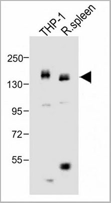
MCSF Receptor (CSF1R), Polyclonal Antibody (Cat# AAA28757)
Full Name
MCSF Receptor (CSF1R) Antibody (C-term)
Gene Names
CSF1R; FMS; CSFR; FIM2; HDLS; C-FMS; CD115; CSF-1R; M-CSF-R
Reactivity
Human
Applications
Western Blot, Flow Cytometry, Immunohistochemistry
Purity
Purified Rabbit Polyclonal Antibody (Pab)
Pricing

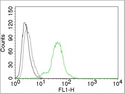
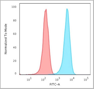
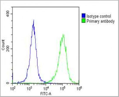
 FCM (Flow Cytometry)
(Flow Cytometric Analysis of human Raji cells using HLA-DR MAb (SPM423) followed by Goat anti-mouse I G-CF488 (Blue); Isotype Control (Red).)
FCM (Flow Cytometry)
(Flow Cytometric Analysis of human Raji cells using HLA-DR MAb (SPM423) followed by Goat anti-mouse I G-CF488 (Blue); Isotype Control (Red).)
HLA-DRB, Monoclonal Antibody (Cat# AAA13826)
Full Name
HLA-DRB (MHC II) Mouse Monoclonal Antibody
Gene Names
HLA-DRB1; SS1; DRB1; DRw10; HLA-DRB; HLA-DR1B
Reactivity
Human, Monkey
Applications
Flow Cytometry, Immunofluorescence, Western Blot, Immunohistochemistry
Pricing
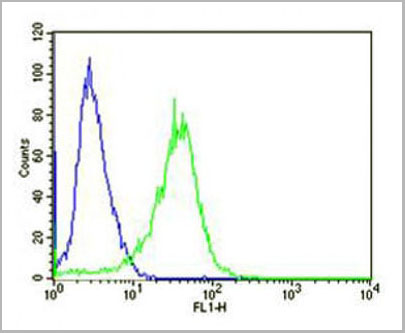
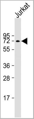
 FCM (Flow Cytometry)
(PCSK9 Antibody (N-term) (Cat. #AAA28774) flow cytometry analysis of Jurkat cells (bottom histogram) compared to a negative control cell(top histogram).FITC-conjugated goat-anti-rabbit secondary antibodies were used for the analysis.)
FCM (Flow Cytometry)
(PCSK9 Antibody (N-term) (Cat. #AAA28774) flow cytometry analysis of Jurkat cells (bottom histogram) compared to a negative control cell(top histogram).FITC-conjugated goat-anti-rabbit secondary antibodies were used for the analysis.)
PCSK9, Polyclonal Antibody (Cat# AAA28774)
Full Name
PCSK9 Antibody (N-term)
Gene Names
PCSK9; FH3; PC9; NARC1; LDLCQ1; NARC-1; HCHOLA3
Reactivity
Human
Applications
Western Blot, Immunohistochemistry, Flow Cytometry
Pricing
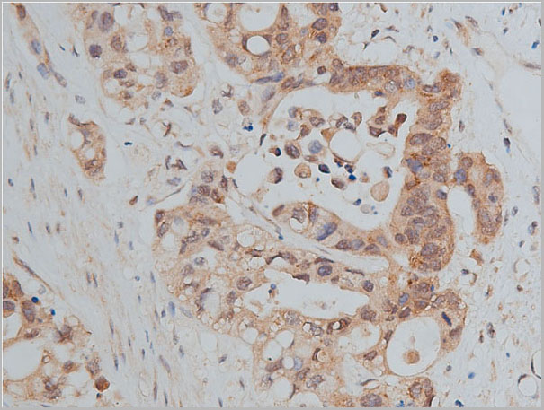
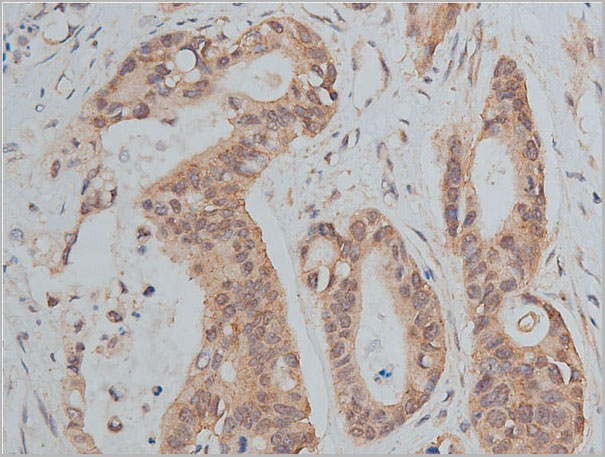
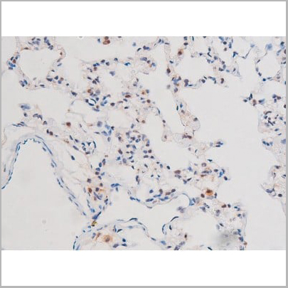
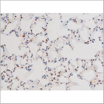
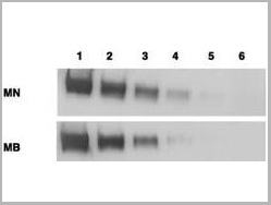 WB (Western Blot)
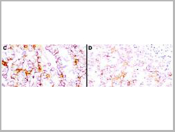
(Anti-Mesothelin Antibodies - Immunohistochemistry. Immunohistochemistry using anti-mesothelin antibodies to detect mesothelin in PEFF human tissue sections treated by antigen retrieval methods. Anti-mesothelin primary antibodies were used to label these sections as follows: C, MAb MB; and D, MAb MN. Reprinted with permission fromClin. Cancer Res. 11(16):5840-6.)
WB (Western Blot)
(Anti-Mesothelin Antibodies - Immunohistochemistry. Immunohistochemistry using anti-mesothelin antibodies to detect mesothelin in PEFF human tissue sections treated by antigen retrieval methods. Anti-mesothelin primary antibodies were used to label these sections as follows: C, MAb MB; and D, MAb MN. Reprinted with permission fromClin. Cancer Res. 11(16):5840-6.)
MSLN / Mesothelin, Monoclonal Antibody (Cat# AAA12374)
Full Name
Mouse Monoclonal [clone MN-1] (IgG) to Human MSLN / Mesothelin
Gene Names
MSLN; MPF; SMRP
Reactivity
Human
Applications
Immunohistochemistry, Western Blot
Purity
Protein A Purified
Pricing
 WB (Western Blot)
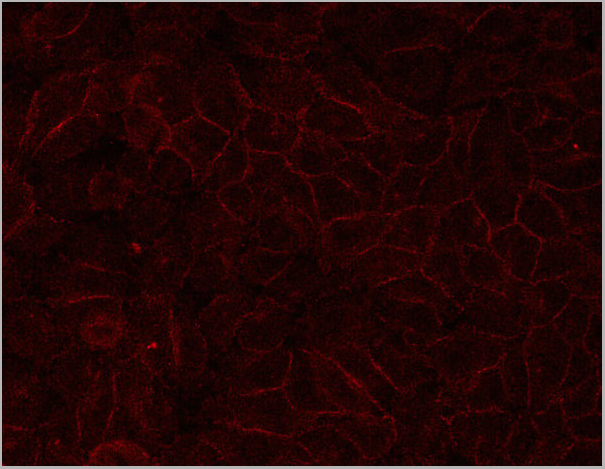
(Western blot analysis on mouse liver tissue lysate using E-cadherin Antibody)
WB (Western Blot)
(Western blot analysis on mouse liver tissue lysate using E-cadherin Antibody)
E-cadherin, Polyclonal Antibody (Cat# AAA30922)
Full Name
E-cadherin Antibody
Gene Names
CDH1; UVO; CDHE; ECAD; LCAM; Arc-1; BCDS1; CD324
Reactivity
Human, Mouse, Rat
Applications
Western Blot, Immunohistochemistry, Immunofluorescence, Immunocytochemistry
Purity
The antiserum was purified by peptide affinity chromatography using SulfoLink Coupling Resin.
Pricing
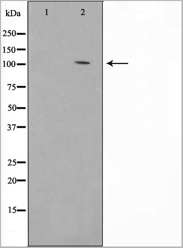
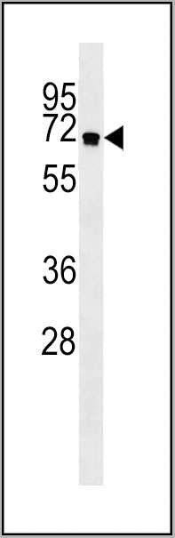
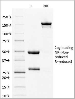
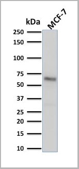
 WB (Western Blot)
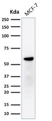
(Western Blot Analysis of human MCF-7 cell lysate using ER-beta1 Mouse Monoclonal Antibody (ERb455).)
WB (Western Blot)
(Western Blot Analysis of human MCF-7 cell lysate using ER-beta1 Mouse Monoclonal Antibody (ERb455).)
ER-beta1 (Estrogen Receptor beta-1), Monoclonal Antibody (Cat# AAA13814)
Full Name
ER-beta1 (Estrogen Receptor beta-1) Mouse Monoclonal Antibody
Gene Names
ESR2; Erb; ESRB; ESTRB; NR3A2; ER-BETA; ESR-BETA
Reactivity
Human
Applications
Flow Cytometry, Immunofluorescence, Western Blot, Immunohistochemistry
Pricing


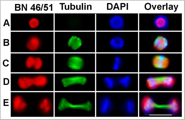
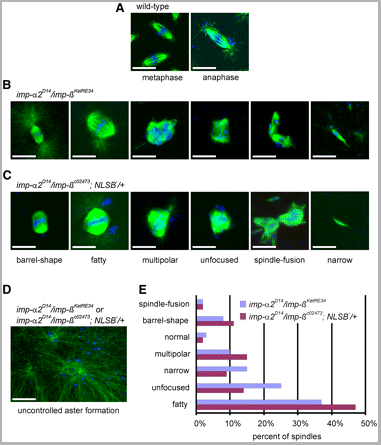
 Application Data
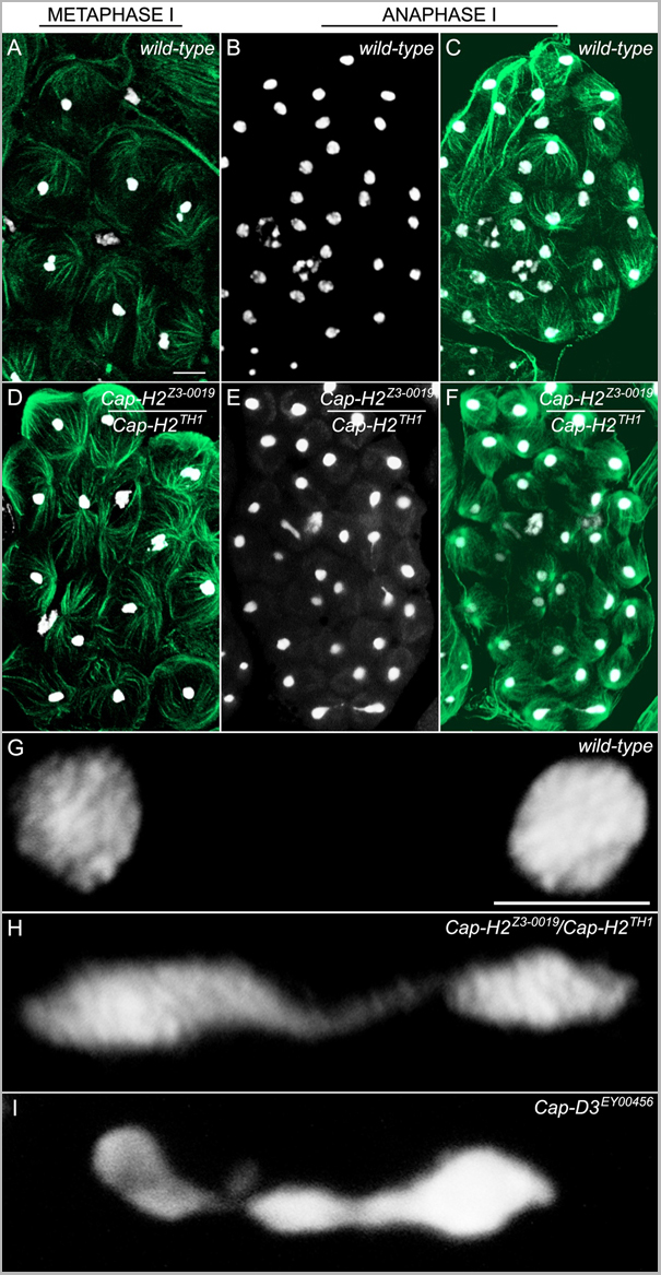
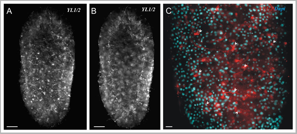
(Published customer image: Spindle abnormalities in embryos derived from imp-a2D14/imp-betaKetRE34 and imp-a2D14/imp-betac02473; NLSB-/+ females. (A -D) Wild-type and mutant embryos stained for a-tubulin (green) and DNA (blue). (A) Mitotic spindles in wild-type embryos at metaphase and anaphase. (B, C) Categories of spindle abnormalities found in embryos derived from (B) imp-a2D14/imp-betaKetRE34 and (C) imp-a2D14/imp-betac02743; NLSB-/+ females. (D) Formation of aster networks found in both genotypes. Scale bar: 10 um. (E) Frequency of spindle defects in embryos from both types of mutant females. Female genotypes are displayed at the upper right corner. At least 200 spindles were scored for both genotypes.From: Specific Cooperation Between Imp-a2 and Imp-beta/Ketel in Spindle Assembly During Drosophila Early Nuclear Divisions Erika Vir¡gh, M¡ty¡s Gorj¡n¡cz, Istv¡n T¶r¶k, Tolga Eichhorn, Sowjanya Kallakuri, Tam¡s Szlanka, Istv¡n Kiss, and Bernard M. Mechler G3 January 2012 2:1-14.)
Application Data
(Published customer image: Spindle abnormalities in embryos derived from imp-a2D14/imp-betaKetRE34 and imp-a2D14/imp-betac02473; NLSB-/+ females. (A -D) Wild-type and mutant embryos stained for a-tubulin (green) and DNA (blue). (A) Mitotic spindles in wild-type embryos at metaphase and anaphase. (B, C) Categories of spindle abnormalities found in embryos derived from (B) imp-a2D14/imp-betaKetRE34 and (C) imp-a2D14/imp-betac02743; NLSB-/+ females. (D) Formation of aster networks found in both genotypes. Scale bar: 10 um. (E) Frequency of spindle defects in embryos from both types of mutant females. Female genotypes are displayed at the upper right corner. At least 200 spindles were scored for both genotypes.From: Specific Cooperation Between Imp-a2 and Imp-beta/Ketel in Spindle Assembly During Drosophila Early Nuclear Divisions Erika Vir¡gh, M¡ty¡s Gorj¡n¡cz, Istv¡n T¶r¶k, Tolga Eichhorn, Sowjanya Kallakuri, Tam¡s Szlanka, Istv¡n Kiss, and Bernard M. Mechler G3 January 2012 2:1-14.)
TUBULIN ALPHA, Monoclonal Antibody (Cat# AAA12232)
Full Name
RAT ANTI TUBULIN ALPHA:HRP
Applications
Immunohistochemistry, Western Blot
Pricing


 Application Data
(Published customer image: Spindle abnormalities in embryos derived from imp-a2D14/imp-betaKetRE34 and imp-a2D14/imp-betac02473; NLSB-/+ females. (A -D) Wild-type and mutant embryos stained for a-tubulin (green) and DNA (blue). (A) Mitotic spindles in wild-type embryos at metaphase and anaphase. (B, C) Categories of spindle abnormalities found in embryos derived from (B) imp-a2D14/imp-betaKetRE34 and (C) imp-a2D14/imp-betac02743; NLSB-/+ females. (D) Formation of aster networks found in both genotypes. Scale bar: 10 um. (E) Frequency of spindle defects in embryos from both types of mutant females. Female genotypes are displayed at the upper right corner. At least 200 spindles were scored for both genotypes.From: Specific Cooperation Between Imp-a2 and Imp-beta/Ketel in Spindle Assembly During Drosophila Early Nuclear Divisions Erika Vir¡gh, M¡ty¡s Gorj¡n¡cz, Istv¡n T¶r¶k, Tolga Eichhorn, Sowjanya Kallakuri, Tam¡s Szlanka, Istv¡n Kiss, and Bernard M. Mechler G3 January 2012 2:1-14.)
Application Data
(Published customer image: Spindle abnormalities in embryos derived from imp-a2D14/imp-betaKetRE34 and imp-a2D14/imp-betac02473; NLSB-/+ females. (A -D) Wild-type and mutant embryos stained for a-tubulin (green) and DNA (blue). (A) Mitotic spindles in wild-type embryos at metaphase and anaphase. (B, C) Categories of spindle abnormalities found in embryos derived from (B) imp-a2D14/imp-betaKetRE34 and (C) imp-a2D14/imp-betac02743; NLSB-/+ females. (D) Formation of aster networks found in both genotypes. Scale bar: 10 um. (E) Frequency of spindle defects in embryos from both types of mutant females. Female genotypes are displayed at the upper right corner. At least 200 spindles were scored for both genotypes.From: Specific Cooperation Between Imp-a2 and Imp-beta/Ketel in Spindle Assembly During Drosophila Early Nuclear Divisions Erika Vir¡gh, M¡ty¡s Gorj¡n¡cz, Istv¡n T¶r¶k, Tolga Eichhorn, Sowjanya Kallakuri, Tam¡s Szlanka, Istv¡n Kiss, and Bernard M. Mechler G3 January 2012 2:1-14.)
TUBULIN ALPHA, Monoclonal Antibody (Cat# AAA12009)
Full Name
RAT ANTI TUBULIN ALPHA
Applications
Immunohistochemistry, Immunofluorescence, Immunoprecipitation, Radioimmunoassay, Western Blot
Pricing

 IF (Immunofluorescence)
Nucleolin, Monoclonal Antibody (Cat# AAA13823)
Full Name
Nucleolin (Marker of Human Cells) Mouse Monoclonal Antibody
Gene Names
NCL; C23
Reactivity
Human.
Does not react with Mouse, Rat and Cow
Does not react with Mouse, Rat and Cow
Applications
Flow Cytometry, Immunofluorescence, Western Blot, Immunohistochemistry
Pricing


 Application Data
(Staining of mouse spleen with Hamster anti Mouse CD81: Alexa Fluor 488)
Application Data
(Staining of mouse spleen with Hamster anti Mouse CD81: Alexa Fluor 488)
CD81, Monoclonal Antibody (Cat# AAA11869)
Full Name
HAMSTER ANTI MOUSE CD81:FITC
Gene Names
Cd81; Tapa1; Tapa-1; Tspan28
Applications
Flow Cytometry
Pricing


 Application Data
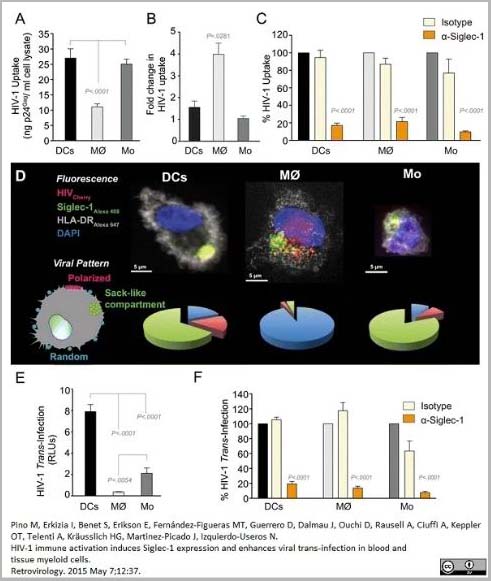
(PE conjugatedMouse anti Human CD169 antibody, clone 7-239 used to block CD169 function on myeloid cells.Image caption:Siglec-1 mediates HIV-1 uptake into a storage compartment and enhances HIV-1 trans-infection specially in IFN?-treated monocytes and DCs. A. Uptake of HIV-1NL4–3 by different myeloid cells exposed to IFN?. Cells were cultured with HIV-1 to measure p24Gag by ELISA. Mean values and SEM from four experiments include cells from 12 donors. B. Fold change in HIV-1NL4–3 uptake of cells treated with bafilomycin A1 compared to untreated cells. Mean values and SEM include cells from three donors. C. Relative uptake of HIV-1NL4–3 by IFN?-treated myeloid cells pre-incubated with the indicated mAbs. Values are normalized to the level of HIV-1 uptake by mock-treated cells (set at 100%). Mean values and SEM from two experiments include cells from six donors. D. Confocal microscopy analysis of different IFN?-treated myeloid cells pulsed with HIV-1Cherry and stained for Siglec-1 (Alexa 488), HLA-DR (Alexa 647) and DAPI. (Top) Representative viral pattern for each kind of myeloid cell analyzed, showing maximum fluorescence intensity of four channels. (Bottom) Percentage of myeloid cells with distinct viral patterns: random distribution, polarized accumulation, and sac-like compartment formation, as illustrated in the left drawing. Mean values of 50 cells from two different donors are shown. E. HIV-1 transmission from IFN?-treated myeloid cells to a luciferase reporter CD4+ cell line. HIV-1 infection was determined by induced luciferase activity in relative light units (RLUs). Mean values and SEM from four experiments include cells from 12 donors. F. Relative HIV-1 transmission from IFN?-treated myeloid cells pre-incubated with the indicated mAbs. Values are normalized to the level of HIV-1 trans-infected by mock-treated cells. Mean values and SEM from two experiments include cells from six donors. Statistical differences were assessed with a paired t test in A and E, and with a one sample t-test in B, C and F.From: Pino M, Erkizia I, Benet S, Erikson E, Fernández-Figueras MT, Guerrero D, Dalmau J, Ouchi D, Rausell A, Ciuffi A, Keppler OT, Telenti A, Kräusslich HG, Martinez-Picado J, Izquierdo-Useros N.HIV-1 immune activation induces Siglec-1 expression and enhances viral trans-infection in blood and tissue myeloid cells.Retrovirology. 2015 May 7;12:37.This image is from an open access article distributed under the terms of the Creative Commons Attribution License.)
Application Data
(PE conjugatedMouse anti Human CD169 antibody, clone 7-239 used to block CD169 function on myeloid cells.Image caption:Siglec-1 mediates HIV-1 uptake into a storage compartment and enhances HIV-1 trans-infection specially in IFN?-treated monocytes and DCs. A. Uptake of HIV-1NL4–3 by different myeloid cells exposed to IFN?. Cells were cultured with HIV-1 to measure p24Gag by ELISA. Mean values and SEM from four experiments include cells from 12 donors. B. Fold change in HIV-1NL4–3 uptake of cells treated with bafilomycin A1 compared to untreated cells. Mean values and SEM include cells from three donors. C. Relative uptake of HIV-1NL4–3 by IFN?-treated myeloid cells pre-incubated with the indicated mAbs. Values are normalized to the level of HIV-1 uptake by mock-treated cells (set at 100%). Mean values and SEM from two experiments include cells from six donors. D. Confocal microscopy analysis of different IFN?-treated myeloid cells pulsed with HIV-1Cherry and stained for Siglec-1 (Alexa 488), HLA-DR (Alexa 647) and DAPI. (Top) Representative viral pattern for each kind of myeloid cell analyzed, showing maximum fluorescence intensity of four channels. (Bottom) Percentage of myeloid cells with distinct viral patterns: random distribution, polarized accumulation, and sac-like compartment formation, as illustrated in the left drawing. Mean values of 50 cells from two different donors are shown. E. HIV-1 transmission from IFN?-treated myeloid cells to a luciferase reporter CD4+ cell line. HIV-1 infection was determined by induced luciferase activity in relative light units (RLUs). Mean values and SEM from four experiments include cells from 12 donors. F. Relative HIV-1 transmission from IFN?-treated myeloid cells pre-incubated with the indicated mAbs. Values are normalized to the level of HIV-1 trans-infected by mock-treated cells. Mean values and SEM from two experiments include cells from six donors. Statistical differences were assessed with a paired t test in A and E, and with a one sample t-test in B, C and F.From: Pino M, Erkizia I, Benet S, Erikson E, Fernández-Figueras MT, Guerrero D, Dalmau J, Ouchi D, Rausell A, Ciuffi A, Keppler OT, Telenti A, Kräusslich HG, Martinez-Picado J, Izquierdo-Useros N.HIV-1 immune activation induces Siglec-1 expression and enhances viral trans-infection in blood and tissue myeloid cells.Retrovirology. 2015 May 7;12:37.This image is from an open access article distributed under the terms of the Creative Commons Attribution License.)
CD169, Monoclonal Antibody (Cat# AAA12265)
Full Name
Mouse Anti Human CD169: RPE
Gene Names
SIGLEC1; SN; CD169; SIGLEC-1
Reactivity
Human
Applications
Flow Cytometry
Purity
>95% by SDS PAGE
Purified IgG prepared by affinity chromatography on Protein A from tissue culture supernatant.
Purified IgG prepared by affinity chromatography on Protein A from tissue culture supernatant.
Pricing


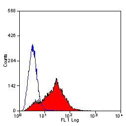 Application Data
(Staining of mouse spleen with Hamster anti Mouse CD81: Alexa Fluor 488)
Application Data
(Staining of mouse spleen with Hamster anti Mouse CD81: Alexa Fluor 488)
CD81, Monoclonal Antibody (Cat# AAA12033)
Full Name
HAMSTER ANTI MOUSE CD81:RPE
Gene Names
Cd81; Tapa1; Tapa-1; Tspan28
Applications
Flow Cytometry
Pricing
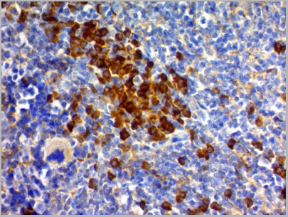
 ICC (Immunocytochemistry)
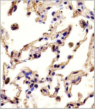
(Immunofluorescence staining of U87MG cells using CD68 Mouse Monoclonal Antibody (C68/684) followed by goat anti-Mouse IgG conjugated to (green). Nuclei are stained with Red dot)
ICC (Immunocytochemistry)
(Immunofluorescence staining of U87MG cells using CD68 Mouse Monoclonal Antibody (C68/684) followed by goat anti-Mouse IgG conjugated to (green). Nuclei are stained with Red dot)
CD68, Antibody (Cat# AAA13807)
Full Name
CD68 (Macrophage Marker) Mouse Monoclonal Antibody
Gene Names
CD68; GP110; LAMP4; SCARD1
Reactivity
Human, Monkey, Mouse, and Rat. Others not known.
Applications
Western Blot, Immunofluorescence, Flow Cytometry, Immunohistochemistry
Pricing
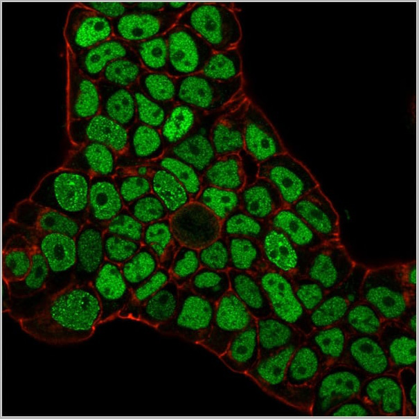

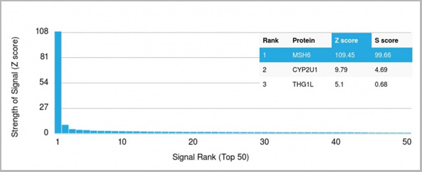 Application Data
(Analysis of Protein Array containing >19,000 full-length human proteins using MSH6 Mouse Monoclonal Antibody (MSH6/3086) Z- and S- Score: The Z-score represents the strength of a signal that a monoclonal antibody (MAb) (in combination with a fluorescently-tagged anti-IgG secondary antibody) produces when binding to a particular protein on the HuProtTM array. Z-scores are described in units of standard deviations (SD’s) above the mean value of all signals generated on that array. If targets on HuProtTM are arranged in descending order of the Z-score, the S-score is the difference (also in units of SD’s) between the Z-score. S-score therefore represents the relative target specificity of a MAb to its intended target. A MAb is considered to specific to its intended target, if the MAb has an S-score of at least 2.5. For example, if a MAb binds to protein X with a Z-score of 43 and to protein Y with a Z-score of 14, then the S-score for the binding of that MAb to protein X is equal to 29.)
Application Data
(Analysis of Protein Array containing >19,000 full-length human proteins using MSH6 Mouse Monoclonal Antibody (MSH6/3086) Z- and S- Score: The Z-score represents the strength of a signal that a monoclonal antibody (MAb) (in combination with a fluorescently-tagged anti-IgG secondary antibody) produces when binding to a particular protein on the HuProtTM array. Z-scores are described in units of standard deviations (SD’s) above the mean value of all signals generated on that array. If targets on HuProtTM are arranged in descending order of the Z-score, the S-score is the difference (also in units of SD’s) between the Z-score. S-score therefore represents the relative target specificity of a MAb to its intended target. A MAb is considered to specific to its intended target, if the MAb has an S-score of at least 2.5. For example, if a MAb binds to protein X with a Z-score of 43 and to protein Y with a Z-score of 14, then the S-score for the binding of that MAb to protein X is equal to 29.)
MSH6, Monoclonal Antibody (Cat# AAA23917)
Full Name
MSH6 (DNA Mismatch Repair Protein)
Gene Names
MSH6; GTBP; HSAP; p160; GTMBP; HNPCC5
Reactivity
Human
Applications
Flow Cytometry, Immunofluorescence, Western Blot, Immunohistochemistry
Purity
Purified Ab with BSA and Azide at 200ug/ml OR Purified Ab WITHOUT BSA and Azide at 1.0mg/ml
Pricing
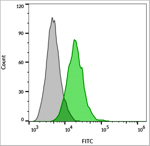
 FCM (Flow Cytometry)
(Overlay histogram showing Hela cells stained with AAA28750 (green line). The cells were fixed with 2% paraformaldehyde (10 min) and then permeabilized with 90% methanol for 10 min. The cells were then icubated in 2% bovine serum albumin to block non-specific protein-protein interactions followed by the antibody (AAA28750, 1:25 dilution) for 60 min at 37ºC. The secondary antibody used was Goat-Anti-Rabbit IgG, DyLight® 488 Conjugated Highly Cross-Adsorbed (1583138) at 1/200 dilution for 40 min at 37°C. Isotype control antibody (blue line) was rabbit IgG1 (1ug/1x106 cells) used under the same conditions. Acquisition of >10, 000 events wasperformed.)
FCM (Flow Cytometry)
(Overlay histogram showing Hela cells stained with AAA28750 (green line). The cells were fixed with 2% paraformaldehyde (10 min) and then permeabilized with 90% methanol for 10 min. The cells were then icubated in 2% bovine serum albumin to block non-specific protein-protein interactions followed by the antibody (AAA28750, 1:25 dilution) for 60 min at 37ºC. The secondary antibody used was Goat-Anti-Rabbit IgG, DyLight® 488 Conjugated Highly Cross-Adsorbed (1583138) at 1/200 dilution for 40 min at 37°C. Isotype control antibody (blue line) was rabbit IgG1 (1ug/1x106 cells) used under the same conditions. Acquisition of >10, 000 events wasperformed.)
WNT5A, Polyclonal Antibody (Cat# AAA28750)
Full Name
WNT5A Antibody (Center)
Gene Names
WNT5A; hWNT5A
Reactivity
Human, Mouse, Rat
Predicted: Rabbit
Predicted: Rabbit
Applications
Immunohistochemistry, Immunohistochemistry, Flow Cytometry, Western Blot
Purity
This antibody is purified through a protein A column, followed by peptide affinity purification.
Pricing


 Application Data
(Staining of mouse spleen with Hamster anti Mouse CD81: Alexa Fluor 488)
Application Data
(Staining of mouse spleen with Hamster anti Mouse CD81: Alexa Fluor 488)
CD81, Monoclonal Antibody (Cat# AAA11942)
Full Name
HAMSTER ANTI MOUSE CD81
Gene Names
Cd81; Tapa1; Tapa-1; Tspan28
Reactivity
Rat
Applications
Immunohistochemistry, Flow Cytometry, Immunoprecipitation, Western Blot
Pricing
 IHC (Immunohistochemistry)
(Immunohistochemical analysis of paraffin-embedded human tonsil tissue using AAA28679 performed on the Leica® BOND RXm. Samples were incubated with primary antibody(1/500) for 1 hours at room temperature. A undiluted biotinylated CRF Anti-Polyvalent HRP Polymer antibody was used as the secondary antibody.)
IHC (Immunohistochemistry)
(Immunohistochemical analysis of paraffin-embedded human tonsil tissue using AAA28679 performed on the Leica® BOND RXm. Samples were incubated with primary antibody(1/500) for 1 hours at room temperature. A undiluted biotinylated CRF Anti-Polyvalent HRP Polymer antibody was used as the secondary antibody.)
HK1 (Hexokinase), Polyclonal Antibody (Cat# AAA28679)
Full Name
HK1 (Hexokinase) Antibody (N-term)
Gene Names
HK1; HKD; HKI; HXK1; HMSNR; HK1-ta; HK1-tb; HK1-tc
Reactivity
Human, Rat
Applications
Western Blot, Immunohistochemistry
Purity
Purified Rabbit Polyclonal Antibody (Pab)
Pricing
 WB (Western Blot)
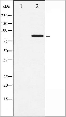
(All lanes : Anti-ERVK-7 Antibody (N-Term) at 1:1000-1:2000 dilutionLane 1: Raji whole cell lysateLane 2: human lung lysateLane 3: human testis lysateLysates/proteins at 20 ug per lane.SecondaryGoat Anti-Rabbit IgG, (H+L), Peroxidase conjugated at 1/10000 dilution.Predicted band size : 67 kDaBlocking/Dilution buffer: 5% NFDM/TBST.)
WB (Western Blot)
(All lanes : Anti-ERVK-7 Antibody (N-Term) at 1:1000-1:2000 dilutionLane 1: Raji whole cell lysateLane 2: human lung lysateLane 3: human testis lysateLysates/proteins at 20 ug per lane.SecondaryGoat Anti-Rabbit IgG, (H+L), Peroxidase conjugated at 1/10000 dilution.Predicted band size : 67 kDaBlocking/Dilution buffer: 5% NFDM/TBST.)
ERVK-7, Polyclonal Antibody (Cat# AAA28797)
Full Name
ERVK-7 Antibody (N-Term)
Reactivity
Human
Applications
Western Blot, Immunohistochemistry
Purity
This antibody is purified through a protein A column, followed by peptide affinity purification.
Pricing
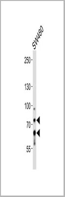
 WB (Western Blot)
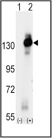
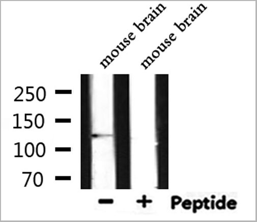
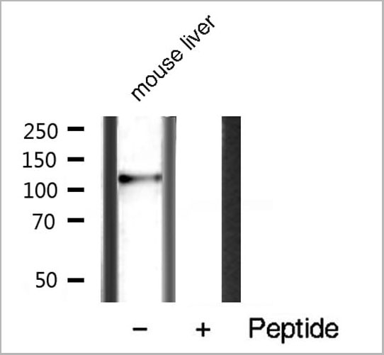
(Western blot analysis of LoVo cell lysate, using PIBF1 Antibody. The lane on the left is treated with the antigen-specific peptide.)
WB (Western Blot)
(Western blot analysis of LoVo cell lysate, using PIBF1 Antibody. The lane on the left is treated with the antigen-specific peptide.)
PIBF1, Polyclonal Antibody (Cat# AAA31130)
Full Name
PIBF1 Antibody
Gene Names
PIBF1; PIBF; CEP90; JBTS33; C13orf24
Reactivity
Human, Mouse, Rat
Applications
Western Blot, Immunohistochemistry, Immunofluorescence, Immunocytochemistry
Purity
The antiserum was purified by peptide affinity chromatography using SulfoLink Coupling Resin (Thermo Fisher Scientific).
Pricing
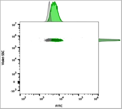
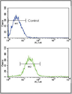
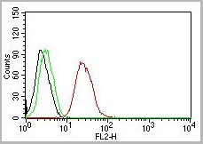
 FCM (Flow Cytometry)
(Flow Cytometric Analysis of PFA-fixed Raji cells with Human Nuclear Antigen Mouse Monoclonal Antibody (235-1); followed by goat anti-mouse IgG-CF488 (Blue); Isotype Control (Red).)
FCM (Flow Cytometry)
(Flow Cytometric Analysis of PFA-fixed Raji cells with Human Nuclear Antigen Mouse Monoclonal Antibody (235-1); followed by goat anti-mouse IgG-CF488 (Blue); Isotype Control (Red).)
Human Nuclear Antigen, Monoclonal Antibody (Cat# AAA13804)
Full Name
Human Nuclear Antigen
Reactivity
Human and Non-human primates. Does not react with mouse, rat, and chicken. Others not known.
Applications
Flow Cytometry, Immunofluorescence, Immunoprecipitation, Immunocytochemistry, Immunohistochemistry
Pricing
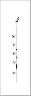
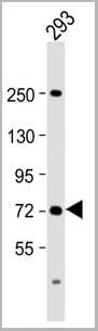 WB (Western Blot)
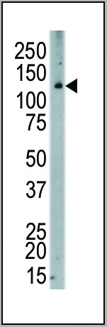
(Anti-SLC6A19 Antibody (C-Term) at 1:2000 dilution + 293 whole cell lysateLysates/proteins at 20 ug per lane. SecondaryGoat Anti-Rabbit IgG, (H+L), Peroxidase conjugated at 1/10000 dilution. Predicted band size : 71 kDaBlocking/Dilution buffer: 5% NFDM/TBST.)
WB (Western Blot)
(Anti-SLC6A19 Antibody (C-Term) at 1:2000 dilution + 293 whole cell lysateLysates/proteins at 20 ug per lane. SecondaryGoat Anti-Rabbit IgG, (H+L), Peroxidase conjugated at 1/10000 dilution. Predicted band size : 71 kDaBlocking/Dilution buffer: 5% NFDM/TBST.)
SLC6A19, Polyclonal Antibody (Cat# AAA28802)
Full Name
SLC6A19 Antibody (C-Term)
Gene Names
SLC6A19; HND; B0AT1
Reactivity
Human
Applications
Western Blot
Purity
This antibody is purified through a protein A column, followed by peptide affinity purification.
Pricing

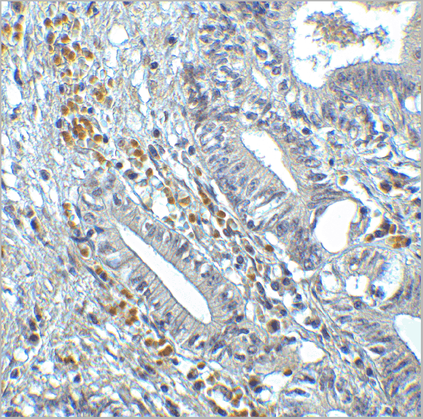
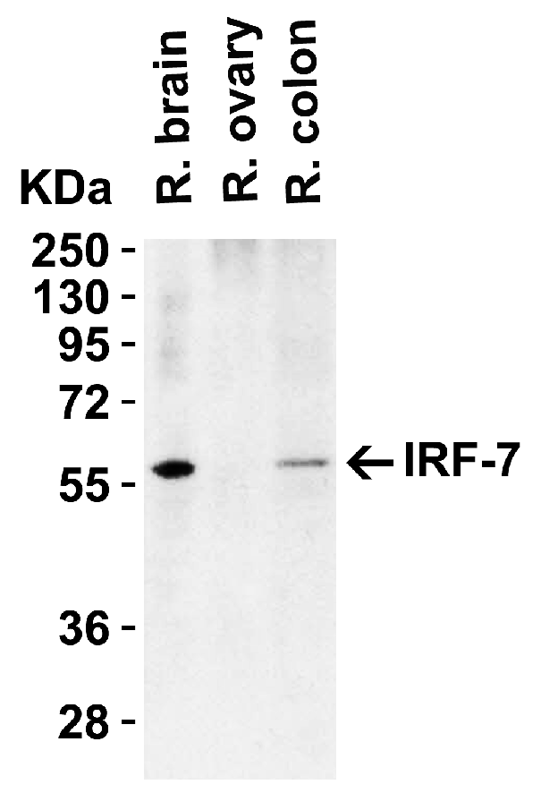
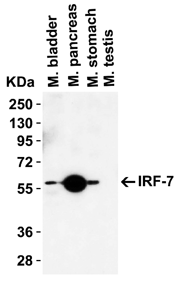
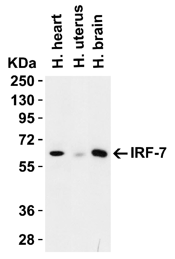
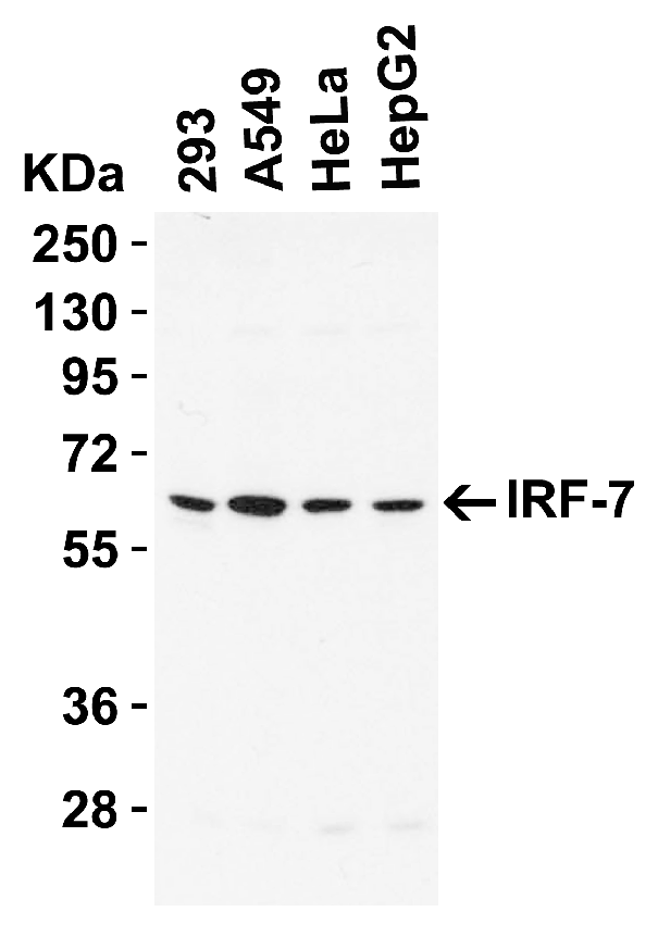
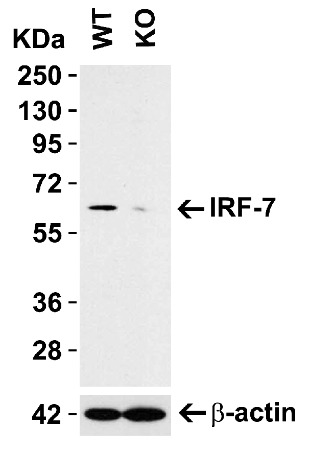
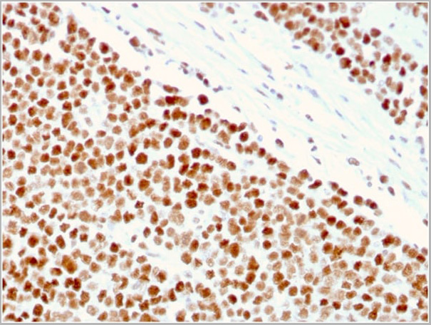
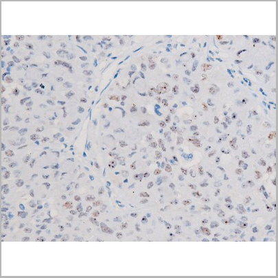
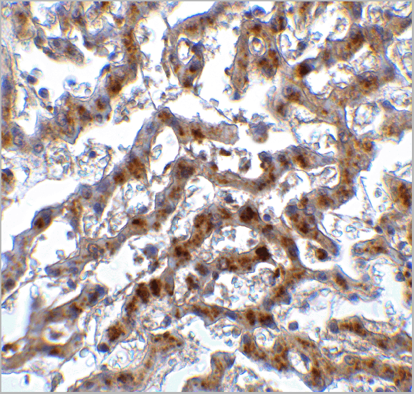 IHC (Immunohistchemistry)
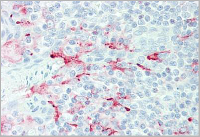
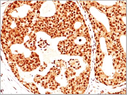
(Figure 9 Immunohistochemistry Validation of IRF7 in Human Liver Tissue Immunohistochemical analysis of paraffin-embedded human liver tissue using anti-IRF7 antibody (8991) at 2 μg/ml. Tissue was fixed with formaldehyde and blocked with 10% serum for 1 h at RT; antigen retrieval was by heat mediation with a citrate buffer (pH6). Samples were incubated with primary antibody overnight at 4˚C. A goat anti-rabbit IgG H&L (HRP) at 1/250 was used as secondary. Counter stained with Hematoxylin.)
IHC (Immunohistchemistry)
(Figure 9 Immunohistochemistry Validation of IRF7 in Human Liver Tissue Immunohistochemical analysis of paraffin-embedded human liver tissue using anti-IRF7 antibody (8991) at 2 μg/ml. Tissue was fixed with formaldehyde and blocked with 10% serum for 1 h at RT; antigen retrieval was by heat mediation with a citrate buffer (pH6). Samples were incubated with primary antibody overnight at 4˚C. A goat anti-rabbit IgG H&L (HRP) at 1/250 was used as secondary. Counter stained with Hematoxylin.)
IRF7, Polyclonal Antibody (Cat# AAA11032)
Full Name
IRF7 Antibody
Gene Names
IRF7; IRF7A; IRF7B; IRF7C; IRF7H; IRF-7H
Reactivity
Human, Mouse, Rat
Applications
Western Blot, Immunohistochemistry
Purity
IRF7 Antibody is Protein A purified.
Pricing
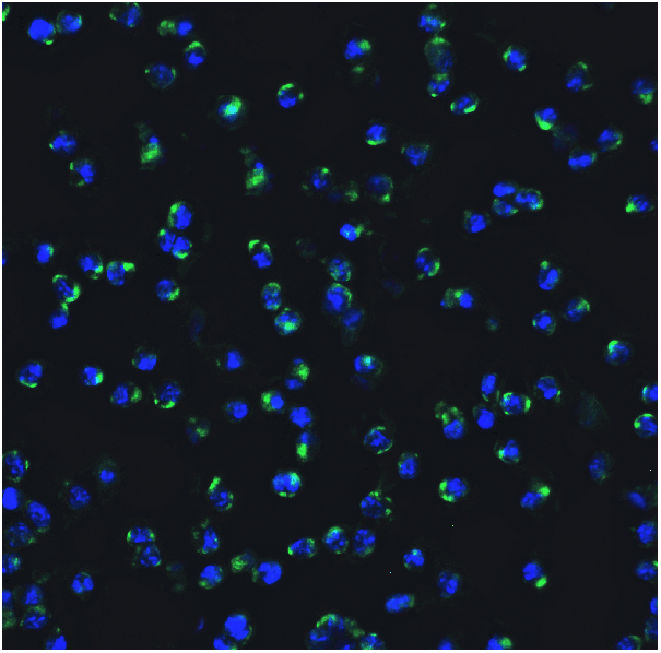
 IF (Immunofluorescence)
(Confocal immunofluorescent analysis of MMP12 Antibody (C-term) with 293 cell followed by Alexa Fluor® 488-conjugated goat anti-rabbit lgG (green). DAPI was used to stain the cell nuclear (blue).)
IF (Immunofluorescence)
(Confocal immunofluorescent analysis of MMP12 Antibody (C-term) with 293 cell followed by Alexa Fluor® 488-conjugated goat anti-rabbit lgG (green). DAPI was used to stain the cell nuclear (blue).)
MMP12, Polyclonal Antibody (Cat# AAA28713)
Full Name
MMP12 Antibody (C-term)
Gene Names
MMP12; ME; HME; MME; MMP-12
Reactivity
Human, mouse
Applications
Western Blot, Immunohistochemistry, Immunofluorescence
Purity
Purified Rabbit Polyclonal Antibody (Pab)
Pricing
 IHC (Immunohistchemistry)
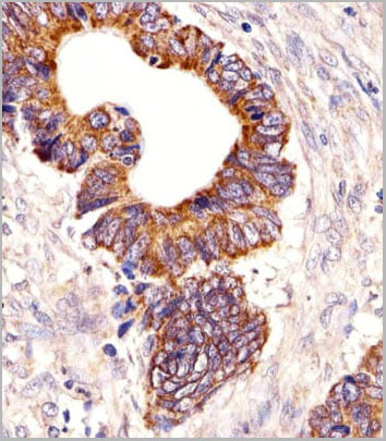
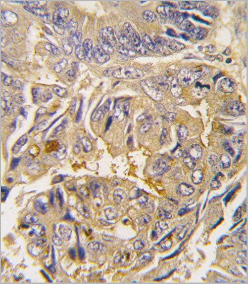
(Formalin-fixed and paraffin-embedded human colon carcinoma tissue reacted with Autophagy APG16L antibody (L176), which was peroxidase-conjugated to the secondary antibody, followed by DAB staining.This data demonstrates the use of this antibody for immunohistochemistry;clinical relevance has not been evaluated.)
IHC (Immunohistchemistry)
(Formalin-fixed and paraffin-embedded human colon carcinoma tissue reacted with Autophagy APG16L antibody (L176), which was peroxidase-conjugated to the secondary antibody, followed by DAB staining.This data demonstrates the use of this antibody for immunohistochemistry;clinical relevance has not been evaluated.)
ATG16L, Polyclonal Antibody (Cat# AAA28729)
Full Name
ATG16L Antibody
Gene Names
ATG16L1; IBD10; WDR30; APG16L; ATG16A; ATG16L
Reactivity
Human, mouse
Applications
Immunofluorescence, Western Blot, Immunohistochemistry
Purity
Purified Rabbit Polyclonal Antibody (Pab)
Pricing

 WB (Western Blot)
(Western blot analysis of STAT3 phosphorylation expression in HeLa whole cell lysates, The lane on the left is treated with the antigen-specific peptide.)
WB (Western Blot)
(Western blot analysis of STAT3 phosphorylation expression in HeLa whole cell lysates, The lane on the left is treated with the antigen-specific peptide.)
STAT3, Polyclonal Antibody (Cat# AAA31042)
Full Name
Phospho-STAT3 (Ser727) Antibody
Gene Names
STAT3; APRF; HIES; ADMIO; ADMIO1
Reactivity
Human, Mouse, Rat
Applications
Western Blot, Immunohistochemistry, Immunofluorescence, Immunocytochemistry, Immunoprecipitation
Purity
From purified rabbit serum by affinity purification via sequential chromatography on phospho-and non-phospho-peptide affinity columns.
Pricing
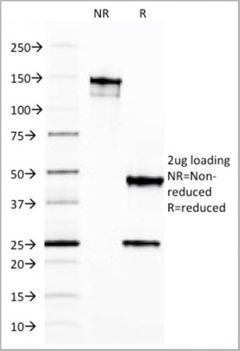
 WB (Western Blot)
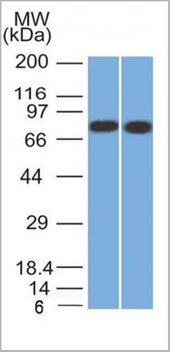
(Western Blot Analysis of Human Kidney lysate using Calnexin Mouse Monoclonal Antibody (CANX/1541).)
WB (Western Blot)
(Western Blot Analysis of Human Kidney lysate using Calnexin Mouse Monoclonal Antibody (CANX/1541).)
Calnexin, Monoclonal Antibody (Cat# AAA23894)
Full Name
Calnexin (Endoplasmic Reticulum Marker)
Gene Names
CANX; CNX; P90; IP90
Reactivity
Human.
Does not react with rat. Others not known.
Does not react with rat. Others not known.
Applications
Western Blot, Immunohistochemistry
Pricing
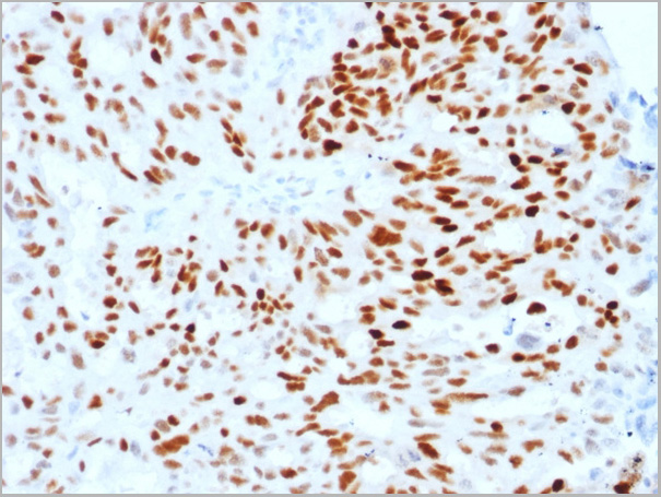
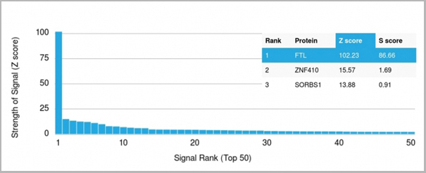
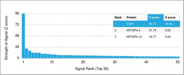 Application Data
(Analysis of Protein Array containing more than 19,000 full-length human proteins using Estrogen Receptor alpha Mouse Monoclonal Antibody (ESR1/1904) Z- and S- Score: The Z-score represents the strength of a signal that a monoclonal antibody (MAb) (in combination with a fluorescently-tagged anti-IgG secondary antibody) produces when binding to a particular protein on the HuProtTM array. Z-scores are described in units of standard deviations (SD’s) above the mean value of all signals generated on that array. If targets on HuProtTM are arranged in descending order of the Z-score, the S-score is the difference (also in units of SD’s) between the Z-score. S-score therefore represents the relative target specificity of a MAb to its intended target. A MAb is considered to specific to its intended target, if the MAb has an S-score of at least 2.5. For example, if a MAb binds to protein X with a Z-score of 43 and to protein Y with a Z-score of 14, then the S-score for the binding of that MAb to protein X is equal to 29.)
Application Data
(Analysis of Protein Array containing more than 19,000 full-length human proteins using Estrogen Receptor alpha Mouse Monoclonal Antibody (ESR1/1904) Z- and S- Score: The Z-score represents the strength of a signal that a monoclonal antibody (MAb) (in combination with a fluorescently-tagged anti-IgG secondary antibody) produces when binding to a particular protein on the HuProtTM array. Z-scores are described in units of standard deviations (SD’s) above the mean value of all signals generated on that array. If targets on HuProtTM are arranged in descending order of the Z-score, the S-score is the difference (also in units of SD’s) between the Z-score. S-score therefore represents the relative target specificity of a MAb to its intended target. A MAb is considered to specific to its intended target, if the MAb has an S-score of at least 2.5. For example, if a MAb binds to protein X with a Z-score of 43 and to protein Y with a Z-score of 14, then the S-score for the binding of that MAb to protein X is equal to 29.)
Estrogen Receptor, alpha, Monoclonal Antibody (Cat# AAA23914)
Full Name
Estrogen Receptor, alpha (Marker of Estrogen Dependence)
Gene Names
ESR1; ER; ESR; Era; ESRA; ESTRR; NR3A1
Reactivity
Human
Applications
Western Blot, Immunohistochemistry
Purity
Purified Ab with BSA and Azide at 200ug/ml OR Purified Ab WITHOUT BSA and Azide at 1.0mg/ml
Pricing
 WB (Western Blot)
(Western Blot Analysis of human kidney cell lysate using Ferritin, Light ChainMouse Monoclonal Antibody (FTL/1387).)
WB (Western Blot)
(Western Blot Analysis of human kidney cell lysate using Ferritin, Light ChainMouse Monoclonal Antibody (FTL/1387).)
Ferritin, Light Chain (FTL), Monoclonal Antibody (Cat# AAA23907)
Full Name
Ferritin, Light Chain (FTL) (Microglia Marker)
Gene Names
FTL; LFTD; NBIA3
Reactivity
Human
Applications
Western Blot, Immunohistochemistry
Pricing
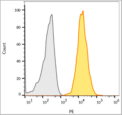
 FCM (Flow Cytometry)
(Overlay histogram showing HeLa cells stained with AAA28782(green line). The cells were fixed with 2% paraformaldehyde (10 min) and then permeabilized with 90% methanol for 10 min. The cells were then icubated in 2% bovine serum albumin to block non-specific protein-protein interactions followed by the antibody (AAA28782, 1:25 dilution) for 60 min at 37ºC. The secondary antibody used was Goat-Anti-Rabbit IgG, DyLight® 488 Conjugated Highly Cross-Adsorbed at 1/200 dilution for 40 min at 37ºC. Isotype control antibody (blue line) was rabbit IgG1 (1ug/1x10^6 cells) used under the same conditions. Acquisition of >10, 000 events was performed.)
FCM (Flow Cytometry)
(Overlay histogram showing HeLa cells stained with AAA28782(green line). The cells were fixed with 2% paraformaldehyde (10 min) and then permeabilized with 90% methanol for 10 min. The cells were then icubated in 2% bovine serum albumin to block non-specific protein-protein interactions followed by the antibody (AAA28782, 1:25 dilution) for 60 min at 37ºC. The secondary antibody used was Goat-Anti-Rabbit IgG, DyLight® 488 Conjugated Highly Cross-Adsorbed at 1/200 dilution for 40 min at 37ºC. Isotype control antibody (blue line) was rabbit IgG1 (1ug/1x10^6 cells) used under the same conditions. Acquisition of >10, 000 events was performed.)
HNRNPR, Polyclonal Antibody (Cat# AAA28782)
Full Name
HNRNPR Antibody (N-term)
Gene Names
HNRNPR; HNRPR; hnRNP-R
Reactivity
Human
Applications
Immunofluorescence, Western Blot, Immunohistochemistry, Flow Cytometry
Purity
Peptide Affinity Purified Rabbit Polyclonal Antibody (Pab)
Pricing


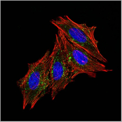
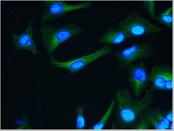
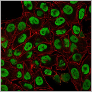
 IF (Immunofluorescence)
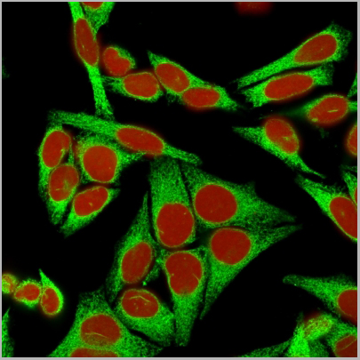
(Confocal immunofluorescence image of HeLa Cells using S100A4 Mouse Monoclonal Antibody (S100A4/1482) followed by goat anti-mouse IgG-CF488 (green). Reddot is used to label the nuclei Red.)
IF (Immunofluorescence)
(Confocal immunofluorescence image of HeLa Cells using S100A4 Mouse Monoclonal Antibody (S100A4/1482) followed by goat anti-mouse IgG-CF488 (green). Reddot is used to label the nuclei Red.)
S100A4, Monoclonal Antibody (Cat# AAA23892)
Full Name
S100A4 (Marker of Tumor Metastasis)
Gene Names
S100A4; 42A; 18A2; CAPL; FSP1; MTS1; P9KA; PEL98
Reactivity
Human, Mouse
Applications
Flow Cytometry, Immunofluorescence, Western Blot, Immunohistochemistry
Pricing


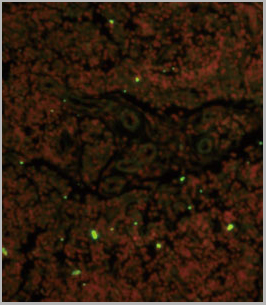
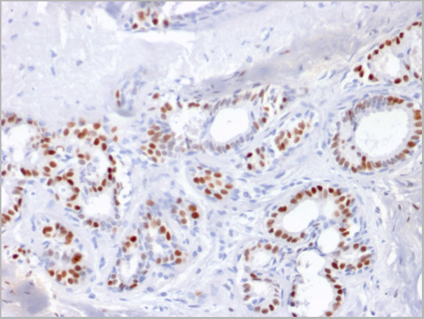
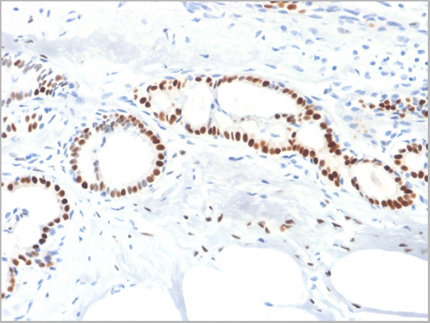
 IHC (Immunohistchemistry)
(Formalin-fixed, paraffin-embedded human Colon Carcinoma stained with AF488 Conjugate of Nucleolin Monoclonal Antibody (364-5 + NCL/902).)
IHC (Immunohistchemistry)
(Formalin-fixed, paraffin-embedded human Colon Carcinoma stained with AF488 Conjugate of Nucleolin Monoclonal Antibody (364-5 + NCL/902).)
Nucleolin, Monoclonal Antibody (Cat# AAA13820)
Full Name
Nucleolin (Marker of Human Cells) Mouse Monoclonal Antibody
Gene Names
NCL; C23
Reactivity
Human.
Does not react with Mouse, Rat and Cow
Does not react with Mouse, Rat and Cow
Applications
Flow Cytometry, Immunofluorescence, Western Blot, Immunohistochemistry
Pricing
MRPL12, Monoclonal Antibody (Cat# AAA24279)
Full Name
MRPL12 (Mitochondrial Ribosomal Protein L12, 39S Ribosomal Protein L12, Mitochondrial, 5c5-2, FLJ60124, L12mt, MGC8610, MRP-L12, MRP-L31/34, MRPL7, MRPL7/L12, RPML12) (AP)
Gene Names
MRPL12; 5c5-2; L12mt; MRPL7; RPML12; MRPL7/L12; MRP-L31/34
Reactivity
Human
Applications
EIA, IHC, WB
Purity
Purified by Protein A Affinity Chromatography.
Pricing