Filters
Clonality
Type
Reactivity
Gene Name
Isotype
Host
Application
Clone
162 results for " TNF" - showing 100-150
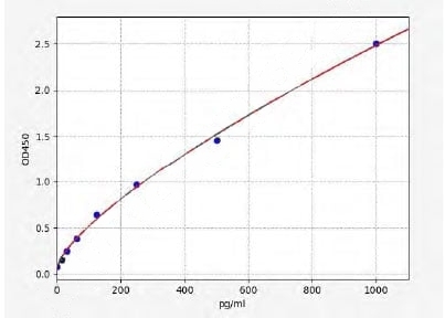 Standard Curve (Sample)
Lymphotoxin-alpha, ELISA Kit (Cat# AAA17685)
Full Name
Mouse Lymphotoxin-alpha ELISA Kit
Gene Names
LTA; LT; TNFB; TNFSF1; TNLG1E
Reactivity
Mouse
Pricing
 Standard Curve (Sample)
Complement 1q, ELISA Kit (Cat# AAA16817)
Full Name
Porcine Complement 1q ELISA Kit
Gene Names
C1QTNF5; CTRP5
Reactivity
Porcine
Pricing
 Standard Curve (Sample)
TNF alpha, ELISA Kit (Cat# AAA17879)
Full Name
Rat TNF alpha ELISA Kit
Gene Names
Tnf; Tnfa; RATTNF; TNF-alpha
Reactivity
Rat
Applications
SE
Pricing
 Standard Curve (Sample)
Tumor necrosis factor, ELISA Kit (Cat# AAA23305)
Full Name
Bovine Tumor necrosis factor ELISA Kit
Gene Names
TNF; TNFa; TNF-a; TNF-alpha
Reactivity
Bovine
Pricing
 Standard Curve (Sample)
TL1A, ELISA Kit (Cat# AAA21803)
Full Name
Human TL1A (Tumor Necrosis Factor Related Ligand 1A) ELISA Kit
Gene Names
TNFSF15; TL1; TL1A; VEGI; VEGI192A
Reactivity
Human
Pricing
 Application Data
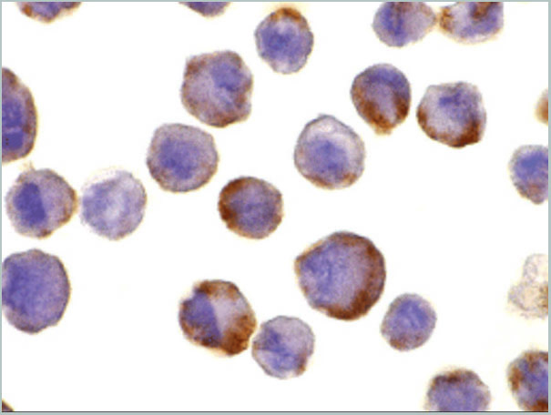
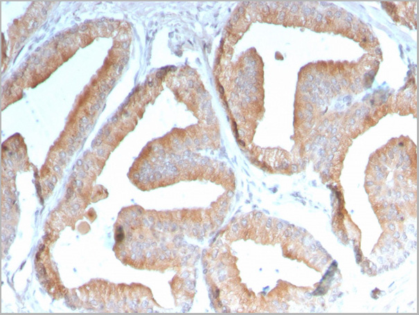
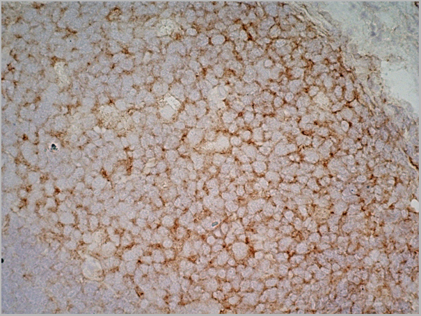
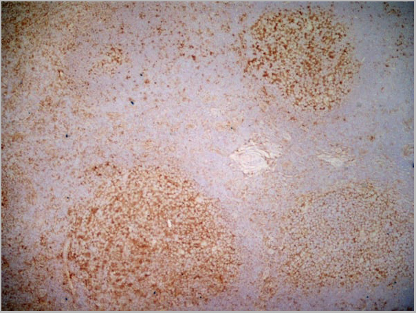
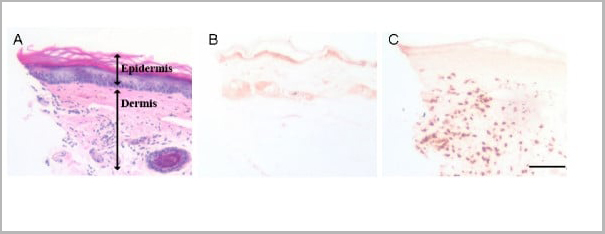
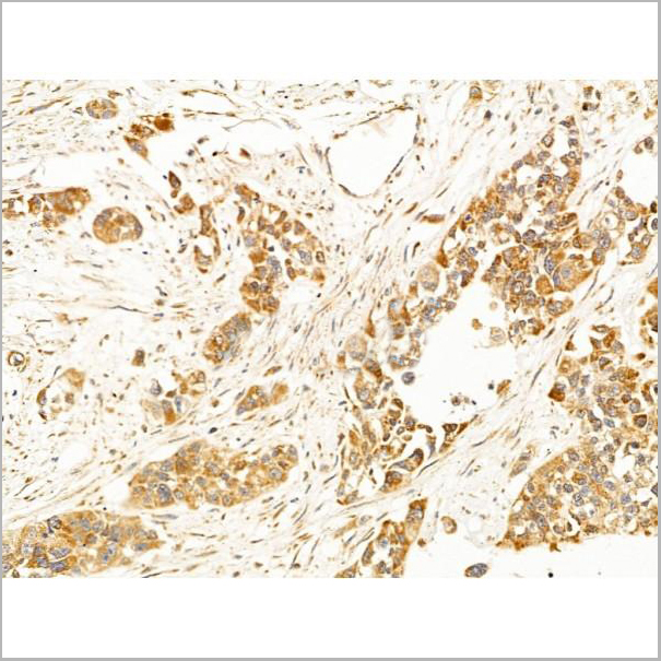
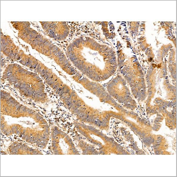
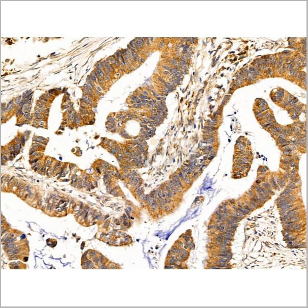
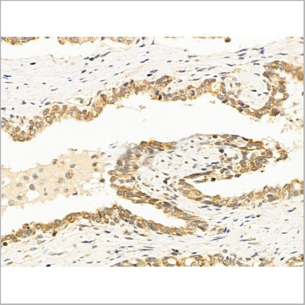
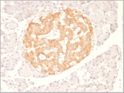
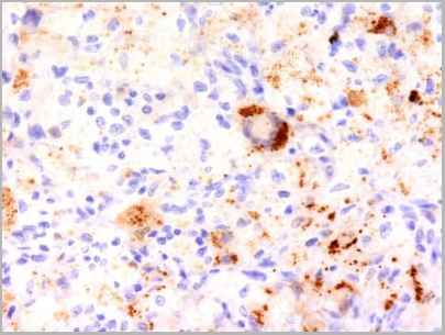
(Figure: Biopsy of tumor lesion from a patient with lymphoma (DLBCL) stained with MAb to APRIL (Aprily-2) . The biopsy was stained by standard immunohistochemistry. The figure shows the selective accumulation of APRIL around tumor cells.Picture courtesy of Dr. Bertrand Huard (University of Geneva).)
Application Data
(Figure: Biopsy of tumor lesion from a patient with lymphoma (DLBCL) stained with MAb to APRIL (Aprily-2) . The biopsy was stained by standard immunohistochemistry. The figure shows the selective accumulation of APRIL around tumor cells.Picture courtesy of Dr. Bertrand Huard (University of Geneva).)
APRIL, Monoclonal Antibody (Cat# AAA14518)
Full Name
APRIL (human) monoclonal antibody (Aprily-2)
Gene Names
TNFSF13; APRIL; CD256; TALL2; ZTNF2; TALL-2; TNLG7B; TRDL-1; UNQ383/PRO715
Reactivity
Human
Applications
Immunohistochemistry
Pricing
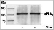
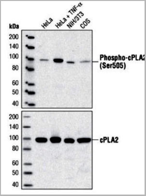 WB (Western Blot)
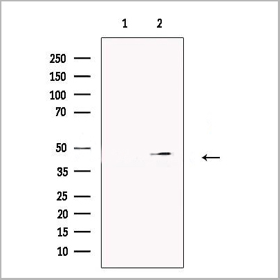
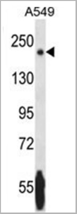
(Western blot analysis of extracts from HeLa, C6 and NIH/3T3 cells, using AAA14717.)
WB (Western Blot)
(Western blot analysis of extracts from HeLa, C6 and NIH/3T3 cells, using AAA14717.)
c-Phospholipase A2, Polyclonal Antibody (Cat# AAA14717)
Full Name
c-Phospholipase A2 (Cytosolic PLA2, cPLA2)
Reactivity
Human
Applications
Western Blot, Immunoprecipitation
Purity
Purified by Protein A and peptide affinity chromatography
Pricing
 Application Data
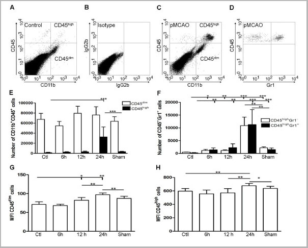
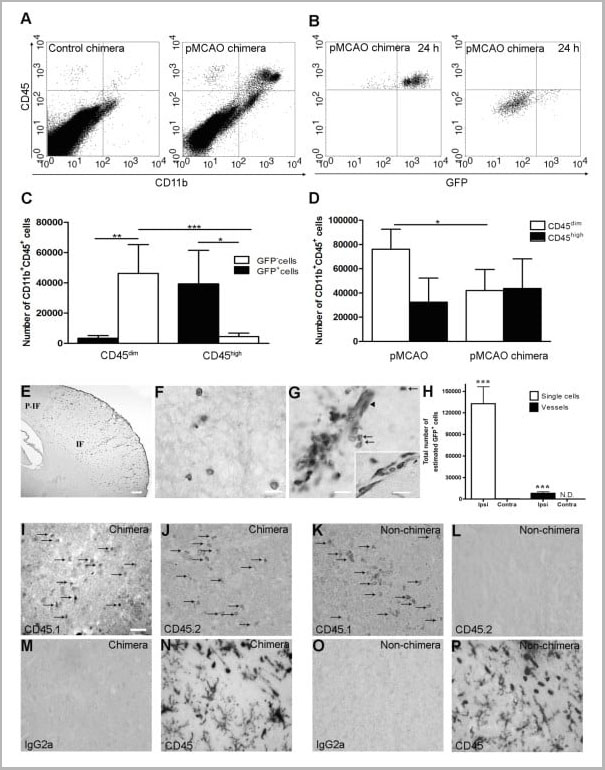
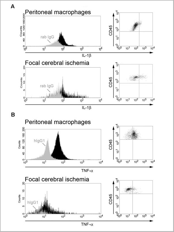
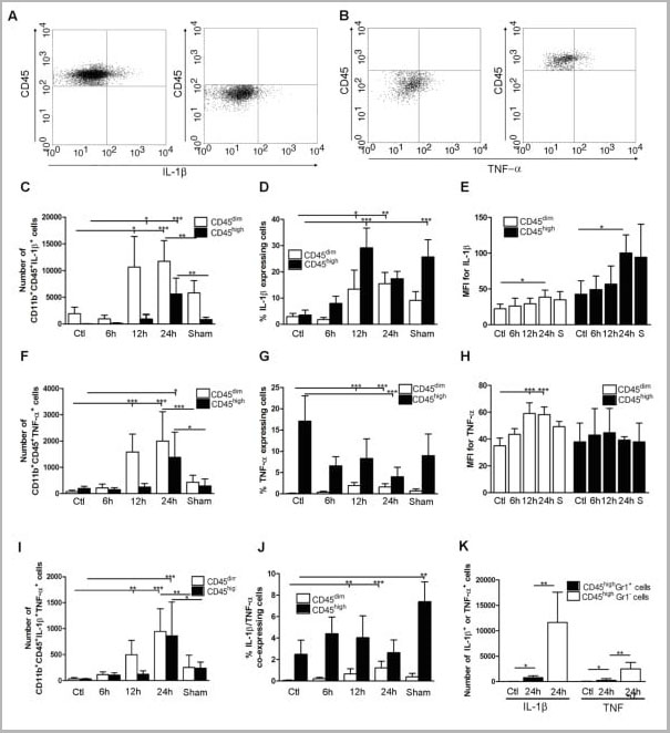
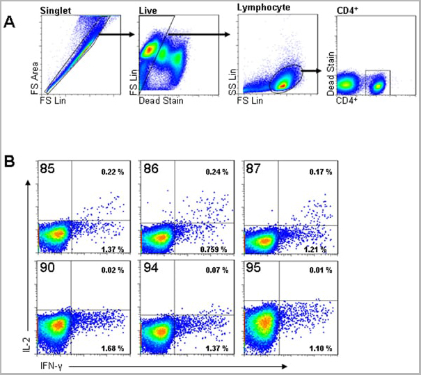
(Published customer image: Cytokine expression in segregated populations of cells following stroke. (A, B) Dot plots showing CD11b+CD45high macrophages/granulocytes (upper right quadrants) and CD11b+CD45dim microglia (bottom right quadrants) expressing IL-1beta (A) or TNF-a (B). (C-J) Bar graphs showing numbers and proportions of IL-1beta (C, D), TNF-a (F, G) and IL-1beta/TNF-a co-expressing (I, J) CD11b+CD45dim microglia and CD11b+CD45high macrophages/granulocytes in unmanipulated control mice (n = 10), in mice 6 (n = 7), 12 (n = 7), or 24 hours after pMCAO (n = 10), and in sham-operated mice 24 hours after pMCAO (n = 7). (E, H) Comparison of the MFI values for IL-1beta (E) and TNF-a (H) in viable CD11b+CD45dim microglia and CD11b+CD45high macrophages/granulocytes in unmanipulated mice, in mice 6, 12, or 24 hours after pMCAO, and in sham-operated mice 24 hours after pMCAO. Macrophages/granulocytes express significantly more IL-1beta than do microglial in unmanipulated mice, in mice 6, 12, or 24 hours after pMCAO, and in sham-operated mice 24 hours after pMCAO (E), whereas microglial cells express significantly higher levels of TNF-a than do macrophages/granulocytes at 12 h and 24 hours, and in sham-operated mice 24 hours after pMCAO (H). (K) CD11b+CD45highGr1- macrophages and not CD11b+CD45highGr1+ granulocytes are the main producers of IL-1beta and TNF-a 24 hours after pMCAO. *P < 0.05, **P < 0.01, and ***P < 0.001.From: http://www.jneuroinflammation.com/content/5/1/46.)
Application Data
(Published customer image: Cytokine expression in segregated populations of cells following stroke. (A, B) Dot plots showing CD11b+CD45high macrophages/granulocytes (upper right quadrants) and CD11b+CD45dim microglia (bottom right quadrants) expressing IL-1beta (A) or TNF-a (B). (C-J) Bar graphs showing numbers and proportions of IL-1beta (C, D), TNF-a (F, G) and IL-1beta/TNF-a co-expressing (I, J) CD11b+CD45dim microglia and CD11b+CD45high macrophages/granulocytes in unmanipulated control mice (n = 10), in mice 6 (n = 7), 12 (n = 7), or 24 hours after pMCAO (n = 10), and in sham-operated mice 24 hours after pMCAO (n = 7). (E, H) Comparison of the MFI values for IL-1beta (E) and TNF-a (H) in viable CD11b+CD45dim microglia and CD11b+CD45high macrophages/granulocytes in unmanipulated mice, in mice 6, 12, or 24 hours after pMCAO, and in sham-operated mice 24 hours after pMCAO. Macrophages/granulocytes express significantly more IL-1beta than do microglial in unmanipulated mice, in mice 6, 12, or 24 hours after pMCAO, and in sham-operated mice 24 hours after pMCAO (E), whereas microglial cells express significantly higher levels of TNF-a than do macrophages/granulocytes at 12 h and 24 hours, and in sham-operated mice 24 hours after pMCAO (H). (K) CD11b+CD45highGr1- macrophages and not CD11b+CD45highGr1+ granulocytes are the main producers of IL-1beta and TNF-a 24 hours after pMCAO. *P < 0.05, **P < 0.01, and ***P < 0.001.From: http://www.jneuroinflammation.com/content/5/1/46.)
CD45, Monoclonal Antibody (Cat# AAA11896)
Full Name
RAT ANTI MOUSE CD45
Gene Names
Ptprc; loc; B220; Cd45; L-CA; Ly-5; T200; CD45R; Lyt-4
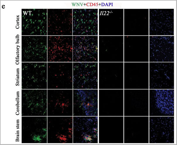
Applications
Immunohistochemistry, Flow Cytometry, Immunofluorescence, Immunoprecipitation
Pricing
OX40L, Recombinant Antibody (Cat# AAA14184)
Full Name
Anti-OX40L [R4930 (Oxelumab)]
Gene Names
TNFSF4; GP34; CD252; OX4OL; TXGP1; CD134L; OX-40L; TNLG2B
Reactivity
Human
Applications
Flow Cytometry
Purity
Protein A Affinity Purified
Pricing

 FCM (Flow Cytometry)
(Flow cytometry analysis of LIGHT overexpressing HEK293 cells using LIGHT antibody and control mouse IgG antibody at 10 μg/ml. Blue: Untransfected HEK293 cells. Yellow: LIGHT overexpressing HEK293 cells.)
FCM (Flow Cytometry)
(Flow cytometry analysis of LIGHT overexpressing HEK293 cells using LIGHT antibody and control mouse IgG antibody at 10 μg/ml. Blue: Untransfected HEK293 cells. Yellow: LIGHT overexpressing HEK293 cells.)
LIGHT, Monoclonal Antibody (Cat# AAA11010)
Full Name
LIGHT Antibody [7B9E12]
Gene Names
TNFSF14; LTg; CD258; HVEML; LIGHT
Reactivity
Human
Applications
Western Blot, Immunohistochemistry, Immunocytochemistry, Immunofluorescence, Flow Cytometry
Purity
Protein A purified
Pricing

 IF (Immunofluorescence)
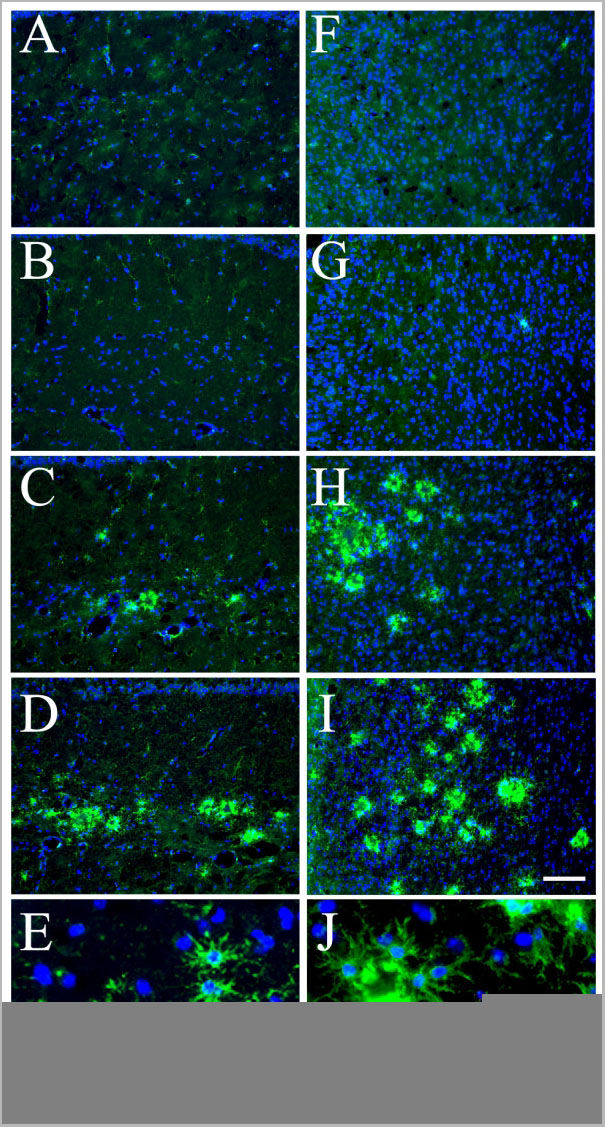
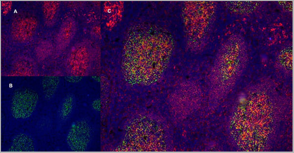
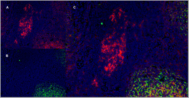
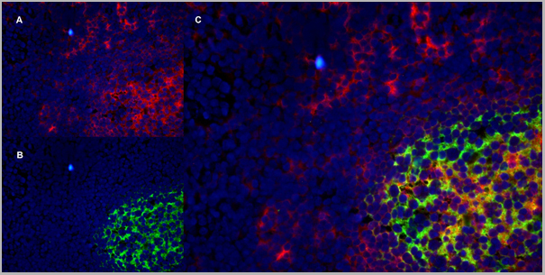
(Figure 10 Immunofluorescence Validation of TACE in Rat Brain (Pradillo et al, 2005)Cellular localization of TACE. Double immunofluorescence staining of brain sections from sham-operated (SHAM; A, C, E) and IPC-exposed animals (IPC; B, D, F) of TACE (red) and the cellular markers (green) NeuN (neurons; A, B), GFAP (astrocytes; C, D) and L. esculentum lectin (microglia and endothelium; E, F). White arrows indicate TACE-positive cells.)
IF (Immunofluorescence)
(Figure 10 Immunofluorescence Validation of TACE in Rat Brain (Pradillo et al, 2005)Cellular localization of TACE. Double immunofluorescence staining of brain sections from sham-operated (SHAM; A, C, E) and IPC-exposed animals (IPC; B, D, F) of TACE (red) and the cellular markers (green) NeuN (neurons; A, B), GFAP (astrocytes; C, D) and L. esculentum lectin (microglia and endothelium; E, F). White arrows indicate TACE-positive cells.)
TACE, Polyclonal Antibody (Cat# AAA10912)
Full Name
TACE Antibody
Gene Names
ADAM17; CSVP; TACE; NISBD; ADAM18; CD156B; NISBD1
Reactivity
Human, Mouse, Rat
Applications
Western Blot, Immunocytochemistry, Immunofluorescence, Flow Cytometry
Purity
TACE Antibody is affinity chromatography purified via peptide column.
Pricing
 FCM (Flow Cytometry)
(Flow cytometry analysis of LIGHT overexpressing HEK293 cells using LIGHT antibody and control mouse IgG antibody at 10 μg/ml. Blue: Untransfected HEK293 cells. Yellow: LIGHT overexpressing HEK293 cells.)
FCM (Flow Cytometry)
(Flow cytometry analysis of LIGHT overexpressing HEK293 cells using LIGHT antibody and control mouse IgG antibody at 10 μg/ml. Blue: Untransfected HEK293 cells. Yellow: LIGHT overexpressing HEK293 cells.)
LIGHT, Monoclonal Antibody (Cat# AAA11008)
Full Name
LIGHT Antibody [7B9F12]
Gene Names
TNFSF14; LTg; CD258; HVEML; LIGHT
Reactivity
Human
Applications
Western Blot, Immunohistochemistry, Immunocytochemistry, Immunofluorescence, Flow Cytometry
Purity
Protein A purified
Pricing


 IF (Immunofluorescence)
(AAA31063 staining HeLa by IF/ICC. The sample were fixed with PFA and permeabilized in 0.1% Triton X-100, then blocked in 10% serum for 45 minutes at 25 degree C. The primary antibody was diluted at 1/200 and incubated with the sample for 1 hour at 37 degree C. An Alexa Fluor 594 conjugated goat anti-rabbit IgG (H+L) Ab, diluted at 1/600, was used as the secondary antibody.)
IF (Immunofluorescence)
(AAA31063 staining HeLa by IF/ICC. The sample were fixed with PFA and permeabilized in 0.1% Triton X-100, then blocked in 10% serum for 45 minutes at 25 degree C. The primary antibody was diluted at 1/200 and incubated with the sample for 1 hour at 37 degree C. An Alexa Fluor 594 conjugated goat anti-rabbit IgG (H+L) Ab, diluted at 1/600, was used as the secondary antibody.)
NF kappaB p65, Polyclonal Antibody (Cat# AAA31063)
Full Name
Phospho-NF kappaB p65 (Ser281) Antibody
Gene Names
RELA; p65; NFKB3
Reactivity
Human, Mouse, Rat
Applications
Western Blot, Immunohistochemistry, Immunofluorescence, Immunocytochemistry
Purity
From purified rabbit serum by affinity purification via sequential chromatography on phospho-and non-phospho-peptide affinity columns.
Pricing

 FCM (Flow Cytometry)
(Flow cytometry analysis of LIGHT overexpressing HEK293 cells using LIGHT antibody and control mouse IgG antibody at 10 μg/ml. Blue: Untransfected HEK293 cells. Yellow: LIGHT overexpressing HEK293 cells.)
FCM (Flow Cytometry)
(Flow cytometry analysis of LIGHT overexpressing HEK293 cells using LIGHT antibody and control mouse IgG antibody at 10 μg/ml. Blue: Untransfected HEK293 cells. Yellow: LIGHT overexpressing HEK293 cells.)
LIGHT, Monoclonal Antibody (Cat# AAA11009)
Full Name
LIGHT Antibody [7B9H9]
Gene Names
TNFSF14; LTg; CD258; HVEML; LIGHT
Reactivity
Human
Applications
Western Blot, Immunohistochemistry, Immunocytochemistry, Immunofluorescence, Flow Cytometry
Purity
Protein A purified
Pricing
 Application Data
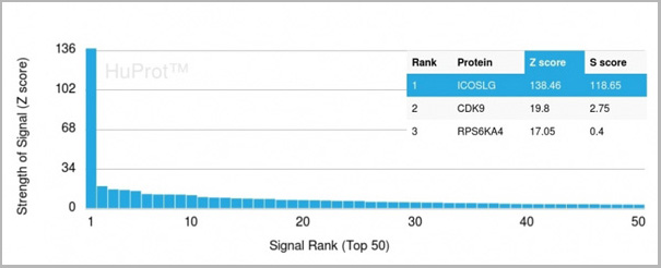
(Analysis of Protein Array containing more than 19,000 full-length human proteins using ICOS-L Mouse Monoclonal Antibody (ICOSL/3111). Z- and S- Score: The Z-score represents the strength of a signal that a monoclonal antibody (MAb) (in combination with a fluorescently-tagged anti-IgG secondary antibody) produces when binding to a particular protein on the HuProtTM array. Z-scores are described in units of standard deviations (SD’s) above the mean value of all signals generated on that array. If targets on HuProtTM are arranged in descending order of the Z-score, the S-score is the difference (also in units of SD’s) between the Z-score. S-score therefore represents the relative target specificity of a MAb to its intended target. A MAb is considered to specific to its intended target, if the MAb has an S-score of at least 2.5. For example, if a MAb binds to protein X with a Z-score of 43 and to protein Y with a Z-score of 14, then the S-score for the binding of that MAb to protein X is equal to 29.)
Application Data
(Analysis of Protein Array containing more than 19,000 full-length human proteins using ICOS-L Mouse Monoclonal Antibody (ICOSL/3111). Z- and S- Score: The Z-score represents the strength of a signal that a monoclonal antibody (MAb) (in combination with a fluorescently-tagged anti-IgG secondary antibody) produces when binding to a particular protein on the HuProtTM array. Z-scores are described in units of standard deviations (SD’s) above the mean value of all signals generated on that array. If targets on HuProtTM are arranged in descending order of the Z-score, the S-score is the difference (also in units of SD’s) between the Z-score. S-score therefore represents the relative target specificity of a MAb to its intended target. A MAb is considered to specific to its intended target, if the MAb has an S-score of at least 2.5. For example, if a MAb binds to protein X with a Z-score of 43 and to protein Y with a Z-score of 14, then the S-score for the binding of that MAb to protein X is equal to 29.)
ICOS-L/ICOS Ligand/B7RP-1 (Immuno-Oncology Target), Monoclonal Antibody (Cat# AAA23916)
Full Name
ICOS-L/ICOS Ligand/B7RP-1 (Immuno-Oncology Target)
Gene Names
ICOSLG; B7h; B7H2; GL50; B7-H2; B7RP1; CD275; ICOSL; LICOS; B7RP-1; ICOS-L
Reactivity
Human
Applications
Flow Cytometry, Immunofluorescence, Immunohistochemistry
Purity
Purified Ab with BSA and Azide at 200ug/ml
Pricing


 Application Data
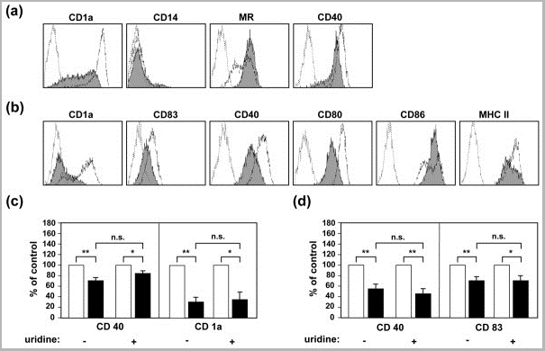
(Staining of human peripheral blood lymphocytes with Mouse anti Human CD40:RPE)
Application Data
(Staining of human peripheral blood lymphocytes with Mouse anti Human CD40:RPE)
CD40, Monoclonal Antibody (Cat# AAA12090)
Full Name
MOUSE ANTI HUMAN CD40
Gene Names
CD40; p50; Bp50; CDW40; TNFRSF5
Applications
Immunohistochemistry, Flow Cytometry, Immunoprecipitation, Immunohistochemistry
Pricing


 Application Data
(Staining of human peripheral blood lymphocytes with Mouse anti Human CD40:RPE)
Application Data
(Staining of human peripheral blood lymphocytes with Mouse anti Human CD40:RPE)
CD40, Monoclonal Antibody (Cat# AAA11867)
Full Name
MOUSE ANTI HUMAN CD40:FITC
Gene Names
CD40; p50; Bp50; CDW40; TNFRSF5
Applications
Flow Cytometry
Pricing


 Application Data
(Staining of human peripheral blood lymphocytes with Mouse anti Human CD40:RPE)
Application Data
(Staining of human peripheral blood lymphocytes with Mouse anti Human CD40:RPE)
CD40, Monoclonal Antibody (Cat# AAA11936)
Full Name
MOUSE ANTI HUMAN CD40
Gene Names
CD40; p50; Bp50; CDW40; TNFRSF5
Reactivity
Dog
Applications
Immunohistochemistry, Flow Cytometry, Immunoprecipitation, Immunohistochemistry
Pricing


 Application Data
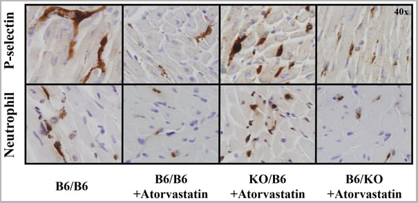
(Staining of New Zealand Black mouse peripheral blood granulocytes with Rat anti Mouse Ly-6B.2 conjugated to FITC Data)
Application Data
(Staining of New Zealand Black mouse peripheral blood granulocytes with Rat anti Mouse Ly-6B.2 conjugated to FITC Data)
Ly-6B.2 ALLOANTIGEN, Monoclonal Antibody (Cat# AAA12197)
Full Name
RAT ANTI MOUSE Ly-6B.2 ALLOANTIGEN:RPE
Applications
Flow Cytometry
Pricing


 Application Data
(Staining of human peripheral blood lymphocytes with Mouse anti Human CD40:RPE)
Application Data
(Staining of human peripheral blood lymphocytes with Mouse anti Human CD40:RPE)
CD40, Monoclonal Antibody (Cat# AAA12089)
Full Name
MOUSE ANTI HUMAN CD40:RPE
Gene Names
CD40; p50; Bp50; CDW40; TNFRSF5
Applications
Flow Cytometry
Pricing


 Application Data
(Staining of New Zealand Black mouse peripheral blood granulocytes with Rat anti Mouse Ly-6B.2 conjugated to FITC Data)
Application Data
(Staining of New Zealand Black mouse peripheral blood granulocytes with Rat anti Mouse Ly-6B.2 conjugated to FITC Data)
Ly-6B.2 ALLOANTIGEN, Monoclonal Antibody (Cat# AAA12193)
Full Name
RAT ANTI MOUSE Ly-6B.2 ALLOANTIGEN:FITC
Applications
Flow Cytometry
Pricing
 Biological Activity
(Human TNF-alpha induces cell cytotoxicity in the L-929 mouse fibroblast cells in the presence of the metabolic inhibitor actinomycin D.)
Biological Activity
(Human TNF-alpha induces cell cytotoxicity in the L-929 mouse fibroblast cells in the presence of the metabolic inhibitor actinomycin D.)
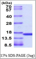
Tumor Necrosis Factor-alpha, Active Protein (Cat# AAA11742)
Full Name
Tumor Necrosis Factor-alpha, 77-233 aa, Human, E Coli (Bioactivity Validated)
Gene Names
TNF; DIF; TNFA; TNFSF2; TNLG1F; TNF-alpha
Applications
SDS-PAGE, Bioactivity
Purity
> 95% by SDS-PAGE
Pricing


 Application Data
(Staining of human peripheral blood lymphocytes with Mouse anti Human CD40:RPE)
Application Data
(Staining of human peripheral blood lymphocytes with Mouse anti Human CD40:RPE)
CD40, Monoclonal Antibody (Cat# AAA12031)
Full Name
MOUSE ANTI HUMAN CD40:RPE
Gene Names
CD40; p50; Bp50; CDW40; TNFRSF5
Applications
Flow Cytometry
Pricing


 Application Data
(Staining of New Zealand Black mouse peripheral blood granulocytes with Rat anti Mouse Ly-6B.2 conjugated to FITC Data)
Application Data
(Staining of New Zealand Black mouse peripheral blood granulocytes with Rat anti Mouse Ly-6B.2 conjugated to FITC Data)
Ly-6B.2 ALLOANTIGEN, Monoclonal Antibody (Cat# AAA12192)
Full Name
RAT ANTI MOUSE Ly-6B.2 ALLOANTIGEN:FITC
Applications
Flow Cytometry
Pricing


 Application Data
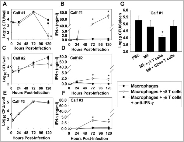
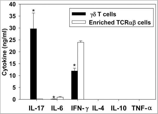
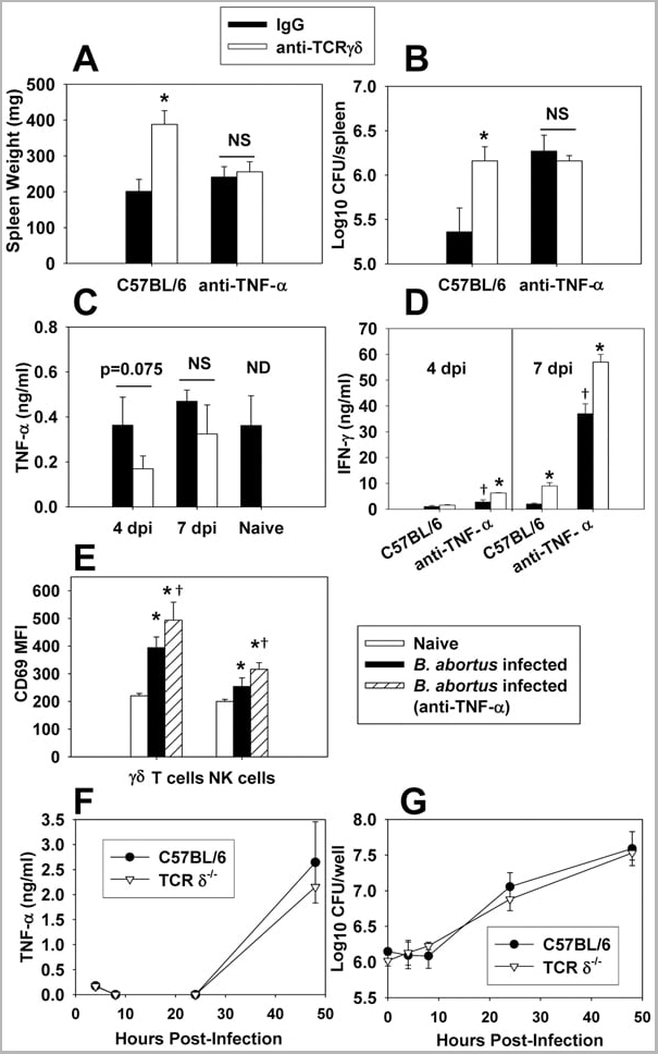
(Published customer image: gamma delta T cells are the primary source of IL-17 during B. abortus infection. C57BL/6 mice were infected i.p. with 5x10⁴ CFUs of B. abortus 2308, and two weeks later gamma delta T cells (>95% purity) and an enriched TCRalphabeta (~55% CD4+, 25% CD8+) cell fraction were isolated from the spleens of infected mice. Cells were stimulated with 500 ng/ml ionomycin and 50 ng/ml PMA for three days, and cell-free supernatants from triplicate wells were assayed for cytokine production via ELISA. The mean +/- SD is shown; * P)
Application Data
(Published customer image: gamma delta T cells are the primary source of IL-17 during B. abortus infection. C57BL/6 mice were infected i.p. with 5x10⁴ CFUs of B. abortus 2308, and two weeks later gamma delta T cells (>95% purity) and an enriched TCRalphabeta (~55% CD4+, 25% CD8+) cell fraction were isolated from the spleens of infected mice. Cells were stimulated with 500 ng/ml ionomycin and 50 ng/ml PMA for three days, and cell-free supernatants from triplicate wells were assayed for cytokine production via ELISA. The mean +/- SD is shown; * P)
IFN GAMMA, Monoclonal Antibody (Cat# AAA12093)
Full Name
MOUSE ANTI BOVINE INTERFERON GAMMA
Reactivity
Dog, Dolphin, Ferret, Fin Whale, Goat, Horse, Human, Mink, Rabbit, Pig, Sheep.
Based on sequence similarity, is expected to react with: Mustelid
N.B. Antibody reactivity and working conditions may vary between species.
Based on sequence similarity, is expected to react with: Mustelid
N.B. Antibody reactivity and working conditions may vary between species.
Applications
Flow Cytometry
Pricing


 Application Data
(Staining of New Zealand Black mouse peripheral blood granulocytes with Rat anti Mouse Ly-6B.2 conjugated to FITC Data)
Application Data
(Staining of New Zealand Black mouse peripheral blood granulocytes with Rat anti Mouse Ly-6B.2 conjugated to FITC Data)
Ly-6B.2 ALLOANTIGEN, Monoclonal Antibody (Cat# AAA12194)
Full Name
RAT ANTI MOUSE Ly-6B.2 ALLOANTIGEN
Applications
Immunohistochemistry, Flow Cytometry, Immunofluorescence, Immunohistochemistry, Western Blot
Pricing


 Application Data
(Staining of New Zealand Black mouse peripheral blood granulocytes with Rat anti Mouse Ly-6B.2 conjugated to FITC Data)
Application Data
(Staining of New Zealand Black mouse peripheral blood granulocytes with Rat anti Mouse Ly-6B.2 conjugated to FITC Data)
Ly-6B.2 ALLOANTIGEN, Monoclonal Antibody (Cat# AAA12198)
Full Name
RAT ANTI MOUSE Ly-6B.2 ALLOANTIGEN:RPE
Applications
Flow Cytometry
Pricing
 Application Data
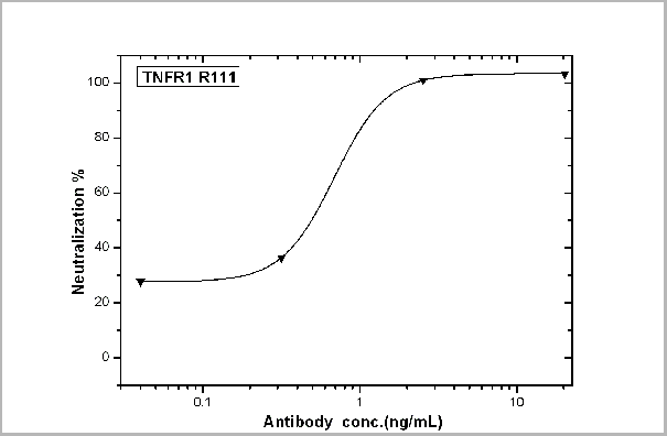
(TNFR1/TNFRSF1A-mediated inhibition of cytotoxicity was Neutralized by Human TNFR1 Antibody. Recombinant Human TNFR1/TNFRSF1A inhibits Recombinant Human TNFa induced cytotoxicity in the L-929 mouse fibroblast cell line. Inhibition of Recombinant Human TNFa (0.2 ng/mL) activity elicited by Recombinant Human TNFR1/TNFRSF1A (0.3 ug/mL) is neutralized by increasing concentrations of Human TNFR1/TNFRSF1A Monoclonal Antibody. The IC50 is typically 0.5-1.5 ug/mL in the presence of the metabolic inhibitor actinomycin D (1 ug/mL).)
Application Data
(TNFR1/TNFRSF1A-mediated inhibition of cytotoxicity was Neutralized by Human TNFR1 Antibody. Recombinant Human TNFR1/TNFRSF1A inhibits Recombinant Human TNFa induced cytotoxicity in the L-929 mouse fibroblast cell line. Inhibition of Recombinant Human TNFa (0.2 ng/mL) activity elicited by Recombinant Human TNFR1/TNFRSF1A (0.3 ug/mL) is neutralized by increasing concentrations of Human TNFR1/TNFRSF1A Monoclonal Antibody. The IC50 is typically 0.5-1.5 ug/mL in the presence of the metabolic inhibitor actinomycin D (1 ug/mL).)
TNFR1/CD120a/TNFRSF1A, Monoclonal Antibody (Cat# AAA27740)
Full Name
TNFR1/CD120a/TNFRSF1A Neutralizing Antibody
Gene Names
TNFRSF1A; FPF; MS5; p55; p60; TBP1; TNF-R; TNFAR; TNFR1; p55-R; CD120a; TNFR55; TNFR60; TNF-R-I; TNF-R55; TNFR1-d2
Applications
Neutralization
Pricing
 Application Data
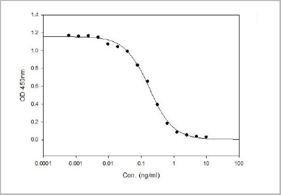
(Sandwich ELISA analysis of CD178 binding using Mouse anti Human CD178 as a capture reagent and biotinylated Mouse anti Human CD178 as a detection reagent with purified human CD178 as antigen for the generation of a standard curve. Detection is by HRP conjugated Streptavidin and substrate. Microtitre plate is read at O.D. 450 nm on the iMark Microplate Absorbance Reader . Plasma (orange) sample is displayed at 1:2 dilution.)
Application Data
(Sandwich ELISA analysis of CD178 binding using Mouse anti Human CD178 as a capture reagent and biotinylated Mouse anti Human CD178 as a detection reagent with purified human CD178 as antigen for the generation of a standard curve. Detection is by HRP conjugated Streptavidin and substrate. Microtitre plate is read at O.D. 450 nm on the iMark Microplate Absorbance Reader . Plasma (orange) sample is displayed at 1:2 dilution.)
CD178, Monoclonal Antibody (Cat# AAA11962)
Full Name
MOUSE ANTI HUMAN CD178
Gene Names
FASLG; APTL; FASL; CD178; CD95L; ALPS1B; CD95-L; TNFSF6; APT1LG1
Reactivity
Human
Applications
Flow Cytometry, Immunoprecipitation
Pricing


 Application Data
(Staining of New Zealand Black mouse peripheral blood granulocytes with Rat anti Mouse Ly-6B.2 conjugated to FITC Data)
Application Data
(Staining of New Zealand Black mouse peripheral blood granulocytes with Rat anti Mouse Ly-6B.2 conjugated to FITC Data)
Ly-6B.2 ALLOANTIGEN, Monoclonal Antibody (Cat# AAA12195)
Full Name
RAT ANTI MOUSE Ly-6B.2 ALLOANTIGEN
Applications
Immunohistochemistry, Flow Cytometry, Immunofluorescence, Immunohistochemistry, Western Blot
Pricing


 Application Data
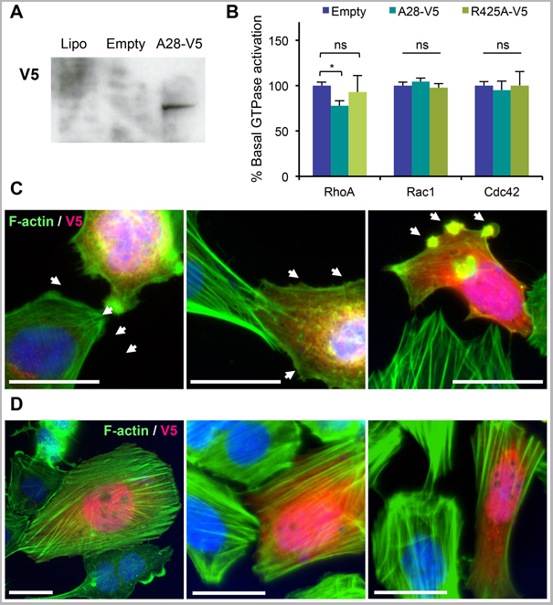
(Published customer image: Mouse anti V5 tag antibody, clone SV5-Pk1 used for the detection of V5 tagged WEEV_nsP3 protein by western blotting and immunofluorescenceImage caption: WEEV nsP3 interaction with host IKKbeta. A) U87MGs were transfected in a 6-well plate with 5 ug of pUC19 and WEEV_nsP3_HA for 24 hours. Cell lysates were resolved using SDS-PAGE and subsequently immunoblotted with V5 antibody and beta-actin served as a loading control. B) U87MGs were transfected with WEEV_nsP3_V5; cells were fixed after 24 hours and stained with antibodies against the endogenous IKKbeta and the V5 tag. Cells were incubated with appropriate secondary Alexa Fluor antibodies and the nuclei stained with DAPI. Co-localization of IKKbeta with WEEV_nsP3_V5 (yellow) was observed as shown by the arrows. B) Panels E -H serve as an example of transfected cells in a given field of view that show co-localization of IKKbeta and WEEV_nsP3_V5 24 hours post transfection. Panels I-L represent magnified images of other cells showing co-localization of IKKbeta and WEEV_nsP3_V5. Panel M is a magnified image of panel L. The co-localization was confirmed by Z-stack analysis. Co-localization was calculated to be approximately in 61% of cells (163 cells were counted of which 44% demonstrated expression of nsP3. Of those cells that expressed nsP3, 61% showed co-localization of both proteins). Images were taken using Nikon Eclipse TE2000-U at 60x magnification and are representative of 2 independent experiments.From: Amaya M, Voss K, Sampey G, Senina S, de la Fuente C, et al. (2014) The Role of IKKbeta in Venezuelan Equine Encephalitis Virus Infection. PLoS ONE 9(2): e86745.)
Application Data
(Published customer image: Mouse anti V5 tag antibody, clone SV5-Pk1 used for the detection of V5 tagged WEEV_nsP3 protein by western blotting and immunofluorescenceImage caption: WEEV nsP3 interaction with host IKKbeta. A) U87MGs were transfected in a 6-well plate with 5 ug of pUC19 and WEEV_nsP3_HA for 24 hours. Cell lysates were resolved using SDS-PAGE and subsequently immunoblotted with V5 antibody and beta-actin served as a loading control. B) U87MGs were transfected with WEEV_nsP3_V5; cells were fixed after 24 hours and stained with antibodies against the endogenous IKKbeta and the V5 tag. Cells were incubated with appropriate secondary Alexa Fluor antibodies and the nuclei stained with DAPI. Co-localization of IKKbeta with WEEV_nsP3_V5 (yellow) was observed as shown by the arrows. B) Panels E -H serve as an example of transfected cells in a given field of view that show co-localization of IKKbeta and WEEV_nsP3_V5 24 hours post transfection. Panels I-L represent magnified images of other cells showing co-localization of IKKbeta and WEEV_nsP3_V5. Panel M is a magnified image of panel L. The co-localization was confirmed by Z-stack analysis. Co-localization was calculated to be approximately in 61% of cells (163 cells were counted of which 44% demonstrated expression of nsP3. Of those cells that expressed nsP3, 61% showed co-localization of both proteins). Images were taken using Nikon Eclipse TE2000-U at 60x magnification and are representative of 2 independent experiments.From: Amaya M, Voss K, Sampey G, Senina S, de la Fuente C, et al. (2014) The Role of IKKbeta in Venezuelan Equine Encephalitis Virus Infection. PLoS ONE 9(2): e86745.)
V5-TAG, Monoclonal Antibody (Cat# AAA12081)
Full Name
MOUSE ANTI V5-TAG:HRP
Applications
Western Blot
Pricing
 IF (Immunofluorescence)
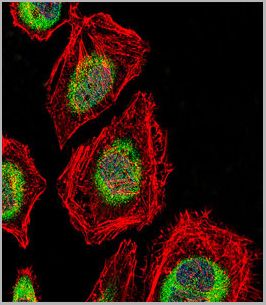
(AAA31246 staining A549 cells by IF/ICC. The samples were fixed with PFA and permeabilized in 0.1% Triton X-100,then blocked in 10% serum for 45 minutes at 25°C. Samples were then incubated with primary Ab(AAA31246) and mouse anti-beta tubulin Ab(T0023) for 1 hour at 37°C. An AlexaFluor594 conjugated goat anti-rabbit IgG(H+L) Ab(Red) and an AlexaFluor488 conjugated goat anti-mouse IgG(H+L) Ab(Green) were used as the secondary antibody.The nuclear counter stain is DAPI (blue).)
IF (Immunofluorescence)
(AAA31246 staining A549 cells by IF/ICC. The samples were fixed with PFA and permeabilized in 0.1% Triton X-100,then blocked in 10% serum for 45 minutes at 25°C. Samples were then incubated with primary Ab(AAA31246) and mouse anti-beta tubulin Ab(T0023) for 1 hour at 37°C. An AlexaFluor594 conjugated goat anti-rabbit IgG(H+L) Ab(Red) and an AlexaFluor488 conjugated goat anti-mouse IgG(H+L) Ab(Green) were used as the secondary antibody.The nuclear counter stain is DAPI (blue).)
TROY, Polyclonal Antibody (Cat# AAA31246)
Full Name
TROY Antibody
Gene Names
TNFRSF19; TAJ; TROY; TRADE; TAJ-alpha
Reactivity
Human, Mouse, Rat
Predicted Reactivity: Horse(88%), Sheep(86%)
Predicted Reactivity: Horse(88%), Sheep(86%)
Applications
ELISA
Purity
The antiserum was purified by peptide affinity chromatography using SulfoLink Coupling Resin (Thermo Fisher Scientific).
Pricing


 Application Data
(Published customer image: gamma delta T cells are the primary source of IL-17 during B. abortus infection. C57BL/6 mice were infected i.p. with 5x10⁴ CFUs of B. abortus 2308, and two weeks later gamma delta T cells (>95% purity) and an enriched TCRalphabeta (~55% CD4+, 25% CD8+) cell fraction were isolated from the spleens of infected mice. Cells were stimulated with 500 ng/ml ionomycin and 50 ng/ml PMA for three days, and cell-free supernatants from triplicate wells were assayed for cytokine production via ELISA. The mean +/- SD is shown; * P)
Application Data
(Published customer image: gamma delta T cells are the primary source of IL-17 during B. abortus infection. C57BL/6 mice were infected i.p. with 5x10⁴ CFUs of B. abortus 2308, and two weeks later gamma delta T cells (>95% purity) and an enriched TCRalphabeta (~55% CD4+, 25% CD8+) cell fraction were isolated from the spleens of infected mice. Cells were stimulated with 500 ng/ml ionomycin and 50 ng/ml PMA for three days, and cell-free supernatants from triplicate wells were assayed for cytokine production via ELISA. The mean +/- SD is shown; * P)
IFN GAMMA, Monoclonal Antibody (Cat# AAA12095)
Full Name
MOUSE ANTI BOVINE INTERFERON GAMMA:FITC
Applications
Flow Cytometry
Pricing


 Application Data
(Published customer image: gamma delta T cells are the primary source of IL-17 during B. abortus infection. C57BL/6 mice were infected i.p. with 5x10⁴ CFUs of B. abortus 2308, and two weeks later gamma delta T cells (>95% purity) and an enriched TCRalphabeta (~55% CD4+, 25% CD8+) cell fraction were isolated from the spleens of infected mice. Cells were stimulated with 500 ng/ml ionomycin and 50 ng/ml PMA for three days, and cell-free supernatants from triplicate wells were assayed for cytokine production via ELISA. The mean +/- SD is shown; * P)
Application Data
(Published customer image: gamma delta T cells are the primary source of IL-17 during B. abortus infection. C57BL/6 mice were infected i.p. with 5x10⁴ CFUs of B. abortus 2308, and two weeks later gamma delta T cells (>95% purity) and an enriched TCRalphabeta (~55% CD4+, 25% CD8+) cell fraction were isolated from the spleens of infected mice. Cells were stimulated with 500 ng/ml ionomycin and 50 ng/ml PMA for three days, and cell-free supernatants from triplicate wells were assayed for cytokine production via ELISA. The mean +/- SD is shown; * P)
IFN GAMMA, Monoclonal Antibody (Cat# AAA12096)
Full Name
MOUSE ANTI BOVINE INTERFERON GAMMA:RPE
Applications
Flow Cytometry
Pricing


 Application Data
(Staining of mouse spleen with Hamster anti Mouse CD81: Alexa Fluor 488)
Application Data
(Staining of mouse spleen with Hamster anti Mouse CD81: Alexa Fluor 488)
CD81, Monoclonal Antibody (Cat# AAA12033)
Full Name
HAMSTER ANTI MOUSE CD81:RPE
Gene Names
Cd81; Tapa1; Tapa-1; Tspan28
Applications
Flow Cytometry
Pricing


 Application Data
(Staining of mouse spleen with Hamster anti Mouse CD81: Alexa Fluor 488)
Application Data
(Staining of mouse spleen with Hamster anti Mouse CD81: Alexa Fluor 488)
CD81, Monoclonal Antibody (Cat# AAA11869)
Full Name
HAMSTER ANTI MOUSE CD81:FITC
Gene Names
Cd81; Tapa1; Tapa-1; Tspan28
Applications
Flow Cytometry
Pricing


 Application Data
(Staining of New Zealand Black mouse peripheral blood granulocytes with Rat anti Mouse Ly-6B.2 conjugated to FITC Data)
Application Data
(Staining of New Zealand Black mouse peripheral blood granulocytes with Rat anti Mouse Ly-6B.2 conjugated to FITC Data)
Ly-6B.2 ALLOANTIGEN, Monoclonal Antibody (Cat# AAA12190)
Full Name
RAT ANTI MOUSE Ly-6B.2 ALLOANTIGEN:FITC
Applications
Flow Cytometry
Pricing


 Application Data
(Staining of New Zealand Black mouse peripheral blood granulocytes with Rat anti Mouse Ly-6B.2 conjugated to FITC Data)
Application Data
(Staining of New Zealand Black mouse peripheral blood granulocytes with Rat anti Mouse Ly-6B.2 conjugated to FITC Data)
Ly-6B.2 ALLOANTIGEN, Monoclonal Antibody (Cat# AAA12189)
Full Name
RAT ANTI MOUSE Ly-6B.2 ALLOANTIGEN:Low Endotoxin
Applications
Immunohistochemistry, Flow Cytometry, Immunofluorescence, Immunohistochemistry, Western Blot
Pricing


 Application Data
(Staining of New Zealand Black mouse peripheral blood granulocytes with Rat anti Mouse Ly-6B.2 conjugated to FITC AAA12191)
Application Data
(Staining of New Zealand Black mouse peripheral blood granulocytes with Rat anti Mouse Ly-6B.2 conjugated to FITC AAA12191)
Ly-6B.2 ALLOANTIGEN, Monoclonal Antibody (Cat# AAA12191)
Full Name
RAT ANTI MOUSE Ly-6B.2 ALLOANTIGEN:FITC
Applications
Flow Cytometry
Pricing


 Application Data
(Staining of New Zealand Black mouse peripheral blood granulocytes with Rat anti Mouse Ly-6B.2 conjugated to FITC Data)
Application Data
(Staining of New Zealand Black mouse peripheral blood granulocytes with Rat anti Mouse Ly-6B.2 conjugated to FITC Data)
Ly-6B.2 ALLOANTIGEN, Monoclonal Antibody (Cat# AAA12188)
Full Name
RAT ANTI MOUSE Ly-6B.2 ALLOANTIGEN:Biotin
Applications
Flow Cytometry
Pricing


 Application Data
(Published customer image: gamma delta T cells are the primary source of IL-17 during B. abortus infection. C57BL/6 mice were infected i.p. with 5x10⁴ CFUs of B. abortus 2308, and two weeks later gamma delta T cells (>95% purity) and an enriched TCRalphabeta (~55% CD4+, 25% CD8+) cell fraction were isolated from the spleens of infected mice. Cells were stimulated with 500 ng/ml ionomycin and 50 ng/ml PMA for three days, and cell-free supernatants from triplicate wells were assayed for cytokine production via ELISA. The mean +/- SD is shown; * P)
Application Data
(Published customer image: gamma delta T cells are the primary source of IL-17 during B. abortus infection. C57BL/6 mice were infected i.p. with 5x10⁴ CFUs of B. abortus 2308, and two weeks later gamma delta T cells (>95% purity) and an enriched TCRalphabeta (~55% CD4+, 25% CD8+) cell fraction were isolated from the spleens of infected mice. Cells were stimulated with 500 ng/ml ionomycin and 50 ng/ml PMA for three days, and cell-free supernatants from triplicate wells were assayed for cytokine production via ELISA. The mean +/- SD is shown; * P)
IFN GAMMA, Monoclonal Antibody (Cat# AAA12094)
Full Name
MOUSE ANTI BOVINE INTERFERON GAMMA:Biotin
Applications
Flow Cytometry
Pricing


 Application Data
(Staining of New Zealand Black mouse peripheral blood granulocytes with Rat anti Mouse Ly-6B.2 conjugated to FITC Data)
Application Data
(Staining of New Zealand Black mouse peripheral blood granulocytes with Rat anti Mouse Ly-6B.2 conjugated to FITC Data)
Ly-6B.2 ALLOANTIGEN, Monoclonal Antibody (Cat# AAA12196)
Full Name
RAT ANTI MOUSE Ly-6B.2 ALLOANTIGEN
Applications
Immunohistochemistry, Flow Cytometry, Immunofluorescence, Immunohistochemistry, Western Blot
Pricing


 Application Data
(Staining of New Zealand Black mouse peripheral blood granulocytes with Rat anti Mouse Ly-6B.2 conjugated to FITC Data)
Application Data
(Staining of New Zealand Black mouse peripheral blood granulocytes with Rat anti Mouse Ly-6B.2 conjugated to FITC Data)
Ly-6B.2 ALLOANTIGEN, Monoclonal Antibody (Cat# AAA12199)
Full Name
RAT ANTI MOUSE Ly-6B.2 ALLOANTIGEN:RPE
Applications
Flow Cytometry
Pricing
 IF (Immunofluorescence)
(Fluorescent confocal image of Hela cell stained with NRK Antibody (Center) (AAA28689). Hela cells were fixed with 4% PFA (20 min), permeabilized with Triton X-100 (0.1%, 10 min), then incubated with NRK primary antibody (1:25, 1 h at 37 degree). For secondary antibody, Alexa Fluor 488 conjugated donkey anti-rabbit antibody (green) was used (1:400, 50 min at 37 degree).Cytoplasmic actin was counterstained with Alexa Fluor 555 (red) conjugated Phalloidin (7units/ml, 1 h at 37 degree). Nuclei were counterstained with DAPI (blue) (10 ug/ml, 10 min). NRK immunoreactivity is localized to Cytoplasm and Nucleus significantly.)
IF (Immunofluorescence)
(Fluorescent confocal image of Hela cell stained with NRK Antibody (Center) (AAA28689). Hela cells were fixed with 4% PFA (20 min), permeabilized with Triton X-100 (0.1%, 10 min), then incubated with NRK primary antibody (1:25, 1 h at 37 degree). For secondary antibody, Alexa Fluor 488 conjugated donkey anti-rabbit antibody (green) was used (1:400, 50 min at 37 degree).Cytoplasmic actin was counterstained with Alexa Fluor 555 (red) conjugated Phalloidin (7units/ml, 1 h at 37 degree). Nuclei were counterstained with DAPI (blue) (10 ug/ml, 10 min). NRK immunoreactivity is localized to Cytoplasm and Nucleus significantly.)
NRK, Polyclonal Antibody (Cat# AAA28689)
Full Name
NRK Antibody (Center)
Gene Names
NRK; NESK
Reactivity
Human
Applications
Western Blot, Immunofluorescence
Purity
This antibody is purified through a protein A column, followed by peptide affinity purification.
Pricing
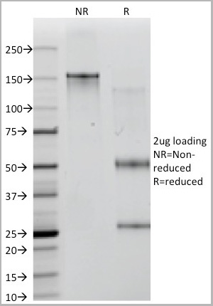
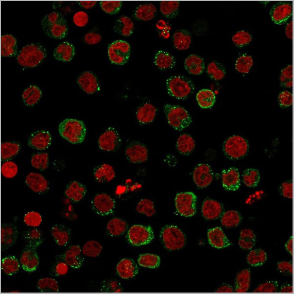
 IF (Immunofluorescence)
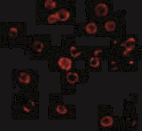
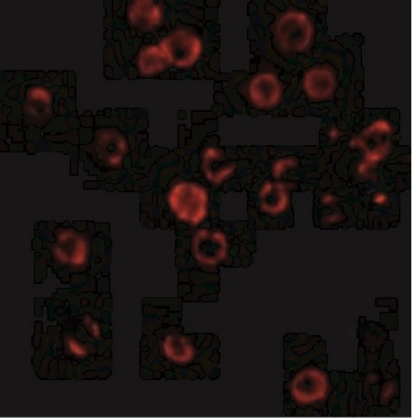
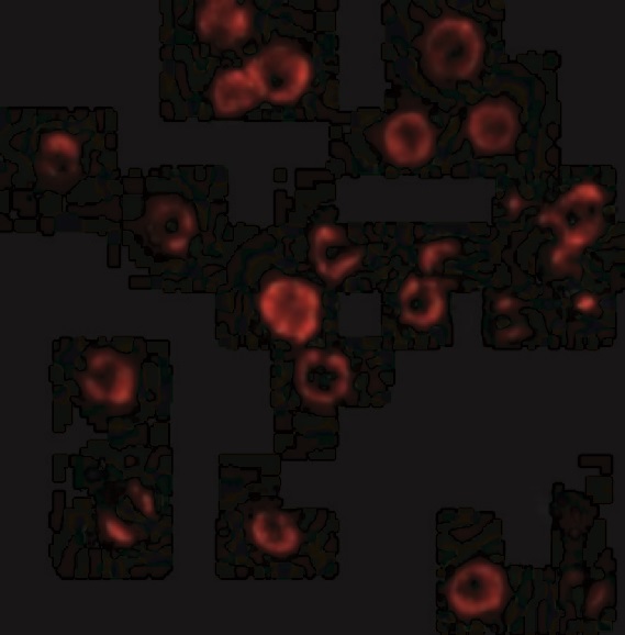
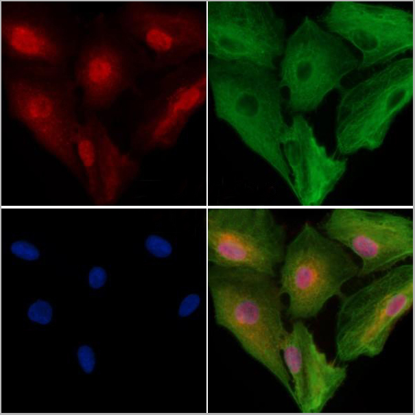
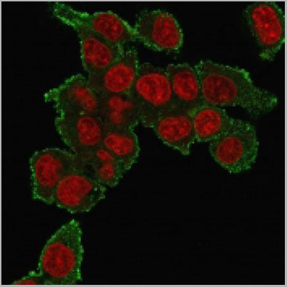
(Immunofluorescence staining of PFA-fixed HePG2 cells using Tumor Necrosis Factor (TNF alpha) Mouse Monoclonal Antibody (TNF706) followed by goat anti-mouse IgG-CF488 (green). Nuclei stained with RedDot.)
IF (Immunofluorescence)
(Immunofluorescence staining of PFA-fixed HePG2 cells using Tumor Necrosis Factor (TNF alpha) Mouse Monoclonal Antibody (TNF706) followed by goat anti-mouse IgG-CF488 (green). Nuclei stained with RedDot.)
TNF-alpha (Tumor Necrosis Factor alpha), Monoclonal Antibody (Cat# AAA13806)
Full Name
TNF-alpha (Tumor Necrosis Factor alpha) Mouse Monoclonal Antibody
Gene Names
TNF; DIF; TNFA; TNFSF2; TNLG1F; TNF-alpha
Reactivity
Human, Mouse, Rat, Rabbit, Cat, Dog, Zebrafish
Applications
Flow Cytometry, Immunofluorescence, Immunohistochemistry
Pricing


 Application Data
(Published customer image: Mouse anti V5 tag antibody, clone SV5-Pk1 used for the detection of V5 tagged WEEV_nsP3 protein by western blotting and immunofluorescenceImage caption: WEEV nsP3 interaction with host IKKbeta. A) U87MGs were transfected in a 6-well plate with 5 ug of pUC19 and WEEV_nsP3_HA for 24 hours. Cell lysates were resolved using SDS-PAGE and subsequently immunoblotted with V5 antibody and beta-actin served as a loading control. B) U87MGs were transfected with WEEV_nsP3_V5; cells were fixed after 24 hours and stained with antibodies against the endogenous IKKbeta and the V5 tag. Cells were incubated with appropriate secondary Alexa Fluor antibodies and the nuclei stained with DAPI. Co-localization of IKKbeta with WEEV_nsP3_V5 (yellow) was observed as shown by the arrows. B) Panels E -H serve as an example of transfected cells in a given field of view that show co-localization of IKKbeta and WEEV_nsP3_V5 24 hours post transfection. Panels I-L represent magnified images of other cells showing co-localization of IKKbeta and WEEV_nsP3_V5. Panel M is a magnified image of panel L. The co-localization was confirmed by Z-stack analysis. Co-localization was calculated to be approximately in 61% of cells (163 cells were counted of which 44% demonstrated expression of nsP3. Of those cells that expressed nsP3, 61% showed co-localization of both proteins). Images were taken using Nikon Eclipse TE2000-U at 60x magnification and are representative of 2 independent experiments.From: Amaya M, Voss K, Sampey G, Senina S, de la Fuente C, et al. (2014) The Role of IKKbeta in Venezuelan Equine Encephalitis Virus Infection. PLoS ONE 9(2): e86745.)
Application Data
(Published customer image: Mouse anti V5 tag antibody, clone SV5-Pk1 used for the detection of V5 tagged WEEV_nsP3 protein by western blotting and immunofluorescenceImage caption: WEEV nsP3 interaction with host IKKbeta. A) U87MGs were transfected in a 6-well plate with 5 ug of pUC19 and WEEV_nsP3_HA for 24 hours. Cell lysates were resolved using SDS-PAGE and subsequently immunoblotted with V5 antibody and beta-actin served as a loading control. B) U87MGs were transfected with WEEV_nsP3_V5; cells were fixed after 24 hours and stained with antibodies against the endogenous IKKbeta and the V5 tag. Cells were incubated with appropriate secondary Alexa Fluor antibodies and the nuclei stained with DAPI. Co-localization of IKKbeta with WEEV_nsP3_V5 (yellow) was observed as shown by the arrows. B) Panels E -H serve as an example of transfected cells in a given field of view that show co-localization of IKKbeta and WEEV_nsP3_V5 24 hours post transfection. Panels I-L represent magnified images of other cells showing co-localization of IKKbeta and WEEV_nsP3_V5. Panel M is a magnified image of panel L. The co-localization was confirmed by Z-stack analysis. Co-localization was calculated to be approximately in 61% of cells (163 cells were counted of which 44% demonstrated expression of nsP3. Of those cells that expressed nsP3, 61% showed co-localization of both proteins). Images were taken using Nikon Eclipse TE2000-U at 60x magnification and are representative of 2 independent experiments.From: Amaya M, Voss K, Sampey G, Senina S, de la Fuente C, et al. (2014) The Role of IKKbeta in Venezuelan Equine Encephalitis Virus Infection. PLoS ONE 9(2): e86745.)
V5-TAG, Monoclonal Antibody (Cat# AAA11864)
Full Name
MOUSE ANTI V5-TAG:FITC
Applications
Immunofluorescence
Pricing


 Application Data
(Published customer image: Mouse anti V5 tag antibody, clone SV5-Pk1 used for the detection of V5 tagged WEEV_nsP3 protein by western blotting and immunofluorescenceImage caption: WEEV nsP3 interaction with host IKKbeta. A) U87MGs were transfected in a 6-well plate with 5 ug of pUC19 and WEEV_nsP3_HA for 24 hours. Cell lysates were resolved using SDS-PAGE and subsequently immunoblotted with V5 antibody and beta-actin served as a loading control. B) U87MGs were transfected with WEEV_nsP3_V5; cells were fixed after 24 hours and stained with antibodies against the endogenous IKKbeta and the V5 tag. Cells were incubated with appropriate secondary Alexa Fluor antibodies and the nuclei stained with DAPI. Co-localization of IKKbeta with WEEV_nsP3_V5 (yellow) was observed as shown by the arrows. B) Panels E -H serve as an example of transfected cells in a given field of view that show co-localization of IKKbeta and WEEV_nsP3_V5 24 hours post transfection. Panels I-L represent magnified images of other cells showing co-localization of IKKbeta and WEEV_nsP3_V5. Panel M is a magnified image of panel L. The co-localization was confirmed by Z-stack analysis. Co-localization was calculated to be approximately in 61% of cells (163 cells were counted of which 44% demonstrated expression of nsP3. Of those cells that expressed nsP3, 61% showed co-localization of both proteins). Images were taken using Nikon Eclipse TE2000-U at 60x magnification and are representative of 2 independent experiments.From: Amaya M, Voss K, Sampey G, Senina S, de la Fuente C, et al. (2014) The Role of IKKbeta in Venezuelan Equine Encephalitis Virus Infection. PLoS ONE 9(2): e86745.)
Application Data
(Published customer image: Mouse anti V5 tag antibody, clone SV5-Pk1 used for the detection of V5 tagged WEEV_nsP3 protein by western blotting and immunofluorescenceImage caption: WEEV nsP3 interaction with host IKKbeta. A) U87MGs were transfected in a 6-well plate with 5 ug of pUC19 and WEEV_nsP3_HA for 24 hours. Cell lysates were resolved using SDS-PAGE and subsequently immunoblotted with V5 antibody and beta-actin served as a loading control. B) U87MGs were transfected with WEEV_nsP3_V5; cells were fixed after 24 hours and stained with antibodies against the endogenous IKKbeta and the V5 tag. Cells were incubated with appropriate secondary Alexa Fluor antibodies and the nuclei stained with DAPI. Co-localization of IKKbeta with WEEV_nsP3_V5 (yellow) was observed as shown by the arrows. B) Panels E -H serve as an example of transfected cells in a given field of view that show co-localization of IKKbeta and WEEV_nsP3_V5 24 hours post transfection. Panels I-L represent magnified images of other cells showing co-localization of IKKbeta and WEEV_nsP3_V5. Panel M is a magnified image of panel L. The co-localization was confirmed by Z-stack analysis. Co-localization was calculated to be approximately in 61% of cells (163 cells were counted of which 44% demonstrated expression of nsP3. Of those cells that expressed nsP3, 61% showed co-localization of both proteins). Images were taken using Nikon Eclipse TE2000-U at 60x magnification and are representative of 2 independent experiments.From: Amaya M, Voss K, Sampey G, Senina S, de la Fuente C, et al. (2014) The Role of IKKbeta in Venezuelan Equine Encephalitis Virus Infection. PLoS ONE 9(2): e86745.)
V5-TAG, Monoclonal Antibody (Cat# AAA11930)
Full Name
MOUSE ANTI V5-TAG
Applications
Immunohistochemistry, Flow Cytometry, Immunofluorescence, Immunoprecipitation, Western Blot, Radioimmunoassay
Pricing

 Assay Principle
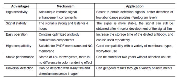
(After years of development on chemiluminescence technology, there have been a variety of luminescence systems, but the most commonly used one in laboratories is still a technology system based on Luminol or its derivatives (isoluminol, etc.). Western luminescent detection reagent ECL immunoblotting chemiluminescence solution is a non-radioactive (horseradish peroxidase) luminescence system designed to detect femtogram-level trace proteins immobilized on a solid membrane (such as NC, PVDF, etc.). It is an experimental auxiliary reagent for the photosensitive recording of its immunoblotting by X-ray film (radiograph).)
Assay Principle
(After years of development on chemiluminescence technology, there have been a variety of luminescence systems, but the most commonly used one in laboratories is still a technology system based on Luminol or its derivatives (isoluminol, etc.). Western luminescent detection reagent ECL immunoblotting chemiluminescence solution is a non-radioactive (horseradish peroxidase) luminescence system designed to detect femtogram-level trace proteins immobilized on a solid membrane (such as NC, PVDF, etc.). It is an experimental auxiliary reagent for the photosensitive recording of its immunoblotting by X-ray film (radiograph).)
West Femto Maximum Sensitivity Substrate, Reagent (Cat# AAA31545)
Full Name
West Femto Maximum Sensitivity Substrate
Applications
Western Blot, Immunoprecipitation
Pricing


 Application Data
(Staining of mouse spleen with Hamster anti Mouse CD81: Alexa Fluor 488)
Application Data
(Staining of mouse spleen with Hamster anti Mouse CD81: Alexa Fluor 488)
CD81, Monoclonal Antibody (Cat# AAA11942)
Full Name
HAMSTER ANTI MOUSE CD81
Gene Names
Cd81; Tapa1; Tapa-1; Tspan28
Reactivity
Rat
Applications
Immunohistochemistry, Flow Cytometry, Immunoprecipitation, Western Blot
Pricing


 Application Data
(Published customer image: Mouse anti V5 tag antibody, clone SV5-Pk1 used for the detection of V5 tagged WEEV_nsP3 protein by western blotting and immunofluorescenceImage caption: WEEV nsP3 interaction with host IKKbeta. A) U87MGs were transfected in a 6-well plate with 5 ug of pUC19 and WEEV_nsP3_HA for 24 hours. Cell lysates were resolved using SDS-PAGE and subsequently immunoblotted with V5 antibody and beta-actin served as a loading control. B) U87MGs were transfected with WEEV_nsP3_V5; cells were fixed after 24 hours and stained with antibodies against the endogenous IKKbeta and the V5 tag. Cells were incubated with appropriate secondary Alexa Fluor antibodies and the nuclei stained with DAPI. Co-localization of IKKbeta with WEEV_nsP3_V5 (yellow) was observed as shown by the arrows. B) Panels E -H serve as an example of transfected cells in a given field of view that show co-localization of IKKbeta and WEEV_nsP3_V5 24 hours post transfection. Panels I-L represent magnified images of other cells showing co-localization of IKKbeta and WEEV_nsP3_V5. Panel M is a magnified image of panel L. The co-localization was confirmed by Z-stack analysis. Co-localization was calculated to be approximately in 61% of cells (163 cells were counted of which 44% demonstrated expression of nsP3. Of those cells that expressed nsP3, 61% showed co-localization of both proteins). Images were taken using Nikon Eclipse TE2000-U at 60x magnification and are representative of 2 independent experiments.From: Amaya M, Voss K, Sampey G, Senina S, de la Fuente C, et al. (2014) The Role of IKKbeta in Venezuelan Equine Encephalitis Virus Infection. PLoS ONE 9(2): e86745.)
Application Data
(Published customer image: Mouse anti V5 tag antibody, clone SV5-Pk1 used for the detection of V5 tagged WEEV_nsP3 protein by western blotting and immunofluorescenceImage caption: WEEV nsP3 interaction with host IKKbeta. A) U87MGs were transfected in a 6-well plate with 5 ug of pUC19 and WEEV_nsP3_HA for 24 hours. Cell lysates were resolved using SDS-PAGE and subsequently immunoblotted with V5 antibody and beta-actin served as a loading control. B) U87MGs were transfected with WEEV_nsP3_V5; cells were fixed after 24 hours and stained with antibodies against the endogenous IKKbeta and the V5 tag. Cells were incubated with appropriate secondary Alexa Fluor antibodies and the nuclei stained with DAPI. Co-localization of IKKbeta with WEEV_nsP3_V5 (yellow) was observed as shown by the arrows. B) Panels E -H serve as an example of transfected cells in a given field of view that show co-localization of IKKbeta and WEEV_nsP3_V5 24 hours post transfection. Panels I-L represent magnified images of other cells showing co-localization of IKKbeta and WEEV_nsP3_V5. Panel M is a magnified image of panel L. The co-localization was confirmed by Z-stack analysis. Co-localization was calculated to be approximately in 61% of cells (163 cells were counted of which 44% demonstrated expression of nsP3. Of those cells that expressed nsP3, 61% showed co-localization of both proteins). Images were taken using Nikon Eclipse TE2000-U at 60x magnification and are representative of 2 independent experiments.From: Amaya M, Voss K, Sampey G, Senina S, de la Fuente C, et al. (2014) The Role of IKKbeta in Venezuelan Equine Encephalitis Virus Infection. PLoS ONE 9(2): e86745.)
V5-TAG, Monoclonal Antibody (Cat# AAA12211)
Full Name
MOUSE ANTI V5-TAG
Applications
Immunohistochemistry, Flow Cytometry, Immunofluorescence, Immunoprecipitation, Western Blot, Radioimmunoassay
Pricing