Filters
Clonality
Type
Reactivity
Gene Name
Isotype
Host
Application
Clone
1523 results for "Binding Protein" - showing 1350-1400
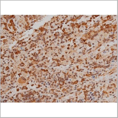
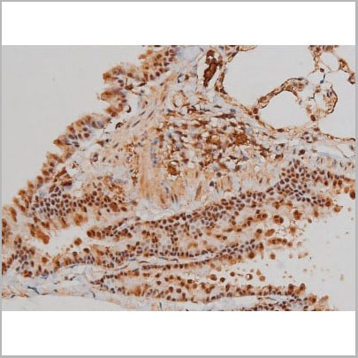
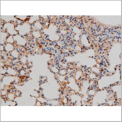
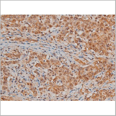
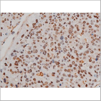
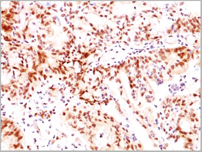
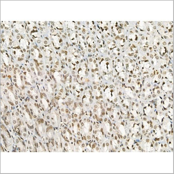
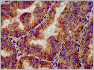
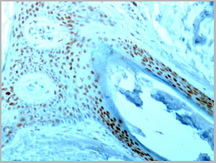
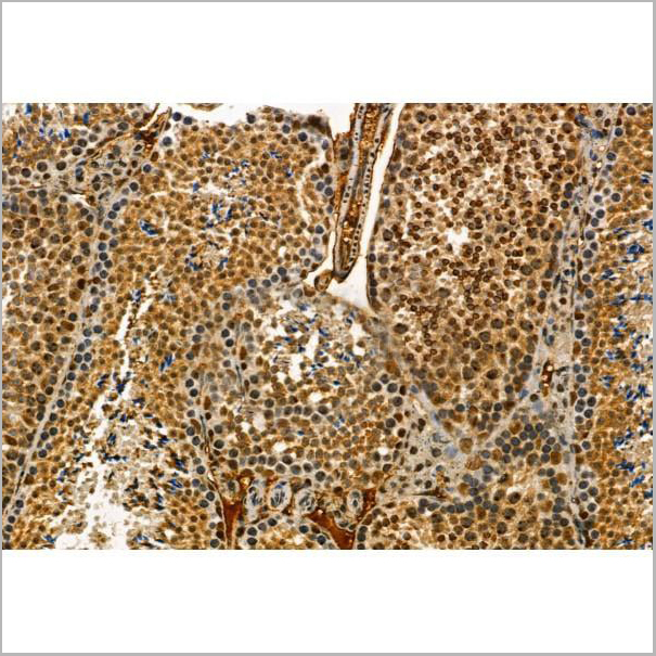
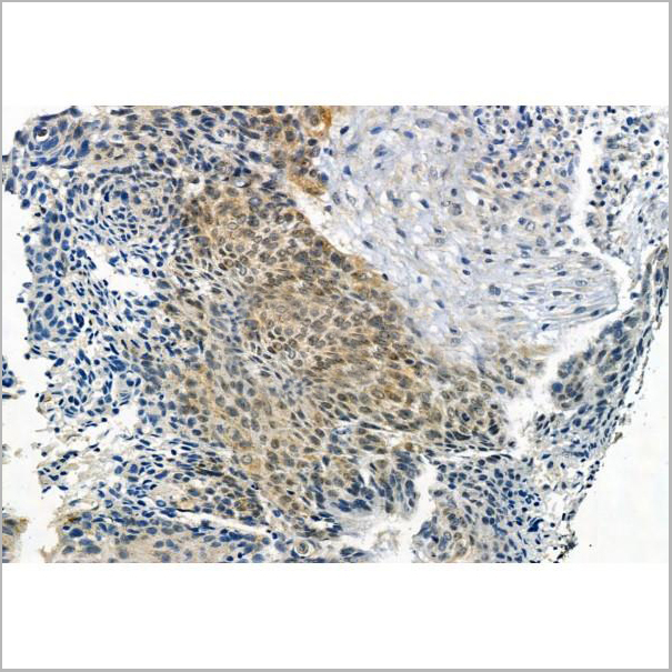
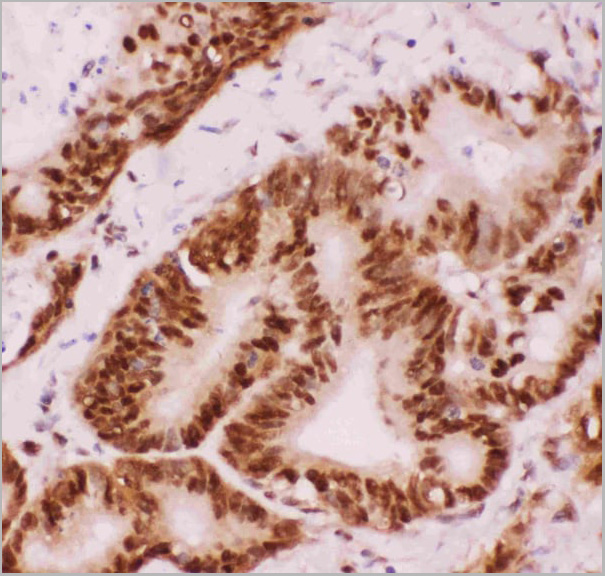
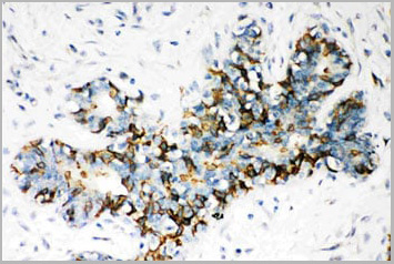
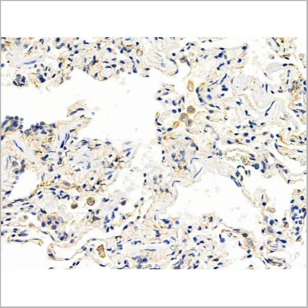 IHC (Immunohistochemistry-Paraffin)
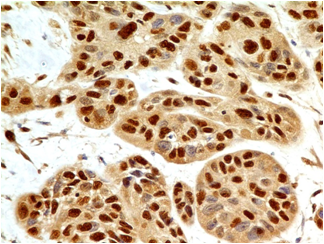
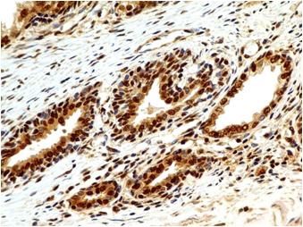
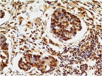
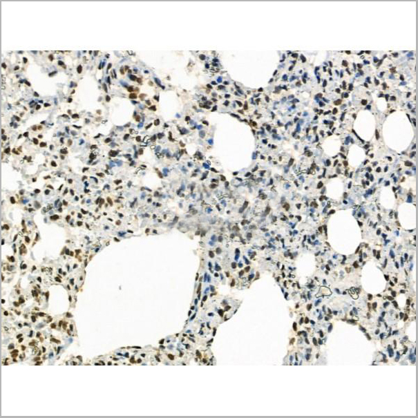
(AAA31201 at 1/100 staining Human lung cancer and adjacent normal tissues by IHC-P. The sample was formaldehyde fixed and a heat mediated antigen retrieval step in citrate buffer was performed. The sample was then blocked and incubated with the primary antibody at 4°C overnight. An HRP conjugated anti-Rabbit antibody was used as the secondary antibody.)
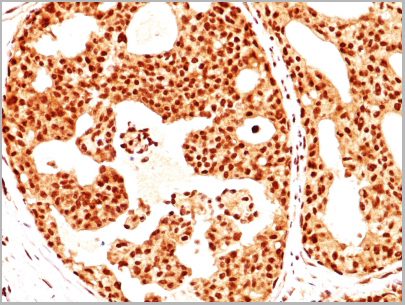
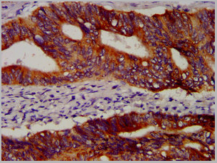
IHC (Immunohistochemistry-Paraffin)
(AAA31201 at 1/100 staining Human lung cancer and adjacent normal tissues by IHC-P. The sample was formaldehyde fixed and a heat mediated antigen retrieval step in citrate buffer was performed. The sample was then blocked and incubated with the primary antibody at 4°C overnight. An HRP conjugated anti-Rabbit antibody was used as the secondary antibody.)
PICALM, Polyclonal Antibody (Cat# AAA31201)
Full Name
PICALM Antibody
Gene Names
PICALM; LAP; CALM; CLTH
Reactivity
Human, Mouse, Rat
Predicted Reactivity: Pig(100%), Bovine(100%), Horse(100%), Sheep(100%), Rabbit(100%), Dog(100%)
Predicted Reactivity: Pig(100%), Bovine(100%), Horse(100%), Sheep(100%), Rabbit(100%), Dog(100%)
Applications
ELISA
Purity
The antiserum was purified by peptide affinity chromatography using SulfoLink Coupling Resin (Thermo Fisher Scientific).
Pricing

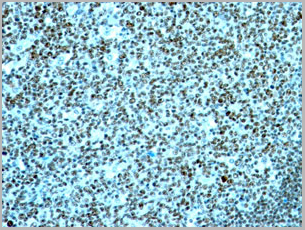
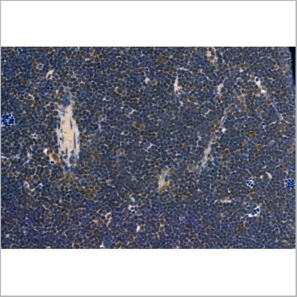
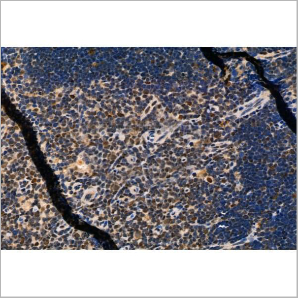
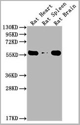
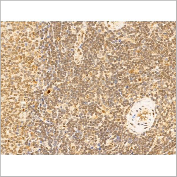
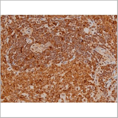 IHC (Immunohistochemistry)
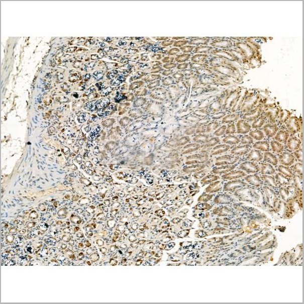
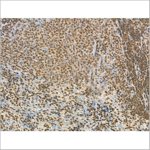
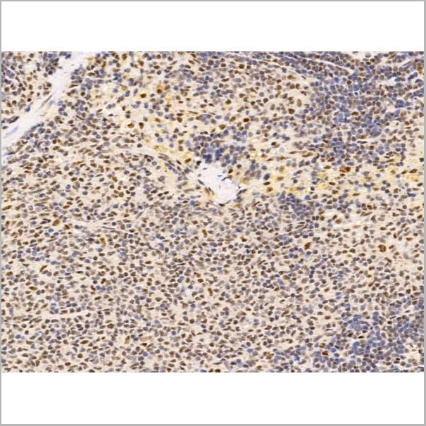
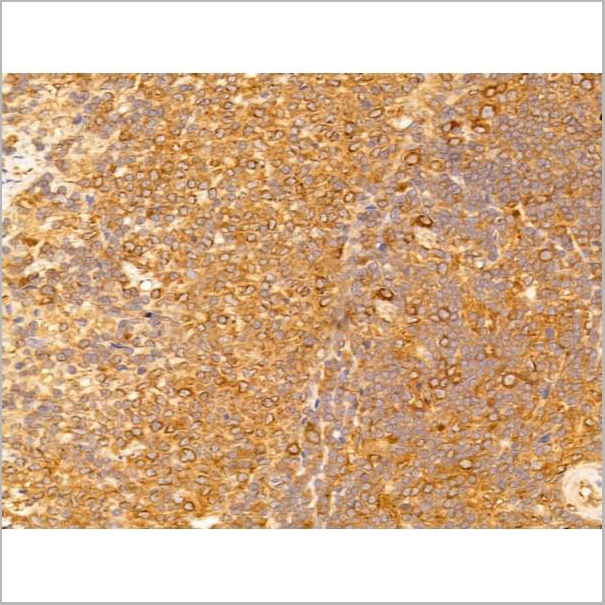
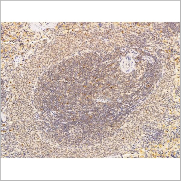
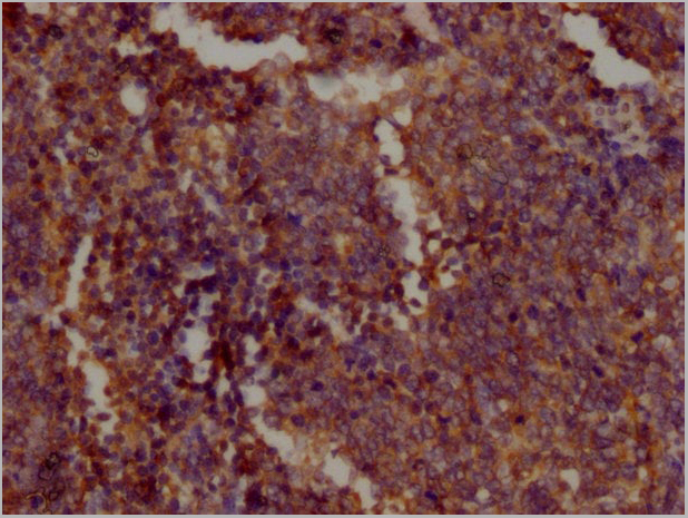
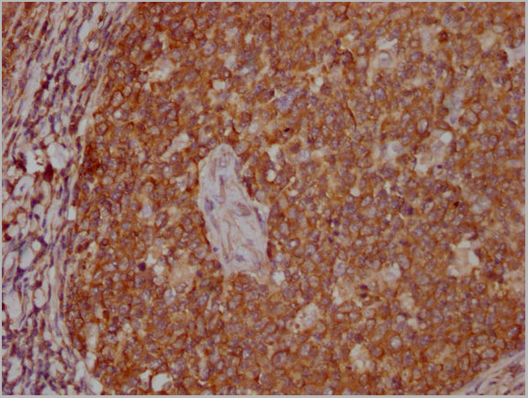
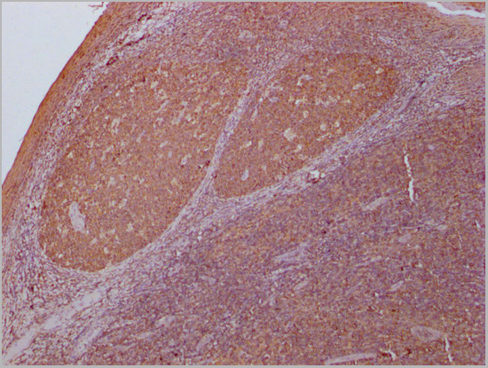
(AAA31022 at 1/200 staining Rat spleen tissue sections by IHC-P. The tissue was formaldehyde fixed and a heat mediated antigen retrieval step in citrate buffer was performed. The tissue was then blocked and incubated with the antibody for 1.5 hours at 22 degree C. An HRP conjugated goat anti-rabbit antibody was used as the secondary.)
IHC (Immunohistochemistry)
(AAA31022 at 1/200 staining Rat spleen tissue sections by IHC-P. The tissue was formaldehyde fixed and a heat mediated antigen retrieval step in citrate buffer was performed. The tissue was then blocked and incubated with the antibody for 1.5 hours at 22 degree C. An HRP conjugated goat anti-rabbit antibody was used as the secondary.)
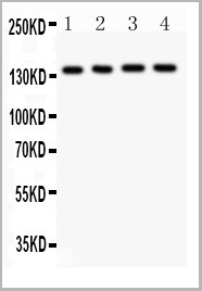
Filamin A, Polyclonal Antibody (Cat# AAA31022)
Full Name
Phospho-Filamin A (Ser2152) Antibody
Gene Names
FLNA; FLN; FMD; MNS; OPD; ABPX; CSBS; CVD1; FGS2; FLN1; NHBP; OPD1; OPD2; XLVD; XMVD; FLN-A; ABP-280
Reactivity
Human, Mouse, Rat
Applications
Western Blot, Immunohistochemistry, Immunofluorescence, Immunocytochemistry
Purity
From purified rabbit serum by affinity purification via sequential chromatography on phospho-and non-phospho-peptide affinity columns.
Pricing
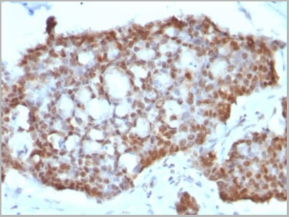
 Application Data
(Analysis of Protein Array containing more than 19,000 full-length human proteins using Estrogen Receptor alpha Mouse Monoclonal Antibody (ESR1/3557) Z- and S- Score: The Z-score represents the strength of a signal that a monoclonal antibody (MAb) (in combination with a fluorescently-tagged anti-IgG secondary antibody) produces when binding to a particular protein on the HuProtTM array. Z-scores are described in units of standard deviations (SD’s) above the mean value of all signals generated on that array. If targets on HuProtTM are arranged in descending order of the Z-score, the S-score is the difference (also in units of SD’s) between the Z-score. S-score therefore represents the relative target specificity of a MAb to its intended target. A MAb is considered to specific to its intended target, if the MAb has an S-score of at least 2.5. For example, if a MAb binds to protein X with a Z-score of 43 and to protein Y with a Z-score of 14, then the S-score for the binding of that MAb to protein X is equal to 29.)
Application Data
(Analysis of Protein Array containing more than 19,000 full-length human proteins using Estrogen Receptor alpha Mouse Monoclonal Antibody (ESR1/3557) Z- and S- Score: The Z-score represents the strength of a signal that a monoclonal antibody (MAb) (in combination with a fluorescently-tagged anti-IgG secondary antibody) produces when binding to a particular protein on the HuProtTM array. Z-scores are described in units of standard deviations (SD’s) above the mean value of all signals generated on that array. If targets on HuProtTM are arranged in descending order of the Z-score, the S-score is the difference (also in units of SD’s) between the Z-score. S-score therefore represents the relative target specificity of a MAb to its intended target. A MAb is considered to specific to its intended target, if the MAb has an S-score of at least 2.5. For example, if a MAb binds to protein X with a Z-score of 43 and to protein Y with a Z-score of 14, then the S-score for the binding of that MAb to protein X is equal to 29.)
Estrogen Receptor, alpha, Monoclonal Antibody (Cat# AAA23915)
Full Name
Estrogen Receptor, alpha (Marker of Estrogen Dependence)
Gene Names
ESR1; ER; ESR; Era; ESRA; ESTRR; NR3A1
Reactivity
Human
Applications
Immunofluorescence, Flow Cytometry, Immunohistochemistry
Purity
Purified Ab with BSA and Azide at 200ug/ml OR Purified Ab WITHOUT BSA and Azide at 1.0mg/ml
Pricing
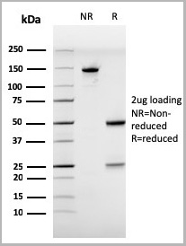
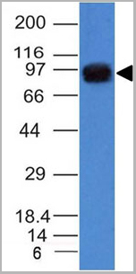
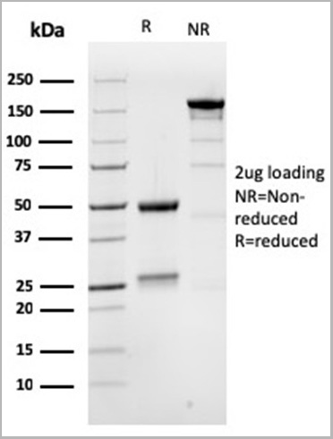
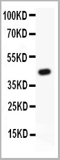
 SDS-PAGE
(Under DTT-reducing SDS-PAGE conditions, the 2019-nCov Nucleocapsid protein NX fragment migrates as a 50 kDa protein (Lane 1).)
SDS-PAGE
(Under DTT-reducing SDS-PAGE conditions, the 2019-nCov Nucleocapsid protein NX fragment migrates as a 50 kDa protein (Lane 1).)
COVID 19 Nucleocapsid (NP) N fragment protein (NX) Coronavirus, Recombinant Protein (Cat# AAA27950)
Full Name
2019-nCov Nucleocapsid protein N fragment protein (NX)
Applications
SDS-Page
Purity
Purity: >95% by SDS-page
Purification: Ni column
Purification: Ni column
Pricing
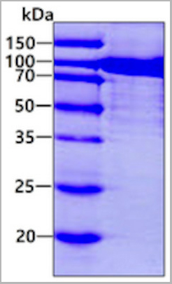
 SDS-PAGE
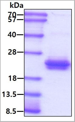
(3ug by SDS-PAGE under reducing condition and visualized by coomassie blue stain.)
SDS-PAGE
(3ug by SDS-PAGE under reducing condition and visualized by coomassie blue stain.)
RAC1, Recombinant Protein (Cat# AAA11741)
Full Name
RAC1, 1-192 aa, Human, E Coli
Gene Names
RAC1; MIG5; MRD48; Rac-1; TC-25; p21-Rac1
Applications
SDS-PAGE
Purity
> 95% by SDS-PAGE
Pricing
 IHC (Immunohistochemistry)
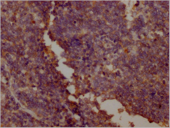
(Immunohistochemical analysis of HMGB1 in normal human spleen tissue using HMGB1 antibody (Clone: ABM24D3) at 1 ug/ml.)
IHC (Immunohistochemistry)
(Immunohistochemical analysis of HMGB1 in normal human spleen tissue using HMGB1 antibody (Clone: ABM24D3) at 1 ug/ml.)
HMGB1, Monoclonal Antibody (Cat# AAA14866)
Full Name
HMGB1 Monoclonal Antibody
Gene Names
HMGB1; HMG1; HMG3; SBP-1
Reactivity
Human (Predicted Reactivity: Mouse
Applications
Western Blot, Immunohistochemistry, Flow Cytometry
Pricing
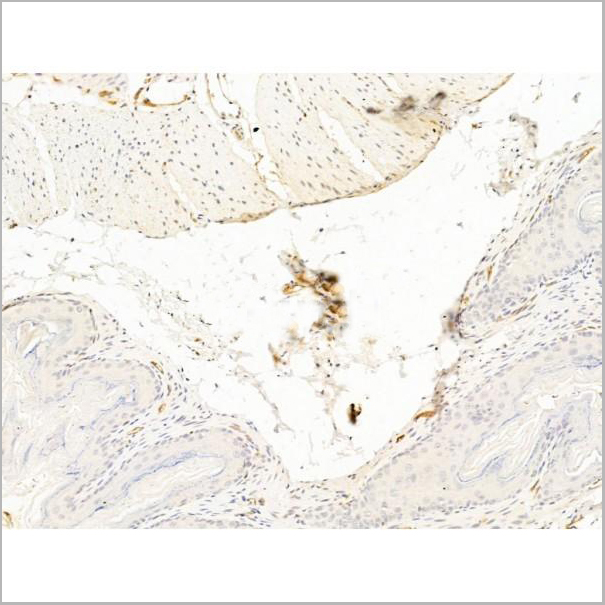
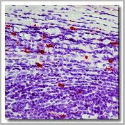 IHC (Immunohistochemistry)
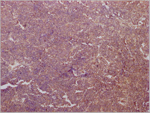
(Immunohistochemistry of formalin-fixed, paraffin-embedded human tonsil using AAA14660 and following by peroxidase-conjugate and AEC chromogen. Note cytoplasmic staining of mast cells.)
IHC (Immunohistochemistry)
(Immunohistochemistry of formalin-fixed, paraffin-embedded human tonsil using AAA14660 and following by peroxidase-conjugate and AEC chromogen. Note cytoplasmic staining of mast cells.)
Mast Cell Chymase, Monoclonal Antibody (Cat# AAA14660)
Full Name
Mast Cell Chymase (BSA & Azide Free)
Gene Names
CMA1; CYH; MCT1; chymase; MGC119890; MGC119891
Reactivity
Human
Applications
Western Blot, Immunohistochemistry
Purity
Purified by Protein G affinity chromatography.
Pricing
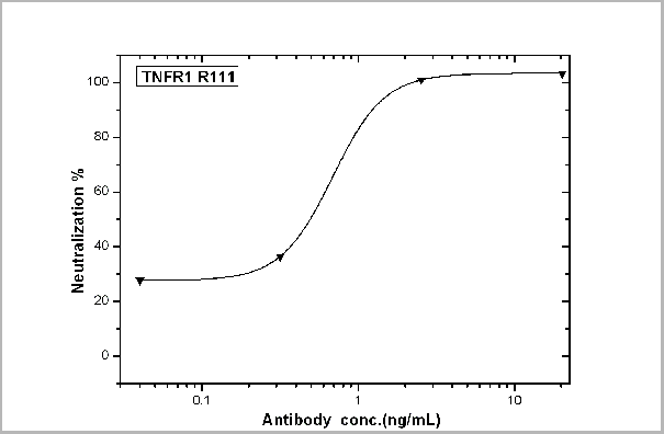 Application Data
(TNFR1/TNFRSF1A-mediated inhibition of cytotoxicity was Neutralized by Human TNFR1 Antibody. Recombinant Human TNFR1/TNFRSF1A inhibits Recombinant Human TNFa induced cytotoxicity in the L-929 mouse fibroblast cell line. Inhibition of Recombinant Human TNFa (0.2 ng/mL) activity elicited by Recombinant Human TNFR1/TNFRSF1A (0.3 ug/mL) is neutralized by increasing concentrations of Human TNFR1/TNFRSF1A Monoclonal Antibody. The IC50 is typically 0.5-1.5 ug/mL in the presence of the metabolic inhibitor actinomycin D (1 ug/mL).)
Application Data
(TNFR1/TNFRSF1A-mediated inhibition of cytotoxicity was Neutralized by Human TNFR1 Antibody. Recombinant Human TNFR1/TNFRSF1A inhibits Recombinant Human TNFa induced cytotoxicity in the L-929 mouse fibroblast cell line. Inhibition of Recombinant Human TNFa (0.2 ng/mL) activity elicited by Recombinant Human TNFR1/TNFRSF1A (0.3 ug/mL) is neutralized by increasing concentrations of Human TNFR1/TNFRSF1A Monoclonal Antibody. The IC50 is typically 0.5-1.5 ug/mL in the presence of the metabolic inhibitor actinomycin D (1 ug/mL).)
TNFR1/CD120a/TNFRSF1A, Monoclonal Antibody (Cat# AAA27740)
Full Name
TNFR1/CD120a/TNFRSF1A Neutralizing Antibody
Gene Names
TNFRSF1A; FPF; MS5; p55; p60; TBP1; TNF-R; TNFAR; TNFR1; p55-R; CD120a; TNFR55; TNFR60; TNF-R-I; TNF-R55; TNFR1-d2
Applications
Neutralization
Pricing


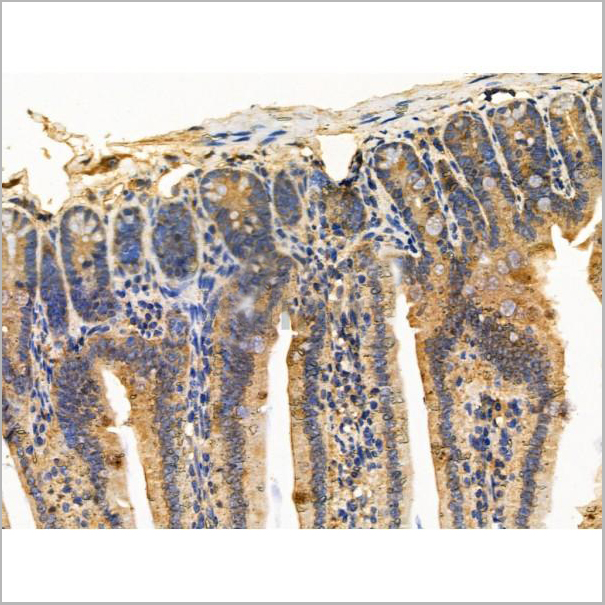
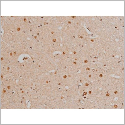
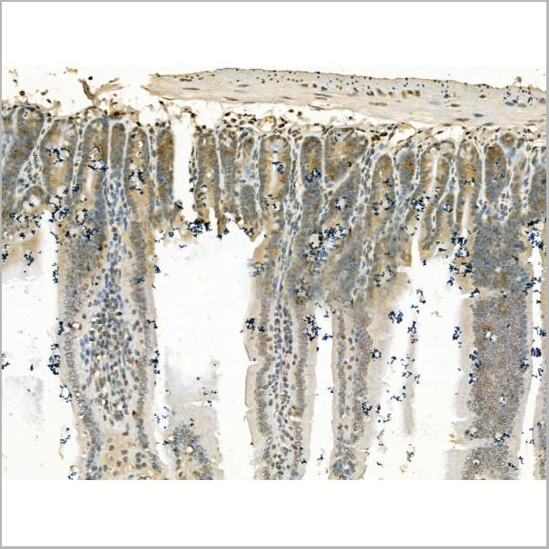
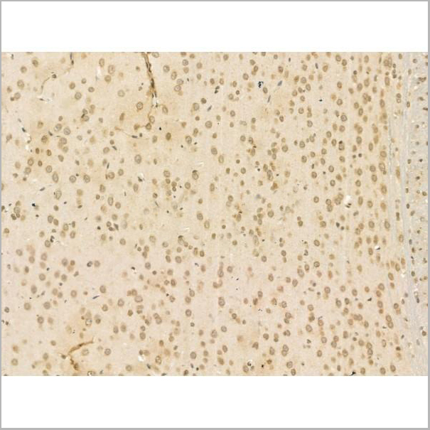
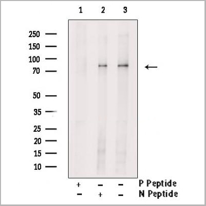
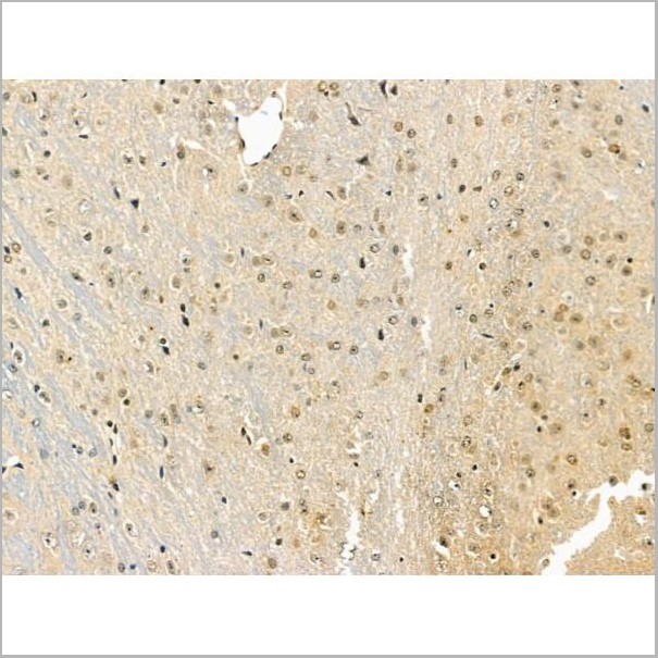
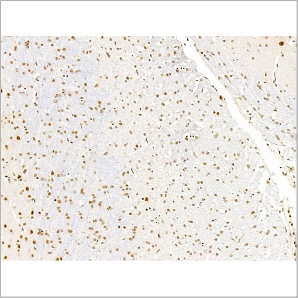
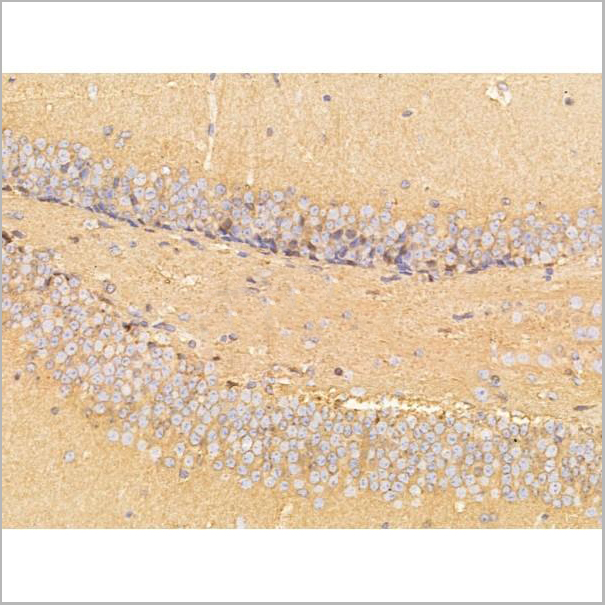
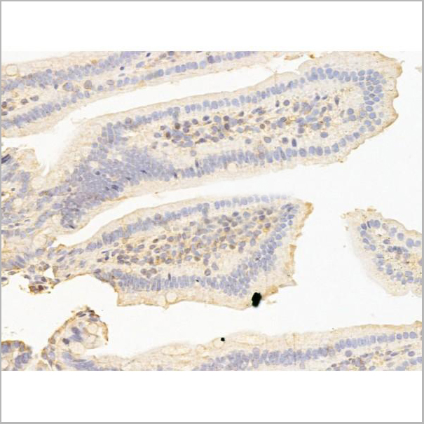
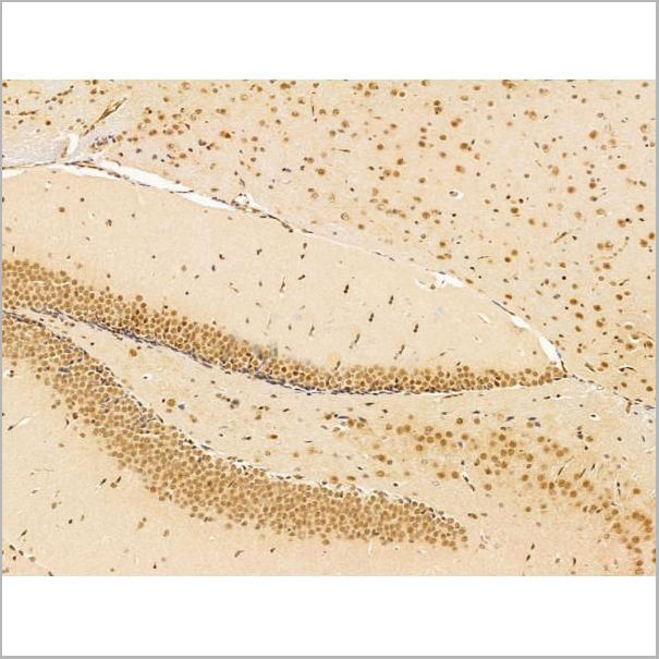
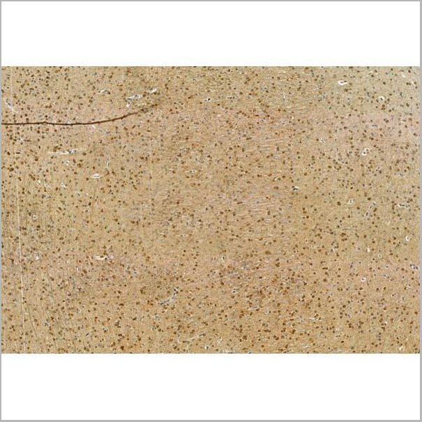
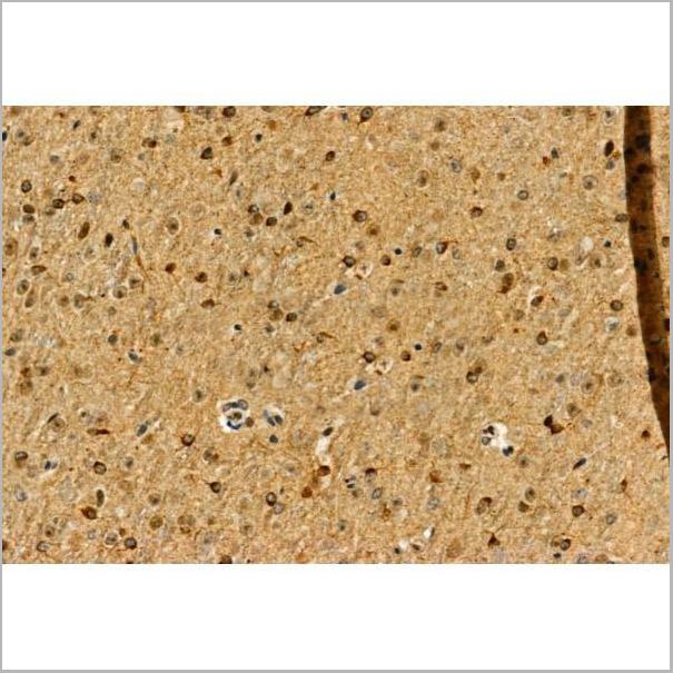
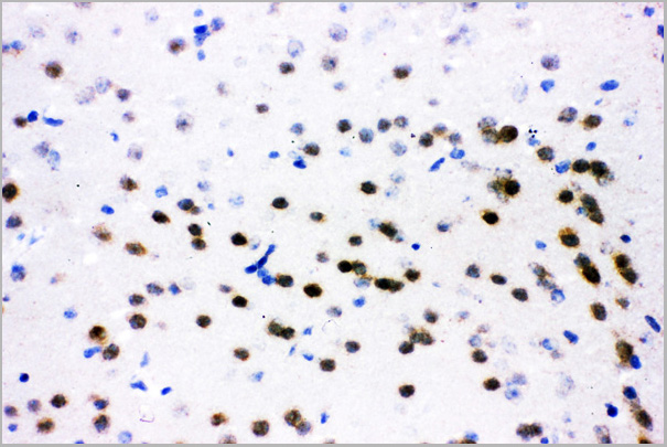
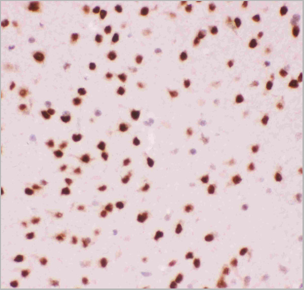
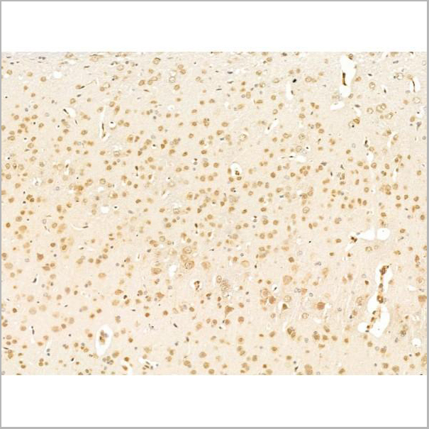
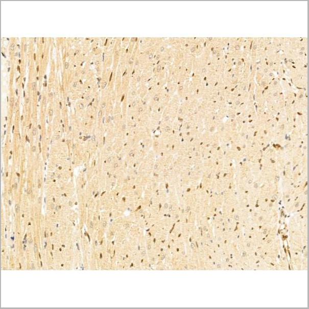
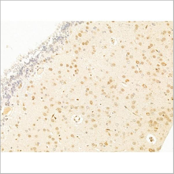
 IHC (Immunohistchemistry-Paraffin)
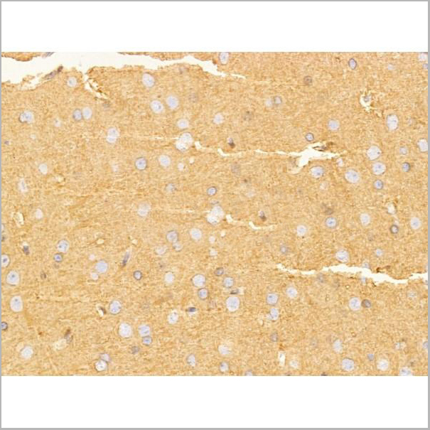
(AAA31218 at 1/100 staining Rat brain tissue by IHC-P. The sample was formaldehyde fixed and a heat mediated antigen retrieval step in citrate buffer was performed. The sample was then blocked and incubated with the primary antibody at 4°C overnight. An HRP conjugated anti-Rabbit antibody was used as the secondary antibody.)
IHC (Immunohistchemistry-Paraffin)
(AAA31218 at 1/100 staining Rat brain tissue by IHC-P. The sample was formaldehyde fixed and a heat mediated antigen retrieval step in citrate buffer was performed. The sample was then blocked and incubated with the primary antibody at 4°C overnight. An HRP conjugated anti-Rabbit antibody was used as the secondary antibody.)
Siglec 7, Polyclonal Antibody (Cat# AAA31218)
Full Name
Siglec 7 Antibody
Gene Names
SIGLEC7; p75; QA79; AIRM1; CD328; CDw328; D-siglec; SIGLEC-7; SIGLECP2; SIGLEC19P; p75/AIRM1
Reactivity
Human, Mouse, Rat
Applications
ELISA
Purity
The antiserum was purified by peptide affinity chromatography using SulfoLink Coupling Resin (Thermo Fisher Scientific).
Pricing
 Application Data
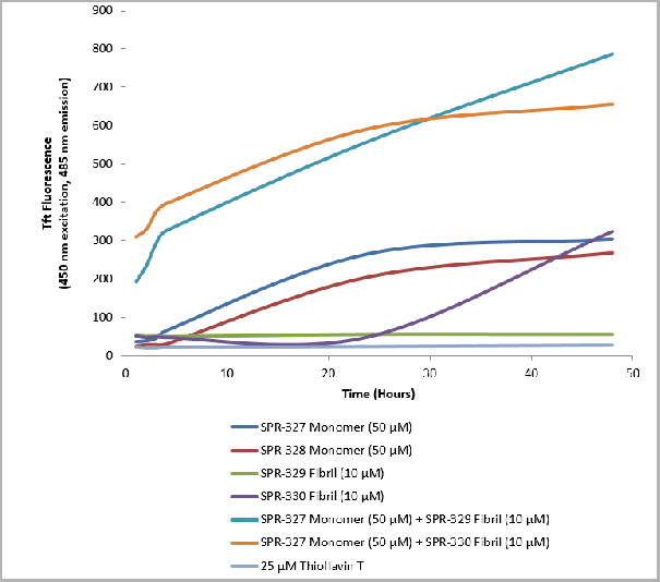
(Thioflavin T is a fluorescent dye that binds to beta sheet-rich structures such as those in tau fibrils. Upon binding, the emission spectrum of the dye experiences a red-shift, and increased fluorescence intensity. Thioflavin T emission curves show increased fluorescence (correlated to tau aggregation) in tau K18 P301L monomers over time. Thioflavin T ex = 450 nm, em = 485 nm.)
Application Data
(Thioflavin T is a fluorescent dye that binds to beta sheet-rich structures such as those in tau fibrils. Upon binding, the emission spectrum of the dye experiences a red-shift, and increased fluorescence intensity. Thioflavin T emission curves show increased fluorescence (correlated to tau aggregation) in tau K18 P301L monomers over time. Thioflavin T ex = 450 nm, em = 485 nm.)
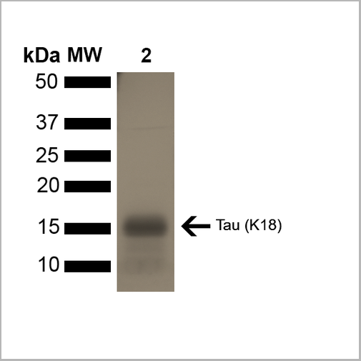
Tau, Active Protein (Cat# AAA27662)
Full Name
Active Human Recombinant Tau (K18), P301L Mutant Protein Monomer
Gene Names
MAPT; TAU; MSTD; PPND; DDPAC; MAPTL; MTBT1; MTBT2; FTDP-17; PPP1R103
Applications
Western Blot
Purity
Purity: >95%
Purification: Ion-exchange Purified
Purification: Ion-exchange Purified
Pricing
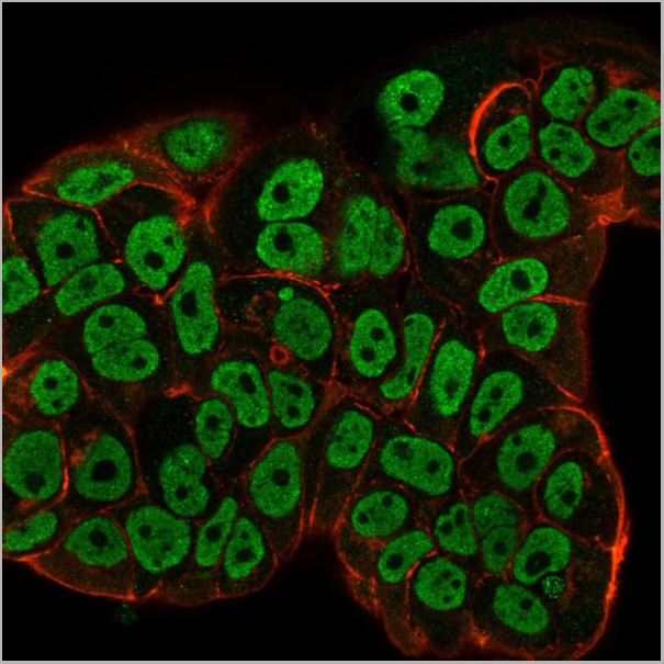
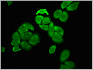
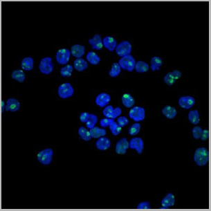
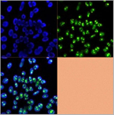
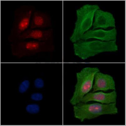
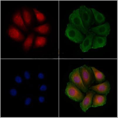
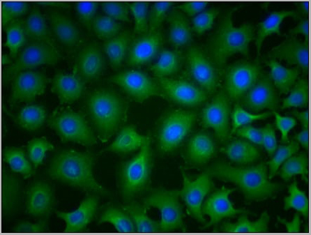
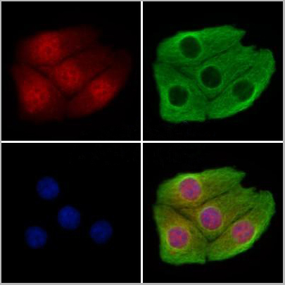 IF (Immunofluorescence)
(AAA31193 staining Hela cells by IF/ICC. The samples were fixed with PFA and permeabilized in 0.1% Triton X-100,then blocked in 10% serum for 45 minutes at 25°C. Samples were then incubated with primary Ab(AAA31193 1:200) and mouse anti-beta tubulin Ab(T0023 1:200) for 1 hour at 37°C. An AlexaFluor594 conjugated goat anti-rabbit IgG(H+L) Ab(Red) and an AlexaFluor488 conjugated goat anti-mouse IgG(H+L) Ab(Green) were used as the secondary antibody.The nuclear counter stain is DAPI(blue).)
IF (Immunofluorescence)
(AAA31193 staining Hela cells by IF/ICC. The samples were fixed with PFA and permeabilized in 0.1% Triton X-100,then blocked in 10% serum for 45 minutes at 25°C. Samples were then incubated with primary Ab(AAA31193 1:200) and mouse anti-beta tubulin Ab(T0023 1:200) for 1 hour at 37°C. An AlexaFluor594 conjugated goat anti-rabbit IgG(H+L) Ab(Red) and an AlexaFluor488 conjugated goat anti-mouse IgG(H+L) Ab(Green) were used as the secondary antibody.The nuclear counter stain is DAPI(blue).)
CHD4, Polyclonal Antibody (Cat# AAA31193)
Full Name
CHD4 Antibody
Gene Names
CHD4; CHD-4; Mi-2b; SIHIWES; Mi2-BETA
Reactivity
Human, Mouse, Rat
Predicted Reactivity: Pig(100%), Horse(100%), Rabbit(100%), Dog(100%), Chicken(100%), Xenopus(100%)
Predicted Reactivity: Pig(100%), Horse(100%), Rabbit(100%), Dog(100%), Chicken(100%), Xenopus(100%)
Applications
Immunofluorescence, Immunocytochemistry
Purity
The antiserum was purified by peptide affinity chromatography using SulfoLink Coupling Resin (Thermo Fisher Scientific).
Pricing
S100A9, Monoclonal Antibody (Cat# AAA13837)
Full Name
S100A9 (Macrophage Marker) Mouse Monoclonal Antibody
Gene Names
S100A9; MIF; NIF; P14; CAGB; CFAG; CGLB; L1AG; LIAG; MRP14; 60B8AG; MAC387
Reactivity
Human and Rat. Others not tested.
Applications
Flow Cytometry, Immunofluorescence, Immunohistochemistry
Pricing
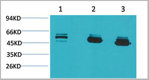
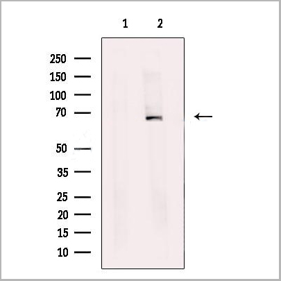
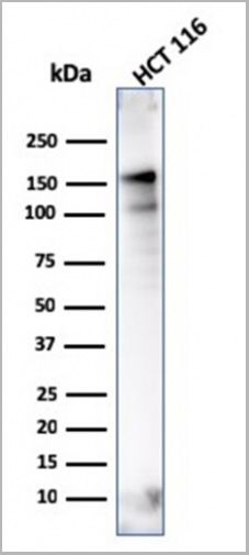
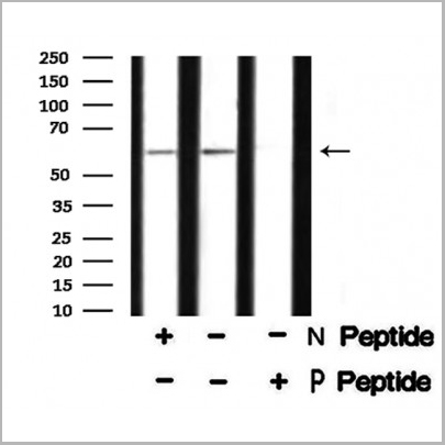
 WB (Western Blot)
(Western Blot Analysis of human MCF-7 cell lysate using ER-beta1 Mouse Monoclonal Antibody (ERb455).)
WB (Western Blot)
(Western Blot Analysis of human MCF-7 cell lysate using ER-beta1 Mouse Monoclonal Antibody (ERb455).)
ER-beta1 (Estrogen Receptor beta-1), Monoclonal Antibody (Cat# AAA13814)
Full Name
ER-beta1 (Estrogen Receptor beta-1) Mouse Monoclonal Antibody
Gene Names
ESR2; Erb; ESRB; ESTRB; NR3A2; ER-BETA; ESR-BETA
Reactivity
Human
Applications
Flow Cytometry, Immunofluorescence, Western Blot, Immunohistochemistry
Pricing
 Application Data
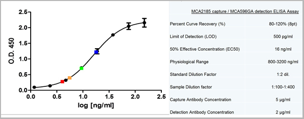
(Sandwich ELISA analysis of CD178 binding using Mouse anti Human CD178 as a capture reagent and biotinylated Mouse anti Human CD178 as a detection reagent with purified human CD178 as antigen for the generation of a standard curve. Detection is by HRP conjugated Streptavidin and substrate. Microtitre plate is read at O.D. 450 nm on the iMark Microplate Absorbance Reader . Plasma (orange) sample is displayed at 1:2 dilution.)
Application Data
(Sandwich ELISA analysis of CD178 binding using Mouse anti Human CD178 as a capture reagent and biotinylated Mouse anti Human CD178 as a detection reagent with purified human CD178 as antigen for the generation of a standard curve. Detection is by HRP conjugated Streptavidin and substrate. Microtitre plate is read at O.D. 450 nm on the iMark Microplate Absorbance Reader . Plasma (orange) sample is displayed at 1:2 dilution.)
CD178, Monoclonal Antibody (Cat# AAA11962)
Full Name
MOUSE ANTI HUMAN CD178
Gene Names
FASLG; APTL; FASL; CD178; CD95L; ALPS1B; CD95-L; TNFSF6; APT1LG1
Reactivity
Human
Applications
Flow Cytometry, Immunoprecipitation
Pricing

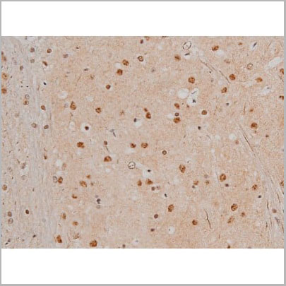
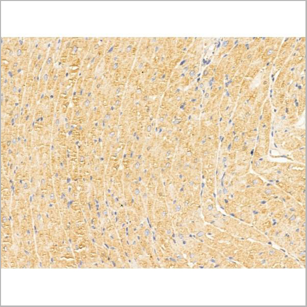
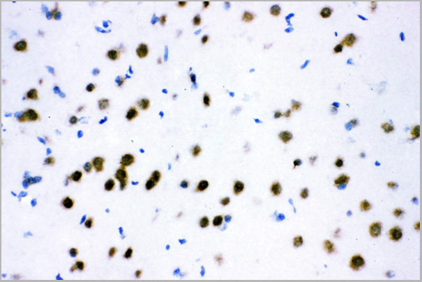
 IHC (Immunohistchemistry)
(At 1/100 staining Mouse brain tissue by IHC-P. The sample was formaldehyde fixed and a heat mediated antigen retrieval step in citrate buffer was performed. The sample was then blocked and incubated with the primary antibody at 4 degree C overnight. An HRP conjugated anti-Rabbit antibody was used as the secondary antibody.)
IHC (Immunohistchemistry)
(At 1/100 staining Mouse brain tissue by IHC-P. The sample was formaldehyde fixed and a heat mediated antigen retrieval step in citrate buffer was performed. The sample was then blocked and incubated with the primary antibody at 4 degree C overnight. An HRP conjugated anti-Rabbit antibody was used as the secondary antibody.)
IRAK1, Polyclonal Antibody (Cat# AAA31368)
Full Name
Phospho-IRAK1 (Thr209) Antibody
Gene Names
IRAK1; IRAK; pelle
Reactivity
Human, Mouse, Rat
Predicted Reactivity: Horse (100%), Dog (82%)
Predicted Reactivity: Horse (100%), Dog (82%)
Applications
Western Blot, Immunohistochemistry, Peptide ELISA
Purity
The antibody is from purified rabbit serum by affinity purification via sequential chromatography on phospho-peptide and non-phospho-peptide affinity columns.
Pricing
COVID 19 Spike RBD Coronavirus, Recombinant Protein (Cat# AAA13542)
Full Name
Recombinant SARS-Cov-2 Spike RBD Protein (Detection)
Applications
Lateral Flow
Purity
>95% by SDS-PAGE
Pricing
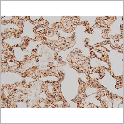
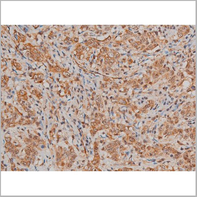
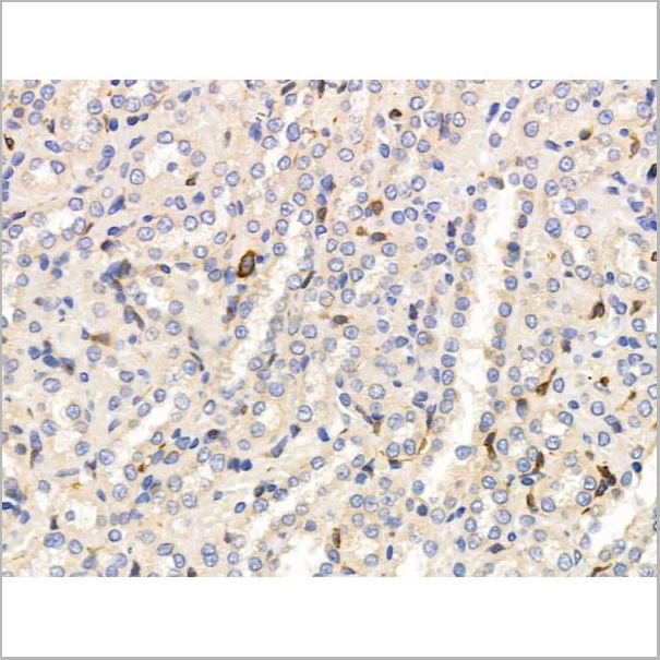
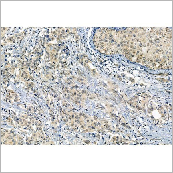
 IHC (Immunohistochemistry)
(Immunohistochemical analysis of paraffin-embedded Human Lung Carcinoma using MICU1 Monoclonal Antibody.)
IHC (Immunohistochemistry)
(Immunohistochemical analysis of paraffin-embedded Human Lung Carcinoma using MICU1 Monoclonal Antibody.)
MICU1, Monoclonal Antibody (Cat# AAA29640)
Full Name
MICU1 Monoclonal Antibody
Gene Names
MICU1; CALC; EFHA3; MPXPS; CBARA1
Reactivity
Human, Mouse, Rat
Applications
Western Blot, Immunohistochemistry
Purity
The antibody was affinity-purified from antiserum by affinity-chromatography
Pricing
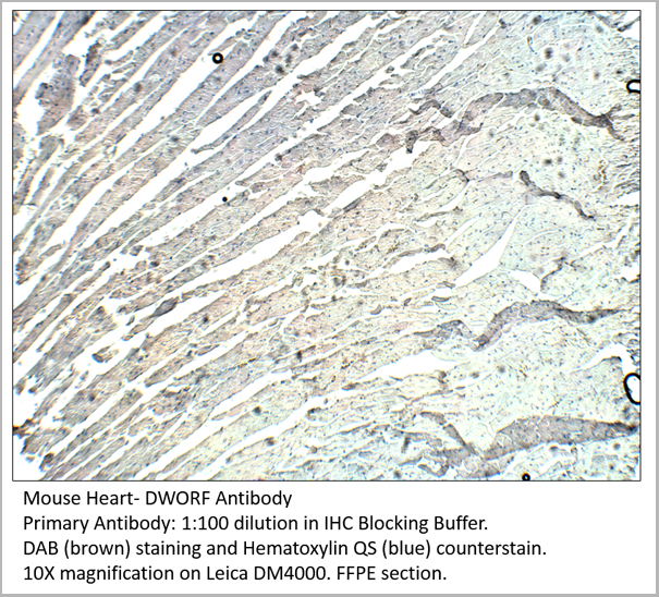
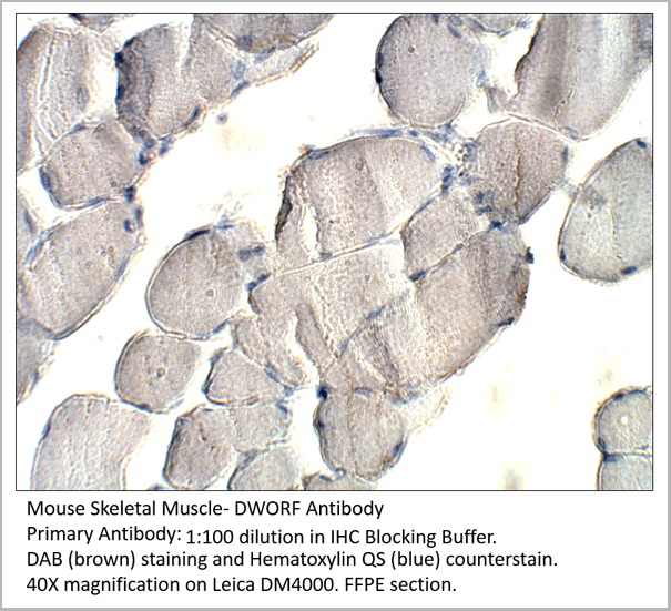
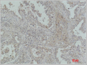
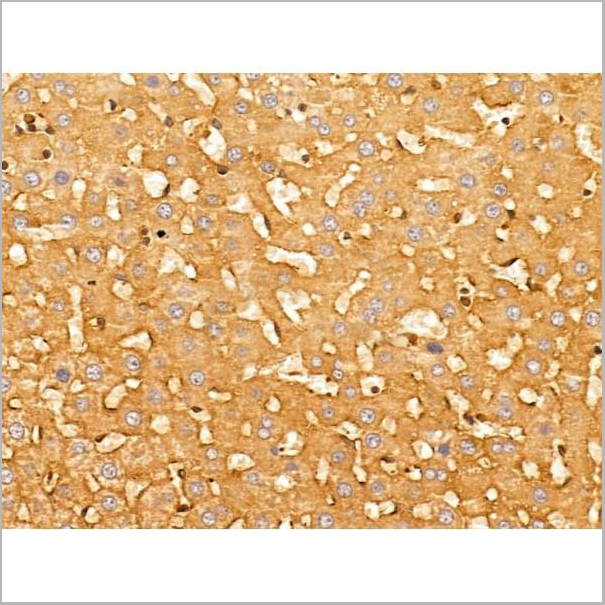
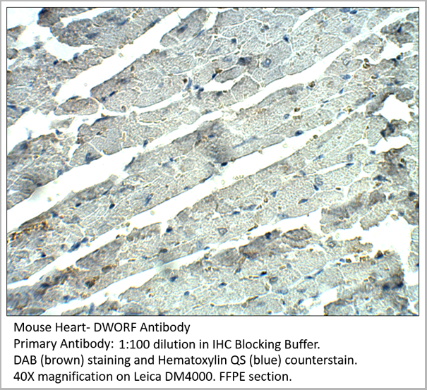 IHC (Immunohistochemistry)
DWORF, Polyclonal Antibody (Cat# AAA14407)
Full Name
DWORF Antibody
Applications
Immunohistochemistry, Immunoprecipitation, Western Blot
Purity
Affinity Purified Immunoglobulins
Pricing
 Application Data
(ELISA plate was coated by N protein, 100 uL/cell, at various concentrations. The direct ELISA analysis was performed by loading 100 uL per well of a anti C-ter His tag HRP conjugated mAb at a concentration of 1:2000. The plate was incubated for 1 hours at 37 degree C, then washed 5 time. Detection was performed using TMB substrate for 10 minutes at room temperature in the dark. The plate was stopped with 2M sulfuric acid. Absorbances were read on a spectrophotometer at 450 nm.)
Application Data
(ELISA plate was coated by N protein, 100 uL/cell, at various concentrations. The direct ELISA analysis was performed by loading 100 uL per well of a anti C-ter His tag HRP conjugated mAb at a concentration of 1:2000. The plate was incubated for 1 hours at 37 degree C, then washed 5 time. Detection was performed using TMB substrate for 10 minutes at room temperature in the dark. The plate was stopped with 2M sulfuric acid. Absorbances were read on a spectrophotometer at 450 nm.)
COVID 19 Nucleocapsid (NP) Coronavirus, Recombinant Protein (Cat# AAA27948)
Full Name
2019-nCov Nucleocapsid Recombinant Protein (full length)
Applications
ELISA
Purity
Purity: >95% by SDS-page
Purification: Ni column
Purification: Ni column
Pricing
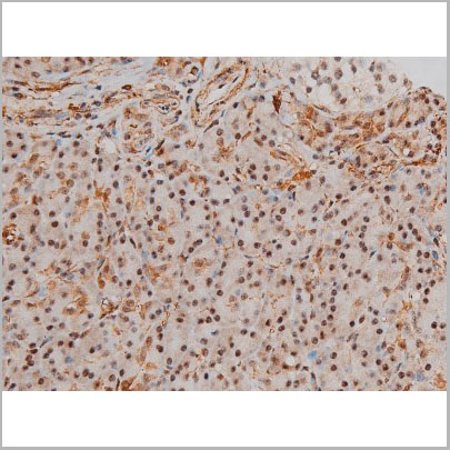
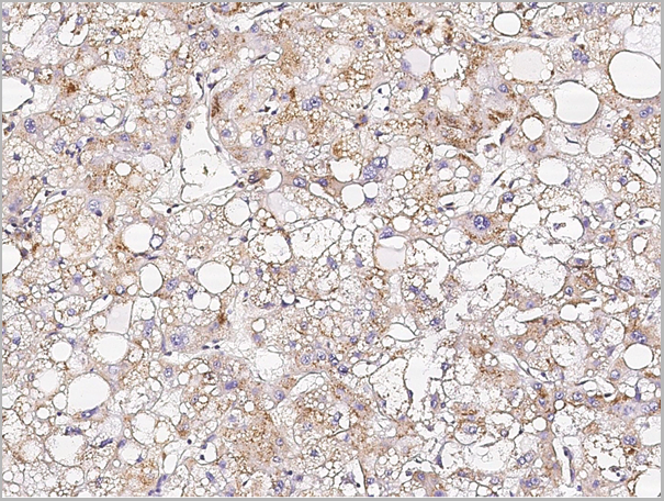 IHC (Immunohistochemistry)
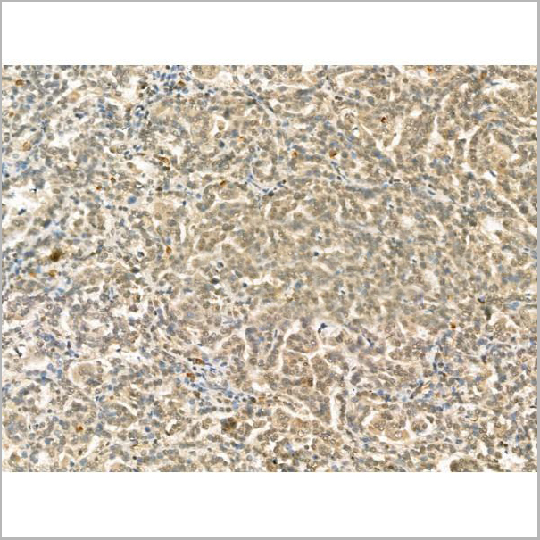
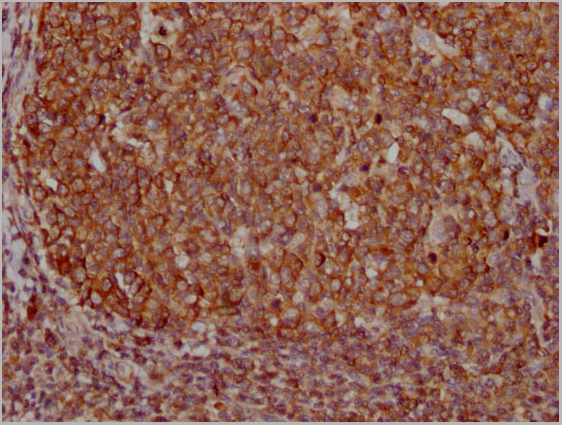
(Immunochemical staining of human SPP1 in human hepatoma with rabbit monoclonal antibody (1:2000, formalin-fixed paraffin embedded sections).)
IHC (Immunohistochemistry)
(Immunochemical staining of human SPP1 in human hepatoma with rabbit monoclonal antibody (1:2000, formalin-fixed paraffin embedded sections).)
Osteopontin, Monoclonal Antibody (Cat# AAA27731)
Full Name
Recombinant Anti-Osteopontin Antibody, Rabbit Monoclonal
Gene Names
SPP1; OPN; BNSP; BSPI; ETA-1; MGC110940
Applications
Immunohistochemistry, Flow Cytometry
Pricing
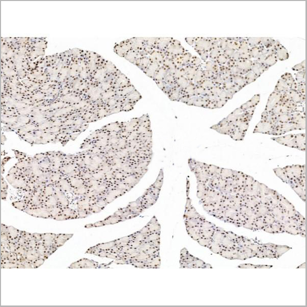
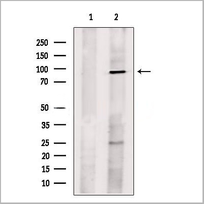
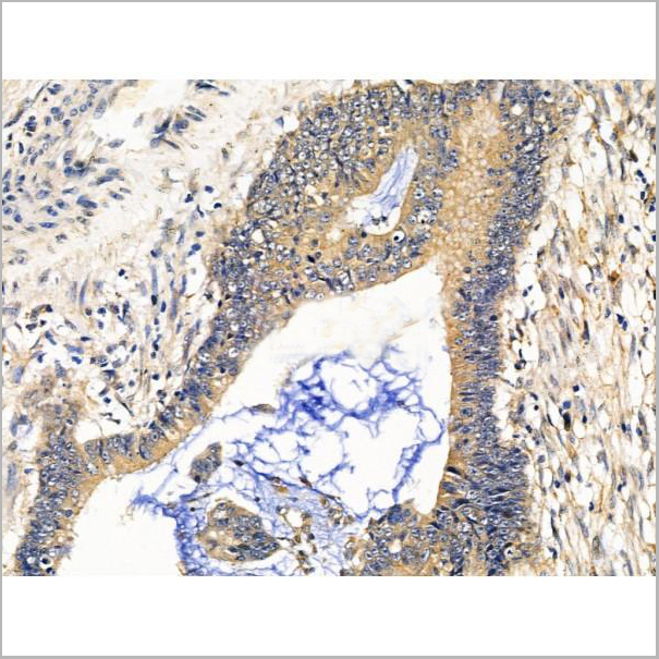
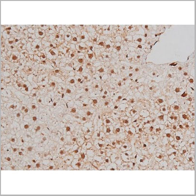
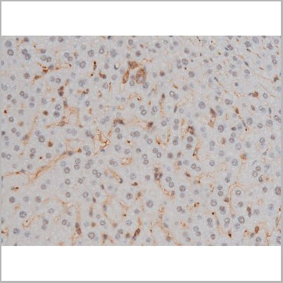
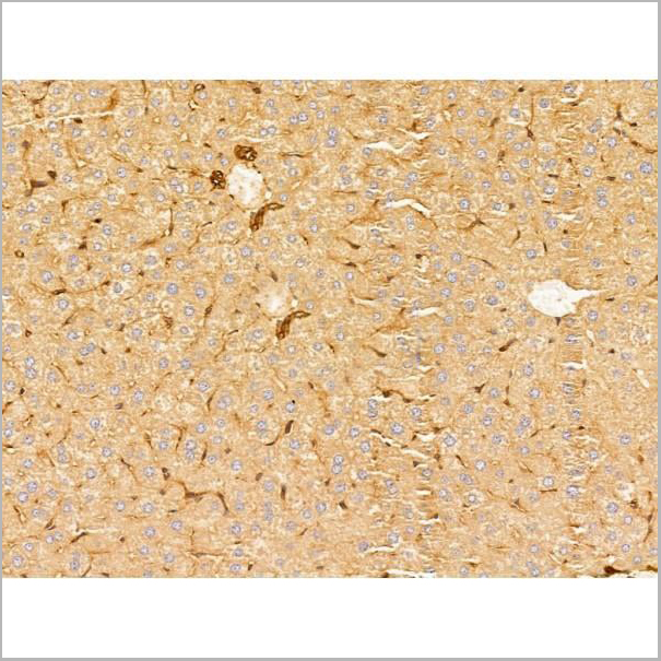
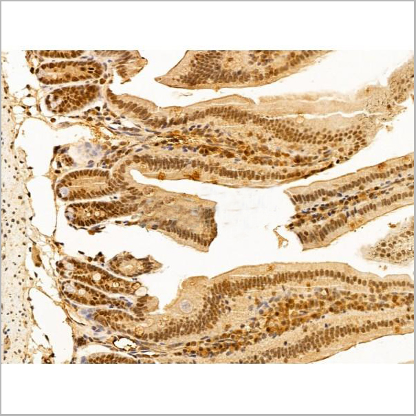
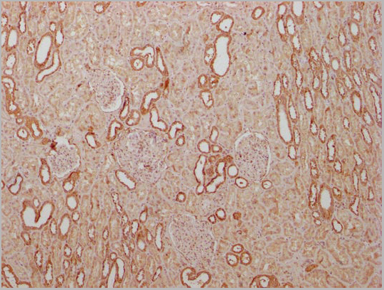
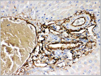
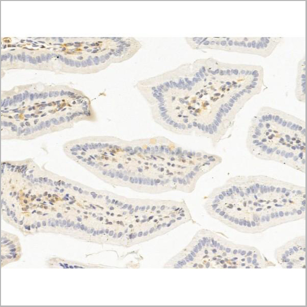
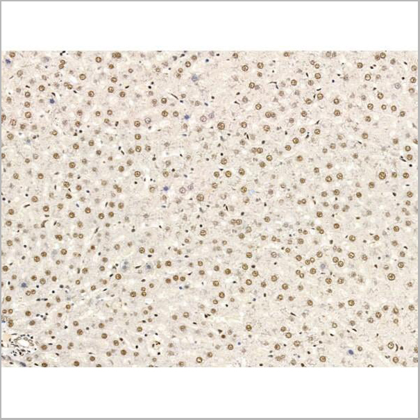 IHC (Immunohistochemistry-Paraffin)
(AAA31258 at 1/100 staining Rat liver tissue by IHC-P. The sample was formaldehyde fixed and a heat mediated antigen retrieval step in citrate buffer was performed. The sample was then blocked and incubated with the primary antibody at 4°C overnight. An HRP conjugated anti-Rabbit antibody was used as the secondary antibody.)
IHC (Immunohistochemistry-Paraffin)
(AAA31258 at 1/100 staining Rat liver tissue by IHC-P. The sample was formaldehyde fixed and a heat mediated antigen retrieval step in citrate buffer was performed. The sample was then blocked and incubated with the primary antibody at 4°C overnight. An HRP conjugated anti-Rabbit antibody was used as the secondary antibody.)
L3MBTL1, Polyclonal Antibody (Cat# AAA31258)
Full Name
L3MBTL1 Antibody
Gene Names
L3MBTL1; L3MBTL; ZC2HC3; H-L(3)MBT; dJ138B7.3
Reactivity
Human, Mouse, Rat
Predicted Reactivity: Horse(100%), Rabbit(100%), Dog(100%), Chicken(100%)
Predicted Reactivity: Horse(100%), Rabbit(100%), Dog(100%), Chicken(100%)
Applications
ELISA
Purity
The antiserum was purified by peptide affinity chromatography using SulfoLink Coupling Resin (Thermo Fisher Scientific).
Pricing
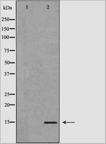
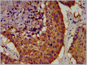
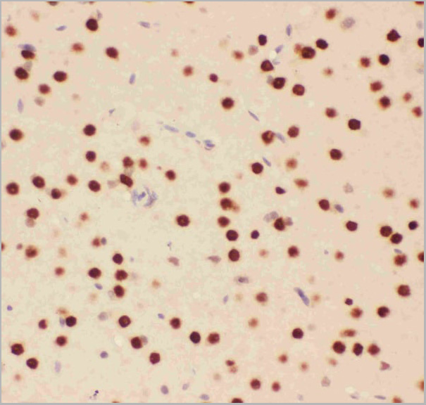
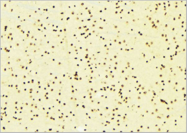 IHC (Immunohistochemistry)
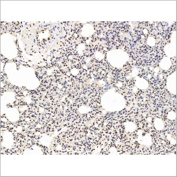
(AAA31121 at 1/100 staining Mouse brain tissue by IHC-P. The sample was formaldehyde fixed and a heat mediated antigen retrieval step in citrate buffer was performed. The sample was then blocked and incubated with the antibody for 1.5 hours at 22 degree C. An HRP conjugated goat anti-rabbit antibody was used as the secondary.)
IHC (Immunohistochemistry)
(AAA31121 at 1/100 staining Mouse brain tissue by IHC-P. The sample was formaldehyde fixed and a heat mediated antigen retrieval step in citrate buffer was performed. The sample was then blocked and incubated with the antibody for 1.5 hours at 22 degree C. An HRP conjugated goat anti-rabbit antibody was used as the secondary.)
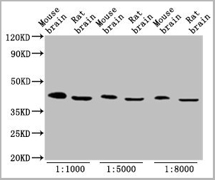
FABP4, Polyclonal Antibody (Cat# AAA31121)
Full Name
FABP4 Antibody
Gene Names
FABP4; aP2; ALBP; AFABP; A-FABP; HEL-S-104
Reactivity
Human, Mouse, Rat
Applications
Western Blot, Immunohistochemistry
Purity
The antiserum was purified by peptide affinity chromatography using SulfoLink Coupling Resin.
Pricing
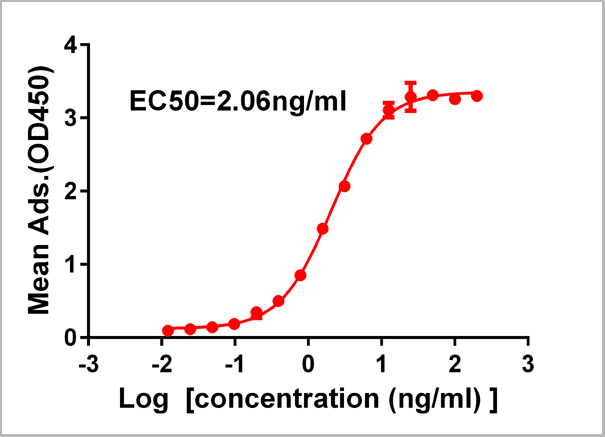
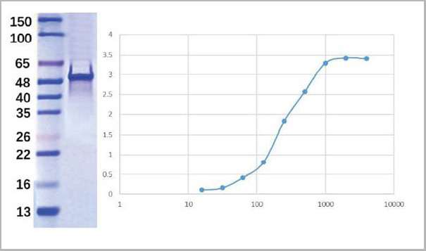
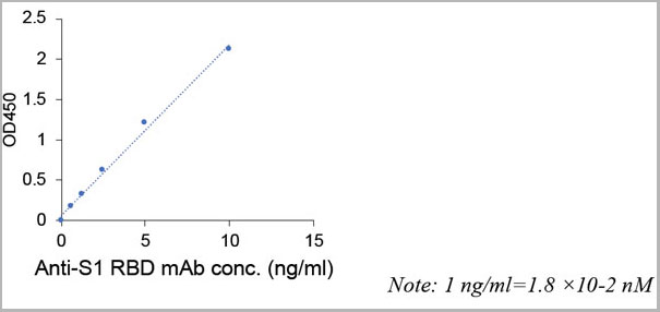
 FCM (Flow Cytometry)
(Figure 1. Elisa plate pre-coated by 2 ug/ml (100ul/well) SARS-CoV-2 RBD protein can bind Rabbit Anti-SARS-CoV-2 RBD monoclonal antibody ( clone:DM35) in a linear range of 0.19-200 ng/ml.)
FCM (Flow Cytometry)
(Figure 1. Elisa plate pre-coated by 2 ug/ml (100ul/well) SARS-CoV-2 RBD protein can bind Rabbit Anti-SARS-CoV-2 RBD monoclonal antibody ( clone:DM35) in a linear range of 0.19-200 ng/ml.)
COVID 19 Spike RBD (DM35) Coronavirus, Monoclonal Antibody (Cat# AAA11707)
Full Name
Anti-SARS-CoV-2 RBD antibody (DM35), Rabbit mAb
Reactivity
SARS-CoV-2
Applications
Flow Cytometry
Purity
Purified from cell culture supernatant by affinity chromatography
Pricing


 IF (Immunofluorescence)
(AAA31213 staining Hela cells by IF/ICC. The samples were fixed with PFA and permeabilized in 0.1% Triton X-100,then blocked in 10% serum for 45 minutes at 25°C. Samples were then incubated with primary Ab(AAA31213 1:200) and mouse anti-beta tubulin Ab(T0023 1:200) for 1 hour at 37°C. An AlexaFluor594 conjugated goat anti-rabbit IgG(H+L) Ab(Red) and an AlexaFluor488 conjugated goat anti-mouse IgG(H+L) Ab(Green) were used as the secondary antibody.The nuclear counter stain is DAPI(blue).)
IF (Immunofluorescence)
(AAA31213 staining Hela cells by IF/ICC. The samples were fixed with PFA and permeabilized in 0.1% Triton X-100,then blocked in 10% serum for 45 minutes at 25°C. Samples were then incubated with primary Ab(AAA31213 1:200) and mouse anti-beta tubulin Ab(T0023 1:200) for 1 hour at 37°C. An AlexaFluor594 conjugated goat anti-rabbit IgG(H+L) Ab(Red) and an AlexaFluor488 conjugated goat anti-mouse IgG(H+L) Ab(Green) were used as the secondary antibody.The nuclear counter stain is DAPI(blue).)
Exportin-5, Polyclonal Antibody (Cat# AAA31213)
Full Name
Exportin-5 Antibody
Gene Names
XPO5; exp5
Reactivity
Human, Mouse, Rat
Predicted Reactivity: Pig(88%), Bovine(100%), Horse(100%), Sheep(100%), Rabbit(88%), Dog(100%), Chicken(100%)
Predicted Reactivity: Pig(88%), Bovine(100%), Horse(100%), Sheep(100%), Rabbit(88%), Dog(100%), Chicken(100%)
Applications
ELISA
Purity
The antiserum was purified by peptide affinity chromatography using SulfoLink Coupling Resin (Thermo Fisher Scientific).
Pricing
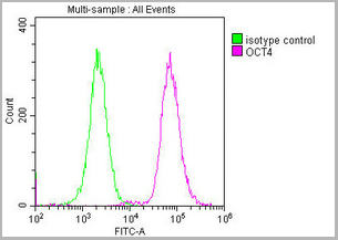
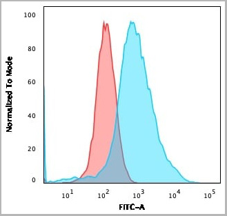
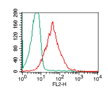
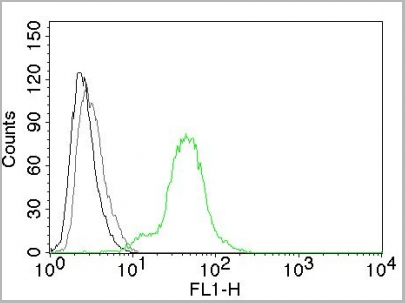
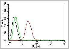
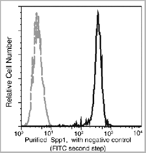
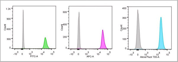
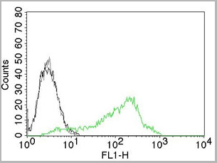
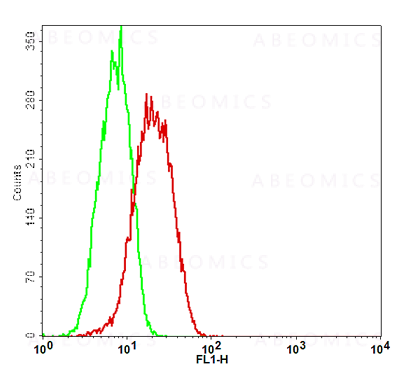
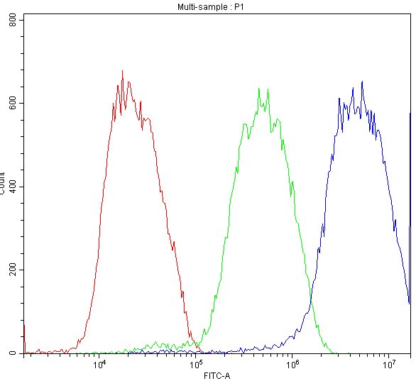
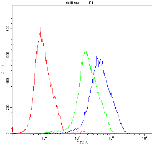
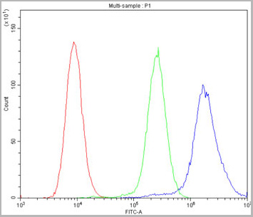
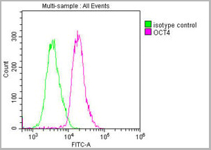 FCM (Flow Cytometry)
(Overlay histogram showing Ntera-2 cells stained with CSB-MA018403A0m (red line) at 1:100. The cells were incubated in 1x PBS /10% normal goat serum to block non-specific protein-protein interactions followed by primary antibody for 1 h at 4 degree C. The secondary antibody used was FITC goat anti-mouse IgG(H+L) at 1/200 dilution for 1 h at 4 degree C. Isotype control antibody (green line) was used under the same conditions. Acquisition of >10,000 events was performed.)
FCM (Flow Cytometry)
(Overlay histogram showing Ntera-2 cells stained with CSB-MA018403A0m (red line) at 1:100. The cells were incubated in 1x PBS /10% normal goat serum to block non-specific protein-protein interactions followed by primary antibody for 1 h at 4 degree C. The secondary antibody used was FITC goat anti-mouse IgG(H+L) at 1/200 dilution for 1 h at 4 degree C. Isotype control antibody (green line) was used under the same conditions. Acquisition of >10,000 events was performed.)
OCT4, Monoclonal Antibody (Cat# AAA27015)
Full Name
OCT4 Monoclonal Antibody
Gene Names
POU5F1; OCT3; OCT4; OTF3; OTF4; OTF-3; Oct-3; Oct-4
Reactivity
Human, Mouse, Rat
Applications
Western Blot, Immunohistochemistry, Immunofluorescence, Flow Cytometry
Purity
>95%
Protein G Purified
Protein G Purified
Pricing
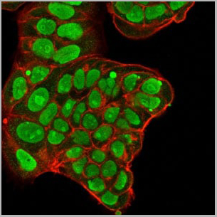
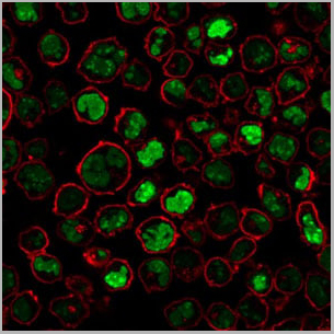
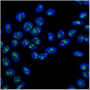

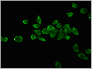
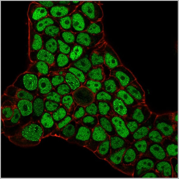
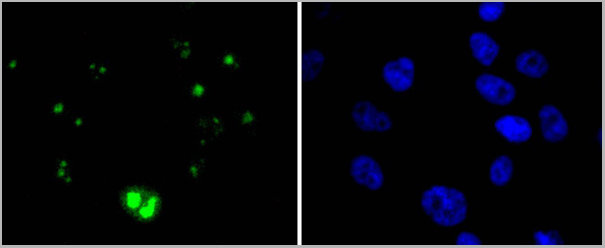
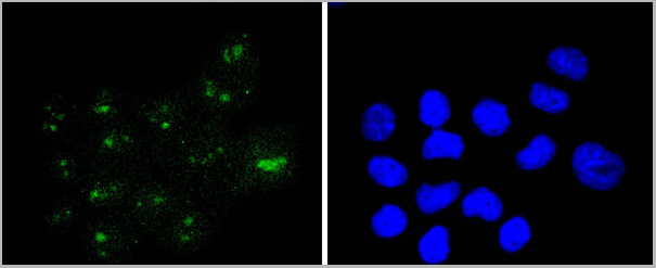
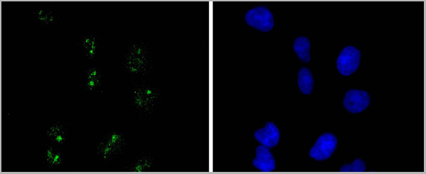
 IF (Immunofluorescence)
Nucleolin, Monoclonal Antibody (Cat# AAA13823)
Full Name
Nucleolin (Marker of Human Cells) Mouse Monoclonal Antibody
Gene Names
NCL; C23
Reactivity
Human.
Does not react with Mouse, Rat and Cow
Does not react with Mouse, Rat and Cow
Applications
Flow Cytometry, Immunofluorescence, Western Blot, Immunohistochemistry
Pricing
 Application Data
(Fig-5: Cell Surface staining of human ACE2 in the ACE2/HEK293 stable cell line. ATTO 488-conjugated anti-hACE2 antibody (clone AC18F; was used at 1ug/ 1x10^6 cells (Red). ATTO 488-conjugated mouse IgG1 was used as isotype control at 1ug/ 1x10^6 cells (Green).)
Application Data
(Fig-5: Cell Surface staining of human ACE2 in the ACE2/HEK293 stable cell line. ATTO 488-conjugated anti-hACE2 antibody (clone AC18F; was used at 1ug/ 1x10^6 cells (Red). ATTO 488-conjugated mouse IgG1 was used as isotype control at 1ug/ 1x10^6 cells (Green).)
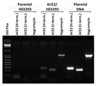
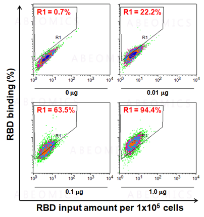
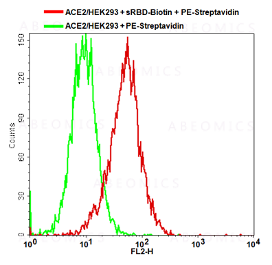
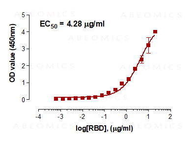
ACE2, Cell Line (Cat# AAA14888)
Full Name
ACE2/HEK293 Stable Cell Line
Gene Names
ACE2; ACEH
Applications
Functional Assay
Pricing
 Application Data
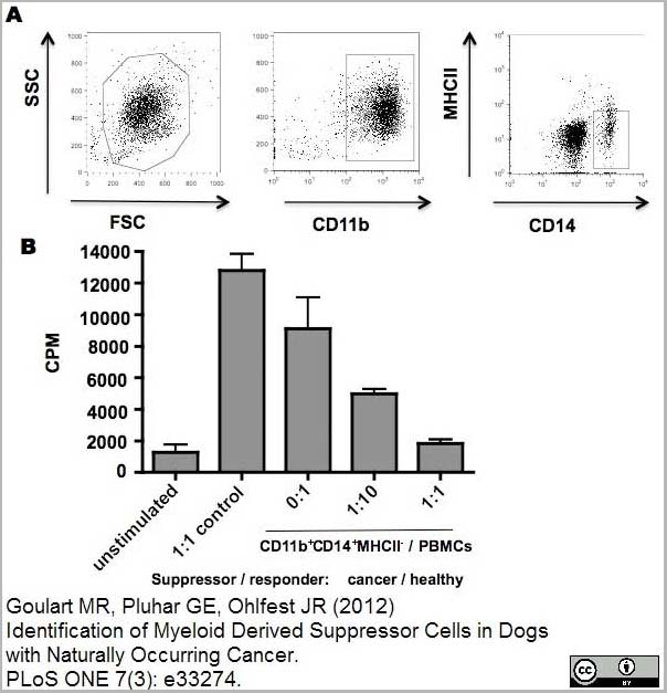
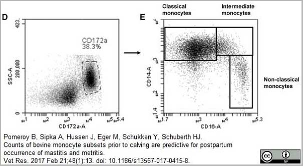
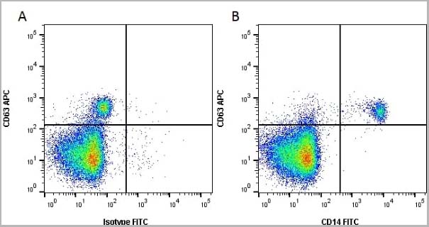
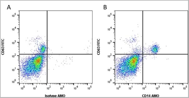
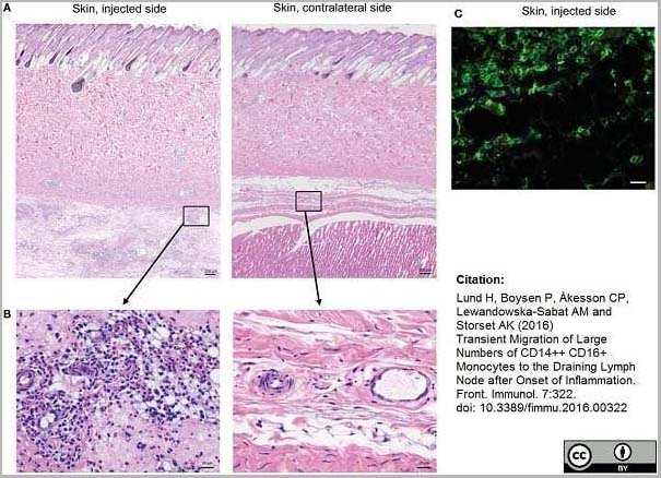
(Mouse anti Human CD14 antibody, clone Tük4 used to identify bovine monocytes in the skin and subcutaneous tissue of adjuvant injected calves by immunofluorescence.Image caption:Cellular recruitment to skin and subcutaneous tissues. (A) HE stained sections of skin with subcutaneous tissue from the side injected with adjuvant and the contralateral side, at 24 h post-injection. Scale bars: 200 ?m. (B) Enlargement of outlined areas in A, as indicated. Scale bars: 20 ?m. (C) Immunofluorescent labeling of subcutaneous tissue on the injected side with antibody against CD14 (green). Scale bar: 20 ?m.From: Lund H, Boysen P, Åkesson CP, Lewandowska-Sabat AM and Storset AK (2016)Transient Migration of Large Numbers of CD14++ CD16+ Monocytes to the Draining Lymph Node after Onset of Inflammation.Front. Immunol. 7:322.This is from an open access article distributed under the terms of the Creative Commons Attribution License.)
Application Data
(Mouse anti Human CD14 antibody, clone Tük4 used to identify bovine monocytes in the skin and subcutaneous tissue of adjuvant injected calves by immunofluorescence.Image caption:Cellular recruitment to skin and subcutaneous tissues. (A) HE stained sections of skin with subcutaneous tissue from the side injected with adjuvant and the contralateral side, at 24 h post-injection. Scale bars: 200 ?m. (B) Enlargement of outlined areas in A, as indicated. Scale bars: 20 ?m. (C) Immunofluorescent labeling of subcutaneous tissue on the injected side with antibody against CD14 (green). Scale bar: 20 ?m.From: Lund H, Boysen P, Åkesson CP, Lewandowska-Sabat AM and Storset AK (2016)Transient Migration of Large Numbers of CD14++ CD16+ Monocytes to the Draining Lymph Node after Onset of Inflammation.Front. Immunol. 7:322.This is from an open access article distributed under the terms of the Creative Commons Attribution License.)
CD14, Monoclonal Antibody (Cat# AAA12266)
Full Name
Mouse Anti Human CD14: Amethyst Orange
Reactivity
Human
Applications
Flow Cytometry
Purity
Purified IgG prepared by affinity chromatography on Protein A from tissue culture supernatant.
Pricing


 Application Data
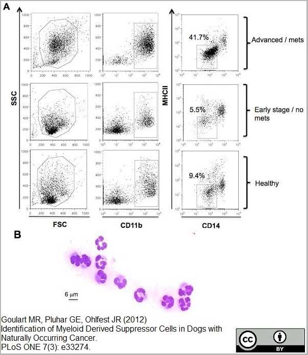
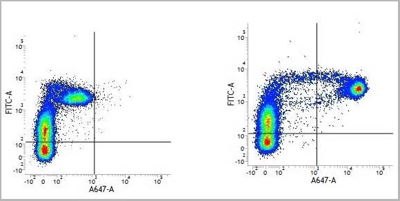
(PE conjugatedMouse anti Human CD169 antibody, clone 7-239 used to block CD169 function on myeloid cells.Image caption:Siglec-1 mediates HIV-1 uptake into a storage compartment and enhances HIV-1 trans-infection specially in IFN?-treated monocytes and DCs. A. Uptake of HIV-1NL4–3 by different myeloid cells exposed to IFN?. Cells were cultured with HIV-1 to measure p24Gag by ELISA. Mean values and SEM from four experiments include cells from 12 donors. B. Fold change in HIV-1NL4–3 uptake of cells treated with bafilomycin A1 compared to untreated cells. Mean values and SEM include cells from three donors. C. Relative uptake of HIV-1NL4–3 by IFN?-treated myeloid cells pre-incubated with the indicated mAbs. Values are normalized to the level of HIV-1 uptake by mock-treated cells (set at 100%). Mean values and SEM from two experiments include cells from six donors. D. Confocal microscopy analysis of different IFN?-treated myeloid cells pulsed with HIV-1Cherry and stained for Siglec-1 (Alexa 488), HLA-DR (Alexa 647) and DAPI. (Top) Representative viral pattern for each kind of myeloid cell analyzed, showing maximum fluorescence intensity of four channels. (Bottom) Percentage of myeloid cells with distinct viral patterns: random distribution, polarized accumulation, and sac-like compartment formation, as illustrated in the left drawing. Mean values of 50 cells from two different donors are shown. E. HIV-1 transmission from IFN?-treated myeloid cells to a luciferase reporter CD4+ cell line. HIV-1 infection was determined by induced luciferase activity in relative light units (RLUs). Mean values and SEM from four experiments include cells from 12 donors. F. Relative HIV-1 transmission from IFN?-treated myeloid cells pre-incubated with the indicated mAbs. Values are normalized to the level of HIV-1 trans-infected by mock-treated cells. Mean values and SEM from two experiments include cells from six donors. Statistical differences were assessed with a paired t test in A and E, and with a one sample t-test in B, C and F.From: Pino M, Erkizia I, Benet S, Erikson E, Fernández-Figueras MT, Guerrero D, Dalmau J, Ouchi D, Rausell A, Ciuffi A, Keppler OT, Telenti A, Kräusslich HG, Martinez-Picado J, Izquierdo-Useros N.HIV-1 immune activation induces Siglec-1 expression and enhances viral trans-infection in blood and tissue myeloid cells.Retrovirology. 2015 May 7;12:37.This image is from an open access article distributed under the terms of the Creative Commons Attribution License.)
Application Data
(PE conjugatedMouse anti Human CD169 antibody, clone 7-239 used to block CD169 function on myeloid cells.Image caption:Siglec-1 mediates HIV-1 uptake into a storage compartment and enhances HIV-1 trans-infection specially in IFN?-treated monocytes and DCs. A. Uptake of HIV-1NL4–3 by different myeloid cells exposed to IFN?. Cells were cultured with HIV-1 to measure p24Gag by ELISA. Mean values and SEM from four experiments include cells from 12 donors. B. Fold change in HIV-1NL4–3 uptake of cells treated with bafilomycin A1 compared to untreated cells. Mean values and SEM include cells from three donors. C. Relative uptake of HIV-1NL4–3 by IFN?-treated myeloid cells pre-incubated with the indicated mAbs. Values are normalized to the level of HIV-1 uptake by mock-treated cells (set at 100%). Mean values and SEM from two experiments include cells from six donors. D. Confocal microscopy analysis of different IFN?-treated myeloid cells pulsed with HIV-1Cherry and stained for Siglec-1 (Alexa 488), HLA-DR (Alexa 647) and DAPI. (Top) Representative viral pattern for each kind of myeloid cell analyzed, showing maximum fluorescence intensity of four channels. (Bottom) Percentage of myeloid cells with distinct viral patterns: random distribution, polarized accumulation, and sac-like compartment formation, as illustrated in the left drawing. Mean values of 50 cells from two different donors are shown. E. HIV-1 transmission from IFN?-treated myeloid cells to a luciferase reporter CD4+ cell line. HIV-1 infection was determined by induced luciferase activity in relative light units (RLUs). Mean values and SEM from four experiments include cells from 12 donors. F. Relative HIV-1 transmission from IFN?-treated myeloid cells pre-incubated with the indicated mAbs. Values are normalized to the level of HIV-1 trans-infected by mock-treated cells. Mean values and SEM from two experiments include cells from six donors. Statistical differences were assessed with a paired t test in A and E, and with a one sample t-test in B, C and F.From: Pino M, Erkizia I, Benet S, Erikson E, Fernández-Figueras MT, Guerrero D, Dalmau J, Ouchi D, Rausell A, Ciuffi A, Keppler OT, Telenti A, Kräusslich HG, Martinez-Picado J, Izquierdo-Useros N.HIV-1 immune activation induces Siglec-1 expression and enhances viral trans-infection in blood and tissue myeloid cells.Retrovirology. 2015 May 7;12:37.This image is from an open access article distributed under the terms of the Creative Commons Attribution License.)
CD169, Monoclonal Antibody (Cat# AAA12265)
Full Name
Mouse Anti Human CD169: RPE
Gene Names
SIGLEC1; SN; CD169; SIGLEC-1
Reactivity
Human
Applications
Flow Cytometry
Purity
>95% by SDS PAGE
Purified IgG prepared by affinity chromatography on Protein A from tissue culture supernatant.
Purified IgG prepared by affinity chromatography on Protein A from tissue culture supernatant.
Pricing
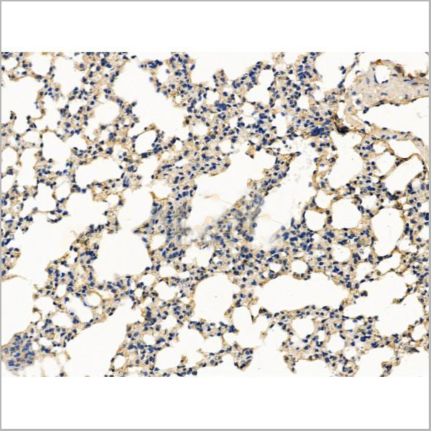
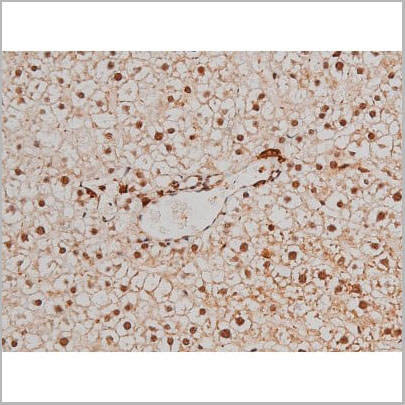
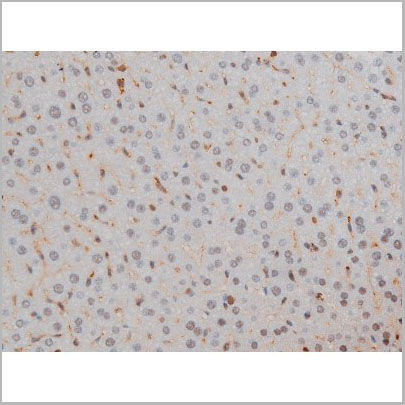
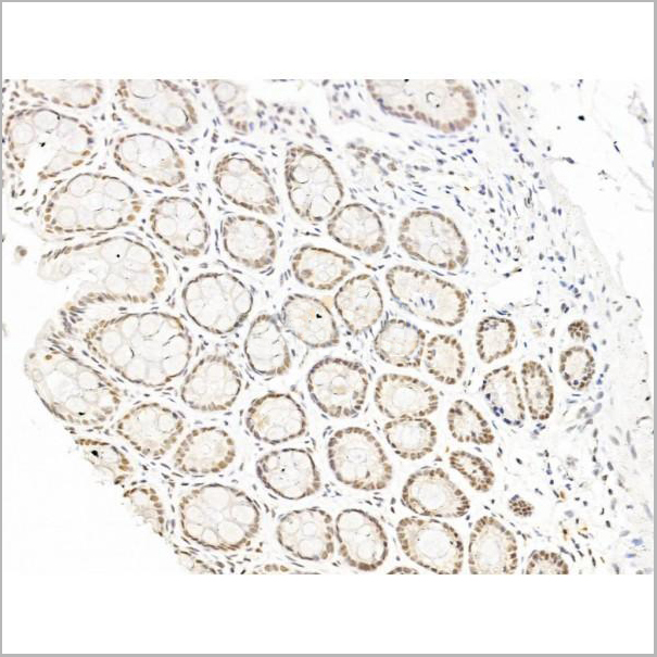
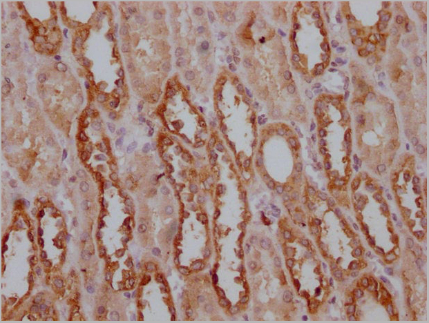
 IHC (Immunohistochemistry-Paraffin)
(AAA31260 at 1/100 staining Mouse liver tissue by IHC-P. The sample was formaldehyde fixed and a heat mediated antigen retrieval step in citrate buffer was performed. The sample was then blocked and incubated with the primary antibody at 4°C overnight. An HRP conjugated anti-Rabbit antibody was used as the secondary antibody.)
IHC (Immunohistochemistry-Paraffin)
(AAA31260 at 1/100 staining Mouse liver tissue by IHC-P. The sample was formaldehyde fixed and a heat mediated antigen retrieval step in citrate buffer was performed. The sample was then blocked and incubated with the primary antibody at 4°C overnight. An HRP conjugated anti-Rabbit antibody was used as the secondary antibody.)
COL4A3BP, Polyclonal Antibody (Cat# AAA31260)
Full Name
COL4A3BP Antibody
Gene Names
CERT1; CERT; GPBP; CERTL; MRD34; STARD11; COL4A3BP
Reactivity
Human, Mouse, Rat
Predicted Reactivity: Zebrafish(100%), Bovine(100%), Horse(100%), Sheep(100%), Rabbit(100%), Dog(100%), Chicken(100%), Xenopus(100%)
Predicted Reactivity: Zebrafish(100%), Bovine(100%), Horse(100%), Sheep(100%), Rabbit(100%), Dog(100%), Chicken(100%), Xenopus(100%)
Applications
ELISA
Purity
The antiserum was purified by peptide affinity chromatography using SulfoLink Coupling Resin (Thermo Fisher Scientific).
Pricing

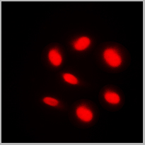 IF (Immunofluorescence)
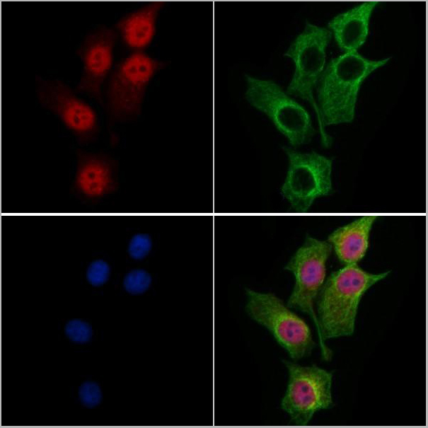
(Immunofluorescent analysis of BAF250B staining in HeLa cells. Formalin-fixed cells were permeabilized with 0.1% Triton X-100 in TBS for 5-10 minutes and blocked with 3% BSA-PBS for 30 minutes at room temperature. Cells were probed with the primary antibody in 3% BSA-PBS and incubated overnight at 4 °C in a humidified chamber. Cells were washed with PBST and incubated with a DyLight 594-conjugated secondary antibody (red) in PBS at room temperature in the dark.)
IF (Immunofluorescence)
(Immunofluorescent analysis of BAF250B staining in HeLa cells. Formalin-fixed cells were permeabilized with 0.1% Triton X-100 in TBS for 5-10 minutes and blocked with 3% BSA-PBS for 30 minutes at room temperature. Cells were probed with the primary antibody in 3% BSA-PBS and incubated overnight at 4 °C in a humidified chamber. Cells were washed with PBST and incubated with a DyLight 594-conjugated secondary antibody (red) in PBS at room temperature in the dark.)
BAF250B, Polyclonal Antibody (Cat# AAA27801)
Full Name
Anti-BAF250B Antibody
Gene Names
ARID1B; OSA2; 6A3-5; DAN15; MRD12; P250R; BRIGHT; BAF250B; ELD/OSA1
Reactivity
Human, Mouse, Rat, Porcine
Applications
Western Blot, Immunohistochemistry, Immunofluorescence, Immunocytochemistry
Purity
The antibody was purified by immunogen affinity chromatography.
Pricing
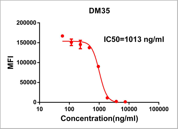
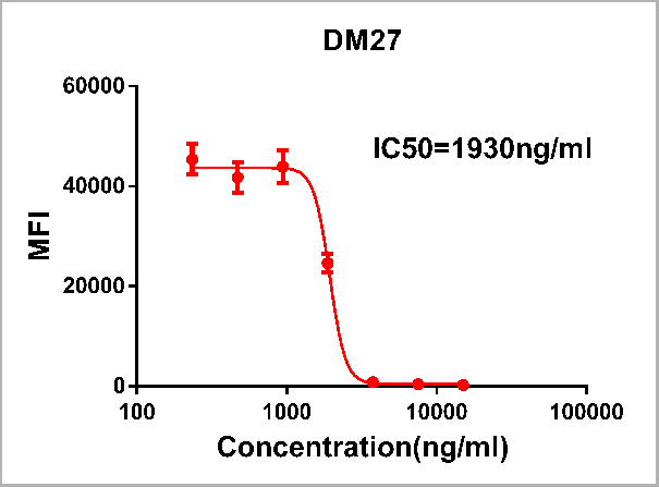 ELISA
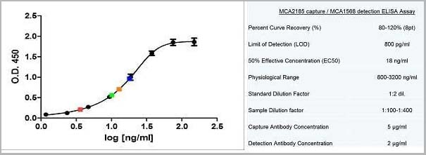
(Figure 1. Competition flow cytometry assay demonstrating Rabbit anti-RBD monoclonal antibody ( clone: DM27) blockade of SARS-CoV-2 (COVID-19) S protein RBD (1ug/ml, [getskuurl sku=PME100497 binding to Expi 293 cell line transfected with human ACE2. IC50=1930ng/ml. The Y-axis represents the geometric mean fluorescence intensity (MFI) while the X-axis represents the concentration of IgG used.)
ELISA
(Figure 1. Competition flow cytometry assay demonstrating Rabbit anti-RBD monoclonal antibody ( clone: DM27) blockade of SARS-CoV-2 (COVID-19) S protein RBD (1ug/ml, [getskuurl sku=PME100497 binding to Expi 293 cell line transfected with human ACE2. IC50=1930ng/ml. The Y-axis represents the geometric mean fluorescence intensity (MFI) while the X-axis represents the concentration of IgG used.)
COVID 19 Spike RBD (DM27) Coronavirus, Monoclonal Antibody (Cat# AAA11706)
Full Name
Anti-SARS-CoV-2 RBD antibody (DM27), Rabbit mAb
Reactivity
SARS-CoV-2
Applications
Flow Cytometry
Purity
Purified from cell culture supernatant by affinity chromatography
Pricing
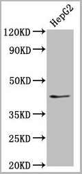
 IF (Immunofluorescence)
(AAA31247 staining HepG2 cells by IF/ICC. The samples were fixed with PFA and permeabilized in 0.1% Triton X-100,then blocked in 10% serum for 45 minutes at 25°C. Samples were then incubated with primary Ab(AAA31247) and mouse anti-beta tubulin Ab(T0023) for 1 hour at 37°C. An AlexaFluor594 conjugated goat anti-rabbit IgG(H+L) Ab(Red) and an AlexaFluor488 conjugated goat anti-mouse IgG(H+L) Ab(Green) were used as the secondary antibody.The nuclear counter stain is DAPI(blue).)
IF (Immunofluorescence)
(AAA31247 staining HepG2 cells by IF/ICC. The samples were fixed with PFA and permeabilized in 0.1% Triton X-100,then blocked in 10% serum for 45 minutes at 25°C. Samples were then incubated with primary Ab(AAA31247) and mouse anti-beta tubulin Ab(T0023) for 1 hour at 37°C. An AlexaFluor594 conjugated goat anti-rabbit IgG(H+L) Ab(Red) and an AlexaFluor488 conjugated goat anti-mouse IgG(H+L) Ab(Green) were used as the secondary antibody.The nuclear counter stain is DAPI(blue).)
MBD4, Polyclonal Antibody (Cat# AAA31247)
Full Name
MBD4 Antibody
Gene Names
MBD4; MED1
Reactivity
Human, Mouse, Rat
Predicted Reactivity: Pig(88%)
Predicted Reactivity: Pig(88%)
Applications
ELISA
Purity
The antiserum was purified by peptide affinity chromatography using SulfoLink Coupling Resin (Thermo Fisher Scientific).
Pricing
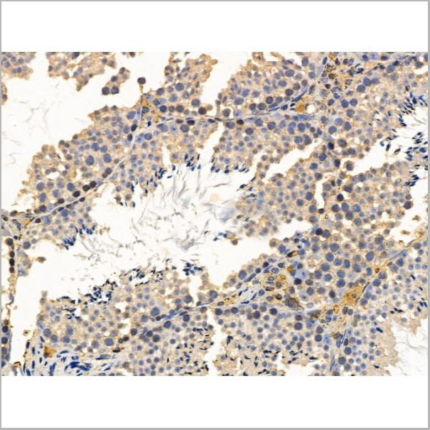
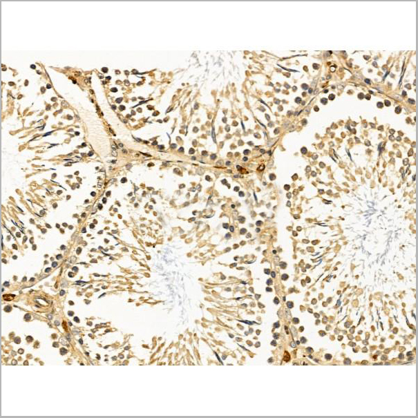
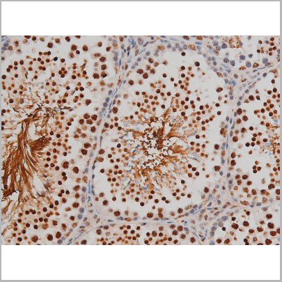
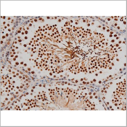
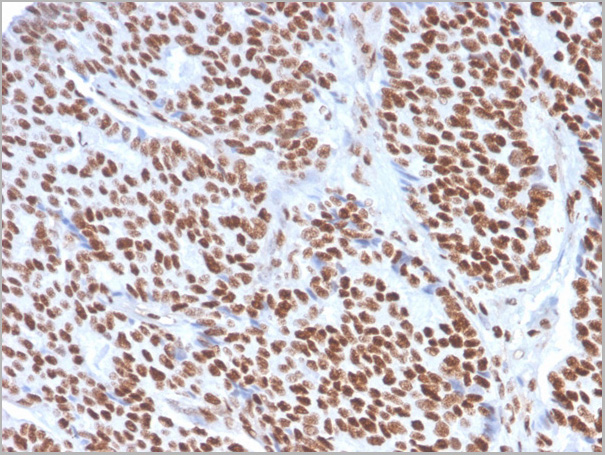
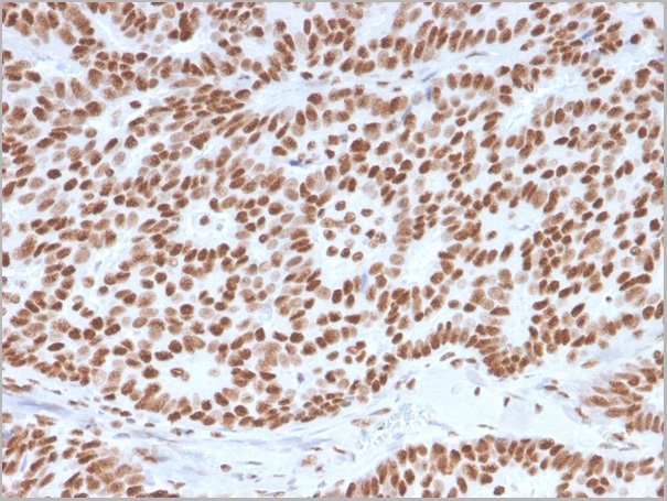
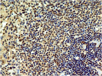
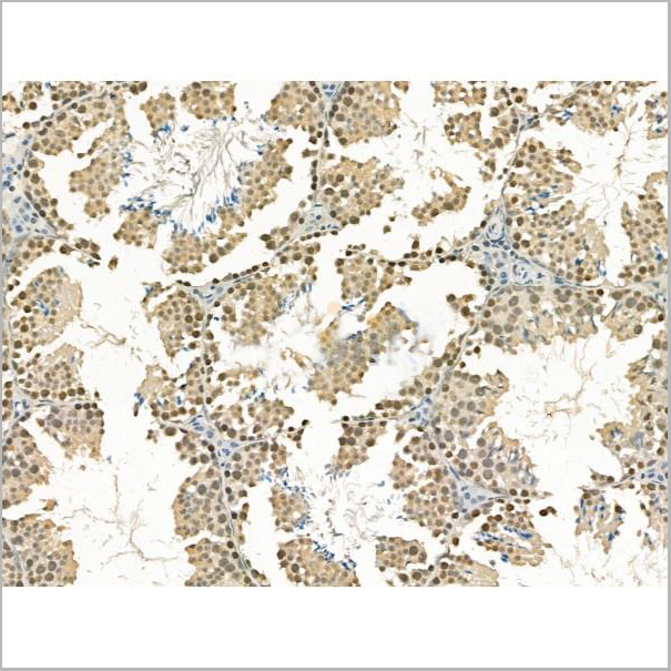
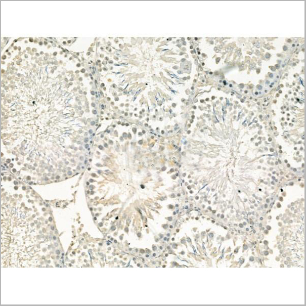
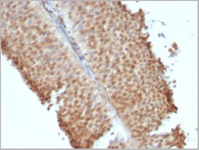
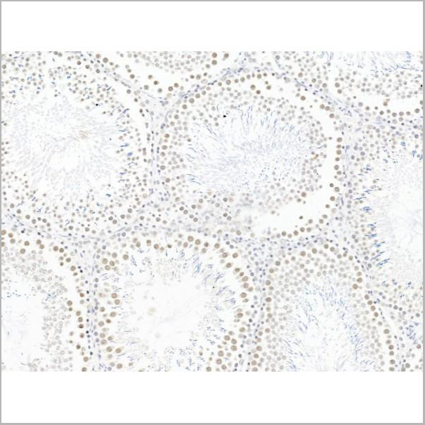
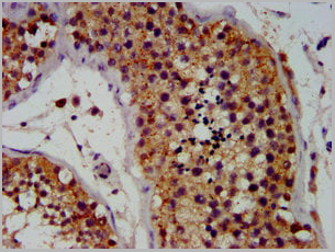
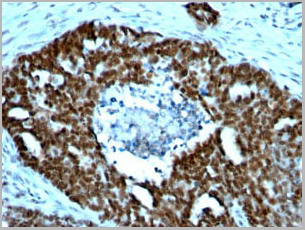
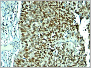
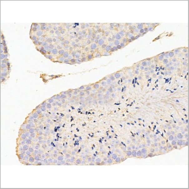
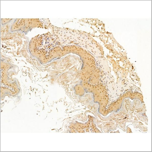
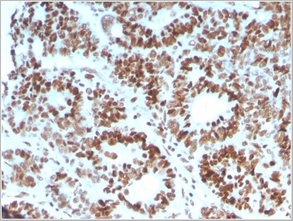
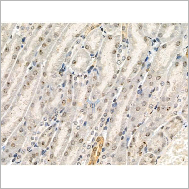
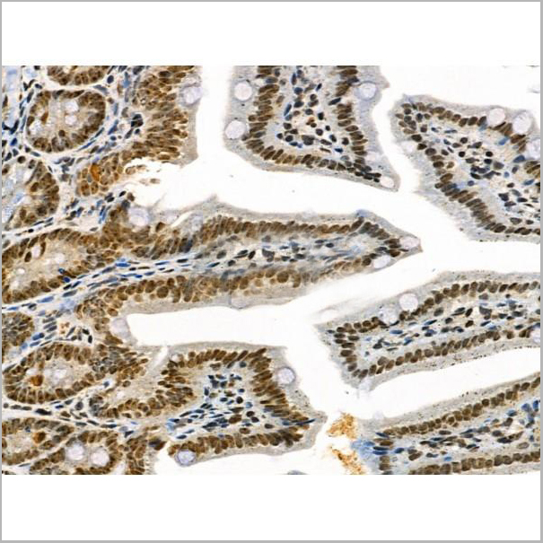
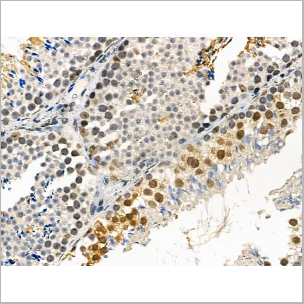
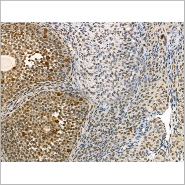
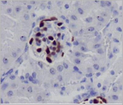
 IHC (Immunohistochemistry)
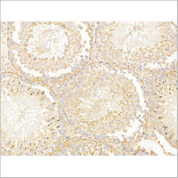
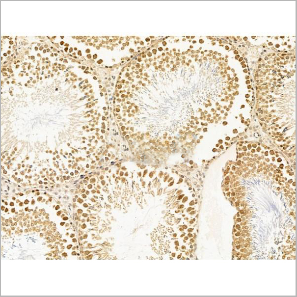
(Anti-NR6A1 / GCNF antibody IHC of human testis. Immunohistochemistry of formalin-fixed, paraffin-embedded tissue after heat-induced antigen retrieval.)
IHC (Immunohistochemistry)
(Anti-NR6A1 / GCNF antibody IHC of human testis. Immunohistochemistry of formalin-fixed, paraffin-embedded tissue after heat-induced antigen retrieval.)
NR6A1 / GCNF, Polyclonal Antibody (Cat# AAA12361)
Full Name
Rabbit Polyclonal to Human NR6A1 / GCNF
Gene Names
NR6A1; RTR; GCNF; NR61; hRTR; CT150; GCNF1; hGCNF
Reactivity
Human, Monkey, Mouse, Rat, Bat, Bovine, Dog, Hamster, Horse, Pig, Rabbit
Applications
Immunohistochemistry
Purity
Immunoaffinity Purified
Pricing

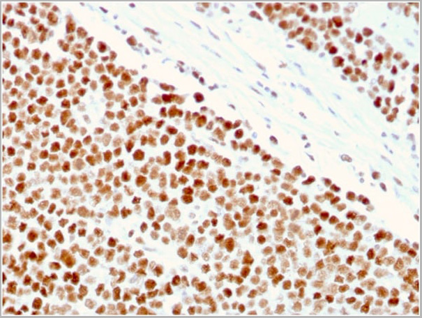
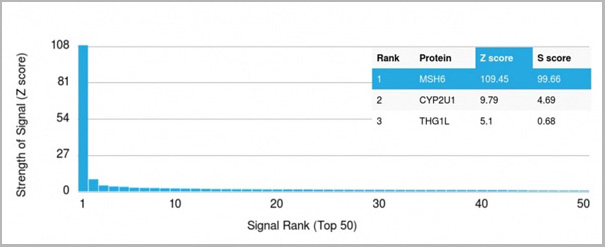 Application Data
(Analysis of Protein Array containing >19,000 full-length human proteins using MSH6 Mouse Monoclonal Antibody (MSH6/3086) Z- and S- Score: The Z-score represents the strength of a signal that a monoclonal antibody (MAb) (in combination with a fluorescently-tagged anti-IgG secondary antibody) produces when binding to a particular protein on the HuProtTM array. Z-scores are described in units of standard deviations (SD’s) above the mean value of all signals generated on that array. If targets on HuProtTM are arranged in descending order of the Z-score, the S-score is the difference (also in units of SD’s) between the Z-score. S-score therefore represents the relative target specificity of a MAb to its intended target. A MAb is considered to specific to its intended target, if the MAb has an S-score of at least 2.5. For example, if a MAb binds to protein X with a Z-score of 43 and to protein Y with a Z-score of 14, then the S-score for the binding of that MAb to protein X is equal to 29.)
Application Data
(Analysis of Protein Array containing >19,000 full-length human proteins using MSH6 Mouse Monoclonal Antibody (MSH6/3086) Z- and S- Score: The Z-score represents the strength of a signal that a monoclonal antibody (MAb) (in combination with a fluorescently-tagged anti-IgG secondary antibody) produces when binding to a particular protein on the HuProtTM array. Z-scores are described in units of standard deviations (SD’s) above the mean value of all signals generated on that array. If targets on HuProtTM are arranged in descending order of the Z-score, the S-score is the difference (also in units of SD’s) between the Z-score. S-score therefore represents the relative target specificity of a MAb to its intended target. A MAb is considered to specific to its intended target, if the MAb has an S-score of at least 2.5. For example, if a MAb binds to protein X with a Z-score of 43 and to protein Y with a Z-score of 14, then the S-score for the binding of that MAb to protein X is equal to 29.)
MSH6, Monoclonal Antibody (Cat# AAA23917)
Full Name
MSH6 (DNA Mismatch Repair Protein)
Gene Names
MSH6; GTBP; HSAP; p160; GTMBP; HNPCC5
Reactivity
Human
Applications
Flow Cytometry, Immunofluorescence, Western Blot, Immunohistochemistry
Purity
Purified Ab with BSA and Azide at 200ug/ml OR Purified Ab WITHOUT BSA and Azide at 1.0mg/ml
Pricing
 Application Data
(At 25 degree C. Samples were then incubated with primary Ab(At 37 degree C. An AlexaFluor594 conjugated goat anti-rabbit IgG(H+L) Ab(Red) and an AlexaFluor488 conjugated goat anti-mouse IgG(H+L) Ab(Green) were used as the secondary antibody.The nuclear counter stain is DAPI(blue).)
Application Data
(At 25 degree C. Samples were then incubated with primary Ab(At 37 degree C. An AlexaFluor594 conjugated goat anti-rabbit IgG(H+L) Ab(Red) and an AlexaFluor488 conjugated goat anti-mouse IgG(H+L) Ab(Green) were used as the secondary antibody.The nuclear counter stain is DAPI(blue).)
IRAK1, Polyclonal Antibody (Cat# AAA31367)
Full Name
Phospho-IRAK1 (Thr100) Antibody
Gene Names
IRAK1; IRAK; pelle
Reactivity
Human, Mouse, Rat
Applications
Western Blot, Immunohistochemistry, Immunofluorescence, Immunocytochemistry, Peptide ELISA
Purity
The antibody is from purified rabbit serum by affinity purification via sequential chromatography on phospho-peptide and non-phospho-peptide affinity columns.
Pricing
 Application Data
(AAA31434 staining Hela cells(4h of LPS treatment) by IF/ICC. The samples were fixed with PFA and permeabilized in 0.1% Triton X-100,then blocked in 10% serum for 45 minutes at 25°C. Samples were then incubated with primary Ab(AAA31434 1:200) and mouse anti-beta tubulin Ab( 1:200) for 1 hour at 37°C. An AlexaFluor594 conjugated goat anti-rabbit IgG(H+L) Ab(Red) and an AlexaFluor488 conjugated goat anti-mouse IgG(H+L) Ab(Green) were used as the secondary antibody. The nuclear counter stain is DAPI(blue).)
Application Data
(AAA31434 staining Hela cells(4h of LPS treatment) by IF/ICC. The samples were fixed with PFA and permeabilized in 0.1% Triton X-100,then blocked in 10% serum for 45 minutes at 25°C. Samples were then incubated with primary Ab(AAA31434 1:200) and mouse anti-beta tubulin Ab( 1:200) for 1 hour at 37°C. An AlexaFluor594 conjugated goat anti-rabbit IgG(H+L) Ab(Red) and an AlexaFluor488 conjugated goat anti-mouse IgG(H+L) Ab(Green) were used as the secondary antibody. The nuclear counter stain is DAPI(blue).)
Smad3, Polyclonal Antibody (Cat# AAA31434)
Full Name
Phospho-Smad3 (Ser423+Ser425) Antibody
Gene Names
SMAD3; LDS3; LDS1C; MADH3; JV15-2; HSPC193; HsT17436
Reactivity
Human, Mouse, Rat
Applications
Western Blot, Immunohistochemistry, Immunofluorescence, Immunocytochemistry
Purity
The antibody is from purified rabbit serum by affinity purification via sequential chromatography on phospho-peptide and non-phospho-peptide affinity columns.
Pricing
 SDS-PAGE
(3ug by SDS-PAGE under reducing condition and visualized by coomassie blue stain.)
SDS-PAGE
(3ug by SDS-PAGE under reducing condition and visualized by coomassie blue stain.)
ACO1, Recombinant Protein (Cat# AAA11797)
Full Name
ACO1, 1-889aa, Human, His tag, E Coli
Gene Names
ACO1; IRP1; ACONS; HEL60; IREB1; IREBP; IREBP1
Applications
SDS-PAGE
Purity
> 90% by SDS-PAGE
Pricing
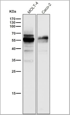
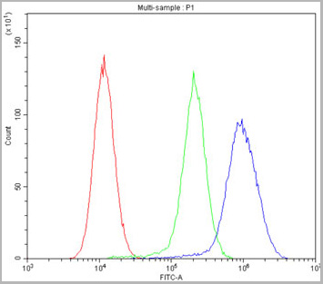
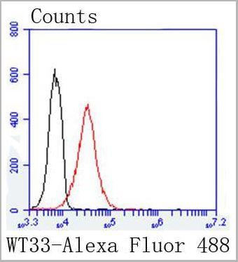 FCM (Flow Cytometry)
(Flow cytometric analysis of K562 cells with Wilms Tumor Protein antibody at 1/50 dilution (red) compared with an unlabelled control (cells without incubation with primary antibody; black). Alexa Fluor 488-conjugated goat anti rabbit IgG was used as the secondary antibody.)
FCM (Flow Cytometry)
(Flow cytometric analysis of K562 cells with Wilms Tumor Protein antibody at 1/50 dilution (red) compared with an unlabelled control (cells without incubation with primary antibody; black). Alexa Fluor 488-conjugated goat anti rabbit IgG was used as the secondary antibody.)
Wilms Tumor Protein, Monoclonal Antibody (Cat# AAA30123)
Full Name
Wilms Tumor Protein Antibody
Gene Names
WT1; GUD; AWT1; WAGR; WT33; NPHS4; WIT-2; EWS-WT1
Reactivity
Human, Mouse
Applications
Western Blot, Immunocytochemistry, Immunofluorescence, Immunohistochemistry, Flow Cytometry
Purity
Affinity-chromatography
Pricing
CIAS1/NALP3, Polyclonal Antibody (Cat# AAA11523)
Full Name
Anti-CIAS1/NALP3 antibody
Gene Names
NLRP3; AII; AVP; FCU; MWS; FCAS; CIAS1; NALP3; C1orf7; CLR1.1; PYPAF1; AGTAVPRL
Reactivity
Human, Rat; Predicted: Mouse
Applications
Western Blot, Immunohistochemistry, Immunocytochemistry, Flow Cytometry
Purity
Immunogen affinity purified
Pricing


 Application Data
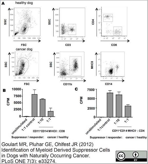
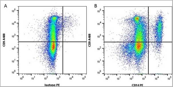
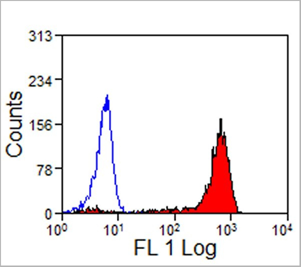
(Staining of human peripheral blood monocytes with Mouse anti Human CD14:RPE)
Application Data
(Staining of human peripheral blood monocytes with Mouse anti Human CD14:RPE)
CD14, Monoclonal Antibody (Cat# AAA12179)
Full Name
MOUSE ANTI HUMAN CD14
Applications
Immunohistochemistry, Flow Cytometry, Immunoprecipitation
Pricing
RAD50, Polyclonal Antibody (Cat# AAA31432)
Full Name
Phospho-RAD50 (Ser635) Antibody
Gene Names
RAD50; NBSLD; RAD502; hRad50
Reactivity
Human, Mouse, Rat
Applications
Western Blot, Immunohistochemistry
Purity
The antibody is from purified rabbit serum by affinity purification via sequential chromatography on phospho-peptide and non-phospho-peptide affinity columns.
Pricing
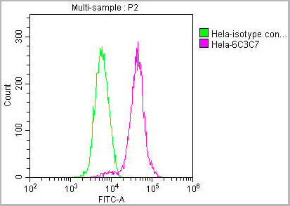
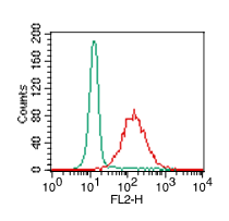
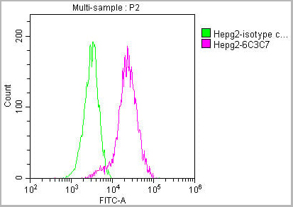 FCM (Flow Cytometry)
(Overlay histogram showing HepG2 cells stained with AAA27041 (red line) at 1:100. The cells were incubated in 1x PBS /10% normal goat serum to block non-specific protein-protein interactions followed by primary antibody for 1 h at 4 degree C. The secondary antibody used was FITC goat anti-mouse IgG(H+L) at 1/200 dilution for 1 h at 4 degree C. Isotype control antibody (green line) was used under the same conditions. Acquisition of >10,000 events was performed.)
FCM (Flow Cytometry)
(Overlay histogram showing HepG2 cells stained with AAA27041 (red line) at 1:100. The cells were incubated in 1x PBS /10% normal goat serum to block non-specific protein-protein interactions followed by primary antibody for 1 h at 4 degree C. The secondary antibody used was FITC goat anti-mouse IgG(H+L) at 1/200 dilution for 1 h at 4 degree C. Isotype control antibody (green line) was used under the same conditions. Acquisition of >10,000 events was performed.)
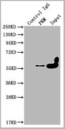
PKM, Monoclonal Antibody (Cat# AAA27041)
Full Name
PKM Monoclonal Antibody
Reactivity
Human, Rat, Mouse, Rabbit
Applications
Western Blot, Immunohistochemistry, Immunofluorescence, Flow Cytometry, Immunoprecipitation
Purity
>95%, Protein G purified
Pricing
 FCM (Flow Cytometry)
(Figure 8. Flow Cytometry analysis of HepG2 cells using anti-ATF2 antibody (AAA11634).Overlay histogram showing HepG2 cells stained with AAA11634 (Blue line).The cells were blocked with 10% normal goat serum. And then incubated with rabbit anti-ATF2 Antibody (AAA11634,1ug/1x10^6 cells) for 30 min at 20 degree C. DyLight ®488 conjugated goat anti-rabbit IgG (5-10ug/1x10^6 cells) was used as secondary antibody for 30 minutes at 20 degree C. Isotype control antibody (Green line) was rabbit IgG (1ug/1x106) used under the same conditions. Unlabelled sample (Red line) was also used as a control.)
FCM (Flow Cytometry)
(Figure 8. Flow Cytometry analysis of HepG2 cells using anti-ATF2 antibody (AAA11634).Overlay histogram showing HepG2 cells stained with AAA11634 (Blue line).The cells were blocked with 10% normal goat serum. And then incubated with rabbit anti-ATF2 Antibody (AAA11634,1ug/1x10^6 cells) for 30 min at 20 degree C. DyLight ®488 conjugated goat anti-rabbit IgG (5-10ug/1x10^6 cells) was used as secondary antibody for 30 minutes at 20 degree C. Isotype control antibody (Green line) was rabbit IgG (1ug/1x106) used under the same conditions. Unlabelled sample (Red line) was also used as a control.)
Cyclic AMP-dependent transcription factor ATF-2, Polyclonal Antibody (Cat# AAA11634)
Full Name
Anti-ATF2 Antibody
Gene Names
ATF2; HB16; CREB2; TREB7; CREB-2; CRE-BP1
Reactivity
Human, Mouse, Rat. No cross reactivity with other proteins.
Applications
Western Blot, Immunohistochemistry, Immunohistochemistry
Purity
Immunogen affinity purified.
Pricing
 FCM (Flow Cytometry)
(Figure 6. Flow Cytometry analysis of A549 cells using anti-MDR3 antibody (AAA11652).Overlay histogram showing A549 cells stained with AAA11652 (Blue line).The cells were blocked with 10% normal goat serum. And then incubated with rabbit anti-MDR3 Antibody (AAA11652,1ug/1x106 cells) for 30 min at 20°C. DyLight®488 conjugated goat anti-rabbit IgG (5-10ug/1x106 cells) was used as secondary antibody for 30 minutes at 20°C. Isotype control antibody (Green line) was rabbit IgG (1ug/1x106) used under the same conditions. Unlabelled sample (Red line) was also used as a control.)
FCM (Flow Cytometry)
(Figure 6. Flow Cytometry analysis of A549 cells using anti-MDR3 antibody (AAA11652).Overlay histogram showing A549 cells stained with AAA11652 (Blue line).The cells were blocked with 10% normal goat serum. And then incubated with rabbit anti-MDR3 Antibody (AAA11652,1ug/1x106 cells) for 30 min at 20°C. DyLight®488 conjugated goat anti-rabbit IgG (5-10ug/1x106 cells) was used as secondary antibody for 30 minutes at 20°C. Isotype control antibody (Green line) was rabbit IgG (1ug/1x106) used under the same conditions. Unlabelled sample (Red line) was also used as a control.)
ABCB4, Polyclonal Antibody (Cat# AAA11652)
Full Name
Anti-ABCB4 Antibody
Gene Names
ABCB4; GBD1; ICP3; MDR2; MDR3; PGY3; ABC21; MDR2/3; PFIC-3
Reactivity
Human, Mouse, Rat
Applications
Western Blot, Immunohistochemistry, Immunofluorescence, Flow Cytometry
Purity
Immunogen Affinity Purified
Pricing
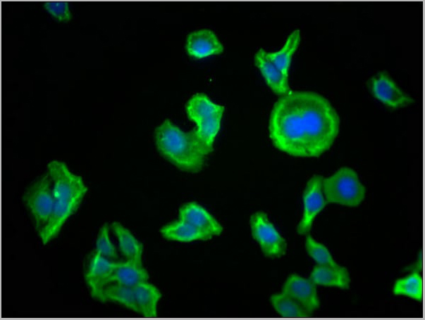
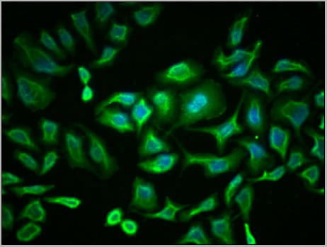
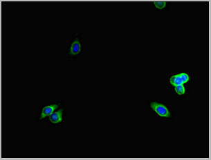 IF (Immunofluorescence)
(Immunofluorescent analysis of HepG2 cells using AAA26954 at a dilution of 1:100 and Alexa Fluor 488-congugated AffiniPure Goat Anti-Rabbit IgG(H+L))
IF (Immunofluorescence)
(Immunofluorescent analysis of HepG2 cells using AAA26954 at a dilution of 1:100 and Alexa Fluor 488-congugated AffiniPure Goat Anti-Rabbit IgG(H+L))
LTBP4, Polyclonal Antibody (Cat# AAA26954)
Full Name
LTBP4 Antibody
Gene Names
LTBP4; ARCL1C; LTBP-4; LTBP4L; LTBP4S
Reactivity
Human
Applications
Immunohistochemistry, Immunofluorescence
Purity
>95%, Protein G purified
Pricing
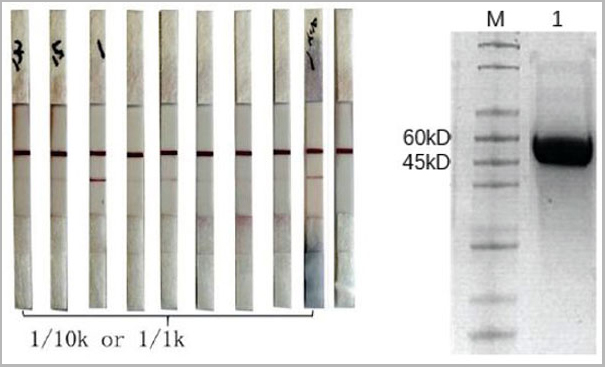
 Affinity Assay
(The binding between immobilized S1RBD and the anti-SARS-CoV-2 S1RBD AAA13617.)
Affinity Assay
(The binding between immobilized S1RBD and the anti-SARS-CoV-2 S1RBD AAA13617.)
COVID 19 S1 Spike RBD Coronavirus, Antibody (Cat# AAA13617)
Full Name
Humanized Monoclonal Antibody against SARS-CoV-2 Spike Protein-1 Receptor-binding Domain (S1 RBD)
Reactivity
Viral
Applications
ELISA
Pricing
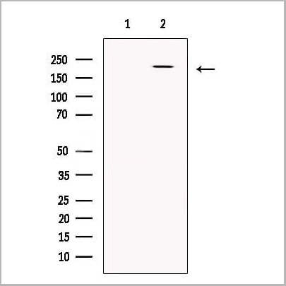
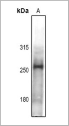
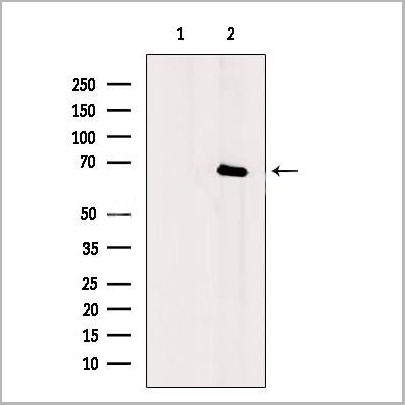
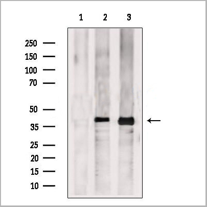
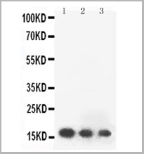 WB (Western Blot)
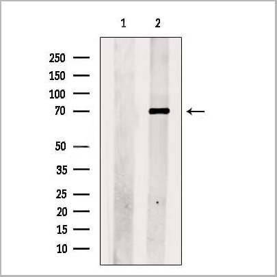
(Anti- FGF2 antibody, AAA11513, Western blottingAll lanes: Anti FGF2(AAA11513) at 0.5ug/mlLane 1: Recombinant Human FGF2 Protein 10ngLane 2: Recombinant Human FGF2 Protein 5ngLane 3: Recombinant Human FGF2 Protein 2.5ngPredicted bind size: 17KDObserved bind size: 17KD)
WB (Western Blot)
(Anti- FGF2 antibody, AAA11513, Western blottingAll lanes: Anti FGF2(AAA11513) at 0.5ug/mlLane 1: Recombinant Human FGF2 Protein 10ngLane 2: Recombinant Human FGF2 Protein 5ngLane 3: Recombinant Human FGF2 Protein 2.5ngPredicted bind size: 17KDObserved bind size: 17KD)
FGF2, Polyclonal Antibody (Cat# AAA11513)
Full Name
Anti-human FGF2 antibody
Gene Names
FGF2; BFGF; FGFB; FGF-2; HBGF-2
Reactivity
Human
Applications
Western Blot, Immunohistochemistry
Purity
Immunogen affinity purified.
Pricing
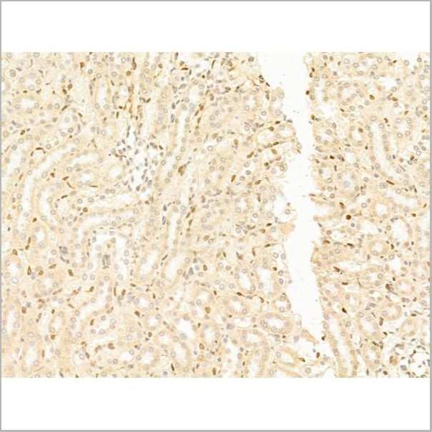
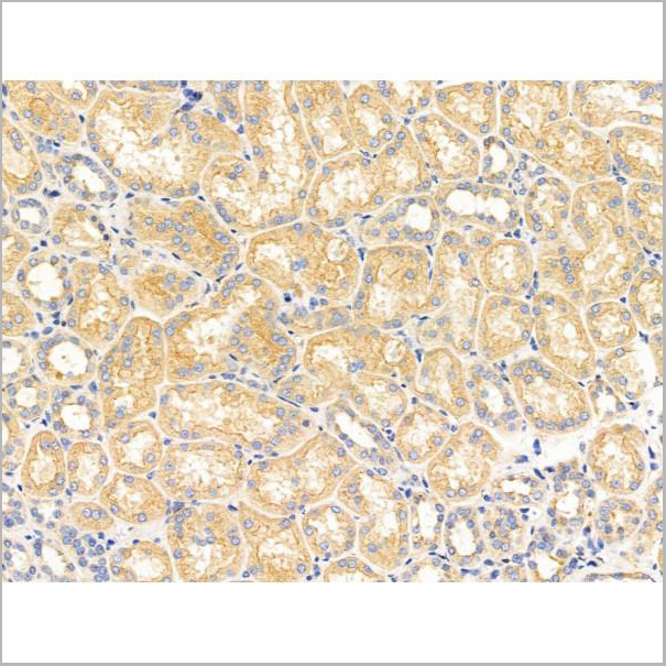
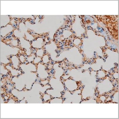
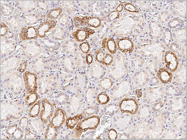
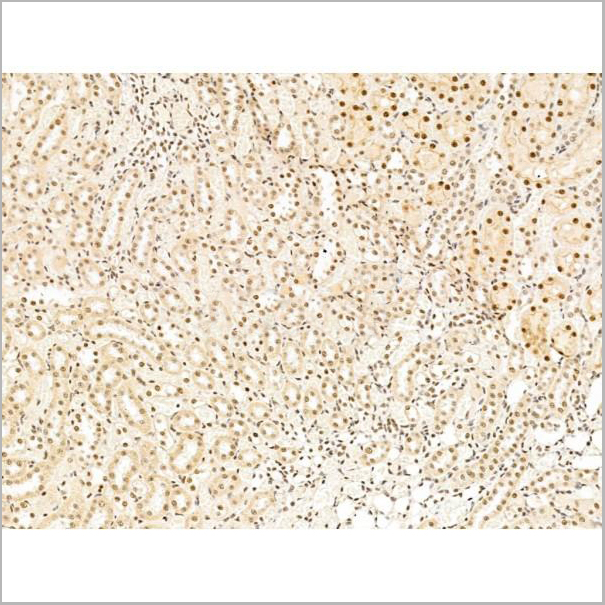
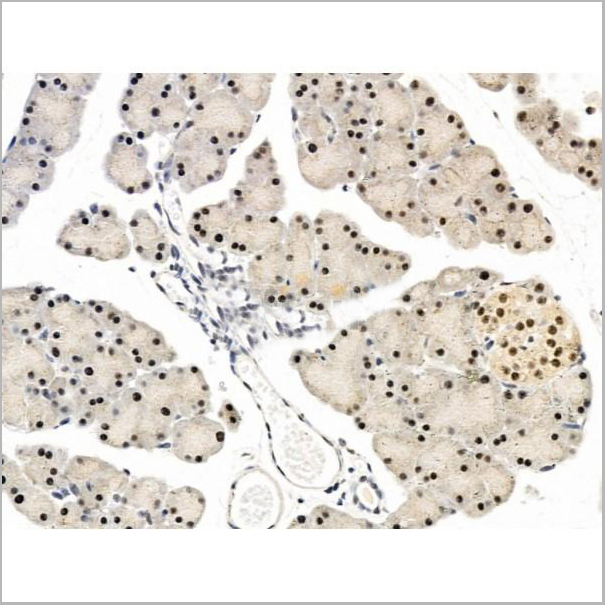
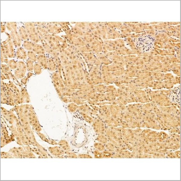
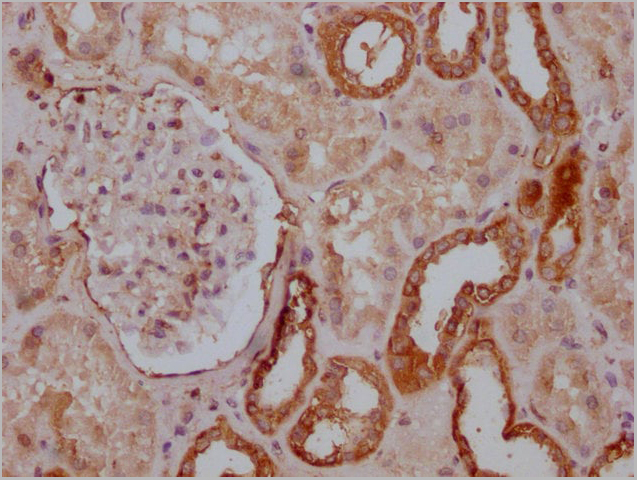
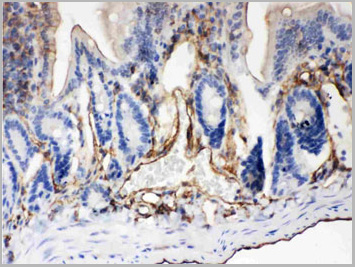
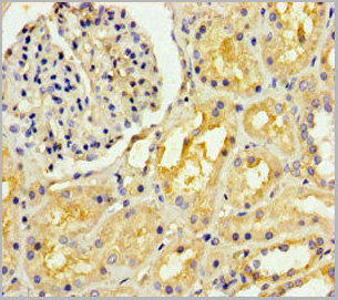
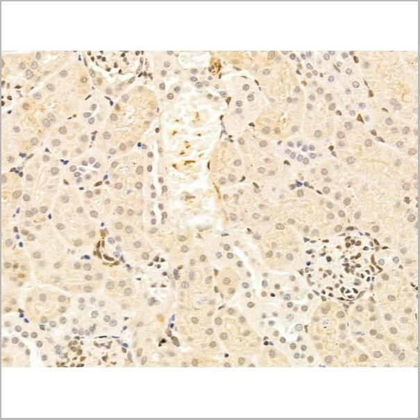 IHC (Immunohistochemistry-Paraffin)
(AAA31250 at 1/100 staining Mouse kidney tissue by IHC-P. The sample was formaldehyde fixed and a heat mediated antigen retrieval step in citrate buffer was performed. The sample was then blocked and incubated with the primary antibody at 4°C overnight. An HRP conjugated anti-Rabbit antibody was used as the secondary antibody.)
IHC (Immunohistochemistry-Paraffin)
(AAA31250 at 1/100 staining Mouse kidney tissue by IHC-P. The sample was formaldehyde fixed and a heat mediated antigen retrieval step in citrate buffer was performed. The sample was then blocked and incubated with the primary antibody at 4°C overnight. An HRP conjugated anti-Rabbit antibody was used as the secondary antibody.)
TFAP4, Polyclonal Antibody (Cat# AAA31250)
Full Name
TFAP4 Antibody
Gene Names
TFAP4; AP-4; bHLHc41
Reactivity
Human, Mouse, Rat
Predicted Reactivity: Pig(100%), Bovine(100%), Horse(100%), Sheep(100%), Rabbit(100%), Dog(100%)
Predicted Reactivity: Pig(100%), Bovine(100%), Horse(100%), Sheep(100%), Rabbit(100%), Dog(100%)
Applications
ELISA
Purity
The antiserum was purified by peptide affinity chromatography using SulfoLink Coupling Resin (Thermo Fisher Scientific).
Pricing


 Application Data
(Staining of human peripheral blood monocytes with Mouse anti Human CD14:RPE)
Application Data
(Staining of human peripheral blood monocytes with Mouse anti Human CD14:RPE)
CD14, Monoclonal Antibody (Cat# AAA11998)
Full Name
MOUSE ANTI HUMAN CD14
Applications
Immunohistochemistry, Flow Cytometry, Immunofluorescence, Immunoprecipitation
Pricing