Filters
Clonality
Type
Reactivity
Gene Name
Isotype
Host
Application
Clone
1507 results for "B cell" - showing 1350-1400


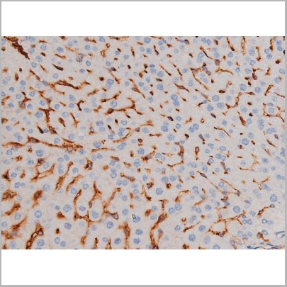
 Application Data
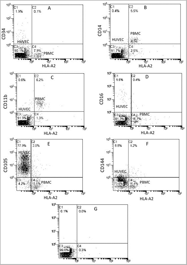
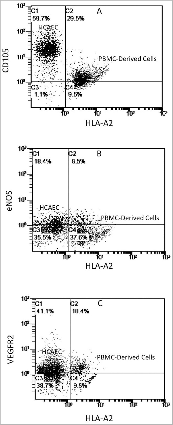
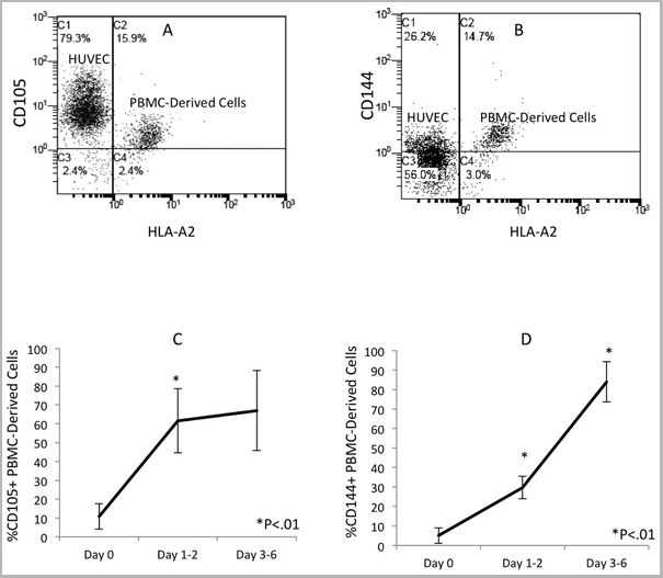
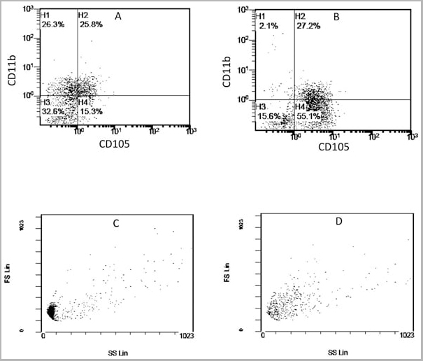
(Published customer image: Mouse anti Human CD105 antibody, clone SN6 used for flow cytometry.Image caption:Phenotype change from HLA-A2+/CD11b+/CD105- to HLA-A2+/CD11b-/CD105+ on endothelium-adherent blood monocyte-derived cells with increase in size and granularity during co-culture. HLA-A2+ PBMCs (1x10⁶ cells/well) were incubated for 2 h (Day 0) with HLA-A2- HUVECs, after which the non-adherent cells were removed by washing. The cell layers were analysed by three-colour flow cytometry staining for HLA-A2, CD11b and CD105 on (A) Day 1 and (B) Day 2. These plots were gated for HLA-A2+ cells. Forward scatter/side scatter dot plots gated for HLA-A2+ cells on Day 0 (C) and Day 2 (D) was shown. These are representative of 2 individual experiments.From: Tso C, Rye K-A, Barter P (2012) Phenotypic and Functional Changes in Blood Monocytes Following Adherence to Endothelium. PLoS ONE 7(5): e37091.)
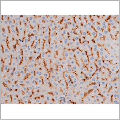
Application Data
(Published customer image: Mouse anti Human CD105 antibody, clone SN6 used for flow cytometry.Image caption:Phenotype change from HLA-A2+/CD11b+/CD105- to HLA-A2+/CD11b-/CD105+ on endothelium-adherent blood monocyte-derived cells with increase in size and granularity during co-culture. HLA-A2+ PBMCs (1x10⁶ cells/well) were incubated for 2 h (Day 0) with HLA-A2- HUVECs, after which the non-adherent cells were removed by washing. The cell layers were analysed by three-colour flow cytometry staining for HLA-A2, CD11b and CD105 on (A) Day 1 and (B) Day 2. These plots were gated for HLA-A2+ cells. Forward scatter/side scatter dot plots gated for HLA-A2+ cells on Day 0 (C) and Day 2 (D) was shown. These are representative of 2 individual experiments.From: Tso C, Rye K-A, Barter P (2012) Phenotypic and Functional Changes in Blood Monocytes Following Adherence to Endothelium. PLoS ONE 7(5): e37091.)
CD105, Monoclonal Antibody (Cat# AAA11933)
Full Name
MOUSE ANTI HUMAN CD105
Gene Names
ENG; END; HHT1; ORW1
Reactivity
Cynomolgus monkey, Horse, Monkey, Primate, Rhesus Monkey
Applications
Immunohistochemistry, Flow Cytometry, Immunoprecipitation, Western Blot
Pricing
 Application Data
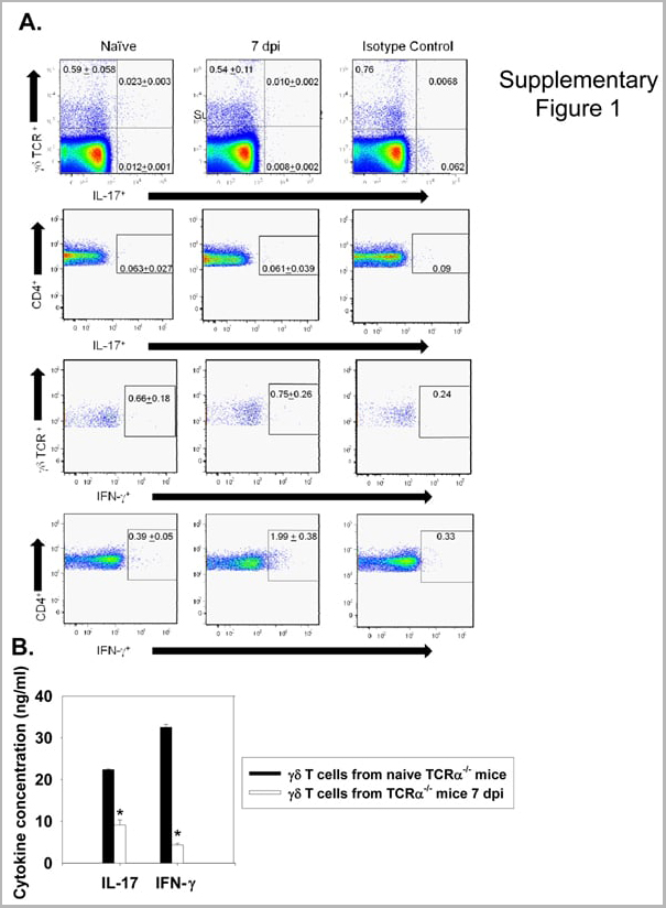
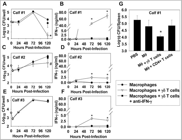
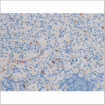
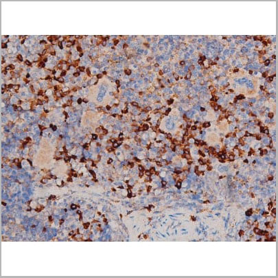
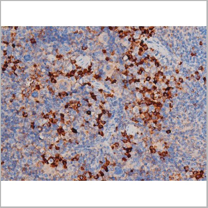
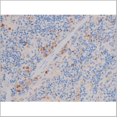
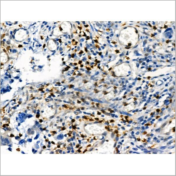
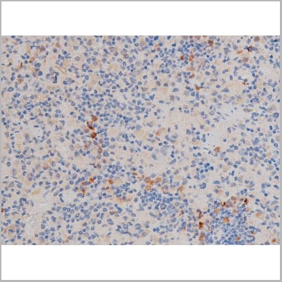
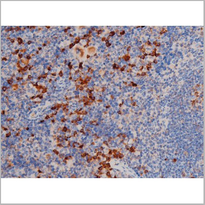
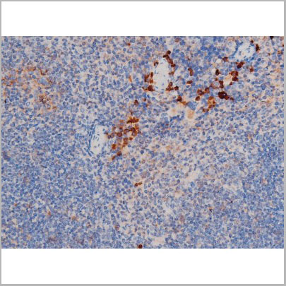
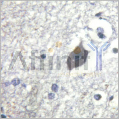
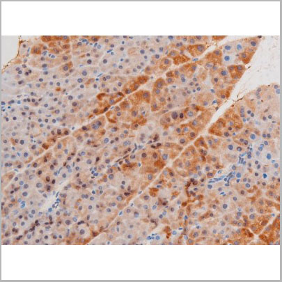
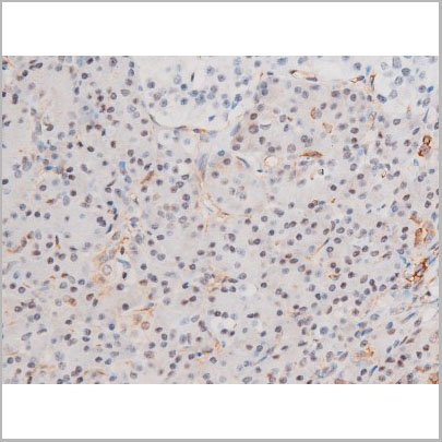
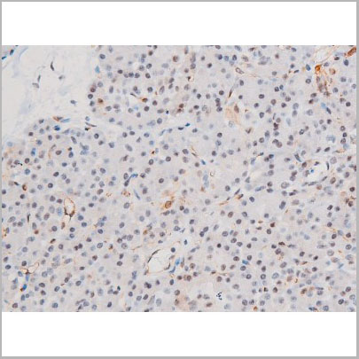
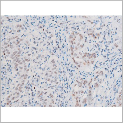
(Published customer image: gamma delta T cells are the primary source of IL-17 during B. abortus infection. C57BL/6 mice were infected i.p. with 5x10⁴ CFUs of B. abortus 2308, and two weeks later gamma delta T cells (>95% purity) and an enriched TCRalphabeta (~55% CD4+, 25% CD8+) cell fraction were isolated from the spleens of infected mice. Cells were stimulated with 500 ng/ml ionomycin and 50 ng/ml PMA for three days, and cell-free supernatants from triplicate wells were assayed for cytokine production via ELISA. The mean +/- SD is shown; * P)
Application Data
(Published customer image: gamma delta T cells are the primary source of IL-17 during B. abortus infection. C57BL/6 mice were infected i.p. with 5x10⁴ CFUs of B. abortus 2308, and two weeks later gamma delta T cells (>95% purity) and an enriched TCRalphabeta (~55% CD4+, 25% CD8+) cell fraction were isolated from the spleens of infected mice. Cells were stimulated with 500 ng/ml ionomycin and 50 ng/ml PMA for three days, and cell-free supernatants from triplicate wells were assayed for cytokine production via ELISA. The mean +/- SD is shown; * P)
IFN GAMMA, Monoclonal Antibody (Cat# AAA12094)
Full Name
MOUSE ANTI BOVINE INTERFERON GAMMA:Biotin
Applications
Flow Cytometry
Pricing
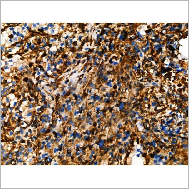
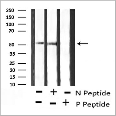
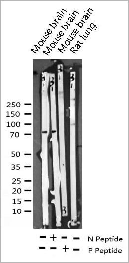
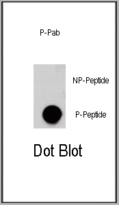
 DB (Dot Blot)
(Dot Blot: 1 ug peptide was blot onto NC membraneA: JAK2 (pY1008, pY1009)B: JAK2 (Non phosphospecific)C: Non-related Phosphopeptide) were blotted at a 1:2000 dilution by:1: Rabbit anti-JAK2(pY1008/pY1009) (AAA14087)2: Rabbit anti-JAK2 (Nonphospho specific).)
DB (Dot Blot)
(Dot Blot: 1 ug peptide was blot onto NC membraneA: JAK2 (pY1008, pY1009)B: JAK2 (Non phosphospecific)C: Non-related Phosphopeptide) were blotted at a 1:2000 dilution by:1: Rabbit anti-JAK2(pY1008/pY1009) (AAA14087)2: Rabbit anti-JAK2 (Nonphospho specific).)
JAK2 (pY1007/Y1008), Antibody (Cat# AAA14087)
Full Name
Rabbit anti Phospho- JAK2 (pTyr 1007/1008)
Gene Names
JAK2; JTK10
Reactivity
Human, Rat, Mouse
Applications
Western Blot, Immunohistochemistry, Immunoprecipitation
Purity
The Rabbit IgG is purified by site-modified Epitope Affinity Purification.
Pricing
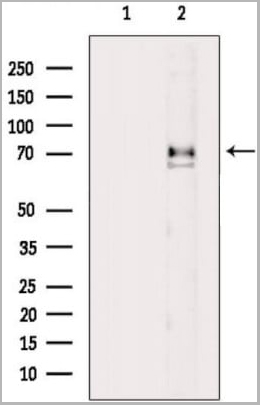
 WB (Western Blot)
(WB: A431 cell lysate was probed with anti-Catenin, beta, active (ABC). Arrow indicates beta-Catenin (92kD))
WB (Western Blot)
(WB: A431 cell lysate was probed with anti-Catenin, beta, active (ABC). Arrow indicates beta-Catenin (92kD))
Catenin, beta, active, Monoclonal Antibody (Cat# AAA14699)
Full Name
Catenin, beta, active (ABC) (BSA, Azide & Glycerol Free)
Reactivity
Human, Mouse, Rat
Applications
Western Blot, Immunohistochemistry, Immunocytochemistry
Purity
Purified by Protein G affinity chromatography.
Pricing
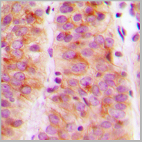
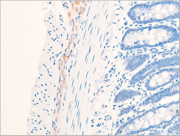
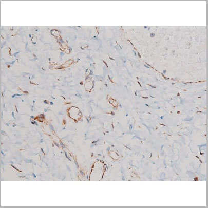
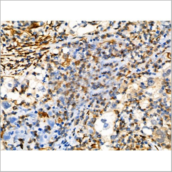
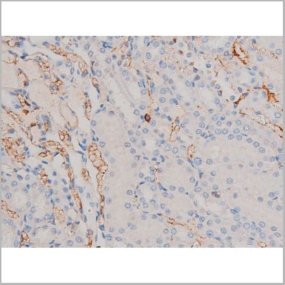
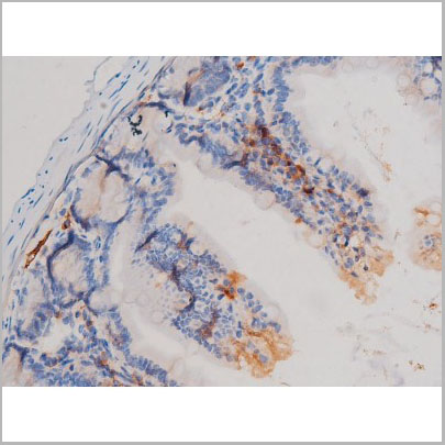
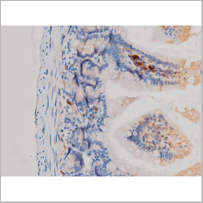
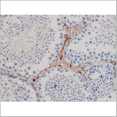
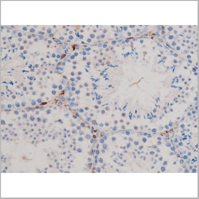
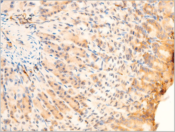
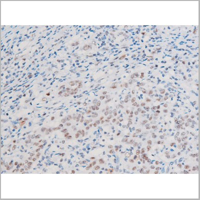
 IHC (Immunohistochemistry)
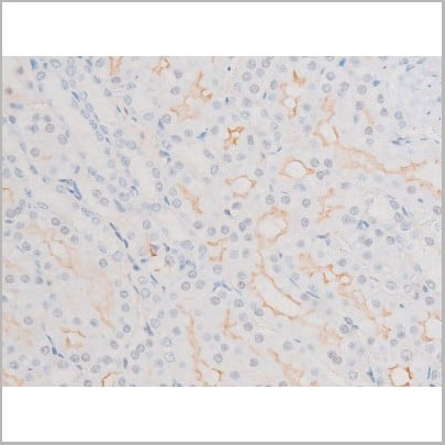
(Immunohistochemical analysis of LPHN2 staining in human breast cancer formalin fixed paraffin embedded tissue section. The section was pre-treated using heat mediated antigen retrieval with sodium citrate buffer (pH 6.0). The section was then incubated with the antibody at room temperature and detected using an HRP conjugated compact polymer system. DAB was used as the chromogen. The section was then counterstained with haematoxylin and mounted with DPX.)
IHC (Immunohistochemistry)
(Immunohistochemical analysis of LPHN2 staining in human breast cancer formalin fixed paraffin embedded tissue section. The section was pre-treated using heat mediated antigen retrieval with sodium citrate buffer (pH 6.0). The section was then incubated with the antibody at room temperature and detected using an HRP conjugated compact polymer system. DAB was used as the chromogen. The section was then counterstained with haematoxylin and mounted with DPX.)
LPHN2, Polyclonal Antibody (Cat# AAA27788)
Full Name
Anti-LPHN2 Antibody
Gene Names
ADGRL2; CL2; LEC1; CIRL2; LPHH1; LPHN2
Reactivity
Human, Mouse, Rat, Bovine
Applications
Western Blot, Immunohistochemistry
Purity
The antibody was purified by immunogen affinity chromatography.
Pricing
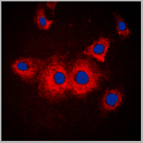
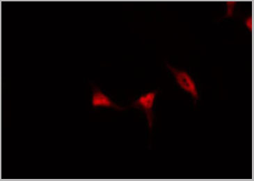
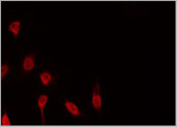
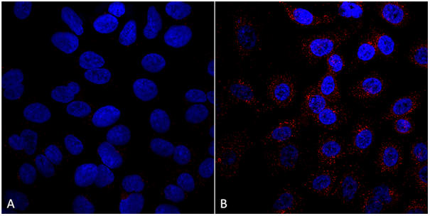
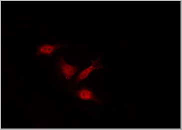
 IF (Immunofluorescence)
(Immunofluorescent analysis of Vasopressin V2 Receptor staining in HEK293T cells. Formalin-fixed cells were permeabilized with 0.1% Triton X-100 in TBS for 5-10 minutes and blocked with 3% BSA-PBS for 30 minutes at room temperature. Cells were probed with the primary antibody in 3% BSA-PBS and incubated overnight at 4 °C in a humidified chamber. Cells were washed with PBST and incubated with a DyLight 594-conjugated secondary antibody (red) in PBS at room temperature in the dark. DAPI was used to stain the cell nuclei (blue).)
IF (Immunofluorescence)
(Immunofluorescent analysis of Vasopressin V2 Receptor staining in HEK293T cells. Formalin-fixed cells were permeabilized with 0.1% Triton X-100 in TBS for 5-10 minutes and blocked with 3% BSA-PBS for 30 minutes at room temperature. Cells were probed with the primary antibody in 3% BSA-PBS and incubated overnight at 4 °C in a humidified chamber. Cells were washed with PBST and incubated with a DyLight 594-conjugated secondary antibody (red) in PBS at room temperature in the dark. DAPI was used to stain the cell nuclei (blue).)
Vasopressin V2 Receptor, Polyclonal Antibody (Cat# AAA27813)
Full Name
Anti-Vasopressin V2 Receptor Antibody
Gene Names
AVPR2; DI1; DIR; NDI; V2R; ADHR; DIR3
Reactivity
Human, Mouse, Rat, Monkey
Applications
Western Blot, Immunofluorescence, Immunocytochemistry
Purity
The antibody was purified by immunogen affinity chromatography.
Pricing
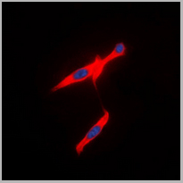
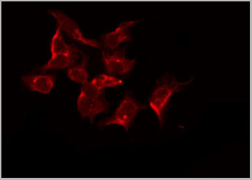
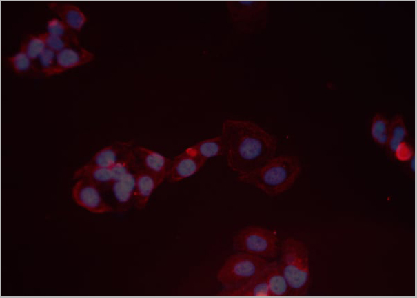
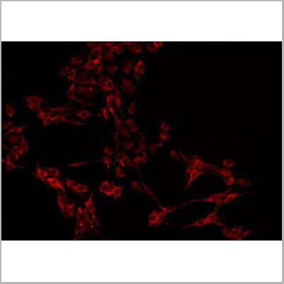
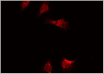
 IF (Immunofluorescence)
(Immunofluorescent analysis of SLK staining in MCF7 cells. Formalin-fixed cells were permeabilized with 0.1% Triton X-100 in TBS for 5-10 minutes and blocked with 3% BSA-PBS for 30 minutes at room temperature. Cells were probed with the primary antibody in 3% BSA-PBS and incubated overnight at 4 °C in a humidified chamber. Cells were washed with PBST and incubated with a DyLight 594-conjugated secondary antibody (red) in PBS at room temperature in the dark. DAPI was used to stain the cell nuclei (blue).)
IF (Immunofluorescence)
(Immunofluorescent analysis of SLK staining in MCF7 cells. Formalin-fixed cells were permeabilized with 0.1% Triton X-100 in TBS for 5-10 minutes and blocked with 3% BSA-PBS for 30 minutes at room temperature. Cells were probed with the primary antibody in 3% BSA-PBS and incubated overnight at 4 °C in a humidified chamber. Cells were washed with PBST and incubated with a DyLight 594-conjugated secondary antibody (red) in PBS at room temperature in the dark. DAPI was used to stain the cell nuclei (blue).)
SLK, Polyclonal Antibody (Cat# AAA27796)
Full Name
Anti-SLK Antibody
Gene Names
SLK; LOSK; STK2; se20-9; bA16H23.1
Reactivity
Human, Mouse, Rat
Applications
Western Blot, Immunohistochemistry, Immunofluorescence
Purity
The antibody was purified by immunogen affinity chromatography.
Pricing
 IF (Immunofluorescence)
(AAA31030 staining HeLa by IF/ICC. The sample were fixed with PFA and permeabilized in 0.1% Triton X-100, then blocked in 10% serum for 45 minutes at 25 degree C. The primary antibody was diluted at 1/200 and incubated with the sample for 1 hour at 37 degree C. An Alexa Fluor 594 conjugated goat anti-rabbit IgG (H+L) Ab, diluted at 1/600, was used as the secondary antibody.)
IF (Immunofluorescence)
(AAA31030 staining HeLa by IF/ICC. The sample were fixed with PFA and permeabilized in 0.1% Triton X-100, then blocked in 10% serum for 45 minutes at 25 degree C. The primary antibody was diluted at 1/200 and incubated with the sample for 1 hour at 37 degree C. An Alexa Fluor 594 conjugated goat anti-rabbit IgG (H+L) Ab, diluted at 1/600, was used as the secondary antibody.)
CDC25A, Polyclonal Antibody (Cat# AAA31030)
Full Name
Phospho-CDC25A (Ser178) Antibody
Gene Names
CDC25A; CDC25A2
Reactivity
Human, Mouse, Rat
Applications
Western Blot, Immunohistochemistry, Immunofluorescence, Immunocytochemistry
Purity
From purified rabbit serum by affinity purification via sequential chromatography on phospho-and non-phospho-peptide affinity columns.
Pricing
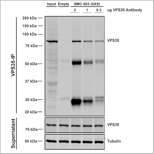


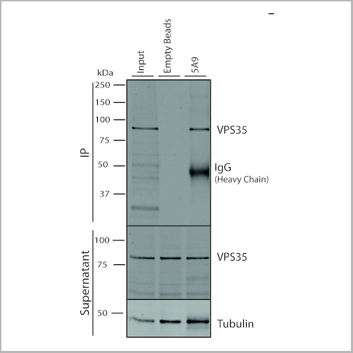

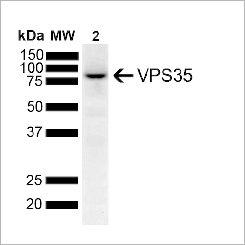
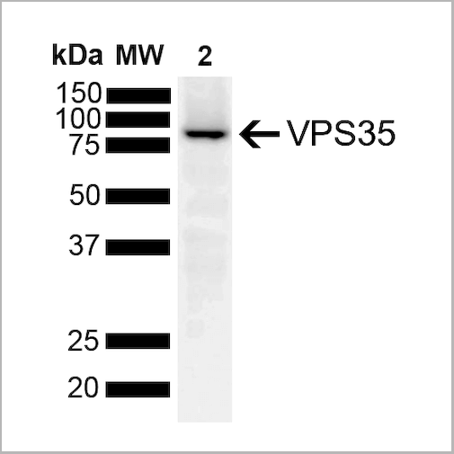
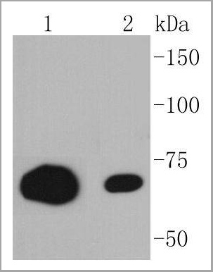
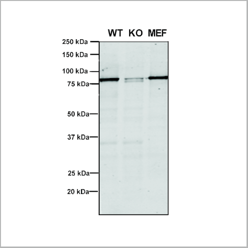
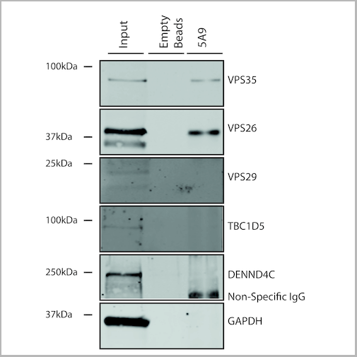
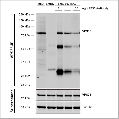 IP (Immunoprecipitation)
(Immunoprecipitation analysis using Mouse Anti-VPS35 Monoclonal Antibody, Clone 5A9. Tissue: embryonic fibroblast. Species: Mouse. Primary Antibody: Mouse Anti-VPS35 Monoclonal Antibody. Three amounts of (3, 1 and 0.3 ug) were non-covalently coupled to 10uL of A/G sepharose beads for 1 hour at 4 degree C and next incubated with 250ug of MEF lysate for 2 hours at 4 degree C.)
IP (Immunoprecipitation)
(Immunoprecipitation analysis using Mouse Anti-VPS35 Monoclonal Antibody, Clone 5A9. Tissue: embryonic fibroblast. Species: Mouse. Primary Antibody: Mouse Anti-VPS35 Monoclonal Antibody. Three amounts of (3, 1 and 0.3 ug) were non-covalently coupled to 10uL of A/G sepharose beads for 1 hour at 4 degree C and next incubated with 250ug of MEF lysate for 2 hours at 4 degree C.)
VPS35, Monoclonal Antibody (Cat# AAA27680)
Full Name
VPS35 Antibody, Clone 5A9: PerCP
Gene Names
VPS35; MEM3; PARK17
Reactivity
Human, Mouse, Rat
Applications
Western Blot, Immunocytochemistry, Immunofluorescence, Immunoprecipitation
Purity
Protein G Purified
Pricing


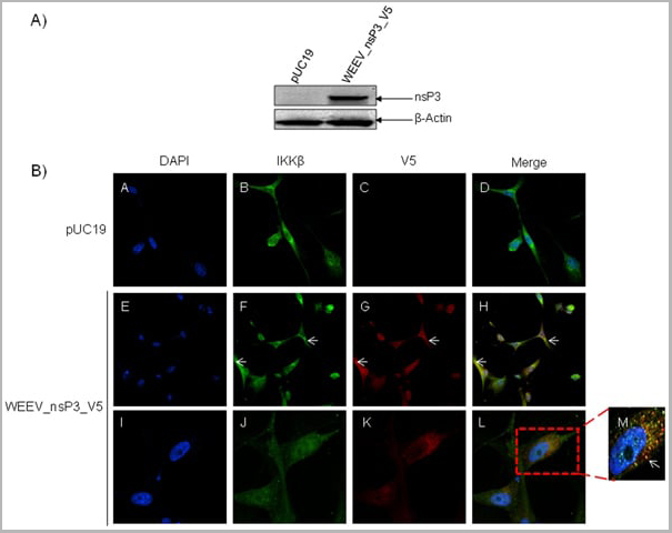 Application Data
(Published customer image: Mouse anti V5 tag antibody, clone SV5-Pk1 used for the detection of V5 tagged WEEV_nsP3 protein by western blotting and immunofluorescenceImage caption: WEEV nsP3 interaction with host IKKbeta. A) U87MGs were transfected in a 6-well plate with 5 ug of pUC19 and WEEV_nsP3_HA for 24 hours. Cell lysates were resolved using SDS-PAGE and subsequently immunoblotted with V5 antibody and beta-actin served as a loading control. B) U87MGs were transfected with WEEV_nsP3_V5; cells were fixed after 24 hours and stained with antibodies against the endogenous IKKbeta and the V5 tag. Cells were incubated with appropriate secondary Alexa Fluor antibodies and the nuclei stained with DAPI. Co-localization of IKKbeta with WEEV_nsP3_V5 (yellow) was observed as shown by the arrows. B) Panels E -H serve as an example of transfected cells in a given field of view that show co-localization of IKKbeta and WEEV_nsP3_V5 24 hours post transfection. Panels I-L represent magnified images of other cells showing co-localization of IKKbeta and WEEV_nsP3_V5. Panel M is a magnified image of panel L. The co-localization was confirmed by Z-stack analysis. Co-localization was calculated to be approximately in 61% of cells (163 cells were counted of which 44% demonstrated expression of nsP3. Of those cells that expressed nsP3, 61% showed co-localization of both proteins). Images were taken using Nikon Eclipse TE2000-U at 60x magnification and are representative of 2 independent experiments.From: Amaya M, Voss K, Sampey G, Senina S, de la Fuente C, et al. (2014) The Role of IKKbeta in Venezuelan Equine Encephalitis Virus Infection. PLoS ONE 9(2): e86745.)
Application Data
(Published customer image: Mouse anti V5 tag antibody, clone SV5-Pk1 used for the detection of V5 tagged WEEV_nsP3 protein by western blotting and immunofluorescenceImage caption: WEEV nsP3 interaction with host IKKbeta. A) U87MGs were transfected in a 6-well plate with 5 ug of pUC19 and WEEV_nsP3_HA for 24 hours. Cell lysates were resolved using SDS-PAGE and subsequently immunoblotted with V5 antibody and beta-actin served as a loading control. B) U87MGs were transfected with WEEV_nsP3_V5; cells were fixed after 24 hours and stained with antibodies against the endogenous IKKbeta and the V5 tag. Cells were incubated with appropriate secondary Alexa Fluor antibodies and the nuclei stained with DAPI. Co-localization of IKKbeta with WEEV_nsP3_V5 (yellow) was observed as shown by the arrows. B) Panels E -H serve as an example of transfected cells in a given field of view that show co-localization of IKKbeta and WEEV_nsP3_V5 24 hours post transfection. Panels I-L represent magnified images of other cells showing co-localization of IKKbeta and WEEV_nsP3_V5. Panel M is a magnified image of panel L. The co-localization was confirmed by Z-stack analysis. Co-localization was calculated to be approximately in 61% of cells (163 cells were counted of which 44% demonstrated expression of nsP3. Of those cells that expressed nsP3, 61% showed co-localization of both proteins). Images were taken using Nikon Eclipse TE2000-U at 60x magnification and are representative of 2 independent experiments.From: Amaya M, Voss K, Sampey G, Senina S, de la Fuente C, et al. (2014) The Role of IKKbeta in Venezuelan Equine Encephalitis Virus Infection. PLoS ONE 9(2): e86745.)
V5-TAG, Monoclonal Antibody (Cat# AAA11930)
Full Name
MOUSE ANTI V5-TAG
Applications
Immunohistochemistry, Flow Cytometry, Immunofluorescence, Immunoprecipitation, Western Blot, Radioimmunoassay
Pricing
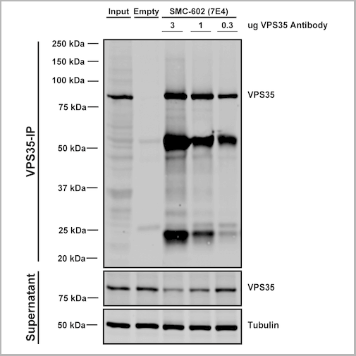
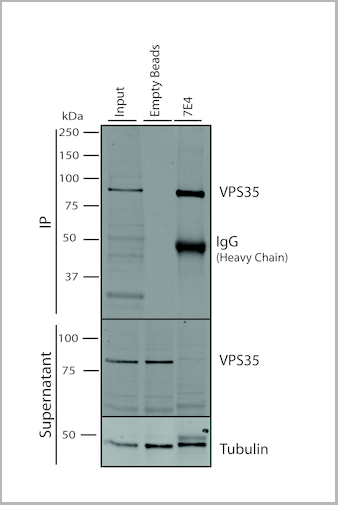
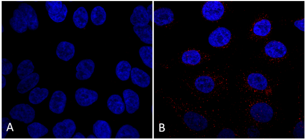
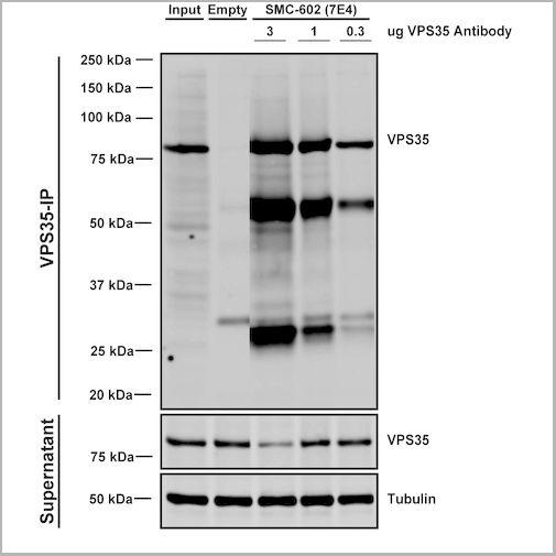 IP (Immunoprecipitation)
(Immunoprecipitation analysis using Mouse Anti-VPS35 Monoclonal Antibody, Clone 7E4. Tissue: embryonic fibroblast. Species: Mouse. Primary Antibody: Mouse Anti-VPS35 Monoclonal Antibody. Three amounts (3, 1 and 0.3 ug) were non-covalently coupled to 10uL of A/G sepharose beads for 1 hour at 4 degree C and next incubated with 250ug of MEF lysate for 2 hours at 4 degree C.)
IP (Immunoprecipitation)
(Immunoprecipitation analysis using Mouse Anti-VPS35 Monoclonal Antibody, Clone 7E4. Tissue: embryonic fibroblast. Species: Mouse. Primary Antibody: Mouse Anti-VPS35 Monoclonal Antibody. Three amounts (3, 1 and 0.3 ug) were non-covalently coupled to 10uL of A/G sepharose beads for 1 hour at 4 degree C and next incubated with 250ug of MEF lysate for 2 hours at 4 degree C.)
VPS35, Monoclonal Antibody (Cat# AAA27671)
Full Name
VPS35 Antibody, Clone 7E4: PerCP
Gene Names
VPS35; MEM3; PARK17
Reactivity
Human, Mouse, Rat
Applications
Western Blot, Immunocytochemistry, Immunofluorescence, Immunoprecipitation
Purity
Protein G Purified
Pricing


 Application Data
(Published customer image: Mouse anti V5 tag antibody, clone SV5-Pk1 used for the detection of V5 tagged WEEV_nsP3 protein by western blotting and immunofluorescenceImage caption: WEEV nsP3 interaction with host IKKbeta. A) U87MGs were transfected in a 6-well plate with 5 ug of pUC19 and WEEV_nsP3_HA for 24 hours. Cell lysates were resolved using SDS-PAGE and subsequently immunoblotted with V5 antibody and beta-actin served as a loading control. B) U87MGs were transfected with WEEV_nsP3_V5; cells were fixed after 24 hours and stained with antibodies against the endogenous IKKbeta and the V5 tag. Cells were incubated with appropriate secondary Alexa Fluor antibodies and the nuclei stained with DAPI. Co-localization of IKKbeta with WEEV_nsP3_V5 (yellow) was observed as shown by the arrows. B) Panels E -H serve as an example of transfected cells in a given field of view that show co-localization of IKKbeta and WEEV_nsP3_V5 24 hours post transfection. Panels I-L represent magnified images of other cells showing co-localization of IKKbeta and WEEV_nsP3_V5. Panel M is a magnified image of panel L. The co-localization was confirmed by Z-stack analysis. Co-localization was calculated to be approximately in 61% of cells (163 cells were counted of which 44% demonstrated expression of nsP3. Of those cells that expressed nsP3, 61% showed co-localization of both proteins). Images were taken using Nikon Eclipse TE2000-U at 60x magnification and are representative of 2 independent experiments.From: Amaya M, Voss K, Sampey G, Senina S, de la Fuente C, et al. (2014) The Role of IKKbeta in Venezuelan Equine Encephalitis Virus Infection. PLoS ONE 9(2): e86745.)
Application Data
(Published customer image: Mouse anti V5 tag antibody, clone SV5-Pk1 used for the detection of V5 tagged WEEV_nsP3 protein by western blotting and immunofluorescenceImage caption: WEEV nsP3 interaction with host IKKbeta. A) U87MGs were transfected in a 6-well plate with 5 ug of pUC19 and WEEV_nsP3_HA for 24 hours. Cell lysates were resolved using SDS-PAGE and subsequently immunoblotted with V5 antibody and beta-actin served as a loading control. B) U87MGs were transfected with WEEV_nsP3_V5; cells were fixed after 24 hours and stained with antibodies against the endogenous IKKbeta and the V5 tag. Cells were incubated with appropriate secondary Alexa Fluor antibodies and the nuclei stained with DAPI. Co-localization of IKKbeta with WEEV_nsP3_V5 (yellow) was observed as shown by the arrows. B) Panels E -H serve as an example of transfected cells in a given field of view that show co-localization of IKKbeta and WEEV_nsP3_V5 24 hours post transfection. Panels I-L represent magnified images of other cells showing co-localization of IKKbeta and WEEV_nsP3_V5. Panel M is a magnified image of panel L. The co-localization was confirmed by Z-stack analysis. Co-localization was calculated to be approximately in 61% of cells (163 cells were counted of which 44% demonstrated expression of nsP3. Of those cells that expressed nsP3, 61% showed co-localization of both proteins). Images were taken using Nikon Eclipse TE2000-U at 60x magnification and are representative of 2 independent experiments.From: Amaya M, Voss K, Sampey G, Senina S, de la Fuente C, et al. (2014) The Role of IKKbeta in Venezuelan Equine Encephalitis Virus Infection. PLoS ONE 9(2): e86745.)
V5-TAG, Monoclonal Antibody (Cat# AAA11864)
Full Name
MOUSE ANTI V5-TAG:FITC
Applications
Immunofluorescence
Pricing
 Application Data
Toxoplasma gondii, Polyclonal Antibody (Cat# AAA13574)
Full Name
Toxoplasma gondii
Reactivity
Human
Applications
Immunohistochemistry
Pricing
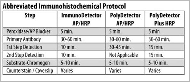
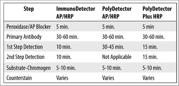 IHC Protocol
CD7, Monoclonal Antibody (Cat# AAA13566)
Full Name
CD7
Gene Names
CD7; GP40; TP41; Tp40; LEU-9
Reactivity
Human
Applications
Immunohistochemistry
Pricing

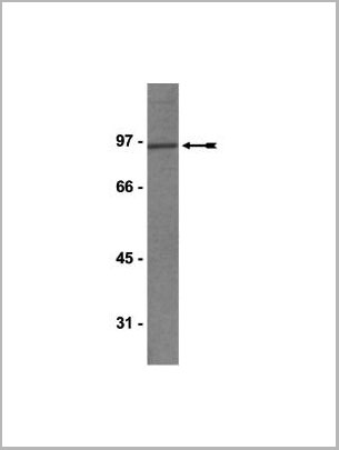
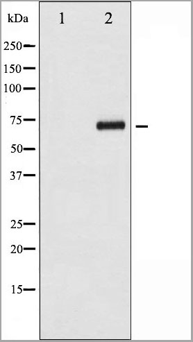
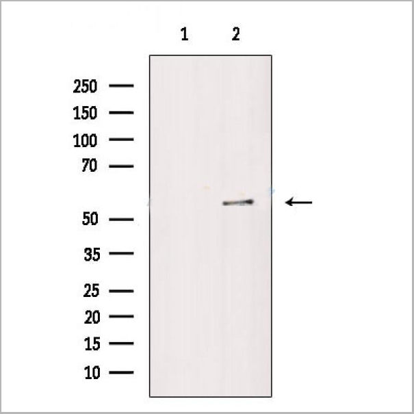
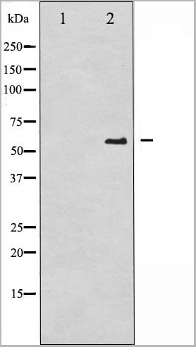
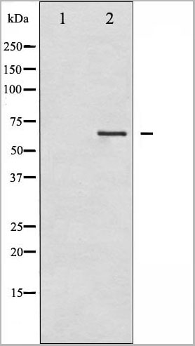
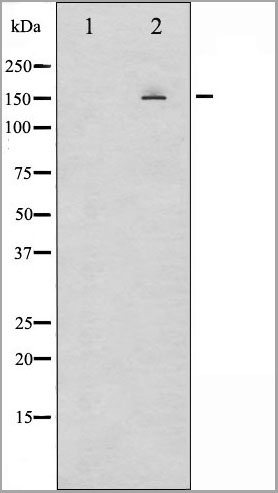 WB (Western Blot)
(Western blot analysis of EGFR phosphorylation expression in TSA treated HeLa whole cell lysates, The lane on the left is treated with the antigen-specific peptide.)
WB (Western Blot)
(Western blot analysis of EGFR phosphorylation expression in TSA treated HeLa whole cell lysates, The lane on the left is treated with the antigen-specific peptide.)
EGFR, Polyclonal Antibody (Cat# AAA30963)
Full Name
Phospho-EGFR (Ser1026) Antibody
Gene Names
EGFR; ERBB; HER1; mENA; ERBB1; PIG61; NISBD2
Reactivity
Human, Mouse, Rat
Applications
Western Blot, Immunohistochemistry, Immunofluorescence, Immunocytochemistry
Purity
From purified rabbit serum by affinity purification via sequential chromatography on phospho-and non-phospho-peptide affinity columns.
Pricing

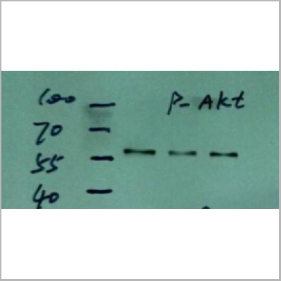
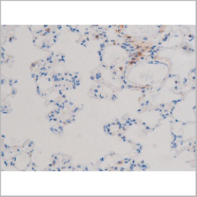
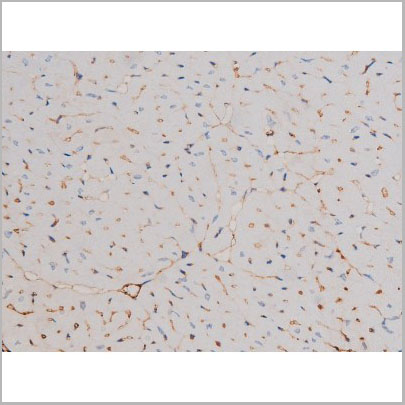
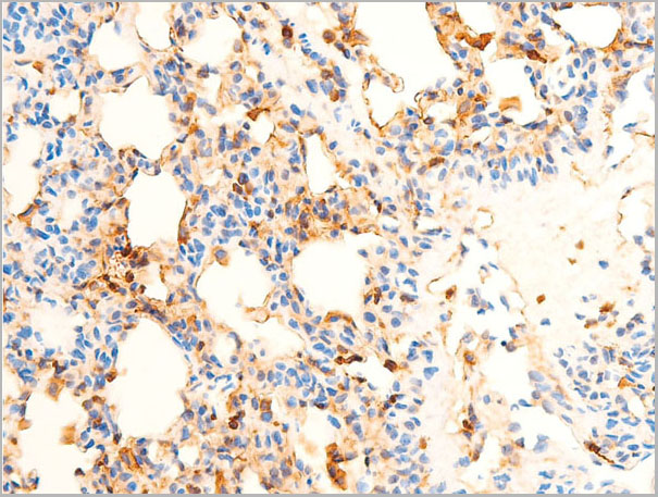
 IHC (Immunohistochemistry)
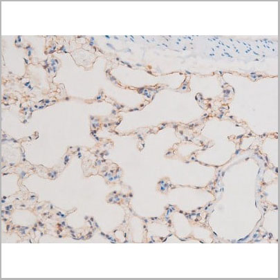
(AAA30920 at 1/200 staining human lung cancer tissue sections by IHC-P. The tissue was formaldehyde fixed and a heat mediated antigen retrieval step in citrate buffer was performed. The tissue was then blocked and incubated with the antibody for 1.5 hours at 22 degree C. An HRP conjugated goat anti-rabbit antibody was used as the secondary.)
IHC (Immunohistochemistry)
(AAA30920 at 1/200 staining human lung cancer tissue sections by IHC-P. The tissue was formaldehyde fixed and a heat mediated antigen retrieval step in citrate buffer was performed. The tissue was then blocked and incubated with the antibody for 1.5 hours at 22 degree C. An HRP conjugated goat anti-rabbit antibody was used as the secondary.)
Akt, Polyclonal Antibody (Cat# AAA30920)
Full Name
Phospho-Akt(Ser473) Antibody
Gene Names
AKT1; AKT; PKB; RAC; CWS6; PRKBA; PKB-ALPHA; RAC-ALPHA
Reactivity
Human, Mouse, Rat
Applications
Western Blot, Immunohistochemistry, Immunofluorescence, Immunocytochemistry
Purity
From purified rabbit serum by affinity purification via sequential chromatography on phospho-and non-phospho-peptide affinity columns.
Pricing


 Application Data
(Published customer image: Mouse anti Human CD105 antibody, clone SN6 used for flow cytometry.Image caption:Phenotype change from HLA-A2+/CD11b+/CD105- to HLA-A2+/CD11b-/CD105+ on endothelium-adherent blood monocyte-derived cells with increase in size and granularity during co-culture. HLA-A2+ PBMCs (1x10⁶ cells/well) were incubated for 2 h (Day 0) with HLA-A2- HUVECs, after which the non-adherent cells were removed by washing. The cell layers were analysed by three-colour flow cytometry staining for HLA-A2, CD11b and CD105 on (A) Day 1 and (B) Day 2. These plots were gated for HLA-A2+ cells. Forward scatter/side scatter dot plots gated for HLA-A2+ cells on Day 0 (C) and Day 2 (D) was shown. These are representative of 2 individual experiments.From: Tso C, Rye K-A, Barter P (2012) Phenotypic and Functional Changes in Blood Monocytes Following Adherence to Endothelium. PLoS ONE 7(5): e37091.)
Application Data
(Published customer image: Mouse anti Human CD105 antibody, clone SN6 used for flow cytometry.Image caption:Phenotype change from HLA-A2+/CD11b+/CD105- to HLA-A2+/CD11b-/CD105+ on endothelium-adherent blood monocyte-derived cells with increase in size and granularity during co-culture. HLA-A2+ PBMCs (1x10⁶ cells/well) were incubated for 2 h (Day 0) with HLA-A2- HUVECs, after which the non-adherent cells were removed by washing. The cell layers were analysed by three-colour flow cytometry staining for HLA-A2, CD11b and CD105 on (A) Day 1 and (B) Day 2. These plots were gated for HLA-A2+ cells. Forward scatter/side scatter dot plots gated for HLA-A2+ cells on Day 0 (C) and Day 2 (D) was shown. These are representative of 2 individual experiments.From: Tso C, Rye K-A, Barter P (2012) Phenotypic and Functional Changes in Blood Monocytes Following Adherence to Endothelium. PLoS ONE 7(5): e37091.)
CD105, Monoclonal Antibody (Cat# AAA12085)
Full Name
MOUSE ANTI HUMAN CD105
Gene Names
ENG; END; HHT1; ORW1
Applications
Immunohistochemistry, Flow Cytometry, Immunoprecipitation, Western Blot
Pricing
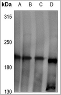
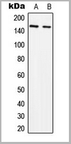
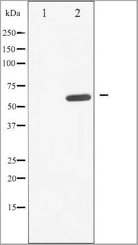
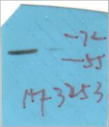
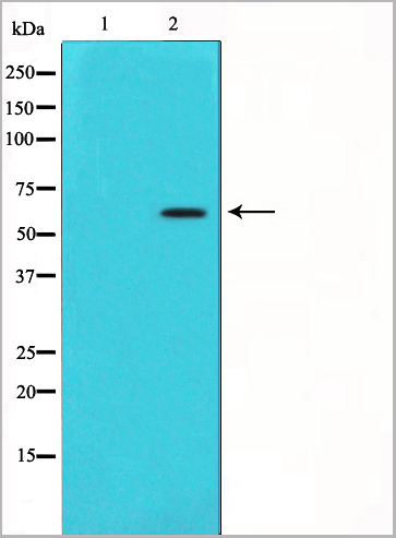
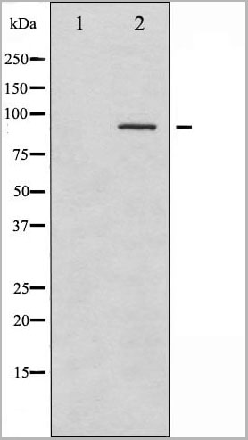
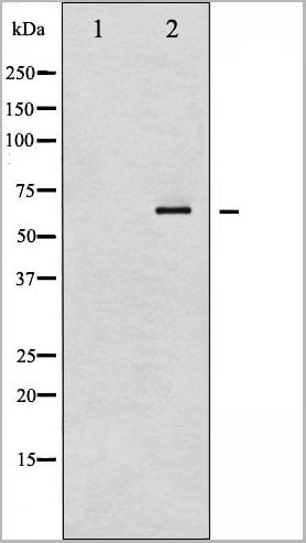
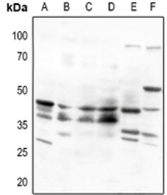
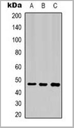
 WB (Western Blot)
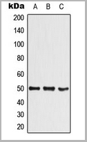
(Western blot analysis of CD298 expression in HEK293T (A), Hela (B), A2780 (C), mouse heart (D), mouse liver (E), rat heart (F), rat liver (G) whole cell lysates.(Predicted band size: 31 kD; Observed band size: 40 kD))
WB (Western Blot)
(Western blot analysis of CD298 expression in HEK293T (A), Hela (B), A2780 (C), mouse heart (D), mouse liver (E), rat heart (F), rat liver (G) whole cell lysates.(Predicted band size: 31 kD; Observed band size: 40 kD))
CD298, Polyclonal Antibody (Cat# AAA27791)
Full Name
Anti-CD298 Antibody
Gene Names
ATP1B3; CD298; ATPB-3
Reactivity
Human, Mouse, Rat, Monkey
Applications
Western Blot
Purity
The antibody was purified by immunogen affinity chromatography.
Pricing
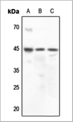
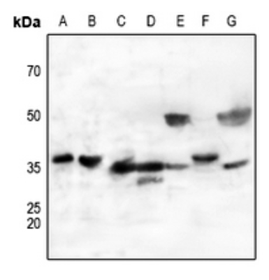
 WB (Western Blot)
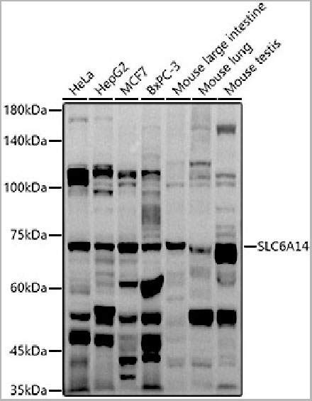
(Western blot analysis of extracts of various cell lines, using SLC6A14 antibody (AAA28230) at 1:1000 dilution.Secondary antibody: HRP Goat Anti-Rabbit IgG (H+L) at 1:10000 dilution. Lysates/proteins: 25ug per lane. Blocking buffer: 3% nonfat dry milk in TBST. Detection: ECL Basic Kit. Exposure time: 90s)
WB (Western Blot)
(Western blot analysis of extracts of various cell lines, using SLC6A14 antibody (AAA28230) at 1:1000 dilution.Secondary antibody: HRP Goat Anti-Rabbit IgG (H+L) at 1:10000 dilution. Lysates/proteins: 25ug per lane. Blocking buffer: 3% nonfat dry milk in TBST. Detection: ECL Basic Kit. Exposure time: 90s)
SLC6A14, Polyclonal Antibody (Cat# AAA28230)
Full Name
SLC6A14 Polyclonal Antibody
Gene Names
SLC6A14; BMIQ11
Reactivity
Human, Mouse
Applications
Western Blot
Purity
Affinity Purification
Pricing
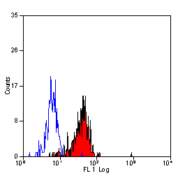
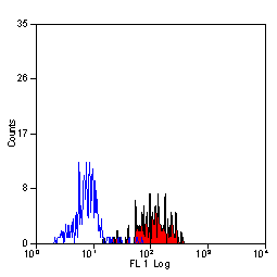
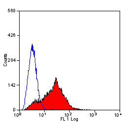
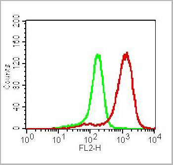 FCM (Flow Cytometry)
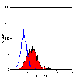
(Figure-1: Cell surface flowcytometry analysis of CLEC1on human PBMC (Monocytes) using 0.5 ug/10^6 Cells of Anti-CLEC1 antibody. Green represent isotype control and red represent Anti human CLEC1 antibody. Goat anti mouse PE conjugated was used as the secondary antibody.)
FCM (Flow Cytometry)
(Figure-1: Cell surface flowcytometry analysis of CLEC1on human PBMC (Monocytes) using 0.5 ug/10^6 Cells of Anti-CLEC1 antibody. Green represent isotype control and red represent Anti human CLEC1 antibody. Goat anti mouse PE conjugated was used as the secondary antibody.)
CLEC1, Monoclonal Antibody (Cat# AAA14882)
Full Name
Monoclonal Antibody to CLEC1 (Clone: ABM2H82)
Gene Names
CLEC1A; CLEC1; CLEC-1
Reactivity
Human
Applications
Flow Cytometry
Purity
Protein G Chromatography Purified
Pricing
 Application Data
HLA Class 1 Antigen A33,B8, Monoclonal Antibody (Cat# AAA14684)
Full Name
Mouse anti-Human HLA Class 1 Antigen A33,B8
Reactivity
Human
Applications
Cell Typing
Purity
Ascites
Pricing
CD22, Active Protein (Cat# AAA14393)
Full Name
CD22 protein
Gene Names
CD22; SIGLEC2; SIGLEC-2
Purity
> 98% by SDS-PAGE and HPLC analyses
Pricing


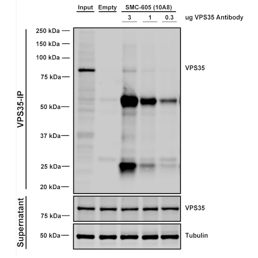
 IP (Immunoprecipitation)
(Immunoprecipitation analysis using Mouse Anti-VPS35 Monoclonal Antibody, Clone 10A8. Tissue: embryonic fibroblast. Species: Mouse. Primary Antibody: Mouse Anti-VPS35 Monoclonal Antibody. Three amounts of (3, 1 and 0.3 ug) were non-covalently coupled to 10uL of A/G sepharose beads for 1 hour at 4 degree C and next incubated with 250ug of MEF lysate for 2 hours at 4 degree C.)
IP (Immunoprecipitation)
(Immunoprecipitation analysis using Mouse Anti-VPS35 Monoclonal Antibody, Clone 10A8. Tissue: embryonic fibroblast. Species: Mouse. Primary Antibody: Mouse Anti-VPS35 Monoclonal Antibody. Three amounts of (3, 1 and 0.3 ug) were non-covalently coupled to 10uL of A/G sepharose beads for 1 hour at 4 degree C and next incubated with 250ug of MEF lysate for 2 hours at 4 degree C.)
VPS35, Monoclonal Antibody (Cat# AAA27697)
Full Name
VPS35 Antibody, Clone 10A8: HRP
Gene Names
VPS35; MEM3; PARK17
Reactivity
Human, Mouse, Rat
Applications
Western Blot, Immunocytochemistry, Immunofluorescence, Immunoprecipitation
Purity
Protein G Purified
Pricing
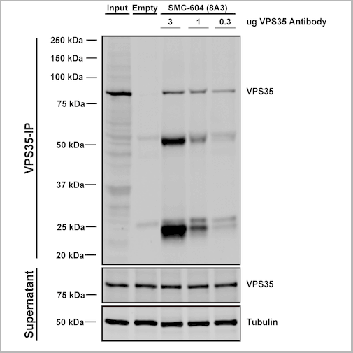

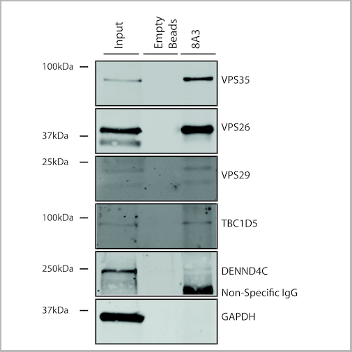

 IP (Immunoprecipitation)
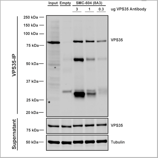
(Immunoprecipitation analysis using Mouse Anti-VPS35 Monoclonal Antibody, Clone 8A3. Tissue: embryonic fibroblast. Species: Mouse. Primary Antibody: Mouse Anti-VPS35 Monoclonal Antibody. Three amounts of (3, 1 and 0.3 ug) were non-covalently coupled to 10uL of A/G sepharose beads for 1 hour at 4 degree C and next incubated with 250ug of MEF lysate for 2 hours at 4 degree C.)
IP (Immunoprecipitation)
(Immunoprecipitation analysis using Mouse Anti-VPS35 Monoclonal Antibody, Clone 8A3. Tissue: embryonic fibroblast. Species: Mouse. Primary Antibody: Mouse Anti-VPS35 Monoclonal Antibody. Three amounts of (3, 1 and 0.3 ug) were non-covalently coupled to 10uL of A/G sepharose beads for 1 hour at 4 degree C and next incubated with 250ug of MEF lysate for 2 hours at 4 degree C.)
VPS35, Monoclonal Antibody (Cat# AAA27687)
Full Name
VPS35 Antibody, Clone 8A3: FITC
Gene Names
VPS35; MEM3; PARK17
Reactivity
Human, Mouse, Rat
Applications
Western Blot, Immunocytochemistry, Immunofluorescence, Immunoprecipitation
Purity
Protein G Purified
Pricing
 IF (Immunofluorescence)
(AAA31031 staining HeLa by IF/ICC. The sample were fixed with PFA and permeabilized in 0.1% Triton X-100, then blocked in 10% serum for 45 minutes at 25 degree C. The primary antibody was diluted at 1/200 and incubated with the sample for 1 hour at 37 degree C. An Alexa Fluor 594 conjugated goat anti-rabbit IgG (H+L) Ab, diluted at 1/600, was used as the secondary antibody.)
IF (Immunofluorescence)
(AAA31031 staining HeLa by IF/ICC. The sample were fixed with PFA and permeabilized in 0.1% Triton X-100, then blocked in 10% serum for 45 minutes at 25 degree C. The primary antibody was diluted at 1/200 and incubated with the sample for 1 hour at 37 degree C. An Alexa Fluor 594 conjugated goat anti-rabbit IgG (H+L) Ab, diluted at 1/600, was used as the secondary antibody.)
CDC25A, Polyclonal Antibody (Cat# AAA31031)
Full Name
Phospho-CDC25A (Ser124) Antibody
Gene Names
CDC25A; CDC25A2
Reactivity
Human, Mouse, Rat
Applications
Western Blot, Immunohistochemistry, Immunofluorescence, Immunocytochemistry
Purity
From purified rabbit serum by affinity purification via sequential chromatography on phospho-and non-phospho-peptide affinity columns.
Pricing


 Application Data
(Published customer image: Mouse anti V5 tag antibody, clone SV5-Pk1 used for the detection of V5 tagged WEEV_nsP3 protein by western blotting and immunofluorescenceImage caption: WEEV nsP3 interaction with host IKKbeta. A) U87MGs were transfected in a 6-well plate with 5 ug of pUC19 and WEEV_nsP3_HA for 24 hours. Cell lysates were resolved using SDS-PAGE and subsequently immunoblotted with V5 antibody and beta-actin served as a loading control. B) U87MGs were transfected with WEEV_nsP3_V5; cells were fixed after 24 hours and stained with antibodies against the endogenous IKKbeta and the V5 tag. Cells were incubated with appropriate secondary Alexa Fluor antibodies and the nuclei stained with DAPI. Co-localization of IKKbeta with WEEV_nsP3_V5 (yellow) was observed as shown by the arrows. B) Panels E -H serve as an example of transfected cells in a given field of view that show co-localization of IKKbeta and WEEV_nsP3_V5 24 hours post transfection. Panels I-L represent magnified images of other cells showing co-localization of IKKbeta and WEEV_nsP3_V5. Panel M is a magnified image of panel L. The co-localization was confirmed by Z-stack analysis. Co-localization was calculated to be approximately in 61% of cells (163 cells were counted of which 44% demonstrated expression of nsP3. Of those cells that expressed nsP3, 61% showed co-localization of both proteins). Images were taken using Nikon Eclipse TE2000-U at 60x magnification and are representative of 2 independent experiments.From: Amaya M, Voss K, Sampey G, Senina S, de la Fuente C, et al. (2014) The Role of IKKbeta in Venezuelan Equine Encephalitis Virus Infection. PLoS ONE 9(2): e86745.)
Application Data
(Published customer image: Mouse anti V5 tag antibody, clone SV5-Pk1 used for the detection of V5 tagged WEEV_nsP3 protein by western blotting and immunofluorescenceImage caption: WEEV nsP3 interaction with host IKKbeta. A) U87MGs were transfected in a 6-well plate with 5 ug of pUC19 and WEEV_nsP3_HA for 24 hours. Cell lysates were resolved using SDS-PAGE and subsequently immunoblotted with V5 antibody and beta-actin served as a loading control. B) U87MGs were transfected with WEEV_nsP3_V5; cells were fixed after 24 hours and stained with antibodies against the endogenous IKKbeta and the V5 tag. Cells were incubated with appropriate secondary Alexa Fluor antibodies and the nuclei stained with DAPI. Co-localization of IKKbeta with WEEV_nsP3_V5 (yellow) was observed as shown by the arrows. B) Panels E -H serve as an example of transfected cells in a given field of view that show co-localization of IKKbeta and WEEV_nsP3_V5 24 hours post transfection. Panels I-L represent magnified images of other cells showing co-localization of IKKbeta and WEEV_nsP3_V5. Panel M is a magnified image of panel L. The co-localization was confirmed by Z-stack analysis. Co-localization was calculated to be approximately in 61% of cells (163 cells were counted of which 44% demonstrated expression of nsP3. Of those cells that expressed nsP3, 61% showed co-localization of both proteins). Images were taken using Nikon Eclipse TE2000-U at 60x magnification and are representative of 2 independent experiments.From: Amaya M, Voss K, Sampey G, Senina S, de la Fuente C, et al. (2014) The Role of IKKbeta in Venezuelan Equine Encephalitis Virus Infection. PLoS ONE 9(2): e86745.)
V5-TAG, Monoclonal Antibody (Cat# AAA12211)
Full Name
MOUSE ANTI V5-TAG
Applications
Immunohistochemistry, Flow Cytometry, Immunofluorescence, Immunoprecipitation, Western Blot, Radioimmunoassay
Pricing


 IP (Immunoprecipitation)
(Immunoprecipitation analysis using Mouse Anti-VPS35 Monoclonal Antibody, Clone 8A3. Tissue: embryonic fibroblast. Species: Mouse. Primary Antibody: Mouse Anti-VPS35 Monoclonal Antibody. Three amounts of (3, 1 and 0.3 ug) were non-covalently coupled to 10uL of A/G sepharose beads for 1 hour at 4 degree C and next incubated with 250ug of MEF lysate for 2 hours at 4 degree C.)
IP (Immunoprecipitation)
(Immunoprecipitation analysis using Mouse Anti-VPS35 Monoclonal Antibody, Clone 8A3. Tissue: embryonic fibroblast. Species: Mouse. Primary Antibody: Mouse Anti-VPS35 Monoclonal Antibody. Three amounts of (3, 1 and 0.3 ug) were non-covalently coupled to 10uL of A/G sepharose beads for 1 hour at 4 degree C and next incubated with 250ug of MEF lysate for 2 hours at 4 degree C.)
VPS35, Monoclonal Antibody (Cat# AAA27683)
Full Name
VPS35 Antibody, Clone 8A3: ATTO 390
Gene Names
VPS35; MEM3; PARK17
Reactivity
Human, Mouse, Rat
Applications
Western Blot, Immunocytochemistry, Immunofluorescence, Immunoprecipitation
Purity
Protein G Purified
Pricing

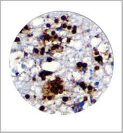
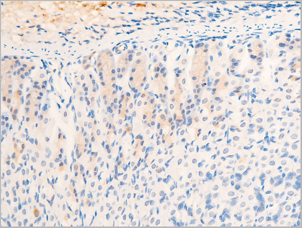
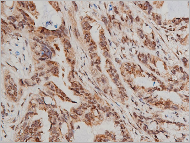
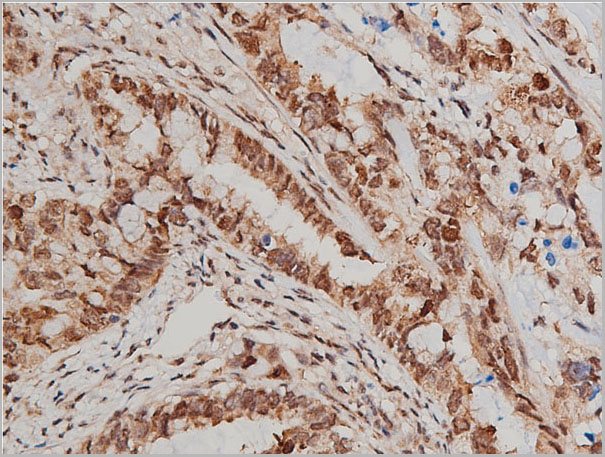
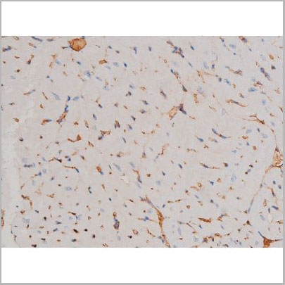
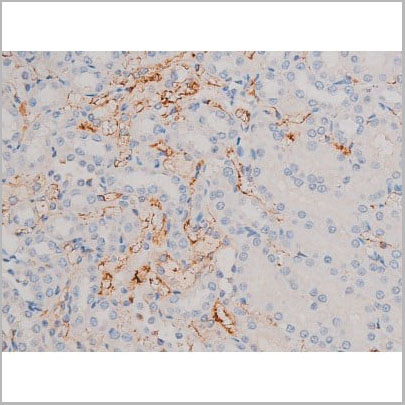
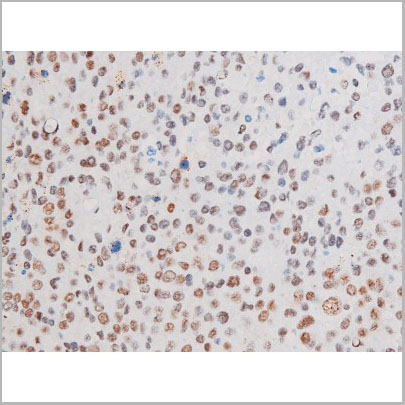
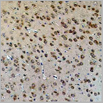
 IHC (Immunohistochemistry)
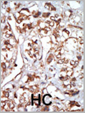
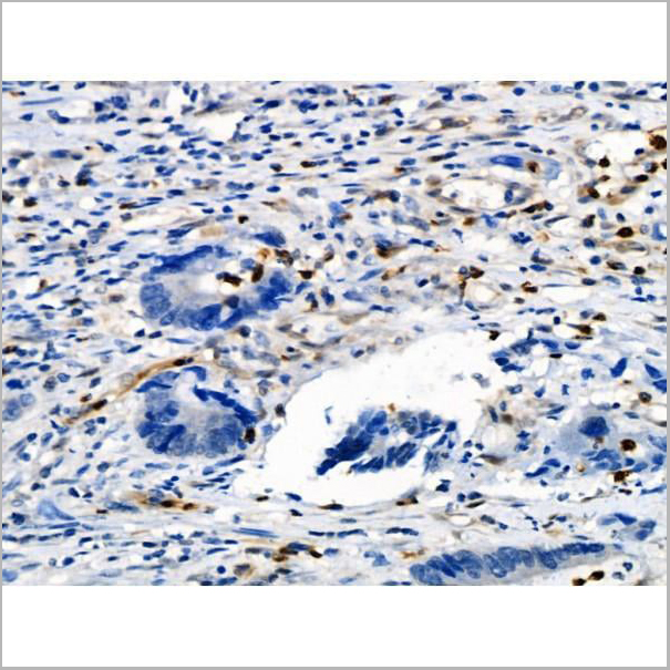
(Formalin-fixed and paraffin-embedded human cancer tissue reacted with the primary antibody, which was peroxidase-conjugated to the secondary antibody, followed by AEC staining. This data demonstrates the use of this antibody for immunohistochemistry; clinical relevance has not been evaluated. BC = breast carcinoma; HC = hepatocarcinoma.)
IHC (Immunohistochemistry)
(Formalin-fixed and paraffin-embedded human cancer tissue reacted with the primary antibody, which was peroxidase-conjugated to the secondary antibody, followed by AEC staining. This data demonstrates the use of this antibody for immunohistochemistry; clinical relevance has not been evaluated. BC = breast carcinoma; HC = hepatocarcinoma.)
HRI, Polyclonal Antibody (Cat# AAA28727)
Full Name
HRI Antibody (N-term)
Gene Names
EIF2AK1; HCR; HRI
Reactivity
Human
Applications
Western Blot, Immunohistochemistry
Purity
Purified Rabbit Polyclonal Antibody (Pab)
Pricing


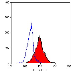
 Application Data
(Published customer image: Mouse anti Human CD105 antibody, clone SN6 used for flow cytometry.Image caption:Phenotype change from HLA-A2+/CD11b+/CD105- to HLA-A2+/CD11b-/CD105+ on endothelium-adherent blood monocyte-derived cells with increase in size and granularity during co-culture. HLA-A2+ PBMCs (1x10⁶ cells/well) were incubated for 2 h (Day 0) with HLA-A2- HUVECs, after which the non-adherent cells were removed by washing. The cell layers were analysed by three-colour flow cytometry staining for HLA-A2, CD11b and CD105 on (A) Day 1 and (B) Day 2. These plots were gated for HLA-A2+ cells. Forward scatter/side scatter dot plots gated for HLA-A2+ cells on Day 0 (C) and Day 2 (D) was shown. These are representative of 2 individual experiments.From: Tso C, Rye K-A, Barter P (2012) Phenotypic and Functional Changes in Blood Monocytes Following Adherence to Endothelium. PLoS ONE 7(5): e37091.)
Application Data
(Published customer image: Mouse anti Human CD105 antibody, clone SN6 used for flow cytometry.Image caption:Phenotype change from HLA-A2+/CD11b+/CD105- to HLA-A2+/CD11b-/CD105+ on endothelium-adherent blood monocyte-derived cells with increase in size and granularity during co-culture. HLA-A2+ PBMCs (1x10⁶ cells/well) were incubated for 2 h (Day 0) with HLA-A2- HUVECs, after which the non-adherent cells were removed by washing. The cell layers were analysed by three-colour flow cytometry staining for HLA-A2, CD11b and CD105 on (A) Day 1 and (B) Day 2. These plots were gated for HLA-A2+ cells. Forward scatter/side scatter dot plots gated for HLA-A2+ cells on Day 0 (C) and Day 2 (D) was shown. These are representative of 2 individual experiments.From: Tso C, Rye K-A, Barter P (2012) Phenotypic and Functional Changes in Blood Monocytes Following Adherence to Endothelium. PLoS ONE 7(5): e37091.)
CD105, Monoclonal Antibody (Cat# AAA12083)
Full Name
MOUSE ANTI HUMAN CD105:FITC
Gene Names
ENG; END; HHT1; ORW1
Applications
Flow Cytometry
Pricing


 Application Data
(Published customer image: Mouse anti Human CD105 antibody, clone SN6 used for flow cytometry.Image caption:Phenotype change from HLA-A2+/CD11b+/CD105- to HLA-A2+/CD11b-/CD105+ on endothelium-adherent blood monocyte-derived cells with increase in size and granularity during co-culture. HLA-A2+ PBMCs (1x10⁶ cells/well) were incubated for 2 h (Day 0) with HLA-A2- HUVECs, after which the non-adherent cells were removed by washing. The cell layers were analysed by three-colour flow cytometry staining for HLA-A2, CD11b and CD105 on (A) Day 1 and (B) Day 2. These plots were gated for HLA-A2+ cells. Forward scatter/side scatter dot plots gated for HLA-A2+ cells on Day 0 (C) and Day 2 (D) was shown. These are representative of 2 individual experiments.From: Tso C, Rye K-A, Barter P (2012) Phenotypic and Functional Changes in Blood Monocytes Following Adherence to Endothelium. PLoS ONE 7(5): e37091.)
Application Data
(Published customer image: Mouse anti Human CD105 antibody, clone SN6 used for flow cytometry.Image caption:Phenotype change from HLA-A2+/CD11b+/CD105- to HLA-A2+/CD11b-/CD105+ on endothelium-adherent blood monocyte-derived cells with increase in size and granularity during co-culture. HLA-A2+ PBMCs (1x10⁶ cells/well) were incubated for 2 h (Day 0) with HLA-A2- HUVECs, after which the non-adherent cells were removed by washing. The cell layers were analysed by three-colour flow cytometry staining for HLA-A2, CD11b and CD105 on (A) Day 1 and (B) Day 2. These plots were gated for HLA-A2+ cells. Forward scatter/side scatter dot plots gated for HLA-A2+ cells on Day 0 (C) and Day 2 (D) was shown. These are representative of 2 individual experiments.From: Tso C, Rye K-A, Barter P (2012) Phenotypic and Functional Changes in Blood Monocytes Following Adherence to Endothelium. PLoS ONE 7(5): e37091.)
CD105, Monoclonal Antibody (Cat# AAA11865)
Full Name
MOUSE ANTI HUMAN CD105:FITC
Gene Names
ENG; END; HHT1; ORW1
Applications
Flow Cytometry
Pricing


 Application Data
(Published customer image: Mouse anti Human CD105 antibody, clone SN6 used for flow cytometry.Image caption:Phenotype change from HLA-A2+/CD11b+/CD105- to HLA-A2+/CD11b-/CD105+ on endothelium-adherent blood monocyte-derived cells with increase in size and granularity during co-culture. HLA-A2+ PBMCs (1x10⁶ cells/well) were incubated for 2 h (Day 0) with HLA-A2- HUVECs, after which the non-adherent cells were removed by washing. The cell layers were analysed by three-colour flow cytometry staining for HLA-A2, CD11b and CD105 on (A) Day 1 and (B) Day 2. These plots were gated for HLA-A2+ cells. Forward scatter/side scatter dot plots gated for HLA-A2+ cells on Day 0 (C) and Day 2 (D) was shown. These are representative of 2 individual experiments.From: Tso C, Rye K-A, Barter P (2012) Phenotypic and Functional Changes in Blood Monocytes Following Adherence to Endothelium. PLoS ONE 7(5): e37091.)
Application Data
(Published customer image: Mouse anti Human CD105 antibody, clone SN6 used for flow cytometry.Image caption:Phenotype change from HLA-A2+/CD11b+/CD105- to HLA-A2+/CD11b-/CD105+ on endothelium-adherent blood monocyte-derived cells with increase in size and granularity during co-culture. HLA-A2+ PBMCs (1x10⁶ cells/well) were incubated for 2 h (Day 0) with HLA-A2- HUVECs, after which the non-adherent cells were removed by washing. The cell layers were analysed by three-colour flow cytometry staining for HLA-A2, CD11b and CD105 on (A) Day 1 and (B) Day 2. These plots were gated for HLA-A2+ cells. Forward scatter/side scatter dot plots gated for HLA-A2+ cells on Day 0 (C) and Day 2 (D) was shown. These are representative of 2 individual experiments.From: Tso C, Rye K-A, Barter P (2012) Phenotypic and Functional Changes in Blood Monocytes Following Adherence to Endothelium. PLoS ONE 7(5): e37091.)
CD105, Monoclonal Antibody (Cat# AAA12029)
Full Name
MOUSE ANTI HUMAN CD105:RPE
Gene Names
ENG; END; HHT1; ORW1
Applications
Flow Cytometry
Pricing
 Application Data
(At 25 degree C. The primary antibody was diluted at 1/200 and incubated with the sample for 1 hour at 37 degree C. An Alexa Fluor 594 conjugated goat anti-rabbit IgG (H+L) Ab, diluted at 1/600, was used as the secondary antibody.)
Application Data
(At 25 degree C. The primary antibody was diluted at 1/200 and incubated with the sample for 1 hour at 37 degree C. An Alexa Fluor 594 conjugated goat anti-rabbit IgG (H+L) Ab, diluted at 1/600, was used as the secondary antibody.)
Vimentin, Polyclonal Antibody (Cat# AAA31419)
Full Name
Phospho-Vimentin (Ser39) Antibody
Reactivity
Human, Mouse, Rat
Applications
Western Blot, Immunohistochemistry, Immunofluorescence, Immunocytochemistry, Peptide ELISA
Purity
The antibody is from purified rabbit serum by affinity purification via sequential chromatography on phospho-peptide and non-phospho-peptide affinity columns.
Pricing
 Application Data
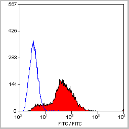
(Staining of mouse spleen with Hamster anti Mouse CD81: Alexa Fluor 488)
Application Data
(Staining of mouse spleen with Hamster anti Mouse CD81: Alexa Fluor 488)
CD81, Monoclonal Antibody (Cat# AAA11942)
Full Name
HAMSTER ANTI MOUSE CD81
Gene Names
Cd81; Tapa1; Tapa-1; Tspan28
Reactivity
Rat
Applications
Immunohistochemistry, Flow Cytometry, Immunoprecipitation, Western Blot
Pricing
Phospholipase B-Like 2, ELISA Kit (Cat# AAA14515)
Full Name
Hamster Phospholipase B-Like 2 (PLBL2) ELISA Kit
Pricing
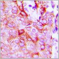
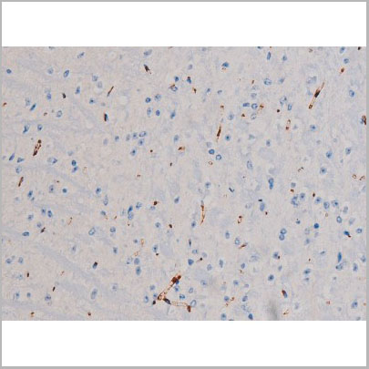
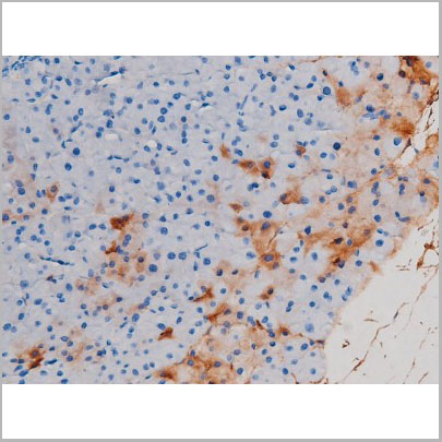
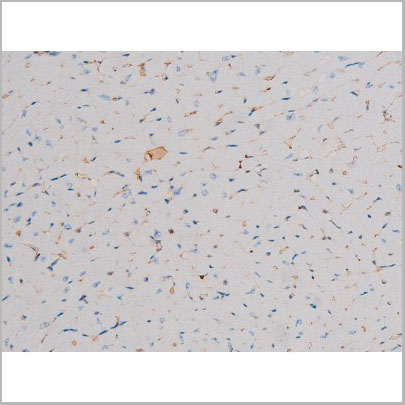
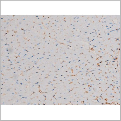
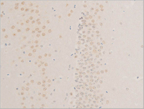
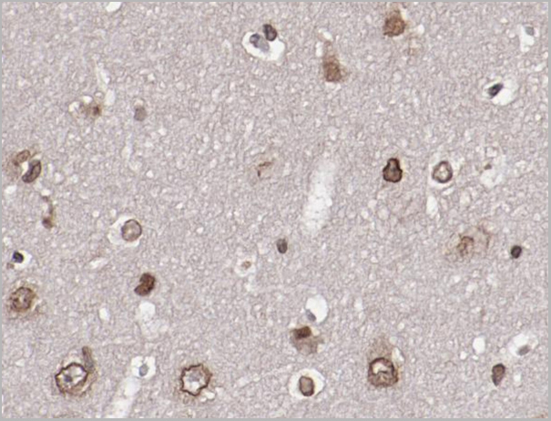
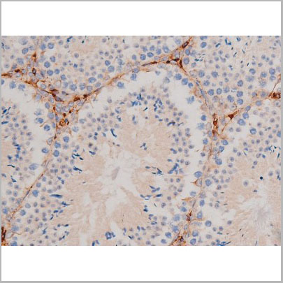
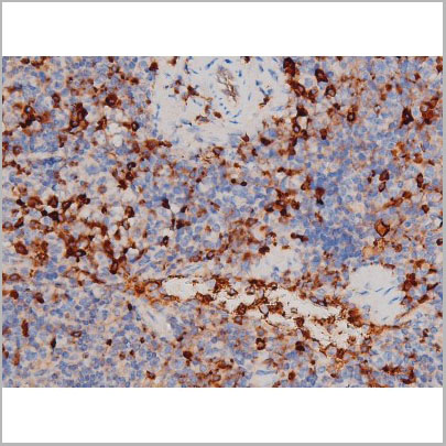
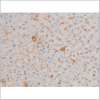
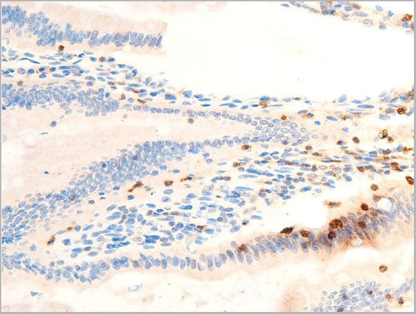
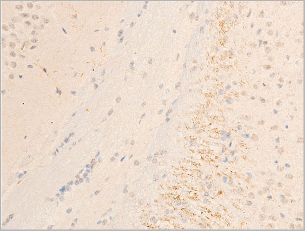
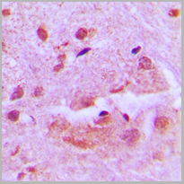 IHC (Immunohistochemistry)
(Immunohistochemical analysis of SHARP1 staining in human brain formalin fixed paraffin embedded tissue section. The section was pre-treated using heat mediated antigen retrieval with sodium citrate buffer (pH 6.0). The section was then incubated with the antibody at room temperature and detected using an HRP conjugated compact polymer system. DAB was used as the chromogen. The section was then counterstained with haematoxylin and mounted with DPX.)
IHC (Immunohistochemistry)
(Immunohistochemical analysis of SHARP1 staining in human brain formalin fixed paraffin embedded tissue section. The section was pre-treated using heat mediated antigen retrieval with sodium citrate buffer (pH 6.0). The section was then incubated with the antibody at room temperature and detected using an HRP conjugated compact polymer system. DAB was used as the chromogen. The section was then counterstained with haematoxylin and mounted with DPX.)
SHARP1, Polyclonal Antibody (Cat# AAA27795)
Full Name
Anti-SHARP1 Antibody
Gene Names
BHLHE41; DEC2; hDEC2; BHLHB3; SHARP1
Reactivity
Human, Mouse, Rat
Applications
Western Blot, Immunohistochemistry
Purity
The antibody was purified by immunogen affinity chromatography.
Pricing
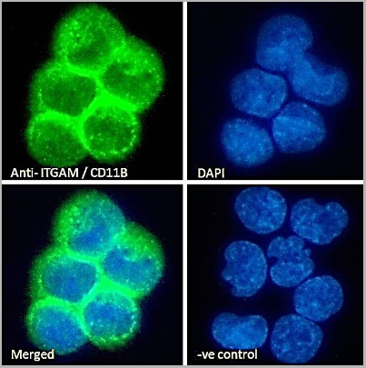
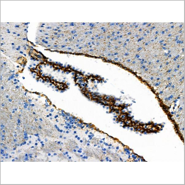
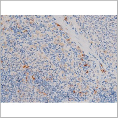
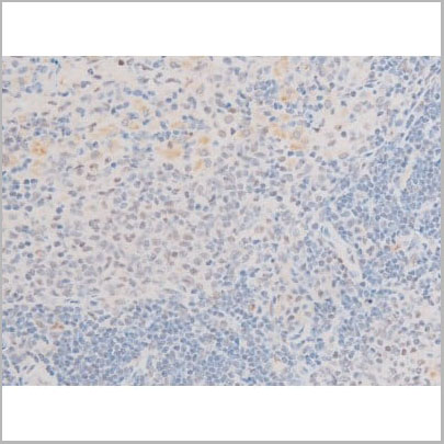
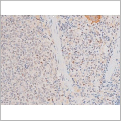
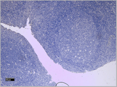 IHC (Immunohistochemistry)
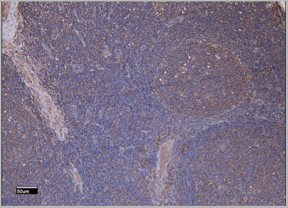
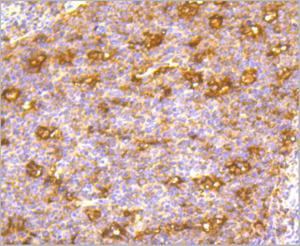
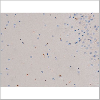
(Negative Control showing staining of paraffin embedded Human Tonsil, with no primary antibody.)
IHC (Immunohistochemistry)
(Negative Control showing staining of paraffin embedded Human Tonsil, with no primary antibody.)
ITGAM/CD11B, Polyclonal Antibody (Cat# AAA13654)
Full Name
Goat anti-ITGAM/CD11B Antibody
Gene Names
ITGAM; CR3A; MO1A; CD11B; MAC-1; MAC1A; SLEB6
Reactivity
Tested: Human
Expected from sequence similarity: Human
Expected from sequence similarity: Human
Applications
Peptide ELISA, Immunofluorescence
Purity
Purified from goat serum by ammonium sulphate precipitation followed by antigen affinity chromatography using the immunizing peptide.
Pricing

 FCM (Flow Cytometry)
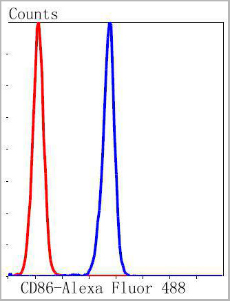
(Flow cytometric analysis of K562 cells with CD86 antibody at 1/50 dilution (blue) compared with an unlabelled control (cells without incubation with primary antibody; red). Alexa Fluor 488-conjugated goat anti rabbit IgG was used as the secondary antibody.)
FCM (Flow Cytometry)
(Flow cytometric analysis of K562 cells with CD86 antibody at 1/50 dilution (blue) compared with an unlabelled control (cells without incubation with primary antibody; red). Alexa Fluor 488-conjugated goat anti rabbit IgG was used as the secondary antibody.)
CD86, Monoclonal Antibody (Cat# AAA30048)
Full Name
CD86 Antibody
Gene Names
CD86; B70; B7-2; B7.2; LAB72; CD28LG2
Reactivity
Human, Rat
Applications
Western Blot, Immunocytochemistry, Immunofluorescence, Immunohistochemistry, Immunoprecipitation, Flow Cytometry
Purity
ProA affinity purified
Pricing


 IP (Immunoprecipitation)
(Immunoprecipitation analysis using Mouse Anti-VPS35 Monoclonal Antibody, Clone 10A8. Tissue: embryonic fibroblast. Species: Mouse. Primary Antibody: Mouse Anti-VPS35 Monoclonal Antibody. Three amounts of (3, 1 and 0.3 ug) were non-covalently coupled to 10uL of A/G sepharose beads for 1 hour at 4 degree C and next incubated with 250ug of MEF lysate for 2 hours at 4 degree C.)
IP (Immunoprecipitation)
(Immunoprecipitation analysis using Mouse Anti-VPS35 Monoclonal Antibody, Clone 10A8. Tissue: embryonic fibroblast. Species: Mouse. Primary Antibody: Mouse Anti-VPS35 Monoclonal Antibody. Three amounts of (3, 1 and 0.3 ug) were non-covalently coupled to 10uL of A/G sepharose beads for 1 hour at 4 degree C and next incubated with 250ug of MEF lysate for 2 hours at 4 degree C.)
VPS35, Monoclonal Antibody (Cat# AAA27693)
Full Name
VPS35 Antibody, Clone 10A8: ATTO 488
Gene Names
VPS35; MEM3; PARK17
Reactivity
Human, Mouse, Rat
Applications
Western Blot, Immunocytochemistry, Immunofluorescence, Immunoprecipitation
Purity
Protein G Purified
Pricing
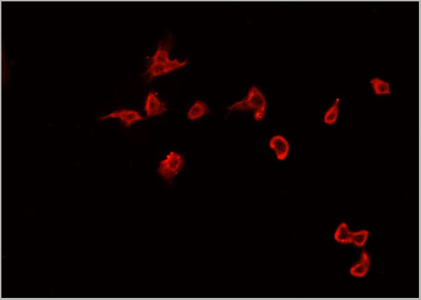
 IF (Immunofluorescence)
(AAA31032 staining HeLa by IF/ICC. The sample were fixed with PFA and permeabilized in 0.1% Triton X-100, then blocked in 10% serum for 45 minutes at 25 degree C. The primary antibody was diluted at 1/200 and incubated with the sample for 1 hour at 37 degree C. An Alexa Fluor 594 conjugated goat anti-rabbit IgG (H+L) Ab, diluted at 1/600, was used as the secondary antibody.)
IF (Immunofluorescence)
(AAA31032 staining HeLa by IF/ICC. The sample were fixed with PFA and permeabilized in 0.1% Triton X-100, then blocked in 10% serum for 45 minutes at 25 degree C. The primary antibody was diluted at 1/200 and incubated with the sample for 1 hour at 37 degree C. An Alexa Fluor 594 conjugated goat anti-rabbit IgG (H+L) Ab, diluted at 1/600, was used as the secondary antibody.)
CDC25A, Polyclonal Antibody (Cat# AAA31032)
Full Name
Phospho-CDC25A (Thr507) Antibody
Gene Names
CDC25A; CDC25A2
Reactivity
Human, Mouse, Rat
Applications
Western Blot, Immunohistochemistry, Immunofluorescence, Immunocytochemistry
Purity
From purified rabbit serum by affinity purification via sequential chromatography on phospho-and non-phospho-peptide affinity columns.
Pricing


 IP (Immunoprecipitation)
(Immunoprecipitation analysis using Mouse Anti-VPS35 Monoclonal Antibody, Clone 5A9. Tissue: embryonic fibroblast. Species: Mouse. Primary Antibody: Mouse Anti-VPS35 Monoclonal Antibody. Three amounts of (3, 1 and 0.3 ug) were non-covalently coupled to 10uL of A/G sepharose beads for 1 hour at 4 degree C and next incubated with 250ug of MEF lysate for 2 hours at 4 degree C.)
IP (Immunoprecipitation)
(Immunoprecipitation analysis using Mouse Anti-VPS35 Monoclonal Antibody, Clone 5A9. Tissue: embryonic fibroblast. Species: Mouse. Primary Antibody: Mouse Anti-VPS35 Monoclonal Antibody. Three amounts of (3, 1 and 0.3 ug) were non-covalently coupled to 10uL of A/G sepharose beads for 1 hour at 4 degree C and next incubated with 250ug of MEF lysate for 2 hours at 4 degree C.)
VPS35, Monoclonal Antibody (Cat# AAA27677)
Full Name
VPS35 Antibody, Clone 5A9: Biotin
Gene Names
VPS35; MEM3; PARK17
Reactivity
Human, Mouse, Rat
Applications
Western Blot, Immunocytochemistry, Immunofluorescence, Immunoprecipitation
Purity
Protein G Purified
Pricing
 IF (Immunofluorescence)
(AAA31029 staining HeLa by IF/ICC. The sample were fixed with PFA and permeabilized in 0.1% Triton X-100, then blocked in 10% serum for 45 minutes at 25 degree C. The primary antibody was diluted at 1/200 and incubated with the sample for 1 hour at 37 degree C. An Alexa Fluor 594 conjugated goat anti-rabbit IgG (H+L) Ab, diluted at 1/600, was used as the secondary antibody.)
IF (Immunofluorescence)
(AAA31029 staining HeLa by IF/ICC. The sample were fixed with PFA and permeabilized in 0.1% Triton X-100, then blocked in 10% serum for 45 minutes at 25 degree C. The primary antibody was diluted at 1/200 and incubated with the sample for 1 hour at 37 degree C. An Alexa Fluor 594 conjugated goat anti-rabbit IgG (H+L) Ab, diluted at 1/600, was used as the secondary antibody.)
CDC25A, Polyclonal Antibody (Cat# AAA31029)
Full Name
Phospho-CDC25A (Ser75) Antibody
Gene Names
CDC25A; CDC25A2
Reactivity
Human, Mouse, Rat
Applications
Western Blot, Immunohistochemistry, Immunofluorescence, Immunocytochemistry
Purity
From purified rabbit serum by affinity purification via sequential chromatography on phospho-and non-phospho-peptide affinity columns.
Pricing
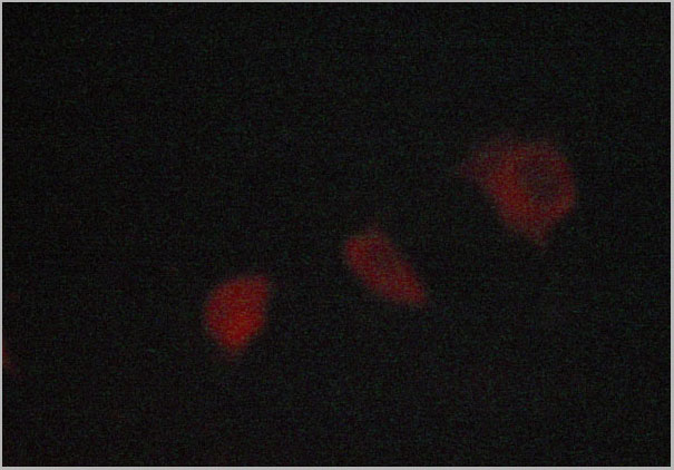
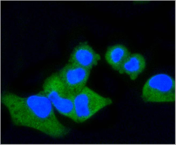
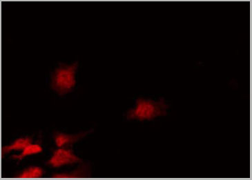
 IF (Immunofluorescence)
(AAA30937 staining A-431 cells by IF/ICC. The sample were fixed with PFA and permeabilized in 0.1% Triton X-100, then blocked in 10% serum for 45 minutes at 25 degree C. The primary antibody was diluted at 1/200 and incubated with the sample for 1 hour at 37 degree C. An Alexa Fluor 594 conjugated goat anti-rabbit IgG (H+L) antibody, diluted at 1/600, was used as secondary antibody.)
IF (Immunofluorescence)
(AAA30937 staining A-431 cells by IF/ICC. The sample were fixed with PFA and permeabilized in 0.1% Triton X-100, then blocked in 10% serum for 45 minutes at 25 degree C. The primary antibody was diluted at 1/200 and incubated with the sample for 1 hour at 37 degree C. An Alexa Fluor 594 conjugated goat anti-rabbit IgG (H+L) antibody, diluted at 1/600, was used as secondary antibody.)
SYK, Polyclonal Antibody (Cat# AAA30937)
Full Name
Phospho-SYK (Tyr323) Antibody
Gene Names
SYK; p72-Syk
Reactivity
Human, Mouse, Rat, Monkey
Applications
Western Blot, Immunohistochemistry, Immunofluorescence, Immunocytochemistry
Purity
From purified rabbit serum by affinity purification via sequential chromatography on phospho-and non-phospho-peptide affinity columns.
Pricing
 IF (Immunoflouorescence)
(AAA31056 staining NIH-3T3 by IF/ICC. The sample were fixed with PFA and permeabilized in 0.1% Triton X-100, then blocked in 10% serum for 45 minutes at 25 degree C. The primary antibody was diluted at 1/200 and incubated with the sample for 1 hour at 37 degree C. An Alexa Fluor 594 conjugated goat anti-rabbit IgG (H+L) Ab, diluted at 1/600, was used as the secondary antibody.)
IF (Immunoflouorescence)
(AAA31056 staining NIH-3T3 by IF/ICC. The sample were fixed with PFA and permeabilized in 0.1% Triton X-100, then blocked in 10% serum for 45 minutes at 25 degree C. The primary antibody was diluted at 1/200 and incubated with the sample for 1 hour at 37 degree C. An Alexa Fluor 594 conjugated goat anti-rabbit IgG (H+L) Ab, diluted at 1/600, was used as the secondary antibody.)
LIMK2, Polyclonal Antibody (Cat# AAA31056)
Full Name
Phospho-LIMK2 (Ser283) Antibody
Reactivity
Human, Mouse, Rat
Applications
Western Blot, Immunohistochemistry, Immunofluorescence, Immunocytochemistry
Purity
From purified rabbit serum by affinity purification via sequential chromatography on phospho-and non-phospho-peptide affinity columns.
Pricing
 IF (Immunofluorescence)
(AAA30936 staining HeLa by IF/ICC. The sample were fixed with PFA and permeabilized in 0.1% Triton X-100, then blocked in 10% serum for 45 minutes at 25 degree C. The primary antibody was diluted at 1/200 and incubated with the sample for 1 hour at 37 degree C. An Alexa Fluor 594 conjugated goat anti-rabbit IgG (H+L) Ab, diluted at 1/600, was used as the secondary antibody.)
IF (Immunofluorescence)
(AAA30936 staining HeLa by IF/ICC. The sample were fixed with PFA and permeabilized in 0.1% Triton X-100, then blocked in 10% serum for 45 minutes at 25 degree C. The primary antibody was diluted at 1/200 and incubated with the sample for 1 hour at 37 degree C. An Alexa Fluor 594 conjugated goat anti-rabbit IgG (H+L) Ab, diluted at 1/600, was used as the secondary antibody.)
NF kappaB p65, Polyclonal Antibody (Cat# AAA30936)
Full Name
Phospho-NF kappaB p65 (Ser536) Antibody
Gene Names
RELA; p65; NFKB3
Reactivity
Human, Mouse, Rat
Applications
Western Blot, Immunohistochemistry, Immunofluorescence, Immunocytochemistry, Immunoprecipitation
Purity
From purified rabbit serum by affinity purification via sequential chromatography on phospho-and non-phospho-peptide affinity columns.
Pricing
 WB (Western Blot)
(Western blot analysis of Beta-3 Adrenergic Receptor expression in HEK293T (A), Hela (B), HepG2 (C), A2780 (D), mouse kidney (E), rat liver (F) whole cell lysates.)
WB (Western Blot)
(Western blot analysis of Beta-3 Adrenergic Receptor expression in HEK293T (A), Hela (B), HepG2 (C), A2780 (D), mouse kidney (E), rat liver (F) whole cell lysates.)
Beta-3 Adrenergic Receptor, Polyclonal Antibody (Cat# AAA27792)
Full Name
Anti-Beta-3 Adrenergic Receptor Antibody
Gene Names
ADRB3; BETA3AR
Reactivity
Human, Mouse, Rat
Applications
Western Blot
Purity
The antibody was purified by immunogen affinity chromatography.
Pricing
 DB (Dot Blot)
(Dot blot analysis of anti-AKT3-pS472 Phospho-specific Pab on nitrocellulose membrane. 50ng of Phospho-peptide or Non Phospho-peptide per dot were adsorbed. Antibody working concentrations are 0.5ug per ml.)
DB (Dot Blot)
(Dot blot analysis of anti-AKT3-pS472 Phospho-specific Pab on nitrocellulose membrane. 50ng of Phospho-peptide or Non Phospho-peptide per dot were adsorbed. Antibody working concentrations are 0.5ug per ml.)
Phospho-AKT3 (S472), Polyclonal Antibody (Cat# AAA28777)
Full Name
Phospho-AKT3 (S472) Antibody
Gene Names
AKT3; MPPH; PKBG; MPPH2; PRKBG; STK-2; PKB-GAMMA; RAC-gamma; RAC-PK-gamma
Reactivity
Human
Predicted: Mouse, Rat.
Predicted: Mouse, Rat.
Applications
Dot Blot
Purity
Peptide Affinity Purified Rabbit Polyclonal Antibody (Pab)
Pricing


 Application Data
(Detection limit for recombinant GST tagged PGR is approximately 0.03ng/ml as a capture antibody.)
Application Data
(Detection limit for recombinant GST tagged PGR is approximately 0.03ng/ml as a capture antibody.)
PGR, Monoclonal Antibody (Cat# AAA25926)
Full Name
PGR (Progesterone Receptor, NR3C3, PR) (AP)
Gene Names
PGR; PR; NR3C3
Applications
Immunohistochemistry, Western Blot
Purity
Purified
Pricing
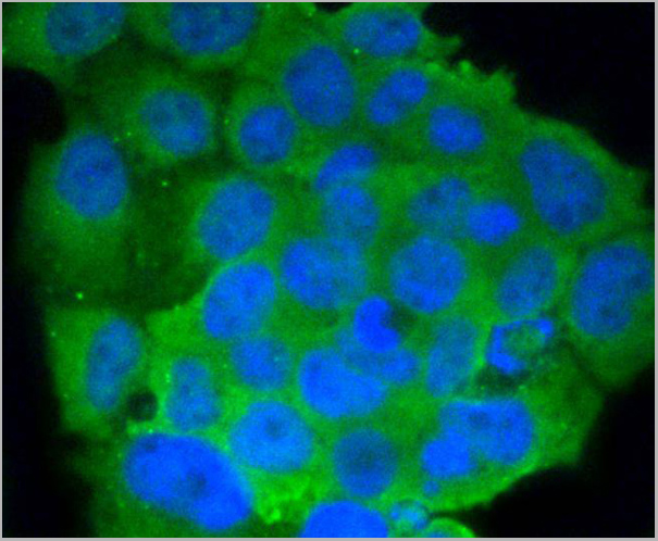
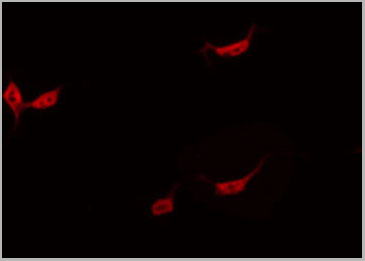
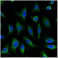 IF (Immunofluorescence)
(Immunofluorescent analysis of Aquaporin 4 staining in Hela cells. Formalin-fixed cells were permeabilized with 0.1% Triton X-100 in TBS for 5-10 minutes and blocked with 3% BSA-PBS for 30 minutes at room temperature. Cells were probed with the primary antibody in 3% BSA-PBS and incubated overnight at 4°C in a humidified chamber. Cells were washed with PBST and incubated with a FITC-conjugated secondary antibody (green) in PBS at room temperature in the dark. DAPI was used to stain the cell nuclei (blue).)
IF (Immunofluorescence)
(Immunofluorescent analysis of Aquaporin 4 staining in Hela cells. Formalin-fixed cells were permeabilized with 0.1% Triton X-100 in TBS for 5-10 minutes and blocked with 3% BSA-PBS for 30 minutes at room temperature. Cells were probed with the primary antibody in 3% BSA-PBS and incubated overnight at 4°C in a humidified chamber. Cells were washed with PBST and incubated with a FITC-conjugated secondary antibody (green) in PBS at room temperature in the dark. DAPI was used to stain the cell nuclei (blue).)
Aquaporin 4, Monoclonal Antibody (Cat# AAA27803)
Full Name
Anti-Aquaporin 4 Antibody
Gene Names
AQP4; MIWC; WCH4
Reactivity
Human, Mouse, Rat
Applications
Western Blot, Immunohistochemistry, Immunofluorescence, Immunocytochemistry
Pricing
GFAP, Antibody (Cat# AAA13651)
Full Name
Goat anti-GFAP Antibody
Gene Names
GFAP; ALXDRD
Reactivity
Tested: Human, Rat
Expected from sequence similarity: Human, Mouse, Rat, Dog
Expected from sequence similarity: Human, Mouse, Rat, Dog
Applications
Peptide ELISA, Western Blot
Purity
Purified from goat serum by ammonium sulphate precipitation followed by antigen affinity chromatography using the immunizing peptide.
Pricing
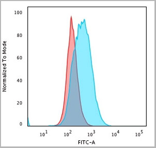 FCM (Flow Cytometry)
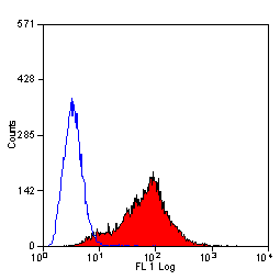
(Flow Cytometric Analysis of Human Raji cells using HLA-DP (MHC II) Monoclonal Antibody (SPM421) followed by Goat anti-Mouse IgG-CF488 (Blue); Isotype control (Red).)
FCM (Flow Cytometry)
(Flow Cytometric Analysis of Human Raji cells using HLA-DP (MHC II) Monoclonal Antibody (SPM421) followed by Goat anti-Mouse IgG-CF488 (Blue); Isotype control (Red).)
HLA-DP, Antibody (Cat# AAA13830)
Full Name
HLA-DP (MHC II) Mouse Monoclonal Antibody
Gene Names
HLA-DPB1; DPB1; HLA-DP; HLA-DPB; HLA-DP1B
Reactivity
Human. Others not known.
Applications
Flow Cytometry, Immunofluorescence
Pricing