Filters
Clonality
Type
Reactivity
Gene Name
Isotype
Host
Application
Clone
1280 results for " Membrane" - showing 1100-1150
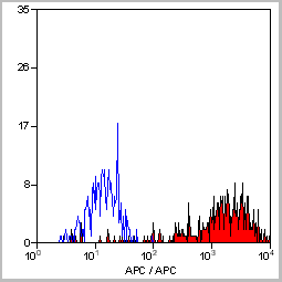
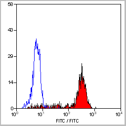
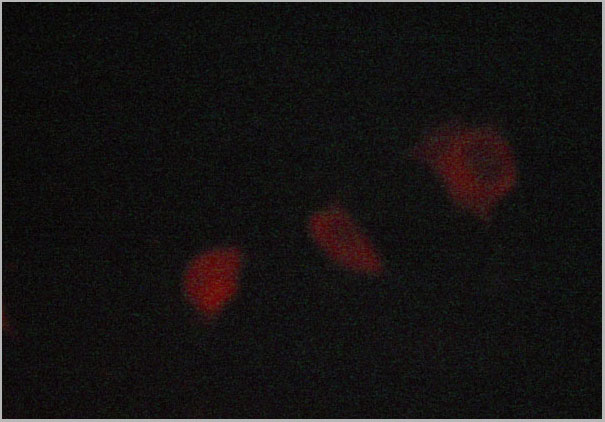
 Application Data
(Staining of human peripheral blood monocytes with Mouse anti Human CD14:RPE)
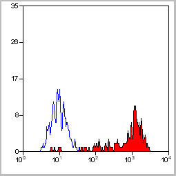
Application Data
(Staining of human peripheral blood monocytes with Mouse anti Human CD14:RPE)
CD14, Monoclonal Antibody (Cat# AAA11998)
Full Name
MOUSE ANTI HUMAN CD14
Applications
Immunohistochemistry, Flow Cytometry, Immunofluorescence, Immunoprecipitation
Pricing
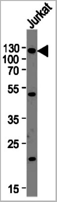
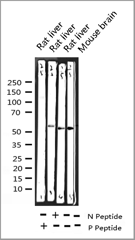
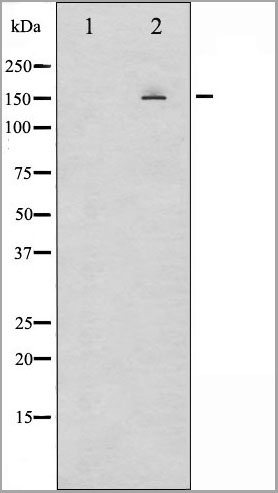
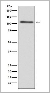
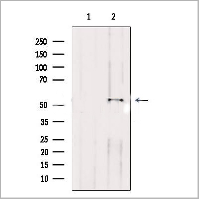
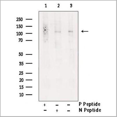 WB (Western Blot)
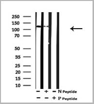
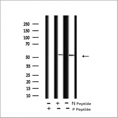
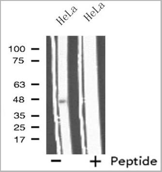
(Western blot analysis of extracts from K562 , using Phospho-IRE1 (Ser724) Antibody. Lane1 was treated with phospho-blocking peptide, Lane2 was treated with non-phospho-blocking peptide.)
WB (Western Blot)
(Western blot analysis of extracts from K562 , using Phospho-IRE1 (Ser724) Antibody. Lane1 was treated with phospho-blocking peptide, Lane2 was treated with non-phospho-blocking peptide.)
IRE1, Polyclonal Antibody (Cat# AAA31151)
Full Name
Phospho-IRE1 (Ser724) Antibody
Gene Names
ERN1; IRE1; IRE1P; IRE1a; hIRE1p
Reactivity
Human, Mouse, Rat
Applications
Western Blot, Immunohistochemistry, Immunofluorescence, Immunocytochemistry, Peptide ELISA
Purity
Peptide affinity purification
Pricing

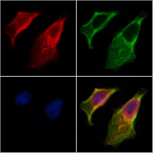
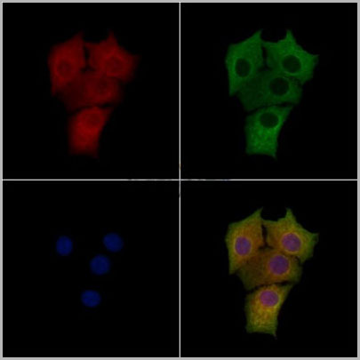
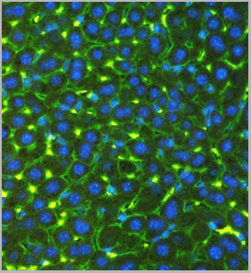
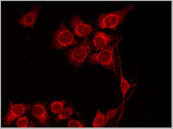
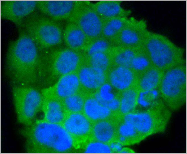
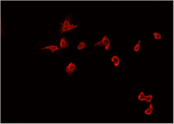
 IF (Immunofluorescence)
(AAA31198 staining Hela cells by IF/ICC. The samples were fixed with PFA and permeabilized in 0.1% Triton X-100,then blocked in 10% serum for 45 minutes at 25°C. Samples were then incubated with primary Ab(AAA31198 1:200) and mouse anti-beta tubulin Ab(T0023 1:200) for 1 hour at 37°C. An AlexaFluor594 conjugated goat anti-rabbit IgG(H+L) Ab(Red) and an AlexaFluor488 conjugated goat anti-mouse IgG(H+L) Ab(Green) were used as the secondary antibody.The nuclear counter stain is DAPI(blue).)
IF (Immunofluorescence)
(AAA31198 staining Hela cells by IF/ICC. The samples were fixed with PFA and permeabilized in 0.1% Triton X-100,then blocked in 10% serum for 45 minutes at 25°C. Samples were then incubated with primary Ab(AAA31198 1:200) and mouse anti-beta tubulin Ab(T0023 1:200) for 1 hour at 37°C. An AlexaFluor594 conjugated goat anti-rabbit IgG(H+L) Ab(Red) and an AlexaFluor488 conjugated goat anti-mouse IgG(H+L) Ab(Green) were used as the secondary antibody.The nuclear counter stain is DAPI(blue).)
SNAP23, Polyclonal Antibody (Cat# AAA31198)
Full Name
SNAP23 Antibody
Gene Names
SNAP23; SNAP-23; SNAP23A; SNAP23B; HsT17016
Reactivity
Human, Mouse, Rat
Predicted Reactivity: Pig(100%), Bovine(100%), Horse(100%), Sheep(100%), Rabbit(100%), Dog(100%), Chicken(83%)
Predicted Reactivity: Pig(100%), Bovine(100%), Horse(100%), Sheep(100%), Rabbit(100%), Dog(100%), Chicken(83%)
Applications
Western Blot, Immunofluorescence, Immunocytochemistry
Purity
The antiserum was purified by peptide affinity chromatography using SulfoLink Coupling Resin (Thermo Fisher Scientific).
Pricing
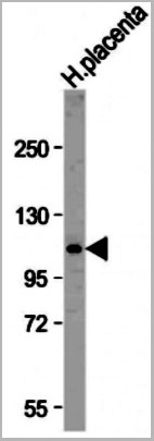
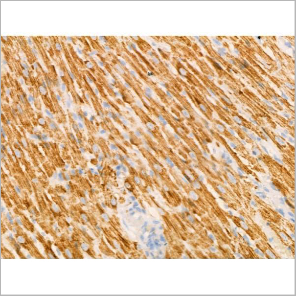
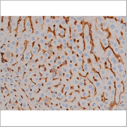
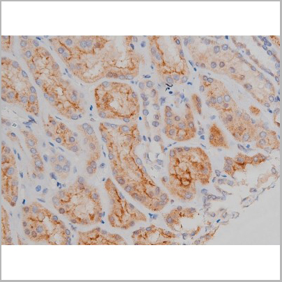
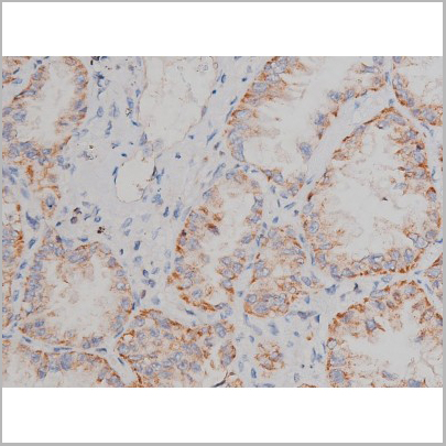
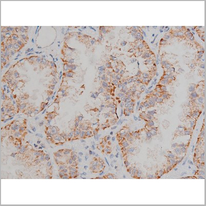
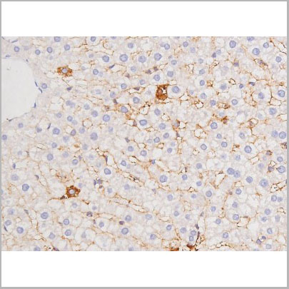
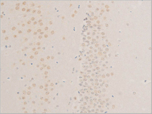
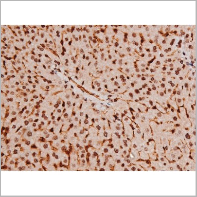
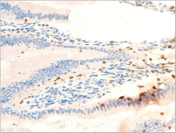
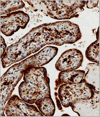 IHC (Immunohistchemistry)
(Immunohistochemical analysis of paraffin-embedded Human placenta tissue using AAA28695 performed on the Leica® BOND RXm. Tissue was fixed with formaldehyde at room temperature, antigen retrieval was by heat mediation with a EDTA buffer (pH9. 0). Samples were incubated with primary antibody(1:500) for 1 hours at room temperature. A undiluted biotinylated CRF Anti-Polyvalent HRP Polymer antibody was used as the secondary antibody.)
IHC (Immunohistchemistry)
(Immunohistochemical analysis of paraffin-embedded Human placenta tissue using AAA28695 performed on the Leica® BOND RXm. Tissue was fixed with formaldehyde at room temperature, antigen retrieval was by heat mediation with a EDTA buffer (pH9. 0). Samples were incubated with primary antibody(1:500) for 1 hours at room temperature. A undiluted biotinylated CRF Anti-Polyvalent HRP Polymer antibody was used as the secondary antibody.)
GAA, Polyclonal Antibody (Cat# AAA28695)
Full Name
GAA Antibody (N-term)
Gene Names
GAA; LYAG
Reactivity
Human
Applications
Western Blot, Immunohistochemistry
Purity
Peptide Affinity Purified Rabbit Polyclonal Antibody (Pab)
Pricing
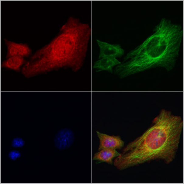
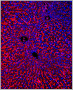
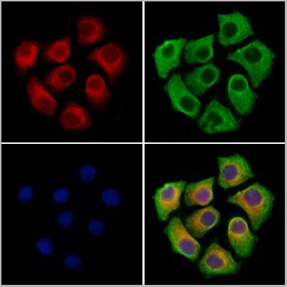
 IF (Immunofluorescence)
(AAA31103 staining Hela cells by IF/ICC. The samples were fixed with PFA and permeabilized in 0.1% Triton X-100,then blocked in 10% serum for 45 minutes at 25°C. Samples were then incubated with primary Ab(AAA31103) and mouse anti-beta tubulin Ab for 1 hour at 37°C. An AlexaFluor594 conjugated goat anti-rabbit IgG(H+L) Ab(Red) and an AlexaFluor488 conjugated goat anti-mouse IgG(H+L) Ab(Green) were used as the secondary antibody.The nuclear counter stain is DAPI (blue).)
IF (Immunofluorescence)
(AAA31103 staining Hela cells by IF/ICC. The samples were fixed with PFA and permeabilized in 0.1% Triton X-100,then blocked in 10% serum for 45 minutes at 25°C. Samples were then incubated with primary Ab(AAA31103) and mouse anti-beta tubulin Ab for 1 hour at 37°C. An AlexaFluor594 conjugated goat anti-rabbit IgG(H+L) Ab(Red) and an AlexaFluor488 conjugated goat anti-mouse IgG(H+L) Ab(Green) were used as the secondary antibody.The nuclear counter stain is DAPI (blue).)
CCL20, Polyclonal Antibody (Cat# AAA31103)
Full Name
CCL20 Antibody
Gene Names
CCL20; CKb4; LARC; ST38; MIP3A; Exodus; MIP-3a; SCYA20; MIP-3-alpha
Reactivity
Human, Mouse
Applications
Western Blot, Immunohistochemistry, Immunofluorescence, Immunocytochemistry
Purity
Purified by peptide affinity chromatography using SulfoLink Coupling Resin
Pricing
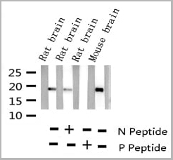
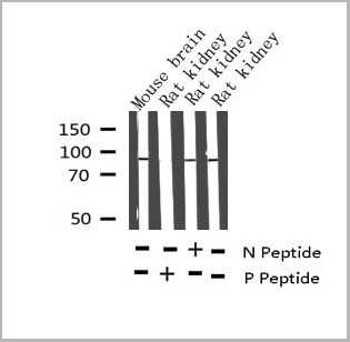
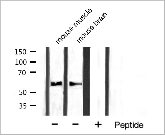
 WB (Western Blot)
(Western blot analysis of Synuclein phosphorylation expression in Mouse brain tissue lysates, The lane on the left is treated with the antigen-specific peptide.)
WB (Western Blot)
(Western blot analysis of Synuclein phosphorylation expression in Mouse brain tissue lysates, The lane on the left is treated with the antigen-specific peptide.)
Synuclein, Polyclonal Antibody (Cat# AAA31040)
Full Name
Phospho-Synuclein (Ser129) Antibody
Gene Names
SNCA; PD1; NACP; PARK1; PARK4
Reactivity
Human, Mouse, Rat
Applications
Western Blot, Immunohistochemistry
Purity
From purified rabbit serum by affinity purification via sequential chromatography on phospho-and non-phospho-peptide affinity columns.
Pricing
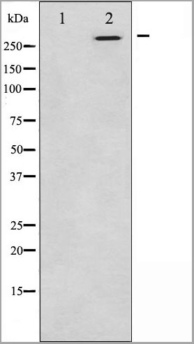
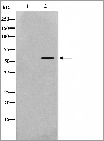
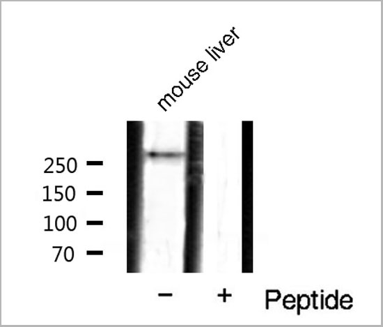 WB (Western Blot)
(Western blot analysis of mTOR phosphorylation expression in mouse liver tissue lysates, The lane on the right is treated with the antigen-specific peptide.)
WB (Western Blot)
(Western blot analysis of mTOR phosphorylation expression in mouse liver tissue lysates, The lane on the right is treated with the antigen-specific peptide.)
mTOR, Polyclonal Antibody (Cat# AAA31048)
Full Name
Phospho-mTOR (Ser2448) Antibody
Gene Names
MTOR; SKS; FRAP; FRAP1; FRAP2; RAFT1; RAPT1
Reactivity
Human, Mouse, Rat, Fish
Applications
Western Blot, Immunohistochemistry
Purity
From purified rabbit serum by affinity purification via sequential chromatography on phospho-and non-phospho-peptide affinity columns.
Pricing

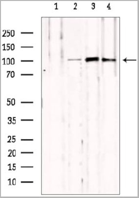
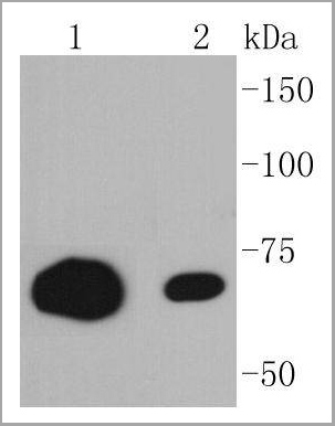
 WB (Western Blot)
(Western blot analysis of Phospho-Tau (Thr212) Antibody expression in mouse brain and rat kidney tissues lysates.)
WB (Western Blot)
(Western blot analysis of Phospho-Tau (Thr212) Antibody expression in mouse brain and rat kidney tissues lysates.)
Tau, Polyclonal Antibody (Cat# AAA31001)
Full Name
Phospho-Tau (Thr212) Antibody
Gene Names
MAPT; TAU; MSTD; PPND; DDPAC; MAPTL; MTBT1; MTBT2; FTDP-17; PPP1R103
Reactivity
Human, Mouse, Rat
Applications
Western Blot, Immunohistochemistry
Purity
From purified rabbit serum by affinity purification via sequential chromatography on phospho-and non-phospho-peptide affinity columns.
Pricing
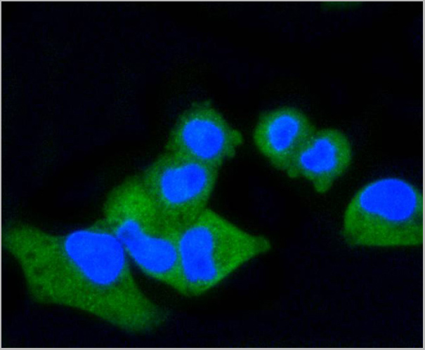
 IF (Immunofluorescence)
(AAA31099 staining A549 cells by IF/ICC. The samples were fixed with PFA and permeabilized in 0.1% Triton X-100,then blocked in 10% serum for 45 minutes at 25°C. Samples were then incubated with primary Ab(AAA31099) and mouse antibeta tubulin Ab for 1 hour at 37°C. An AlexaFluor594 conjugated goat anti-rabbit IgG(H+L) Ab(Red) and an AlexaFluor488 conjugated goat anti-mouse IgG(H+L) Ab(Green) were used as the secondary antibody. The nuclear counter stain is DAPI (blue))
IF (Immunofluorescence)
(AAA31099 staining A549 cells by IF/ICC. The samples were fixed with PFA and permeabilized in 0.1% Triton X-100,then blocked in 10% serum for 45 minutes at 25°C. Samples were then incubated with primary Ab(AAA31099) and mouse antibeta tubulin Ab for 1 hour at 37°C. An AlexaFluor594 conjugated goat anti-rabbit IgG(H+L) Ab(Red) and an AlexaFluor488 conjugated goat anti-mouse IgG(H+L) Ab(Green) were used as the secondary antibody. The nuclear counter stain is DAPI (blue))
NOD2, Polyclonal Antibody (Cat# AAA31099)
Full Name
NOD2 Antibody
Gene Names
NOD2; CD; ACUG; BLAU; IBD1; YAOS; BLAUS; NLRC2; NOD2B; CARD15; CLR16.3; PSORAS1
Reactivity
Human, Mouse, Rat
Applications
Western Blot, Immunohistochemistry, Immunofluorescence, Immunocytochemistry
Purity
Peptide affinity chromatography using SulfoLink Coupling Resin (Thermo Fisher Scientific)
Pricing
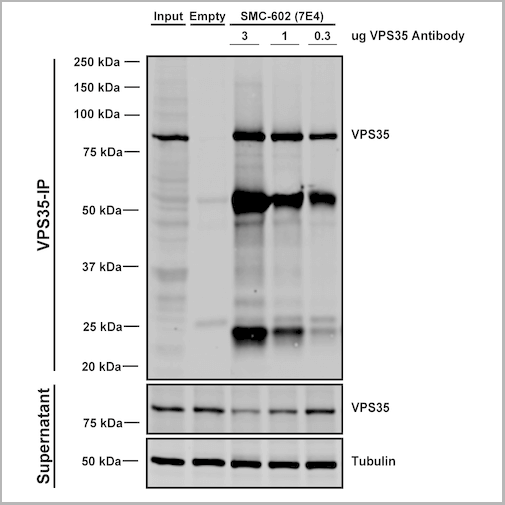
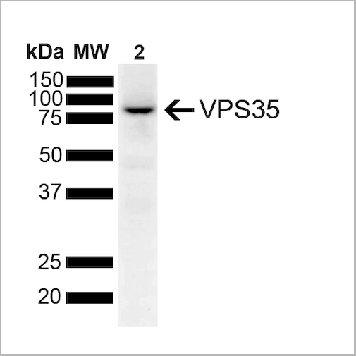
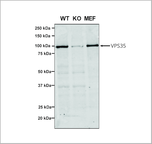
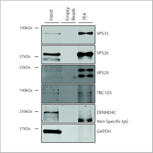
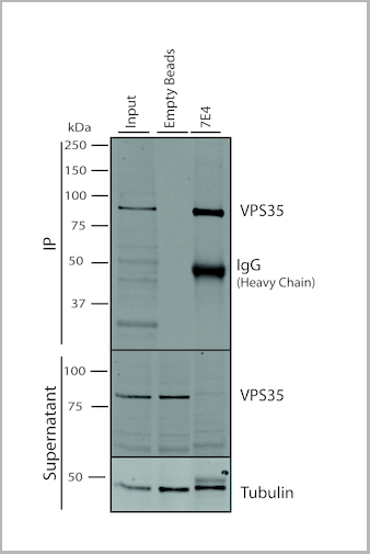
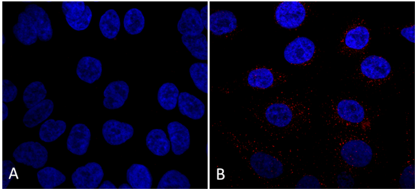
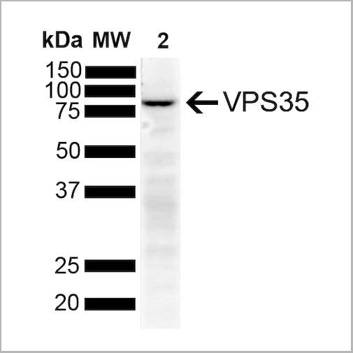
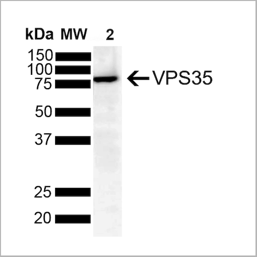
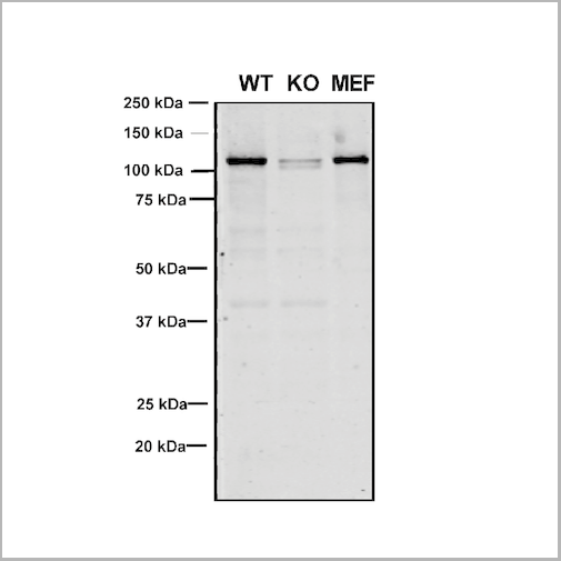
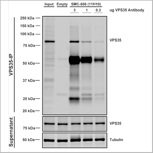
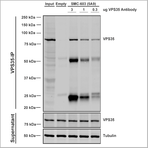
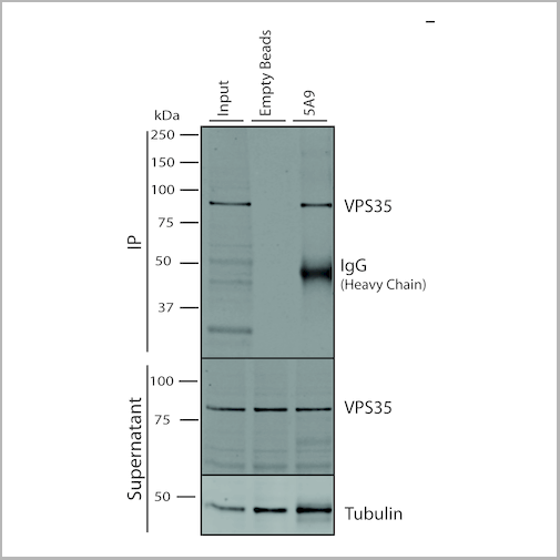
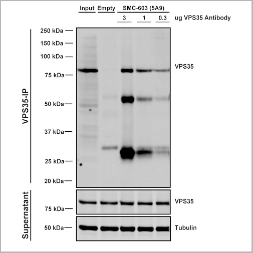
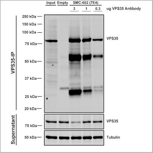 IP (Immunoprecipitation)
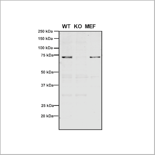
(Immunoprecipitation analysis using Mouse Anti-VPS35 Monoclonal Antibody, Clone 7E4. Tissue: embryonic fibroblast. Species: Mouse. Primary Antibody: Mouse Anti-VPS35 Monoclonal Antibody. Three amounts (3, 1 and 0.3 ug) were non-covalently coupled to 10uL of A/G sepharose beads for 1 hour at 4 degree C and next incubated with 250ug of MEF lysate for 2 hours at 4 degree C.)
IP (Immunoprecipitation)
(Immunoprecipitation analysis using Mouse Anti-VPS35 Monoclonal Antibody, Clone 7E4. Tissue: embryonic fibroblast. Species: Mouse. Primary Antibody: Mouse Anti-VPS35 Monoclonal Antibody. Three amounts (3, 1 and 0.3 ug) were non-covalently coupled to 10uL of A/G sepharose beads for 1 hour at 4 degree C and next incubated with 250ug of MEF lysate for 2 hours at 4 degree C.)
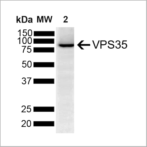
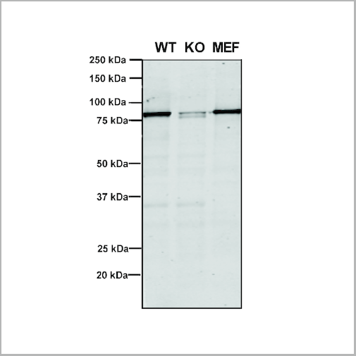
VPS35, Monoclonal Antibody (Cat# AAA27671)
Full Name
VPS35 Antibody, Clone 7E4: PerCP
Gene Names
VPS35; MEM3; PARK17
Reactivity
Human, Mouse, Rat
Applications
Western Blot, Immunocytochemistry, Immunofluorescence, Immunoprecipitation
Purity
Protein G Purified
Pricing
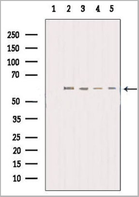
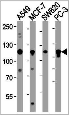
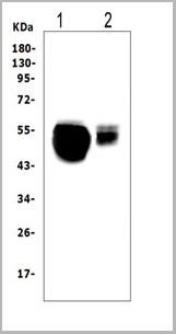
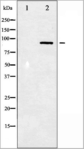
 WB (Western Blot)
(Western blot analysis of extracts of various tissue sample, using Src Antibody.)
WB (Western Blot)
(Western blot analysis of extracts of various tissue sample, using Src Antibody.)
Src, Polyclonal Antibody (Cat# AAA31086)
Full Name
Src Antibody
Gene Names
SRC; ASV; SRC1; THC6; c-SRC; p60-Src
Reactivity
Human, Mouse, Rat, Monkey
Applications
Western Blot, Immunohistochemistry, Immunofluorescence, Immunocytochemistry
Purity
Peptide affinity purification
Pricing
 IF (Immunofluorescence)
(AAA31237 staining Hela cells by IF/ICC. The samples were fixed with PFA and permeabilized in 0.1% Triton X-100,then blocked in 10% serum for 45 minutes at 25°C. Samples were then incubated with primary Ab(AAA31237 1:200) and mouse anti-beta tubulin Ab(T0023 1:200) for 1 hour at 37°C. An AlexaFluor594 conjugated goat anti-rabbit IgG(H+L) Ab(Red) and an AlexaFluor488 conjugated goat anti-mouse IgG(H+L) Ab(Green) were used as the secondary antibody.The nuclear counter stain is DAPI(blue).)
IF (Immunofluorescence)
(AAA31237 staining Hela cells by IF/ICC. The samples were fixed with PFA and permeabilized in 0.1% Triton X-100,then blocked in 10% serum for 45 minutes at 25°C. Samples were then incubated with primary Ab(AAA31237 1:200) and mouse anti-beta tubulin Ab(T0023 1:200) for 1 hour at 37°C. An AlexaFluor594 conjugated goat anti-rabbit IgG(H+L) Ab(Red) and an AlexaFluor488 conjugated goat anti-mouse IgG(H+L) Ab(Green) were used as the secondary antibody.The nuclear counter stain is DAPI(blue).)
DBT, Polyclonal Antibody (Cat# AAA31237)
Full Name
DBT Antibody
Gene Names
DBT; E2; E2B; BCATE2; BCKADE2; BCKAD-E2; BCOADC-E2
Reactivity
Human,Mouse,Ra
Applications
Western Blot, Immunofluorescence, Immunocytochemistry, Immunohistochemistry
Purity
Peptide affinity purification
Pricing
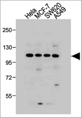
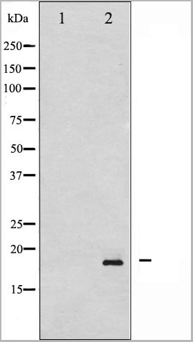
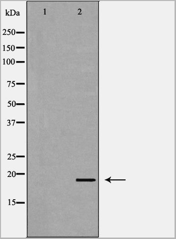
 WB (Western Blot)
(Western blot analysis of SGK phosphorylation expression in Insulin treated HeLa whole cell lysates, The lane on the left is treated with the antigen-specific peptide.)
WB (Western Blot)
(Western blot analysis of SGK phosphorylation expression in Insulin treated HeLa whole cell lysates, The lane on the left is treated with the antigen-specific peptide.)
SGK, Polyclonal Antibody (Cat# AAA30942)
Full Name
Phospho-SGK (Ser422) Antibody
Gene Names
SGK1; SGK
Reactivity
Human, Mouse, Rat
Applications
Western Blot, Immunohistochemistry, Immunofluorescence, Immunocytochemistry
Purity
From purified rabbit serum by affinity purification via sequential chromatography on phospho-and non-phospho-peptide affinity columns.
Pricing

 WB (Western Blot)
(Western blot analysis of EGFR phosphorylation expression in TSA treated HeLa whole cell lysates, The lane on the left is treated with the antigen-specific peptide.)
WB (Western Blot)
(Western blot analysis of EGFR phosphorylation expression in TSA treated HeLa whole cell lysates, The lane on the left is treated with the antigen-specific peptide.)
EGFR, Polyclonal Antibody (Cat# AAA30963)
Full Name
Phospho-EGFR (Ser1026) Antibody
Gene Names
EGFR; ERBB; HER1; mENA; ERBB1; PIG61; NISBD2
Reactivity
Human, Mouse, Rat
Applications
Western Blot, Immunohistochemistry, Immunofluorescence, Immunocytochemistry
Purity
From purified rabbit serum by affinity purification via sequential chromatography on phospho-and non-phospho-peptide affinity columns.
Pricing
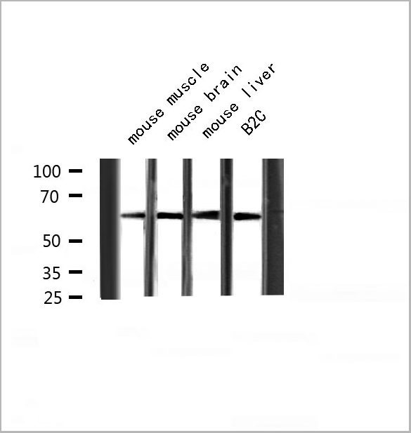
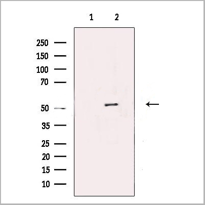
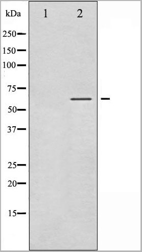
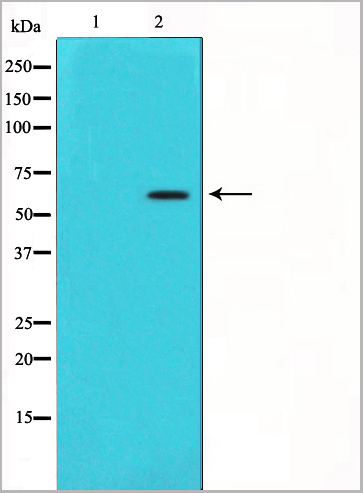
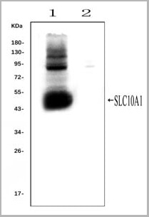 WB (Western Blot)
(Western blot analysis of SLC10A1 using anti- SLC10A1 antibody (AAA11653).Electrophoresis was performed on a 5-20% SDS-PAGE gel at 70V (Stacking gel) / 90V (Resolving gel) for 2-3 hours. The sample well of each lane was loaded with 50ug of sample under reducing conditions.Lane 1: rat liver tissue lysates (positive control), Lane 2: rat kidney tissue lysates, (negative control)After Electrophoresis, proteins were transferred to a Nitrocellulose membrane at 150mA for 50-90 minutes. Blocked the membrane with 5% Non-fat Milk/ TBS for 1.5 hour at RT. The membrane was incubated with rabbit anti-SLC10A1 antigen affinity purified polyclonal antibody (AAA11653) at 0.25ug/mL overnight at 4°C, then washed with TBS-0.1%Tween 3 times with 5 minutes each and probed with a goat anti-rabbit IgG-HRP secondary antibody at a dilution of 1:10000 for 1.5 hour at RT. The signal is developed using an Enhanced Chemiluminescent detection (ECL) kit with Tanon 5200 system. A specific band was detected for SLC10A1 at approximately 50KD. The expected band size for SLC10A1 is at 38KD.)
WB (Western Blot)
(Western blot analysis of SLC10A1 using anti- SLC10A1 antibody (AAA11653).Electrophoresis was performed on a 5-20% SDS-PAGE gel at 70V (Stacking gel) / 90V (Resolving gel) for 2-3 hours. The sample well of each lane was loaded with 50ug of sample under reducing conditions.Lane 1: rat liver tissue lysates (positive control), Lane 2: rat kidney tissue lysates, (negative control)After Electrophoresis, proteins were transferred to a Nitrocellulose membrane at 150mA for 50-90 minutes. Blocked the membrane with 5% Non-fat Milk/ TBS for 1.5 hour at RT. The membrane was incubated with rabbit anti-SLC10A1 antigen affinity purified polyclonal antibody (AAA11653) at 0.25ug/mL overnight at 4°C, then washed with TBS-0.1%Tween 3 times with 5 minutes each and probed with a goat anti-rabbit IgG-HRP secondary antibody at a dilution of 1:10000 for 1.5 hour at RT. The signal is developed using an Enhanced Chemiluminescent detection (ECL) kit with Tanon 5200 system. A specific band was detected for SLC10A1 at approximately 50KD. The expected band size for SLC10A1 is at 38KD.)
SLC10A1, Polyclonal Antibody (Cat# AAA11653)
Full Name
Anti-SLC10A1 Antibody
Gene Names
Slc10a1; Ntcp
Reactivity
Mouse, Rat
Applications
Flow Cytometry, Immunofluorescence, Immunohistochemistry, Immunohistochemistry, Immunocytochemistry, Western Blot
Purity
Immunogen affinity purified.
Pricing

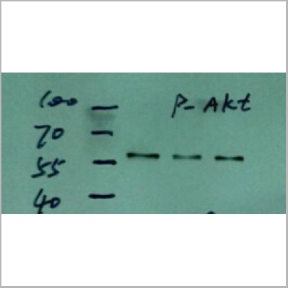
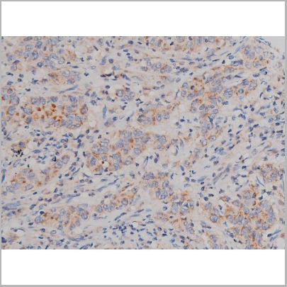
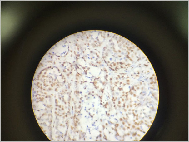
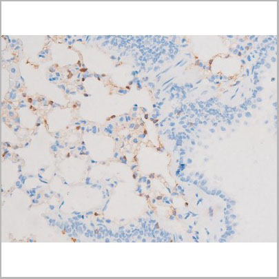
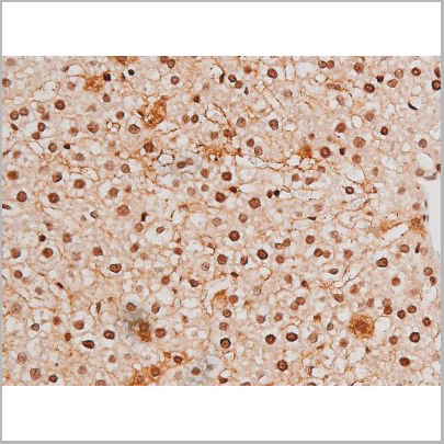
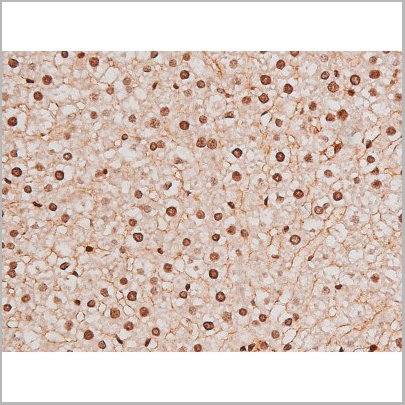
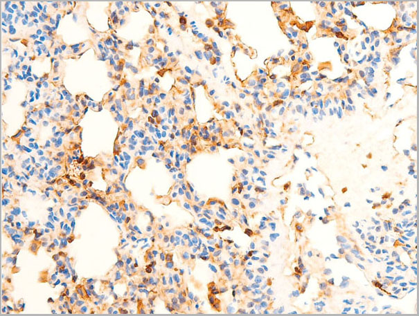
 IHC (Immunohistochemistry)
(AAA30920 at 1/200 staining human lung cancer tissue sections by IHC-P. The tissue was formaldehyde fixed and a heat mediated antigen retrieval step in citrate buffer was performed. The tissue was then blocked and incubated with the antibody for 1.5 hours at 22 degree C. An HRP conjugated goat anti-rabbit antibody was used as the secondary.)
IHC (Immunohistochemistry)
(AAA30920 at 1/200 staining human lung cancer tissue sections by IHC-P. The tissue was formaldehyde fixed and a heat mediated antigen retrieval step in citrate buffer was performed. The tissue was then blocked and incubated with the antibody for 1.5 hours at 22 degree C. An HRP conjugated goat anti-rabbit antibody was used as the secondary.)
Akt, Polyclonal Antibody (Cat# AAA30920)
Full Name
Phospho-Akt(Ser473) Antibody
Gene Names
AKT1; AKT; PKB; RAC; CWS6; PRKBA; PKB-ALPHA; RAC-ALPHA
Reactivity
Human, Mouse, Rat
Applications
Western Blot, Immunohistochemistry, Immunofluorescence, Immunocytochemistry
Purity
From purified rabbit serum by affinity purification via sequential chromatography on phospho-and non-phospho-peptide affinity columns.
Pricing


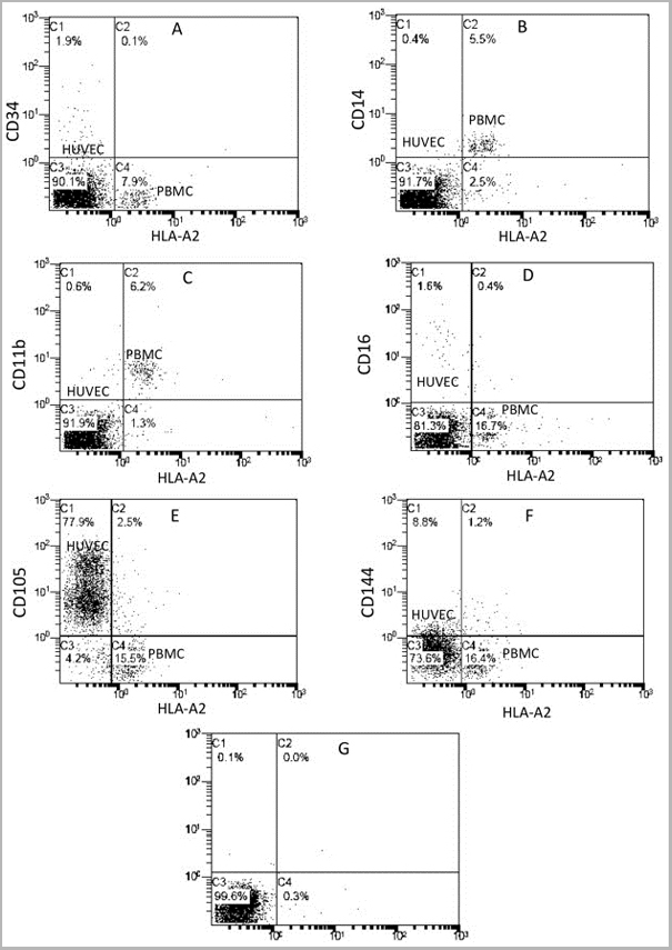
 Application Data
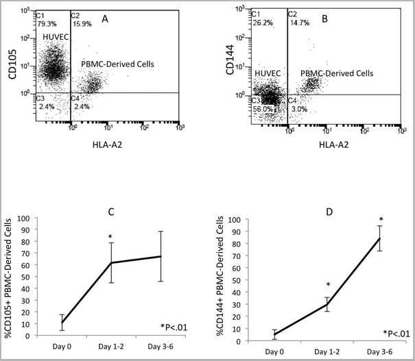
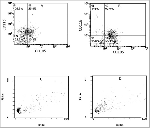
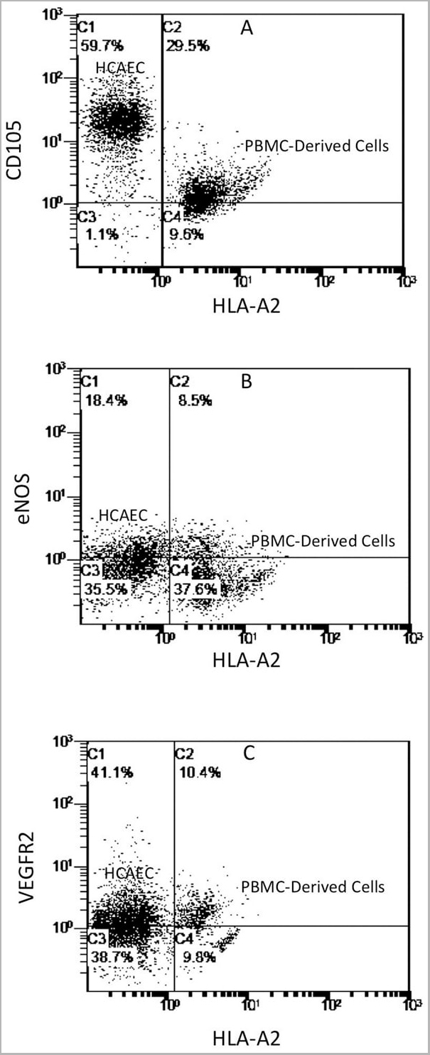
(Published customer image: Mouse anti Human CD105 antibody, clone SN6 used for flow cytometry.Image caption:Phenotype change from HLA-A2+/CD11b+/CD105- to HLA-A2+/CD11b-/CD105+ on endothelium-adherent blood monocyte-derived cells with increase in size and granularity during co-culture. HLA-A2+ PBMCs (1x10⁶ cells/well) were incubated for 2 h (Day 0) with HLA-A2- HUVECs, after which the non-adherent cells were removed by washing. The cell layers were analysed by three-colour flow cytometry staining for HLA-A2, CD11b and CD105 on (A) Day 1 and (B) Day 2. These plots were gated for HLA-A2+ cells. Forward scatter/side scatter dot plots gated for HLA-A2+ cells on Day 0 (C) and Day 2 (D) was shown. These are representative of 2 individual experiments.From: Tso C, Rye K-A, Barter P (2012) Phenotypic and Functional Changes in Blood Monocytes Following Adherence to Endothelium. PLoS ONE 7(5): e37091.)
Application Data
(Published customer image: Mouse anti Human CD105 antibody, clone SN6 used for flow cytometry.Image caption:Phenotype change from HLA-A2+/CD11b+/CD105- to HLA-A2+/CD11b-/CD105+ on endothelium-adherent blood monocyte-derived cells with increase in size and granularity during co-culture. HLA-A2+ PBMCs (1x10⁶ cells/well) were incubated for 2 h (Day 0) with HLA-A2- HUVECs, after which the non-adherent cells were removed by washing. The cell layers were analysed by three-colour flow cytometry staining for HLA-A2, CD11b and CD105 on (A) Day 1 and (B) Day 2. These plots were gated for HLA-A2+ cells. Forward scatter/side scatter dot plots gated for HLA-A2+ cells on Day 0 (C) and Day 2 (D) was shown. These are representative of 2 individual experiments.From: Tso C, Rye K-A, Barter P (2012) Phenotypic and Functional Changes in Blood Monocytes Following Adherence to Endothelium. PLoS ONE 7(5): e37091.)
CD105, Monoclonal Antibody (Cat# AAA12085)
Full Name
MOUSE ANTI HUMAN CD105
Gene Names
ENG; END; HHT1; ORW1
Applications
Immunohistochemistry, Flow Cytometry, Immunoprecipitation, Western Blot
Pricing
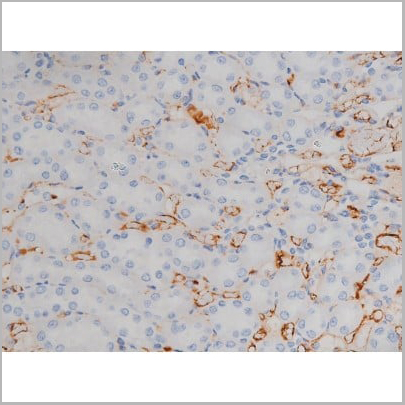
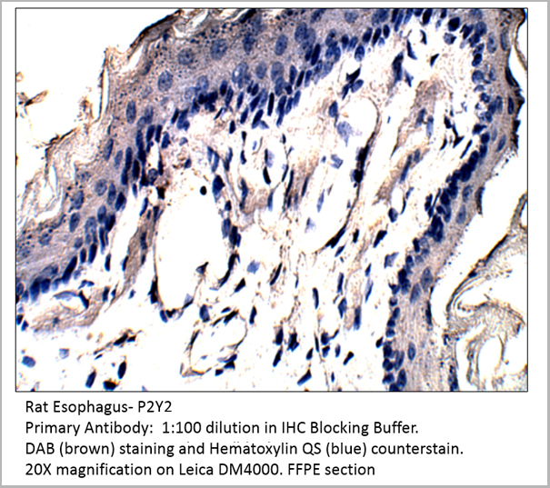 IHC (Immunohistochemistry)
P2Y2, Polyclonal Antibody (Cat# AAA14402)
Full Name
P2Y2 Antibody
Gene Names
P2RY2; P2U; HP2U; P2U1; P2UR; P2Y2; P2RU1; P2Y2R
Reactivity
Cat, Human, Mouse, Rat
Applications
Confocal Microscopy, Immunocytochemistry, Immunofluorescence, Immunohistochemistry, Immunoprecipitation, Western Blot
Pricing

 Assay Principle
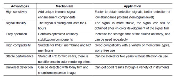
(After years of development on chemiluminescence technology, there have been a variety of luminescence systems, but the most commonly used one in laboratories is still a technology system based on Luminol or its derivatives (isoluminol, etc.). Western luminescent detection reagent ECL immunoblotting chemiluminescence solution is a non-radioactive (horseradish peroxidase) luminescence system designed to detect femtogram-level trace proteins immobilized on a solid membrane (such as NC, PVDF, etc.). It is an experimental auxiliary reagent for the photosensitive recording of its immunoblotting by X-ray film (radiograph).)
Assay Principle
(After years of development on chemiluminescence technology, there have been a variety of luminescence systems, but the most commonly used one in laboratories is still a technology system based on Luminol or its derivatives (isoluminol, etc.). Western luminescent detection reagent ECL immunoblotting chemiluminescence solution is a non-radioactive (horseradish peroxidase) luminescence system designed to detect femtogram-level trace proteins immobilized on a solid membrane (such as NC, PVDF, etc.). It is an experimental auxiliary reagent for the photosensitive recording of its immunoblotting by X-ray film (radiograph).)
West Femto Maximum Sensitivity Substrate, Reagent (Cat# AAA31545)
Full Name
West Femto Maximum Sensitivity Substrate
Applications
Western Blot, Immunoprecipitation
Pricing
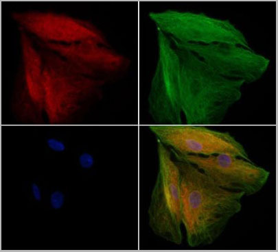
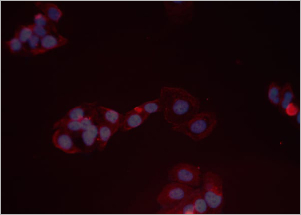
 IF (Immunofluorescence)
(AAA31114 staining A549 cells by IF/ICC. The samples were fixed with PFA and permeabilized in 0.1% Triton X-100,then blocked in 10% serum for 45 minutes at 25°C. Samples were then incubated with primary Ab(AAA31114) and mouse anti-beta tubulin Ab for 1 hour at 37°C. An AlexaFluor594 conjugated goat anti-rabbit IgG(H+L) Ab(Red) and an AlexaFluor488 conjugated goat anti-mouse IgG(H+L) Ab(Green) were used as the secondary antibody. The nuclear counter stain is DAPI (blue).)
IF (Immunofluorescence)
(AAA31114 staining A549 cells by IF/ICC. The samples were fixed with PFA and permeabilized in 0.1% Triton X-100,then blocked in 10% serum for 45 minutes at 25°C. Samples were then incubated with primary Ab(AAA31114) and mouse anti-beta tubulin Ab for 1 hour at 37°C. An AlexaFluor594 conjugated goat anti-rabbit IgG(H+L) Ab(Red) and an AlexaFluor488 conjugated goat anti-mouse IgG(H+L) Ab(Green) were used as the secondary antibody. The nuclear counter stain is DAPI (blue).)
ARX, Polyclonal Antibody (Cat# AAA31114)
Full Name
ARX Antibody
Gene Names
ARX; ISSX; PRTS; CT121; EIEE1; MRX29; MRX32; MRX33; MRX36; MRX38; MRX43; MRX54; MRX76; MRX87; MRXS1
Reactivity
Human, Mouse, Rat
Applications
Western Blot, Immunofluorescence, Immunocytochemistry
Purity
Peptide affinity chromatography using SulfoLink Coupling Resin (Thermo Fisher Scientific)
Pricing
 WB (Western Blot)
(Western blot analysis of C3 expression in HepG2 cell lysate (AAA11693). Electrophoresis was performed on a 5-20% SDS-PAGE gel at 70V (stacking gel) / 90V (resolving gel) for 2-3 hours. The sample well of each lane was loaded with 50ug of sample under reducing conditions. After Electrophoresis, proteins were transferred to a Nitrocellulose membrane at 150mA for 50-90 minutes. Blocked the membrane with 5% Non-fat Milk/TBS for 1.5 hour at RT. The membrane was incubated with rabbit anti-C3 monoclonal antibody (AAA11693) overnight at 4°C, then washed with TBS-0.1%Tween 3 times with 5 minutes each and probed with goat anti-rabbit IgG-HRP secondary antibody at a dilution of 1:10000 for 1.5 hour at RT. The signal is developed using an Enhanced Chemiluminescent detection (ECL) kit (please inquire) with Tanon 5200 system. A specific band was detected for C3.)
WB (Western Blot)
(Western blot analysis of C3 expression in HepG2 cell lysate (AAA11693). Electrophoresis was performed on a 5-20% SDS-PAGE gel at 70V (stacking gel) / 90V (resolving gel) for 2-3 hours. The sample well of each lane was loaded with 50ug of sample under reducing conditions. After Electrophoresis, proteins were transferred to a Nitrocellulose membrane at 150mA for 50-90 minutes. Blocked the membrane with 5% Non-fat Milk/TBS for 1.5 hour at RT. The membrane was incubated with rabbit anti-C3 monoclonal antibody (AAA11693) overnight at 4°C, then washed with TBS-0.1%Tween 3 times with 5 minutes each and probed with goat anti-rabbit IgG-HRP secondary antibody at a dilution of 1:10000 for 1.5 hour at RT. The signal is developed using an Enhanced Chemiluminescent detection (ECL) kit (please inquire) with Tanon 5200 system. A specific band was detected for C3.)
C3/Complement C3, Monoclonal Antibody (Cat# AAA11693)
Full Name
Anti-C3/Complement C3 Rabbit Monoclonal Antibody
Gene Names
C3; ASP; C3a; C3b; AHUS5; ARMD9; CPAMD1; HEL-S-62p
Reactivity
Human
Applications
Flow Cytometry, Immunofluorescence, Immunocytochemistry, Western Blot
Purity
Affinity-chromatography
Pricing
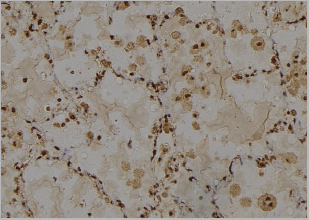
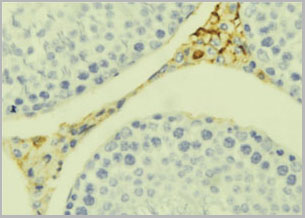
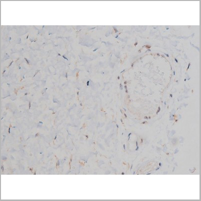
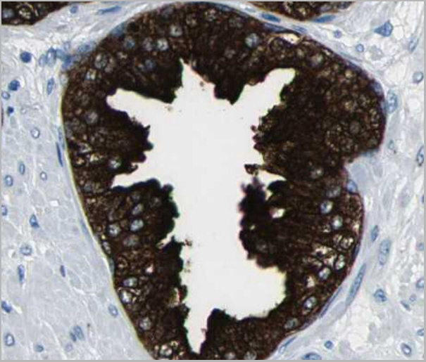
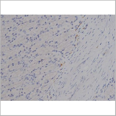
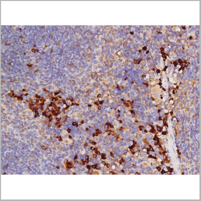
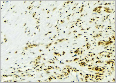
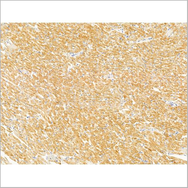
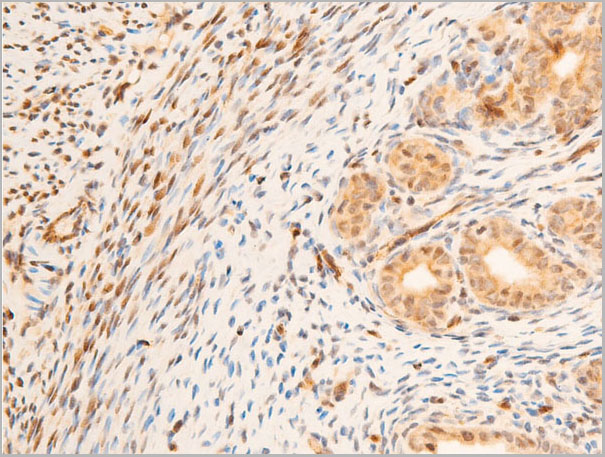
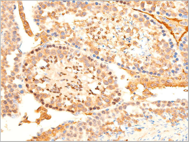
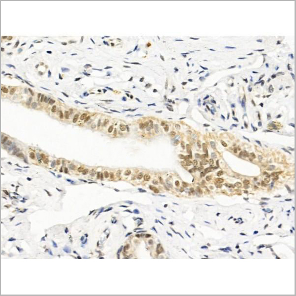
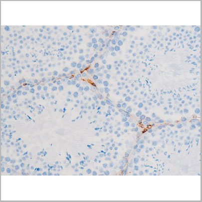
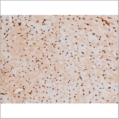
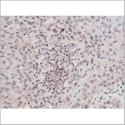
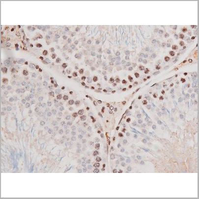
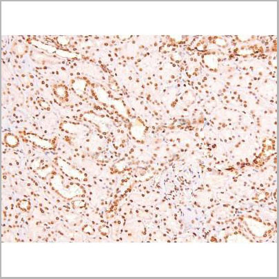
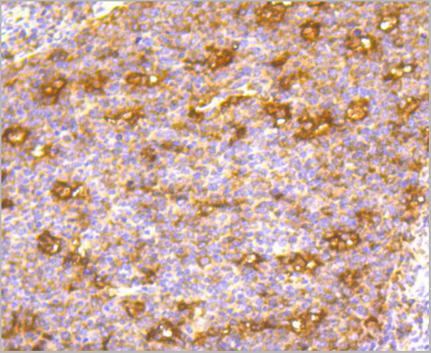
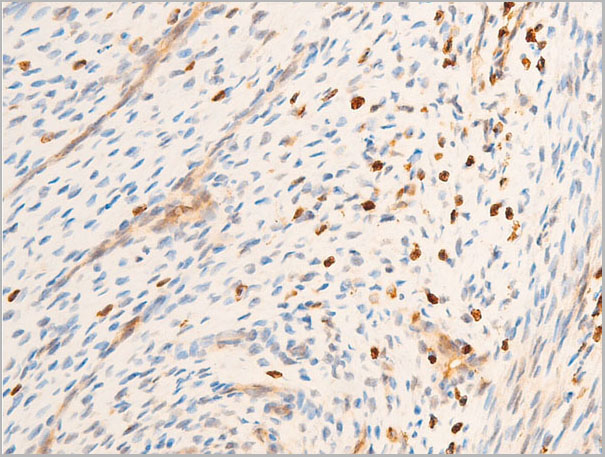
 IHC (Immunohistochemistry)
(AAA31129 at 1/100 staining Rat ovary tissue by IHC-P. The sample was formaldehyde fixed and a heat mediated antigen retrieval step in citrate buffer was performed. The sample was then blocked and incubated with the primary antibody at 4°C overnight. An HRP conjugated anti-Rabbit antibody was used as the secondary antibody.)
IHC (Immunohistochemistry)
(AAA31129 at 1/100 staining Rat ovary tissue by IHC-P. The sample was formaldehyde fixed and a heat mediated antigen retrieval step in citrate buffer was performed. The sample was then blocked and incubated with the primary antibody at 4°C overnight. An HRP conjugated anti-Rabbit antibody was used as the secondary antibody.)
IL10, Polyclonal Antibody (Cat# AAA31129)
Full Name
IL10 Antibody
Gene Names
IL10; CSIF; TGIF; GVHDS; IL-10; IL10A
Reactivity
Human, Mouse, Rat
Applications
Western Blot, Immunohistochemistry, Immunofluorescence, Immunocytochemistry
Purity
Peptide affinity chromatography using SulfoLink Coupling Resin
Pricing


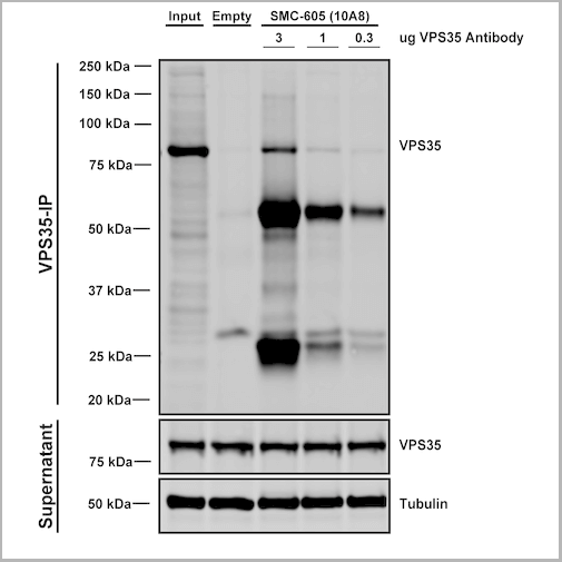
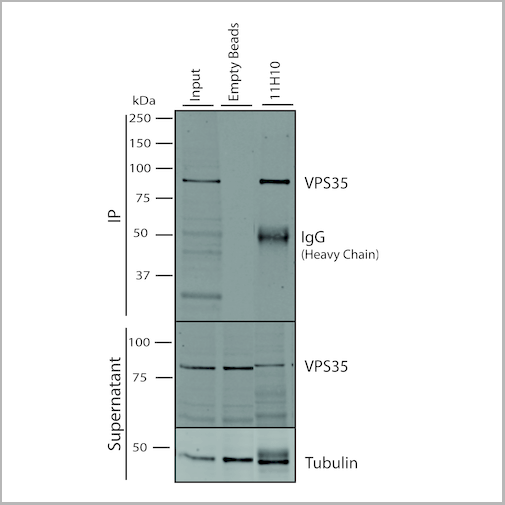
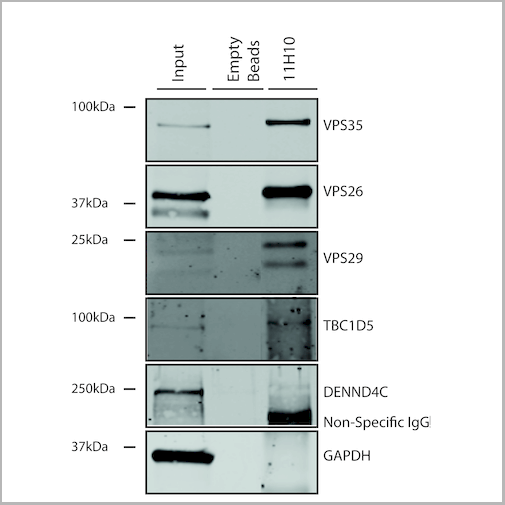
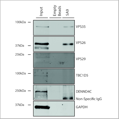
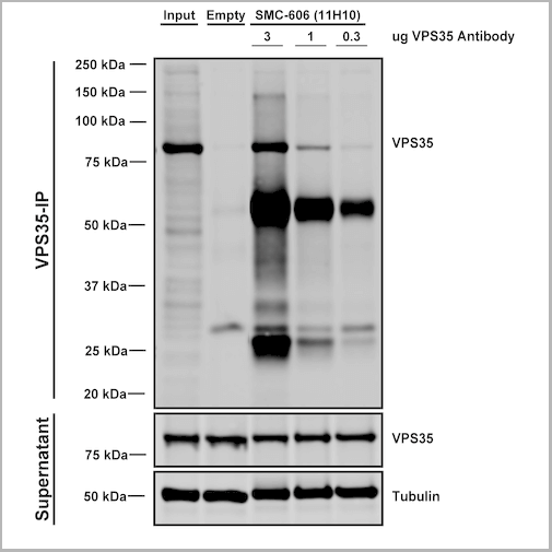 IP (Immunoprecipitation)
(Immunoprecipitation analysis using Mouse Anti-VPS35 Monoclonal Antibody, Clone 11H10. Tissue: embryonic fibroblast. Species: Mouse. Primary Antibody: Mouse Anti-VPS35 Monoclonal Antibody. Three amounts of (3, 1 and 0.3 ug) were non-covalently coupled to 10uL of A/G sepharose beads for 1 hour at 4 degree C and next incubated with 250ug of MEF lysate for 2 hours at 4 degree C.)
IP (Immunoprecipitation)
(Immunoprecipitation analysis using Mouse Anti-VPS35 Monoclonal Antibody, Clone 11H10. Tissue: embryonic fibroblast. Species: Mouse. Primary Antibody: Mouse Anti-VPS35 Monoclonal Antibody. Three amounts of (3, 1 and 0.3 ug) were non-covalently coupled to 10uL of A/G sepharose beads for 1 hour at 4 degree C and next incubated with 250ug of MEF lysate for 2 hours at 4 degree C.)
VPS35, Monoclonal Antibody (Cat# AAA27702)
Full Name
VPS35 Antibody, Clone 11H10: ATTO 488
Gene Names
VPS35; MEM3; PARK17
Reactivity
Human, Mouse, Rat
Applications
Western Blot, Immunoprecipitation
Purity
Protein G Purified
Pricing


 IP (Immunoprecipitation)
(Immunoprecipitation analysis using Mouse Anti-VPS35 Monoclonal Antibody, Clone 11H10. Tissue: embryonic fibroblast. Species: Mouse. Primary Antibody: Mouse Anti-VPS35 Monoclonal Antibody. Three amounts of (3, 1 and 0.3 ug) were non-covalently coupled to 10uL of A/G sepharose beads for 1 hour at 4 degree C and next incubated with 250ug of MEF lysate for 2 hours at 4 degree C.)
IP (Immunoprecipitation)
(Immunoprecipitation analysis using Mouse Anti-VPS35 Monoclonal Antibody, Clone 11H10. Tissue: embryonic fibroblast. Species: Mouse. Primary Antibody: Mouse Anti-VPS35 Monoclonal Antibody. Three amounts of (3, 1 and 0.3 ug) were non-covalently coupled to 10uL of A/G sepharose beads for 1 hour at 4 degree C and next incubated with 250ug of MEF lysate for 2 hours at 4 degree C.)
VPS35, Monoclonal Antibody (Cat# AAA27700)
Full Name
VPS35 Antibody, Clone 11H10
Gene Names
VPS35; MEM3; PARK17
Reactivity
Human, Mouse, Rat
Applications
Western Blot, Immunoprecipitation
Purity
Protein G Purified
Pricing

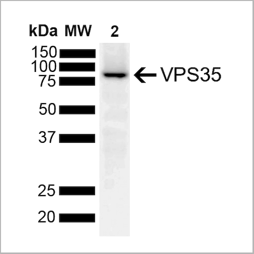

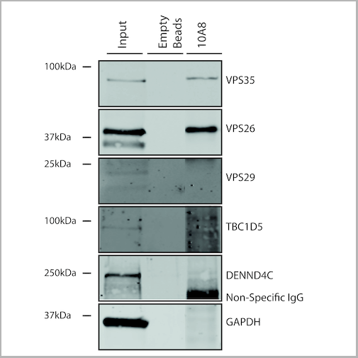
 IP (Immunoprecipitation)
(Immunoprecipitation analysis using Mouse Anti-VPS35 Monoclonal Antibody, Clone 10A8. Tissue: embryonic fibroblast. Species: Mouse. Primary Antibody: Mouse Anti-VPS35 Monoclonal Antibody. Three amounts of (3, 1 and 0.3 ug) were non-covalently coupled to 10uL of A/G sepharose beads for 1 hour at 4 degree C and next incubated with 250ug of MEF lysate for 2 hours at 4 degree C.)
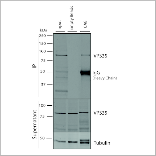
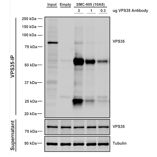
IP (Immunoprecipitation)
(Immunoprecipitation analysis using Mouse Anti-VPS35 Monoclonal Antibody, Clone 10A8. Tissue: embryonic fibroblast. Species: Mouse. Primary Antibody: Mouse Anti-VPS35 Monoclonal Antibody. Three amounts of (3, 1 and 0.3 ug) were non-covalently coupled to 10uL of A/G sepharose beads for 1 hour at 4 degree C and next incubated with 250ug of MEF lysate for 2 hours at 4 degree C.)
VPS35, Monoclonal Antibody (Cat# AAA27697)
Full Name
VPS35 Antibody, Clone 10A8: HRP
Gene Names
VPS35; MEM3; PARK17
Reactivity
Human, Mouse, Rat
Applications
Western Blot, Immunocytochemistry, Immunofluorescence, Immunoprecipitation
Purity
Protein G Purified
Pricing
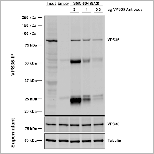

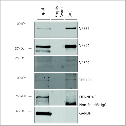

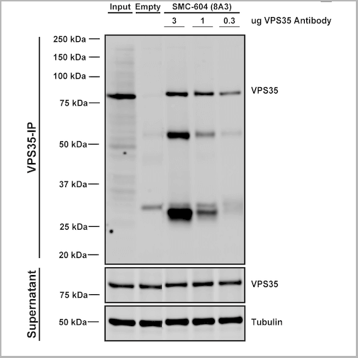
 IP (Immunoprecipitation)
(Immunoprecipitation analysis using Mouse Anti-VPS35 Monoclonal Antibody, Clone 8A3. Tissue: embryonic fibroblast. Species: Mouse. Primary Antibody: Mouse Anti-VPS35 Monoclonal Antibody. Three amounts of (3, 1 and 0.3 ug) were non-covalently coupled to 10uL of A/G sepharose beads for 1 hour at 4 degree C and next incubated with 250ug of MEF lysate for 2 hours at 4 degree C.)
IP (Immunoprecipitation)
(Immunoprecipitation analysis using Mouse Anti-VPS35 Monoclonal Antibody, Clone 8A3. Tissue: embryonic fibroblast. Species: Mouse. Primary Antibody: Mouse Anti-VPS35 Monoclonal Antibody. Three amounts of (3, 1 and 0.3 ug) were non-covalently coupled to 10uL of A/G sepharose beads for 1 hour at 4 degree C and next incubated with 250ug of MEF lysate for 2 hours at 4 degree C.)
VPS35, Monoclonal Antibody (Cat# AAA27687)
Full Name
VPS35 Antibody, Clone 8A3: FITC
Gene Names
VPS35; MEM3; PARK17
Reactivity
Human, Mouse, Rat
Applications
Western Blot, Immunocytochemistry, Immunofluorescence, Immunoprecipitation
Purity
Protein G Purified
Pricing
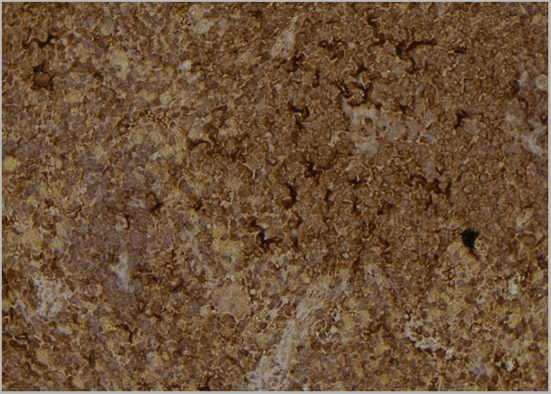
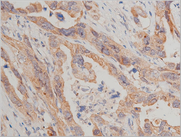
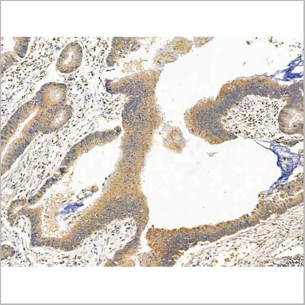
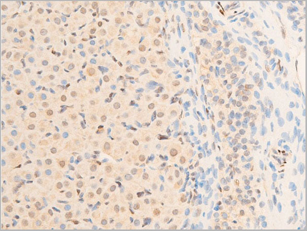
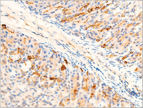
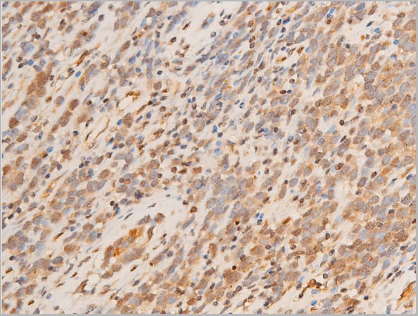
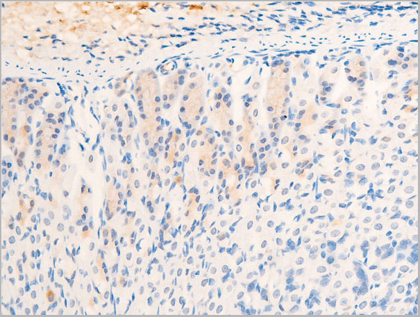
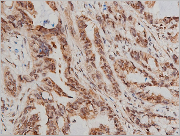
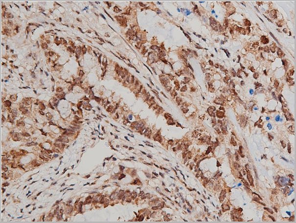
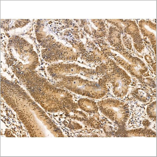
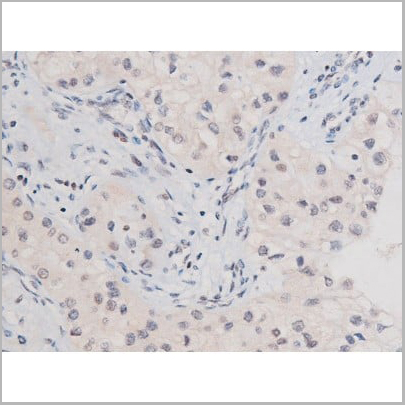
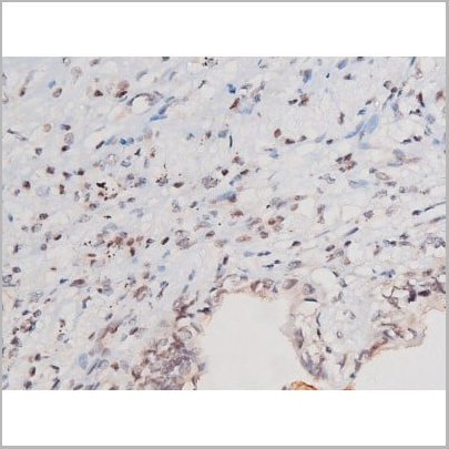
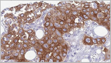
 IHC (Immunohistochemistry-Paraffin)
(AAA31101at 1/100 staining human mammary cancer tissue byIHC-P. The sample was formaldehyde fixed and a heatmediated antigen retrieval step in citrate buffer wasperformed. The sample was then blocked and incubated withthe antibody for 1.5 hours at 22°C. An HRP conjugated goatanti-rabbit antibody was used as the secondary antibody.)
IHC (Immunohistochemistry-Paraffin)
(AAA31101at 1/100 staining human mammary cancer tissue byIHC-P. The sample was formaldehyde fixed and a heatmediated antigen retrieval step in citrate buffer wasperformed. The sample was then blocked and incubated withthe antibody for 1.5 hours at 22°C. An HRP conjugated goatanti-rabbit antibody was used as the secondary antibody.)
xCT, Polyclonal Antibody (Cat# AAA31101)
Full Name
xCT Antibody
Gene Names
SLC7A11; xCT; CCBR1
Reactivity
Human, Mouse, Monkey
Applications
Western Blot, Immunohistochemistry, Immunofluorescence, Immunocytochemistry
Purity
The antiserum was purified by peptide affinity chromatography using SulfoLink™ Coupling Resin (Thermo Fisher Scientific).
Pricing


 IP (Immunoprecipitation)
(Immunoprecipitation analysis using Mouse Anti-VPS35 Monoclonal Antibody, Clone 8A3. Tissue: embryonic fibroblast. Species: Mouse. Primary Antibody: Mouse Anti-VPS35 Monoclonal Antibody. Three amounts of (3, 1 and 0.3 ug) were non-covalently coupled to 10uL of A/G sepharose beads for 1 hour at 4 degree C and next incubated with 250ug of MEF lysate for 2 hours at 4 degree C.)
IP (Immunoprecipitation)
(Immunoprecipitation analysis using Mouse Anti-VPS35 Monoclonal Antibody, Clone 8A3. Tissue: embryonic fibroblast. Species: Mouse. Primary Antibody: Mouse Anti-VPS35 Monoclonal Antibody. Three amounts of (3, 1 and 0.3 ug) were non-covalently coupled to 10uL of A/G sepharose beads for 1 hour at 4 degree C and next incubated with 250ug of MEF lysate for 2 hours at 4 degree C.)
VPS35, Monoclonal Antibody (Cat# AAA27683)
Full Name
VPS35 Antibody, Clone 8A3: ATTO 390
Gene Names
VPS35; MEM3; PARK17
Reactivity
Human, Mouse, Rat
Applications
Western Blot, Immunocytochemistry, Immunofluorescence, Immunoprecipitation
Purity
Protein G Purified
Pricing
 IF (Immunofluorescence)
(AAA31270 staining Hela cells by IF/ICC. The samples were fixed with PFA and permeabilized in 0.1% Triton X-100,then blocked in 10% serum for 45 minutes at 25°C. Samples were then incubated with primary Ab(AAA31270 1:200) and mouse anti-beta tubulin Ab( 1:200) for 1 hour at 37°C. An AlexaFluor594 conjugated goat anti-rabbit IgG(H+L) Ab(Red) and an AlexaFluor488 conjugated goat anti-mouse IgG(H+L) Ab(Green) were used as the secondary antibody.The nuclear counter stain is DAPI(blue).)
IF (Immunofluorescence)
(AAA31270 staining Hela cells by IF/ICC. The samples were fixed with PFA and permeabilized in 0.1% Triton X-100,then blocked in 10% serum for 45 minutes at 25°C. Samples were then incubated with primary Ab(AAA31270 1:200) and mouse anti-beta tubulin Ab( 1:200) for 1 hour at 37°C. An AlexaFluor594 conjugated goat anti-rabbit IgG(H+L) Ab(Red) and an AlexaFluor488 conjugated goat anti-mouse IgG(H+L) Ab(Green) were used as the secondary antibody.The nuclear counter stain is DAPI(blue).)
Hyaluronan synthase 2, Polyclonal Antibody (Cat# AAA31270)
Full Name
Hyaluronan synthase 2 Antibody
Reactivity
Human, Mouse, Rat
Applications
Immunofluorescence, Immunocytochemistry, Western Blot, Immunohistochemistry
Purity
Peptide affinity purification
Pricing
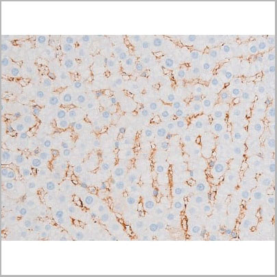
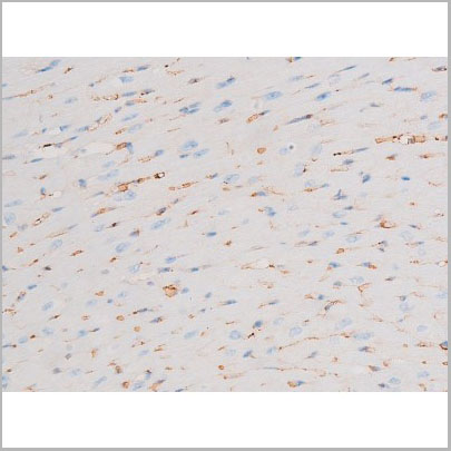
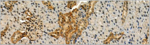
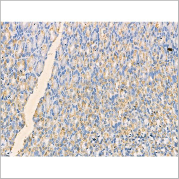
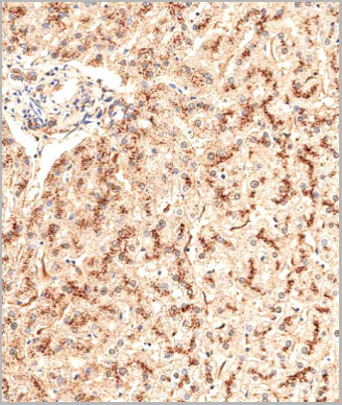
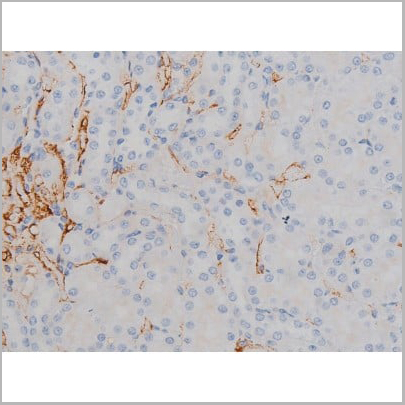
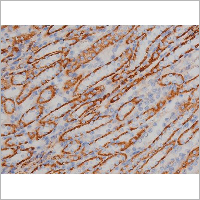
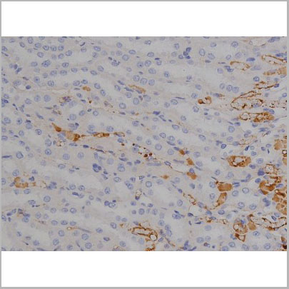
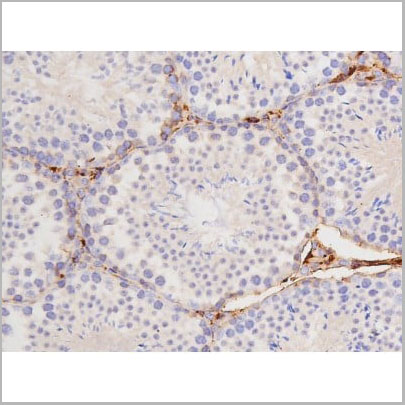
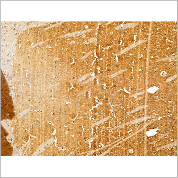
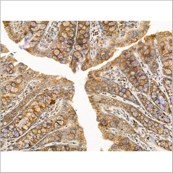
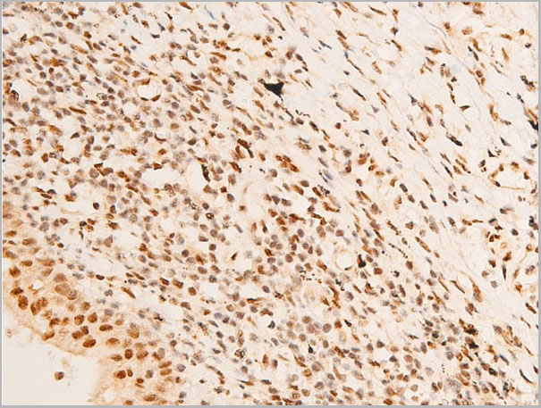
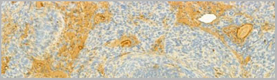
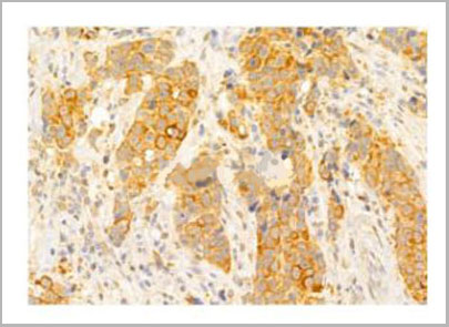
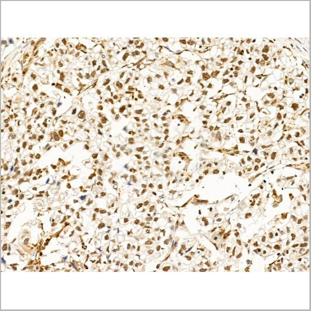
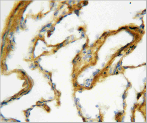
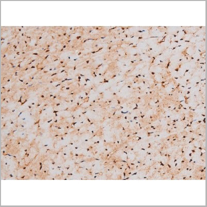
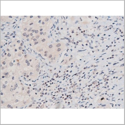
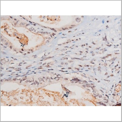
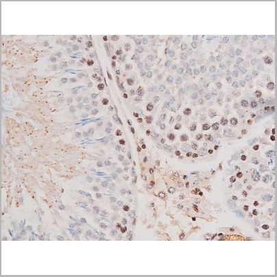
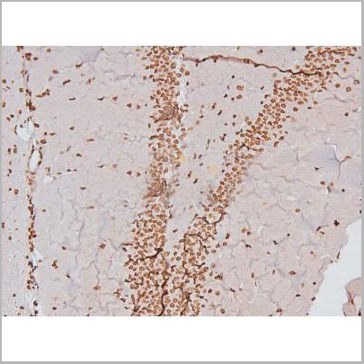
 IHC (Immunohistchemistry)
(AAA31064 at 1/200 staining Rat lung tissue sections by IHC-P. The tissue was formaldehyde fixed and a heat mediated antigen retrieval step in citrate buffer was performed. The tissue was then blocked and incubated with the antibody for 1.5 hours at 22 degree C. An HRP conjugated goat anti-rabbit antibody was used as the secondary.)
IHC (Immunohistchemistry)
(AAA31064 at 1/200 staining Rat lung tissue sections by IHC-P. The tissue was formaldehyde fixed and a heat mediated antigen retrieval step in citrate buffer was performed. The tissue was then blocked and incubated with the antibody for 1.5 hours at 22 degree C. An HRP conjugated goat anti-rabbit antibody was used as the secondary.)
PKC theta, Polyclonal Antibody (Cat# AAA31064)
Full Name
Phospho-PKC theta (Ser676) Antibody
Gene Names
PRKCQ; PRKCT; nPKC-theta
Reactivity
Human, Mouse, Rat
Applications
Western Blot, Immunohistochemistry, Immunofluorescence, Immunocytochemistry
Purity
From purified rabbit serum by affinity purification via sequential chromatography on phospho-and non-phospho-peptide affinity columns.
Pricing
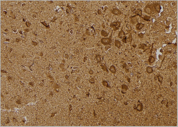
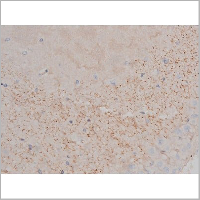
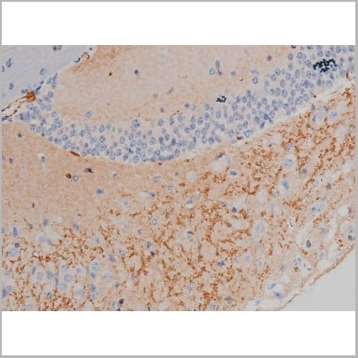
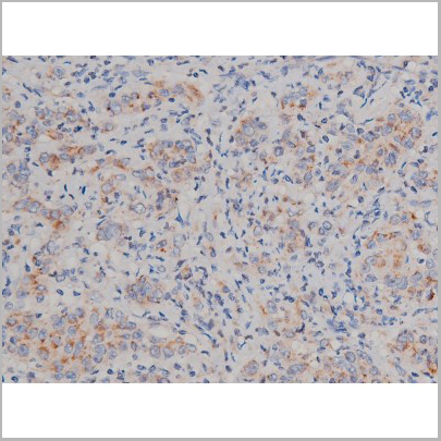
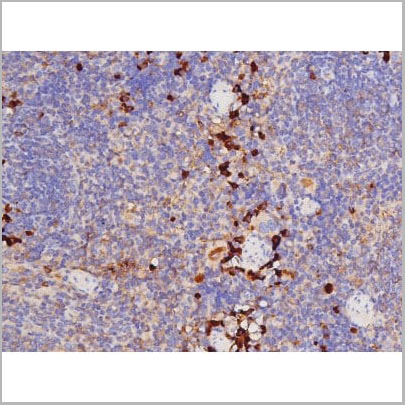
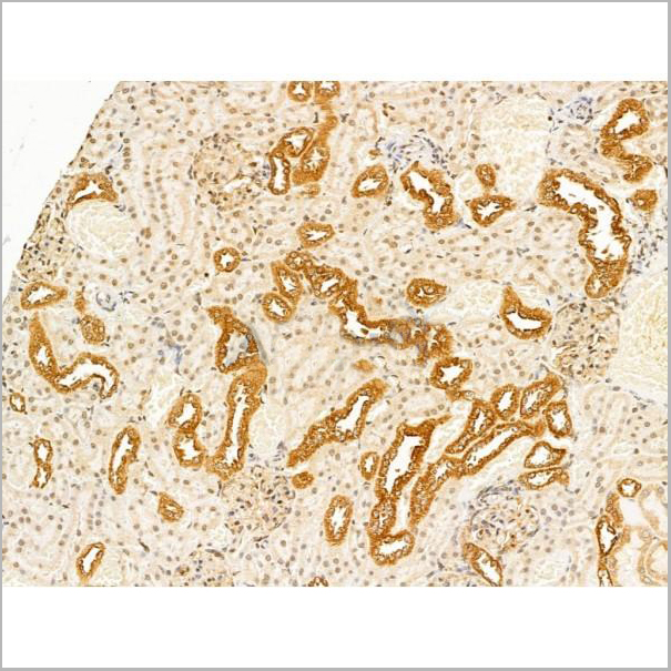
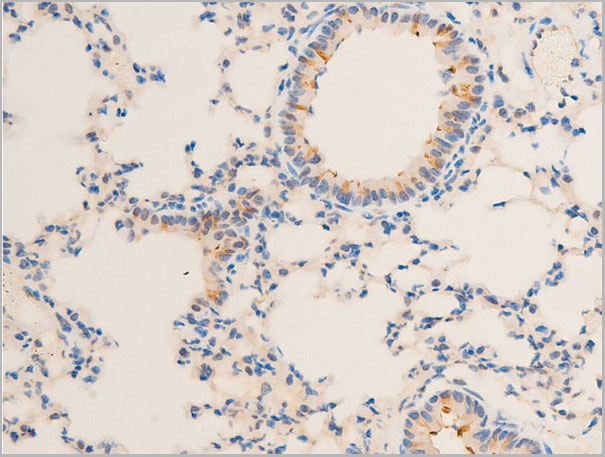
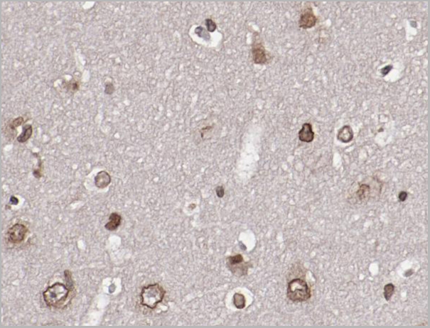
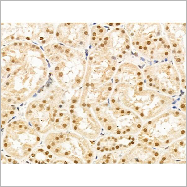
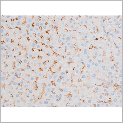
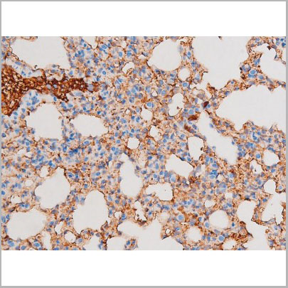
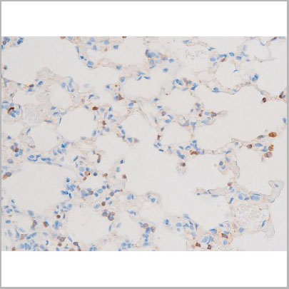
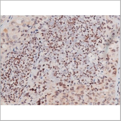
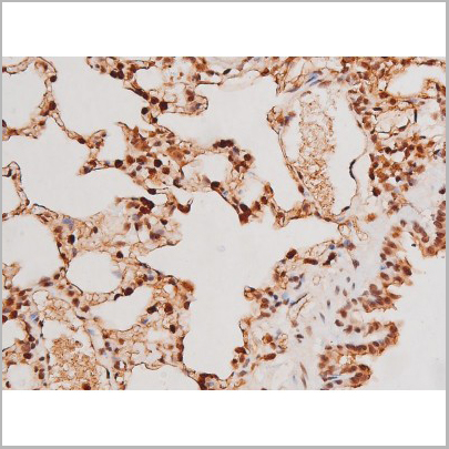
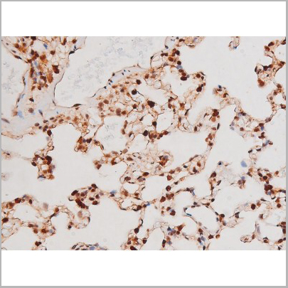
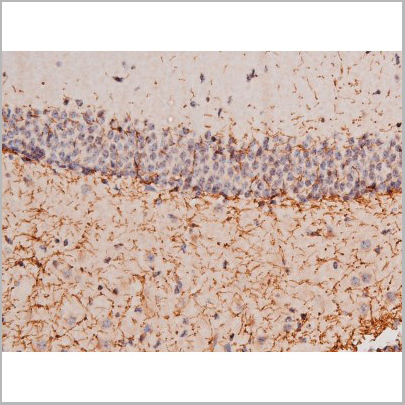
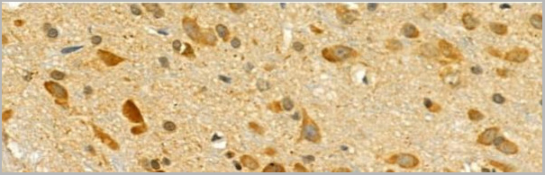
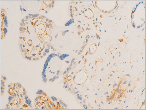
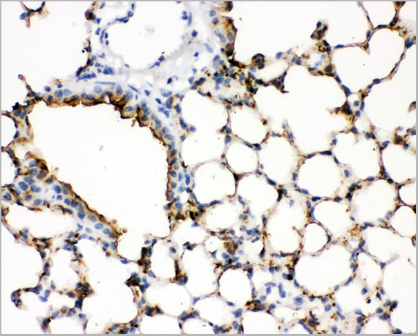 IHC (Immunohistochemistry)
(Figure 3. IHC analysis of SFTPA1 using anti- SFTPA1 antibody (AAA11521).SFTPA1 was detected in paraffin-embedded section of mouse lung tissues. Heat mediated antigen retrieval was performed in citrate buffer (pH6, epitope retrieval solution) for 20 mins. The tissue section was blocked with 10% goat serum. The tissue section was then incubated with 1ug/ml rabbit anti- SFTPA1 Antibody (AAA11521) overnight at 4 degree C. Biotinylated goat anti-rabbit IgG was used as secondary antibody and incubated for 30 minutes at 37 degree C. The tissue section was developed using Strepavidin-Biotin-Complex (SABC) with DAB as the chromogen.)
IHC (Immunohistochemistry)
(Figure 3. IHC analysis of SFTPA1 using anti- SFTPA1 antibody (AAA11521).SFTPA1 was detected in paraffin-embedded section of mouse lung tissues. Heat mediated antigen retrieval was performed in citrate buffer (pH6, epitope retrieval solution) for 20 mins. The tissue section was blocked with 10% goat serum. The tissue section was then incubated with 1ug/ml rabbit anti- SFTPA1 Antibody (AAA11521) overnight at 4 degree C. Biotinylated goat anti-rabbit IgG was used as secondary antibody and incubated for 30 minutes at 37 degree C. The tissue section was developed using Strepavidin-Biotin-Complex (SABC) with DAB as the chromogen.)
Surfactant Protein A, Polyclonal Antibody (Cat# AAA11521)
Full Name
Polyclonal Anti-SFTPA1 Antibody
Gene Names
Sftpa1; SP-A; Sftp1; Sftp-1
Reactivity
Mouse, Rat
Applications
Western Blot, Immunohistochemistry
Purity
Immunogen affinity purified.
Pricing
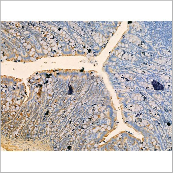
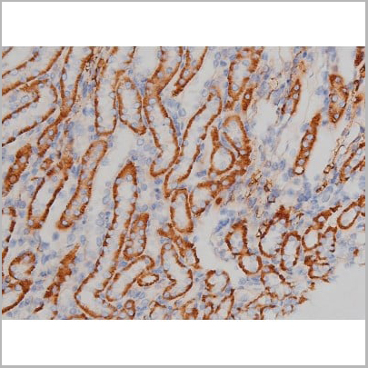
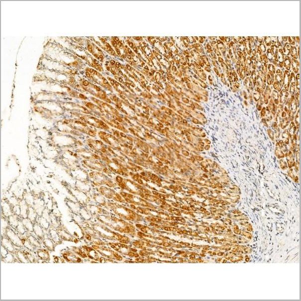
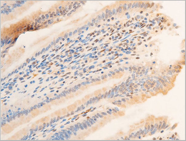
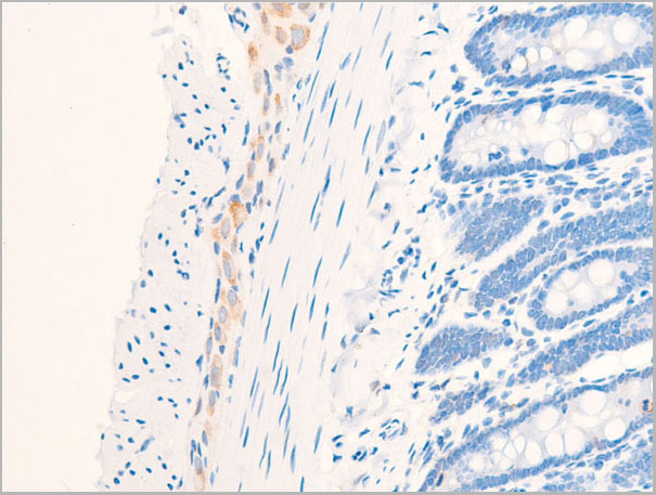
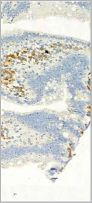 IHC-P (Immunohistochemistry Paraffin)
(AAA31041 at 1/100 staining Mouse colon tissue by IHC-P. The sample was formaldehyde fixed and a heat mediated antigen retrieval step in citrate buffer was performed. The sample was then blocked and incubated with the primary antibody at 4°C overnight. An HRP conjugated anti-Rabbit antibody was use das the secondary antibody.)
IHC-P (Immunohistochemistry Paraffin)
(AAA31041 at 1/100 staining Mouse colon tissue by IHC-P. The sample was formaldehyde fixed and a heat mediated antigen retrieval step in citrate buffer was performed. The sample was then blocked and incubated with the primary antibody at 4°C overnight. An HRP conjugated anti-Rabbit antibody was use das the secondary antibody.)
STAT3, Polyclonal Antibody (Cat# AAA31041)
Full Name
Phospho-STAT3 (Tyr705) Antibody
Gene Names
STAT3; APRF; HIES; ADMIO; ADMIO1
Reactivity
Human, Mouse, Rat
Applications
Western Blot, Immunohistochemistry, Immunofluorescence, Immunoprecipitation, Peptide ELISA
Purity
Peptide affinity purification.
Pricing
 Application Data
(At 25 degree C. The primary antibody was diluted at 1/200 and incubated with the sample for 1 hour at 37 degree C. An Alexa Fluor 594 conjugated goat anti-rabbit IgG (H+L) antibody(Red), diluted at 1/600, was used as secondary antibody.)
Application Data
(At 25 degree C. The primary antibody was diluted at 1/200 and incubated with the sample for 1 hour at 37 degree C. An Alexa Fluor 594 conjugated goat anti-rabbit IgG (H+L) antibody(Red), diluted at 1/600, was used as secondary antibody.)
CSK, Polyclonal Antibody (Cat# AAA31403)
Full Name
Phospho-CSK (Tyr304) Antibody
Reactivity
Human, Mouse, Rat
Applications
Western Blot, Immunohistochemistry, Immunofluorescence, Immunocytochemistry, Peptide ELISA
Purity
The antibody is from purified rabbit serum by affinity purification via sequential chromatography on phospho-peptide and non-phospho-peptide affinity columns.
Pricing


 Application Data
(Published customer image: Mouse anti Human CD105 antibody, clone SN6 used for flow cytometry.Image caption:Phenotype change from HLA-A2+/CD11b+/CD105- to HLA-A2+/CD11b-/CD105+ on endothelium-adherent blood monocyte-derived cells with increase in size and granularity during co-culture. HLA-A2+ PBMCs (1x10⁶ cells/well) were incubated for 2 h (Day 0) with HLA-A2- HUVECs, after which the non-adherent cells were removed by washing. The cell layers were analysed by three-colour flow cytometry staining for HLA-A2, CD11b and CD105 on (A) Day 1 and (B) Day 2. These plots were gated for HLA-A2+ cells. Forward scatter/side scatter dot plots gated for HLA-A2+ cells on Day 0 (C) and Day 2 (D) was shown. These are representative of 2 individual experiments.From: Tso C, Rye K-A, Barter P (2012) Phenotypic and Functional Changes in Blood Monocytes Following Adherence to Endothelium. PLoS ONE 7(5): e37091.)
Application Data
(Published customer image: Mouse anti Human CD105 antibody, clone SN6 used for flow cytometry.Image caption:Phenotype change from HLA-A2+/CD11b+/CD105- to HLA-A2+/CD11b-/CD105+ on endothelium-adherent blood monocyte-derived cells with increase in size and granularity during co-culture. HLA-A2+ PBMCs (1x10⁶ cells/well) were incubated for 2 h (Day 0) with HLA-A2- HUVECs, after which the non-adherent cells were removed by washing. The cell layers were analysed by three-colour flow cytometry staining for HLA-A2, CD11b and CD105 on (A) Day 1 and (B) Day 2. These plots were gated for HLA-A2+ cells. Forward scatter/side scatter dot plots gated for HLA-A2+ cells on Day 0 (C) and Day 2 (D) was shown. These are representative of 2 individual experiments.From: Tso C, Rye K-A, Barter P (2012) Phenotypic and Functional Changes in Blood Monocytes Following Adherence to Endothelium. PLoS ONE 7(5): e37091.)
CD105, Monoclonal Antibody (Cat# AAA12083)
Full Name
MOUSE ANTI HUMAN CD105:FITC
Gene Names
ENG; END; HHT1; ORW1
Applications
Flow Cytometry
Pricing


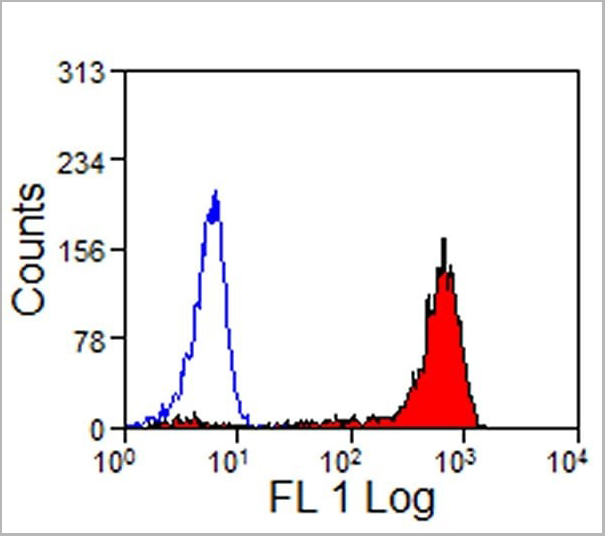
 Application Data
(Published customer image: Mouse anti Human CD105 antibody, clone SN6 used for flow cytometry.Image caption:Phenotype change from HLA-A2+/CD11b+/CD105- to HLA-A2+/CD11b-/CD105+ on endothelium-adherent blood monocyte-derived cells with increase in size and granularity during co-culture. HLA-A2+ PBMCs (1x10⁶ cells/well) were incubated for 2 h (Day 0) with HLA-A2- HUVECs, after which the non-adherent cells were removed by washing. The cell layers were analysed by three-colour flow cytometry staining for HLA-A2, CD11b and CD105 on (A) Day 1 and (B) Day 2. These plots were gated for HLA-A2+ cells. Forward scatter/side scatter dot plots gated for HLA-A2+ cells on Day 0 (C) and Day 2 (D) was shown. These are representative of 2 individual experiments.From: Tso C, Rye K-A, Barter P (2012) Phenotypic and Functional Changes in Blood Monocytes Following Adherence to Endothelium. PLoS ONE 7(5): e37091.)
Application Data
(Published customer image: Mouse anti Human CD105 antibody, clone SN6 used for flow cytometry.Image caption:Phenotype change from HLA-A2+/CD11b+/CD105- to HLA-A2+/CD11b-/CD105+ on endothelium-adherent blood monocyte-derived cells with increase in size and granularity during co-culture. HLA-A2+ PBMCs (1x10⁶ cells/well) were incubated for 2 h (Day 0) with HLA-A2- HUVECs, after which the non-adherent cells were removed by washing. The cell layers were analysed by three-colour flow cytometry staining for HLA-A2, CD11b and CD105 on (A) Day 1 and (B) Day 2. These plots were gated for HLA-A2+ cells. Forward scatter/side scatter dot plots gated for HLA-A2+ cells on Day 0 (C) and Day 2 (D) was shown. These are representative of 2 individual experiments.From: Tso C, Rye K-A, Barter P (2012) Phenotypic and Functional Changes in Blood Monocytes Following Adherence to Endothelium. PLoS ONE 7(5): e37091.)
CD105, Monoclonal Antibody (Cat# AAA11865)
Full Name
MOUSE ANTI HUMAN CD105:FITC
Gene Names
ENG; END; HHT1; ORW1
Applications
Flow Cytometry
Pricing


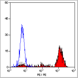
 Application Data
(Published customer image: Mouse anti Human CD105 antibody, clone SN6 used for flow cytometry.Image caption:Phenotype change from HLA-A2+/CD11b+/CD105- to HLA-A2+/CD11b-/CD105+ on endothelium-adherent blood monocyte-derived cells with increase in size and granularity during co-culture. HLA-A2+ PBMCs (1x10⁶ cells/well) were incubated for 2 h (Day 0) with HLA-A2- HUVECs, after which the non-adherent cells were removed by washing. The cell layers were analysed by three-colour flow cytometry staining for HLA-A2, CD11b and CD105 on (A) Day 1 and (B) Day 2. These plots were gated for HLA-A2+ cells. Forward scatter/side scatter dot plots gated for HLA-A2+ cells on Day 0 (C) and Day 2 (D) was shown. These are representative of 2 individual experiments.From: Tso C, Rye K-A, Barter P (2012) Phenotypic and Functional Changes in Blood Monocytes Following Adherence to Endothelium. PLoS ONE 7(5): e37091.)
Application Data
(Published customer image: Mouse anti Human CD105 antibody, clone SN6 used for flow cytometry.Image caption:Phenotype change from HLA-A2+/CD11b+/CD105- to HLA-A2+/CD11b-/CD105+ on endothelium-adherent blood monocyte-derived cells with increase in size and granularity during co-culture. HLA-A2+ PBMCs (1x10⁶ cells/well) were incubated for 2 h (Day 0) with HLA-A2- HUVECs, after which the non-adherent cells were removed by washing. The cell layers were analysed by three-colour flow cytometry staining for HLA-A2, CD11b and CD105 on (A) Day 1 and (B) Day 2. These plots were gated for HLA-A2+ cells. Forward scatter/side scatter dot plots gated for HLA-A2+ cells on Day 0 (C) and Day 2 (D) was shown. These are representative of 2 individual experiments.From: Tso C, Rye K-A, Barter P (2012) Phenotypic and Functional Changes in Blood Monocytes Following Adherence to Endothelium. PLoS ONE 7(5): e37091.)
CD105, Monoclonal Antibody (Cat# AAA12029)
Full Name
MOUSE ANTI HUMAN CD105:RPE
Gene Names
ENG; END; HHT1; ORW1
Applications
Flow Cytometry
Pricing
 Application Data
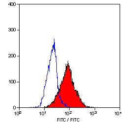
(Staining of mouse spleen with Hamster anti Mouse CD81: Alexa Fluor 488)
Application Data
(Staining of mouse spleen with Hamster anti Mouse CD81: Alexa Fluor 488)
CD81, Monoclonal Antibody (Cat# AAA11942)
Full Name
HAMSTER ANTI MOUSE CD81
Gene Names
Cd81; Tapa1; Tapa-1; Tspan28
Reactivity
Rat
Applications
Immunohistochemistry, Flow Cytometry, Immunoprecipitation, Western Blot
Pricing
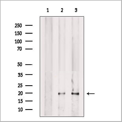
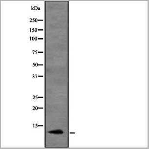
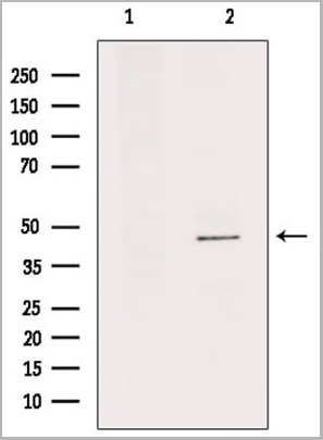
 WB (Western Blot)
(Positive WB detected in Recombinant proteinAll lanes: L1R antibody at 1:2000Secondary Goat polyclonal to rabbit IgG at 1/50000 dilutionPredicted band size: 26.8 kDabserved band size: 29 kDa)
WB (Western Blot)
(Positive WB detected in Recombinant proteinAll lanes: L1R antibody at 1:2000Secondary Goat polyclonal to rabbit IgG at 1/50000 dilutionPredicted band size: 26.8 kDabserved band size: 29 kDa)
L1R, Polyclonal Antibody (Cat# AAA27054)
Full Name
Rabbit anti-Vaccinia virus (strain Copenhagen)(VACV) L1R Polyclonal Antibody
Reactivity
Vaccinia virus
Applications
Western Blot
Purity
Protein G
Pricing
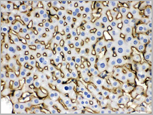
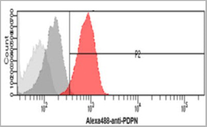 FCM (Flow Cytometry)
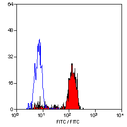
(Flow cytometry analysis of Podoplanin in HeLa cells. The cell was stained with (AAA11728) at 2-5ug for 1x10^6cells (red). A Goat anti mouse IgG (Alexa fluor 488) was used as the secondary antibody. Mouse monoclonal IgG was used as the isotype control (dark gray), cells without incubation with primary and secondary antibody was used as the negative control (light gray).)
FCM (Flow Cytometry)
(Flow cytometry analysis of Podoplanin in HeLa cells. The cell was stained with (AAA11728) at 2-5ug for 1x10^6cells (red). A Goat anti mouse IgG (Alexa fluor 488) was used as the secondary antibody. Mouse monoclonal IgG was used as the isotype control (dark gray), cells without incubation with primary and secondary antibody was used as the negative control (light gray).)
PDPN, Monoclonal Antibody (Cat# AAA11728)
Full Name
PDPN antibody
Gene Names
PDPN; T1A; GP36; GP40; Gp38; OTS8; T1A2; TI1A; T1A-2; AGGRUS; HT1A-1; PA2.26
Reactivity
Human
Applications
Flow Cytometry, Immunocytochemistry, Immunofluorescence
Purity
By protein-G affinity chromatography
Pricing
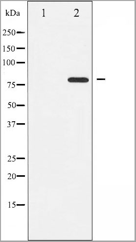
 WB (Western Blot)
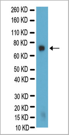
(Western Blot analysis for galactocerebroside in PC12 lysate. PC12 lysate was resolved by electrophoresis, transferred to PVDF membrane and probed with AAA14685 at 1:500 dilution. Proteins were visualized using a goat anti-mouse secondary antibody conjugated to HRP and a chemiluminescence detection system. Arrow indicates protein Galactocerebroside (~75kD).)
WB (Western Blot)
(Western Blot analysis for galactocerebroside in PC12 lysate. PC12 lysate was resolved by electrophoresis, transferred to PVDF membrane and probed with AAA14685 at 1:500 dilution. Proteins were visualized using a goat anti-mouse secondary antibody conjugated to HRP and a chemiluminescence detection system. Arrow indicates protein Galactocerebroside (~75kD).)
Galactocerebroside, Monoclonal Antibody (Cat# AAA14685)
Full Name
Mouse anti-Bovine Galactocerebroside
Applications
Immunohistochemistry, Immunocytochemistry
Purity
Purified by Protein A affinity chromatography.
Pricing

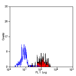
 FCM (Flow Cytometry)
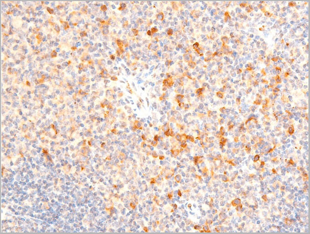
(Flow cytometric analysis of K562 cells with CD86 antibody at 1/50 dilution (blue) compared with an unlabelled control (cells without incubation with primary antibody; red). Alexa Fluor 488-conjugated goat anti rabbit IgG was used as the secondary antibody.)
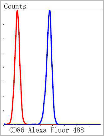
FCM (Flow Cytometry)
(Flow cytometric analysis of K562 cells with CD86 antibody at 1/50 dilution (blue) compared with an unlabelled control (cells without incubation with primary antibody; red). Alexa Fluor 488-conjugated goat anti rabbit IgG was used as the secondary antibody.)
CD86, Monoclonal Antibody (Cat# AAA30048)
Full Name
CD86 Antibody
Gene Names
CD86; B70; B7-2; B7.2; LAB72; CD28LG2
Reactivity
Human, Rat
Applications
Western Blot, Immunocytochemistry, Immunofluorescence, Immunohistochemistry, Immunoprecipitation, Flow Cytometry
Purity
ProA affinity purified
Pricing


 IP (Immunoprecipitation)
(Immunoprecipitation analysis using Mouse Anti-VPS35 Monoclonal Antibody, Clone 10A8. Tissue: embryonic fibroblast. Species: Mouse. Primary Antibody: Mouse Anti-VPS35 Monoclonal Antibody. Three amounts of (3, 1 and 0.3 ug) were non-covalently coupled to 10uL of A/G sepharose beads for 1 hour at 4 degree C and next incubated with 250ug of MEF lysate for 2 hours at 4 degree C.)
IP (Immunoprecipitation)
(Immunoprecipitation analysis using Mouse Anti-VPS35 Monoclonal Antibody, Clone 10A8. Tissue: embryonic fibroblast. Species: Mouse. Primary Antibody: Mouse Anti-VPS35 Monoclonal Antibody. Three amounts of (3, 1 and 0.3 ug) were non-covalently coupled to 10uL of A/G sepharose beads for 1 hour at 4 degree C and next incubated with 250ug of MEF lysate for 2 hours at 4 degree C.)
VPS35, Monoclonal Antibody (Cat# AAA27693)
Full Name
VPS35 Antibody, Clone 10A8: ATTO 488
Gene Names
VPS35; MEM3; PARK17
Reactivity
Human, Mouse, Rat
Applications
Western Blot, Immunocytochemistry, Immunofluorescence, Immunoprecipitation
Purity
Protein G Purified
Pricing
 FCM (Flow Cytometry)
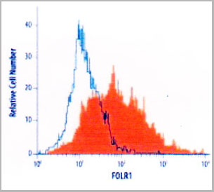
(Flow Cytometry detection of FOLR1 in MCF-7 human cell line using AAA14691. MCF-7 human breast cancer cell line was stained with AAA14691 (filled histogram) or isotype control antibody (open histogram), followed byPhycoerythrin-conjugated Anti-MouseIgG Secondary Antibody.)
FCM (Flow Cytometry)
(Flow Cytometry detection of FOLR1 in MCF-7 human cell line using AAA14691. MCF-7 human breast cancer cell line was stained with AAA14691 (filled histogram) or isotype control antibody (open histogram), followed byPhycoerythrin-conjugated Anti-MouseIgG Secondary Antibody.)
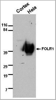
FOLR1, Monoclonal Antibody (Cat# AAA14691)
Full Name
FOLR1 (Folate receptor 1 (adult) Adult folate-binding protein, FBP, Folate receptor, adult, Folate receptor 1, Folate receptor alpha, FOLR, FR-alpha, KB cells FBP, MOv18, Ovarian tumor-associated antigen MOv18)
Gene Names
FOLR1; FBP; FOLR
Reactivity
Human
Applications
Flow Cytometry, Western Blot, Immunocytochemistry
Purity
Purified by Protein G affinity chromatography from hybridoma culture supernatant.
Pricing


 IP (Immunoprecipitation)
(Immunoprecipitation analysis using Mouse Anti-VPS35 Monoclonal Antibody, Clone 11H10. Tissue: embryonic fibroblast. Species: Mouse. Primary Antibody: Mouse Anti-VPS35 Monoclonal Antibody. Three amounts of (3, 1 and 0.3 ug) were non-covalently coupled to 10uL of A/G sepharose beads for 1 hour at 4 degree C and next incubated with 250ug of MEF lysate for 2 hours at 4 degree C.)
IP (Immunoprecipitation)
(Immunoprecipitation analysis using Mouse Anti-VPS35 Monoclonal Antibody, Clone 11H10. Tissue: embryonic fibroblast. Species: Mouse. Primary Antibody: Mouse Anti-VPS35 Monoclonal Antibody. Three amounts of (3, 1 and 0.3 ug) were non-covalently coupled to 10uL of A/G sepharose beads for 1 hour at 4 degree C and next incubated with 250ug of MEF lysate for 2 hours at 4 degree C.)
VPS35, Monoclonal Antibody (Cat# AAA27701)
Full Name
VPS35 Antibody, Clone 11H10: ATTO 390
Gene Names
VPS35; MEM3; PARK17
Reactivity
Human, Mouse, Rat
Applications
Western Blot, Immunoprecipitation
Purity
Protein G Purified
Pricing
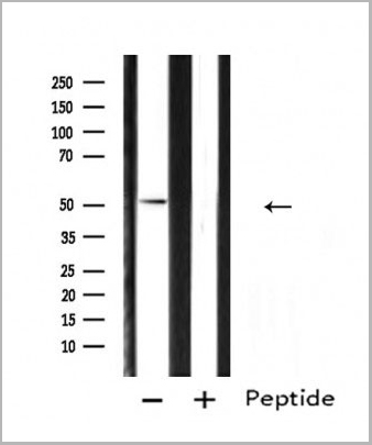
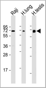 WB (Western Blot)
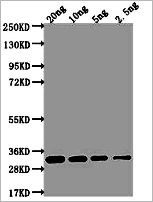
(All lanes : Anti-ERVK-7 Antibody (N-Term) at 1:1000-1:2000 dilutionLane 1: Raji whole cell lysateLane 2: human lung lysateLane 3: human testis lysateLysates/proteins at 20 ug per lane.SecondaryGoat Anti-Rabbit IgG, (H+L), Peroxidase conjugated at 1/10000 dilution.Predicted band size : 67 kDaBlocking/Dilution buffer: 5% NFDM/TBST.)
WB (Western Blot)
(All lanes : Anti-ERVK-7 Antibody (N-Term) at 1:1000-1:2000 dilutionLane 1: Raji whole cell lysateLane 2: human lung lysateLane 3: human testis lysateLysates/proteins at 20 ug per lane.SecondaryGoat Anti-Rabbit IgG, (H+L), Peroxidase conjugated at 1/10000 dilution.Predicted band size : 67 kDaBlocking/Dilution buffer: 5% NFDM/TBST.)
ERVK-7, Polyclonal Antibody (Cat# AAA28797)
Full Name
ERVK-7 Antibody (N-Term)
Reactivity
Human
Applications
Western Blot, Immunohistochemistry
Purity
This antibody is purified through a protein A column, followed by peptide affinity purification.
Pricing
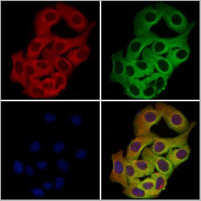
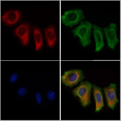
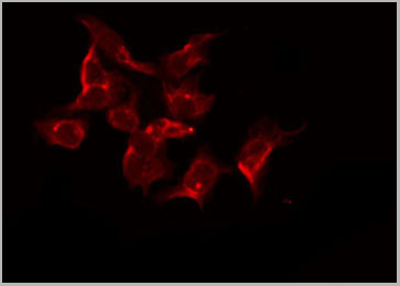
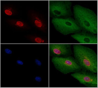
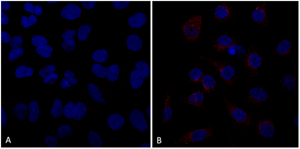
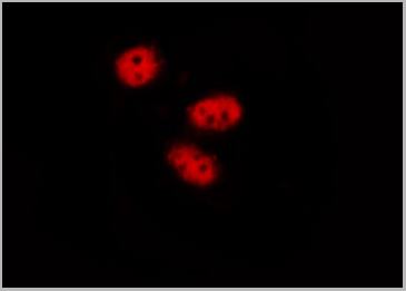
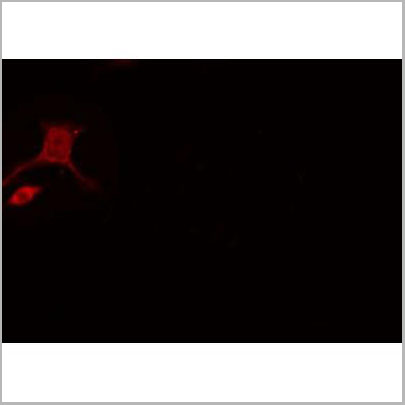
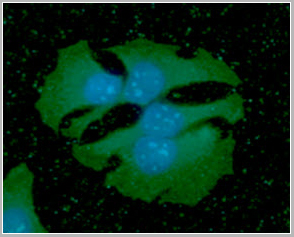
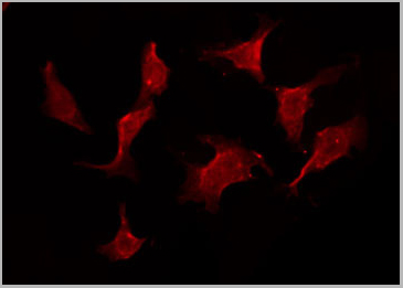 IF (Immunofluorescence)
(AAA31115 staining HeLa by IF/ICC. The sample were fixed with PFA and permeabilized in 0.1% Triton X-100, then blocked in 10% serum for 45 minutes at 25 degree C. The primary antibody was diluted at 1/200 and incubated with the sample for 1 hour at 37 degree C. An Alexa Fluor 594 conjugated goat anti-rabbit IgG (H+L) Ab, diluted at 1/600, was used as the secondary antibody.)
IF (Immunofluorescence)
(AAA31115 staining HeLa by IF/ICC. The sample were fixed with PFA and permeabilized in 0.1% Triton X-100, then blocked in 10% serum for 45 minutes at 25 degree C. The primary antibody was diluted at 1/200 and incubated with the sample for 1 hour at 37 degree C. An Alexa Fluor 594 conjugated goat anti-rabbit IgG (H+L) Ab, diluted at 1/600, was used as the secondary antibody.)
IL11RA, Polyclonal Antibody (Cat# AAA31115)
Full Name
IL11RA Antibody
Gene Names
IL11RA; CRSDA
Reactivity
Human, Mouse, Rat
Applications
Western Blot, Immunohistochemistry, Immunofluorescence, Immunocytochemistry
Purity
Peptide affinity purification
Pricing


 IP (Immunoprecipitation)
(Immunoprecipitation analysis using Mouse Anti-VPS35 Monoclonal Antibody, Clone 11H10. Tissue: embryonic fibroblast. Species: Mouse. Primary Antibody: Mouse Anti-VPS35 Monoclonal Antibody. Three amounts of (3, 1 and 0.3 ug) were non-covalently coupled to 10uL of A/G sepharose beads for 1 hour at 4 degree C and next incubated with 250ug of MEF lysate for 2 hours at 4 degree C.)
IP (Immunoprecipitation)
(Immunoprecipitation analysis using Mouse Anti-VPS35 Monoclonal Antibody, Clone 11H10. Tissue: embryonic fibroblast. Species: Mouse. Primary Antibody: Mouse Anti-VPS35 Monoclonal Antibody. Three amounts of (3, 1 and 0.3 ug) were non-covalently coupled to 10uL of A/G sepharose beads for 1 hour at 4 degree C and next incubated with 250ug of MEF lysate for 2 hours at 4 degree C.)
VPS35, Monoclonal Antibody (Cat# AAA27706)
Full Name
VPS35 Antibody, Clone 11H10: HRP
Gene Names
VPS35; MEM3; PARK17
Reactivity
Human, Mouse, Rat
Applications
Western Blot, Immunoprecipitation
Purity
Protein G Purified
Pricing


 IP (Immunoprecipitation)
(Immunoprecipitation analysis using Mouse Anti-VPS35 Monoclonal Antibody, Clone 11H10. Tissue: embryonic fibroblast. Species: Mouse. Primary Antibody: Mouse Anti-VPS35 Monoclonal Antibody. Three amounts of (3, 1 and 0.3 ug) were non-covalently coupled to 10uL of A/G sepharose beads for 1 hour at 4 degree C and next incubated with 250ug of MEF lysate for 2 hours at 4 degree C.)
IP (Immunoprecipitation)
(Immunoprecipitation analysis using Mouse Anti-VPS35 Monoclonal Antibody, Clone 11H10. Tissue: embryonic fibroblast. Species: Mouse. Primary Antibody: Mouse Anti-VPS35 Monoclonal Antibody. Three amounts of (3, 1 and 0.3 ug) were non-covalently coupled to 10uL of A/G sepharose beads for 1 hour at 4 degree C and next incubated with 250ug of MEF lysate for 2 hours at 4 degree C.)
VPS35, Monoclonal Antibody (Cat# AAA27708)
Full Name
VPS35 Antibody, Clone 11H10: RPE
Gene Names
VPS35; MEM3; PARK17
Reactivity
Human, Mouse, Rat
Applications
Western Blot, Immunoprecipitation
Purity
Protein G Purified
Pricing
 IP (Immunoprecipitation)
(Immunoprecipitation analysis using Mouse Anti-VPS35 Monoclonal Antibody, Clone 5A9. Tissue: embryonic fibroblast. Species: Mouse. Primary Antibody: Mouse Anti-VPS35 Monoclonal Antibody. Three amounts of (3, 1 and 0.3 ug) were non-covalently coupled to 10uL of A/G sepharose beads for 1 hour at 4 degree C and next incubated with 250ug of MEF lysate for 2 hours at 4 degree C.)
IP (Immunoprecipitation)
(Immunoprecipitation analysis using Mouse Anti-VPS35 Monoclonal Antibody, Clone 5A9. Tissue: embryonic fibroblast. Species: Mouse. Primary Antibody: Mouse Anti-VPS35 Monoclonal Antibody. Three amounts of (3, 1 and 0.3 ug) were non-covalently coupled to 10uL of A/G sepharose beads for 1 hour at 4 degree C and next incubated with 250ug of MEF lysate for 2 hours at 4 degree C.)
VPS35, Monoclonal Antibody (Cat# AAA27677)
Full Name
VPS35 Antibody, Clone 5A9: Biotin
Gene Names
VPS35; MEM3; PARK17
Reactivity
Human, Mouse, Rat
Applications
Western Blot, Immunocytochemistry, Immunofluorescence, Immunoprecipitation
Purity
Protein G Purified
Pricing
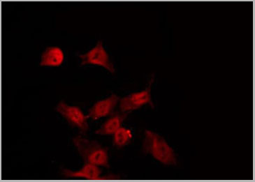
 IF (Immunofluorescence)
(AAA30937 staining A-431 cells by IF/ICC. The sample were fixed with PFA and permeabilized in 0.1% Triton X-100, then blocked in 10% serum for 45 minutes at 25 degree C. The primary antibody was diluted at 1/200 and incubated with the sample for 1 hour at 37 degree C. An Alexa Fluor 594 conjugated goat anti-rabbit IgG (H+L) antibody, diluted at 1/600, was used as secondary antibody.)
IF (Immunofluorescence)
(AAA30937 staining A-431 cells by IF/ICC. The sample were fixed with PFA and permeabilized in 0.1% Triton X-100, then blocked in 10% serum for 45 minutes at 25 degree C. The primary antibody was diluted at 1/200 and incubated with the sample for 1 hour at 37 degree C. An Alexa Fluor 594 conjugated goat anti-rabbit IgG (H+L) antibody, diluted at 1/600, was used as secondary antibody.)
SYK, Polyclonal Antibody (Cat# AAA30937)
Full Name
Phospho-SYK (Tyr323) Antibody
Gene Names
SYK; p72-Syk
Reactivity
Human, Mouse, Rat, Monkey
Applications
Western Blot, Immunohistochemistry, Immunofluorescence, Immunocytochemistry
Purity
From purified rabbit serum by affinity purification via sequential chromatography on phospho-and non-phospho-peptide affinity columns.
Pricing