Filters
Clonality
Type
Reactivity
Gene Name
Isotype
Host
Application
Clone
128 results for "Mouse Anti Monkey IgG" - showing 50-100


 Application Data
(Published customer image:Phycoerythrin conjugated Mouse anti Human CD83 antibody, clone HB15e used for the evaluation of CD83 expression on monocyte derived dendritic cells by flow cytometry.Phenotypic characterization of immunogenic and tolerogenic moDC populations by flow cytometry. Monocytes were negatively selected from PBMC using magnetic beads. Immature moDC were generated with IL-4 and GM-CSF for 6 days. 15d-PGJ2 (PGJ2 DC) and dexamethasone plus 1alpha,25-dihydroxyvitamin were added to generate tolerogenic moDC, respectively (PGJ2 DC and Dex/VD3 DC). To generate immunogenic moDC, immature moDC were stimulated for 24 h with LPS, polyI:C and a cytokine cocktail containing TNF-alpha, IL-1beta, IL-6 and PGE2, respectively. The phenotypes of the cells were analyzed by flow cytometry. Live cells were gated according to FSC/SSC. One representative experiment out of three is shown.From: Sprater F, Hovden A-O, Appel S (2012)Expression of ESE-3 Isoforms in Immunogenic and Tolerogenic Human Monocyte-Derived Dendritic Cells.PLoS ONE 7(11): e49577.)
Application Data
(Published customer image:Phycoerythrin conjugated Mouse anti Human CD83 antibody, clone HB15e used for the evaluation of CD83 expression on monocyte derived dendritic cells by flow cytometry.Phenotypic characterization of immunogenic and tolerogenic moDC populations by flow cytometry. Monocytes were negatively selected from PBMC using magnetic beads. Immature moDC were generated with IL-4 and GM-CSF for 6 days. 15d-PGJ2 (PGJ2 DC) and dexamethasone plus 1alpha,25-dihydroxyvitamin were added to generate tolerogenic moDC, respectively (PGJ2 DC and Dex/VD3 DC). To generate immunogenic moDC, immature moDC were stimulated for 24 h with LPS, polyI:C and a cytokine cocktail containing TNF-alpha, IL-1beta, IL-6 and PGE2, respectively. The phenotypes of the cells were analyzed by flow cytometry. Live cells were gated according to FSC/SSC. One representative experiment out of three is shown.From: Sprater F, Hovden A-O, Appel S (2012)Expression of ESE-3 Isoforms in Immunogenic and Tolerogenic Human Monocyte-Derived Dendritic Cells.PLoS ONE 7(11): e49577.)
CD83, Monoclonal Antibody (Cat# AAA12280)
Full Name
MOUSE ANTI HUMAN CD83
Reactivity
Human
Applications
Flow Cytometry, Immunohistochemistry, Immunohistochemistry, Immunoprecipitation, Immunofluorescence
Pricing


 Application Data
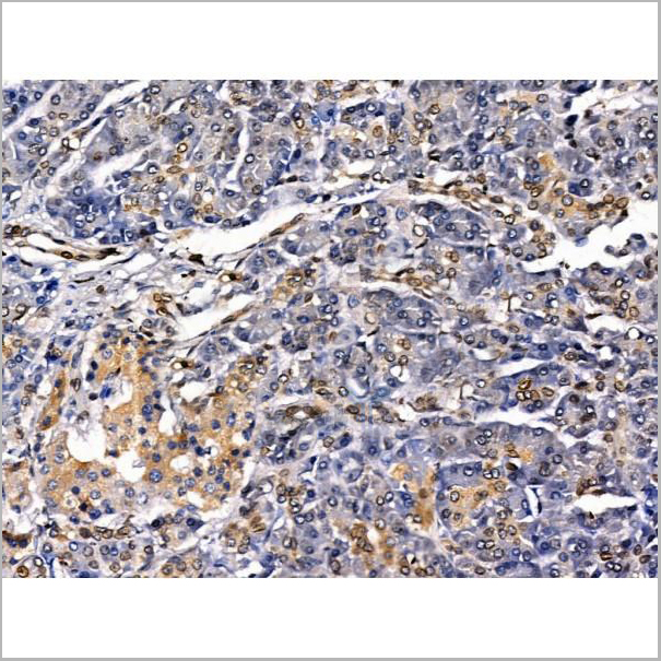
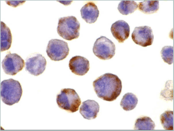
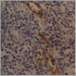
(Formalin fixed, paraffin embedded human breast cancer biopsy stained with Mouse anti Human CD44 antibody followed by HRP-polymer detection and DAB substrate development (high power) following antigen retrieval using citrate buffer at pH6.2)
Application Data
(Formalin fixed, paraffin embedded human breast cancer biopsy stained with Mouse anti Human CD44 antibody followed by HRP-polymer detection and DAB substrate development (high power) following antigen retrieval using citrate buffer at pH6.2)
CD44, Monoclonal Antibody (Cat# AAA12016)
Full Name
MOUSE ANTI HUMAN CD44
Gene Names
CD44; IN; LHR; MC56; MDU2; MDU3; MIC4; Pgp1; CDW44; CSPG8; HCELL; HUTCH-I; ECMR-III
Reactivity
Cynomolgus monkey
Applications
Immunohistochemistry, Flow Cytometry, Immunofluorescence, Immunohistochemistry
Pricing


 FCM (Flow Cytometry)
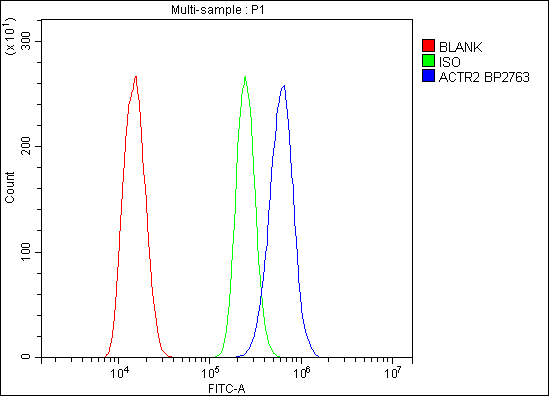
(Figure 6. Flow Cytometry analysis of C6 cells using anti-Arp2/ACTR2 antibody (AAA19284).Overlay histogram showing C6 cells stained with AAA19284 (Blue line). The cells were blocked with 10% normal goat serum. And then incubated with rabbit anti-Arp2/ACTR2 Antibody (AAA19284,1μg/1x106 cells) for 30 min at 20 degree C. DyLight®488 conjugated goat anti-rabbit IgG (5-10μg/1x106 cells) was used as secondary antibody for 30 minutes at 20 degree C. Isotype control antibody (Green line) was rabbit IgG (1μg/1x106) used under the same conditions. Unlabelled sample (Red line) was also used as a control.)
FCM (Flow Cytometry)
(Figure 6. Flow Cytometry analysis of C6 cells using anti-Arp2/ACTR2 antibody (AAA19284).Overlay histogram showing C6 cells stained with AAA19284 (Blue line). The cells were blocked with 10% normal goat serum. And then incubated with rabbit anti-Arp2/ACTR2 Antibody (AAA19284,1μg/1x106 cells) for 30 min at 20 degree C. DyLight®488 conjugated goat anti-rabbit IgG (5-10μg/1x106 cells) was used as secondary antibody for 30 minutes at 20 degree C. Isotype control antibody (Green line) was rabbit IgG (1μg/1x106) used under the same conditions. Unlabelled sample (Red line) was also used as a control.)
Arp2/ACTR2, Polyclonal Antibody (Cat# AAA19284)
Full Name
Anti-Arp2/ACTR2 Antibody
Gene Names
ACTR2; ARP2
Reactivity
Human, Mouse, Monkey, Rat
Applications
WB, IHC-P, ICC, IF, FC/FACS/FCM, EIA
Purity
Immunogen affinity purified.
Pricing


 FCM (Flow Cytometry)
(Figure 8. Flow Cytometry analysis of HEPA1-6 cells using anti-PI-16/PI16 antibody (AAA19330).Overlay histogram showing HEPA1-6 cells stained with AAA19330 (Blue line). The cells were blocked with 10% normal goat serum. And then incubated with rabbit anti-PI-16/PI16 Antibody (AAA19330, 1μg/1x106 cells) for 30 min at 20 degree C. DyLight®488 conjugated goat anti-rabbit IgG (5-10μg/1x106 cells) was used as secondary antibody for 30 minutes at 20 degree C. Isotype control antibody (Green line) was rabbit IgG (1μg/1x106) used under the same conditions. Unlabelled sample (Red line) was also used as a control.)
FCM (Flow Cytometry)
(Figure 8. Flow Cytometry analysis of HEPA1-6 cells using anti-PI-16/PI16 antibody (AAA19330).Overlay histogram showing HEPA1-6 cells stained with AAA19330 (Blue line). The cells were blocked with 10% normal goat serum. And then incubated with rabbit anti-PI-16/PI16 Antibody (AAA19330, 1μg/1x106 cells) for 30 min at 20 degree C. DyLight®488 conjugated goat anti-rabbit IgG (5-10μg/1x106 cells) was used as secondary antibody for 30 minutes at 20 degree C. Isotype control antibody (Green line) was rabbit IgG (1μg/1x106) used under the same conditions. Unlabelled sample (Red line) was also used as a control.)
PI-16/PI16, Polyclonal Antibody (Cat# AAA19330)
Full Name
Anti-PI-16/PI16 Antibody
Gene Names
PI16; PSPBP; CRISP9; MSMBBP
Reactivity
Human, Mouse, Rat, Monkey
Applications
WB, IHC-P, FC/FACS/FCM, EIA
Purity
Immunogen affinity purified.
Pricing


 FCM (Flow Cytometry)
(Figure 7. Flow Cytometry analysis of Hela cells using anti-Aquaporin 3 antibody (AAA11516).Overlay histogram showing Hela cells stained with AAA11516 (Blue line).The cells were blocked with 10% normal goat serum. And then incubated with rabbit anti-Aquaporin 3 Antibody (AAA11516,1ug/1x10^6 cells) for 30 min at 20 degree C. DyLight®488 conjugated goat anti-rabbit IgG (5-10ug/1x10^6 cells) was used as secondary antibody for 30 minutes at 20 degree C. Isotype control antibody (Green line) was rabbit IgG (1ug/1x106) used under the same conditions. Unlabelled sample (Red line) was also used as a control.)
FCM (Flow Cytometry)
(Figure 7. Flow Cytometry analysis of Hela cells using anti-Aquaporin 3 antibody (AAA11516).Overlay histogram showing Hela cells stained with AAA11516 (Blue line).The cells were blocked with 10% normal goat serum. And then incubated with rabbit anti-Aquaporin 3 Antibody (AAA11516,1ug/1x10^6 cells) for 30 min at 20 degree C. DyLight®488 conjugated goat anti-rabbit IgG (5-10ug/1x10^6 cells) was used as secondary antibody for 30 minutes at 20 degree C. Isotype control antibody (Green line) was rabbit IgG (1ug/1x106) used under the same conditions. Unlabelled sample (Red line) was also used as a control.)
Aquaporin 3, Polyclonal Antibody (Cat# AAA11516)
Full Name
Anti-Aquaporin 3 antibody
Gene Names
AQP3; GIL; AQP-3
Reactivity
Human, Mouse, Rat
Applications
Western Blot, Immunohistochemistry, Immunohistochemistry, Immunocytochemistry
Purity
Immunogen affinity purified.
Pricing


 Application Data
(Staining of human peripheral blood granulocytes with Mouse anti Human CD13: Alexa Fluor 647 (AAA11927A647))
Application Data
(Staining of human peripheral blood granulocytes with Mouse anti Human CD13: Alexa Fluor 647 (AAA11927A647))
CD13, Monoclonal Antibody (Cat# AAA11927)
Full Name
MOUSE ANTI HUMAN CD13
Gene Names
ANPEP; APN; CD13; LAP1; P150; PEPN; GP150
Reactivity
Rhesus Monkey
Applications
Immunohistochemistry, Flow Cytometry, Immunoprecipitation
Pricing


 IF (Immunofluorescence)
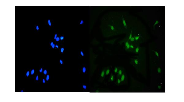
(Figure 7 Immunofluorescence Validation of TMPRSS2 in Rat BrainImmunofluorescent analysis of 4% paraformaldehyde-fixed rat brain labeling TMPRSS2 at 20ug/mL, followed by goat anti-rabbit IgG secondary antibody at 1/500 dilution (green) and DAPI staining (blue).)
IF (Immunofluorescence)
(Figure 7 Immunofluorescence Validation of TMPRSS2 in Rat BrainImmunofluorescent analysis of 4% paraformaldehyde-fixed rat brain labeling TMPRSS2 at 20ug/mL, followed by goat anti-rabbit IgG secondary antibody at 1/500 dilution (green) and DAPI staining (blue).)
TMPRSS2, Polyclonal Antibody (Cat# AAA11038)
Full Name
TMPRSS2 (IN) Antibody
Gene Names
TMPRSS2; PP9284; PRSS10
Reactivity
Human, Mouse, Rat
Predicted species reactivity based on immunogen sequence: Horse (100%); Rabbit (100%); Monkey (100%); Sheep (100%); Gorilla (100%); Cat (100%).
Predicted species reactivity based on immunogen sequence: Horse (100%); Rabbit (100%); Monkey (100%); Sheep (100%); Gorilla (100%); Cat (100%).
Applications
Immunofluorescence, Western Blot
Purity
TMPRSS2 Antibody is affinity chromatography purified via peptide column.
Pricing


 FCM (Flow Cytometry)
(Figure 8. Flow Cytometry analysis of SiHa cells using anti- ASS1 antibody (AAA19369).Overlay histogram showing SiHa cells stained with AAA19369 (Blue line). The cells were blocked with 10% normal goat serum. And then incubated with mouse anti-ASS1 Antibody (AAA19369, 1μg/1x106 cells) for 30 min at 20 degree C. DyLight®488 conjugated goat anti-mouse IgG (BA1126, 5-10μg/1x106 cells) was used as secondary antibody for 30 minutes at 20 degree C. Isotype control antibody (Green line) was mouse IgG (1μg/1x106) used under the same conditions. Unlabelled sample (Red line) was also used as a control.)
FCM (Flow Cytometry)
(Figure 8. Flow Cytometry analysis of SiHa cells using anti- ASS1 antibody (AAA19369).Overlay histogram showing SiHa cells stained with AAA19369 (Blue line). The cells were blocked with 10% normal goat serum. And then incubated with mouse anti-ASS1 Antibody (AAA19369, 1μg/1x106 cells) for 30 min at 20 degree C. DyLight®488 conjugated goat anti-mouse IgG (BA1126, 5-10μg/1x106 cells) was used as secondary antibody for 30 minutes at 20 degree C. Isotype control antibody (Green line) was mouse IgG (1μg/1x106) used under the same conditions. Unlabelled sample (Red line) was also used as a control.)
ASS1, Monoclonal Antibody (Cat# AAA19369)
Full Name
Anti-ASS1 Antibody (monoclonal, 5I5)
Gene Names
ASS1; ASS; CTLN1
Reactivity
Human, Mouse, Rat, Monkey
Applications
WB, IHC-P, ICC, IF, FC/FACS/FCM
Purity
Immunogen affinity purified.
Pricing


 Application Data
(Staining of human peripheral blood lymphocytes with Mouse anti Human HLA ABC:Alexa Fluor 647 (AAA12013A647))
Application Data
(Staining of human peripheral blood lymphocytes with Mouse anti Human HLA ABC:Alexa Fluor 647 (AAA12013A647))
HLA ABC, Monoclonal Antibody (Cat# AAA12013)
Full Name
MOUSE ANTI HUMAN HLA ABC
Reactivity
Baboon, Bovine, Chimpanzee, Cynomolgus monkey, Gorilla, Macaque, Rhesus Monkey, Shrew
Applications
Immunohistochemistry, Flow Cytometry, Immunofluorescence, Immunoprecipitation
Pricing


 Application Data
(Staining of human peripheral blood monocytes with Mouse anti Human CD284: Low Endotoxin)
Application Data
(Staining of human peripheral blood monocytes with Mouse anti Human CD284: Low Endotoxin)
CD284, Monoclonal Antibody (Cat# AAA11948)
Full Name
MOUSE ANTI HUMAN CD284
Gene Names
TLR4; TOLL; CD284; TLR-4; ARMD10
Reactivity
Dog, Guinea Pig, Pig, Rhesus Monkey
Applications
Flow Cytometry, Immunoprecipitation, Western Blot
Pricing


 Application Data
(C:FGFR2/isolectinB4 (C) and FGFR1/isolectinB4 (D) staining of apparent mesenchymal cells and the subpopulation of endothelial cells. Virtually all other dispersed apparent mesenchymal cells express FGFR1 and FGFR2 (merged image in E). F: FGFR2 (F) and FGFR1 (G) staining in clustered cells of epithelial origin (inferred by morphology here) demonstrating that epithelial cells express both FGFR1 and FGFR2 (merged image with DAPI staining in H).)
Application Data
(C:FGFR2/isolectinB4 (C) and FGFR1/isolectinB4 (D) staining of apparent mesenchymal cells and the subpopulation of endothelial cells. Virtually all other dispersed apparent mesenchymal cells express FGFR1 and FGFR2 (merged image in E). F: FGFR2 (F) and FGFR1 (G) staining in clustered cells of epithelial origin (inferred by morphology here) demonstrating that epithelial cells express both FGFR1 and FGFR2 (merged image with DAPI staining in H).)
FGFR2, Polyclonal Antibody (Cat# AAA26855)
Full Name
FGFR2, NT (FGFR2, BEK, KGFR, KSAM, Fibroblast growth factor receptor 2, K-sam, Keratinocyte growth factor receptor, CD332) (Azide free) (HRP)
Gene Names
FGFR2; BEK; JWS; BBDS; CEK3; CFD1; ECT1; KGFR; TK14; TK25; BFR-1; CD332; K-SAM
Reactivity
Human, Monkey, Mouse, Rat
Applications
IHC, EIA, WB
Purity
Purified by Protein G Affinity Chromatography.
Pricing


 FCM (Flow Cytometry)
(Figure 6. Flow Cytometry analysis of HL-60 cells using anti-MCM6 antibody (AAA19264).Overlay histogram showing HL-60 cells stained with AAA19264 (Blue line). The cells were blocked with 10% normal goat serum. And then incubated with rabbit anti-MCM6 Antibody (AAA19264,1μg/1x106 cells) for 30 min at 20 degree C. DyLight®488 conjugated goat anti-rabbit IgG (5-10μg/1x106 cells) was used as secondary antibody for 30 minutes at 20 degree C. Isotype control antibody (Green line) was rabbit IgG (1μg/1x106) used under the same conditions. Unlabelled sample (Red line) was also used as a control.)
FCM (Flow Cytometry)
(Figure 6. Flow Cytometry analysis of HL-60 cells using anti-MCM6 antibody (AAA19264).Overlay histogram showing HL-60 cells stained with AAA19264 (Blue line). The cells were blocked with 10% normal goat serum. And then incubated with rabbit anti-MCM6 Antibody (AAA19264,1μg/1x106 cells) for 30 min at 20 degree C. DyLight®488 conjugated goat anti-rabbit IgG (5-10μg/1x106 cells) was used as secondary antibody for 30 minutes at 20 degree C. Isotype control antibody (Green line) was rabbit IgG (1μg/1x106) used under the same conditions. Unlabelled sample (Red line) was also used as a control.)
MCM6, Polyclonal Antibody (Cat# AAA19264)
Full Name
Anti-MCM6 Antibody
Gene Names
MCM6; Mis5; P105MCM; MCG40308
Reactivity
Human, Mouse, Rat, Monkey
Applications
Western Blot, Immunohistochemistry, Immunocytochemistry, Immunofluorescence, Flow Cytometry, Direct ELISA
Purity
Immunogen affinity purified.
Pricing


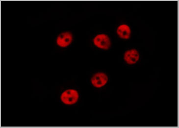
 IF (Immunofluorescence)
(Figure 6. IF analysis of UNG using anti-UNG antibody (AAA19245).UNG was detected in immunocytochemical section of MCF-7 cells. Enzyme antigen retrieval was performed using IHC enzyme antigen retrieval reagent for 15 mins. The cells were blocked with 10% goat serum. And then incubated with 5μg/mL rabbit anti- UNG Antibody (AAA19245) overnight at 4 degree C. DyLight®594 Conjugated Goat Anti-Rabbit IgG (BA1142) was used as secondary antibody at 1:100 dilution and incubated for 30 minutes at 37 degree C. The section was counterstained with DAPI. Visualize using a fluorescence microscope and filter sets appropriate for the label used.)
IF (Immunofluorescence)
(Figure 6. IF analysis of UNG using anti-UNG antibody (AAA19245).UNG was detected in immunocytochemical section of MCF-7 cells. Enzyme antigen retrieval was performed using IHC enzyme antigen retrieval reagent for 15 mins. The cells were blocked with 10% goat serum. And then incubated with 5μg/mL rabbit anti- UNG Antibody (AAA19245) overnight at 4 degree C. DyLight®594 Conjugated Goat Anti-Rabbit IgG (BA1142) was used as secondary antibody at 1:100 dilution and incubated for 30 minutes at 37 degree C. The section was counterstained with DAPI. Visualize using a fluorescence microscope and filter sets appropriate for the label used.)
UNG, Polyclonal Antibody (Cat# AAA19245)
Full Name
Anti-UNG Antibody
Gene Names
UNG; DGU; UDG; UNG1; UNG2; HIGM4; HIGM5; UNG15
Reactivity
Human, Mouse, Rat, Monkey
Applications
Western Blot, Immunohistochemistry, Immunocytochemistry, Immunofluorescence, Flow Cytometry, Direct ELISA
Purity
Immunogen affinity purified.
Pricing


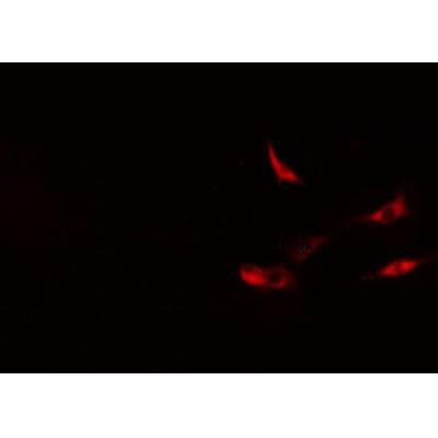 Application Data
(At 25 degree C. The primary antibody was diluted at 1/200 and incubated with the sample for 1 hour at 37 degree C. An Alexa Fluor 594 conjugated goat anti-rabbit IgG (H+L) antibody(Red), diluted at 1/600, was used as secondary antibody.)
Application Data
(At 25 degree C. The primary antibody was diluted at 1/200 and incubated with the sample for 1 hour at 37 degree C. An Alexa Fluor 594 conjugated goat anti-rabbit IgG (H+L) antibody(Red), diluted at 1/600, was used as secondary antibody.)
JAK3, Polyclonal Antibody (Cat# AAA31405)
Full Name
Phospho-JAK3 (Tyr981) Antibody
Gene Names
JAK3; JAKL; LJAK; JAK-3; L-JAK; JAK3_HUMAN
Reactivity
Human, Mouse, Rat, Monkey
Predicted Reactivity: Pig (100%), Bovine (91%), Horse (100%), Sheep (100%), Dog (100%)
Predicted Reactivity: Pig (100%), Bovine (91%), Horse (100%), Sheep (100%), Dog (100%)
Applications
Western Blot, Immunohistochemistry, Immunofluorescence, Immunocytochemistry, Peptide ELISA
Purity
The antibody is from purified rabbit serum by affinity purification via sequential chromatography on phospho-peptide and non-phospho-peptide affinity columns.
Pricing


 FCM (Flow Cytometry)
(Figure 6. Flow Cytometry analysis of MCF-7 cells using anti- PCK2 antibody (AAA19384).Overlay histogram showing MCF-7 cells stained with AAA19384 (Blue line). The cells were blocked with 10% normal goat serum. And then incubated with mouse anti-PCK2 Antibody (AAA19384, 1μg/1x106 cells) for 30 min at 20 degree C. DyLight®488 conjugated goat anti-mouse IgG (BA1126, 5-10μg/1x106 cells) was used as secondary antibody for 30 minutes at 20 degree C. Isotype control antibody (Green line) was mouse IgG (1μg/1x106) used under the same conditions. Unlabelled sample (Red line) was also used as a control.)
FCM (Flow Cytometry)
(Figure 6. Flow Cytometry analysis of MCF-7 cells using anti- PCK2 antibody (AAA19384).Overlay histogram showing MCF-7 cells stained with AAA19384 (Blue line). The cells were blocked with 10% normal goat serum. And then incubated with mouse anti-PCK2 Antibody (AAA19384, 1μg/1x106 cells) for 30 min at 20 degree C. DyLight®488 conjugated goat anti-mouse IgG (BA1126, 5-10μg/1x106 cells) was used as secondary antibody for 30 minutes at 20 degree C. Isotype control antibody (Green line) was mouse IgG (1μg/1x106) used under the same conditions. Unlabelled sample (Red line) was also used as a control.)
PCK2, Monoclonal Antibody (Cat# AAA19384)
Full Name
Anti-PCK2 Antibody (monoclonal, 3F7)
Gene Names
PCK2; PEPCK; PEPCK2; PEPCK-M
Reactivity
Human, Mouse, Rat, Monkey
Applications
WB, IHC-P, ICC, IF, FC/FACS/FCM
Purity
Immunogen affinity purified.
Pricing


 FCM (Flow Cytometry)
(Figure 7. Flow Cytometry analysis of SiHa cells using anti-Aconitase 1/ACO1 antibody (AAA19267).Overlay histogram showing SiHa cells stained with AAA19267 (Blue line). The cells were blocked with 10% normal goat serum. And then incubated with rabbit anti-Aconitase 1/ACO1 Antibody (AAA19267,1μg/1x106 cells) for 30 min at 20 degree C. DyLight®488 conjugated goat anti-rabbit IgG (5-10μg/1x106 cells) was used as secondary antibody for 30 minutes at 20 degree C. Isotype control antibody (Green line) was rabbit IgG (1μg/1x106) used under the same conditions. Unlabelled sample (Red line) was also used as a control.)
FCM (Flow Cytometry)
(Figure 7. Flow Cytometry analysis of SiHa cells using anti-Aconitase 1/ACO1 antibody (AAA19267).Overlay histogram showing SiHa cells stained with AAA19267 (Blue line). The cells were blocked with 10% normal goat serum. And then incubated with rabbit anti-Aconitase 1/ACO1 Antibody (AAA19267,1μg/1x106 cells) for 30 min at 20 degree C. DyLight®488 conjugated goat anti-rabbit IgG (5-10μg/1x106 cells) was used as secondary antibody for 30 minutes at 20 degree C. Isotype control antibody (Green line) was rabbit IgG (1μg/1x106) used under the same conditions. Unlabelled sample (Red line) was also used as a control.)
Aconitase 1/ACO1, Polyclonal Antibody (Cat# AAA19267)
Full Name
Anti-Aconitase 1/ACO1 Antibody
Gene Names
ACO1; IRP1; ACONS; IREB1; IREBP; IREBP1
Reactivity
Human, Mouse, Monkey, Rat
Applications
WB, IHC-P, ICC, IF, FC/FACS/FCM, EIA
Purity
Immunogen affinity purified.
Pricing


 FCM (Flow Cytometry)
(Figure 6. Flow Cytometry analysis of PC-3 cells using anti-MitoNEET/CISD1 antibody (AAA19293).Overlay histogram showing PC-3 cells stained with AAA19293 (Blue line). The cells were blocked with 10% normal goat serum. And then incubated with rabbit anti-MitoNEET/CISD1 Antibody (AAA19293,1μg/1x106 cells) for 30 min at 20 degree C. DyLight®488 conjugated goat anti-rabbit IgG (5-10μg/1x106 cells) was used as secondary antibody for 30 minutes at 20 degree C. Isotype control antibody (Green line) was rabbit IgG (1μg/1x106) used under the same conditions. Unlabelled sample (Red line) was also used as a control.)
FCM (Flow Cytometry)
(Figure 6. Flow Cytometry analysis of PC-3 cells using anti-MitoNEET/CISD1 antibody (AAA19293).Overlay histogram showing PC-3 cells stained with AAA19293 (Blue line). The cells were blocked with 10% normal goat serum. And then incubated with rabbit anti-MitoNEET/CISD1 Antibody (AAA19293,1μg/1x106 cells) for 30 min at 20 degree C. DyLight®488 conjugated goat anti-rabbit IgG (5-10μg/1x106 cells) was used as secondary antibody for 30 minutes at 20 degree C. Isotype control antibody (Green line) was rabbit IgG (1μg/1x106) used under the same conditions. Unlabelled sample (Red line) was also used as a control.)
MitoNEET/CISD1, Polyclonal Antibody (Cat# AAA19293)
Full Name
Anti-MitoNEET/CISD1 Antibody
Gene Names
CISD1; ZCD1; MDS029; C10orf70; mitoNEET
Reactivity
Human, Mouse, Rat, Monkey
Applications
WB, IHC-P, FC/FACS/FCM, EIA
Purity
Immunogen affinity purified.
Pricing


 FCM (Flow Cytometry)
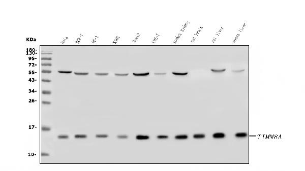
(Figure 10. Flow Cytometry analysis of A431 cells using anti-TIMM8A/DDP antibody (AAA19317).Overlay histogram showing A431 cells stained with AAA19317 (Blue line). The cells were blocked with 10% normal goat serum. And then incubated with rabbit anti-TIMM8A/DDP Antibody (AAA19317, 1μg/1x106 cells) for 30 min at 20 degree C. DyLight®488 conjugated goat anti-rabbit IgG (5-10μg/1x106 cells) was used as secondary antibody for 30 minutes at 20 degree C. Isotype control antibody (Green line) was rabbit IgG (1μg/1x106) used under the same conditions. Unlabelled sample (Red line) was also used as a control.)
FCM (Flow Cytometry)
(Figure 10. Flow Cytometry analysis of A431 cells using anti-TIMM8A/DDP antibody (AAA19317).Overlay histogram showing A431 cells stained with AAA19317 (Blue line). The cells were blocked with 10% normal goat serum. And then incubated with rabbit anti-TIMM8A/DDP Antibody (AAA19317, 1μg/1x106 cells) for 30 min at 20 degree C. DyLight®488 conjugated goat anti-rabbit IgG (5-10μg/1x106 cells) was used as secondary antibody for 30 minutes at 20 degree C. Isotype control antibody (Green line) was rabbit IgG (1μg/1x106) used under the same conditions. Unlabelled sample (Red line) was also used as a control.)
TIMM8A/DDP, Polyclonal Antibody (Cat# AAA19317)
Full Name
Anti-TIMM8A/DDP Antibody
Gene Names
TIMM8A; DDP; MTS; DDP1; DFN1; TIM8
Reactivity
Human, Mouse, Rat, Monkey
Applications
WB, IHC-P, ICC, IF, EIA
Purity
Immunogen affinity purified.
Pricing


 FCM (Flow Cytometry)
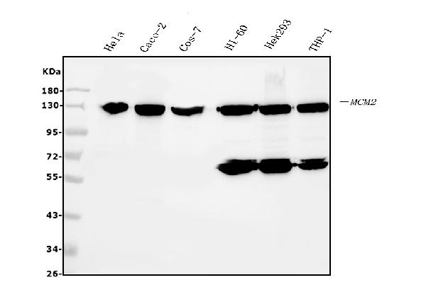
(Figure 6. Flow Cytometry analysis of HL-60 cells using anti- MCM2 antibody (AAA19351).Overlay histogram showing HL-60 cells stained with AAA19351 (Blue line). The cells were blocked with 10% normal goat serum. And then incubated with mouse anti-MCM2 Antibody (AAA19351, 1μg/1x106 cells) for 30 min at 20 degree C. DyLight®488 conjugated goat anti-mouse IgG (BA1126, 5-10μg/1x106 cells) was used as secondary antibody for 30 minutes at 20 degree C. Isotype control antibody (Green line) was mouse IgG (1μg/1x106) used under the same conditions. Unlabelled sample (Red line) was also used as a control.)
FCM (Flow Cytometry)
(Figure 6. Flow Cytometry analysis of HL-60 cells using anti- MCM2 antibody (AAA19351).Overlay histogram showing HL-60 cells stained with AAA19351 (Blue line). The cells were blocked with 10% normal goat serum. And then incubated with mouse anti-MCM2 Antibody (AAA19351, 1μg/1x106 cells) for 30 min at 20 degree C. DyLight®488 conjugated goat anti-mouse IgG (BA1126, 5-10μg/1x106 cells) was used as secondary antibody for 30 minutes at 20 degree C. Isotype control antibody (Green line) was mouse IgG (1μg/1x106) used under the same conditions. Unlabelled sample (Red line) was also used as a control.)
MCM2, Monoclonal Antibody (Cat# AAA19351)
Full Name
Anti-MCM2 Antibody (monoclonal, 11C4)
Gene Names
MCM2; BM28; CCNL1; CDCL1; cdc19; D3S3194; MITOTIN
Reactivity
Human, Monkey
Applications
WB, IHC-P, ICC, IF, FC/FACS/FCM
Purity
Immunogen affinity purified.
Pricing


 Application Data
(C:FGFR2/isolectinB4 (C) and FGFR1/isolectinB4 (D) staining of apparent mesenchymal cells and the subpopulation of endothelial cells. Virtually all other dispersed apparent mesenchymal cells express FGFR1 and FGFR2 (merged image in E). F: FGFR2 (F) and FGFR1 (G) staining in clustered cells of epithelial origin (inferred by morphology here) demonstrating that epithelial cells express both FGFR1 and FGFR2 (merged image with DAPI staining in H).)
Application Data
(C:FGFR2/isolectinB4 (C) and FGFR1/isolectinB4 (D) staining of apparent mesenchymal cells and the subpopulation of endothelial cells. Virtually all other dispersed apparent mesenchymal cells express FGFR1 and FGFR2 (merged image in E). F: FGFR2 (F) and FGFR1 (G) staining in clustered cells of epithelial origin (inferred by morphology here) demonstrating that epithelial cells express both FGFR1 and FGFR2 (merged image with DAPI staining in H).)
FGFR2, Polyclonal Antibody (Cat# AAA14790)
Full Name
FGFR2, NT (FGFR2, BEK, KGFR, KSAM, Fibroblast growth factor receptor 2, K-sam, Keratinocyte growth factor receptor, CD332)
Gene Names
FGFR2; BEK; JWS; BBDS; CEK3; CFD1; ECT1; KGFR; TK14; TK25; BFR-1; CD332; K-SAM
Reactivity
Human, Monkey, Mouse, Rat
Applications
EL/EIA, WB, IHC, FC/FACS, IF
Purity
Affinity Purified
Purified by Protein A affinity chromatography.
Purified by Protein A affinity chromatography.
Pricing


 FCM (Flow Cytometry)
(Figure 9. Flow Cytometry analysis of THP-1 cells using anti-CHCHD10 antibody (AAA19310).Overlay histogram showing THP-1 cells stained with AAA19310 (Blue line). The cells were blocked with 10% normal goat serum. And then incubated with rabbit anti-CHCHD10 Antibody (AAA19310, 1μg/1x106 cells) for 30 min at 20 degree C. DyLight®488 conjugated goat anti-rabbit IgG (5-10μg/1x106 cells) was used as secondary antibody for 30 minutes at 20 degree C. Isotype control antibody (Green line) was rabbit IgG (1μg/1x106) used under the same conditions. Unlabelled sample (Red line) was also used as a control.)
FCM (Flow Cytometry)
(Figure 9. Flow Cytometry analysis of THP-1 cells using anti-CHCHD10 antibody (AAA19310).Overlay histogram showing THP-1 cells stained with AAA19310 (Blue line). The cells were blocked with 10% normal goat serum. And then incubated with rabbit anti-CHCHD10 Antibody (AAA19310, 1μg/1x106 cells) for 30 min at 20 degree C. DyLight®488 conjugated goat anti-rabbit IgG (5-10μg/1x106 cells) was used as secondary antibody for 30 minutes at 20 degree C. Isotype control antibody (Green line) was rabbit IgG (1μg/1x106) used under the same conditions. Unlabelled sample (Red line) was also used as a control.)
CHCHD10, Polyclonal Antibody (Cat# AAA19310)
Full Name
Anti-CHCHD10 Antibody
Gene Names
CHCHD10; FTDALS2; N27C7-4; C22orf16
Reactivity
Human, Mouse, Rat, Monkey
Applications
WB, IHC-P, ICC, IF, FC/FACS/FCM, EIA
Purity
Immunogen affinity purified.
Pricing


 WB (Western Blot)
(The anti-SUMO2/3 C-term Pab is used in Western blot to detect SUMO2/3 in HeLa cell lysate.)
WB (Western Blot)
(The anti-SUMO2/3 C-term Pab is used in Western blot to detect SUMO2/3 in HeLa cell lysate.)
SUMO2/3, Polyclonal Antibody (Cat# AAA28663)
Full Name
SUMO2/3 Antibody (C-term)
Gene Names
SUMO3; SMT3A; Smt3B; SMT3H1; SUMO-3
Reactivity
Human, mouse (Predicted Reactivity: Xenopus, Zebrafish, Bovine, Chicken, Hamster, Monkey, Pig, Rat)
Applications
IF, EIA, WB, IHC
Purity
Purified Rabbit Polyclonal Antibody (Pab)
Pricing


 Application Data
(C:FGFR2/isolectinB4 (C) and FGFR1/isolectinB4 (D) staining of apparent mesenchymal cells and the subpopulation of endothelial cells. Virtually all other dispersed apparent mesenchymal cells express FGFR1 and FGFR2 (merged image in E). F: FGFR2 (F) and FGFR1 (G) staining in clustered cells of epithelial origin (inferred by morphology here) demonstrating that epithelial cells express both FGFR1 and FGFR2 (merged image with DAPI staining in H).)
Application Data
(C:FGFR2/isolectinB4 (C) and FGFR1/isolectinB4 (D) staining of apparent mesenchymal cells and the subpopulation of endothelial cells. Virtually all other dispersed apparent mesenchymal cells express FGFR1 and FGFR2 (merged image in E). F: FGFR2 (F) and FGFR1 (G) staining in clustered cells of epithelial origin (inferred by morphology here) demonstrating that epithelial cells express both FGFR1 and FGFR2 (merged image with DAPI staining in H).)
FGFR2, Polyclonal Antibody (Cat# AAA26854)
Full Name
FGFR2, NT (FGFR2, BEK, KGFR, KSAM, Fibroblast growth factor receptor 2, K-sam, Keratinocyte growth factor receptor, CD332) (FITC)
Gene Names
FGFR2; BEK; JWS; BBDS; CEK3; CFD1; ECT1; KGFR; TK14; TK25; BFR-1; CD332; K-SAM
Reactivity
Human, Monkey, Mouse, Rat
Applications
WB, IHC, IF, FC/FACS
Purity
Purified by Protein G Affinity Chromatography.
Pricing


 Application Data
(C:FGFR2/isolectinB4 (C) and FGFR1/isolectinB4 (D) staining of apparent mesenchymal cells and the subpopulation of endothelial cells. Virtually all other dispersed apparent mesenchymal cells express FGFR1 and FGFR2 (merged image in E). F: FGFR2 (F) and FGFR1 (G) staining in clustered cells of epithelial origin (inferred by morphology here) demonstrating that epithelial cells express both FGFR1 and FGFR2 (merged image with DAPI staining in H).)
Application Data
(C:FGFR2/isolectinB4 (C) and FGFR1/isolectinB4 (D) staining of apparent mesenchymal cells and the subpopulation of endothelial cells. Virtually all other dispersed apparent mesenchymal cells express FGFR1 and FGFR2 (merged image in E). F: FGFR2 (F) and FGFR1 (G) staining in clustered cells of epithelial origin (inferred by morphology here) demonstrating that epithelial cells express both FGFR1 and FGFR2 (merged image with DAPI staining in H).)
FGFR2, Polyclonal Antibody (Cat# AAA26851)
Full Name
FGFR2, NT (FGFR2, BEK, KGFR, KSAM, Fibroblast growth factor receptor 2, K-sam, Keratinocyte growth factor receptor, CD332) (AP)
Gene Names
FGFR2; BEK; JWS; BBDS; CEK3; CFD1; ECT1; KGFR; TK14; TK25; BFR-1; CD332; K-SAM
Reactivity
Human, Monkey, Mouse, Rat
Applications
IF, EIA, IHC, WB
Purity
Purified by Protein G Affinity Chromatography.
Pricing


 Application Data
(Published customer image: Mouse anti Human CD49d antibody, clone HP2/1 used for binding efficiency determinationImage caption:Binding efficiencies (BE) of different a4beta7 molecules composed of distinct a4 mutants to monoclonal antibodies against a4, beta7 or the a4beta7 heterodimer. Binding efficiency is determined by the ratio between the mean fluorescence of antibody binding to each a4 molecule and of the binding in a mock-transfected cell culture (see Materials and Methods for details). Dark gray bars represent binding to the human (wild type) a4 clone, whereas light gray bars are those of binding to the different a4 mutants (as shown in the x-axis). a4 mutants which included substitutions at codon 201 are boxed. A, binding of anti-a4 2b4 antibody. B, binding of anti-a4 HP2/1 antibody. C, BE of different anti-a4 and beta7 antibodies to the human a4 and the quintuple a4 mutant (5 aa mut). Bars represent the range of standard errors deduced from triplicate experiments. p-values of Student's t tests are shown above each comparison. NS, non-significant (> 0.05).From:Darc M, Hait SH, Soares EA, Cicala C, Seuanez HN, et al. (2011) Polymorphisms in the a4 Integrin of Neotropical Primates: Insights for Binding of Natural Ligands and HIV-1 gp120 to the Human a4beta7. PLoS ONE 6(9): e24461.)
Application Data
(Published customer image: Mouse anti Human CD49d antibody, clone HP2/1 used for binding efficiency determinationImage caption:Binding efficiencies (BE) of different a4beta7 molecules composed of distinct a4 mutants to monoclonal antibodies against a4, beta7 or the a4beta7 heterodimer. Binding efficiency is determined by the ratio between the mean fluorescence of antibody binding to each a4 molecule and of the binding in a mock-transfected cell culture (see Materials and Methods for details). Dark gray bars represent binding to the human (wild type) a4 clone, whereas light gray bars are those of binding to the different a4 mutants (as shown in the x-axis). a4 mutants which included substitutions at codon 201 are boxed. A, binding of anti-a4 2b4 antibody. B, binding of anti-a4 HP2/1 antibody. C, BE of different anti-a4 and beta7 antibodies to the human a4 and the quintuple a4 mutant (5 aa mut). Bars represent the range of standard errors deduced from triplicate experiments. p-values of Student's t tests are shown above each comparison. NS, non-significant (> 0.05).From:Darc M, Hait SH, Soares EA, Cicala C, Seuanez HN, et al. (2011) Polymorphisms in the a4 Integrin of Neotropical Primates: Insights for Binding of Natural Ligands and HIV-1 gp120 to the Human a4beta7. PLoS ONE 6(9): e24461.)
CD49d, Monoclonal Antibody (Cat# AAA12002)
Full Name
MOUSE ANTI HUMAN CD49d
Gene Names
ITGA4; IA4; CD49D
Reactivity
Bovine, Cat, Cynomolgus monkey, Goat, Horse, Llama, Mink, Mustelid, Pig, Rabbit, Rat, Rhesus Monkey
Applications
Immunohistochemistry, Flow Cytometry, Functional Assay, Immunoprecipitation
Pricing


 IF (Immunofluorescence)
(Figure 15. IF analysis of LSM2 using anti- LSM2 antibody (AAA19316).LSM2 was detected in immunocytochemical section of MCF-7 cells. Enzyme antigen retrieval was performed using IHC enzyme antigen retrieval reagent for 15 mins. The cells were blocked with 10% goat serum. And then incubated with 5μg/mL rabbit anti- LSM2 Antibody (AAA19316) overnight at 4 degree C. DyLight®488 Conjugated Goat Anti-Rabbit IgG was used as secondary antibody at 1:100 dilution and incubated for 30 minutes at 37 degree C. The section was counterstained with DAPI. Visualize using a fluorescence microscope and filter sets appropriate for the label used.)
IF (Immunofluorescence)
(Figure 15. IF analysis of LSM2 using anti- LSM2 antibody (AAA19316).LSM2 was detected in immunocytochemical section of MCF-7 cells. Enzyme antigen retrieval was performed using IHC enzyme antigen retrieval reagent for 15 mins. The cells were blocked with 10% goat serum. And then incubated with 5μg/mL rabbit anti- LSM2 Antibody (AAA19316) overnight at 4 degree C. DyLight®488 Conjugated Goat Anti-Rabbit IgG was used as secondary antibody at 1:100 dilution and incubated for 30 minutes at 37 degree C. The section was counterstained with DAPI. Visualize using a fluorescence microscope and filter sets appropriate for the label used.)
LSM2, Polyclonal Antibody (Cat# AAA19316)
Full Name
Anti-LSM2 Antibody
Gene Names
LSM2; G7B; snRNP; C6orf28; YBL026W
Reactivity
Human, Mouse, Rat, Monkey
Applications
WB, IHC-P, ICC, IF, FC/FACS/FCM
Purity
Immunogen affinity purified.
Pricing


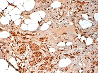
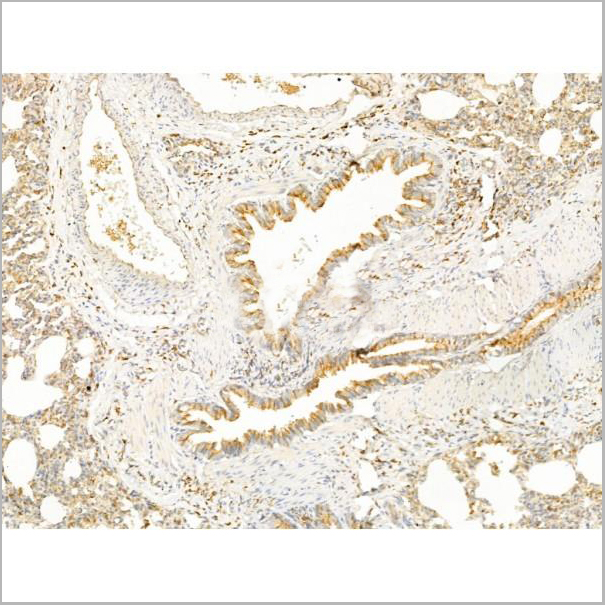
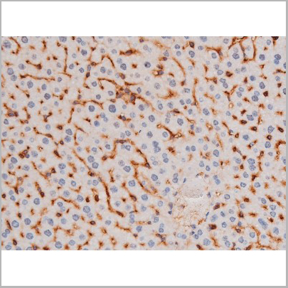
 IHC (Immunohistchemistry)
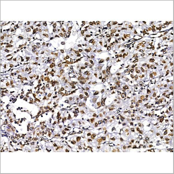
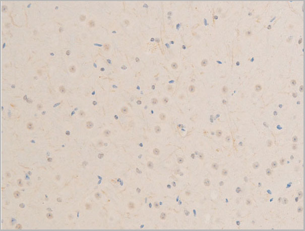
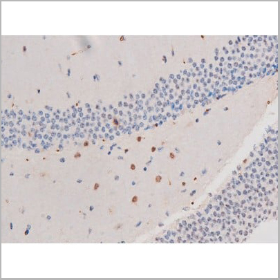
(Figure 6. IHC analysis of PP2A-alpha/PPP2CA using anti-PP2A-alpha/PPP2CA antibody (AAA19366).PP2A-alpha/PPP2CA was detected in paraffin-embedded section of rat brain tissue. Heat mediated antigen retrieval was performed in EDTA buffer (pH8. 0, epitope retrieval solution). The tissue section was blocked with 10% goat serum. The tissue section was then incubated with 1μg/ml mouse anti-PP2A-alpha/PPP2CA Antibody (AAA19366) overnight at 4 degree C. Biotinylated goat anti-mouse IgG was used as secondary antibody and incubated for 30 minutes at 37 degree C. The tissue section was developed using Strepavidin-Biotin-Complex (SABC) (Catalog # with DAB as the chromogen.)
IHC (Immunohistchemistry)
(Figure 6. IHC analysis of PP2A-alpha/PPP2CA using anti-PP2A-alpha/PPP2CA antibody (AAA19366).PP2A-alpha/PPP2CA was detected in paraffin-embedded section of rat brain tissue. Heat mediated antigen retrieval was performed in EDTA buffer (pH8. 0, epitope retrieval solution). The tissue section was blocked with 10% goat serum. The tissue section was then incubated with 1μg/ml mouse anti-PP2A-alpha/PPP2CA Antibody (AAA19366) overnight at 4 degree C. Biotinylated goat anti-mouse IgG was used as secondary antibody and incubated for 30 minutes at 37 degree C. The tissue section was developed using Strepavidin-Biotin-Complex (SABC) (Catalog # with DAB as the chromogen.)
PP2A-alpha/PPP2CA, Monoclonal Antibody (Cat# AAA19366)
Full Name
Anti-PP2A-alpha/PPP2CA Antibody (monoclonal, 3B6)
Gene Names
PPP2CA; RP-C; PP2Ac; PP2CA; PP2Calpha
Reactivity
Human, Monkey, Mouse, Rat
Applications
WB, IHC-P, ICC, IF, FC/FACS/FCM
Purity
Immunogen affinity purified.
Pricing


 FCM (Flow Cytometry)
(Figure 8. Flow Cytometry analysis of SiHa cells using anti- ASS1 antibody (AAA19370).Overlay histogram showing SiHa cells stained with AAA19370 (Blue line). The cells were blocked with 10% normal goat serum. And then incubated with mouse anti-ASS1 Antibody (AAA19370, 1μg/1x106 cells) for 30 min at 20 degree C. DyLight®488 conjugated goat anti-mouse IgG (BA1126, 5-10μg/1x106 cells) was used as secondary antibody for 30 minutes at 20 degree C. Isotype control antibody (Green line) was mouse IgG (1μg/1x106) used under the same conditions. Unlabelled sample (Red line) was also used as a control.)
FCM (Flow Cytometry)
(Figure 8. Flow Cytometry analysis of SiHa cells using anti- ASS1 antibody (AAA19370).Overlay histogram showing SiHa cells stained with AAA19370 (Blue line). The cells were blocked with 10% normal goat serum. And then incubated with mouse anti-ASS1 Antibody (AAA19370, 1μg/1x106 cells) for 30 min at 20 degree C. DyLight®488 conjugated goat anti-mouse IgG (BA1126, 5-10μg/1x106 cells) was used as secondary antibody for 30 minutes at 20 degree C. Isotype control antibody (Green line) was mouse IgG (1μg/1x106) used under the same conditions. Unlabelled sample (Red line) was also used as a control.)
ASS1, Monoclonal Antibody (Cat# AAA19370)
Full Name
Anti-ASS1 Antibody (monoclonal, 7I9)
Gene Names
ASS1; ASS; CTLN1
Reactivity
Human, Mouse, Rat, Monkey
Applications
WB, IHC-P, ICC, IF, FC/FACS/FCM
Purity
Immunogen affinity purified.
Pricing


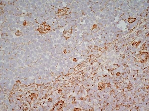
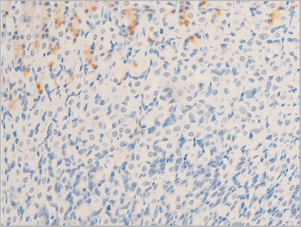
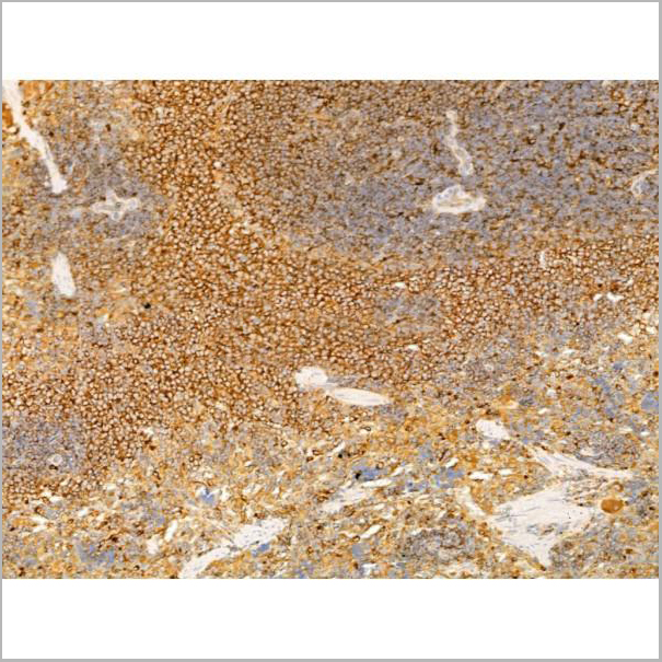
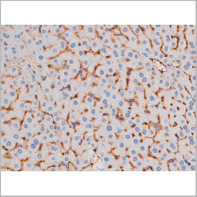
 IHC (Immunohistchemistry)
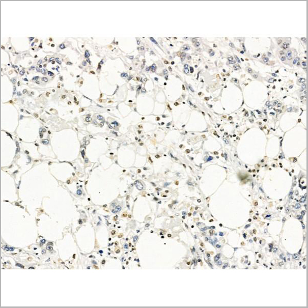
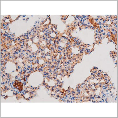
(Figure 6. IHC analysis of CD13/ANPEP using anti-CD13/ANPEP antibody (AAA19371).CD13/ANPEP was detected in paraffin-embedded section of rat kidney tissue. Heat mediated antigen retrieval was performed in EDTA buffer (pH8. 0, epitope retrieval solution). The tissue section was blocked with 10% goat serum. The tissue section was then incubated with 2μg/ml mouse anti-CD13/ANPEP Antibody (AAA19371) overnight at 4 degree C. Biotinylated goat anti-mouse IgG was used as secondary antibody and incubated for 30 minutes at 37 degree C. The tissue section was developed using Strepavidin-Biotin-Complex (SABC) (Catalog # with DAB as the chromogen.)
IHC (Immunohistchemistry)
(Figure 6. IHC analysis of CD13/ANPEP using anti-CD13/ANPEP antibody (AAA19371).CD13/ANPEP was detected in paraffin-embedded section of rat kidney tissue. Heat mediated antigen retrieval was performed in EDTA buffer (pH8. 0, epitope retrieval solution). The tissue section was blocked with 10% goat serum. The tissue section was then incubated with 2μg/ml mouse anti-CD13/ANPEP Antibody (AAA19371) overnight at 4 degree C. Biotinylated goat anti-mouse IgG was used as secondary antibody and incubated for 30 minutes at 37 degree C. The tissue section was developed using Strepavidin-Biotin-Complex (SABC) (Catalog # with DAB as the chromogen.)
CD13/ANPEP, Monoclonal Antibody (Cat# AAA19371)
Full Name
Anti-CD13/ANPEP Antibody (monoclonal, 5B9)
Gene Names
ANPEP; APN; CD13; LAP1; P150; PEPN; GP150
Reactivity
Human, Rat, Monkey
Applications
WB, IHC-P
Purity
Immunogen affinity purified.
Pricing


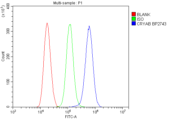 FCM (Flow Cytometry)
(Figure 13. Flow Cytometry analysis of THP-1 cells using anti-Alpha B Crystallin/CRYAB antibody (AAA19277).Overlay histogram showing THP-1 cells stained with AAA19277 (Blue line). The cells were blocked with 10% normal goat serum. And then incubated with rabbit anti-Alpha B Crystallin/CRYAB Antibody (AAA19277, 1μg/1x106 cells) for 30 min at 20 degree C. DyLight®488 conjugated goat anti-rabbit IgG (5-10μg/1x106 cells) was used as secondary antibody for 30 minutes at 20 degree C. Isotype control antibody (Green line) was rabbit IgG (1μg/1x106) used under the same conditions. Unlabelled sample (Red line) was also used as a control.)
FCM (Flow Cytometry)
(Figure 13. Flow Cytometry analysis of THP-1 cells using anti-Alpha B Crystallin/CRYAB antibody (AAA19277).Overlay histogram showing THP-1 cells stained with AAA19277 (Blue line). The cells were blocked with 10% normal goat serum. And then incubated with rabbit anti-Alpha B Crystallin/CRYAB Antibody (AAA19277, 1μg/1x106 cells) for 30 min at 20 degree C. DyLight®488 conjugated goat anti-rabbit IgG (5-10μg/1x106 cells) was used as secondary antibody for 30 minutes at 20 degree C. Isotype control antibody (Green line) was rabbit IgG (1μg/1x106) used under the same conditions. Unlabelled sample (Red line) was also used as a control.)
Alpha B Crystallin/CRYAB, Polyclonal Antibody (Cat# AAA19277)
Full Name
Anti-Alpha B Crystallin/CRYAB Antibody
Gene Names
CRYAB; CRYA2; CTPP2; HSPB5; CMD1II
Reactivity
Human, Mouse, Rat, Monkey
Applications
WB, IHC-P, ICC, IF, FC/FACS/FCM, EIA
Purity
Immunogen affinity purified.
Pricing


 Application Data
(C:FGFR2/isolectinB4 (C) and FGFR1/isolectinB4 (D) staining of apparent mesenchymal cells and the subpopulation of endothelial cells. Virtually all other dispersed apparent mesenchymal cells express FGFR1 and FGFR2 (merged image in E). F: FGFR2 (F) and FGFR1 (G) staining in clustered cells of epithelial origin (inferred by morphology here) demonstrating that epithelial cells express both FGFR1 and FGFR2 (merged image with DAPI staining in H).)
Application Data
(C:FGFR2/isolectinB4 (C) and FGFR1/isolectinB4 (D) staining of apparent mesenchymal cells and the subpopulation of endothelial cells. Virtually all other dispersed apparent mesenchymal cells express FGFR1 and FGFR2 (merged image in E). F: FGFR2 (F) and FGFR1 (G) staining in clustered cells of epithelial origin (inferred by morphology here) demonstrating that epithelial cells express both FGFR1 and FGFR2 (merged image with DAPI staining in H).)
FGFR2, Polyclonal Antibody (Cat# AAA26856)
Full Name
FGFR2, NT (FGFR2, BEK, KGFR, KSAM, Fibroblast growth factor receptor 2, K-sam, Keratinocyte growth factor receptor, CD332) (PE)
Gene Names
FGFR2; BEK; JWS; BBDS; CEK3; CFD1; ECT1; KGFR; TK14; TK25; BFR-1; CD332; K-SAM
Reactivity
Human, Monkey, Mouse, Rat
Applications
WB, IHC, IF, FC/FACS
Purity
Purified by Protein G Affinity Chromatography.
Pricing


 FCM (Flow Cytometry)
(Figure 7. Flow Cytometry analysis of HEPG2 cells using anti-ALDH1L1 antibody (AAA19297).Overlay histogram showing HEPG2 cells stained with AAA19297 (Blue line). The cells were blocked with 10% normal goat serum. And then incubated with rabbit anti-ALDH1L1 Antibody (AAA19297 1μg/1x106 cells) for 30 min at 20 degree C. DyLight®488 conjugated goat anti-rabbit IgG (5-10μg/1x106 cells) was used as secondary antibody for 30 minutes at 20 degree C. Isotype control antibody (Green line) was rabbit IgG (1μg/1x106) used under the same conditions. Unlabelled sample (Red line) was also used as a control.)
FCM (Flow Cytometry)
(Figure 7. Flow Cytometry analysis of HEPG2 cells using anti-ALDH1L1 antibody (AAA19297).Overlay histogram showing HEPG2 cells stained with AAA19297 (Blue line). The cells were blocked with 10% normal goat serum. And then incubated with rabbit anti-ALDH1L1 Antibody (AAA19297 1μg/1x106 cells) for 30 min at 20 degree C. DyLight®488 conjugated goat anti-rabbit IgG (5-10μg/1x106 cells) was used as secondary antibody for 30 minutes at 20 degree C. Isotype control antibody (Green line) was rabbit IgG (1μg/1x106) used under the same conditions. Unlabelled sample (Red line) was also used as a control.)
ALDH1L1, Polyclonal Antibody (Cat# AAA19297)
Full Name
Anti-ALDH1L1 Antibody
Gene Names
ALDH1L1; FDH; FTHFD; 10-fTHF; 10-FTHFDH
Reactivity
Human, Mouse, Rat, Monkey
Applications
WB, IHC-P, ICC, IF, FC/FACS/FCM, EIA
Purity
Immunogen affinity purified.
Pricing


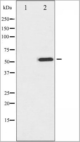
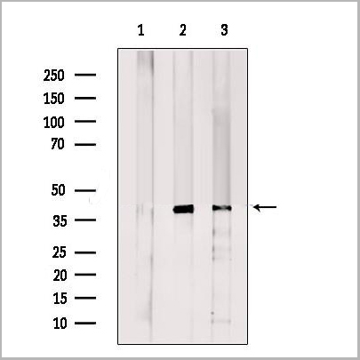
 WB (Western Blot)
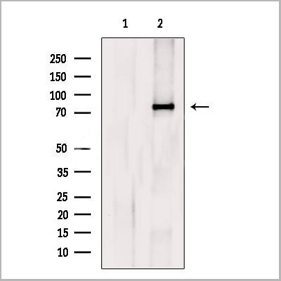
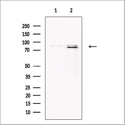
(Figure 1. Western blot analysis of CISD2 using anti-CISD2 antibody (AAA19311).Electrophoresis was performed on a 5-20% SDS-PAGE gel at 70V (Stacking gel) / 90V (Resolving gel) for 2-3 hours. The sample well of each lane was loaded with 50ug of sample under reducing conditions.Lane 1: human HEK293 whole cell lysatesLane 2: human HELA whole cell lysatesLane 3: human MCF-7 whole cell lysatesLane 4: monkey kidney tissue lysatesLane 5: human SW620 whole cell lysatesLane 6: human Raji whole cell lysatesLane 7: rat kidney tissue lysatesLane 8: mouse kidney tissue lysates.After Electrophoresis, proteins were transferred to a Nitrocellulose membrane at 150mA for 50-90 minutes. Blocked the membrane with 5% Non-fat Milk/ TBS for 1. 5 hour at RT. The membrane was incubated with rabbit anti-CISD2 antigen affinity purified polyclonal antibody (Catalog # AAA19311) at 0. 5 μg/mL overnight at 4 degree C, then washed with TBS-0. 1%Tween 3 times with 5 minutes each and probed with a goat anti-rabbit IgG-HRP secondary antibody at a dilution of 1:10000 for 1. 5 hour at RT. The signal is developed using an Enhanced Chemiluminescent detection (ECL) kit (Catalog # with Tanon 5200 system. A specific band was detected for CISD2 at approximately 15KD. The expected band size for CISD2 is at 15KD.)
WB (Western Blot)
(Figure 1. Western blot analysis of CISD2 using anti-CISD2 antibody (AAA19311).Electrophoresis was performed on a 5-20% SDS-PAGE gel at 70V (Stacking gel) / 90V (Resolving gel) for 2-3 hours. The sample well of each lane was loaded with 50ug of sample under reducing conditions.Lane 1: human HEK293 whole cell lysatesLane 2: human HELA whole cell lysatesLane 3: human MCF-7 whole cell lysatesLane 4: monkey kidney tissue lysatesLane 5: human SW620 whole cell lysatesLane 6: human Raji whole cell lysatesLane 7: rat kidney tissue lysatesLane 8: mouse kidney tissue lysates.After Electrophoresis, proteins were transferred to a Nitrocellulose membrane at 150mA for 50-90 minutes. Blocked the membrane with 5% Non-fat Milk/ TBS for 1. 5 hour at RT. The membrane was incubated with rabbit anti-CISD2 antigen affinity purified polyclonal antibody (Catalog # AAA19311) at 0. 5 μg/mL overnight at 4 degree C, then washed with TBS-0. 1%Tween 3 times with 5 minutes each and probed with a goat anti-rabbit IgG-HRP secondary antibody at a dilution of 1:10000 for 1. 5 hour at RT. The signal is developed using an Enhanced Chemiluminescent detection (ECL) kit (Catalog # with Tanon 5200 system. A specific band was detected for CISD2 at approximately 15KD. The expected band size for CISD2 is at 15KD.)
CISD2, Polyclonal Antibody (Cat# AAA19311)
Full Name
Anti-CISD2 Antibody
Gene Names
CISD2; ERIS; WFS2; ZCD2; NAF-1; Miner1
Reactivity
Human, Mouse, Rat, Monkey
Applications
WB, IHC-P, ICC, IF, FC/FACS/FCM, EIA
Purity
Immunogen affinity purified.
Pricing


 FCM (Flow Cytometry)
(Figure 13. Flow Cytometry analysis of MCF-7 cells using anti-EHD3 antibody (AAA19295).Overlay histogram showing MCF-7 cells stained with AAA19295 (Blue line). The cells were blocked with 10% normal goat serum. And then incubated with rabbit anti-EHD3 Antibody (AAA19295, 1μg/1x106 cells) for 30 min at 20 degree C. DyLight®488 conjugated goat anti-rabbit IgG (5-10μg/1x106 cells) was used as secondary antibody for 30 minutes at 20 degree C. Isotype control antibody (Green line) was rabbit IgG (1μg/1x106) used under the same conditions. Unlabelled sample (Red line) was also used as a control.)
FCM (Flow Cytometry)
(Figure 13. Flow Cytometry analysis of MCF-7 cells using anti-EHD3 antibody (AAA19295).Overlay histogram showing MCF-7 cells stained with AAA19295 (Blue line). The cells were blocked with 10% normal goat serum. And then incubated with rabbit anti-EHD3 Antibody (AAA19295, 1μg/1x106 cells) for 30 min at 20 degree C. DyLight®488 conjugated goat anti-rabbit IgG (5-10μg/1x106 cells) was used as secondary antibody for 30 minutes at 20 degree C. Isotype control antibody (Green line) was rabbit IgG (1μg/1x106) used under the same conditions. Unlabelled sample (Red line) was also used as a control.)
EHD3, Polyclonal Antibody (Cat# AAA19295)
Full Name
Anti-EHD3 Antibody
Gene Names
EHD2; PAST2
Reactivity
Human, Monkey, Mouse, Rat
Applications
WB, IHC-P, ICC, IF, FC/FACS/FCM
Purity
Immunogen affinity purified.
Pricing


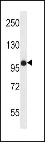
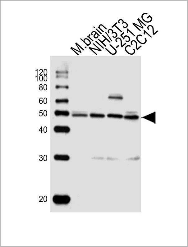
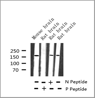
 WB (Western Blot)
(Western blot analysis of lysates from 293, Hela, mouse NIH/3T3, rat PC-12 cell line and rat brain tissue lysate(from left to right), using RPS6 Antibody (N-term). AAA28643 was diluted at 1:2000 at each lane. A goat anti-mouse IgG H&L(HRP) at 1:3000 dilution was used as the secondary antibody. Lysates at 35ug per lane.)
WB (Western Blot)
(Western blot analysis of lysates from 293, Hela, mouse NIH/3T3, rat PC-12 cell line and rat brain tissue lysate(from left to right), using RPS6 Antibody (N-term). AAA28643 was diluted at 1:2000 at each lane. A goat anti-mouse IgG H&L(HRP) at 1:3000 dilution was used as the secondary antibody. Lysates at 35ug per lane.)
RPS6, Monoclonal Antibody (Cat# AAA28643)
Full Name
RPS6 Antibody (N-term)
Gene Names
RPS6; S6
Reactivity
Human, rat (Predicted Reactivity: Monkey, Mouse)
Applications
EIA, IHC, WB, FC/FACS, IF
Purity
Purified Mouse Monoclonal Antibody (Mab)
Pricing


 FCM (Flow Cytometry)
(Figure 7. Flow Cytometry analysis of A549 cells using anti-Hsp90 alpha antibody (AAA19359).Overlay histogram showing A549 cells stained with AAA19359 (Blue line). The cells were blocked with 10% normal goat serum. And then incubated with mouse anti-Hsp90 alpha Antibody (AAA19359, 1μg/1x106 cells) for 30 min at 20 degree C. DyLight®488 conjugated goat anti-mouse IgG (BA1126, 5-10μg/1x106 cells) was used as secondary antibody for 30 minutes at 20 degree C. Isotype control antibody (Green line) was mouse IgG (1μg/1x106) used under the same conditions. Unlabelled sample (Red line) was also used as a control.)
FCM (Flow Cytometry)
(Figure 7. Flow Cytometry analysis of A549 cells using anti-Hsp90 alpha antibody (AAA19359).Overlay histogram showing A549 cells stained with AAA19359 (Blue line). The cells were blocked with 10% normal goat serum. And then incubated with mouse anti-Hsp90 alpha Antibody (AAA19359, 1μg/1x106 cells) for 30 min at 20 degree C. DyLight®488 conjugated goat anti-mouse IgG (BA1126, 5-10μg/1x106 cells) was used as secondary antibody for 30 minutes at 20 degree C. Isotype control antibody (Green line) was mouse IgG (1μg/1x106) used under the same conditions. Unlabelled sample (Red line) was also used as a control.)
Hsp90 alpha, Monoclonal Antibody (Cat# AAA19359)
Full Name
Anti-Hsp90 alpha Antibody (monoclonal, 6B5)
Gene Names
HSP90AA1; EL52; HSPN; LAP2; HSP86; HSPC1; HSPCA; Hsp89; Hsp90; HSP89A; HSP90A; HSP90N; HSPCAL1; HSPCAL4
Reactivity
Human, Mouse, Rat, Monkey
Applications
WB, IHC-P, ICC, IF, FC/FACS/FCM
Purity
Immunogen affinity purified.
Pricing


 Application Data
(C:FGFR2/isolectinB4 (C) and FGFR1/isolectinB4 (D) staining of apparent mesenchymal cells and the subpopulation of endothelial cells. Virtually all other dispersed apparent mesenchymal cells express FGFR1 and FGFR2 (merged image in E). F: FGFR2 (F) and FGFR1 (G) staining in clustered cells of epithelial origin (inferred by morphology here) demonstrating that epithelial cells express both FGFR1 and FGFR2 (merged image with DAPI staining in H).)
Application Data
(C:FGFR2/isolectinB4 (C) and FGFR1/isolectinB4 (D) staining of apparent mesenchymal cells and the subpopulation of endothelial cells. Virtually all other dispersed apparent mesenchymal cells express FGFR1 and FGFR2 (merged image in E). F: FGFR2 (F) and FGFR1 (G) staining in clustered cells of epithelial origin (inferred by morphology here) demonstrating that epithelial cells express both FGFR1 and FGFR2 (merged image with DAPI staining in H).)
FGFR2, Polyclonal Antibody (Cat# AAA26852)
Full Name
FGFR2, NT (FGFR2, BEK, KGFR, KSAM, Fibroblast growth factor receptor 2, K-sam, Keratinocyte growth factor receptor, CD332) (APC)
Gene Names
FGFR2; BEK; JWS; BBDS; CEK3; CFD1; ECT1; KGFR; TK14; TK25; BFR-1; CD332; K-SAM
Reactivity
Human, Monkey, Mouse, Rat
Applications
FC/FACS, IF, IHC, WB
Purity
Purified by Protein G Affinity Chromatography.
Pricing
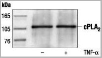
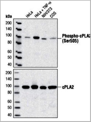 WB (Western Blot)
(Western blot analysis of extracts from HeLa, C6 and NIH/3T3 cells, using AAA14717.)
WB (Western Blot)
(Western blot analysis of extracts from HeLa, C6 and NIH/3T3 cells, using AAA14717.)
c-Phospholipase A2, Polyclonal Antibody (Cat# AAA14717)
Full Name
c-Phospholipase A2 (Cytosolic PLA2, cPLA2)
Reactivity
Human
Applications
Western Blot, Immunoprecipitation
Purity
Purified by Protein A and peptide affinity chromatography
Pricing
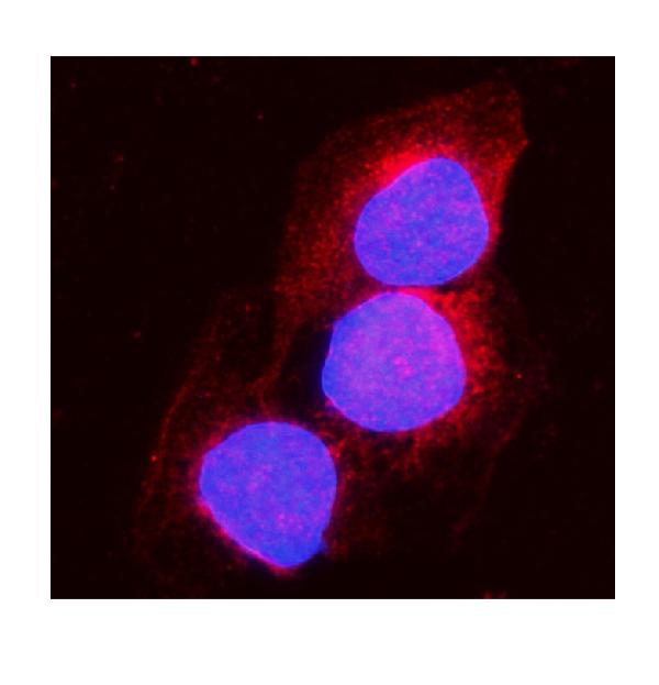
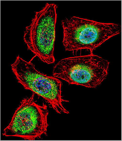
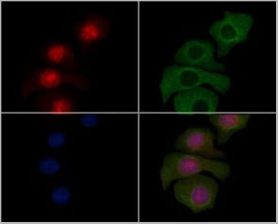
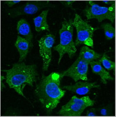
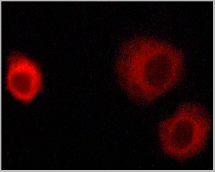
 IF (Immunofluorescence)
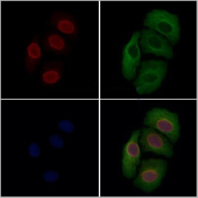
(AAA31214 staining Hela cells by IF/ICC. The samples were fixed with PFA and permeabilized in 0.1% Triton X-100,then blocked in 10% serum for 45 minutes at 25°C. Samples were then incubated with primary Ab(AAA31214 1:200) and mouse anti-beta tubulin Ab(T0023 1:200) for 1 hour at 37°C. An AlexaFluor594 conjugated goat anti-rabbit IgG(H+L) Ab(Red) and an AlexaFluor488 conjugated goat anti-mouse IgG(H+L) Ab(Green) were used as the secondary antibody.The nuclear counter stain is DAPI(blue).)
IF (Immunofluorescence)
(AAA31214 staining Hela cells by IF/ICC. The samples were fixed with PFA and permeabilized in 0.1% Triton X-100,then blocked in 10% serum for 45 minutes at 25°C. Samples were then incubated with primary Ab(AAA31214 1:200) and mouse anti-beta tubulin Ab(T0023 1:200) for 1 hour at 37°C. An AlexaFluor594 conjugated goat anti-rabbit IgG(H+L) Ab(Red) and an AlexaFluor488 conjugated goat anti-mouse IgG(H+L) Ab(Green) were used as the secondary antibody.The nuclear counter stain is DAPI(blue).)
MUM1, Polyclonal Antibody (Cat# AAA31214)
Full Name
MUM1 Antibody
Gene Names
PWWP3A; MUM1; MUM-1; EXPAND1; HSPC211
Reactivity
Human, Mouse, Rat, Monkey
Predicted Reactivity: Pig(100%), Bovine(100%), Horse(88%), Sheep(100%)
Predicted Reactivity: Pig(100%), Bovine(100%), Horse(88%), Sheep(100%)
Applications
ELISA
Purity
The antiserum was purified by peptide affinity chromatography using SulfoLink Coupling Resin (Thermo Fisher Scientific).
Pricing

 IF (Immunofluorescence)
(Figure 10 Immunofluorescence Validation of TACE in Rat Brain (Pradillo et al, 2005)Cellular localization of TACE. Double immunofluorescence staining of brain sections from sham-operated (SHAM; A, C, E) and IPC-exposed animals (IPC; B, D, F) of TACE (red) and the cellular markers (green) NeuN (neurons; A, B), GFAP (astrocytes; C, D) and L. esculentum lectin (microglia and endothelium; E, F). White arrows indicate TACE-positive cells.)
IF (Immunofluorescence)
(Figure 10 Immunofluorescence Validation of TACE in Rat Brain (Pradillo et al, 2005)Cellular localization of TACE. Double immunofluorescence staining of brain sections from sham-operated (SHAM; A, C, E) and IPC-exposed animals (IPC; B, D, F) of TACE (red) and the cellular markers (green) NeuN (neurons; A, B), GFAP (astrocytes; C, D) and L. esculentum lectin (microglia and endothelium; E, F). White arrows indicate TACE-positive cells.)
TACE, Polyclonal Antibody (Cat# AAA10912)
Full Name
TACE Antibody
Gene Names
ADAM17; CSVP; TACE; NISBD; ADAM18; CD156B; NISBD1
Reactivity
Human, Mouse, Rat
Applications
Western Blot, Immunocytochemistry, Immunofluorescence, Flow Cytometry
Purity
TACE Antibody is affinity chromatography purified via peptide column.
Pricing
 FCM (Flow Cytometry)
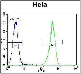
(ENOA Antibody (N-term) (Catalog Number: AAA28805) flow cytometric analysis of Hela cells (right histogram) compared to a negative control cell (left histogram). FITC-conjugated goat-anti-rabbit secondary antibodies were used for the analysis.)
FCM (Flow Cytometry)
(ENOA Antibody (N-term) (Catalog Number: AAA28805) flow cytometric analysis of Hela cells (right histogram) compared to a negative control cell (left histogram). FITC-conjugated goat-anti-rabbit secondary antibodies were used for the analysis.)
ENOA, Polyclonal Antibody (Cat# AAA28805)
Full Name
ENOA Antibody (N-term)
Gene Names
ENO1; NNE; PPH; MPB1; ENO1L1; HEL-S-17
Reactivity
Human, Mouse, Rat
Predicted: Bovine, Monkey
Predicted: Bovine, Monkey
Applications
Immunofluorescence, Western Blot, Flow Cytometry
Purity
Purified Rabbit Polyclnal Antibody (Pab)
Pricing
 FCM (Flow Cytometry)
(Staining of thrombin activated platelets with Mouse anti Human CD62P)
FCM (Flow Cytometry)
(Staining of thrombin activated platelets with Mouse anti Human CD62P)
CD62P, Monoclonal Antibody (Cat# AAA12247)
Full Name
MOUSE ANTI HUMAN CD62P
Gene Names
SELP; CD62; GRMP; PSEL; CD62P; GMP140; LECAM3; PADGEM
Applications
Immunohistochemistry, Flow Cytometry, Immunofluorescence, Immunohistochemistry, Western Blot
Pricing

 IF (Immunofluorescence)
(AAA30956 staining HepG2 cells(4h of LPS treatment) by IF/ICC. The samples were fixed with PFA and permeabilized in 0.1% Triton X-100,then blocked in 10% serum for 45 minutes at 25°C. Samples were then incubated with primary Ab(AAA30956 1:200) and mouse anti-beta tubulin Ab( 1:200) for 1 hour at 37°C. An AlexaFluor594 conjugated goat anti-rabbit IgG(H+L) Ab(Red) and an AlexaFluor488 conjugated goat anti-mouse IgG(H+L) Ab(Green) were used as the secondary Ab. The nuclear counter stain is DAPI(blue).)
IF (Immunofluorescence)
(AAA30956 staining HepG2 cells(4h of LPS treatment) by IF/ICC. The samples were fixed with PFA and permeabilized in 0.1% Triton X-100,then blocked in 10% serum for 45 minutes at 25°C. Samples were then incubated with primary Ab(AAA30956 1:200) and mouse anti-beta tubulin Ab( 1:200) for 1 hour at 37°C. An AlexaFluor594 conjugated goat anti-rabbit IgG(H+L) Ab(Red) and an AlexaFluor488 conjugated goat anti-mouse IgG(H+L) Ab(Green) were used as the secondary Ab. The nuclear counter stain is DAPI(blue).)
MITF, Polyclonal Antibody (Cat# AAA30956)
Full Name
Phospho-MITF (Ser180/73) Antibody
Gene Names
MITF; MI; WS2; CMM8; WS2A; COMMAD; bHLHe32
Reactivity
Human, Mouse, Rat, Monkey
Applications
Western Blot, Immunohistochemistry, Immunofluorescence, Immunocytochemistry
Purity
Peptide affinity puriification
Pricing
 IF (Immunofluorescence)
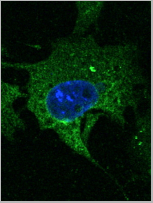
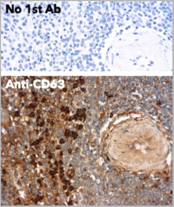
(Immunofluorescence – anti-CD63 Ab in Hepa1-6 cells at 1/50 dilution; cells were fixed with 4% of PFA;)
IF (Immunofluorescence)
(Immunofluorescence – anti-CD63 Ab in Hepa1-6 cells at 1/50 dilution; cells were fixed with 4% of PFA;)
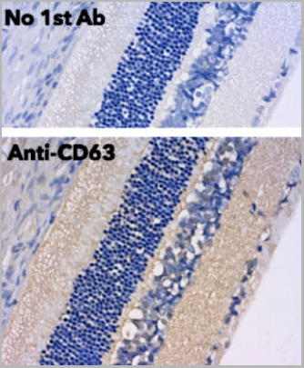
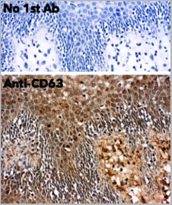
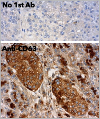
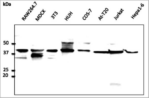
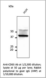
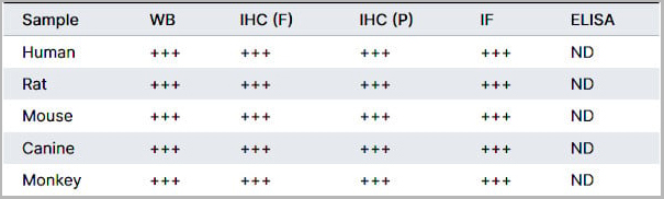
CD63, Polyclonal Antibody (Cat# AAA13873)
Full Name
CD63 Polyclonal Antibody
Gene Names
CD63; MLA1; ME491; LAMP-3; OMA81H; TSPAN30
Reactivity
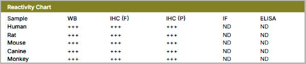
Reacts against human, rat, mouse, canine and monkey proteins
Applications
Immunofluorescence, Western Blot, Immunohistochemistry
Purity
This antibody is epitope-affinity purified from goat antiserum.
Pricing
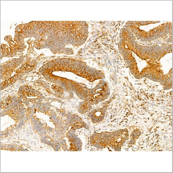
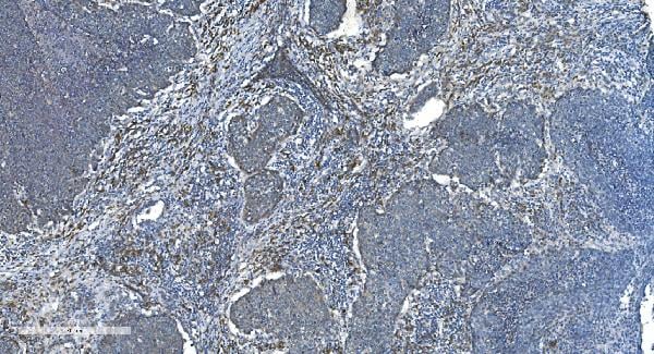
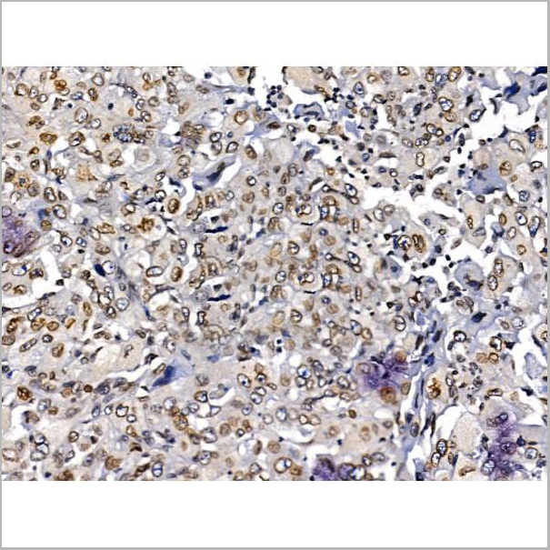
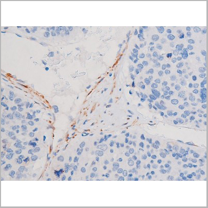
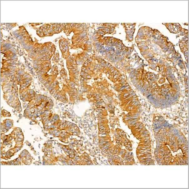 IHC (Immunohistchemistry-Paraffin)
(AAA31276 at 1/100 staining Human colorectal cancer and adjacent normal tissues by IHC-P. The sample was formaldehyde fixed and a heat mediated antigen retrieval step in citrate buffer was performed. The sample was then blocked and incubated with the primary antibody at 4°C overnight. An HRP conjugated anti-Rabbit antibody was used as the secondary antibody.)
IHC (Immunohistchemistry-Paraffin)
(AAA31276 at 1/100 staining Human colorectal cancer and adjacent normal tissues by IHC-P. The sample was formaldehyde fixed and a heat mediated antigen retrieval step in citrate buffer was performed. The sample was then blocked and incubated with the primary antibody at 4°C overnight. An HRP conjugated anti-Rabbit antibody was used as the secondary antibody.)
SCAMP2, Polyclonal Antibody (Cat# AAA31276)
Full Name
SCAMP2 Antibody
Reactivity
Human, Mouse, Rat, Monkey
Predicted Reactivity: Pig(100%), Zebrafish(100%), Bovine(100%), Horse(100%), Sheep(100%), Rabbit(100%), Dog(100%), Chicken(100%), Xenopus(100%)
Predicted Reactivity: Pig(100%), Zebrafish(100%), Bovine(100%), Horse(100%), Sheep(100%), Rabbit(100%), Dog(100%), Chicken(100%), Xenopus(100%)
Applications
ELISA
Purity
The antiserum was purified by peptide affinity chromatography using SulfoLink Coupling Resin (Thermo Fisher Scientific).
Pricing
Mouse Anti-Monkey IgG, Monoclonal Secondary Antibody (Cat# AAA14895)
Full Name
Mouse Anti-Monkey IgG-HRP
Reactivity
Monkey
Applications
ELISA
Pricing
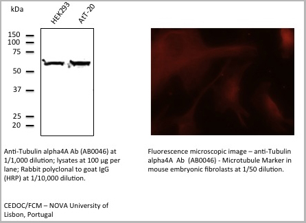
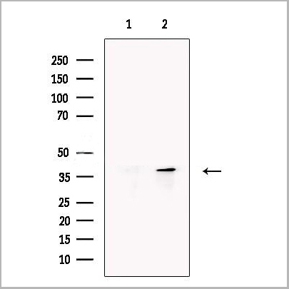
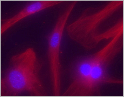 IF (Immunofluorescence)
(Immunofluorescence – anti-Tubulin alpha4A Ab - Microtubule Marker in mouse embryonic fibrolasts at 1/50 dilution;)
IF (Immunofluorescence)
(Immunofluorescence – anti-Tubulin alpha4A Ab - Microtubule Marker in mouse embryonic fibrolasts at 1/50 dilution;)
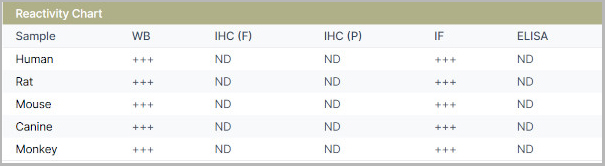
TUBA4A, Polyclonal Antibody (Cat# AAA13868)
Full Name
Tubulin Alpha4A Chain Polyclonal Antibody
Gene Names
TUBA4A; TUBA1; H2-ALPHA
Reactivity
Reacts against human, rat, mouse, canine and monkey proteins.
Applications
Immunofluorescence, Western Blot
Pricing
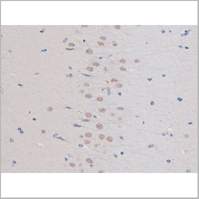
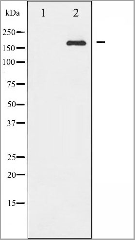
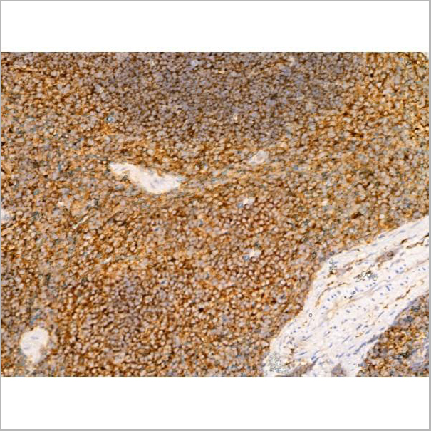
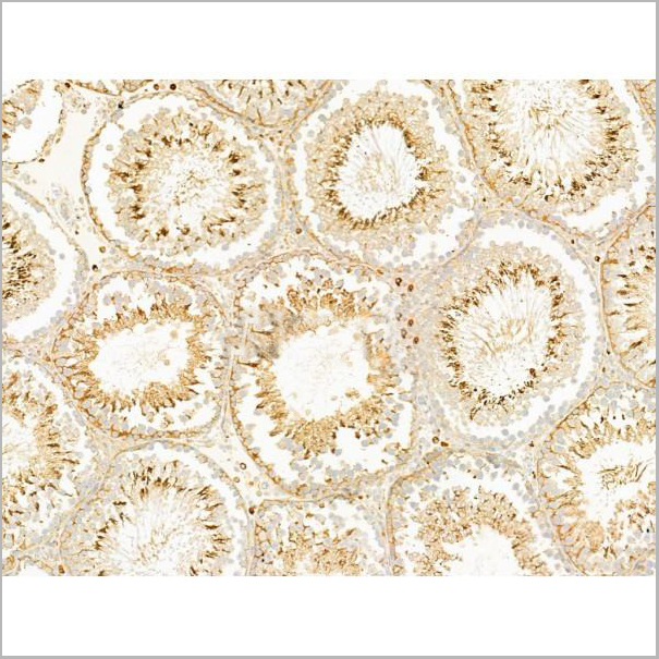
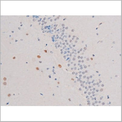 IHC (Immunohistochemistry)
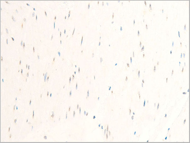
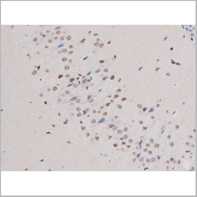
(AAA31037 at 1/200 staining Rat brain tissue sections by IHC-P. The tissue was formaldehyde fixed and a heat mediated antigen retrieval step in citrate buffer was performed. The tissue was then blocked and incubated with the antibody for 1.5 hours at 22 degree C. An HRP conjugated goat anti-rabbit antibody was used as the secondary.)
IHC (Immunohistochemistry)
(AAA31037 at 1/200 staining Rat brain tissue sections by IHC-P. The tissue was formaldehyde fixed and a heat mediated antigen retrieval step in citrate buffer was performed. The tissue was then blocked and incubated with the antibody for 1.5 hours at 22 degree C. An HRP conjugated goat anti-rabbit antibody was used as the secondary.)
IRS-1, Polyclonal Antibody (Cat# AAA31037)
Full Name
Phospho-IRS-1 (Ser636) Antibody
Gene Names
IRS1; HIRS-1
Reactivity
Human, Mouse, Rat, Monkey
Applications
Western Blot, Immunohistochemistry, Immunofluorescence, Immunocytochemistry
Purity
From purified rabbit serum by affinity purification via sequential chromatography on phospho-and non-phospho-peptide affinity columns.
Pricing
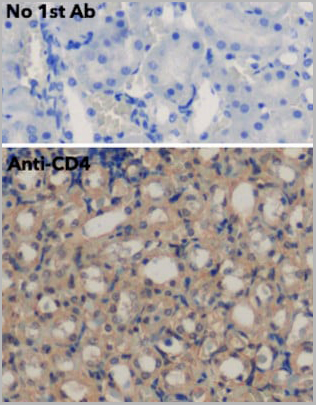
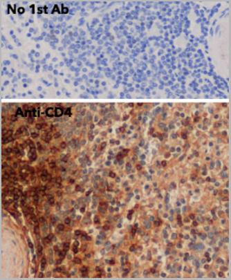
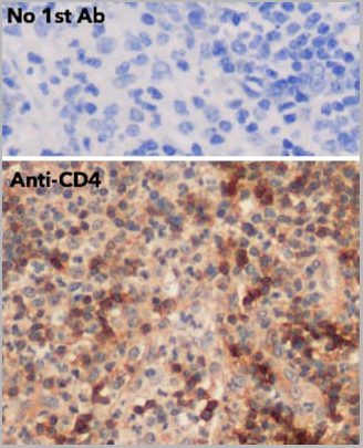
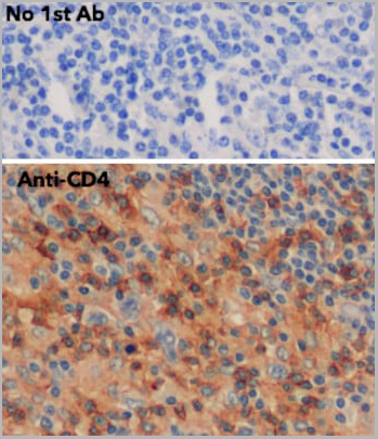
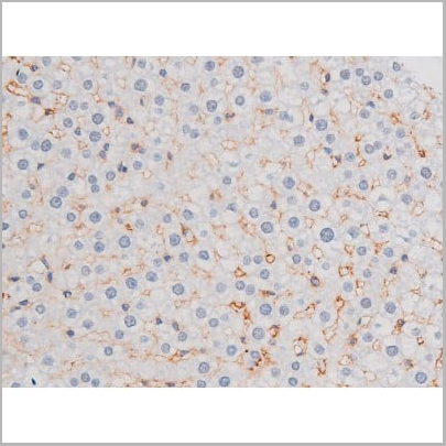
 IHC (Immunhistochemistry)
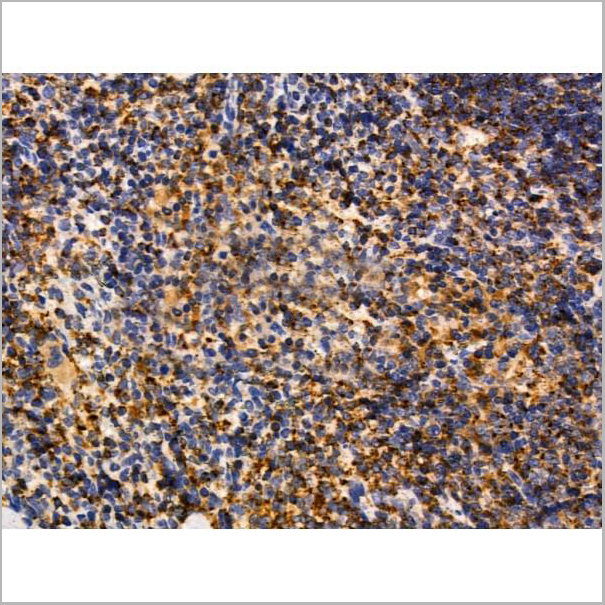
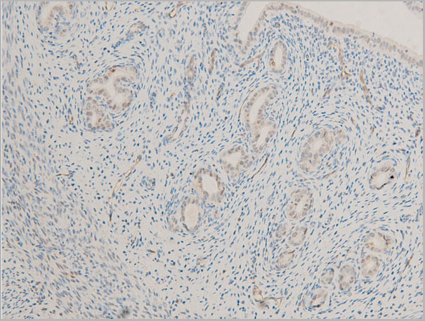
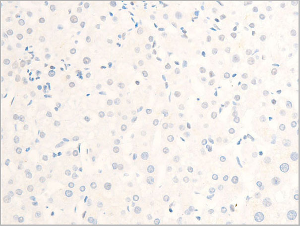
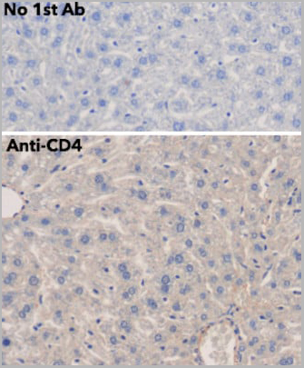
(IHC of mouse liver using anti-CD4 antibody and FFPE tissue after heat-induced antigen retrieval. Anti-CD4 Ab at 1:750/DAB detection.)
IHC (Immunhistochemistry)
(IHC of mouse liver using anti-CD4 antibody and FFPE tissue after heat-induced antigen retrieval. Anti-CD4 Ab at 1:750/DAB detection.)
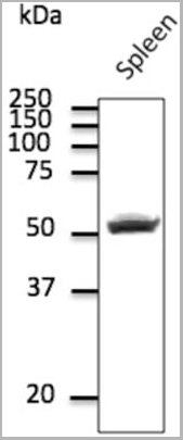
CD4, Polyclonal Antibody (Cat# AAA13858)
Full Name
CD4 Polyclonal Antibody
Gene Names
CD4; CD4mut
Reactivity
Reacts against human, rat, mouse, canine and monkey proteins.
Applications
Western Blot
Pricing
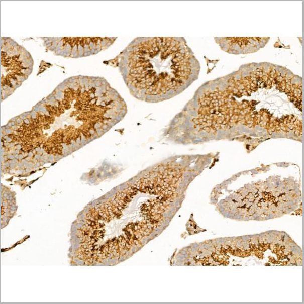
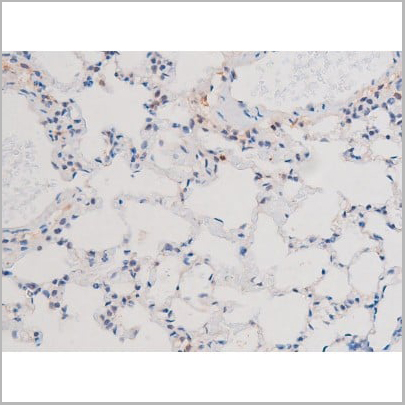
 IHC (Immunohistochemistry)
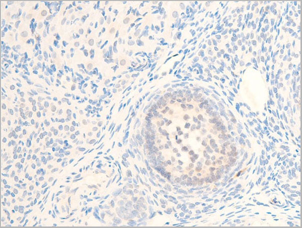
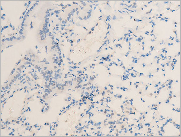
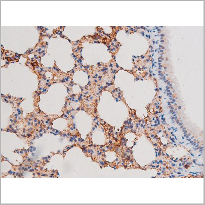
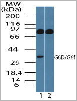
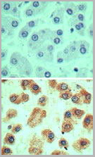
(Immunohistochemical analysis of G6D in formalin-fixed, paraffin-embedded human liver tissue using an isotype control (top) and (bottom) AAA24064 at 5ug/ml.)
IHC (Immunohistochemistry)
(Immunohistochemical analysis of G6D in formalin-fixed, paraffin-embedded human liver tissue using an isotype control (top) and (bottom) AAA24064 at 5ug/ml.)
LY6G6D, Polyclonal Antibody (Cat# AAA24064)
Full Name
LY6G6D (G6D, Lymphocyte Antigen 6 Complex Locus Protein G6f, C6orf21, G6F, LY6G6D, NG32)
Gene Names
LY6G6D; G6D; NG25; LY6-D; MEGT1; C6orf23
Reactivity
Human
Applications
Western Blot, Immunohistochemistry
Purity
Purified by Protein A affinity chromatography.
Pricing