Filters
Clonality
Type
Reactivity
Gene Name
Isotype
Host
Application
Clone
132 results for "Labeling" - showing 50-100


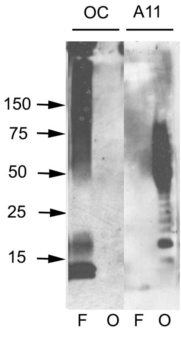


 DB (Dot Blot)
(Dot blot analysis using Rabbit Anti-Amyloid Fibrils (OC) Polyclonal Antibody (SPC-507). Tissue: Cell lysates. Species: Human. Primary Antibody: Rabbit Anti-Amyloid Fibrils (OC) Polyclonal Antibody (SPC-507) at 1:500, 1:5000. Beta Amyloid HEPES-NaCl aggregation, showing 1:500 (L) and 1:5000 (R) time lapse dot blot.)
DB (Dot Blot)
(Dot blot analysis using Rabbit Anti-Amyloid Fibrils (OC) Polyclonal Antibody (SPC-507). Tissue: Cell lysates. Species: Human. Primary Antibody: Rabbit Anti-Amyloid Fibrils (OC) Polyclonal Antibody (SPC-507) at 1:500, 1:5000. Beta Amyloid HEPES-NaCl aggregation, showing 1:500 (L) and 1:5000 (R) time lapse dot blot.)
Amyloid Fibrils (OC), Polyclonal Antibody (Cat# AAA17802)
Full Name
Amyloid Fibrils (OC) Antibody: RPE
Reactivity
Human. Potentially mouse and rat based on species homology.
Applications
IP, ICC, IHC, EIA, WB, DB
Pricing


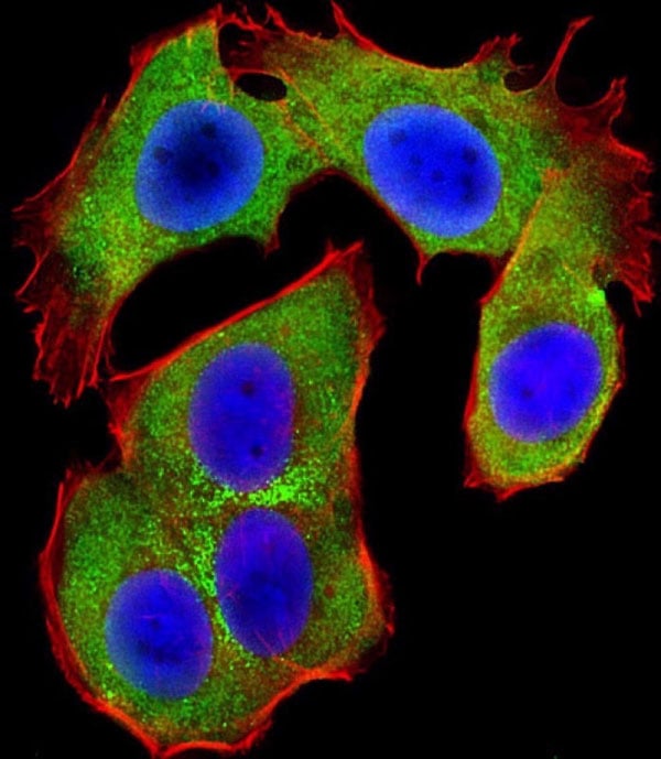
 IF (Immunofluorescence)
(Immunofluorescent analysis of 4% paraformaldehyde-fixed, 0.1% Triton X-100 permeabilized MCF-7 (human breast cancer cell line) cells labeling Pdx1 with at 1:25 dilution, followed by DyLight 488-conjugated IgG goat anti-rabbit secondary antibody at 1:200 dilution (green). Immunofluorescence image showing cytoplasm staining on MCF-7 cell line. Cytoplasmic actin is detected with DyLight 554 Phalloidin (PD18466410) at 1:100 dilution (red). The nuclear counter stain is DAPI (blue).)
IF (Immunofluorescence)
(Immunofluorescent analysis of 4% paraformaldehyde-fixed, 0.1% Triton X-100 permeabilized MCF-7 (human breast cancer cell line) cells labeling Pdx1 with at 1:25 dilution, followed by DyLight 488-conjugated IgG goat anti-rabbit secondary antibody at 1:200 dilution (green). Immunofluorescence image showing cytoplasm staining on MCF-7 cell line. Cytoplasmic actin is detected with DyLight 554 Phalloidin (PD18466410) at 1:100 dilution (red). The nuclear counter stain is DAPI (blue).)
OPN-a/b, Polyclonal Antibody (Cat# AAA26857)
Full Name
OPN-a/b, NT (SPP1, BNSP, OPN, Osteopontin, Bone sialoprotein 1, Nephropontin, Secreted phosphoprotein 1, Urinary stone protein, Uropontin) (APC)
Gene Names
SPP1; OPN; BNSP; BSPI; ETA-1
Reactivity
Human
Applications
WB, IHC, IF
Purity
Purified by Protein A and Peptide Affinity Chromatography.
Pricing


 IF (Immunofluorescence)
(Immunofluorescent analysis of 4% paraformaldehyde-fixed, 0.1% Triton X-100 permeabilized MCF-7 (human breast cancer cell line) cells labeling Pdx1 with at 1:25 dilution, followed by DyLight 488-conjugated IgG goat anti-rabbit secondary antibody at 1:200 dilution (green). Immunofluorescence image showing cytoplasm staining on MCF-7 cell line. Cytoplasmic actin is detected with DyLight 554 Phalloidin (PD18466410) at 1:100 dilution (red). The nuclear counter stain is DAPI (blue).)
IF (Immunofluorescence)
(Immunofluorescent analysis of 4% paraformaldehyde-fixed, 0.1% Triton X-100 permeabilized MCF-7 (human breast cancer cell line) cells labeling Pdx1 with at 1:25 dilution, followed by DyLight 488-conjugated IgG goat anti-rabbit secondary antibody at 1:200 dilution (green). Immunofluorescence image showing cytoplasm staining on MCF-7 cell line. Cytoplasmic actin is detected with DyLight 554 Phalloidin (PD18466410) at 1:100 dilution (red). The nuclear counter stain is DAPI (blue).)
OPN-a/b, Polyclonal Antibody (Cat# AAA26861)
Full Name
OPN-a/b, NT (SPP1, BNSP, OPN, Osteopontin, Bone sialoprotein 1, Nephropontin, Secreted phosphoprotein 1, Urinary stone protein, Uropontin) (MaxLight 490)
Gene Names
SPP1; OPN; BNSP; BSPI; ETA-1
Reactivity
Human
Applications
WB, IHC, IF
Purity
Purified by Protein A and Peptide Affinity Chromatography.
Pricing


 IF (Immunofluorescence)
(Immunofluorescent analysis of 4% paraformaldehyde-fixed, 0.1% Triton X-100 permeabilized MCF-7 (human breast cancer cell line) cells labeling Pdx1 with at 1:25 dilution, followed by DyLight 488-conjugated IgG goat anti-rabbit secondary antibody at 1:200 dilution (green). Immunofluorescence image showing cytoplasm staining on MCF-7 cell line. Cytoplasmic actin is detected with DyLight 554 Phalloidin (PD18466410) at 1:100 dilution (red). The nuclear counter stain is DAPI (blue).)
IF (Immunofluorescence)
(Immunofluorescent analysis of 4% paraformaldehyde-fixed, 0.1% Triton X-100 permeabilized MCF-7 (human breast cancer cell line) cells labeling Pdx1 with at 1:25 dilution, followed by DyLight 488-conjugated IgG goat anti-rabbit secondary antibody at 1:200 dilution (green). Immunofluorescence image showing cytoplasm staining on MCF-7 cell line. Cytoplasmic actin is detected with DyLight 554 Phalloidin (PD18466410) at 1:100 dilution (red). The nuclear counter stain is DAPI (blue).)
OPN-a/b, Polyclonal Antibody (Cat# AAA26863)
Full Name
OPN-a/b, NT (SPP1, BNSP, OPN, Osteopontin, Bone sialoprotein 1, Nephropontin, Secreted phosphoprotein 1, Urinary stone protein, Uropontin) (MaxLight 650)
Gene Names
SPP1; OPN; BNSP; BSPI; ETA-1
Reactivity
Human
Applications
WB, IHC, IF
Purity
Purified by Protein A and Peptide Affinity Chromatography.
Pricing


 IF (Immunofluorescence)
(Immunofluorescent analysis of 4% paraformaldehyde-fixed, 0.1% Triton X-100 permeabilized MCF-7 (human breast cancer cell line) cells labeling Pdx1 with at 1:25 dilution, followed by DyLight 488-conjugated IgG goat anti-rabbit secondary antibody at 1:200 dilution (green). Immunofluorescence image showing cytoplasm staining on MCF-7 cell line. Cytoplasmic actin is detected with DyLight 554 Phalloidin (PD18466410) at 1:100 dilution (red). The nuclear counter stain is DAPI (blue).)
IF (Immunofluorescence)
(Immunofluorescent analysis of 4% paraformaldehyde-fixed, 0.1% Triton X-100 permeabilized MCF-7 (human breast cancer cell line) cells labeling Pdx1 with at 1:25 dilution, followed by DyLight 488-conjugated IgG goat anti-rabbit secondary antibody at 1:200 dilution (green). Immunofluorescence image showing cytoplasm staining on MCF-7 cell line. Cytoplasmic actin is detected with DyLight 554 Phalloidin (PD18466410) at 1:100 dilution (red). The nuclear counter stain is DAPI (blue).)
OPN-a/b, Polyclonal Antibody (Cat# AAA26864)
Full Name
OPN-a/b, NT (SPP1, BNSP, OPN, Osteopontin, Bone sialoprotein 1, Nephropontin, Secreted phosphoprotein 1, Urinary stone protein, Uropontin) (MaxLight 750)
Gene Names
SPP1; OPN; BNSP; BSPI; ETA-1
Reactivity
Human
Applications
WB, IHC, IF
Purity
Purified by Protein A and Peptide Affinity Chromatography.
Pricing


 IF (Immunofluorescence)
(Immunofluorescent analysis of 4% paraformaldehyde-fixed, 0.1% Triton X-100 permeabilized MCF-7 (human breast cancer cell line) cells labeling Pdx1 with at 1:25 dilution, followed by DyLight 488-conjugated IgG goat anti-rabbit secondary antibody at 1:200 dilution (green). Immunofluorescence image showing cytoplasm staining on MCF-7 cell line. Cytoplasmic actin is detected with DyLight 554 Phalloidin (PD18466410) at 1:100 dilution (red). The nuclear counter stain is DAPI (blue).)
IF (Immunofluorescence)
(Immunofluorescent analysis of 4% paraformaldehyde-fixed, 0.1% Triton X-100 permeabilized MCF-7 (human breast cancer cell line) cells labeling Pdx1 with at 1:25 dilution, followed by DyLight 488-conjugated IgG goat anti-rabbit secondary antibody at 1:200 dilution (green). Immunofluorescence image showing cytoplasm staining on MCF-7 cell line. Cytoplasmic actin is detected with DyLight 554 Phalloidin (PD18466410) at 1:100 dilution (red). The nuclear counter stain is DAPI (blue).)
OPN-a/b, Polyclonal Antibody (Cat# AAA26862)
Full Name
OPN-a/b, NT (SPP1, BNSP, OPN, Osteopontin, Bone sialoprotein 1, Nephropontin, Secreted phosphoprotein 1, Urinary stone protein, Uropontin) (MaxLight 550)
Gene Names
SPP1; OPN; BNSP; BSPI; ETA-1
Reactivity
Human
Applications
WB, IHC, IF
Purity
Purified by Protein A and Peptide Affinity Chromatography.
Pricing
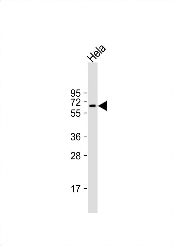
 WB (Western Blot)
Phospholipase A2, Group IID (PLA2G2D), Polyclonal Antibody (Cat# AAA20317)
Full Name
FITC-linked Antibody to Phospholipase A2, Group IID (PLA2G2D)
Gene Names
PLA2G2D; SPLASH; sPLA2S; PLA2IID; sPLA2-IID
Reactivity
Mouse
Applications
WB, IHC, ICC, IF
Purity
Antigen-specific Affinity Chromatography.
Pricing


 DB (Dot Blot)
(Dot blot analysis using Rabbit Anti-Amyloid Fibrils (OC) Polyclonal Antibody (SPC-507). Tissue: Cell lysates. Species: Human. Primary Antibody: Rabbit Anti-Amyloid Fibrils (OC) Polyclonal Antibody (SPC-507) at 1:500, 1:5000. Beta Amyloid HEPES-NaCl aggregation, showing 1:500 (L) and 1:5000 (R) time lapse dot blot.)
DB (Dot Blot)
(Dot blot analysis using Rabbit Anti-Amyloid Fibrils (OC) Polyclonal Antibody (SPC-507). Tissue: Cell lysates. Species: Human. Primary Antibody: Rabbit Anti-Amyloid Fibrils (OC) Polyclonal Antibody (SPC-507) at 1:500, 1:5000. Beta Amyloid HEPES-NaCl aggregation, showing 1:500 (L) and 1:5000 (R) time lapse dot blot.)
Amyloid Fibrils (OC), Polyclonal Antibody (Cat# AAA17801)
Full Name
Amyloid Fibrils (OC) Antibody: ATTO 594
Reactivity
Human. Potentially mouse and rat based on species homology.
Applications
IP, ICC, IHC, EIA, WB, DB
Pricing


 DB (Dot Blot)
(Dot blot analysis using Rabbit Anti-Amyloid Fibrils (OC) Polyclonal Antibody (SPC-507). Tissue: Cell lysates. Species: Human. Primary Antibody: Rabbit Anti-Amyloid Fibrils (OC) Polyclonal Antibody (SPC-507) at 1:500, 1:5000. Beta Amyloid HEPES-NaCl aggregation, showing 1:500 (L) and 1:5000 (R) time lapse dot blot.)
DB (Dot Blot)
(Dot blot analysis using Rabbit Anti-Amyloid Fibrils (OC) Polyclonal Antibody (SPC-507). Tissue: Cell lysates. Species: Human. Primary Antibody: Rabbit Anti-Amyloid Fibrils (OC) Polyclonal Antibody (SPC-507) at 1:500, 1:5000. Beta Amyloid HEPES-NaCl aggregation, showing 1:500 (L) and 1:5000 (R) time lapse dot blot.)
Amyloid Fibrils (OC), Polyclonal Antibody (Cat# AAA17797)
Full Name
Amyloid Fibrils (OC) Antibody: FITC
Reactivity
Human. Potentially mouse and rat based on species homology.
Applications
IP, ICC, IHC, EIA, WB, DB
Pricing


 IF (Immunofluorescence)
(Immunofluorescent analysis of 4% paraformaldehyde-fixed, 0.1% Triton X-100 permeabilized MCF-7 (human breast cancer cell line) cells labeling Pdx1 with at 1:25 dilution, followed by DyLight 488-conjugated IgG goat anti-rabbit secondary antibody at 1:200 dilution (green). Immunofluorescence image showing cytoplasm staining on MCF-7 cell line. Cytoplasmic actin is detected with DyLight 554 Phalloidin (PD18466410) at 1:100 dilution (red). The nuclear counter stain is DAPI (blue).)
IF (Immunofluorescence)
(Immunofluorescent analysis of 4% paraformaldehyde-fixed, 0.1% Triton X-100 permeabilized MCF-7 (human breast cancer cell line) cells labeling Pdx1 with at 1:25 dilution, followed by DyLight 488-conjugated IgG goat anti-rabbit secondary antibody at 1:200 dilution (green). Immunofluorescence image showing cytoplasm staining on MCF-7 cell line. Cytoplasmic actin is detected with DyLight 554 Phalloidin (PD18466410) at 1:100 dilution (red). The nuclear counter stain is DAPI (blue).)
OPN-a/b, Polyclonal Antibody (Cat# AAA26859)
Full Name
OPN-a/b, NT (SPP1, BNSP, OPN, Osteopontin, Bone sialoprotein 1, Nephropontin, Secreted phosphoprotein 1, Urinary stone protein, Uropontin) (FITC)
Gene Names
SPP1; OPN; BNSP; BSPI; ETA-1
Reactivity
Human
Applications
WB, IHC, IF
Purity
Purified by Protein A and Peptide Affinity Chromatography.
Pricing


 DB (Dot Blot)
(Dot blot analysis using Rabbit Anti-Amyloid Fibrils (OC) Polyclonal Antibody (SPC-507). Tissue: Cell lysates. Species: Human. Primary Antibody: Rabbit Anti-Amyloid Fibrils (OC) Polyclonal Antibody (SPC-507) at 1:500, 1:5000. Beta Amyloid HEPES-NaCl aggregation, showing 1:500 (L) and 1:5000 (R) time lapse dot blot.)
DB (Dot Blot)
(Dot blot analysis using Rabbit Anti-Amyloid Fibrils (OC) Polyclonal Antibody (SPC-507). Tissue: Cell lysates. Species: Human. Primary Antibody: Rabbit Anti-Amyloid Fibrils (OC) Polyclonal Antibody (SPC-507) at 1:500, 1:5000. Beta Amyloid HEPES-NaCl aggregation, showing 1:500 (L) and 1:5000 (R) time lapse dot blot.)
Amyloid Fibrils (OC), Polyclonal Antibody (Cat# AAA17798)
Full Name
Amyloid Fibrils (OC) Antibody: ATTO 390
Reactivity
Human. Potentially mouse and rat based on species homology.
Applications
IP, ICC, IHC, EIA, WB, DB
Pricing


 IF (Immunofluorescence)
(Immunofluorescent analysis of 4% paraformaldehyde-fixed, 0.1% Triton X-100 permeabilized MCF-7 (human breast cancer cell line) cells labeling Pdx1 with at 1:25 dilution, followed by DyLight 488-conjugated IgG goat anti-rabbit secondary antibody at 1:200 dilution (green). Immunofluorescence image showing cytoplasm staining on MCF-7 cell line. Cytoplasmic actin is detected with DyLight 554 Phalloidin (PD18466410) at 1:100 dilution (red). The nuclear counter stain is DAPI (blue).)
IF (Immunofluorescence)
(Immunofluorescent analysis of 4% paraformaldehyde-fixed, 0.1% Triton X-100 permeabilized MCF-7 (human breast cancer cell line) cells labeling Pdx1 with at 1:25 dilution, followed by DyLight 488-conjugated IgG goat anti-rabbit secondary antibody at 1:200 dilution (green). Immunofluorescence image showing cytoplasm staining on MCF-7 cell line. Cytoplasmic actin is detected with DyLight 554 Phalloidin (PD18466410) at 1:100 dilution (red). The nuclear counter stain is DAPI (blue).)
OPN-a/b, Polyclonal Antibody (Cat# AAA26860)
Full Name
OPN-a/b, NT (SPP1, BNSP, OPN, Osteopontin, Bone sialoprotein 1, Nephropontin, Secreted phosphoprotein 1, Urinary stone protein, Uropontin) (MaxLight 405)
Gene Names
SPP1; OPN; BNSP; BSPI; ETA-1
Reactivity
Human
Applications
WB, IHC, IF
Purity
Purified by Protein A and Peptide Affinity Chromatography.
Pricing


 IF (Immunofluorescence)
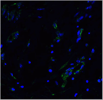
(Immunofluorescent analysis of 4% paraformaldehyde-fixed, 0.1% Triton X-100 permeabilized U-2 OS (human osteosarcoma cell line) cells labeling DLL3 with (1:25), followed by Dylight 488- conjugated goat anti- rabbit IgG secondary antibody at (1:200) (green). Immunofluorescence image showing nucleus and weak cytoplasm staining on U-2 OScell line. Cytoplasmic actin is detected with Dylight 554 Phalloidin (1:100) (red).)
IF (Immunofluorescence)
(Immunofluorescent analysis of 4% paraformaldehyde-fixed, 0.1% Triton X-100 permeabilized U-2 OS (human osteosarcoma cell line) cells labeling DLL3 with (1:25), followed by Dylight 488- conjugated goat anti- rabbit IgG secondary antibody at (1:200) (green). Immunofluorescence image showing nucleus and weak cytoplasm staining on U-2 OScell line. Cytoplasmic actin is detected with Dylight 554 Phalloidin (1:100) (red).)
DLL3, Polyclonal Antibody (Cat# AAA26850)
Full Name
DLL3, CT (DLL3, Delta-like protein 3, Drosophila Delta homolog 3) (APC)
Gene Names
DLL3; SCDO1
Reactivity
Human
Applications
IF, IHC, WB
Purity
Purified by Protein A Affinity Chromatography.
Pricing


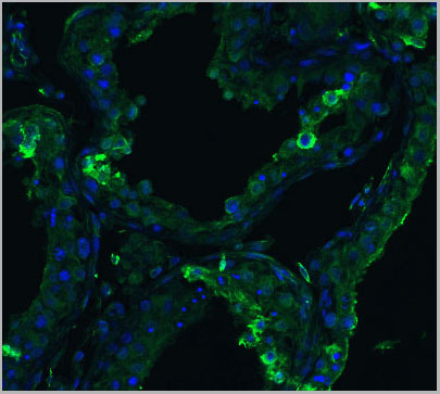
 IF (Immunofluorescence)
(Immunofluorescent analysis of 4% paraformaldehyde-fixed, 0.1% Triton X-100 permeabilized MCF-7 (human breast cancer cell line) cells labeling Pdx1 with AAA14796 at 1:25 dilution, followed by Dylight® 488-conjugated IgG goat anti-rabbit secondary antibody at 1:200 dilution (green). Immunofluorescence image showing cytoplasm staining on MCF-7 cell line. Cytoplasmic actin is detected with Dylight® 554 Phalloidin (PD18466410) at 1:100 dilution (red). The nuclear counter stain is DAPI (blue).)
IF (Immunofluorescence)
(Immunofluorescent analysis of 4% paraformaldehyde-fixed, 0.1% Triton X-100 permeabilized MCF-7 (human breast cancer cell line) cells labeling Pdx1 with AAA14796 at 1:25 dilution, followed by Dylight® 488-conjugated IgG goat anti-rabbit secondary antibody at 1:200 dilution (green). Immunofluorescence image showing cytoplasm staining on MCF-7 cell line. Cytoplasmic actin is detected with Dylight® 554 Phalloidin (PD18466410) at 1:100 dilution (red). The nuclear counter stain is DAPI (blue).)
OPN-a/b, Polyclonal Antibody (Cat# AAA14796)
Full Name
OPN-a/b, NT (SPP1, BNSP, OPN, Osteopontin, Bone sialoprotein 1, Nephropontin, Secreted phosphoprotein 1, Urinary stone protein, Uropontin)
Reactivity
Human
Applications
EL/EIA, WB, IHC, IF
Purity
Affinity Purified
Purified by Protein A affinity chromatography.
Purified by Protein A affinity chromatography.
Pricing


 IF (Immunofluorescence)
(Immunofluorescent analysis of 4% paraformaldehyde-fixed, 0.1% Triton X-100 permeabilized MCF-7 (human breast cancer cell line) cells labeling Pdx1 with at 1:25 dilution, followed by DyLight 488-conjugated IgG goat anti-rabbit secondary antibody at 1:200 dilution (green). Immunofluorescence image showing cytoplasm staining on MCF-7 cell line. Cytoplasmic actin is detected with DyLight 554 Phalloidin (PD18466410) at 1:100 dilution (red). The nuclear counter stain is DAPI (blue).)
IF (Immunofluorescence)
(Immunofluorescent analysis of 4% paraformaldehyde-fixed, 0.1% Triton X-100 permeabilized MCF-7 (human breast cancer cell line) cells labeling Pdx1 with at 1:25 dilution, followed by DyLight 488-conjugated IgG goat anti-rabbit secondary antibody at 1:200 dilution (green). Immunofluorescence image showing cytoplasm staining on MCF-7 cell line. Cytoplasmic actin is detected with DyLight 554 Phalloidin (PD18466410) at 1:100 dilution (red). The nuclear counter stain is DAPI (blue).)
OPN-a/b, Polyclonal Antibody (Cat# AAA26858)
Full Name
OPN-a/b, NT (SPP1, BNSP, OPN, Osteopontin, Bone sialoprotein 1, Nephropontin, Secreted phosphoprotein 1, Urinary stone protein, Uropontin) (Biotin)
Gene Names
SPP1; OPN; BNSP; BSPI; ETA-1
Reactivity
Human
Applications
WB, IHC, IF, EIA
Purity
Purified by Protein A and Peptide Affinity Chromatography.
Pricing

 SDS-PAGE
Tumor Protein p53, Recombinant Protein (Cat# AAA20978)
Full Name
Recombinant Tumor Protein p53 (TP53)
Gene Names
TP53; P53; BCC7; LFS1; TRP53
Reactivity
Rat
Applications
May be suitable for use in other assays to be determined by the end user.
Purity
>98%
Pricing


 IF (Immunofluorescence)
(Immunofluorescence Analysis of PFA fixed U87 cells labeling VCL-Monospecific Mouse Monoclonal Antibody (VCL/3617) followed by Goat anti-mouse IgG-CF488 (Green).)
IF (Immunofluorescence)
(Immunofluorescence Analysis of PFA fixed U87 cells labeling VCL-Monospecific Mouse Monoclonal Antibody (VCL/3617) followed by Goat anti-mouse IgG-CF488 (Green).)
Vinculin, Monoclonal Antibody (Cat# AAA23953)
Full Name
Vinculin (Marker of Age-related Macular Degeneration)
Gene Names
VCL; MV; MVCL; CMD1W; CMH15; HEL114
Reactivity
Human, Mouse, Rat, Cow, Pig, Rabbit, Frog, Fish, Bird
Applications
FC/FACS, IF, IHC
Purity
Purified Ab with BSA and Azide at 200ug/ml or Purified Ab with BSA and Azide at 200ug/ml or Purified Ab WITHOUT BSA and Azide at 1.0mg/ml
Pricing


 DB (Dot Blot)
(Dot blot analysis using Rabbit Anti-Amyloid Fibrils (OC) Polyclonal Antibody (SPC-507). Tissue: Cell lysates. Species: Human. Primary Antibody: Rabbit Anti-Amyloid Fibrils (OC) Polyclonal Antibody (SPC-507) at 1:500, 1:5000. Beta Amyloid HEPES-NaCl aggregation, showing 1:500 (L) and 1:5000 (R) time lapse dot blot.)
DB (Dot Blot)
(Dot blot analysis using Rabbit Anti-Amyloid Fibrils (OC) Polyclonal Antibody (SPC-507). Tissue: Cell lysates. Species: Human. Primary Antibody: Rabbit Anti-Amyloid Fibrils (OC) Polyclonal Antibody (SPC-507) at 1:500, 1:5000. Beta Amyloid HEPES-NaCl aggregation, showing 1:500 (L) and 1:5000 (R) time lapse dot blot.)
Amyloid Fibrils (OC), Polyclonal Antibody (Cat# AAA17804)
Full Name
Amyloid Fibrils (OC) Antibody
Reactivity
Human. Potentially mouse and rat based on species homology.
Applications
IP, ICC, IHC, EIA, WB, DB
Pricing
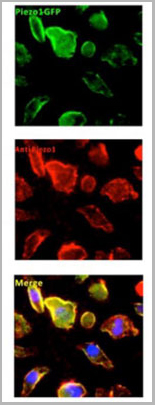
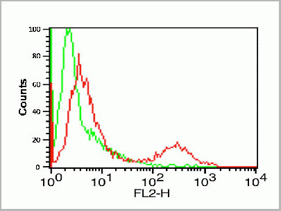
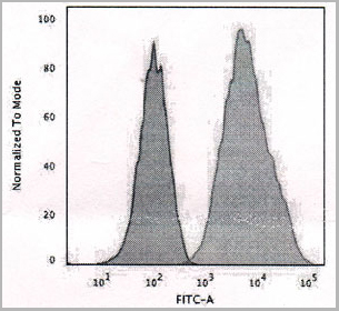
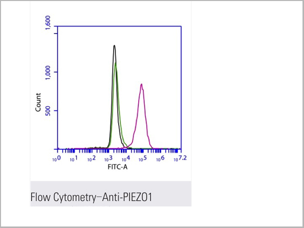 FCM (Flow Cytometry)
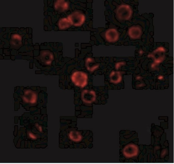
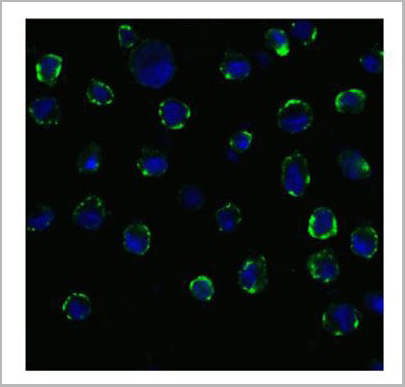
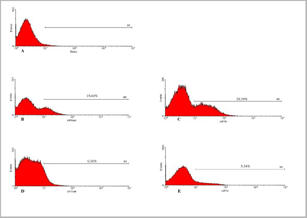
(Flow cytometric analysis of 2% paraformaldehyde-fixed THP1 (Human acute monocytic leukemia cell line) cells labeling PIEZO1 with AAA13800 at 1/200 dilution (red) compared with a mouse monoclonal IgG isotype control (black) and an unlabelled control (cells without incubation with primary antibody, green). Goat anti-mouse IgG (FITC) at 1/300 dilution was used as the secondary antibody.)
FCM (Flow Cytometry)
(Flow cytometric analysis of 2% paraformaldehyde-fixed THP1 (Human acute monocytic leukemia cell line) cells labeling PIEZO1 with AAA13800 at 1/200 dilution (red) compared with a mouse monoclonal IgG isotype control (black) and an unlabelled control (cells without incubation with primary antibody, green). Goat anti-mouse IgG (FITC) at 1/300 dilution was used as the secondary antibody.)
PIEZO1, Monoclonal Antibody (Cat# AAA13800)
Full Name
PIEZO1 Mouse mAb
Gene Names
PIEZO1; DHS; Mib; LMPH3; FAM38A
Reactivity
Homo Sapiens
Applications
Immunoprecipitation, Immunofluorescence, Flow Cytometry
Pricing
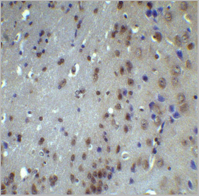
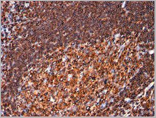
 IHC (Immunohistochemistry)
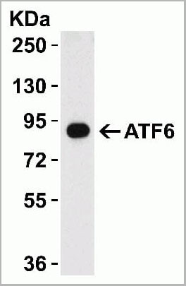
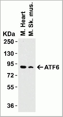
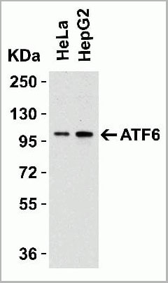
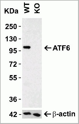
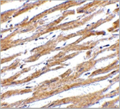
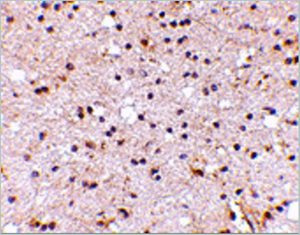
(Figure 8 Immunohistochemistry Validation of ATF6 in Rat Brain Tissue Immunohistochemical analysis of paraffin-embedded Rat brain tissue using anti-ATF6 antibody (AAA10937) at 5 ug/ml. Tissue was fixed with formaldehyde and blocked with 10% serum for 1 h at RT; antigen retrieval was by heat mediation with a citrate buffer (pH6). Samples were incubated with primary antibody overnight at 4°C. A goat anti-rabbit IgG H&L (HRP) at 1/250 was used as secondary. Counter stained with Hematoxylin.)
IHC (Immunohistochemistry)
(Figure 8 Immunohistochemistry Validation of ATF6 in Rat Brain Tissue Immunohistochemical analysis of paraffin-embedded Rat brain tissue using anti-ATF6 antibody (AAA10937) at 5 ug/ml. Tissue was fixed with formaldehyde and blocked with 10% serum for 1 h at RT; antigen retrieval was by heat mediation with a citrate buffer (pH6). Samples were incubated with primary antibody overnight at 4°C. A goat anti-rabbit IgG H&L (HRP) at 1/250 was used as secondary. Counter stained with Hematoxylin.)
ATF6, Polyclonal Antibody (Cat# AAA10937)
Full Name
ATF6 Antibody
Gene Names
ATF6; ATF6A
Reactivity
Human, Mouse, Rat
Applications
Western Blot, Immunohistochemistry, Immunofluorescence
Purity
ATF6 Antibody is affinity chromatography purified via peptide column.
Pricing
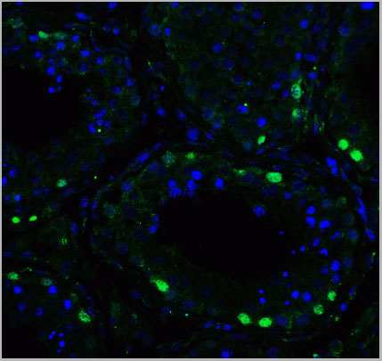
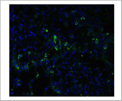
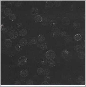
 IF (Immunofluorescence)
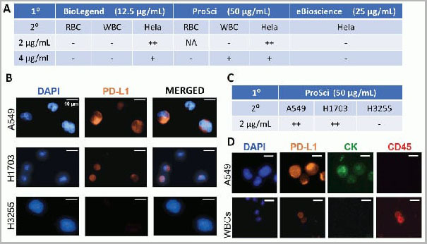
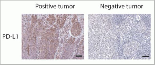
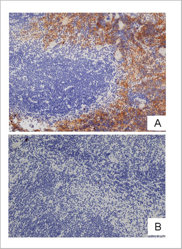
(Figure 12 Immunohistochemistry Validation of PD-L1in Human Tumors (Gadiot et al., 2011)Immunohistochemical analysis of patient tumors labeling PD-L1 with anti-PD-L1 antibodies (AAA10941). Several anti-PD-L1 antibodies were tested for staining, “Only 1 antibody gave no background staining and was competitively blocked by the addition of PD-L1Fc protein (AAA10941)”.)
IF (Immunofluorescence)
(Figure 12 Immunohistochemistry Validation of PD-L1in Human Tumors (Gadiot et al., 2011)Immunohistochemical analysis of patient tumors labeling PD-L1 with anti-PD-L1 antibodies (AAA10941). Several anti-PD-L1 antibodies were tested for staining, “Only 1 antibody gave no background staining and was competitively blocked by the addition of PD-L1Fc protein (AAA10941)”.)
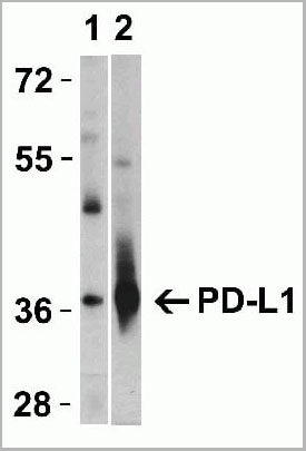
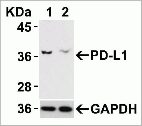
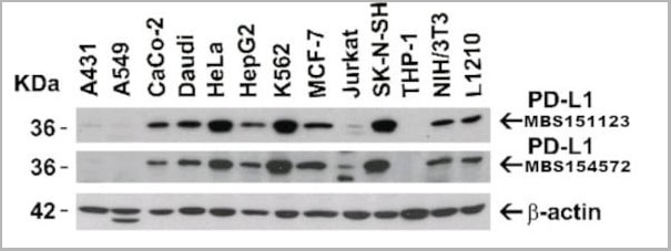
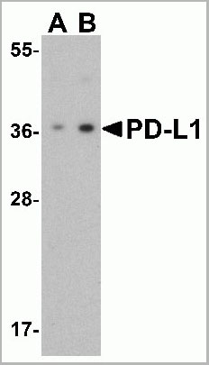
PDL-1, Polyclonal Antibody (Cat# AAA10941)
Full Name
PDL-1 Antibody
Gene Names
CD274; B7-H; B7H1; PDL1; PD-L1; PDCD1L1; PDCD1LG1
Applications
Western Blot, Immunohistochemistry, Immunofluorescence, Flow Cytometry
Purity
PD-L1 Antibody is affinity chromatography purified via peptide column.
Pricing
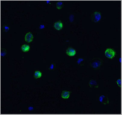
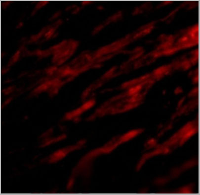
 IF (Immunofluorescence)
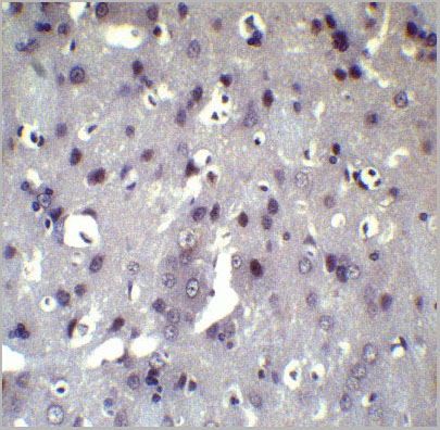
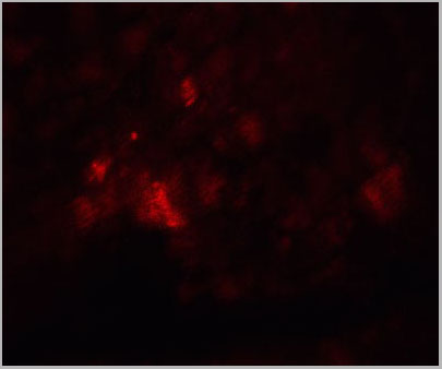
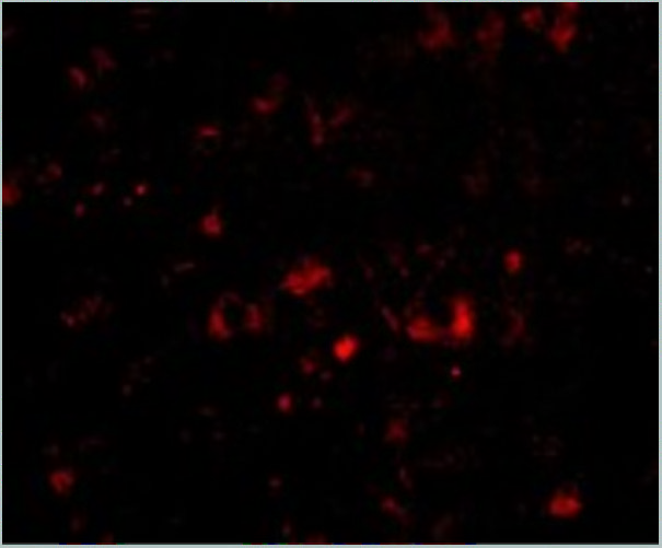
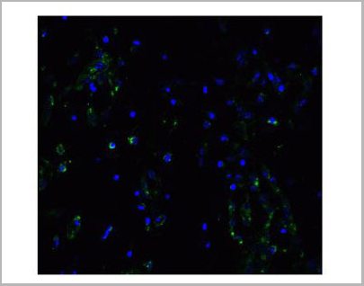
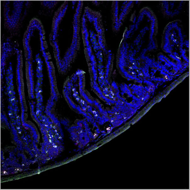
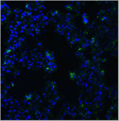
(Immunofluorescent analysis of 4% paraformaldehyde-fixed mouse brain issue labeling NPTX2 with AAA10933 at 20ug/mL, followed by goat anti-rabbit IgG secondaryantibody at 1/500 dilution (red) and DAPI staining (blue).)
IF (Immunofluorescence)
(Immunofluorescent analysis of 4% paraformaldehyde-fixed mouse brain issue labeling NPTX2 with AAA10933 at 20ug/mL, followed by goat anti-rabbit IgG secondaryantibody at 1/500 dilution (red) and DAPI staining (blue).)
NPTX2, Polyclonal Antibody (Cat# AAA10933)
Full Name
NPTX2 Antibody
Gene Names
NPTX2; NP2; NARP; NP-II
Reactivity
Human, Mouse, Rat
Applications
Western Blot, Immunohistochemistry, Immunofluorescence
Purity
NPTX2 Antibody is affinity chromatography purified via peptide column.
Pricing
 IF (Immunofluorescence)
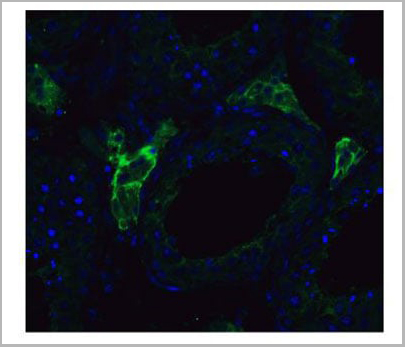
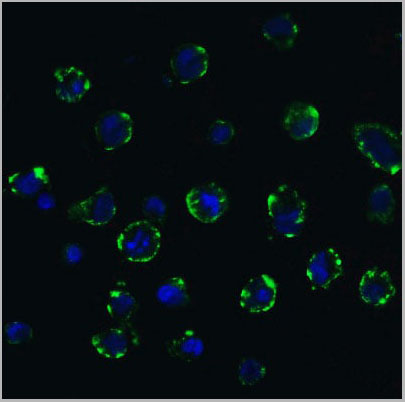
(Figure 11 Immunofluorescence Validation of ACE2 In Caco2 CellsImmunofluorescent analysis of 4% paraformaldehyde-fixed Caco2 cells labeling ACE2 with AAA10945 at 20 ug/mL, followed by goat anti-rabbit IgG secondary antibody at 1/500 dilution (green) and DAPI staining (blue). Imageshowing membrane staining on Caco2 cells.)
IF (Immunofluorescence)
(Figure 11 Immunofluorescence Validation of ACE2 In Caco2 CellsImmunofluorescent analysis of 4% paraformaldehyde-fixed Caco2 cells labeling ACE2 with AAA10945 at 20 ug/mL, followed by goat anti-rabbit IgG secondary antibody at 1/500 dilution (green) and DAPI staining (blue). Imageshowing membrane staining on Caco2 cells.)
ACE2, Polyclonal Antibody (Cat# AAA10945)
Full Name
ACE2 Antibody
Gene Names
ACE2; ACEH
Reactivity
Human, Mouse, Rat
Applications
Western Blot, Immunohistochemistry, Immunofluorescence
Purity
ACE2 Antibody is affinity chromatography purified via peptide column.
Pricing
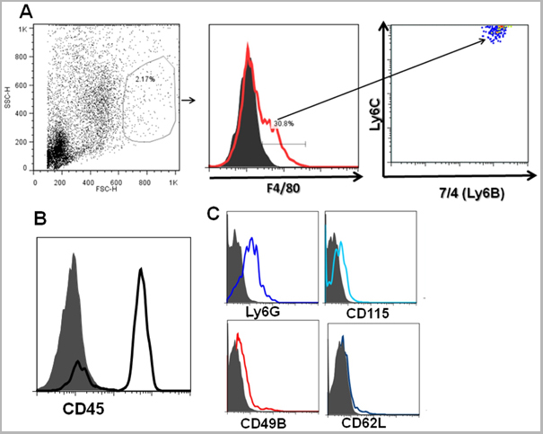
 Application Data
(Staining of J774 cells with Rat anti Mouse F4/80 antigen Biotin)
Application Data
(Staining of J774 cells with Rat anti Mouse F4/80 antigen Biotin)
F4/80, Monoclonal Antibody (Cat# AAA12225)
Full Name
RAT ANTI MOUSE F4/80
Gene Names
Emr1; Ly71; F4/80; Gpf480; TM7LN3; DD7A5-7; EGF-TM7
Applications
Flow Cytometry, Immunohistochemistry, Immunohistochemistry, Immunohistochemistry, Immunoprecipitation, Immunofluorescence, Radioimmunoassay, Fluorescence Microscopy
Pricing
Granzyme K (GZMK), Antibody (Cat# AAA21049)
Full Name
PE-Linked Polyclonal Antibody to Granzyme K (GZMK)
Gene Names
GZMK; TRYP2
Reactivity
Mouse
Applications
Westen Blot, Immunohistochemistry, Immunocytochemistry, Immunofluorescence
Purity
Antigen-specific Affinity Chromatography
Pricing
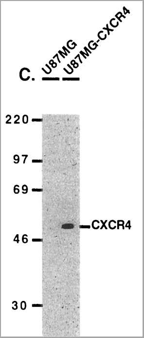

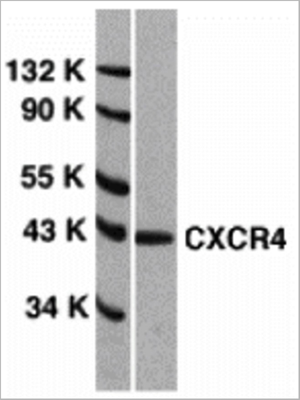
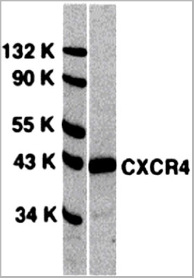
 WB (Western Blot)
(Figure 9 WB Validation of CXCR4 in metastatic melanoma (Scala et al., 2006)CXCR4 protein was detected in the human metastatic melanoma cell lines and human melanoma cell line (colo38), but not in the human primary melanocytes (MPR1) with anti-CXCR4 antibodies.)
WB (Western Blot)
(Figure 9 WB Validation of CXCR4 in metastatic melanoma (Scala et al., 2006)CXCR4 protein was detected in the human metastatic melanoma cell lines and human melanoma cell line (colo38), but not in the human primary melanocytes (MPR1) with anti-CXCR4 antibodies.)
CXCR4, Polyclonal Antibody (Cat# AAA10962)
Full Name
CXCR4 Antibody
Gene Names
CXCR4; FB22; HM89; LAP3; LCR1; NPYR; WHIM; CD184; LAP-3; LESTR; NPY3R; NPYRL; HSY3RR; NPYY3R; D2S201E
Reactivity
Human, Mouse
Applications
Western Blot, Immunofluorescence
Purity
CXCR4 Antibody is Protein A purified.
Pricing
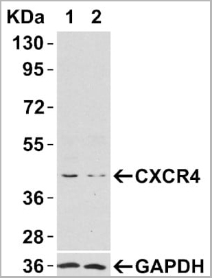
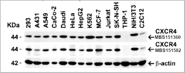
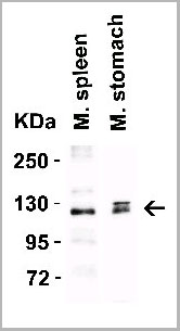
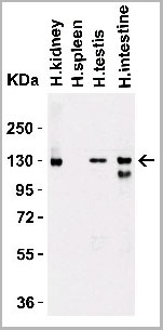
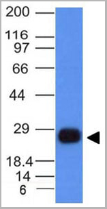
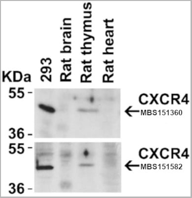 WB (Western Blot)
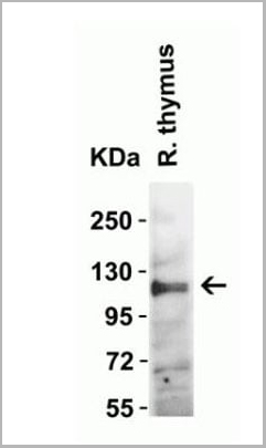
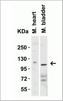
(Figure 4 Animal Species ReactivityLoading: Lysates/proteins at 20 μg per lane. Antibodies: 1009 (2 μg/mL) or 1012 (2 μg/mL). 1 h incubation at RT in 5% NFDM/TBST. Secondary: Goat anti-rabbit IgG HRP conjugate at 1:10000 dilution.)
WB (Western Blot)
(Figure 4 Animal Species ReactivityLoading: Lysates/proteins at 20 μg per lane. Antibodies: 1009 (2 μg/mL) or 1012 (2 μg/mL). 1 h incubation at RT in 5% NFDM/TBST. Secondary: Goat anti-rabbit IgG HRP conjugate at 1:10000 dilution.)
CXCR4, Polyclonal Antibody (Cat# AAA10951)
Full Name
CXCR4 Antibody
Gene Names
CXCR4; FB22; HM89; LAP3; LCR1; NPYR; WHIM; CD184; LAP-3; LESTR; NPY3R; NPYRL; HSY3RR; NPYY3R; D2S201E
Reactivity
Human, Mouse, Rat
Applications
Western Blot, Immunocytochemistry, Immunoprecipitation, Immunofluorescence, Immunohistochemistry, Flow Cytometry
Purity
CXCR4 Antibody is affinity chromatography purified via peptide column.
Pricing

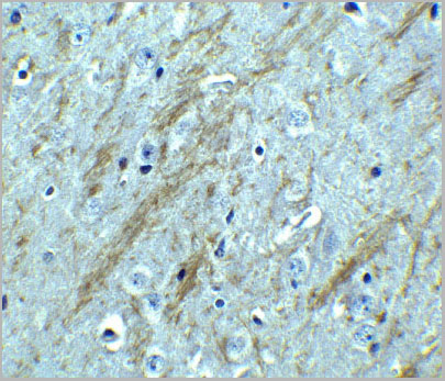
 IF (Immunofluorescence)
(Figure 10 KD Validation of BACE in DRG (Hyun, 2007)Decreased BACE1 expression in DRG following siRNA3 transfection. DRG neurons were transfected with 1 ug siRNA3 plasmid and incubated for 48 hours in 37°˚C. DRG neurons were stained for BACE1 us¬ing the Anti-BACE antibody (a,b) Neurons transfected with the control plas¬mid pSUPER-EGFP (green) did not display any changes in BACE1 expression (red). (c,d) DRG neurons transfected with siR¬NA3 displayed reduced BACE1 expression in the axon.)
IF (Immunofluorescence)
(Figure 10 KD Validation of BACE in DRG (Hyun, 2007)Decreased BACE1 expression in DRG following siRNA3 transfection. DRG neurons were transfected with 1 ug siRNA3 plasmid and incubated for 48 hours in 37°˚C. DRG neurons were stained for BACE1 us¬ing the Anti-BACE antibody (a,b) Neurons transfected with the control plas¬mid pSUPER-EGFP (green) did not display any changes in BACE1 expression (red). (c,d) DRG neurons transfected with siR¬NA3 displayed reduced BACE1 expression in the axon.)
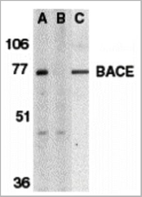
BACE, Polyclonal Antibody (Cat# AAA10918)
Full Name
BACE Antibody
Gene Names
BACE1; ASP2; BACE; HSPC104
Reactivity
Human, Mouse
Applications
Immunocytochemistry, Immunofluorescence, Immunohistochemistry, Western Blot
Purity
BACE Antibody is affinity chromatography purified via peptide column.
Pricing
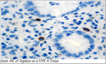
 Application Data
Tryptase, Monoclonal Antibody (Cat# AAA13565)
Full Name
Tryptase
Gene Names
MCT7; PMCT7; TPSB1
Reactivity
Human
Applications
Immunohistochemistry
Pricing


 Application Data
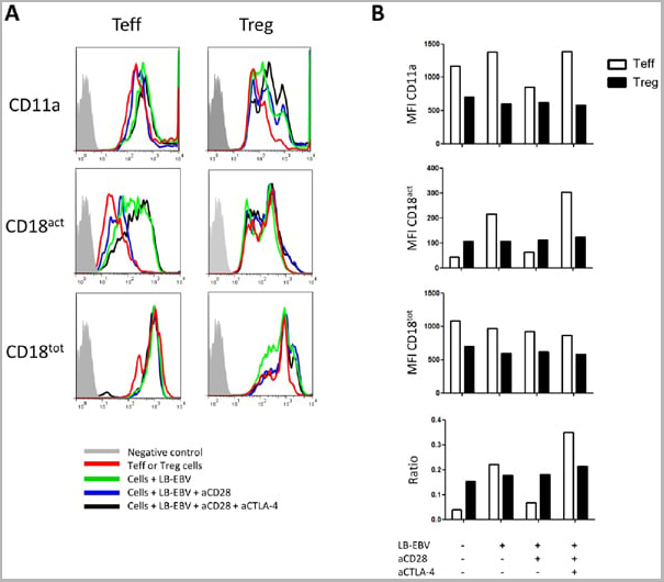
(Staining of human peripheral blood granulocytes with CD18: Alexa Fluor 647)
Application Data
(Staining of human peripheral blood granulocytes with CD18: Alexa Fluor 647)
CD18, Monoclonal Antibody (Cat# AAA12051)
Full Name
RAT ANTI HUMAN CD18:RPE
Gene Names
ITGB2; LAD; CD18; MF17; MFI7; LCAMB; LFA-1; MAC-1
Applications
Flow Cytometry
Pricing


 Application Data
(Staining of human peripheral blood granulocytes with CD18: Alexa Fluor 647 (AAA11986A647))
Application Data
(Staining of human peripheral blood granulocytes with CD18: Alexa Fluor 647 (AAA11986A647))
CD18, Monoclonal Antibody (Cat# AAA11986)
Full Name
RAT ANTI HUMAN CD18
Gene Names
ITGB2; LAD; CD18; MF17; MFI7; LCAMB; LFA-1; MAC-1
Reactivity
Dog, Guinea Pig
Applications
Immunohistochemistry, Flow Cytometry, Immunoprecipitation
Pricing
ApoC-III, Native Protein (Cat# AAA14372)
Full Name
ApoC-III protein
Gene Names
APOC3; HALP2; APOCIII
Purity
> 98% by SDS-PAGE
Pricing
 IF (Immuofluorescence)
(Figure 9 Immunofluorescence Validation of ACE2 In Caco2 Cells Immunofluorescent analysis of 4% paraformaldehyde-fixed Caco2 cells labeling ACE2 with AAA10928 at 20 ug/mL,followed by goat anti-rabbit IgG secondary antibody at1/500 dilution (green) and DAPI staining (blue). Imageshowing membrane staining on Caco2 cells.)
IF (Immuofluorescence)
(Figure 9 Immunofluorescence Validation of ACE2 In Caco2 Cells Immunofluorescent analysis of 4% paraformaldehyde-fixed Caco2 cells labeling ACE2 with AAA10928 at 20 ug/mL,followed by goat anti-rabbit IgG secondary antibody at1/500 dilution (green) and DAPI staining (blue). Imageshowing membrane staining on Caco2 cells.)
ACE2, Polyclonal Antibody (Cat# AAA10928)
Full Name
ACE2 Antibody
Gene Names
ACE2; ACEH
Reactivity
Human, Mouse, Rat
Applications
Western Blot, Immunohistochemistry, Immunofluorescence
Purity
ACE2 Antibody is affinity chromatography purified via peptide column.
Pricing
 IHC (Immunohistochemistry)
(IHC of an FFPE Breast Tissue)
IHC (Immunohistochemistry)
(IHC of an FFPE Breast Tissue)
CA15-3, Monoclonal Antibody (Cat# AAA13555)
Full Name
CA15-3
Reactivity
Paraffin, frozen
Applications
Immunohistochemistry, Immunohistochemistry
Pricing
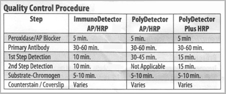 QC (Quality Control)
Treponema pallidum, Polyclonal Antibody (Cat# AAA13577)
Full Name
Treponema pallidum
Reactivity
Eubacteria
Applications
Immunohistochemistry
Pricing


 IF (Immunofluorescence)
(Figure 8 Immunofluorescence Validation of JMJD3 in K562 Cells Immunofluorescent analysis of 4% paraformaldehyde fixed K562 cells labeling JMJD3 with AAA10925 at 20 ug/mL, followed by goat anti-rabbit IgG secondary antibody at 1/500 dilution (red).)
IF (Immunofluorescence)
(Figure 8 Immunofluorescence Validation of JMJD3 in K562 Cells Immunofluorescent analysis of 4% paraformaldehyde fixed K562 cells labeling JMJD3 with AAA10925 at 20 ug/mL, followed by goat anti-rabbit IgG secondary antibody at 1/500 dilution (red).)
JMJD3, Antibody (Cat# AAA10925)
Full Name
JMJD3 Antibody
Gene Names
KDM6B; JMJD3
Reactivity
Human, Mouse, Rat
Applications
Immunocytochemistry, Immunofluorescence, Western Blot
Purity
JMJD3 Antibody is affinity chromatography purified via peptide column.
Pricing
 Application Data
(The cancer cell lines were stably expressing DYKDDDDK-tagged RIP3 and RIP expression was detected by anti-RIP3 antibodies (AAA10922) in RIP3-overexpressed cells.)
Application Data
(The cancer cell lines were stably expressing DYKDDDDK-tagged RIP3 and RIP expression was detected by anti-RIP3 antibodies (AAA10922) in RIP3-overexpressed cells.)
RIP3, Polyclonal Antibody (Cat# AAA10922)
Full Name
RIP3 Antibody
Gene Names
Ripk3; Rip3; AW107945; 2610528K09Rik
Reactivity
Human, Mouse, Rat
Applications
Immunofluorescence, Immunohistochemistry, Immunoprecipitation, Western Blot
Purity
RIP3 Antibody is affinity chromatography purified via peptide column.
Pricing


 Application Data
(Staining of human peripheral blood granulocytes with CD18: Alexa Fluor 647)
Application Data
(Staining of human peripheral blood granulocytes with CD18: Alexa Fluor 647)
CD18, Monoclonal Antibody (Cat# AAA11881)
Full Name
RAT ANTI HUMAN CD18:FITC
Gene Names
ITGB2; LAD; CD18; MF17; MFI7; LCAMB; LFA-1; MAC-1
Applications
Flow Cytometry
Pricing


 Application Data
(Staining of human peripheral blood granulocytes with CD18: Alexa Fluor 647)
Application Data
(Staining of human peripheral blood granulocytes with CD18: Alexa Fluor 647)
CD18, Monoclonal Antibody (Cat# AAA11987)
Full Name
RAT ANTI HUMAN CD18
Gene Names
ITGB2; LAD; CD18; MF17; MFI7; LCAMB; LFA-1; MAC-1
Applications
Immunohistochemistry, Flow Cytometry, Immunoprecipitation
Pricing
 WB (Western Blot)
Diacylglycerol (DAG), Polyclonal Antibody (Cat# AAA20308)
Full Name
Biotin-Linked Antibody to Diacylglycerol (DAG)
Reactivity
General
Applications
Immunohistochemistry, Immunocytochemistry, Immunoprecipitation
Purity
Antigen-specific Affinity Chromatography.
Pricing
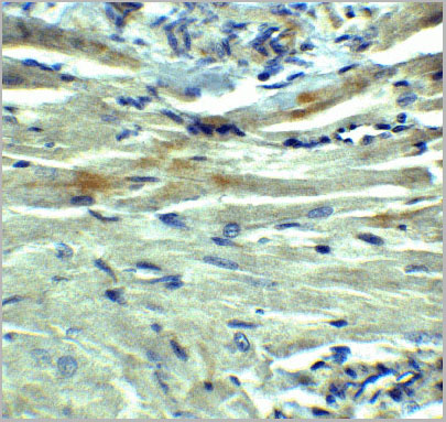
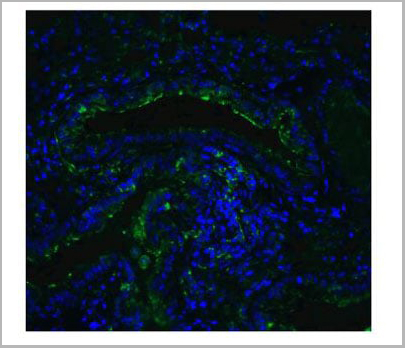
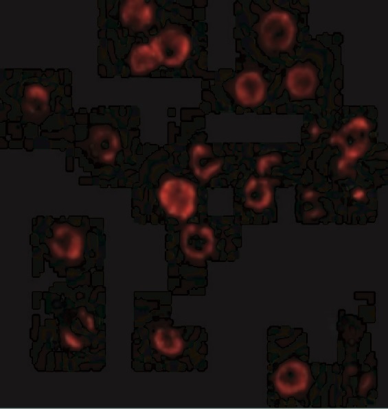
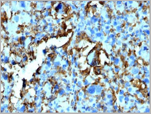
 IHC (Immunohistochemistry)
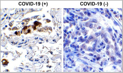
(Immunohistochemistry Validation of SARSCoV-2 (COVID-19) Spike RBD in COVID-19 Patient Lung Immunohistochemical analysis of paraffin-embedded COVID-19 patient lung tissue using anti- SARS-CoV-2 (COVID-19) Spike RBD antibody (AAA11030, 0.5 µg/mL). Tissue was fixed with formaldehyde and blocked with 10% serum for 1 h at RT; antigen retrieval was by heat mediation with a citrate buffer (pH6). Samples were incubated with primary antibody overnight at 4°C. A goat anti-rabbit IgG H&L (HRP) at 1/250 was used as secondary. Counter stained with Hematoxylin. Strong signal of SARS-COV-2 Spike RBD protein was observed in macrophage of COVID-19 patient lung, but not in non-COVID-19 patient lung.)
IHC (Immunohistochemistry)
(Immunohistochemistry Validation of SARSCoV-2 (COVID-19) Spike RBD in COVID-19 Patient Lung Immunohistochemical analysis of paraffin-embedded COVID-19 patient lung tissue using anti- SARS-CoV-2 (COVID-19) Spike RBD antibody (AAA11030, 0.5 µg/mL). Tissue was fixed with formaldehyde and blocked with 10% serum for 1 h at RT; antigen retrieval was by heat mediation with a citrate buffer (pH6). Samples were incubated with primary antibody overnight at 4°C. A goat anti-rabbit IgG H&L (HRP) at 1/250 was used as secondary. Counter stained with Hematoxylin. Strong signal of SARS-COV-2 Spike RBD protein was observed in macrophage of COVID-19 patient lung, but not in non-COVID-19 patient lung.)
COVID 19 Spike RBD Coronavirus, Polyclonal Antibody (Cat# AAA11030)
Full Name
SARS-CoV-2 (COVID-19) Spike RBD Antibody
Gene Names
S; spike glycoprotein
Reactivity
Virus
Applications
Immunofluorescence, Immunohistochemistry, Western Blot
Purity
SARS-CoV-2 (COVID-19) Spike RBD antibody is affinity chromatography purified via peptide column.
Pricing
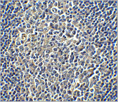
 IHC (Immunohistochemistry)
(ICH of Macrophage HAM-56 on an FFPE Liver Tissue)
IHC (Immunohistochemistry)
(ICH of Macrophage HAM-56 on an FFPE Liver Tissue)
Macrophage HAM-56, Monoclonal Antibody (Cat# AAA13562)
Full Name
Macrophage HAM-56
Reactivity
Human, Monkey
Applications
Immunohistochemistry
Pricing


 Application Data
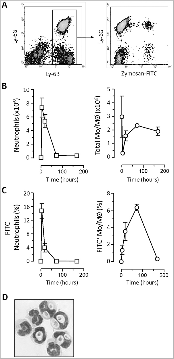
(Staining of New Zealand Black mouse peripheral blood granulocytes with Rat anti Mouse Ly-6B.2 conjugated to FITC Data)
Application Data
(Staining of New Zealand Black mouse peripheral blood granulocytes with Rat anti Mouse Ly-6B.2 conjugated to FITC Data)
Ly-6B.2 ALLOANTIGEN, Monoclonal Antibody (Cat# AAA12197)
Full Name
RAT ANTI MOUSE Ly-6B.2 ALLOANTIGEN:RPE
Applications
Flow Cytometry
Pricing
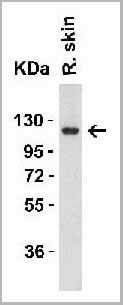
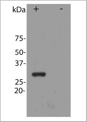 WB (Western Blot)
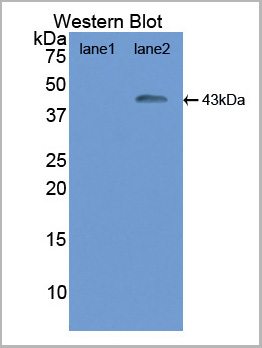
(Image: Blot of HEK293 cells transfected with pFin-EF1-mCherry vector, in the lane marked "+". HEK293 cells which were not transfected with this vector show no protein band in lane marked)
WB (Western Blot)
(Image: Blot of HEK293 cells transfected with pFin-EF1-mCherry vector, in the lane marked "+". HEK293 cells which were not transfected with this vector show no protein band in lane marked)
mCherry, Antibody (Cat# AAA14440)
Full Name
mCherry, Affinity Purified Antibody, Rabbit
Reactivity
All mammalian
Applications
Immunofluorescence, Western Blot
Pricing
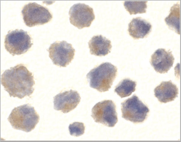
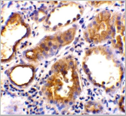
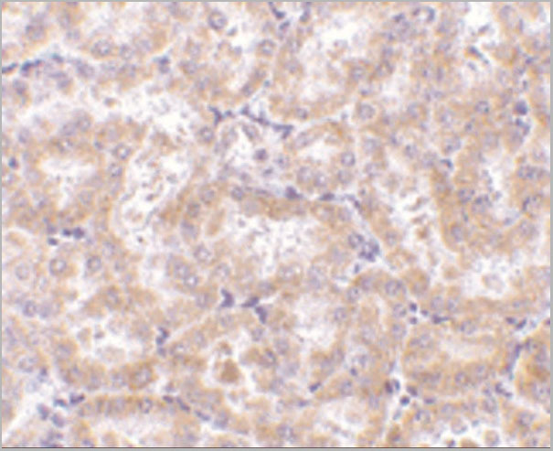
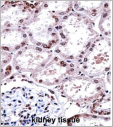 IHC (Immunohistochemistry)
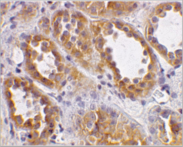
(FOXD1 Antibody (N-term) (AAA28675) immunohistochemistry analysis in formalin fixed and paraffin embedded human kidney tissue followed by peroxidase conjugation of the secondary antibody and DAB staining.This data demonstrates the use of FOXD1 Antibody (N-term) for immunohistochemistry. Clinical relevance has not been evaluated.)
IHC (Immunohistochemistry)
(FOXD1 Antibody (N-term) (AAA28675) immunohistochemistry analysis in formalin fixed and paraffin embedded human kidney tissue followed by peroxidase conjugation of the secondary antibody and DAB staining.This data demonstrates the use of FOXD1 Antibody (N-term) for immunohistochemistry. Clinical relevance has not been evaluated.)
FOXD1, Polyclonal Antibody (Cat# AAA28675)
Full Name
FOXD1 Antibody (N-term)
Gene Names
FOXD1; FKHL8; FREAC4; FREAC-4
Reactivity
Human, Mouse; Predicted: Mouse
Applications
Western Blot, Immunohistochemistry, Immunofluorescence
Purity
This antibody is purified through a protein A column, followed by peptide affinity purification.
Pricing

 FCM (Flow Cytometry)
(Flow Cytometric Analysis of human Raji cells using HLA-DR MAb (SPM423) followed by Goat anti-mouse I G-CF488 (Blue); Isotype Control (Red).)
FCM (Flow Cytometry)
(Flow Cytometric Analysis of human Raji cells using HLA-DR MAb (SPM423) followed by Goat anti-mouse I G-CF488 (Blue); Isotype Control (Red).)
HLA-DRB, Monoclonal Antibody (Cat# AAA13826)
Full Name
HLA-DRB (MHC II) Mouse Monoclonal Antibody
Gene Names
HLA-DRB1; SS1; DRB1; DRw10; HLA-DRB; HLA-DR1B
Reactivity
Human, Monkey
Applications
Flow Cytometry, Immunofluorescence, Western Blot, Immunohistochemistry
Pricing


 Application Data
(Staining of New Zealand Black mouse peripheral blood granulocytes with Rat anti Mouse Ly-6B.2 conjugated to FITC Data)
Application Data
(Staining of New Zealand Black mouse peripheral blood granulocytes with Rat anti Mouse Ly-6B.2 conjugated to FITC Data)
Ly-6B.2 ALLOANTIGEN, Monoclonal Antibody (Cat# AAA12193)
Full Name
RAT ANTI MOUSE Ly-6B.2 ALLOANTIGEN:FITC
Applications
Flow Cytometry
Pricing


 Application Data
(Staining of New Zealand Black mouse peripheral blood granulocytes with Rat anti Mouse Ly-6B.2 conjugated to FITC Data)
Application Data
(Staining of New Zealand Black mouse peripheral blood granulocytes with Rat anti Mouse Ly-6B.2 conjugated to FITC Data)
Ly-6B.2 ALLOANTIGEN, Monoclonal Antibody (Cat# AAA12192)
Full Name
RAT ANTI MOUSE Ly-6B.2 ALLOANTIGEN:FITC
Applications
Flow Cytometry
Pricing
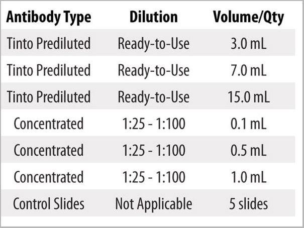
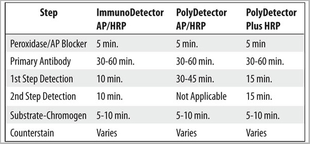
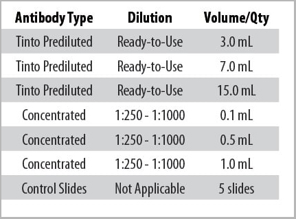
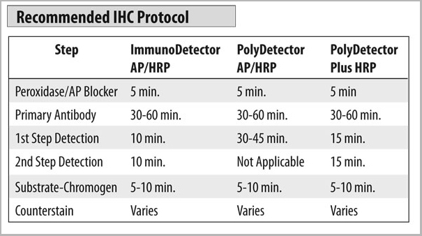 Staining Procedure
(1. Cut and mount 3-5 micron formalin-fixed paraffin-embedded tissues on positive charged slides such as Hydrophilic Plus Slides.2. Air dry for 2 hours at 58° C.3. Deparaffinize, dehydrate and rehydrate tissues.4. Subject tissues to heat epitope retrieval using a suitable retrieval solution such as ImmunoDNA Retriever with Citrate or EDTA.5. Any of three heating methods may be used:a. TintoRetriever Pressure Cooker or EquivalentPlace tissues/slides in a staining dish or coplin jar containing the ImmunoDNA Retriever with Citrate or EDTA, and place in the pressure cooker. Add 1-2 inches of distilled water to the pressure cooker and turn heat to high. Incubate for 15 minutes. Open and immediately transfer slides to room temperature. b. TintoRetriever PT Module or Water Bath Method Place tissues/slides in a pre-warmed staining dish or coplin jar containing the ImmunoDNA Retriever with Citrate or EDTA at 95°-99° C. Incubate for 30-60 minutes.c. Conventional Steamer MethodPlace tissues/slides in a pre-warmed staining dish or coplin jar containing the ImmunoDNA Retriever with Citrate or EDTA in a Steamer, cover and steam for 30-60 minutes.6. After heat treatment, transfer slides in ImmunoDNA Retriever with Citrate or EDTA to room temperature and let stand for 15-20 minutes.7. For manual staining, perform antibody incubation at ambient temperature. For automated staining methods, perform antibody incubation according to instrument manufacturer’s instructions. 8. Wash slides with ImmunoDNA washer or DI water. 9. Continue IHC staining protocol.)
Staining Procedure
(1. Cut and mount 3-5 micron formalin-fixed paraffin-embedded tissues on positive charged slides such as Hydrophilic Plus Slides.2. Air dry for 2 hours at 58° C.3. Deparaffinize, dehydrate and rehydrate tissues.4. Subject tissues to heat epitope retrieval using a suitable retrieval solution such as ImmunoDNA Retriever with Citrate or EDTA.5. Any of three heating methods may be used:a. TintoRetriever Pressure Cooker or EquivalentPlace tissues/slides in a staining dish or coplin jar containing the ImmunoDNA Retriever with Citrate or EDTA, and place in the pressure cooker. Add 1-2 inches of distilled water to the pressure cooker and turn heat to high. Incubate for 15 minutes. Open and immediately transfer slides to room temperature. b. TintoRetriever PT Module or Water Bath Method Place tissues/slides in a pre-warmed staining dish or coplin jar containing the ImmunoDNA Retriever with Citrate or EDTA at 95°-99° C. Incubate for 30-60 minutes.c. Conventional Steamer MethodPlace tissues/slides in a pre-warmed staining dish or coplin jar containing the ImmunoDNA Retriever with Citrate or EDTA in a Steamer, cover and steam for 30-60 minutes.6. After heat treatment, transfer slides in ImmunoDNA Retriever with Citrate or EDTA to room temperature and let stand for 15-20 minutes.7. For manual staining, perform antibody incubation at ambient temperature. For automated staining methods, perform antibody incubation according to instrument manufacturer’s instructions. 8. Wash slides with ImmunoDNA washer or DI water. 9. Continue IHC staining protocol.)
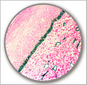
Podoplanin/D2-40, Monoclonal Antibody (Cat# AAA13558)
Full Name
Podoplanin/D2-40
Reactivity
Human, Rat, Mouse
Applications
Immunohistochemistry
Pricing


 Application Data
(Staining of New Zealand Black mouse peripheral blood granulocytes with Rat anti Mouse Ly-6B.2 conjugated to FITC Data)
Application Data
(Staining of New Zealand Black mouse peripheral blood granulocytes with Rat anti Mouse Ly-6B.2 conjugated to FITC Data)
Ly-6B.2 ALLOANTIGEN, Monoclonal Antibody (Cat# AAA12194)
Full Name
RAT ANTI MOUSE Ly-6B.2 ALLOANTIGEN
Applications
Immunohistochemistry, Flow Cytometry, Immunofluorescence, Immunohistochemistry, Western Blot
Pricing