Filters
Clonality
Type
Reactivity
Gene Name
Isotype
Host
Application
Clone
194 results for " tumor antigen" - showing 50-100


 ICC (Immunocytochemistry)
(ICC staining CD147 in SKOV-3 cells (red). The nuclear counter stain is DAPI (blue). Cells were fixed in paraformaldehyde, permeabilised with 0.25% Triton X100/PBS.)
ICC (Immunocytochemistry)
(ICC staining CD147 in SKOV-3 cells (red). The nuclear counter stain is DAPI (blue). Cells were fixed in paraformaldehyde, permeabilised with 0.25% Triton X100/PBS.)
CD147, Monoclonal Antibody (Cat# AAA30287)
Full Name
CD147 Antibody
Gene Names
BSG; M6; OK; 5F7; TCSF; CD147; EMMPRIN
Reactivity
Human, Mouse, Rat
Applications
Western Blot, Immunocytochemistry, Immunofluorescence, Immunohistochemistry
Purity
ProA affinity purified
Pricing

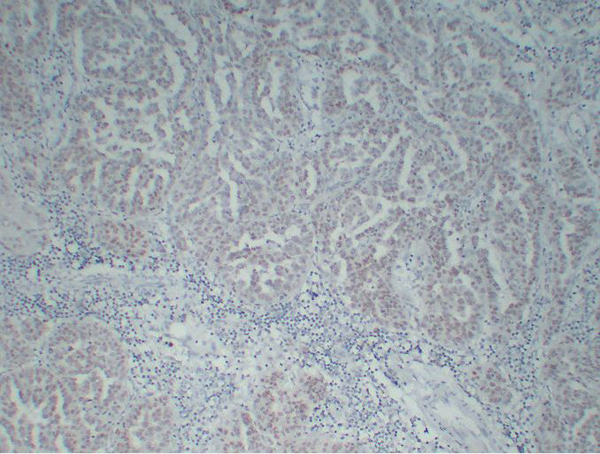


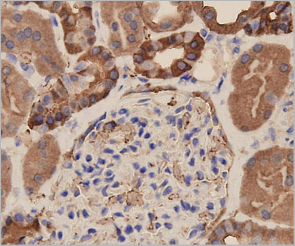
 IHC (Immunohistchemistry)
(Immunohistochemistry analysis of Formalin-fixed, paraffin-embedded Human Wilms' tumor using WT1, Wilms' Tumor 1 antibody.)
IHC (Immunohistchemistry)
(Immunohistochemistry analysis of Formalin-fixed, paraffin-embedded Human Wilms' tumor using WT1, Wilms' Tumor 1 antibody.)
Wilms' Tumor 1 (WT1), Monoclonal Antibody (Cat# AAA30741)
Full Name
Anti-Wilms' Tumor 1 (WT1) Antibody [ABT-WT1]
Gene Names
WT1; GUD; AWT1; WAGR; WT33; NPHS4; WIT-2
Reactivity
Human
Applications
Immunohistochemistry
Purity
Immunogen Affinity Purified
Pricing
 WB (Western Blot)
(Western Blot analysis of extracts from NIH-3T3 cells using MUC1 Antibody.)
WB (Western Blot)
(Western Blot analysis of extracts from NIH-3T3 cells using MUC1 Antibody.)
MUC1, Polyclonal Antibody (Cat# AAA30739)
Full Name
MUC1 Antibody
Gene Names
MUC1; EMA; MCD; PEM; PUM; KL-6; MAM6; MCKD; PEMT; CD227; H23AG; MCKD1; MUC-1; ADMCKD; ADMCKD1; CA 15-3; MUC-1/X; MUC1/ZD; MUC-1/SEC
Reactivity
Human, Mouse, Rat
Applications
Western Blot, Immunohistochemistry, Immunohistochemistry
Purity
The antibody was affinity-purified from rabbit antiserum by affinity-
chromatography using epitope-specific immunogen.
Pricing

 IHC (Immunohistochemistry)
(GCC2 antibody (C-term) (AAA28751) immunohistochemistry analysis in formalin fixed and paraffin embedded human brain tissue followed by peroxidase conjugation of the secondary antibody and DAB staining. This data demonstrates the use of the GCC2 antibody (C-term) for immunohistochemistry. Clinical relevance has not been evaluated.)
IHC (Immunohistochemistry)
(GCC2 antibody (C-term) (AAA28751) immunohistochemistry analysis in formalin fixed and paraffin embedded human brain tissue followed by peroxidase conjugation of the secondary antibody and DAB staining. This data demonstrates the use of the GCC2 antibody (C-term) for immunohistochemistry. Clinical relevance has not been evaluated.)
GCC2, Polyclonal Antibody (Cat# AAA28751)
Full Name
GCC2 Antibody (C-term)
Gene Names
GCC2; REN53; GCC185; RANBP2L4
Reactivity
Human, mouse (Predicted Reactivity: Rat)
Applications
Western Blot, Immunohistochemistry
Purity
This antibody is purified through a protein A column, followed by peptide affinity purification.
Pricing


 Application Data
(At 25 degree C. The primary antibody was diluted at 1/200 and incubated with the sample for 1 hour at 37 degree C. An Alexa Fluor 594 conjugated goat anti-rabbit IgG (H+L) Ab, diluted at 1/600, was used as the secondary antibody.)
Application Data
(At 25 degree C. The primary antibody was diluted at 1/200 and incubated with the sample for 1 hour at 37 degree C. An Alexa Fluor 594 conjugated goat anti-rabbit IgG (H+L) Ab, diluted at 1/600, was used as the secondary antibody.)
Retinoblastoma, Polyclonal Antibody (Cat# AAA31401)
Full Name
Phospho-Retinoblastoma (Ser788) Antibody
Gene Names
RB1; RB; pRb; OSRC; pp110; p105-Rb
Reactivity
Human, Mouse, Rat
Predicted Reactivity: Pig (100%), Bovine (100%), Horse (100%), Sheep (100%), Rabbit (100%), Dog (100%), Chicken (92%), Xenopus (92%)
Predicted Reactivity: Pig (100%), Bovine (100%), Horse (100%), Sheep (100%), Rabbit (100%), Dog (100%), Chicken (92%), Xenopus (92%)
Applications
Western Blot, Immunohistochemistry, Immunofluorescence, Immunocytochemistry, Peptide ELISA
Purity
The antibody is from purified rabbit serum by affinity purification via sequential chromatography on phospho-peptide and non-phospho-peptide affinity columns.
Pricing


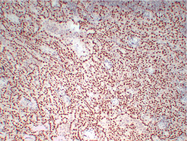
 IHC (Immunohistochemistry)
(At 1/100 staining Human colorectal cancer by IHC-P. The sample was formaldehyde fixed and a heat mediated antigen retrieval step in citrate buffer was performed. The sample was then blocked and incubated with the primary antibody at 4 degree C overnight. An HRP conjugated anti-Rabbit antibody was used as the secondary antibody.)
IHC (Immunohistochemistry)
(At 1/100 staining Human colorectal cancer by IHC-P. The sample was formaldehyde fixed and a heat mediated antigen retrieval step in citrate buffer was performed. The sample was then blocked and incubated with the primary antibody at 4 degree C overnight. An HRP conjugated anti-Rabbit antibody was used as the secondary antibody.)
CD227/MUC1, Polyclonal Antibody (Cat# AAA31426)
Full Name
Phospho-CD227/MUC1 (Ser1227) Antibody
Gene Names
MUC1; EMA; PEM; PUM; KL-6; MAM6; PEMT; CD227; H23AG; MUC-1; CA 15-3; MUC-1/X; MUC1/ZD; MUC-1/SEC
Reactivity
Human, Mouse, Rat
Applications
Western Blot, Immunohistochemistry, Peptide ELISA
Purity
The antibody is from purified rabbit serum by affinity purification via sequential chromatography on phospho-peptide and non-phospho-peptide affinity columns.
Pricing


 Application Data
(Detection limit for recombinant GST tagged TP53 is approximately 1ng/ml as a capture antibody.)
Application Data
(Detection limit for recombinant GST tagged TP53 is approximately 1ng/ml as a capture antibody.)
TP53, Monoclonal Antibody (Cat# AAA26173)
Full Name
TP53 (Tumor Protein p53, FLJ92943, LFS1, TRP53, p53) (Biotin)
Gene Names
TP53; P53; BCC7; LFS1; BMFS5; TRP53
Applications
Immunofluorescence, Immunohistochemistry, Immunoprecipitation, Western Blot
Purity
Purified
Pricing


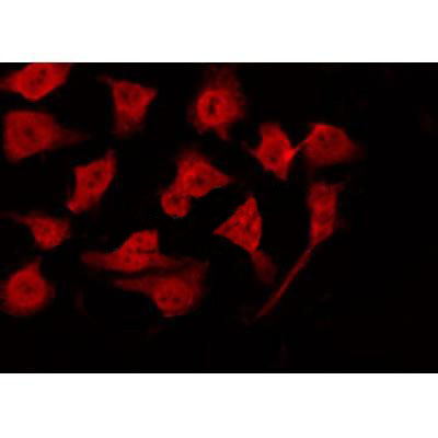
 ICC (Immunocytochemistry)
(ICC staining EpCAM in SW1990 cells (green). The nuclear counter stain is DAPI (blue). Cells were fixed in paraformaldehyde, permeabilised with 0.25% Triton X100/PBS.)
ICC (Immunocytochemistry)
(ICC staining EpCAM in SW1990 cells (green). The nuclear counter stain is DAPI (blue). Cells were fixed in paraformaldehyde, permeabilised with 0.25% Triton X100/PBS.)
EP-CAM, Monoclonal Antibody (Cat# AAA29891)
Full Name
EP-CAM Antibody
Gene Names
EPCAM; ESA; KSA; M4S1; MK-1; DIAR5; EGP-2; EGP40; KS1/4; MIC18; TROP1; EGP314; HNPCC8; TACSTD1
Reactivity
Human, Mouse
Applications
Western Blot, Immunocytochemistry, Immunohistochemistry, Flow Cytometry
Purity
ProL affinity purified
Pricing


 IF (Immunofluorescence)
(Immunofluorescence analysis of Raw264.7 cells using CD44 antibody at dilution of 1:100. Blue: DAPI for nuclear staining.)
IF (Immunofluorescence)
(Immunofluorescence analysis of Raw264.7 cells using CD44 antibody at dilution of 1:100. Blue: DAPI for nuclear staining.)
CD44, Polyclonal Antibody (Cat# AAA28336)
Full Name
CD44 Rabbit pAb
Gene Names
CD44; IN; LHR; MC56; MDU2; MDU3; MIC4; Pgp1; CDW44; CSPG8; HCELL; HUTCH-I; ECMR-III
Reactivity
Human, Mouse, Rat
Applications
Western Blot, Immunohistochemistry, Immunofluorescence
Purity
Affinity purification
Pricing


 IF (Immunofluorescence)
(Confocal Immunofluorescent analysis of SK-OV-3 cells using AF488-labeled Isotype Control Monoclonal Antibody (IgG1) (Green).DAPI was used to stain the cell nuclei (blue). (Negative Control))
IF (Immunofluorescence)
(Confocal Immunofluorescent analysis of SK-OV-3 cells using AF488-labeled Isotype Control Monoclonal Antibody (IgG1) (Green).DAPI was used to stain the cell nuclei (blue). (Negative Control))
Ep-CAM /CD326, Monoclonal Antibody (Cat# AAA13838)
Full Name
Ep-CAM /CD326 (Epithelial Marker) Mouse Monoclonal Antibody
Gene Names
EPCAM; ESA; KSA; M4S1; MK-1; DIAR5; EGP-2; EGP40; KS1/4; MIC18; TROP1; EGP314; HNPCC8; TACSTD1
Reactivity
Human.
Does not react with Mouse and Rat
Does not react with Mouse and Rat
Applications
Flow Cytometry, Immunofluorescence, Western Blot, Immunohistochemistry
Pricing


 FCM (Flow Cytometry)
(Flow cytometric analysis of THP-1 cells with eEF1A1 antibody at 1/100 dilution (red) compared with an unlabelled control (cells without incubation with primary antibody; black). Alexa Fluor 488-conjugated goat anti-rabbit IgG was used as the secondary antibody.)
FCM (Flow Cytometry)
(Flow cytometric analysis of THP-1 cells with eEF1A1 antibody at 1/100 dilution (red) compared with an unlabelled control (cells without incubation with primary antibody; black). Alexa Fluor 488-conjugated goat anti-rabbit IgG was used as the secondary antibody.)
EEF1A1, Monoclonal Antibody (Cat# AAA30467)
Full Name
EEF1A1 Antibody
Gene Names
EEF1A1; CCS3; EF1A; PTI1; CCS-3; EE1A1; EEF-1; EEF1A; EF-Tu; LENG7; eEF1A-1; GRAF-1EF; HNGC:16303
Reactivity
Human, Mouse, Rat
Applications
Western Blot, Immunocytochemistry, Immunofluorescence, Immunohistochemistry, Flow Cytometry, Immunoprecipitation
Purity
ProA affinity purified
Pricing


 Application Data
(Published customer image: Histological staining of control and HBOT wounds at post-wounding days 7 and 29. A -D) H&E staining. E -H) CD34 immunohistochemistry. I+J) CD68 immunohistochemistry.From: uk B, Tong M, Fijneman EMG, van Neck JW (2014) Hyperbaric Oxygen Therapy to Treat Diabetes Impaired Wound Healing in Rats. PLoS ONE 9(10): e108533.)
Application Data
(Published customer image: Histological staining of control and HBOT wounds at post-wounding days 7 and 29. A -D) H&E staining. E -H) CD34 immunohistochemistry. I+J) CD68 immunohistochemistry.From: uk B, Tong M, Fijneman EMG, van Neck JW (2014) Hyperbaric Oxygen Therapy to Treat Diabetes Impaired Wound Healing in Rats. PLoS ONE 9(10): e108533.)
CD68, Monoclonal Antibody (Cat# AAA12151)
Full Name
MOUSE ANTI RAT CD68
Applications
Immunohistochemistry, Flow Cytometry, Immunofluorescence, Immunoprecipitation, Immunohistochemistry, Radioimmunoassay, Western Blot
Pricing


 FCM (Flow Cytometry)
(Flow cytometric analysis of SW480 cells with DR5 antibody at 1/100 dilution (red) compared with an unlabelled control (cells without incubation with primary antibody; black).)
FCM (Flow Cytometry)
(Flow cytometric analysis of SW480 cells with DR5 antibody at 1/100 dilution (red) compared with an unlabelled control (cells without incubation with primary antibody; black).)
DR5, Monoclonal Antibody (Cat# AAA30353)
Full Name
DR5 Antibody
Gene Names
TNFRSF10B; DR5; CD262; KILLER; TRICK2; TRICKB; ZTNFR9; TRAILR2; TRICK2A; TRICK2B; TRAIL-R2; KILLER/DR5
Reactivity
Human
Applications
Western Blot, Immunoprecipitation, Immunohistochemistry, Flow Cytometry
Purity
ProA affinity purified
Pricing

 WB (Western Blot)
(Peptide-+ Western blot analysis of extracts from RAW264.7 cells, HT-29 cells and LOVO cells, using TNF Receptor II antibody. Immunofluorescence analysis of HeLa cells, using TNF Receptor II antibody.)
WB (Western Blot)
(Peptide-+ Western blot analysis of extracts from RAW264.7 cells, HT-29 cells and LOVO cells, using TNF Receptor II antibody. Immunofluorescence analysis of HeLa cells, using TNF Receptor II antibody.)
TNF Receptor II, Antibody (Cat# AAA17967)
Full Name
TNF Receptor II Antibody
Gene Names
TNFRSF1B; p75; TBPII; TNFBR; TNFR2; CD120b; TNFR1B; TNFR80; TNF-R75; p75TNFR; TNF-R-II
Reactivity
Human, Mouse, Rat
Applications
Western Blot, Immunofluorescence
Purity
Affinity-purified from rabbit antiserum by affinity-chromatography using epitope-specific immunogen.
Pricing


 Application Data
(Published customer image Infiltration of GFP+ BM-cells in infarct and peri-infarct regions. (A-B) Dot plots of viable macrophages/granulocytes (CD11b+CD45high, top right quadrants) and microglia (CD11b+CD45dim, bottom right quadrants) in cortex from BM-chimeric unmanipulated mice and mice exposed to pMCAO. (C) Bar graph showing mean numbers of CD11b+CD45dim microglia and CD11b+CD45high macrophages/granulocytes in BM-chimeric mice 24 hours after pMCAO, subdivided based on expression of GFP (n = 5). Approximately 92% of of the CD45high population were GFP+. (D) Estimation and comparison of mean numbers of CD11b+CD45dim microglia in non-chimeric (n = 10) versus BM-chimeric mice (n = 5) 24 hours after of pMCAO shows significantly fewer CD11b+CD45dim microglial cells in irradiated mice. (E) Overview, showing distribution of infiltrating GFP+ BM-derived cells into infarct (IF) and peri-infarct (P-IF) regions 24 hours after pMCAO. (E-G) By 24 hours, GFP+ single cells (F) and vessel-associated aggregates of GFP+ cells (arrows in G) were observed in infarct and peri-infarct regions. Some of the vessel-associated cells were round, leukocyte-like cells (arrows) while others were elongated cells lining the vasculature (arrow heads in G and in insert). (H) Bar graph showing mean numbers of single GFP+ cells and vessel-associated aggregates of GFP+ cells in ipsi- and contralateral cortex 24 hours after surgery (n = 10). (I-P) Immunohistochemical staining of CD45.1 (I, K), CD45.2 (J, L), IgG2a (M, O) and CD45 (N, P) in ischemic tissue in BM-chimeric (I, J, M, N) and non-chimeric mice (K, L, O, P) 24 hours after pMCAO. N.D, none detected. Scale bars: 200 um (A), 10 um (B, C). 50 um (I-P) *P < 0.05, **P < 0.01, and ***P < 0.001.From: Clausen BH, Lambertsen KL, Babcock AA, Holm TH, Dagnaes-Hansen F, Finsen B. Interleukin-1beta and tumor necrosis factor-alpha are expressed by different subsets of microglia and macrophages after ischemic stroke in mice. J Neuroinflammation. 2008 Oct 23;5:46.)
Application Data
(Published customer image Infiltration of GFP+ BM-cells in infarct and peri-infarct regions. (A-B) Dot plots of viable macrophages/granulocytes (CD11b+CD45high, top right quadrants) and microglia (CD11b+CD45dim, bottom right quadrants) in cortex from BM-chimeric unmanipulated mice and mice exposed to pMCAO. (C) Bar graph showing mean numbers of CD11b+CD45dim microglia and CD11b+CD45high macrophages/granulocytes in BM-chimeric mice 24 hours after pMCAO, subdivided based on expression of GFP (n = 5). Approximately 92% of of the CD45high population were GFP+. (D) Estimation and comparison of mean numbers of CD11b+CD45dim microglia in non-chimeric (n = 10) versus BM-chimeric mice (n = 5) 24 hours after of pMCAO shows significantly fewer CD11b+CD45dim microglial cells in irradiated mice. (E) Overview, showing distribution of infiltrating GFP+ BM-derived cells into infarct (IF) and peri-infarct (P-IF) regions 24 hours after pMCAO. (E-G) By 24 hours, GFP+ single cells (F) and vessel-associated aggregates of GFP+ cells (arrows in G) were observed in infarct and peri-infarct regions. Some of the vessel-associated cells were round, leukocyte-like cells (arrows) while others were elongated cells lining the vasculature (arrow heads in G and in insert). (H) Bar graph showing mean numbers of single GFP+ cells and vessel-associated aggregates of GFP+ cells in ipsi- and contralateral cortex 24 hours after surgery (n = 10). (I-P) Immunohistochemical staining of CD45.1 (I, K), CD45.2 (J, L), IgG2a (M, O) and CD45 (N, P) in ischemic tissue in BM-chimeric (I, J, M, N) and non-chimeric mice (K, L, O, P) 24 hours after pMCAO. N.D, none detected. Scale bars: 200 um (A), 10 um (B, C). 50 um (I-P) *P < 0.05, **P < 0.01, and ***P < 0.001.From: Clausen BH, Lambertsen KL, Babcock AA, Holm TH, Dagnaes-Hansen F, Finsen B. Interleukin-1beta and tumor necrosis factor-alpha are expressed by different subsets of microglia and macrophages after ischemic stroke in mice. J Neuroinflammation. 2008 Oct 23;5:46.)
CD11b, Monoclonal Antibody (Cat# AAA12186)
Full Name
RAT ANTI MOUSE CD11b:RPE
Gene Names
Itgam; CR3; CR3A; MAC1; Cd11b; Ly-40; Mac-1; Mac-1a; CD11b/CD18; F730045J24Rik
Applications
Flow Cytometry
Pricing
 SDS-PAGE
Cellular tumor antigen p53 (Tp53), Recombinant Protein (Cat# AAA18501)
Full Name
Recombinant Mouse Cellular tumor antigen p53 (Tp53)
Gene Names
Trp53; bbl; bfy; bhy; p44; p53; Tp53
Purity
Greater or equal to 85% purity as determined by SDS-PAGE.
Pricing


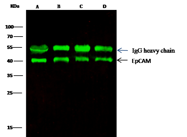 IP (Immunoprecipitation)
(EpCAM was immunoprecipitated using:Lane A:0.5 mg MCF-7 Whole Cell LysateLane B:0.5 mg A431 Whole Cell LysateLane C:0.5 mg HepG2 Whole Cell LysateLane D:0.5 mg Caco-2 Whole Cell Lysate0.5 uL anti-EpCAM rabbit monoclonal antibody and 15 ul of 50 % Protein G agarose.Primary antibody:Anti-EpCAM rabbit monoclonal antibody,at 1:5000 dilution Secondary antibody:Dylight 800-labeled antibody to rabbit IgG (H+L), at 1:5000 dilution Developed using the odssey technique.Performed under reducing conditions.Predicted band size: 40 kDaObserved band size: 40 kDa)
IP (Immunoprecipitation)
(EpCAM was immunoprecipitated using:Lane A:0.5 mg MCF-7 Whole Cell LysateLane B:0.5 mg A431 Whole Cell LysateLane C:0.5 mg HepG2 Whole Cell LysateLane D:0.5 mg Caco-2 Whole Cell Lysate0.5 uL anti-EpCAM rabbit monoclonal antibody and 15 ul of 50 % Protein G agarose.Primary antibody:Anti-EpCAM rabbit monoclonal antibody,at 1:5000 dilution Secondary antibody:Dylight 800-labeled antibody to rabbit IgG (H+L), at 1:5000 dilution Developed using the odssey technique.Performed under reducing conditions.Predicted band size: 40 kDaObserved band size: 40 kDa)
EpCAM, Monoclonal Antibody (Cat# AAA27736)
Full Name
Recombinant Anti-EpCAM Antibody, Rabbit Monoclonal
Gene Names
EPCAM; ESA; KSA; M4S1; MK-1; DIAR5; EGP-2; EGP40; KS1/4; MIC18; TROP1; EGP314; HNPCC8; TACSTD1
Reactivity
Human
Applications
Western Blot, Immunohistochemistry, Flow Cytometry, Immunocytochemistry, Immunofluorescence, Immunoprecipitation
Purity
Protein A
Pricing
 Standard Curve (Sample)
CD248 molecule, endosialin, ELISA Kit (Cat# AAA18107)
Full Name
Human Endosialin, CD248 ELISA Kit
Gene Names
CD248; TEM1; CD164L1
Reactivity
Human
Pricing


 Application Data
(Published customer image Infiltration of GFP+ BM-cells in infarct and peri-infarct regions. (A-B) Dot plots of viable macrophages/granulocytes (CD11b+CD45high, top right quadrants) and microglia (CD11b+CD45dim, bottom right quadrants) in cortex from BM-chimeric unmanipulated mice and mice exposed to pMCAO. (C) Bar graph showing mean numbers of CD11b+CD45dim microglia and CD11b+CD45high macrophages/granulocytes in BM-chimeric mice 24 hours after pMCAO, subdivided based on expression of GFP (n = 5). Approximately 92% of of the CD45high population were GFP+. (D) Estimation and comparison of mean numbers of CD11b+CD45dim microglia in non-chimeric (n = 10) versus BM-chimeric mice (n = 5) 24 hours after of pMCAO shows significantly fewer CD11b+CD45dim microglial cells in irradiated mice. (E) Overview, showing distribution of infiltrating GFP+ BM-derived cells into infarct (IF) and peri-infarct (P-IF) regions 24 hours after pMCAO. (E-G) By 24 hours, GFP+ single cells (F) and vessel-associated aggregates of GFP+ cells (arrows in G) were observed in infarct and peri-infarct regions. Some of the vessel-associated cells were round, leukocyte-like cells (arrows) while others were elongated cells lining the vasculature (arrow heads in G and in insert). (H) Bar graph showing mean numbers of single GFP+ cells and vessel-associated aggregates of GFP+ cells in ipsi- and contralateral cortex 24 hours after surgery (n = 10). (I-P) Immunohistochemical staining of CD45.1 (I, K), CD45.2 (J, L), IgG2a (M, O) and CD45 (N, P) in ischemic tissue in BM-chimeric (I, J, M, N) and non-chimeric mice (K, L, O, P) 24 hours after pMCAO. N.D, none detected. Scale bars: 200 um (A), 10 um (B, C). 50 um (I-P) *P < 0.05, **P < 0.01, and ***P < 0.001.From: Clausen BH, Lambertsen KL, Babcock AA, Holm TH, Dagnaes-Hansen F, Finsen B. Interleukin-1beta and tumor necrosis factor-alpha are expressed by different subsets of microglia and macrophages after ischemic stroke in mice. J Neuroinflammation. 2008 Oct 23;5:46.)
Application Data
(Published customer image Infiltration of GFP+ BM-cells in infarct and peri-infarct regions. (A-B) Dot plots of viable macrophages/granulocytes (CD11b+CD45high, top right quadrants) and microglia (CD11b+CD45dim, bottom right quadrants) in cortex from BM-chimeric unmanipulated mice and mice exposed to pMCAO. (C) Bar graph showing mean numbers of CD11b+CD45dim microglia and CD11b+CD45high macrophages/granulocytes in BM-chimeric mice 24 hours after pMCAO, subdivided based on expression of GFP (n = 5). Approximately 92% of of the CD45high population were GFP+. (D) Estimation and comparison of mean numbers of CD11b+CD45dim microglia in non-chimeric (n = 10) versus BM-chimeric mice (n = 5) 24 hours after of pMCAO shows significantly fewer CD11b+CD45dim microglial cells in irradiated mice. (E) Overview, showing distribution of infiltrating GFP+ BM-derived cells into infarct (IF) and peri-infarct (P-IF) regions 24 hours after pMCAO. (E-G) By 24 hours, GFP+ single cells (F) and vessel-associated aggregates of GFP+ cells (arrows in G) were observed in infarct and peri-infarct regions. Some of the vessel-associated cells were round, leukocyte-like cells (arrows) while others were elongated cells lining the vasculature (arrow heads in G and in insert). (H) Bar graph showing mean numbers of single GFP+ cells and vessel-associated aggregates of GFP+ cells in ipsi- and contralateral cortex 24 hours after surgery (n = 10). (I-P) Immunohistochemical staining of CD45.1 (I, K), CD45.2 (J, L), IgG2a (M, O) and CD45 (N, P) in ischemic tissue in BM-chimeric (I, J, M, N) and non-chimeric mice (K, L, O, P) 24 hours after pMCAO. N.D, none detected. Scale bars: 200 um (A), 10 um (B, C). 50 um (I-P) *P < 0.05, **P < 0.01, and ***P < 0.001.From: Clausen BH, Lambertsen KL, Babcock AA, Holm TH, Dagnaes-Hansen F, Finsen B. Interleukin-1beta and tumor necrosis factor-alpha are expressed by different subsets of microglia and macrophages after ischemic stroke in mice. J Neuroinflammation. 2008 Oct 23;5:46.)
CD11b, Monoclonal Antibody (Cat# AAA12231)
Full Name
RAT ANTI MOUSE CD11b:Low Endotoxin
Gene Names
Itgam; CR3; CR3A; MAC1; Cd11b; Ly-40; Mac-1; Mac-1a; CD11b/CD18; F730045J24Rik
Applications
Immunohistochemistry, Flow Cytometry, Functional Assay, Immunofluorescence, Immunoprecipitation
Pricing


 FCM (Flow Cytometry)
(Flow cytometric analysis of PC-3M cells with BAP31 antibody at 1/100 dilution (purple) compared with an unlabelled control (cells without incubation with primary antibody; yellow). Alexa Fluor 488-conjugated goat anti-rabbit IgG was used as the secondary antibody.)
FCM (Flow Cytometry)
(Flow cytometric analysis of PC-3M cells with BAP31 antibody at 1/100 dilution (purple) compared with an unlabelled control (cells without incubation with primary antibody; yellow). Alexa Fluor 488-conjugated goat anti-rabbit IgG was used as the secondary antibody.)
BAP31, Monoclonal Antibody (Cat# AAA30501)
Full Name
BAP31 Antibody
Gene Names
BCAP31; CDM; BAP31; 6C6-AG; DXS1357E
Reactivity
Human, Mouse
Applications
Western Blot, Immunocytochemistry, Immunofluorescence, Immunohistochemistry, Flow Cytometry
Purity
ProA affinity purified
Pricing


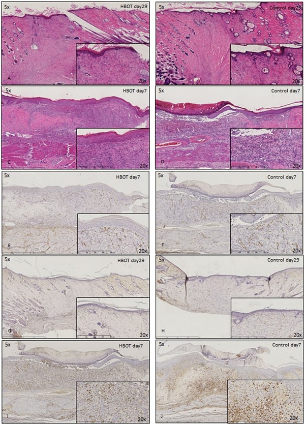 Application Data
(Published customer image: Histological staining of control and HBOT wounds at post-wounding days 7 and 29. A -D) H&E staining. E -H) CD34 immunohistochemistry. I+J) CD68 immunohistochemistry.From: uk B, Tong M, Fijneman EMG, van Neck JW (2014) Hyperbaric Oxygen Therapy to Treat Diabetes Impaired Wound Healing in Rats. PLoS ONE 9(10): e108533.)
Application Data
(Published customer image: Histological staining of control and HBOT wounds at post-wounding days 7 and 29. A -D) H&E staining. E -H) CD34 immunohistochemistry. I+J) CD68 immunohistochemistry.From: uk B, Tong M, Fijneman EMG, van Neck JW (2014) Hyperbaric Oxygen Therapy to Treat Diabetes Impaired Wound Healing in Rats. PLoS ONE 9(10): e108533.)
CD68, Monoclonal Antibody (Cat# AAA12148)
Full Name
MOUSE ANTI RAT CD68:FITC
Applications
Flow Cytometry
Pricing


 Application Data
(Published customer image Infiltration of GFP+ BM-cells in infarct and peri-infarct regions. (A-B) Dot plots of viable macrophages/granulocytes (CD11b+CD45high, top right quadrants) and microglia (CD11b+CD45dim, bottom right quadrants) in cortex from BM-chimeric unmanipulated mice and mice exposed to pMCAO. (C) Bar graph showing mean numbers of CD11b+CD45dim microglia and CD11b+CD45high macrophages/granulocytes in BM-chimeric mice 24 hours after pMCAO, subdivided based on expression of GFP (n = 5). Approximately 92% of of the CD45high population were GFP+. (D) Estimation and comparison of mean numbers of CD11b+CD45dim microglia in non-chimeric (n = 10) versus BM-chimeric mice (n = 5) 24 hours after of pMCAO shows significantly fewer CD11b+CD45dim microglial cells in irradiated mice. (E) Overview, showing distribution of infiltrating GFP+ BM-derived cells into infarct (IF) and peri-infarct (P-IF) regions 24 hours after pMCAO. (E-G) By 24 hours, GFP+ single cells (F) and vessel-associated aggregates of GFP+ cells (arrows in G) were observed in infarct and peri-infarct regions. Some of the vessel-associated cells were round, leukocyte-like cells (arrows) while others were elongated cells lining the vasculature (arrow heads in G and in insert). (H) Bar graph showing mean numbers of single GFP+ cells and vessel-associated aggregates of GFP+ cells in ipsi- and contralateral cortex 24 hours after surgery (n = 10). (I-P) Immunohistochemical staining of CD45.1 (I, K), CD45.2 (J, L), IgG2a (M, O) and CD45 (N, P) in ischemic tissue in BM-chimeric (I, J, M, N) and non-chimeric mice (K, L, O, P) 24 hours after pMCAO. N.D, none detected. Scale bars: 200 um (A), 10 um (B, C). 50 um (I-P) *P < 0.05, **P < 0.01, and ***P < 0.001.From: Clausen BH, Lambertsen KL, Babcock AA, Holm TH, Dagnaes-Hansen F, Finsen B. Interleukin-1beta and tumor necrosis factor-alpha are expressed by different subsets of microglia and macrophages after ischemic stroke in mice. J Neuroinflammation. 2008 Oct 23;5:46.)
Application Data
(Published customer image Infiltration of GFP+ BM-cells in infarct and peri-infarct regions. (A-B) Dot plots of viable macrophages/granulocytes (CD11b+CD45high, top right quadrants) and microglia (CD11b+CD45dim, bottom right quadrants) in cortex from BM-chimeric unmanipulated mice and mice exposed to pMCAO. (C) Bar graph showing mean numbers of CD11b+CD45dim microglia and CD11b+CD45high macrophages/granulocytes in BM-chimeric mice 24 hours after pMCAO, subdivided based on expression of GFP (n = 5). Approximately 92% of of the CD45high population were GFP+. (D) Estimation and comparison of mean numbers of CD11b+CD45dim microglia in non-chimeric (n = 10) versus BM-chimeric mice (n = 5) 24 hours after of pMCAO shows significantly fewer CD11b+CD45dim microglial cells in irradiated mice. (E) Overview, showing distribution of infiltrating GFP+ BM-derived cells into infarct (IF) and peri-infarct (P-IF) regions 24 hours after pMCAO. (E-G) By 24 hours, GFP+ single cells (F) and vessel-associated aggregates of GFP+ cells (arrows in G) were observed in infarct and peri-infarct regions. Some of the vessel-associated cells were round, leukocyte-like cells (arrows) while others were elongated cells lining the vasculature (arrow heads in G and in insert). (H) Bar graph showing mean numbers of single GFP+ cells and vessel-associated aggregates of GFP+ cells in ipsi- and contralateral cortex 24 hours after surgery (n = 10). (I-P) Immunohistochemical staining of CD45.1 (I, K), CD45.2 (J, L), IgG2a (M, O) and CD45 (N, P) in ischemic tissue in BM-chimeric (I, J, M, N) and non-chimeric mice (K, L, O, P) 24 hours after pMCAO. N.D, none detected. Scale bars: 200 um (A), 10 um (B, C). 50 um (I-P) *P < 0.05, **P < 0.01, and ***P < 0.001.From: Clausen BH, Lambertsen KL, Babcock AA, Holm TH, Dagnaes-Hansen F, Finsen B. Interleukin-1beta and tumor necrosis factor-alpha are expressed by different subsets of microglia and macrophages after ischemic stroke in mice. J Neuroinflammation. 2008 Oct 23;5:46.)
CD11b, Monoclonal Antibody (Cat# AAA12181)
Full Name
RAT ANTI MOUSE CD11b
Gene Names
Itgam; CR3; CR3A; MAC1; Cd11b; Ly-40; Mac-1; Mac-1a; CD11b/CD18; F730045J24Rik
Reactivity
Human
Applications
Immunohistochemistry, Flow Cytometry, Immunofluorescence, Immunoprecipitation
Pricing


 IHC (Immunohistchemistry)
(Immunohistochemistry of TM4SF1 in mouse lung tissue with TM4SF1 antibody at 2 μg/mL.)
IHC (Immunohistchemistry)
(Immunohistochemistry of TM4SF1 in mouse lung tissue with TM4SF1 antibody at 2 μg/mL.)
TM4SF1, Polyclonal Antibody (Cat# AAA10947)
Full Name
TM4SF1 Antibody
Gene Names
TM4SF1; L6; H-L6; M3S1; TAAL6
Reactivity
Human, Mouse, Rat
Applications
Immunofluorescence, Immunohistochemistry, Western Blot
Purity
TM4SF1 Antibody is affinity chromatography purified via peptide column.
Pricing


 IHC (Immunohistochemistry)
(Anti-PGES / PTGES antibody IHC of human Lung, Non-Small Cell Carcinoma. Immunohistochemistry of formalin-fixed, paraffin-embedded tissue after heat-induced antigen retrieval.)
IHC (Immunohistochemistry)
(Anti-PGES / PTGES antibody IHC of human Lung, Non-Small Cell Carcinoma. Immunohistochemistry of formalin-fixed, paraffin-embedded tissue after heat-induced antigen retrieval.)
PGES / PTGES, Polyclonal Antibody (Cat# AAA12311)
Full Name
Rabbit Polyclonal to Human PGES / PTGES
Gene Names
PTGES; PGES; MPGES; PIG12; PP102; PP1294; MGST-IV; MGST1L1; TP53I12; mPGES-1; MGST1-L1
Reactivity
Gibbon, Gorilla, Human, Rabbit
Predicted Reactivity: Monkey, Dog (at least 90% immunogen sequence identity)
Predicted Reactivity: Monkey, Dog (at least 90% immunogen sequence identity)
Applications
Immunohistochemistry
Purity
Immunoaffinity Purified
Pricing


 Application Data
(Detection limit for recombinant GST tagged TP53 is approximately 3ng/ml as a capture antibody.)
Application Data
(Detection limit for recombinant GST tagged TP53 is approximately 3ng/ml as a capture antibody.)
TP53, Monoclonal Antibody (Cat# AAA24685)
Full Name
TP53 (Tumor Suppressor p53, Cellular Tumor Antigen p53, TRP53, Antigen NY-CO-13, BCC7, LFS1, Phosphoprotein p53) APC
Gene Names
TP53; P53; BCC7; LFS1; TRP53
Reactivity
Human
Applications
Immunohistochemistry, Western Blot
Purity
Purified by Protein A Affinity Chromatography.
Pricing

 WB (Western Blot)
((0.5ug/ml) staining of A549 (A), HepG2 (B) and K562 (C) lysates (35ug protein in RIPA buffer). Primary incubation was 1 hour. Detected by chemiluminescence.)
WB (Western Blot)
((0.5ug/ml) staining of A549 (A), HepG2 (B) and K562 (C) lysates (35ug protein in RIPA buffer). Primary incubation was 1 hour. Detected by chemiluminescence.)
TNFRSF1A/TNFR1, Polyclonal Antibody (Cat# AAA13672)
Full Name
Goat anti-TNFRSF1A/TNFR1 Antibody
Gene Names
TNFRSF1A; FPF; MS5; p55; p60; TBP1; TNF-R; TNFAR; TNFR1; p55-R; CD120a; TNFR55; TNFR60; TNF-R-I; TNF-R55; TNFR1-d2
Reactivity
Tested: Human; Expected from sequence similarity: Human, Rat, Pig
Applications
Peptide ELISA, Western Blot
Purity
Purified from goat serum by ammonium sulphate precipitation followed by antigen affinity chromatography using the immunizing peptide.
Pricing


 WB (Western Blot)
(Western BlotSample: Recombinant GAL9, Human;Antibody: Rabbit Anti-Human GAL9 Ab)
WB (Western Blot)
(Western BlotSample: Recombinant GAL9, Human;Antibody: Rabbit Anti-Human GAL9 Ab)
Galectin 9 (GAL9), Active Protein (Cat# AAA21103)
Full Name
Active Galectin 9 (GAL9)
Gene Names
LGALS9; HUAT; LGALS9A
Reactivity
Homo sapiens (Human)
Applications
Cell culture; Activity Assays.
Purity
>90%
Pricing
 Standard Curve (Sample)
heat shock protein 90kDa alpha (cytosolic) , class B member 1, ELISA Kit (Cat# AAA18180)
Full Name
Mouse Heat shock protein HSP 90-beta, HSP90AB1 ELISA Kit
Gene Names
Hsp90ab1; 90kDa; Hsp84; Hsp90; Hspcb; C81438; Hsp84-1; AL022974
Reactivity
Mouse
Pricing


 Application Data
(Analysis of Protein Array containing >19, 000 full-length human proteins using EpCAM Mouse Monoclonal Antibody (EGP40/1372) Z- and S- Score: The Z-score represents the strength of a signal that a monoclonal antibody (MAb) (in combination with a fluorescently-tagged anti-IgG secondary antibody) produces when binding to a particular protein on the HuProtTM array. Z-scores are described in units of standard deviations (SD's) above the mean value of all signals generated on that array. If targets on HuProtTM are arranged in descending order of the Z-score, the S-score is the difference (also in units of SD's) between the Z-score. S-score therefore represents the relative target specificity of a MAb to its intended target. A MAb is considered to specific to its intended target, if the MAb has an S-score of at least 2.5. For example, if a MAb binds to protein X with a Z-score of 43 and to protein Y with a Z-score of 14, then the S-score for the binding of that MAb to protein X is equal to 29.)
Application Data
(Analysis of Protein Array containing >19, 000 full-length human proteins using EpCAM Mouse Monoclonal Antibody (EGP40/1372) Z- and S- Score: The Z-score represents the strength of a signal that a monoclonal antibody (MAb) (in combination with a fluorescently-tagged anti-IgG secondary antibody) produces when binding to a particular protein on the HuProtTM array. Z-scores are described in units of standard deviations (SD's) above the mean value of all signals generated on that array. If targets on HuProtTM are arranged in descending order of the Z-score, the S-score is the difference (also in units of SD's) between the Z-score. S-score therefore represents the relative target specificity of a MAb to its intended target. A MAb is considered to specific to its intended target, if the MAb has an S-score of at least 2.5. For example, if a MAb binds to protein X with a Z-score of 43 and to protein Y with a Z-score of 14, then the S-score for the binding of that MAb to protein X is equal to 29.)
Ep-CAM/CD326, Monoclonal Antibody (Cat# AAA23890)
Full Name
Ep-CAM/CD326 (Extracellular Domain) (Epithelial Marker)
Gene Names
EPCAM; ESA; KSA; M4S1; MK-1; DIAR5; EGP-2; EGP40; KS1/4; MIC18; TROP1; EGP314; HNPCC8; TACSTD1
Reactivity
Human. Others not known.
Applications
Flow Cytometry, Immunofluorescence, Western Blot, Immunohistochemistry
Pricing
 Standard Curve (Sample)
Centrosomal protein of 290 kDa (CEP290), ELISA Kit (Cat# AAA27144)
Full Name
Human Centrosomal protein of 290 kDa (CEP290) ELISA Kit
Gene Names
CEP290; CT87; MKS4; POC3; rd16; BBS14; JBTS5; LCA10; NPHP6; SLSN6; 3H11Ag
Reactivity
Human
Pricing
 Standard Curve (Sample)
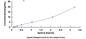
Integrin Associated Protein (IAP), ELISA Kit (Cat# AAA20650)
Full Name
Human Integrin Associated Protein (IAP) ELISA Kit
Gene Names
CD47; IAP; OA3; MER6
Reactivity
Human
Pricing
 Standard Curve (Sample)
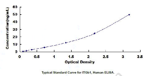
Integrin Beta 1 (ITGb1), ELISA Kit (Cat# AAA20576)
Full Name
Human Integrin Beta 1 (ITGb1) ELISA Kit
Gene Names
ITGB1; CD29; FNRB; MDF2; VLAB; GPIIA; MSK12; VLA-BETA
Reactivity
Human
Pricing


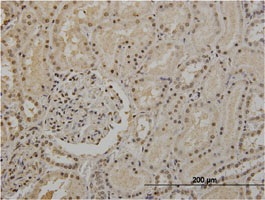
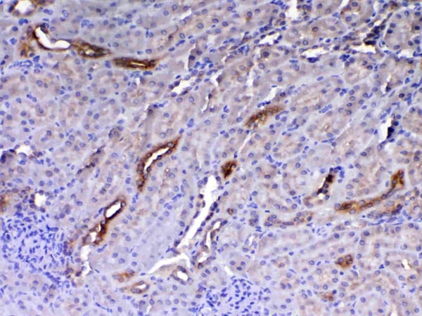
 IHC (Immunohistochemistry)
(IHC image diluted at 1:1000 and staining in paraffin-embedded human kidney tissue performed on a Leica BondTM system. After dewaxing and hydration, antigen retrieval was mediated by high pressure in a citrate buffer (pH 6.0). Section was blocked with 10% normal goat serum 30min at RT. Then primary antibody (1% BSA) was incubated at 4 degree C overnight. The primary is detected by a biotinylated secondary antibody and visualized using an HRP conjugated SP system.)
IHC (Immunohistochemistry)
(IHC image diluted at 1:1000 and staining in paraffin-embedded human kidney tissue performed on a Leica BondTM system. After dewaxing and hydration, antigen retrieval was mediated by high pressure in a citrate buffer (pH 6.0). Section was blocked with 10% normal goat serum 30min at RT. Then primary antibody (1% BSA) was incubated at 4 degree C overnight. The primary is detected by a biotinylated secondary antibody and visualized using an HRP conjugated SP system.)
PKM, Monoclonal Antibody (Cat# AAA27048)
Full Name
PKM Monoclonal Antibody
Gene Names
PKM; PK3; TCB; OIP3; PKM2; CTHBP; THBP1
Reactivity
Human, Mouse
Applications
Western Blot, Immunohistochemistry, Immunofluorescence, Flow Cytometry, Immunoprecipitation
Purity
>95%, Protein G purified
Pricing
 Standard Curve (Sample)
Galectin-3-binding protein, ELISA Kit (Cat# AAA17744)
Full Name
Human Galectin-3-binding protein ELISA Kit
Gene Names
LGALS3BP; 90K; M2BP; gp90; CyCAP; BTBD17B; MAC-2-BP; TANGO10B
Reactivity
Human
Pricing


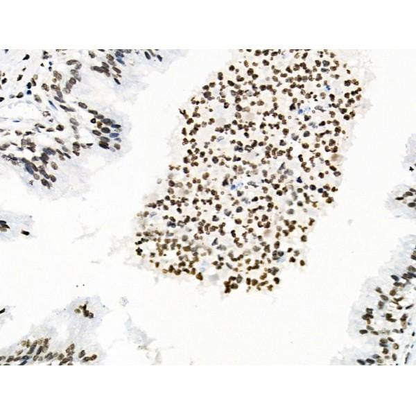
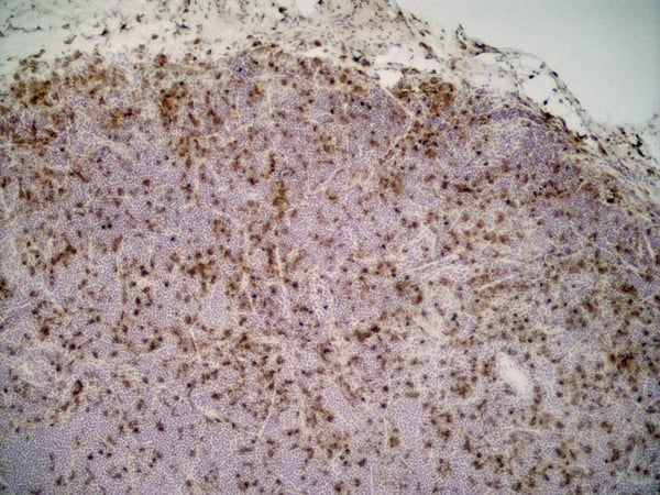
 IHC (Immunohistchemistry)
(Figure 6. IHC analysis of HE4 using anti-HE4 antibody (AAA19161).HE4 was detected in paraffin-embedded section of rat small intestine tissue. Heat mediated antigen retrieval was performed in citrate buffer (pH6, epitope retrieval solution) for 20 mins. The tissue section was blocked with 10% goat serum. The tissue section was then incubated with 1ug/ml rabbit anti-HE4 Antibody (AAA19161) overnight at 4 degree C. Biotinylated goat anti-rabbit IgG was used as secondary antibody and incubated for 30 minutes at 37 degree C. The tissue section was developed using Strepavidin-Biotin-Complex (SABC) with DAB as the chromogen.)
IHC (Immunohistchemistry)
(Figure 6. IHC analysis of HE4 using anti-HE4 antibody (AAA19161).HE4 was detected in paraffin-embedded section of rat small intestine tissue. Heat mediated antigen retrieval was performed in citrate buffer (pH6, epitope retrieval solution) for 20 mins. The tissue section was blocked with 10% goat serum. The tissue section was then incubated with 1ug/ml rabbit anti-HE4 Antibody (AAA19161) overnight at 4 degree C. Biotinylated goat anti-rabbit IgG was used as secondary antibody and incubated for 30 minutes at 37 degree C. The tissue section was developed using Strepavidin-Biotin-Complex (SABC) with DAB as the chromogen.)
HE4, Polyclonal Antibody (Cat# AAA19161)
Full Name
Anti-HE4 Picoband Antibody
Gene Names
Wfdc2; re4
Reactivity
Mouse, Rat
No cross reactivity with other proteins.
No cross reactivity with other proteins.
Applications
IHC, WB
Purity
Immunogen affinity purified
Pricing


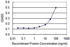
 Application Data
(Sandwich ELISA analysis of CD178 binding using Mouse anti Human CD178 as a capture reagent and biotinylated Mouse anti Human CD178 as a detection reagent with purified human CD178 as antigen for the generation of a standard curve. Detection is by HRP conjugated Streptavidin and substrate. Microtitre plate is read at O.D. 450 nm on the iMark Microplate Absorbance Reader . Plasma (orange) sample is displayed at 1:2 dilution.)
Application Data
(Sandwich ELISA analysis of CD178 binding using Mouse anti Human CD178 as a capture reagent and biotinylated Mouse anti Human CD178 as a detection reagent with purified human CD178 as antigen for the generation of a standard curve. Detection is by HRP conjugated Streptavidin and substrate. Microtitre plate is read at O.D. 450 nm on the iMark Microplate Absorbance Reader . Plasma (orange) sample is displayed at 1:2 dilution.)
CD178, Monoclonal Antibody (Cat# AAA11961)
Full Name
MOUSE ANTI HUMAN CD178
Gene Names
FASLG; APTL; FASL; CD178; CD95L; ALPS1B; CD95-L; TNFSF6; APT1LG1
Applications
Flow Cytometry, Immunoprecipitation
Pricing
 Standard Curve (Sample)
Tumor Necrosis Factor Alpha (TNFa), ELISA Kit (Cat# AAA20696)
Full Name
High Sensitivity Human Tumor Necrosis Factor Alpha (TNFa) ELISA Kit
Gene Names
TNF; DIF; TNFA; TNFSF2; TNF-alpha
Reactivity
Human
Pricing
 SDS-PAGE
Cellular tumor antigen p53 (TP53), Recombinant Protein (Cat# AAA18532)
Full Name
Recombinant Human Cellular tumor antigen p53 (TP53)
Gene Names
TP53; P53; BCC7; LFS1; TRP53
Purity
Greater or equal to 85% purity as determined by SDS-PAGE.
Pricing


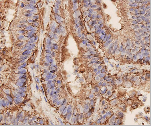
 IHC (Immunohistchemistry)
(Formalin-fixed, paraffin-embedded human Breast Carcinoma stained with MUC1 / EMA Monoclonal Antibody (MUC1/845).)
IHC (Immunohistchemistry)
(Formalin-fixed, paraffin-embedded human Breast Carcinoma stained with MUC1 / EMA Monoclonal Antibody (MUC1/845).)
MUC1 / EMA /CD227, Monoclonal Antibody (Cat# AAA13841)
Full Name
MUC1 / EMA /CD227 (Epithelial Marker) Mouse Monoclonal Antibody
Gene Names
MUC1; EMA; MCD; PEM; PUM; KL-6; MAM6; MCKD; PEMT; CD227; H23AG; MCKD1; MUC-1; ADMCKD; ADMCKD1; CA 15-3; MUC-1/X; MUC1/ZD; MUC-1/SEC
Reactivity
Human
Applications
Flow Cytometry, Immunofluorescence, Immunohistochemistry
Pricing
 Standard Curve (Sample)
Trefoil Factor 3, Intestinal (TFF3), ELISA Kit (Cat# AAA20374)
Full Name
Human Trefoil Factor 3, Intestinal (TFF3) ELISA Kit
Gene Names
TFF3; ITF; P1B; TFI
Reactivity
Human
Pricing
 Standard Curve (Sample)
Mucin 1 (MUC1), DIY ELISA Kit (Cat# AAA21080)
Full Name
Human Mucin 1 (MUC1) ELISA Kit DIY Materials
Gene Names
MUC1; EMA; MCD; PEM; PUM; KL-6; MAM6; MCKD; PEMT; CD227; H23AG; MCKD1; MUC-1; ADMCKD; ADMCKD1; CA 15-3; MUC-1/X; MUC1/ZD; MUC-1/SEC
Reactivity
Human
Pricing


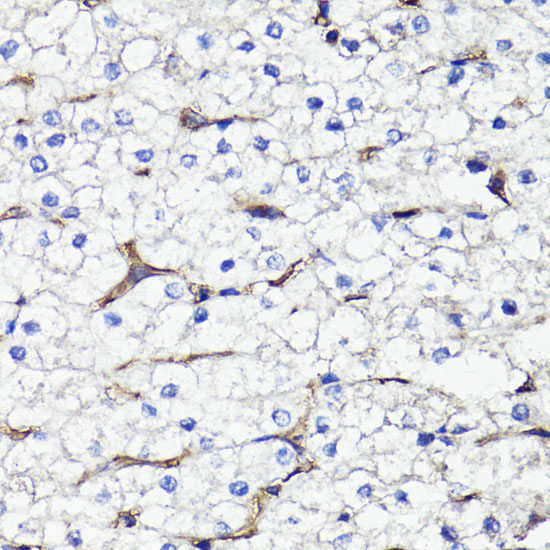
 IHC (Immunohistochemistry)
(DAB staining on IHC-P; Samples: Human Kidney Tissue.)
IHC (Immunohistochemistry)
(DAB staining on IHC-P; Samples: Human Kidney Tissue.)
Extracellular Matrix Metalloproteinase Inducer, Polyclonal Antibody (Cat# AAA20867)
Full Name
Polyclonal Antibody to Extracellular Matrix Metalloproteinase Inducer (EMMPRIN)
Gene Names
BSG; OK; 5F7; TCSF; CD147; EMMPRIN
Reactivity
Human, Mouse, Rat
Applications
WB, ICC, IHC, EIA
Purity
Affinity Chromatography
Pricing
 Standard Curve (Sample)
Major Histocompatibility Complex Class II DQ Beta 1 (MHCDQb1), ELISA Kit (Cat# AAA20552)
Full Name
Human Major Histocompatibility Complex Class II DQ Beta 1 (MHCDQb1) ELISA Kit
Gene Names
HLA-DQB1; IDDM1; CELIAC1; HLA-DQB
Reactivity
Human
Pricing


 FCM (Flow Cytometry)
(Figure 6. Flow Cytometry analysis of A549 cells using anti-Ch TOG/CKAP5 antibody (AAA19386).Overlay histogram showing A549 cells stained with AAA19386 (Blue line). The cells were blocked with 10% normal goat serum. And then incubated with mouse anti- Ch TOG/CKAP5 Antibody (AAA19386, 1μg/1x106 cells) for 30 min at 20 degree C. DyLight®488 conjugated goat anti-mouse IgG (BA1126, 5-10μg/1x106 cells) was used as secondary antibody for 30 minutes at 20 degree C. Isotype control antibody (Green line) was mouse IgG (1μg/1x106) used under the same conditions. Unlabelled sample (Red line) was also used as a control.)
FCM (Flow Cytometry)
(Figure 6. Flow Cytometry analysis of A549 cells using anti-Ch TOG/CKAP5 antibody (AAA19386).Overlay histogram showing A549 cells stained with AAA19386 (Blue line). The cells were blocked with 10% normal goat serum. And then incubated with mouse anti- Ch TOG/CKAP5 Antibody (AAA19386, 1μg/1x106 cells) for 30 min at 20 degree C. DyLight®488 conjugated goat anti-mouse IgG (BA1126, 5-10μg/1x106 cells) was used as secondary antibody for 30 minutes at 20 degree C. Isotype control antibody (Green line) was mouse IgG (1μg/1x106) used under the same conditions. Unlabelled sample (Red line) was also used as a control.)
ch TOG/CKAP5, Monoclonal Antibody (Cat# AAA19386)
Full Name
Anti-ch TOG/CKAP5 Antibody (monoclonal, 3C13)
Gene Names
CKAP5; TOG; MSPS; TOGp; CHTOG; ch-TOG
Reactivity
Human, Mouse, Rat
Applications
WB, IHC-P, ICC, IF, FC/FACS/FCM
Purity
Immunogen affinity purified.
Pricing


 WB (Western Blot)
(WB Suggested Anti-FOLR1 antibody Titration: 1 ug/mLSample Type: Human liver)
WB (Western Blot)
(WB Suggested Anti-FOLR1 antibody Titration: 1 ug/mLSample Type: Human liver)
FOLR1, Polyclonal Antibody (Cat# AAA23491)
Full Name
FOLR1 antibody - middle region
Gene Names
FOLR1; FBP; FOLR
Reactivity
Cow, Dog, Guinea Pig, Horse, Human, Mouse, Rabbit, Rat
Applications
WB, IHC
Purity
Affinity Purified
Pricing
 Standard Curve (Sample)
Olfactomedin 4 (OLFM4), ELISA Kit (Cat# AAA20557)
Full Name
Human Olfactomedin 4 (OLFM4) ELISA Kit
Gene Names
OLFM4; GC1; OLM4; OlfD; GW112; hGC-1; hOLfD; UNQ362; bA209J19.1
Reactivity
Human
Pricing


 WB (Western Blot)
(WB Suggested Anti-RBM26 Antibody Titration: 0.2-1 ug/mlELISA Titer: 1:62500Positive Control: HepG2 cell lysateThere is BioGPS gene expression data showing that RBM26 is expressed in HepG2)
WB (Western Blot)
(WB Suggested Anti-RBM26 Antibody Titration: 0.2-1 ug/mlELISA Titer: 1:62500Positive Control: HepG2 cell lysateThere is BioGPS gene expression data showing that RBM26 is expressed in HepG2)
RBM26, Polyclonal Antibody (Cat# AAA23487)
Full Name
RBM26 antibody - middle region
Gene Names
RBM26; ARRS2; SE70-2; ZC3H17; PRO1777; C13orf10; PPP1R132
Reactivity
Cow, Dog, Horse, Human, Mouse, Pig, Rabbit, Rat, Zebrafish
Applications
WB
Purity
Affinity Purified
Pricing


 Application Data
(Detection limit for recombinant GST tagged TP53 is approximately 1ng/ml as a capture antibody.)
Application Data
(Detection limit for recombinant GST tagged TP53 is approximately 1ng/ml as a capture antibody.)
TP53, Monoclonal Antibody (Cat# AAA26306)
Full Name
TP53 (Tumor Protein p53, FLJ92943, LFS1, TRP53, p53) (FITC)
Gene Names
TP53; P53; BCC7; LFS1; BMFS5; TRP53
Applications
IF, IHC, IP, WB
Purity
Purified
Pricing
 Standard Curve (Sample)
Lymphocyte Function Associated Antigen 1 Alpha (LFA1a), ELISA Kit (Cat# AAA20549)
Full Name
Human Lymphocyte Function Associated Antigen 1 Alpha (LFA1a) ELISA Kit
Reactivity
Human
Pricing


 IHC (Immunohistochemistry)
(Immunohistochemistry of paraffin-embedded human colon cancer using AAA18859 at dilution of 1:100)
IHC (Immunohistochemistry)
(Immunohistochemistry of paraffin-embedded human colon cancer using AAA18859 at dilution of 1:100)
Cellular tumor antigen p53, Polyclonal Antibody (Cat# AAA18859)
Full Name
Rabbit anti-human Cellular tumor antigen p53 polyclonal Antibody
Gene Names
TP53; P53; BCC7; LFS1; TRP53
Reactivity
Human
Applications
EIA, WB, IHC
Purity
Caprylic Acid Ammonium Sulfate Precipitation Purified
Pricing