Filters
Clonality
Type
Reactivity
Gene Name
Isotype
Host
Application
Clone
488 results for " tumor" - showing 50-100


 FCM (Flow Cytometry)
(Flow cytometric analysis of K562 cells with TSG101 antibody at 1/50 dilution (red) compared with an unlabelled control (cells without incubation with primary antibody; black). Alexa Fluor 488-conjugated goat anti rabbit IgG was used as the secondary antibody.)
FCM (Flow Cytometry)
(Flow cytometric analysis of K562 cells with TSG101 antibody at 1/50 dilution (red) compared with an unlabelled control (cells without incubation with primary antibody; black). Alexa Fluor 488-conjugated goat anti rabbit IgG was used as the secondary antibody.)
TSG101, Monoclonal Antibody (Cat# AAA30238)
Full Name
TSG101 Antibody
Gene Names
TSG101; TSG10; VPS23
Reactivity
Human, Mouse, Rat
Applications
Western Blot, Immunocytochemistry, Immunofluorescence, Immunohistochemistry, Flow Cytometry
Purity
ProA affinity purified
Pricing


 WB (Western Blot)
(Figure 9 KD Validation in HeLa Cells (Horinaka et al., 2005)HeLa cells were transfected with DR4siRNA or LacZ control siRNA. At 24 h after transfection, the cells were treated with or without 20 μM luteolin for 24 h. Western blot analysis was carried out with anti-DR4 antibodies. DR4 expression was markedly reduced after DR4 knockdown.)
WB (Western Blot)
(Figure 9 KD Validation in HeLa Cells (Horinaka et al., 2005)HeLa cells were transfected with DR4siRNA or LacZ control siRNA. At 24 h after transfection, the cells were treated with or without 20 μM luteolin for 24 h. Western blot analysis was carried out with anti-DR4 antibodies. DR4 expression was markedly reduced after DR4 knockdown.)
DR4, Polyclonal Antibody (Cat# AAA10950)
Full Name
DR4 Antibody
Gene Names
TNFRSF10A; DR4; APO2; CD261; TRAILR1; TRAILR-1
Reactivity
Human
Applications
Western Blot
Purity
DR4 Antibody is affinity chromatography purified via peptide column.
Pricing


 Application Data
(At 25 degree C. Samples were then incubated with primary Ab(At 37 degree C. An AlexaFluor594 conjugated goat anti-rabbit IgG(H+L) Ab(Red) and an AlexaFluor488 conjugated goat anti-mouse IgG(H+L) Ab(Green) were used as the secondary antibody.The nuclear counter stain is DAPI (blue).)
Application Data
(At 25 degree C. Samples were then incubated with primary Ab(At 37 degree C. An AlexaFluor594 conjugated goat anti-rabbit IgG(H+L) Ab(Red) and an AlexaFluor488 conjugated goat anti-mouse IgG(H+L) Ab(Green) were used as the secondary antibody.The nuclear counter stain is DAPI (blue).)
SIRT2, Polyclonal Antibody (Cat# AAA31286)
Full Name
Phospho-SIRT2 (Ser368) Antibody
Gene Names
SIRT2; SIR2; SIR2L; SIR2L2
Reactivity
Human, Mouse, Rat
Applications
Immunohistochemistry, Immunofluorescence, Immunocytochemistry, Peptide ELISA
Purity
The antibody is from purified rabbit serum by affinity purification via sequential chromatography on phospho-peptide and non-phospho-peptide affinity columns.
Pricing


 SDS-PAGE
Mucin 1 (MUC1), Recombinant Protein (Cat# AAA20240)
Full Name
Recombinant Mucin 1 (MUC1)
Gene Names
MUC1; EMA; PEM; PUM; KL-6; MAM6; PEMT; CD227; H23AG; MCKD1; MUC-1; CA 15-3; MUC-1/X; MUC1/ZD; MUC-1/SEC
Reactivity
Homo sapiens (Human)
Applications
Western Blot
Purity
> 80%
Pricing


 Application Data
(Staining of canine peripheral blood lymphocytes with Rat anti Canine CD4: Alexa Fluor 488)
Application Data
(Staining of canine peripheral blood lymphocytes with Rat anti Canine CD4: Alexa Fluor 488)
CD4, Monoclonal Antibody (Cat# AAA12064)
Full Name
RAT ANTI DOG CD4:FITC
Applications
Flow Cytometry
Pricing


 Application Data
(Published customer image: Histological staining of control and HBOT wounds at post-wounding days 7 and 29. A -D) H&E staining. E -H) CD34 immunohistochemistry. I+J) CD68 immunohistochemistry.From: uk B, Tong M, Fijneman EMG, van Neck JW (2014) Hyperbaric Oxygen Therapy to Treat Diabetes Impaired Wound Healing in Rats. PLoS ONE 9(10): e108533.)
Application Data
(Published customer image: Histological staining of control and HBOT wounds at post-wounding days 7 and 29. A -D) H&E staining. E -H) CD34 immunohistochemistry. I+J) CD68 immunohistochemistry.From: uk B, Tong M, Fijneman EMG, van Neck JW (2014) Hyperbaric Oxygen Therapy to Treat Diabetes Impaired Wound Healing in Rats. PLoS ONE 9(10): e108533.)
CD68, Monoclonal Antibody (Cat# AAA12147)
Full Name
MOUSE ANTI RAT CD68:Biotin
Applications
Flow Cytometry
Pricing
 Application Data
Interferon-a Receptor, Type I, Subunit I (Ser535,539), Polyclonal Antibody (Cat# AAA14231)
Full Name
Anti-Phospho-Ser535,539 Interferon-alpha Receptor, Type I, Subunit I
Gene Names
IFNAR1; AVP; IFRC; IFNAR; IFNBR; IFN-alpha-REC
Reactivity
Rat
Applications
Western Blot, Immunohistochemistry
Purity
Affinity Purified (Prepared from rabbit serum by affinity purification via sequential chromatography on phospho- and dephosphopeptide affinity columns.)
Pricing


 FCM (Flow Cytometry)
(Figure 6. Flow Cytometry analysis of Hela cells using anti-CD147/Emmprin antibody (AAA11678).Overlay histogram showing Hela cells stained with AAA11678 (Blue line).The cells were blocked with 10% normal goat serum. And then incubated with rabbit anti-CD147/Emmprin Antibody (AAA11678,1ug/1x10^6 cells) for 30 min at 20 degree C. DyLight 488 conjugated goat anti-rabbit IgG (5-10ug/1x10^6 cells) was used as secondary antibody for 30 minutes at 20 degree C. Isotype control antibody (Green line) was rabbit IgG (1ug/1x106) used under the same conditions. Unlabelled sample (Red line) was also used as a control.)
FCM (Flow Cytometry)
(Figure 6. Flow Cytometry analysis of Hela cells using anti-CD147/Emmprin antibody (AAA11678).Overlay histogram showing Hela cells stained with AAA11678 (Blue line).The cells were blocked with 10% normal goat serum. And then incubated with rabbit anti-CD147/Emmprin Antibody (AAA11678,1ug/1x10^6 cells) for 30 min at 20 degree C. DyLight 488 conjugated goat anti-rabbit IgG (5-10ug/1x10^6 cells) was used as secondary antibody for 30 minutes at 20 degree C. Isotype control antibody (Green line) was rabbit IgG (1ug/1x106) used under the same conditions. Unlabelled sample (Red line) was also used as a control.)
CD147/Emmprin, Polyclonal Antibody (Cat# AAA11678)
Full Name
Anti-CD147/Emmprin Antibody
Gene Names
BSG; OK; 5F7; TCSF; CD147; EMMPRIN
Reactivity
Human, Mouse, Rat
Applications
Western Blot, Immunohistochemistry
Purity
Immunogen affinity purified.
Pricing


 WB (Western Blot)
(WB Suggested Anti-CRBN Antibody Titration: 0.2-1 ug/mlPositive Control: COLO205 cell lysate)
WB (Western Blot)
(WB Suggested Anti-CRBN Antibody Titration: 0.2-1 ug/mlPositive Control: COLO205 cell lysate)
CRBN, Polyclonal Antibody (Cat# AAA23573)
Full Name
CRBN antibody - N-terminal region
Gene Names
CRBN; MRT2; MRT2A
Reactivity
Cow, Guinea Pig, Horse, Human, Mouse, Rabbit, Rat, Zebrafish
Applications
Western Blot
Purity
Affinity Purified
Pricing
 Standard Curve (Sample)
Interleukin 1 Receptor Associated Kinase 4 (IRAK4), ELISA Kit (Cat# AAA20690)
Full Name
Human Interleukin 1 Receptor Associated Kinase 4 (IRAK4) ELISA Kit
Gene Names
IRAK4; IPD1; REN64; IRAK-4; NY-REN-64
Reactivity
Human
Pricing


 Application Data
(Staining of mouse splenocytes with Rat anti Mouse CD40: RPE)
Application Data
(Staining of mouse splenocytes with Rat anti Mouse CD40: RPE)
CD40, Monoclonal Antibody (Cat# AAA11919)
Full Name
RAT ANTI MOUSE CD40
Gene Names
Cd40; IGM; p50; Bp50; GP39; IMD3; TRAP; HIGM1; T-BAM; Tnfrsf5; AI326936
Applications
Immunohistochemistry, Flow Cytometry
Pricing


 FCM (Flow Cytometry)
(Flow cytometry of Jurkat cells, using anti-BSG antibody, (Red), compared to a nonspecific negative control antibody, (Blue).)
FCM (Flow Cytometry)
(Flow cytometry of Jurkat cells, using anti-BSG antibody, (Red), compared to a nonspecific negative control antibody, (Blue).)
Basigin / Emmprin / CD147, Monoclonal Antibody (Cat# AAA12375)
Full Name
Anti-Basigin / Emmprin / CD147 Antibody (clone 10E10) IHC-plus
Gene Names
BSG; M6; OK; 5F7; TCSF; CD147; EMMPRIN
Reactivity
Dog, Rat, Human, Monkey
Applications
Immunohistochemistry, Immunofluorescence, Western Blot, Flow Cytometry
Purity
Protein A/G purified
Pricing


 Application Data
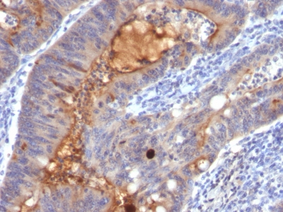
(Figure 7. Indirect immunoperoxidase staining of human colon adenocarcinoma paraffin tissue section (RCK108; Mouse anti keratin 19). Dilution 1:50 and microwave pretreatment. Specific staining of the epithelial tumor cells. No reactivity in connective tissues.)
Application Data
(Figure 7. Indirect immunoperoxidase staining of human colon adenocarcinoma paraffin tissue section (RCK108; Mouse anti keratin 19). Dilution 1:50 and microwave pretreatment. Specific staining of the epithelial tumor cells. No reactivity in connective tissues.)
CytokeRatin 19 / KeRatin K19, Monoclonal Antibody (Cat# AAA14562)
Full Name
Mouse anti CytokeRatin 19 / KeRatin K19
Reactivity
Human, Zebrafish
Applications
Flow Cytometry, Immunocytochemistry, Immunohistochemistry, Immunohistochemistry, Western Blot
Pricing


 Strength of Signal
(Analysis of Protein Array containing more than 19,000 full-length human proteins using Vimentin (VIM) Mouse Monoclonal Antibody (VM452)Z- and S- Score: The Z-score represents the strength of a signal that a monoclonal antibody (MAb) (in combination with a fluorescently-tagged anti-IgG secondary antibody) produces when binding to a particular protein on the HuProtTM array. Z-scores are described in units of standard deviations (SD's) above the mean value of all signals generated on that array. If targets on HuProtTM are arranged in descending order of the Z-score, the S-score is the difference (also in units of SD's) between the Z-score. S-score therefore represents the relative target specificity of a MAb to its intended target. A MAb is considered to specific to its intended target, if the MAb has an S-score of at least 2.5. For example, if a MAb binds to protein X with a Z-score of 43 and to protein Y with a Z-score of 14, then the S-score for the binding of that MAb to protein X is equal to 29.)
Strength of Signal
(Analysis of Protein Array containing more than 19,000 full-length human proteins using Vimentin (VIM) Mouse Monoclonal Antibody (VM452)Z- and S- Score: The Z-score represents the strength of a signal that a monoclonal antibody (MAb) (in combination with a fluorescently-tagged anti-IgG secondary antibody) produces when binding to a particular protein on the HuProtTM array. Z-scores are described in units of standard deviations (SD's) above the mean value of all signals generated on that array. If targets on HuProtTM are arranged in descending order of the Z-score, the S-score is the difference (also in units of SD's) between the Z-score. S-score therefore represents the relative target specificity of a MAb to its intended target. A MAb is considered to specific to its intended target, if the MAb has an S-score of at least 2.5. For example, if a MAb binds to protein X with a Z-score of 43 and to protein Y with a Z-score of 14, then the S-score for the binding of that MAb to protein X is equal to 29.)
Vimentin, Monoclonal Antibody (Cat# AAA13810)
Full Name
Vimentin (Mesenchymal Cell Marker) Mouse Monoclonal Antibody
Gene Names
VIM; HEL113; CTRCT30
Reactivity
Human, Cow, Dog, Cat, Pig, Goat, Chicken.
Does not react with Mouse and Rat
Does not react with Mouse and Rat
Applications
Flow Cytometry, Immunofluorescence, Western Blot, Immunohistochemistry
Pricing


 IHC (Immunohistchemistry)
(At 1/100 staining Human ovarian cancer by IHC-P. The sample was formaldehyde fixed and a heat mediated antigen retrieval step in citrate buffer was performed. The sample was then blocked and incubated with the primary antibody at 4 degree C overnight. An HRP conjugated anti-Rabbit antibody was used as the secondary antibody.)
IHC (Immunohistchemistry)
(At 1/100 staining Human ovarian cancer by IHC-P. The sample was formaldehyde fixed and a heat mediated antigen retrieval step in citrate buffer was performed. The sample was then blocked and incubated with the primary antibody at 4 degree C overnight. An HRP conjugated anti-Rabbit antibody was used as the secondary antibody.)
Niban, Polyclonal Antibody (Cat# AAA31335)
Full Name
Niban Antibody
Gene Names
FAM129A; GIG39; NIBAN; C1orf24
Reactivity
Human, Rat
Predicted Reactivity: Pig (100%), Bovine (100%), Horse (100%), Sheep (100%), Rabbit (92%), Dog (100%)
Predicted Reactivity: Pig (100%), Bovine (100%), Horse (100%), Sheep (100%), Rabbit (92%), Dog (100%)
Applications
Western Blot, Immunohistochemistry, Immunofluorescence, Immunocytochemistry, Peptide ELISA
Purity
The antiserum was purified by peptide affinity chromatography using SulfoLink Coupling Resin
Pricing


 IHC (Immunohistochemistry)
(At 1/100 staining Mouse lung tissue by IHC-P. The sample was formaldehyde fixed and a heat mediated antigen retrieval step in citrate buffer was performed. The sample was then blocked and incubated with the primary antibody at 4 degree C overnight. An HRP conjugated anti-Rabbit antibody was used as the secondary antibody.)
IHC (Immunohistochemistry)
(At 1/100 staining Mouse lung tissue by IHC-P. The sample was formaldehyde fixed and a heat mediated antigen retrieval step in citrate buffer was performed. The sample was then blocked and incubated with the primary antibody at 4 degree C overnight. An HRP conjugated anti-Rabbit antibody was used as the secondary antibody.)
Histone H2A, Polyclonal Antibody (Cat# AAA31347)
Full Name
Acetyl-Histone H2A (Lys5) Antibody
Gene Names
HIST1H2AB; H2A/m; H2AFM
Reactivity
Human, Mouse, Rat
Applications
Western Blot, Immunohistochemistry, Peptide ELISA
Purity
The antiserum was purified by peptide affinity chromatography using SulfoLink Coupling Resin
Pricing
 Standard Curve (Sample)
Tumor Necrosis Factor Alpha, ELISA Kit (Cat# AAA17469)
Full Name
Guinea pig Tumor Necrosis Factor Alpha ELISA Kit
Reactivity
Guinea pig
Pricing
 Standard Curve (Sample)
Hepcidin, ELISA Kit (Cat# AAA16721)
Full Name
Bovine Hepcidin ELISA Kit
Gene Names
HAMP; HEPC; PLTR; HFE2B; LEAP1
Reactivity
Bovine
Pricing


 Application Data
Ep-CAM /CD326, Monoclonal Antibody (Cat# AAA13831)
Full Name
Ep-CAM /CD326 (Epithelial Marker) Mouse Monoclonal Antibody
Gene Names
EPCAM; ESA; KSA; M4S1; MK-1; DIAR5; EGP-2; EGP40; KS1/4; MIC18; TROP1; EGP314; HNPCC8; TACSTD1
Reactivity
Human, Mouse, Rat
Applications
Flow Cytometry, Immunofluorescence, Western Blot, Immunohistochemistry
Pricing


 WB (Western Blot)
(WB Suggested Anti-HNF4A antibody Titration: 1 ug/mLSample Type: Human Liver)
WB (Western Blot)
(WB Suggested Anti-HNF4A antibody Titration: 1 ug/mLSample Type: Human Liver)
HNF4A, Polyclonal Antibody (Cat# AAA23401)
Full Name
HNF4A Antibody - N-terminal region
Gene Names
HNF4A; TCF; HNF4; MODY; FRTS4; MODY1; NR2A1; TCF14; HNF4a7; HNF4a8; HNF4a9; NR2A21; HNF4alpha
Reactivity
Cow, Dog, Guinea Pig, Horse, Human, Mouse, Rabbit, Rat, Sheep, Zebrafish
Applications
Western Blot
Purity
Affinity Purified
Pricing


 Application Data
(Analysis of Protein Array containing more than 19,000 full-length human proteins using ERCC1 Mouse Monoclonal Antibody (ERCC1/2318). Z- and S- Score: The Z-score represents the strength of a signal that a monoclonal antibody (MAb) (in combination with a fluorescently-tagged anti-IgG secondary antibody) produces when binding to a particular protein on the HuProtTM array. Z-scores are described in units of standard deviations (SD's) above the mean value of all signals generated on that array. If targets on HuProtTM are arranged in descending order of the Z-score, the S-score is the difference (also in units of SD's) between the Z-score. S-score therefore represents the relative target specificity of a MAb to its intended target. A MAb is considered to specific to its intended target, if the MAb has an S-score of at least 2.5. For example, if a MAb binds to protein X with a Z-score of 43 and to protein Y with a Z-score of 14, then the S-score for the binding of that MAb to protein X is equal to 29.)
Application Data
(Analysis of Protein Array containing more than 19,000 full-length human proteins using ERCC1 Mouse Monoclonal Antibody (ERCC1/2318). Z- and S- Score: The Z-score represents the strength of a signal that a monoclonal antibody (MAb) (in combination with a fluorescently-tagged anti-IgG secondary antibody) produces when binding to a particular protein on the HuProtTM array. Z-scores are described in units of standard deviations (SD's) above the mean value of all signals generated on that array. If targets on HuProtTM are arranged in descending order of the Z-score, the S-score is the difference (also in units of SD's) between the Z-score. S-score therefore represents the relative target specificity of a MAb to its intended target. A MAb is considered to specific to its intended target, if the MAb has an S-score of at least 2.5. For example, if a MAb binds to protein X with a Z-score of 43 and to protein Y with a Z-score of 14, then the S-score for the binding of that MAb to protein X is equal to 29.)
ERCC1/RAD10, Monoclonal Antibody (Cat# AAA23913)
Full Name
ERCC1/RAD10 (Tumor Progression Marker)
Gene Names
ERCC1; UV20; COFS4; RAD10
Reactivity
Human
Applications
Immunohistochemistry
Purity
Purified Ab with BSA and Azide at 200ug/ml OR Purified Ab WITHOUT BSA and Azide at 1.0mg/ml
Pricing
 Standard Curve (Sample)
Tumor Fibrin and Fibrinogen Degradation Products (tFFDP), ELISA Kit (Cat# AAA22532)
Full Name
Human Tumor Fibrin and Fibrinogen Degradation Products, tFFDP ELISA Kit
Reactivity
Human
Pricing


 Application Data
(Detection limit for recombinant GST tagged TP53 is approximately 1ng/ml as a capture antibody.)
Application Data
(Detection limit for recombinant GST tagged TP53 is approximately 1ng/ml as a capture antibody.)
TP53, Monoclonal Antibody (Cat# AAA26439)
Full Name
TP53 (Tumor Protein p53, FLJ92943, LFS1, TRP53, p53) (HRP)
Gene Names
TP53; P53; BCC7; LFS1; BMFS5; TRP53
Applications
Immunofluorescence, Immunohistochemistry, Immunoprecipitation, Western Blot
Purity
Purified
Pricing


 IF (Immunofluorescence)
(Immunofluorescence analysis of Raw264.7 cells using eEF1A1 antibody at dilution of 1:100. Blue: DAPI for nuclear staining.)
IF (Immunofluorescence)
(Immunofluorescence analysis of Raw264.7 cells using eEF1A1 antibody at dilution of 1:100. Blue: DAPI for nuclear staining.)
eEF1A1, Polyclonal Antibody (Cat# AAA28354)
Full Name
eEF1A1 Rabbit pAb
Gene Names
EEF1A1; CCS3; EF1A; PTI1; CCS-3; EE1A1; EEF-1; EEF1A; EF-Tu; LENG7; eEF1A-1; GRAF-1EF; HNGC:16303
Reactivity
Human, Mouse, Rat
Applications
Western Blot, Immunohistochemistry, Immunofluorescence
Purity
Affinity purification
Pricing


 FCM (Flow Cytometry)
(Flow cytometric analysis of A431 cells with Securin antibody at 1/100 dilution (red) compared with an unlabelled control (cells without incubation with primary antibody; black). Alexa Fluor 488-conjugated goat anti-rabbit IgG was used as the secondary antibody.)
FCM (Flow Cytometry)
(Flow cytometric analysis of A431 cells with Securin antibody at 1/100 dilution (red) compared with an unlabelled control (cells without incubation with primary antibody; black). Alexa Fluor 488-conjugated goat anti-rabbit IgG was used as the secondary antibody.)
Securin, Monoclonal Antibody (Cat# AAA30455)
Full Name
Securin Antibody
Gene Names
PTTG1; EAP1; PTTG; HPTTG; TUTR1
Reactivity
Human
Applications
Western Blot, Immunocytochemistry, Immunofluorescence, Immunohistochemistry, Flow Cytometry, Immunoprecipitation
Purity
ProA affinity purified
Pricing


 Application Data
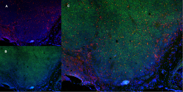
(Published customer image Infiltration of GFP+ BM-cells in infarct and peri-infarct regions. (A-B) Dot plots of viable macrophages/granulocytes (CD11b+CD45high, top right quadrants) and microglia (CD11b+CD45dim, bottom right quadrants) in cortex from BM-chimeric unmanipulated mice and mice exposed to pMCAO. (C) Bar graph showing mean numbers of CD11b+CD45dim microglia and CD11b+CD45high macrophages/granulocytes in BM-chimeric mice 24 hours after pMCAO, subdivided based on expression of GFP (n = 5). Approximately 92% of of the CD45high population were GFP+. (D) Estimation and comparison of mean numbers of CD11b+CD45dim microglia in non-chimeric (n = 10) versus BM-chimeric mice (n = 5) 24 hours after of pMCAO shows significantly fewer CD11b+CD45dim microglial cells in irradiated mice. (E) Overview, showing distribution of infiltrating GFP+ BM-derived cells into infarct (IF) and peri-infarct (P-IF) regions 24 hours after pMCAO. (E-G) By 24 hours, GFP+ single cells (F) and vessel-associated aggregates of GFP+ cells (arrows in G) were observed in infarct and peri-infarct regions. Some of the vessel-associated cells were round, leukocyte-like cells (arrows) while others were elongated cells lining the vasculature (arrow heads in G and in insert). (H) Bar graph showing mean numbers of single GFP+ cells and vessel-associated aggregates of GFP+ cells in ipsi- and contralateral cortex 24 hours after surgery (n = 10). (I-P) Immunohistochemical staining of CD45.1 (I, K), CD45.2 (J, L), IgG2a (M, O) and CD45 (N, P) in ischemic tissue in BM-chimeric (I, J, M, N) and non-chimeric mice (K, L, O, P) 24 hours after pMCAO. N.D, none detected. Scale bars: 200 um (A), 10 um (B, C). 50 um (I-P) *P < 0.05, **P < 0.01, and ***P < 0.001.From: Clausen BH, Lambertsen KL, Babcock AA, Holm TH, Dagnaes-Hansen F, Finsen B. Interleukin-1beta and tumor necrosis factor-alpha are expressed by different subsets of microglia and macrophages after ischemic stroke in mice. J Neuroinflammation. 2008 Oct 23;5:46.)
Application Data
(Published customer image Infiltration of GFP+ BM-cells in infarct and peri-infarct regions. (A-B) Dot plots of viable macrophages/granulocytes (CD11b+CD45high, top right quadrants) and microglia (CD11b+CD45dim, bottom right quadrants) in cortex from BM-chimeric unmanipulated mice and mice exposed to pMCAO. (C) Bar graph showing mean numbers of CD11b+CD45dim microglia and CD11b+CD45high macrophages/granulocytes in BM-chimeric mice 24 hours after pMCAO, subdivided based on expression of GFP (n = 5). Approximately 92% of of the CD45high population were GFP+. (D) Estimation and comparison of mean numbers of CD11b+CD45dim microglia in non-chimeric (n = 10) versus BM-chimeric mice (n = 5) 24 hours after of pMCAO shows significantly fewer CD11b+CD45dim microglial cells in irradiated mice. (E) Overview, showing distribution of infiltrating GFP+ BM-derived cells into infarct (IF) and peri-infarct (P-IF) regions 24 hours after pMCAO. (E-G) By 24 hours, GFP+ single cells (F) and vessel-associated aggregates of GFP+ cells (arrows in G) were observed in infarct and peri-infarct regions. Some of the vessel-associated cells were round, leukocyte-like cells (arrows) while others were elongated cells lining the vasculature (arrow heads in G and in insert). (H) Bar graph showing mean numbers of single GFP+ cells and vessel-associated aggregates of GFP+ cells in ipsi- and contralateral cortex 24 hours after surgery (n = 10). (I-P) Immunohistochemical staining of CD45.1 (I, K), CD45.2 (J, L), IgG2a (M, O) and CD45 (N, P) in ischemic tissue in BM-chimeric (I, J, M, N) and non-chimeric mice (K, L, O, P) 24 hours after pMCAO. N.D, none detected. Scale bars: 200 um (A), 10 um (B, C). 50 um (I-P) *P < 0.05, **P < 0.01, and ***P < 0.001.From: Clausen BH, Lambertsen KL, Babcock AA, Holm TH, Dagnaes-Hansen F, Finsen B. Interleukin-1beta and tumor necrosis factor-alpha are expressed by different subsets of microglia and macrophages after ischemic stroke in mice. J Neuroinflammation. 2008 Oct 23;5:46.)
CD11b, Monoclonal Antibody (Cat# AAA12185)
Full Name
RAT ANTI MOUSE CD11b
Gene Names
Itgam; CR3; CR3A; MAC1; Cd11b; Ly-40; Mac-1; Mac-1a; CD11b/CD18; F730045J24Rik
Applications
Immunohistochemistry, Flow Cytometry, Immunofluorescence, Immunoprecipitation
Pricing


 Application Data
(Immunoperoxidase staining of a human tonsil cryosection with Mouse anti Human CD163 antibody, clone EDHu-1 followed by the Histar detection system . Low power)
Application Data
(Immunoperoxidase staining of a human tonsil cryosection with Mouse anti Human CD163 antibody, clone EDHu-1 followed by the Histar detection system . Low power)
CD163, Monoclonal Antibody (Cat# AAA12100)
Full Name
MOUSE ANTI HUMAN CD163
Gene Names
CD163; M130; MM130
Applications
Immunohistochemistry, Flow Cytometry, Immunofluorescence, Immunoassay, Immunohistochemistry, Western Blot
Pricing


 Application Data
(Published customer image Infiltration of GFP+ BM-cells in infarct and peri-infarct regions. (A-B) Dot plots of viable macrophages/granulocytes (CD11b+CD45high, top right quadrants) and microglia (CD11b+CD45dim, bottom right quadrants) in cortex from BM-chimeric unmanipulated mice and mice exposed to pMCAO. (C) Bar graph showing mean numbers of CD11b+CD45dim microglia and CD11b+CD45high macrophages/granulocytes in BM-chimeric mice 24 hours after pMCAO, subdivided based on expression of GFP (n = 5). Approximately 92% of of the CD45high population were GFP+. (D) Estimation and comparison of mean numbers of CD11b+CD45dim microglia in non-chimeric (n = 10) versus BM-chimeric mice (n = 5) 24 hours after of pMCAO shows significantly fewer CD11b+CD45dim microglial cells in irradiated mice. (E) Overview, showing distribution of infiltrating GFP+ BM-derived cells into infarct (IF) and peri-infarct (P-IF) regions 24 hours after pMCAO. (E-G) By 24 hours, GFP+ single cells (F) and vessel-associated aggregates of GFP+ cells (arrows in G) were observed in infarct and peri-infarct regions. Some of the vessel-associated cells were round, leukocyte-like cells (arrows) while others were elongated cells lining the vasculature (arrow heads in G and in insert). (H) Bar graph showing mean numbers of single GFP+ cells and vessel-associated aggregates of GFP+ cells in ipsi- and contralateral cortex 24 hours after surgery (n = 10). (I-P) Immunohistochemical staining of CD45.1 (I, K), CD45.2 (J, L), IgG2a (M, O) and CD45 (N, P) in ischemic tissue in BM-chimeric (I, J, M, N) and non-chimeric mice (K, L, O, P) 24 hours after pMCAO. N.D, none detected. Scale bars: 200 um (A), 10 um (B, C). 50 um (I-P) *P < 0.05, **P < 0.01, and ***P < 0.001.From: Clausen BH, Lambertsen KL, Babcock AA, Holm TH, Dagnaes-Hansen F, Finsen B. Interleukin-1beta and tumor necrosis factor-alpha are expressed by different subsets of microglia and macrophages after ischemic stroke in mice. J Neuroinflammation. 2008 Oct 23;5:46.)
Application Data
(Published customer image Infiltration of GFP+ BM-cells in infarct and peri-infarct regions. (A-B) Dot plots of viable macrophages/granulocytes (CD11b+CD45high, top right quadrants) and microglia (CD11b+CD45dim, bottom right quadrants) in cortex from BM-chimeric unmanipulated mice and mice exposed to pMCAO. (C) Bar graph showing mean numbers of CD11b+CD45dim microglia and CD11b+CD45high macrophages/granulocytes in BM-chimeric mice 24 hours after pMCAO, subdivided based on expression of GFP (n = 5). Approximately 92% of of the CD45high population were GFP+. (D) Estimation and comparison of mean numbers of CD11b+CD45dim microglia in non-chimeric (n = 10) versus BM-chimeric mice (n = 5) 24 hours after of pMCAO shows significantly fewer CD11b+CD45dim microglial cells in irradiated mice. (E) Overview, showing distribution of infiltrating GFP+ BM-derived cells into infarct (IF) and peri-infarct (P-IF) regions 24 hours after pMCAO. (E-G) By 24 hours, GFP+ single cells (F) and vessel-associated aggregates of GFP+ cells (arrows in G) were observed in infarct and peri-infarct regions. Some of the vessel-associated cells were round, leukocyte-like cells (arrows) while others were elongated cells lining the vasculature (arrow heads in G and in insert). (H) Bar graph showing mean numbers of single GFP+ cells and vessel-associated aggregates of GFP+ cells in ipsi- and contralateral cortex 24 hours after surgery (n = 10). (I-P) Immunohistochemical staining of CD45.1 (I, K), CD45.2 (J, L), IgG2a (M, O) and CD45 (N, P) in ischemic tissue in BM-chimeric (I, J, M, N) and non-chimeric mice (K, L, O, P) 24 hours after pMCAO. N.D, none detected. Scale bars: 200 um (A), 10 um (B, C). 50 um (I-P) *P < 0.05, **P < 0.01, and ***P < 0.001.From: Clausen BH, Lambertsen KL, Babcock AA, Holm TH, Dagnaes-Hansen F, Finsen B. Interleukin-1beta and tumor necrosis factor-alpha are expressed by different subsets of microglia and macrophages after ischemic stroke in mice. J Neuroinflammation. 2008 Oct 23;5:46.)
CD11b, Monoclonal Antibody (Cat# AAA12183)
Full Name
RAT ANTI MOUSE CD11b:FITC
Gene Names
Itgam; CR3; CR3A; MAC1; Cd11b; Ly-40; Mac-1; Mac-1a; CD11b/CD18; F730045J24Rik
Applications
Flow Cytometry
Pricing


 FCM (Flow Cytometry)
(Flow cytometric analysis of Hela cells with 53BP1 antibody at 1/50 dilution (red) compared with an unlabelled control (cells without incubation with primary antibody; black). Alexa Fluor 488-conjugated goat anti rabbit IgG was used as the secondary antibody.)
FCM (Flow Cytometry)
(Flow cytometric analysis of Hela cells with 53BP1 antibody at 1/50 dilution (red) compared with an unlabelled control (cells without incubation with primary antibody; black). Alexa Fluor 488-conjugated goat anti rabbit IgG was used as the secondary antibody.)
53BP1, Monoclonal Antibody (Cat# AAA30337)
Full Name
53BP1 Antibody
Gene Names
TP53BP1; p202; 53BP1
Reactivity
Human, Mouse, Rat
Applications
Western Blot, Immunocytochemistry, Immunofluorescence, Immunohistochemistry, Flow Cytometry
Purity
ProA affinity purified
Pricing
 Standard Curve (Sample)
insulin-like growth factor binding protein 7, ELISA Kit (Cat# AAA15846)
Full Name
Human Insulin-like growth factor-binding protein 7, IGFBP7/MAC25/PSF ELISA Kit
Gene Names
IGFBP7; AGM; PSF; TAF; FSTL2; IBP-7; MAC25; IGFBP-7; RAMSVPS; IGFBP-7v; IGFBPRP1
Reactivity
Human
Pricing


 ICC (Immunocytochemistry)
(ICC staining CD147 in SKOV-3 cells (red). The nuclear counter stain is DAPI (blue). Cells were fixed in paraformaldehyde, permeabilised with 0.25% Triton X100/PBS.)
ICC (Immunocytochemistry)
(ICC staining CD147 in SKOV-3 cells (red). The nuclear counter stain is DAPI (blue). Cells were fixed in paraformaldehyde, permeabilised with 0.25% Triton X100/PBS.)
CD147, Monoclonal Antibody (Cat# AAA30287)
Full Name
CD147 Antibody
Gene Names
BSG; M6; OK; 5F7; TCSF; CD147; EMMPRIN
Reactivity
Human, Mouse, Rat
Applications
Western Blot, Immunocytochemistry, Immunofluorescence, Immunohistochemistry
Purity
ProA affinity purified
Pricing
 SDS-PAGE
(SDS-PAGE Analysis Purified Secretory Component Mouse Monoclonal Antibody (SPM217). Confirmation of Purity and Integrity of Antibody.)
SDS-PAGE
(SDS-PAGE Analysis Purified Secretory Component Mouse Monoclonal Antibody (SPM217). Confirmation of Purity and Integrity of Antibody.)
IgA Secretory Component / ECM1, Monoclonal Antibody (Cat# AAA13802)
Full Name
IgA Secretory Component / ECM1 Mouse Monoclonal Antibody
Gene Names
ECM1; URBWD
Reactivity
Human, Rat
Applications
Flow Cytometry, Immunofluorescence, Immunohistochemistry
Pricing


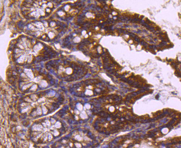
 IHC (Immunohistchemistry)
(Immunohistochemistry analysis of Formalin-fixed, paraffin-embedded Human Wilms' tumor using WT1, Wilms' Tumor 1 antibody.)
IHC (Immunohistchemistry)
(Immunohistochemistry analysis of Formalin-fixed, paraffin-embedded Human Wilms' tumor using WT1, Wilms' Tumor 1 antibody.)
Wilms' Tumor 1 (WT1), Monoclonal Antibody (Cat# AAA30741)
Full Name
Anti-Wilms' Tumor 1 (WT1) Antibody [ABT-WT1]
Gene Names
WT1; GUD; AWT1; WAGR; WT33; NPHS4; WIT-2
Reactivity
Human
Applications
Immunohistochemistry
Purity
Immunogen Affinity Purified
Pricing


 IHC (Immunohistchemistry)
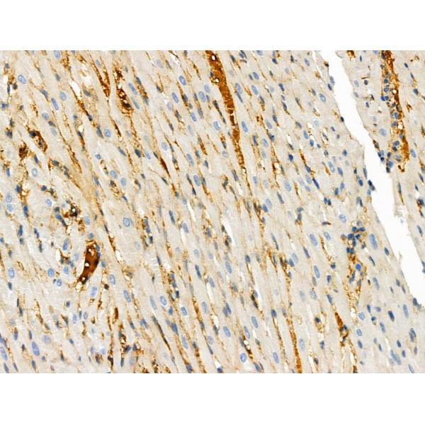
(Immunohistochemistry analysis using Mouse Anti-Hsp70 Monoclonal Antibody, Clone BB70 . Tissue: hepatocytes. Species: Rat. Fixation: Paraffin Embedded. Primary Antibody: Mouse Anti-Hsp70 Monoclonal Antibody at 1:200. Liver sections were paraffin embedded. First pictures in series show two hours after exposure to stress, the second shows the control. Courtesy of: G. Matic, University of Belgrade, Serbia.)
IHC (Immunohistchemistry)
(Immunohistochemistry analysis using Mouse Anti-Hsp70 Monoclonal Antibody, Clone BB70 . Tissue: hepatocytes. Species: Rat. Fixation: Paraffin Embedded. Primary Antibody: Mouse Anti-Hsp70 Monoclonal Antibody at 1:200. Liver sections were paraffin embedded. First pictures in series show two hours after exposure to stress, the second shows the control. Courtesy of: G. Matic, University of Belgrade, Serbia.)
HSP70/HSC70, Monoclonal Antibody (Cat# AAA27659)
Full Name
HSP70/HSC70 Antibody
Gene Names
HSPA2; HSP70
Reactivity
Human, Mouse, Rat, Bovine, Sheep, Dog, Beluga, Fish, Guinea Pig, Pig, Hamster, Rabbit, Chicken, African Clawed Frog, Fruit Fly, Yeast
Applications
Western Blot, Immunocytochemistry, Immunofluorescence, Immunohistochemistry, Immunoprecipitation
Purity
Protein G Purified
Pricing


 SDS-PAGE
(SDS-PAGE Analysis Purified HSP60 Mouse Monoclonal Antibody (CPTC-HSPD1-1). Confirmation of Purity and Integrity of Antibody.)
SDS-PAGE
(SDS-PAGE Analysis Purified HSP60 Mouse Monoclonal Antibody (CPTC-HSPD1-1). Confirmation of Purity and Integrity of Antibody.)
HSP60 (Heat Shock Protein 60), Monoclonal Antibody (Cat# AAA23919)
Full Name
HSP60 (Heat Shock Protein 60) (Mitochondrial Marker)
Gene Names
HSPD1; HLD4; CPN60; GROEL; HSP60; HSP65; SPG13; HSP-60; HuCHA60
Reactivity
Human
Applications
Flow Cytometry, Immunofluorescence, Western Blot, Immunohistochemistry
Purity
Purified Ab with BSA and Azide at 200ug/ml OR Purified Ab WITHOUT BSA and Azide at 1.0mg/ml
Pricing
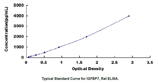 Standard Curve (Sample)
Insulin Like Growth Factor Binding Protein 7 (IGFBP7), ELISA Kit (Cat# AAA23011)
Full Name
Rat Insulin Like Growth Factor Binding Protein 7 (IGFBP7) ELISA Kit
Gene Names
IGFBP7; AGM; PSF; TAF; FSTL2; IBP-7; MAC25; IGFBP-7; RAMSVPS; IGFBP-7v; IGFBPRP1
Reactivity
Rat
Pricing
 WB (Western Blot)
(Western Blot analysis of extracts from NIH-3T3 cells using MUC1 Antibody.)
WB (Western Blot)
(Western Blot analysis of extracts from NIH-3T3 cells using MUC1 Antibody.)
MUC1, Polyclonal Antibody (Cat# AAA30739)
Full Name
MUC1 Antibody
Gene Names
MUC1; EMA; MCD; PEM; PUM; KL-6; MAM6; MCKD; PEMT; CD227; H23AG; MCKD1; MUC-1; ADMCKD; ADMCKD1; CA 15-3; MUC-1/X; MUC1/ZD; MUC-1/SEC
Reactivity
Human, Mouse, Rat
Applications
Western Blot, Immunohistochemistry, Immunohistochemistry
Purity
The antibody was affinity-purified from rabbit antiserum by affinity-
chromatography using epitope-specific immunogen.
Pricing


 WB (Western Blot)
(WB Suggested Anti-YTHDF1 Antibody Titration: 0.2-1 ug/mlPositive Control: HepG2 cell lysateYTHDF1 is supported by BioGPS gene expression data to be expressed in HepG2)
WB (Western Blot)
(WB Suggested Anti-YTHDF1 Antibody Titration: 0.2-1 ug/mlPositive Control: HepG2 cell lysateYTHDF1 is supported by BioGPS gene expression data to be expressed in HepG2)
YTHDF1, Polyclonal Antibody (Cat# AAA23574)
Full Name
YTHDF1 antibody - middle region
Gene Names
YTHDF1; C20orf21
Reactivity
Dog, Guinea Pig, Horse, Human, Mouse, Rabbit, Rat, Yeast
Applications
Western Blot
Purity
Affinity Purified
Pricing


 WB (Western Blot)
(WB Suggested Anti-PTX3 Antibody Titration: 0.2-1 ug/mlELISA Titer: 1:312500Positive Control: MCF7 cell lysate)
WB (Western Blot)
(WB Suggested Anti-PTX3 Antibody Titration: 0.2-1 ug/mlELISA Titer: 1:312500Positive Control: MCF7 cell lysate)
PTX3, Polyclonal Antibody (Cat# AAA23569)
Full Name
PTX3 antibody - N-terminal region
Gene Names
PTX3; TSG-14; TNFAIP5
Reactivity
Cow, Guinea Pig, Human, Mouse, Rabbit, Rat
Applications
Immunohistochemistry, Western Blot
Purity
Affinity Purified
Pricing

 IHC (Immunohistochemistry)
(GCC2 antibody (C-term) (AAA28751) immunohistochemistry analysis in formalin fixed and paraffin embedded human brain tissue followed by peroxidase conjugation of the secondary antibody and DAB staining. This data demonstrates the use of the GCC2 antibody (C-term) for immunohistochemistry. Clinical relevance has not been evaluated.)
IHC (Immunohistochemistry)
(GCC2 antibody (C-term) (AAA28751) immunohistochemistry analysis in formalin fixed and paraffin embedded human brain tissue followed by peroxidase conjugation of the secondary antibody and DAB staining. This data demonstrates the use of the GCC2 antibody (C-term) for immunohistochemistry. Clinical relevance has not been evaluated.)
GCC2, Polyclonal Antibody (Cat# AAA28751)
Full Name
GCC2 Antibody (C-term)
Gene Names
GCC2; REN53; GCC185; RANBP2L4
Reactivity
Human, mouse (Predicted Reactivity: Rat)
Applications
Western Blot, Immunohistochemistry
Purity
This antibody is purified through a protein A column, followed by peptide affinity purification.
Pricing


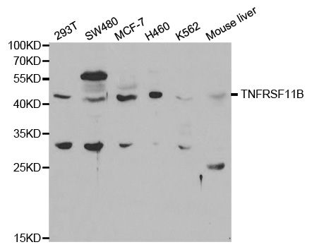
 IHC (Immunohistochemistry)
(Immunohistochemistry of paraffin-embedded mouse spleen using TNFRSF11B Antibody at dilution of 1:200 (40x lens).)
IHC (Immunohistochemistry)
(Immunohistochemistry of paraffin-embedded mouse spleen using TNFRSF11B Antibody at dilution of 1:200 (40x lens).)
TNFRSF11B, Polyclonal Antibody (Cat# AAA10748)
Full Name
TNFRSF11B Polyclonal Antibody
Gene Names
TNFRSF11B; OPG; TR1; OCIF
Reactivity
Human, Mouse, Rat
Applications
Western Blot, Immunohistochemistry, Immunofluorescence
Purity
Affinity Purification
Pricing


 ICC (Immunocytochemistry)
(ICC staining p53DINP1 in HUVEC cells (green). The nuclear counter stain is DAPI (blue). Cells were fixed in paraformaldehyde, permeabilised with 0.25% Triton X100/PBS.)
ICC (Immunocytochemistry)
(ICC staining p53DINP1 in HUVEC cells (green). The nuclear counter stain is DAPI (blue). Cells were fixed in paraformaldehyde, permeabilised with 0.25% Triton X100/PBS.)
p53DINP1, Monoclonal Antibody (Cat# AAA30494)
Full Name
p53 DINP1 Antibody
Gene Names
TP53INP1; SIP; Teap; p53DINP1; TP53DINP1; TP53INP1A; TP53INP1B
Reactivity
Human, Mouse, Rat
Applications
Western Blot, Immunocytochemistry, Immunofluorescence, Immunohistochemistry
Purity
ProA affinity purified
Pricing


 Application Data
(At 25 degree C. The primary antibody was diluted at 1/200 and incubated with the sample for 1 hour at 37 degree C. An Alexa Fluor 594 conjugated goat anti-rabbit IgG (H+L) Ab, diluted at 1/600, was used as the secondary antibody.)
Application Data
(At 25 degree C. The primary antibody was diluted at 1/200 and incubated with the sample for 1 hour at 37 degree C. An Alexa Fluor 594 conjugated goat anti-rabbit IgG (H+L) Ab, diluted at 1/600, was used as the secondary antibody.)
Retinoblastoma, Polyclonal Antibody (Cat# AAA31401)
Full Name
Phospho-Retinoblastoma (Ser788) Antibody
Gene Names
RB1; RB; pRb; OSRC; pp110; p105-Rb
Reactivity
Human, Mouse, Rat
Predicted Reactivity: Pig (100%), Bovine (100%), Horse (100%), Sheep (100%), Rabbit (100%), Dog (100%), Chicken (92%), Xenopus (92%)
Predicted Reactivity: Pig (100%), Bovine (100%), Horse (100%), Sheep (100%), Rabbit (100%), Dog (100%), Chicken (92%), Xenopus (92%)
Applications
Western Blot, Immunohistochemistry, Immunofluorescence, Immunocytochemistry, Peptide ELISA
Purity
The antibody is from purified rabbit serum by affinity purification via sequential chromatography on phospho-peptide and non-phospho-peptide affinity columns.
Pricing


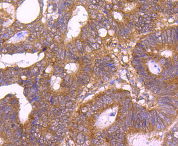
 IHC (Immunohistochemistry)
(At 1/100 staining Human colorectal cancer by IHC-P. The sample was formaldehyde fixed and a heat mediated antigen retrieval step in citrate buffer was performed. The sample was then blocked and incubated with the primary antibody at 4 degree C overnight. An HRP conjugated anti-Rabbit antibody was used as the secondary antibody.)
IHC (Immunohistochemistry)
(At 1/100 staining Human colorectal cancer by IHC-P. The sample was formaldehyde fixed and a heat mediated antigen retrieval step in citrate buffer was performed. The sample was then blocked and incubated with the primary antibody at 4 degree C overnight. An HRP conjugated anti-Rabbit antibody was used as the secondary antibody.)
CD227/MUC1, Polyclonal Antibody (Cat# AAA31426)
Full Name
Phospho-CD227/MUC1 (Ser1227) Antibody
Gene Names
MUC1; EMA; PEM; PUM; KL-6; MAM6; PEMT; CD227; H23AG; MUC-1; CA 15-3; MUC-1/X; MUC1/ZD; MUC-1/SEC
Reactivity
Human, Mouse, Rat
Applications
Western Blot, Immunohistochemistry, Peptide ELISA
Purity
The antibody is from purified rabbit serum by affinity purification via sequential chromatography on phospho-peptide and non-phospho-peptide affinity columns.
Pricing


 Application Data
(Detection limit for recombinant GST tagged TP53 is approximately 1ng/ml as a capture antibody.)
Application Data
(Detection limit for recombinant GST tagged TP53 is approximately 1ng/ml as a capture antibody.)
TP53, Monoclonal Antibody (Cat# AAA26173)
Full Name
TP53 (Tumor Protein p53, FLJ92943, LFS1, TRP53, p53) (Biotin)
Gene Names
TP53; P53; BCC7; LFS1; BMFS5; TRP53
Applications
Immunofluorescence, Immunohistochemistry, Immunoprecipitation, Western Blot
Purity
Purified
Pricing
 Standard Curve (Sample)
LY6/PLAUR domain containing 1, ELISA Kit (Cat# AAA18292)
Full Name
Human Ly6/PLAUR domain-containing protein 1, LYPD1 ELISA Kit
Gene Names
LYPD1; PHTS; LYPDC1
Reactivity
Human
Pricing
 Standard Curve (Sample)
Antioncogene p16 protein, ELISA Kit (Cat# AAA16041)
Full Name
Rat Antioncogene p16 protein ELISA Kit
Gene Names
CDKN2A; ARF; MLM; P14; P16; P19; CMM2; INK4; MTS1; TP16; CDK4I; CDKN2; INK4A; MTS-1; P14ARF; P19ARF; P16INK4; P16INK4A; P16-INK4A
Reactivity
Rat
Pricing
 Standard Curve (Sample)
Fibroblast Growth Factor 23, ELISA Kit (Cat# AAA22576)
Full Name
Canine Fibroblast Growth Factor 23 (FGF23) ELISA Kit
Gene Names
FGF23; ADHR; FGFN; HYPF; HPDR2; PHPTC
Reactivity
Canine
Pricing
 FCM (Flow Cytometry)
(Test Results:Tissue Distribution by Flow Cytometry Analysis:Mouse Strain: BALB/cCell Concentration: 1 x 10^6 cells per test.Antibody Concentration Used: 1.0ug/10^6 cells)
FCM (Flow Cytometry)
(Test Results:Tissue Distribution by Flow Cytometry Analysis:Mouse Strain: BALB/cCell Concentration: 1 x 10^6 cells per test.Antibody Concentration Used: 1.0ug/10^6 cells)
CD134L/OX40L, Monoclonal Antibody (Cat# AAA14267)
Full Name
Anti-Mouse CD134L/OX40L, Purified (Clone OX89) (rat IgG1)
Reactivity
Mouse
Applications
Immunohistochemistry, Functional Assay
Purity
Purified from ascitic fluid via Protein G Chromatography
Pricing
 Standard Curve (Sample)
tumor necrosis factor (TNF superfamily, member 2), ELISA Kit (Cat# AAA15045)
Full Name
Rat TNF-alpha ELISA Kit
Gene Names
TNF; TNFA; TNFSF2
Reactivity
Rat
Pricing